The Skeletal System - cookhealthscience.weebly.com · skeletal system, bone structure, and types of...
71
Bell Work Read and highlight the review worksheets given to you paying attention to the functions of the skeletal system, bone structure, and types of bones.
Transcript of The Skeletal System - cookhealthscience.weebly.com · skeletal system, bone structure, and types of...

Bell Work
Read and highlight the review worksheets given to
you paying attention to the functions of the
skeletal system, bone structure, and types of
bones.

The
Skeletal
System

Standards
Review the gross and cellular anatomy and physiology of the musculoskeletal, nervous, and cardiovascular systems.
13) Review the concepts of kinesiology and biomechanics from the Rehabilitation Careers course. Explain how joint and bone movement, body motion, and levers can have positive or negative effects on an athlete’s performance and development. In a presentation or speech intended for an audience of young athletes, describe the effects of overtraining on the musculoskeletal system, and relate the importance of adopting safe biomechanical practices when training.

Objectives
Review the gross anatomy and physiology of the axial skeleton
by labeling a diagram and completing a group research
activity.


The Axial Skeleton
The axial skeleton is blueand includes the:
skull
vertebral column
sternum
ribs
hyoid bone (or laryngeal)
ossicles (inner ear)

The Axial Skeleton
Are there any bones
that might be
confused as to being
part of the axial
skeleton, but are not?

The Skull
(Lateral view)
*Cranium
*Facial bones
What is the
difference?
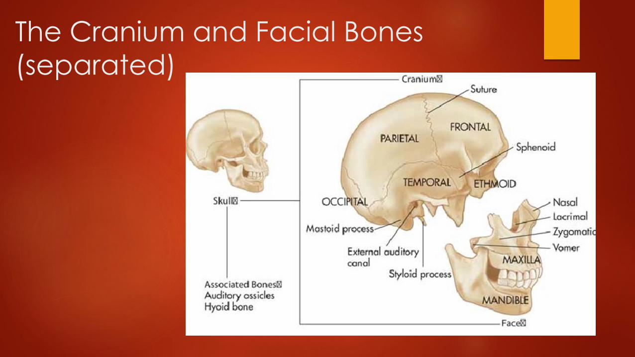
The Cranium and Facial Bones
(separated)

Skull fractures

Facial fractures

The
Vertebral
Column
(The
Spinal
Column)

Cervical Spine OR C-Spine
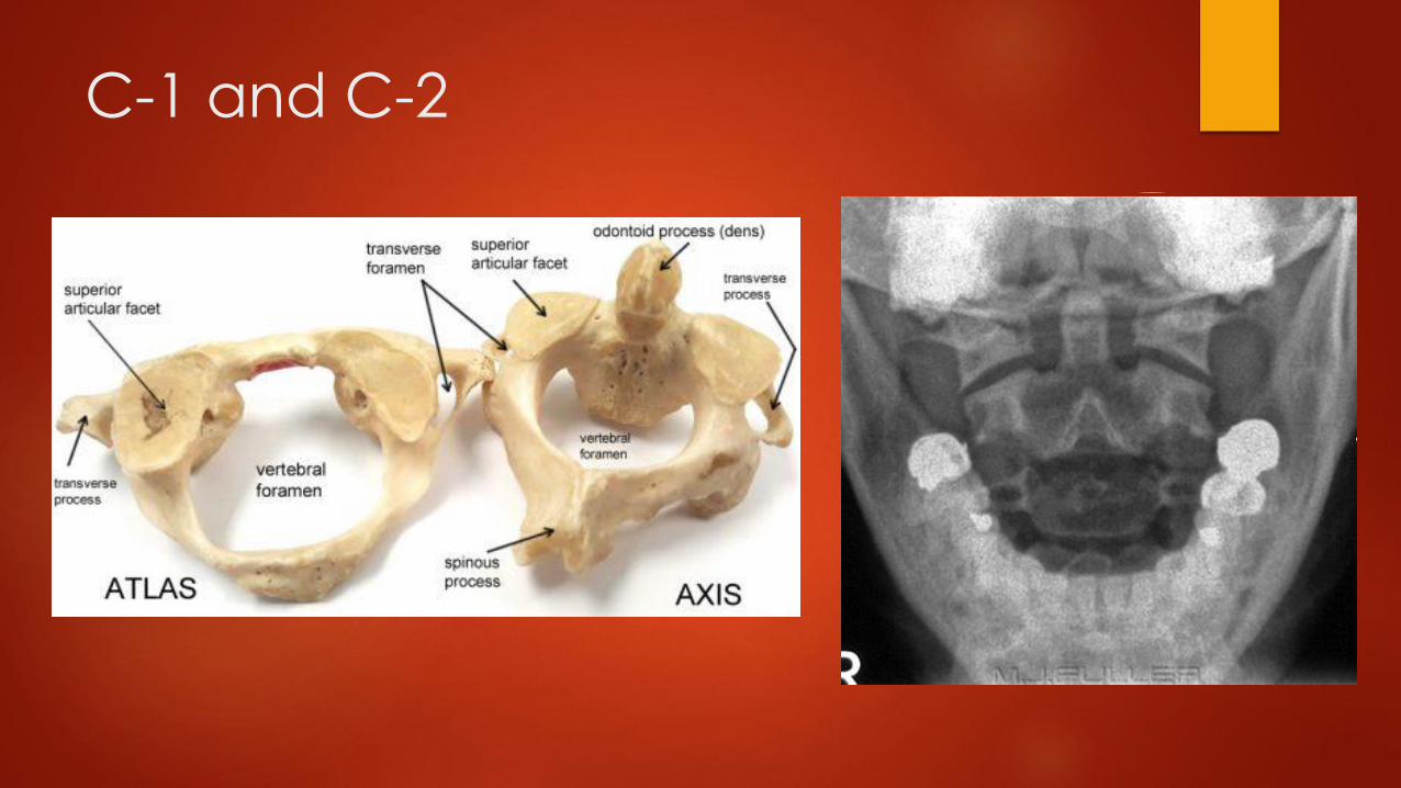
C-1 and C-2

https://peperperspective.com/2014/11/20/cellphone-harm-cervical-spine-
stress-and-increase-risk-of-brain-cancer/
What problems are related to posture and smart phone use?

Thoracic Spine OR T-Spine http://omega-rehab.com/2017/04/25/shoulder-pain-thoracic-spine/

AP View
What do you notice about this
thoracic spine x-ray?
What organs do you see?
What organs or bones do you
think you should see but do not?
Can you figure out which
vertebral body is T-12?
This image is “collimated” in,
leaving the rest of the chest
unseen. Why would the image, which was correctly obtained
need to “collimate”?

Lumbar Spine OR L-Spine

Sacrum and Coccyx
Activity
(turn to page 316 in the new book)
Draw and describe the following conditions
related to the spine:
Kyphosis
Lordosis
Scoliosis


Problems with Alignment

Sternum and Rib Cage
12 sets of
Ribs
True,
False,
Floating
What body cavity do the ribs protect?

What do
you notice
about the
floating
ribs?
Why would
we name
the false
ribs such?

Sternum and ribs

Identify the parts of the axial
skeleton that are possibly fractured
or damaged!

Identify the parts of the axial
skeleton that are possibly fractured
or damaged!

Identify the parts of the axial
skeleton that are possibly fractured
or damaged!

Identify the parts of the axial
skeleton that are possibly fractured
or damaged!

Identify the parts of the axial
skeleton that are possibly fractured
or damaged!

Activities
Label and color the worksheets of the skull.
Use various colors to help you distinguish between the terminology.
Read the professional journal on Facial Fractures and answer the
questions.
Also, Write a three paragraph summary describing the:
Epidemiology of facial fractures
Different kinds of facial fractures
Treatment of facial fractures

Optional Activities
Create a PowerPoint of the Station 5 chart located under Skeletal System Lab Activities.
-OR-
Create a PowerPoint outlining the differences in the bone structure of ethnic groups, gender and pediatrics, and those with disabilities.
At least 10 slides.
Include at least 5 pictures.
Distinguish Caucasian, African-American, Asian, and Native American ethnic groups.
Distinguish between men, women, and children.
Address what healthcare professionals look for in ultrasound pre-natal assessments concerning bone anatomy.

Exit Ticket
Name the bones within the axial skeleton?
Name the three types of ribs.
How many thoracic spine are there?
Where is the hyoid bone located?

Bell Work
Continue working on your skull and
spine diagrams and extended
learning questions.

Standards
8) Review the gross and cellular anatomy and physiology of the
musculoskeletal, nervous, and cardiovascular systems.
13) Review the concepts of kinesiology and biomechanics from the
Rehabilitation Careers course. Explain how joint and bone movement,
body motion, and levers can have positive or negative effects on an
athlete’s performance and development. In a presentation or speech
intended for an audience of young athletes, describe the effects of
overtraining on the musculoskeletal system, and relate the importance
of adopting safe biomechanical practices when training.

Objectives
Identify the bones of the upper and lower extremities as well as their
attachments by examining a model and labeling a diagram.
Research the negative affects of body movement by comparing
normal and abnormal appendicular imaging.

The Appendicular Skeleton
The appendicular skeleton is beigeand includes
shoulder girdles
arms
wrists
hands and fingers
pelvic girdle
legs
ankles
feet and toes
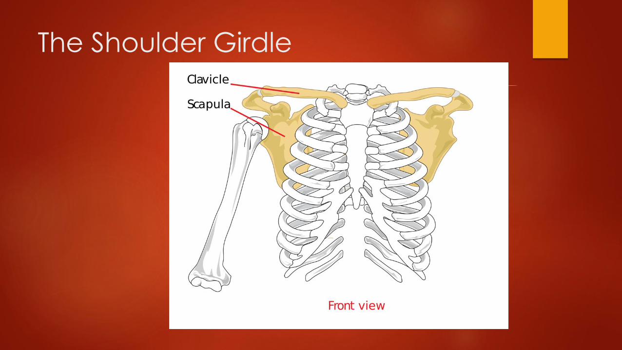
The Shoulder Girdle

The Scapula and
Humerus

Humerus (upper arm)

Ulna and Radius
(lower arm)

The Humerus,
Radius, and
Ulna
What do we
call this area
where these
three bones
communicate?

The Hand and Fingers

The Hand and Fingers

The Pelvic Girdle
Why do you think the shape of the pelvis
Is different between males and females?

Which is the pediatric pelvis?

Femur, Tibia, and Fibula
Upper Leg Lower Leg
------Patella

The Femur, Tibia, Fibula, and Patella
(Knee joint)

Feet and toes

Feet and toes

r
What are the bumps on the sides of the feet near
the toes called?
With a partner! Create a PPT.
Exercise Science
Research types of fractures.
Describe them.
Insert pictures.
Explain treatment and
rehabilitation options.
Also explain the difference
between open, closed,
complete, and incomplete.
Transverse
Oblique
Spiral
Comminuted
Impacted
Avulsion
Fissure
Greenstick
With a partner!! (A & P only)
1st Create an acrostic
of the carpal (wrist)
bones to help you
memorize their names
and positions.
You may start with
anyone of them, as
long as you include all
EIGHT in your acrostic.
2nd Create an acrostic of the
SEVEN tarsal (ankle) bones the
same way.
What is an acrostic? Here is an
example they may be familiar to
you:
Order of solving mathematical
equations -
Please Excuse My Dear Aunt Sally
– Parenthesis, Exponents,
Multiplication, Division,
Addition, Subtraction

Individual extended learning…
Continue working on your skull and spine diagrams.
Color and add labels to the hands and feet diagrams
as well.
Complete the exploration activity given on a link from
the class website.

Bell Work
Synarthroses-immoveable joint connected by tough
fibrous connective tissue.
Amphiarthroses-partially moveable joints with cartilage
between their articular surfaces.
Diarthroses-moveable joints consisting of articular
cartilage, a bursa , and a synovial cavity.

Objectives
Distinguish the three classifications of joints and examples
of each.
Identify joint structures and explore their functions
through a Range of Motion activity.
Explore possible problems and treatments related to the
joints.
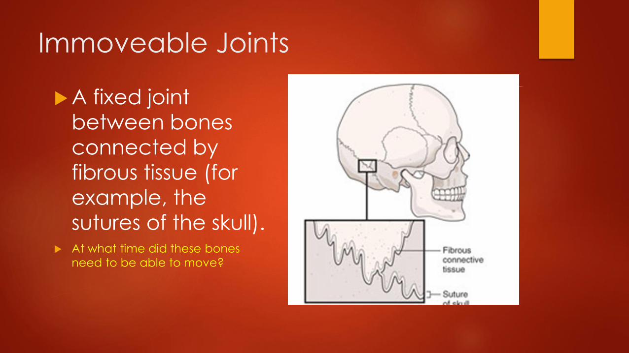
Immoveable Joints
A fixed joint
between bones
connected by
fibrous tissue (for
example, the
sutures of the skull). At what time did these bones
need to be able to move?


Cartilaginous Joint: the joint space is covered in dense connective tissue
In males this may
shift slightly at
times.
In females this joint
is vital to provide
room during
vaginal childbirth.

Moveable JointsJoint structures
Tendons:
Cords of fibrous connective tissue, like bungee cords, connecting muscle to bone.
Ligaments:
tough, whitish bands that connect from bone to boneand can withstand heavy stress.
Cartilage:
Acts as cushion between bones; articular cartilage
located on ends of bones and acts as shock
absorber, preventing ends from grinding together
when you move.

Joint structures Bursae-thin, lubricated cushions
located at points of friction between a bone and the surrounding soft tissue, such as skin, muscles, ligaments and tendons; like a tiny water balloon with only a few drops of fluid in it, wedged between two surfaces.
Synovial cavity(synovial joints)-allow for movement. Where the bones meet to form a synovial joint, the bones' surfaces are covered with a thin layer of strong, smooth articular cartilage. A very thin layer of slippery, viscous joint fluid, called synovial fluid, separates and lubricates the two cartilage-covered bone surfaces.

Moveable
Bone Joints
-Saddle
-Ball and Socket
-Pivot
-Hinge
-Ellipsoidal
-Gliding

ARTHRITIS: ARTHR/O=JOINT ITIS=INFLAMMATION
Signs and symptoms may include: Pain, Stiffness, Swelling, Redness, and Decreased range of motion
Osteoarthritis OSTEO=BONE
The most common type, wear-and-tear damage to your joint's cartilage — the hard, slick coating on the ends of bones. Enough damage can result in bone grinding directly on bone, which causes pain and restricted movement.
Rheumatoid arthritis
The body's immune system attacks the lining of the joint capsule, a tough membrane that encloses all the joint parts. This lining, known as the synovial membrane, becomes inflamed and swollen. The disease process can eventually destroy cartilage and bone within the joint.

Compare
the types: Treatments
Analgesics (pain meds)
Nonsteroidal anti-inflammatory drugs (NSAIDs)
Disease-modifying antirheumatic drugs (DMARDs)
Counterirritants (menthol type creams)
Corticosteroids (suppresses immune system)Physical Therapy
Surgery

The Vertebral Column/Spine
The joints in the spine are commonly called Facet Joints. anther
name for these joints are Apophyseal Joints.
Each vertebra has two sets of facet joints. One pair faces
upward (superior articular facet) and one downward (inferior
articular facet). There is one joint on each side (right and left).
Facet joints are hinge–like and link vertebrae together. They are
located at the back of the spine (posterior).

Vertebral Column/Spine
Facet joints are synovial joints. This means each
joint is surrounded by a capsule of connective tissue and produces a fluid to nourish and
lubricate the joint. The joint surfaces are coated
with cartilage allowing joints to move or glide
smoothly (articulate) against each other.
These joints allow flexion (bend forward), extension
(bend backward), and twisting motion. Certain
types of movement are restricted. The spine is made more stable due to the interlocking nature
to adjacent vertebrae.

OSTEOPOROSIS Oste/o=bone por/ous= pores in the bone
osis=process/condition
…causes bones to become weak and brittle — so brittle that a fall or even mild stresses like bending over or coughing can cause a fracture.
Your bones are in a constant state of renewal — new bone is made and old bone is broken down. When you're young, your body makes new bone faster than it breaks down old bone and your bone mass increases. Most people reach their peak bone mass by their early 20s. As people age, bone mass is lost faster than it's created.

OSTEOPOROSIS TREATMENT
Bisphosphonates (increases bone density)
Drugs that promote bone growth
Hormone-related therapy (estrogen especially in women after menopause)
Increase of vitamins, calcium
Vertebroplasty or Kyphoplasty(process of injected bone cement into the vertebral bodies to increase height, also this reduces pain from the bone grinding together. (This procedure can be done in surgery or better yet Interventional Radiology)!!!!
A FRACTURE IN THE SPINAL COLUNN IS CALLED A COMPRESSION FRACTURE

Group Activities
Complete Meet Me at the Joint Worksheet
Next continue working on your PPT project. After completing the
fracture research…
Research possible joint injuries: dislocations and hyperextension
(including torn ligaments.)
Include pictures and possible treatments for the various types of
joints we have discussed.
If completed…
Treatment of bone/joint injuries worksheet.

Activity with a partner…
Go to class website and choose the Range of Motion (ROM) Activity
Make sure to define ALL of the terms including:
The FOUR main types, and
The SIXTEEN movements
Then practice directing and assisting these movements with a partner.

Directed Reading Activity In your group of three choose one of the following
directed reading from the website:
Care Considerations with Patients with Spinal Cord Injuries
Total Knee Replacement and Imaging
Computed Tomography of Facial Fractures
Each person in your group will choose a different
directed reading. You may not do the same one.
After you answer the questions, then go to the Extended
Learning Assignments tab on the class website.
Complete the task for the corresponding professional
journal.