The role of nanopore shape in surface-induced...
5
LETTERS PUBLISHED ONLINE: 11 SEPTEMBER 2011 | DOI: 10.1038/NMAT3117 The role of nanopore shape in surface-induced crystallization Ying Diao, Takuya Harada † , Allan S. Myerson, T. Alan Hatton and Bernhardt L. Trout * Crystallization of a molecular liquid from solution often initiates at solid–liquid interfaces 1–3 , and nucleation rates are generally believed to be enhanced by surface roughness 4,5 . Here we show that, on a rough surface, the shape of surface nanopores can also alter nucleation kinetics. Using lithographic methods, we patterned polymer films with nanopores of various shapes and found that spherical nanopores 15–120 nm in diameter hindered nucleation of aspirin crystals, whereas angular nanopores of the same size promoted it. We also show that favourable surface–solute interactions are required for angular nanopores to promote nucleation, and propose that pore shape affects nucleation kinetics through the alteration of the orientational order of the crystallizing molecule near the angles of the pores. Our findings have clear technological implications, for instance in the control of pharmaceutical polymorphism and in the design of ‘seed’ particles for the regulation of crystallization of fine chemicals. It is well recognized that surfaces play a crucial role in liquid– solid phase transformations, and surface morphology has been shown to impact nucleation and crystallization significantly 6–8 . Current fundamental understanding is insufficient, however, to allow the rational design of surfaces for nucleation/crystallization control. Roughening of the surface in a crystallization system leads to accelerated nucleation, and in industrial practice surface scratching has long been used to promote nucleation 9 . However, without knowledge of the geometrical features of the surface cavities at a microscopic scale relevant to nucleation, the surface roughness alone, as a macroscopic parameter, may be insufficient, and even misleading, in describing the effect of surface morphology on nucleation. Recently, there has been an increase in the number of studies on crystal nucleation in sub-100 nm pores, which were demonstrated to affect nucleation kinetics 7,10,11 , polymorphism 12 and crystal orientation 13 . These studies focused mainly on the effect of pore size in the context of nanoscopic confinement, but the role of pore shape has been neglected. The lack of systematic investigation on the effect of pore shape is due, in no small part, to the challenges in making macroscopic material with nanopores of tunable geometry, particularly with pores under 100 nm in size. Here, we present the first experimental evidence that nanopore shape plays a key role in determining the kinetics of nucleation from solution. We are particularly interested in comparing the effects of angular pores to those of spherical pores of similar size. For this purpose, a fabrication technique is required to control both the surface pore geometry and the pore size down to length scales relevant to nucleation, and especially to enable surface patterning with pores from a few to hundreds of nanometres. Nanoscopic pores with high area density are preferred, providing a sufficient number of pores to ensure statistical Department of Chemical Engineering, Massachusetts Institute of Technology, 77 Massachusetts Avenue, E19-502b, Cambridge, Massachusetts 02139, USA. † Present address: Nanotechnology Center, Yokohama R&D Laboratories, Furukawa Electric Co., Ltd., 2-4-3, Okano, Nishi-ku, Yokohama 220-0073, Japan. *e-mail: [email protected]. significance of the observed effects on nucleation. Sub-10 nm pores are avoided because reported volume confinement effects on nucleation 11,14,15 may mask the effects of pore shape. In addition, the resolution requirement for the fabrication technique is set by the length scale of molecular events preceding nucleation, namely the molecular clustering and re-orientation that occur in domains of, probably, a few nanometres for small organic molecules. To meet these requirements, we developed ‘Nanoparticle Imprint Lithography’ (NpIL), drawing inspiration from Nanoimprint Lithography (NIL; ref. 16) and Nanosphere Lithography (NSL; ref. 17). NpIL can be used to fabricate nanopatterned polymer surfaces with nanopore arrays of various shapes, ranging from ten to hundreds of nanometres, using nanoparticle assemblies as templates. The fabrication of polymer films with spherical nanopores by NpIL is illustrated in Fig. 1. First, spherical silica nanoparticles were self-assembled on a quartz slide driven by capillary forces during water evaporation 17 , and then anchored to the substrate via calcination to form the imprint mould (Fig. 1a). Second, a mixture of monomer, crosslinker and initiator was sandwiched between the imprint mould and the substrate, and subsequently polymerized under ultraviolet irradiation. The imprint mould was then easily peeled off to reveal a polymer film conforming to the substrate, with the nanopattern inversely transferred from the imprint mould (Fig. 1b). Polymer films with spherical nanopores ranging from 15 nm to 300 nm were fabricated in this manner (Fig. 1c), templated by commercially available monodispersed colloidal silica of various sizes. This method combines many of the advantages of NSL and ultraviolet-assisted NIL, such as low cost, high throughput 18 , and high resolution 16 . Moreover, in contrast to the commonly practiced NSL technique, where hydrofluoric acid is needed to dissolve the silica nanoparticles 17 , our method removes the template nondestructively by a simple liftoff from the polymer film, allowing the mask to be recovered easily and reused. Polymer films with hexagonal pores (Fig. 2a) were also prepared by NpIL following a similar procedure (see Methods section), templated with iron oxide magnetic nanocrystals with well- defined facets (Fig. 2b). For making square nanopores (Fig. 2c), square-shaped nanoposts (Fig. 2d) were fabricated by Achromatic Interference Lithography (AIL; ref. 19) as the imprint mould. The imprinted square pores are comparable to the spherical ones in width and depth (Fig. 2e), with sharply delineated pore angles (radius of curvature <3 nm, Supplementary Fig. S1). In addition, the nanopatterning procedures employed in this study preserved the molecular level surface roughness with respect to the nonporous polymer surface (Supplementary Table S1), which enables unambiguous differentiation of the effects of pore shape on crystal nucleation. NATURE MATERIALS | VOL 10 | NOVEMBER 2011 | www.nature.com/naturematerials 867 © 2011 Macmillan Publishers Limited. All rights reserved
Transcript of The role of nanopore shape in surface-induced...

LETTERSPUBLISHED ONLINE: 11 SEPTEMBER 2011 | DOI: 10.1038/NMAT3117
The role of nanopore shape in surface-inducedcrystallizationYing Diao, Takuya Harada†, Allan S. Myerson, T. Alan Hatton and Bernhardt L. Trout*Crystallization of a molecular liquid from solution ofteninitiates at solid–liquid interfaces1–3, and nucleation rates aregenerally believed to be enhanced by surface roughness4,5.Here we show that, on a rough surface, the shape of surfacenanopores can also alter nucleation kinetics. Using lithographicmethods, we patterned polymer films with nanopores ofvarious shapes and found that spherical nanopores 15–120 nmin diameter hindered nucleation of aspirin crystals, whereasangular nanopores of the same size promoted it. We also showthat favourable surface–solute interactions are required forangular nanopores to promote nucleation, and propose thatpore shape affects nucleation kinetics through the alterationof the orientational order of the crystallizing molecule nearthe angles of the pores. Our findings have clear technologicalimplications, for instance in the control of pharmaceuticalpolymorphism and in the design of ‘seed’ particles for theregulation of crystallization of fine chemicals.
It is well recognized that surfaces play a crucial role in liquid–solid phase transformations, and surface morphology has beenshown to impact nucleation and crystallization significantly6–8.Current fundamental understanding is insufficient, however, toallow the rational design of surfaces for nucleation/crystallizationcontrol. Roughening of the surface in a crystallization systemleads to accelerated nucleation, and in industrial practice surfacescratching has long been used to promote nucleation9. However,without knowledge of the geometrical features of the surface cavitiesat a microscopic scale relevant to nucleation, the surface roughnessalone, as a macroscopic parameter, may be insufficient, and evenmisleading, in describing the effect of surface morphology onnucleation. Recently, there has been an increase in the numberof studies on crystal nucleation in sub-100 nm pores, which weredemonstrated to affect nucleation kinetics7,10,11, polymorphism12
and crystal orientation13. These studies focused mainly on theeffect of pore size in the context of nanoscopic confinement, butthe role of pore shape has been neglected. The lack of systematicinvestigation on the effect of pore shape is due, in no small part, tothe challenges in making macroscopic material with nanopores oftunable geometry, particularly with pores under 100 nm in size.
Here, we present the first experimental evidence that nanoporeshape plays a key role in determining the kinetics of nucleationfrom solution. We are particularly interested in comparing theeffects of angular pores to those of spherical pores of similarsize. For this purpose, a fabrication technique is required tocontrol both the surface pore geometry and the pore sizedown to length scales relevant to nucleation, and especially toenable surface patterning with pores from a few to hundreds ofnanometres. Nanoscopic pores with high area density are preferred,providing a sufficient number of pores to ensure statistical
Department of Chemical Engineering, Massachusetts Institute of Technology, 77 Massachusetts Avenue, E19-502b, Cambridge, Massachusetts 02139,USA. †Present address: Nanotechnology Center, Yokohama R&D Laboratories, Furukawa Electric Co., Ltd., 2-4-3, Okano, Nishi-ku, Yokohama 220-0073,Japan. *e-mail: [email protected].
significance of the observed effects on nucleation. Sub-10 nmpores are avoided because reported volume confinement effectson nucleation11,14,15 may mask the effects of pore shape. Inaddition, the resolution requirement for the fabrication techniqueis set by the length scale of molecular events precedingnucleation, namely the molecular clustering and re-orientationthat occur in domains of, probably, a few nanometres for smallorganic molecules. To meet these requirements, we developed‘Nanoparticle Imprint Lithography’ (NpIL), drawing inspirationfrom Nanoimprint Lithography (NIL; ref. 16) and NanosphereLithography (NSL; ref. 17). NpIL can be used to fabricatenanopatterned polymer surfaces with nanopore arrays of variousshapes, ranging from ten to hundreds of nanometres, usingnanoparticle assemblies as templates.
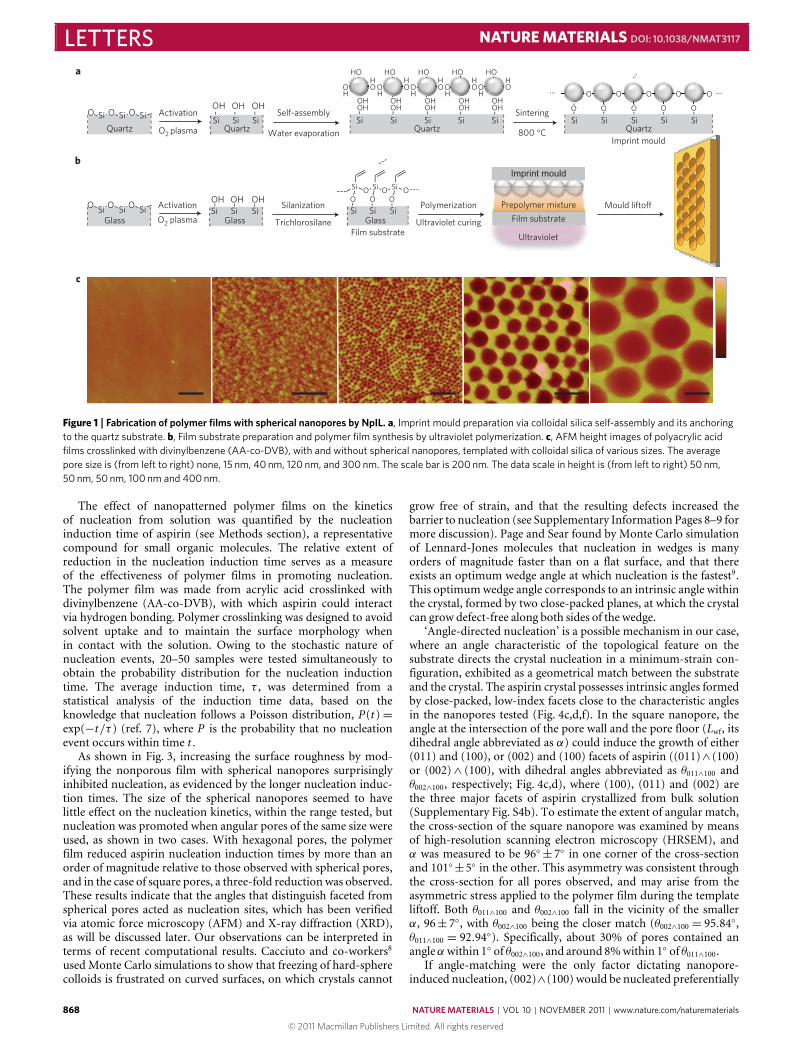
The fabrication of polymer films with spherical nanopores byNpIL is illustrated in Fig. 1. First, spherical silica nanoparticleswere self-assembled on a quartz slide driven by capillary forcesduring water evaporation17, and then anchored to the substrate viacalcination to form the imprint mould (Fig. 1a). Second, a mixtureof monomer, crosslinker and initiator was sandwiched between theimprint mould and the substrate, and subsequently polymerizedunder ultraviolet irradiation. The imprint mould was then easilypeeled off to reveal a polymer film conforming to the substrate,with the nanopattern inversely transferred from the imprint mould(Fig. 1b). Polymer films with spherical nanopores ranging from15 nm to 300 nmwere fabricated in thismanner (Fig. 1c), templatedby commercially available monodispersed colloidal silica of varioussizes. This method combines many of the advantages of NSLand ultraviolet-assisted NIL, such as low cost, high throughput18,and high resolution16. Moreover, in contrast to the commonlypracticed NSL technique, where hydrofluoric acid is needed todissolve the silica nanoparticles17, ourmethod removes the templatenondestructively by a simple liftoff from the polymer film, allowingthe mask to be recovered easily and reused.
Polymer films with hexagonal pores (Fig. 2a) were also preparedby NpIL following a similar procedure (see Methods section),templated with iron oxide magnetic nanocrystals with well-defined facets (Fig. 2b). For making square nanopores (Fig. 2c),square-shaped nanoposts (Fig. 2d) were fabricated by AchromaticInterference Lithography (AIL; ref. 19) as the imprint mould.The imprinted square pores are comparable to the sphericalones in width and depth (Fig. 2e), with sharply delineated poreangles (radius of curvature <3 nm, Supplementary Fig. S1). Inaddition, the nanopatterning procedures employed in this studypreserved the molecular level surface roughness with respect tothe nonporous polymer surface (Supplementary Table S1), whichenables unambiguous differentiation of the effects of pore shapeon crystal nucleation.
NATURE MATERIALS | VOL 10 | NOVEMBER 2011 | www.nature.com/naturematerials 867
© 2011 Macmillan Publishers Limited. All rights reserved
LETTERS NATURE MATERIALS DOI: 10.1038/NMAT3117
Si SiO Si OO ActivationOHOHOH
Self-assemblyOH OH OH OH OHOH OH OH OH OH
Sintering
......
...
------
---
a
b
c
O2 plasma Water evaporation
O O O O OH H H H H
H H H H HO O O O O
800 °C
O O O O O
Imprint mould
Mould liftoffPolymerization
Ultraviolet curingFilm substrate
GlassSi Si SiO O O
O O O
Silanization
TrichlorosilaneSi Si SiOH OH OH
Glass
Activation
O2 plasmaGlassSi Si Si
Quartz Quartz Quartz QuartzSi SiSi Si SiSi Si Si Si SiSi Si Si
O O O O O
HO HO HO HO HO
O O O
Si Si Si
Imprint mould
Prepolymer mixture
Film substrate
Ultraviolet
Figure 1 | Fabrication of polymer films with spherical nanopores by NpIL. a, Imprint mould preparation via colloidal silica self-assembly and its anchoringto the quartz substrate. b, Film substrate preparation and polymer film synthesis by ultraviolet polymerization. c, AFM height images of polyacrylic acidfilms crosslinked with divinylbenzene (AA-co-DVB), with and without spherical nanopores, templated with colloidal silica of various sizes. The averagepore size is (from left to right) none, 15 nm, 40 nm, 120 nm, and 300 nm. The scale bar is 200 nm. The data scale in height is (from left to right) 50 nm,50 nm, 50 nm, 100 nm and 400 nm.
The effect of nanopatterned polymer films on the kineticsof nucleation from solution was quantified by the nucleationinduction time of aspirin (see Methods section), a representativecompound for small organic molecules. The relative extent ofreduction in the nucleation induction time serves as a measureof the effectiveness of polymer films in promoting nucleation.The polymer film was made from acrylic acid crosslinked withdivinylbenzene (AA-co-DVB), with which aspirin could interactvia hydrogen bonding. Polymer crosslinking was designed to avoidsolvent uptake and to maintain the surface morphology whenin contact with the solution. Owing to the stochastic nature ofnucleation events, 20–50 samples were tested simultaneously toobtain the probability distribution for the nucleation inductiontime. The average induction time, τ , was determined from astatistical analysis of the induction time data, based on theknowledge that nucleation follows a Poisson distribution, P(t )=exp(−t/τ ) (ref. 7), where P is the probability that no nucleationevent occurs within time t .
As shown in Fig. 3, increasing the surface roughness by mod-ifying the nonporous film with spherical nanopores surprisinglyinhibited nucleation, as evidenced by the longer nucleation induc-tion times. The size of the spherical nanopores seemed to havelittle effect on the nucleation kinetics, within the range tested, butnucleation was promoted when angular pores of the same size wereused, as shown in two cases. With hexagonal pores, the polymerfilm reduced aspirin nucleation induction times by more than anorder of magnitude relative to those observed with spherical pores,and in the case of square pores, a three-fold reduction was observed.These results indicate that the angles that distinguish faceted fromspherical pores acted as nucleation sites, which has been verifiedvia atomic force microscopy (AFM) and X-ray diffraction (XRD),as will be discussed later. Our observations can be interpreted interms of recent computational results. Cacciuto and co-workers8used Monte Carlo simulations to show that freezing of hard-spherecolloids is frustrated on curved surfaces, on which crystals cannot
grow free of strain, and that the resulting defects increased thebarrier to nucleation (see Supplementary Information Pages 8–9 formore discussion). Page and Sear found by Monte Carlo simulationof Lennard-Jones molecules that nucleation in wedges is manyorders of magnitude faster than on a flat surface, and that thereexists an optimum wedge angle at which nucleation is the fastest9.This optimumwedge angle corresponds to an intrinsic angle withinthe crystal, formed by two close-packed planes, at which the crystalcan grow defect-free along both sides of the wedge.
‘Angle-directed nucleation’ is a possible mechanism in our case,where an angle characteristic of the topological feature on thesubstrate directs the crystal nucleation in a minimum-strain con-figuration, exhibited as a geometrical match between the substrateand the crystal. The aspirin crystal possesses intrinsic angles formedby close-packed, low-index facets close to the characteristic anglesin the nanopores tested (Fig. 4c,d,f). In the square nanopore, theangle at the intersection of the pore wall and the pore floor (Lwf, itsdihedral angle abbreviated as α) could induce the growth of either(011) and (100), or (002) and (100) facets of aspirin ((011)∧ (100)or (002)∧ (100), with dihedral angles abbreviated as θ011∧100 andθ002∧100, respectively; Fig. 4c,d), where (100), (011) and (002) arethe three major facets of aspirin crystallized from bulk solution(Supplementary Fig. S4b). To estimate the extent of angular match,the cross-section of the square nanopore was examined by meansof high-resolution scanning electron microscopy (HRSEM), andα was measured to be 96◦± 7◦ in one corner of the cross-sectionand 101◦±5◦ in the other. This asymmetry was consistent throughthe cross-section for all pores observed, and may arise from theasymmetric stress applied to the polymer film during the templateliftoff. Both θ011∧100 and θ002∧100 fall in the vicinity of the smallerα, 96± 7◦, with θ002∧100 being the closer match (θ002∧100 = 95.84◦,θ011∧100 = 92.94◦). Specifically, about 30% of pores contained anangleαwithin 1◦ of θ002∧100, and around 8%within 1◦ of θ011∧100.
If angle-matching were the only factor dictating nanopore-induced nucleation, (002)∧(100) would be nucleated preferentially
868 NATURE MATERIALS | VOL 10 | NOVEMBER 2011 | www.nature.com/naturematerials
© 2011 Macmillan Publishers Limited. All rights reserved

NATURE MATERIALS DOI: 10.1038/NMAT3117 LETTERS
Square pores
Spherical pores
a b
c d
e
Figure 2 | Angular nanopores on AA-co-DVB polymer films and theirtemplates. a, AFM height image of hexagonal nanopores on the polymersurface templated with iron oxide magnetic nanocrystals by means of NpIL.The scale bar is 50 nm. (Inset) Higher resolution image of a hexagonalnanopore. The scale bar is 10 nm. b, Transmission electron microscopyimage of iron oxide magnetic nanocrystals as synthesized. The scale bar is50 nm. c, AFM height image of square nanopores on the polymer surfacetemplated with Si square posts. The scale bar is 200 nm. d, High-resolutionSEM image of Si square posts on a Si wafer fabricated by AIL for templatingsquare pores. The scale bar is 200 nm. e, Depth profiles of square andspherical nanopores of similar sizes. The scale bar is 200 nm. The squarepores are 125 nm in width and 48 nm in depth, and the spherical pores are120 nm in width and 45 nm in depth, on average.
from Lwf within the pore. The AFM images of aspirin crystals grownfrom the pores suggest it was the (011)∧(100) facets that emanatedfrom Lwf, whereas the (002) facet was not in contact with the poresurface (Fig. 4a,b; Supplementary Fig. S2; see Supplementary Infor-mation Page 5 for assignment of crystal facets). Layered growth ofaspirin parallel to the pore floor is evident in both the crystal grownout from the pore (Fig. 4a) and the crystals contained in the pore(Fig. 4b), which originates from the aspirin dimerization throughthe carboxyl group within the (100) layer, and a much weaker vanderWaals interaction between the layers. Figure 4b shows that these(100) crystal layers seem to extend from the pore wall with whichthe (011) face is in contact. Moreover, the layer extension direction,as denoted by white arrows, is consistent in all pores containingcrystals, indicating nucleation occurs predominantly from one sideof the pore. In addition, only a fraction of the pores induced nucle-ation. These observations provide evidence that the (011)∧ (100)and not (002)∧ (100) facets were nucleated from Lwf, but onlyfrom those with the appropriate angle α. These growth patterns canbe attributed to the favourable interactions between (011)∧ (100)and the polymer surface, as inferred from the characteristicfunctionalities exhibited on their respective surfaces (Fig. 4c–e).(011) and (100) planes, rich in carboxyl and carbonyl groups, canform hydrogen bonds with the carboxyl groups on the AA-co-DVBpolymer surface, whereas the nonpolar (002) plane is likely tointeract with the polymer much more weakly. This result suggeststhat solute–polymer interactions, and not just the geometricalmatch, play an important role in determining nucleation behaviourin angular pores. Directed by both favourable interactions and
(min
)
0
100
200
300
400
500
600
No pore 15 nm 40 nm 120 nm
τ
Figure 3 | Effect of the nanopore shape in AA-co-DVB polymer films onthe nucleation kinetics of aspirin: spherical pores versus hexagonal poresand square pores of the same size. Nanopatterned surfaces are comparedagainst flat and smooth surfaces without pores, labelled as ‘no pore’. τ isthe average nucleation induction time. The standard errors of τ werecalculated from the regression on the induction time probabilitydistribution following the Poisson distribution.
angular match, the single crystals in square nanopores exhibiteda high degree of alignment (Supplementary Figs S4, S5), providingfurther evidence for nucleation at pore angles.
Following the principle of angle-directed nucleation assistedby favourable interactions, we propose that the corners withinhexagonal pores acted as nucleation sites to induce the growthof (011) ∧ (011̄) ∧ (100), where (100) was in contact with thepore floor, and (011)∧ (011̄) were in contact with the pore walls(Fig. 4f). This is plausible because the angle mismatch is verysmall in this configuration, and all three faces of aspirin couldinteract with the polymer surface through hydrogen bonding. Ifnucleation ensued from the corner, the growth thereafter wouldhave resulted in an aspirin crystallite that fitted comfortably insidethe pore and took on the shape of a hexagon, given that theother intrinsic angles of the crystal also matched fairly well withthe pore geometry (Fig. 4h). Indeed, crystallites with comparableshape and size to those of the pore were observed via AFM onthe surface of aspirin crystals detached from the polymer film(Fig. 4g,i). In addition, XRD results verified that the (100) face wasin contact with the pore floor (Supplementary Fig. S3). Moreover,in-plane alignment of hexagonal crystallites was also evident in localdomains (Supplementary Fig. S6). These observations support ourhypothesis of corner-induced nucleation fromhexagonal pores.
On the basis of the experimental and computational evidence,we propose a molecular mechanism to interpret the pore shapeeffect on nucleation. Crystal nucleation from solution is precededby molecular cluster formation through density fluctuations andmolecular re-orientation through structure fluctuations; both arenecessary for nucleation20–23. The rate of nucleation can bemodifiedin two ways by the presence of an amorphous, nanoporoussurface in a metastable solution. First, favourable surface–soluteinteractions enrich solute concentrations near the surface, andmolecular recognition events between the surface and the soluteinduce partial orientational order in the enriched solute layers; botheffects could facilitate nucleus formation7,24,25. Second, angles inthe pore further enhance the orientational order of the solute indomains close to the surface by means of geometrical confinement,which facilitates the solute molecular realignment during nucleusformation. When the molecular orientation imposed by the anglegeometry resembles that in the crystal, the rate of nucleation isincreased to the greatest extent, the macroscopic expression ofwhich is angle-directed nucleation.
As implied by our hypothesis, favourable surface–soluteinteraction is a prerequisite for angular nanopores to promotenucleation. To verify this point, we changed the chemical makeupof the polymer film from AA-co-DVB to AM-co-DVB (Fig. 5).
NATURE MATERIALS | VOL 10 | NOVEMBER 2011 | www.nature.com/naturematerials 869
© 2011 Macmillan Publishers Limited. All rights reserved

LETTERS NATURE MATERIALS DOI: 10.1038/NMAT3117
(011)
119.4°
(100)
(002)
HH H
15 nm
(100)
20 nm
d e
f
h
Square nanopore
g i
(100)
(002)
(002)
(011)
120.2
9°
(002)(011)
(002)
(011)
(002)
(011)
100 nm
a(011)
(002)
(011
)
(100)
(002)
(100)
92.9°
(011)
(100)
Growthdirection
b
Pores with crystalsEmpty pores
(01 1)
120.3°
(002) (01 1)
(002)
(01 1)(0 1 1)
(0 1 1)
(01 1)
Polymer film Polymer film
(011) (100)
C C
C CO O O O
O O OH3C
95.8°
c
Figure 4 | Angle-directed nucleation of aspirin crystals induced by angular nanopores. a, AFM phase image of aspirin crystals grown out from the squarepores. b, AFM phase image showing (100) layers of aspirin crystals nucleated at ledges in the square pores, indicated with white lines for all porescontaining crystals. The scale bar is 100 nm. c,d, Possible configurations of aspirin crystal facets in the square pore, with the cross-section depicted.e, Proposed aspirin–polymer interactions at the crystal-polymer interface. Between (100) and the polymer, the methyl hydrogen of (100) could interactwith the carbonyl oxygen of the polymer via a secondary hydrogen bond, which is not shown in the depiction. f, Proposed configuration of aspirin crystalfacets at the corner of a hexagonal pore. g,h, AFM phase image of an aspirin crystallite grown from the 15 nm hexagonal pores and its possible orientation.i, AFM height image of the surface of an aspirin crystal grown on and detached from the AA-co-DVB polymer film with hexagonal pores. The contours ofthe crystallites are traced at a small distance from the crystal edges so as not to obscure them.
This chemistry was selected out of the polymer films testedbecause, in the absence of pores, it exhibited no effect on aspirinnucleation from butyl acetate, indicating that surface–soluteinteractions are not sufficiently strong to affect nucleation underthese conditions (Supplementary Fig. S7). As expected, patterningof the AM-co-DVB surface with the same angular nanopores didnot lead to enhanced nucleation kinetics relative to nucleation onnonporous films (Fig. 5). With our new insight, nanostructuredmaterials can be designed to cater for a variety of applications,from controlling pharmaceutical polymorphism to inhibiting icenucleation on airplanes.
MethodsFabrication of polymer films with spherical nanopores. Quartz slides(75mm×25mm) were treated with O2 plasma to enrich the surface in hydroxylgroups. Two hundred microlitres of 5w% colloidal silica (commercially available)
were spread on the quartz slide and allowed to self-assemble during slowwater evaporation over 12 h. The self-assembled SiO2 and the quartz slidewere then sintered at 800 ◦C for 5min to coalesce the particles with the quartzslide and form the imprint mould. The film substrate (25mm×5mm) wasprepared by treating a glass slide with O2 plasma, followed by silanization withtrichlorosilane in a vacuum oven at 40 ◦C. Silanization is necessary to ‘glue’the polymer film to the substrate via covalent bonds and thereby avoid filmcracking and peeling from the substrates. One microlitre prepolymer mixtureof monomer acrylic acid (AA), crosslinker divinylbenzene (DVB), and initiatorIRGACURE 2022 were sandwiched between the imprint mould and the filmsubstrate. The molar ratio of monomer to DVB was 2:1. The concentrationof IRGACURE 2022 was 4 v% with respect to DVB. The prepolymer mixturewas then polymerized under ultraviolet irradiation for 15min, at 72mWcm−2.After irradiation, the imprint mould was peeled off and the polymer filmswere annealed at 70 ◦C in a vacuum oven for 3 h to remove unreacted species.Whenever possible, parts were pre-cleaned and assembled in a Bio-Safety cabinetto reduce contamination by impurities, which can interfere with polymer filminduced nucleation.
870 NATURE MATERIALS | VOL 10 | NOVEMBER 2011 | www.nature.com/naturematerials
© 2011 Macmillan Publishers Limited. All rights reserved

NATURE MATERIALS DOI: 10.1038/NMAT3117 LETTERS
OHO N
O
O
No pore
AA-co-DVB
AM-co-DVB
0
100
200
300
400
500
600
15 nm 125 nm AMAA
(min
)τ
Figure 5 | Effect of polymer surface chemistry on the kinetics of angularnanopore-induced nucleation of aspirin: AA-co-DVB versus AM-co-DVB.τ is the average nucleation induction time. AM denotes4-acryloylmorpholine. AA denotes acrylic acid. AM-co-DVB denotes poly4-acryloylmorpholine crosslinked with divinylbenzene.
Fabrication of polymer films with angular nanopores. Polymer films withhexagonal nanopores were synthesized following a procedure similar to thatdescribed above, templated with iron oxide magnetic nanoparticles (MNP).The nanoparticle synthesis scheme is included on Page 1 of the SupplementaryInformation. The presence of sufficient surfactants (oleic acid) during synthesiswas important for obtaining sharply defined facets of MNP crystals. As in the caseof producing spherical nanopores, colloidal self-assembly was used to preparethe imprint mould, which was made by spreading 20 µl MNP-decane solution(∼9w%) on a plasma cleaned quartz slide (75mm×25mm) and allowing thedecane to evaporate over a period of 6 h. The excessive surfactants present inthe nanocrystal dispersion also participated in the assembly process, leavingspace for polymers to form between the nanocrystals. After polymerization,the imprint mould was peeled off from the polymer film, the nanocrystals onthe film were subsequently dissolved with dilute hydrocholoric acid (∼1N),and the film was rinsed with deionized water, then with acetone, and vacuumdried. The imprint mould for making square pores was fabricated by (AIL;ref. 19) at the MIT Research Laboratory of Electronics. The mould took theform of 125 nm Si square pillar arrays with a 200 nm pitch covering a 3-inch Siwafer. The top edges of the pillars were sharply defined, with radii of curvatureless than 5 nm. Large area patterning is necessary to make sufficient copiesof polymer films to obtain the induction time probability distribution. Thepolymer-film synthesis and post-processing procedures were the same as thoseused in the preparation of spherical nanopores. The effects of polymer filmswith pores on nucleation kinetics were compared against those in the absenceof pores, which were synthesized following the same procedure, with the quartzsurface as the template.
Nucleation induction time measurement. Once synthesized, the polymer filmwith its substrate was inserted vertically into a 1ml glass shell vial containing200 µl 47mgml−1 aspirin solution in butyl acetate. For each polymer sample,20–50 vials were assembled and immersed in a circulator stabilized at 50±0.1 ◦Cto dissolve any pre-existing crystals, and then the solution was quench cooledto 5±0.1 ◦C by immersing into a second circulator. The supersaturation at thestart of each experiment was 2.2, defined as the ratio of starting concentrationto the equilibrium concentration at the crystallization temperature. The numberof vials in which crystallization occurred was recorded as a function of time.All the operations involving exposing polymer films, aspirin solutions and shellvials to the atmosphere were conducted inside a Bio-Safety cabinet to reduceimpurity contamination to the lowest possible level. Efforts were made to cleanall components before use, and aspirin solutions were filtered with an Acrodisc0.2 µm PTFE syringe filter.
Characterization. AFM and XRD were employed to study the aspirin crystalorientation inside the angular nanopores on the polymer films following thenucleation induction time study. AFM images were obtained with a Dimension3100 XY closed loop scanner (Nanoscope IV, VEECO) equipped with NanoMansoftware. Height and phase images were obtained in tapping mode in ambientair with silicon tips (VEECO). The crystal orientation was verified with XRD toidentify the specific crystallographic planes parallel to the polymer film. The X-raydiffraction patterns were recorded with a PANalytical X’Pert PRO Theta/ThetaPowder X-Ray Diffraction System with a Cu tube and X’Celerator high-speeddetector. No fewer than five polymer films were examined with XRD on eachtype of polymer sample.
Received 7 April 2011; accepted 10 August 2011; published online11 September 2011
References1. Debenedetti, P. G.Metastable Liquids: Concepts and Principles (Princeton Univ.
Press, 1996).2. Mullin, J. W. Crystallization 4th edn (Butterworth-Heinemann, 2001).3. Turnbull, D. Kinetics of heterogeneous nucleation. J. Chem. Phys. 18,
198–203 (1950).4. Curcio, E., Curcio, V., Di Profio, G., Fontananova, E. & Drioli, E. Energetics
of protein nucleation on rough polymeric surfaces. J. Phys. Chem. B 114,13650–13655 (2010).
5. Briseno, A. L. et al. Patterning organic single-crystal transistor arrays. Nature444, 913–917 (2006).
6. Ward, M. D. Bulk crystals to surfaces: Combining X-ray diffraction and atomicforce microscopy to probe the structure and formation of crystal interfaces.Chem. Rev. 101, 1697–1725 (2001).
7. Diao, Y., Myerson, A. S., Hatton, T. A. & Trout, B. L. Surface design forcontrolled crystallization: The role of surface chemistry and nanoscale pores inheterogeneous nucleation. Langmuir 27, 5324–5334 (2011).
8. Cacciuto, A., Auer, S. & Frenkel, D. Onset of heterogeneous crystal nucleationin colloidal suspensions. Nature 428, 404–406 (2004).
9. Page, A. J. & Sear, R. P. Crystallization controlled by the geometry of a surface.J. Am. Chem. Soc. 131, 17550–17551 (2009).
10. Chayen, N. E., Saridakis, E. & Sear, R. P. Experiment and theory forheterogeneous nucleation of protein crystals in a porous medium. Proc. NatlAcad. Sci. USA 103, 597–601 (2006).
11. Diao, Y. et al. Controlled nucleation from solution using polymer microgels.J. Am. Chem. Soc. 133, 3756–3759 (2011).
12. Ha, J. M., Wolf, J. H., Hillmyer, M. A. & Ward, M. D. Polymorph selectivityunder nanoscopic confinement. J. Am. Chem. Soc. 126, 3382–3383 (2004).
13. Hamilton, B. D., Weissbuch, I., Lahav, M., Hillmyer, M. A. & Ward,M. D. Manipulating crystal orientation in nanoscale cylindrical pores bystereochemical inhibition. J. Am. Chem. Soc. 131, 2588–2596 (2009).
14. Jackson, C. L. & McKenna, G. B. Vitrification and crystallization of organicliquids confined to nanoscale pores. Chem. Mater. 8, 2128–2137 (1996).
15. Maheshwari, P. et al. Effect of interfacial hydrogen bonding on thefreezing/melting behavior of nanoconfined liquids. J. Phys. Chem. C 114,4966–4972 (2010).
16. Stewart, M. D. & Willson, C. G. Imprint materials for nanoscale devices.MRS Bull. 30, 947–951 (2005).
17. Xia, Y. N., Gates, B., Yin, Y. D. & Lu, Y. Monodispersed colloidal spheres: Oldmaterials with new applications. Adv. Mater. 12, 693–713 (2000).
18. Trujillo, N. J., Baxamusa, S. H. & Gleason, K. K. Grafted functional polymernanostructures patterned bottom-up by colloidal lithography and initiatedchemical vapor deposition (iCVD). Chem. Mater. 21, 742–750 (2009).
19. Savas, T. A., Schattenburg, M. L., Carter, J. M. & Smith, H. I. Large-areaachromatic interferometric lithography for 100 nm period gratings and grids.J. Vacuum Sci. Technol. B 14, 4167–4170 (1996).
20. tenWolde, P. R. & Frenkel, D. Enhancement of protein crystal nucleation bycritical density fluctuations. Science 277, 1975–1978 (1997).
21. Erdemir, D., Lee, A. Y. & Myerson, A. S. Nucleation of crystals from solution:Classical and two-step models. Acc. Chem. Res. 42, 621–629 (2009).
22. Santiso, E. E. & Trout, B. L. A general set of order parameters for molecularcrystals. J. Chem. Phys. 134, 064109 (2011).
23. Vekilov, P. G. Dense liquid precursor for the nucleation of ordered solid phasesfrom solution. Cryst. Growth Des. 4, 671–685 (2004).
24. van Meel, J. A., Sear, R. P. & Frenkel, D. Design principles for broad-spectrumprotein-crystal nucleants with nanoscale pits. Phys. Rev. Lett. 105,205501 (2010).
25. Dey, A. et al. The role of prenucleation clusters in surface-induced calciumphosphate crystallization. Nature Mater. 9, 1010–1014 (2010).
AcknowledgementsWe acknowledge the Novartis-MIT Center for Continuous Manufacturing for funding.We are grateful to T. Savas at MIT Research Laboratory of Electronics for fabricating theimprint mould with Si square nanopillars and to K. Gleason for use of her equipment forplasma treatment and glass silanization.
Author contributionsY.D. designed, carried out the experiments and wrote the manuscript. B.L.T.,T.A.H. and A.S.M. supervised the work, guided and revised the manuscript. T.H.synthesized and characterized the Fe3O4 magnetic nanoparticles and co-wrote theSupplementary Information.
Additional informationThe authors declare no competing financial interests. Supplementary informationaccompanies this paper on www.nature.com/naturematerials. Reprints and permissionsinformation is available online at http://www.nature.com/reprints. Correspondence andrequests for materials should be addressed to B.L.T.
NATURE MATERIALS | VOL 10 | NOVEMBER 2011 | www.nature.com/naturematerials 871
© 2011 Macmillan Publishers Limited. All rights reserved