The Role of Hypoxia-Inducible Factor-1 in Acetaminophen...
11
The Role of Hypoxia-Inducible Factor-1 in Acetaminophen Hepatotoxicity Erica M. Sparkenbaugh, Yogesh Saini, Krista K. Greenwood, John J. LaPres, James P. Luyendyk, Bryan L. Copple, Jane F. Maddox, Patricia E. Ganey, and Robert A. Roth Departments of Pharmacology and Toxicology (E.M.S., J.F.M., P.E.G., R.A.R.) and Biochemistry and Molecular Biology (Y.S., K.K.G., J.J.L.), and Center for Integrative Toxicology (E.M.S., Y.S., K.K.G., J.J.L., J.F.M., P.E.G., R.A.R.), Michigan State University, East Lansing, Michigan; and Department of Pharmacology, Toxicology, and Therapeutics, University of Kansas Medical Center, Kansas City, Kansas (J.P.L., B.L.C.) Received February 12, 2011; accepted May 12, 2011 ABSTRACT Hypoxia-inducible factor-1 (HIF-1) is a critical transcription factor that controls oxygen homeostasis in response to hyp- oxia, inflammation, and oxidative stress. HIF has been impli- cated in the pathogenesis of liver injury in which these events play a role, including acetaminophen (APAP) overdose, which is the leading cause of acute liver failure in the United States. APAP overdose has been reported to activate HIF-1 in mouse livers and isolated hepatocytes downstream of oxidative stress. HIF-1 signaling controls many factors that contribute to APAP hepatotoxicity, including mitochondrial cell death, inflamma- tion, and hemostasis. Therefore, we tested the hypothesis that HIF-1 contributes to APAP hepatotoxicity. Conditional HIF-1 deletion was generated in mice using an inducible Cre-lox system. Control (HIF-1-sufficient) mice developed severe liver injury 6 and 24 h after APAP overdose (400 mg/kg). HIF-1- deficient mice were protected from APAP hepatotoxicity at 6 h, but developed severe liver injury by 24 h, suggesting that HIF-1 is involved in the early stage of APAP toxicity. In further studies, HIF-1-deficient mice had attenuated thrombin gener- ation and reduced plasminogen activator inhibitor-1 production compared with control mice, indicating that HIF-1 signaling contributes to hemostasis in APAP hepatotoxicity. Finally, HIF- 1-deficient animals had decreased hepatic neutrophil accu- mulation and plasma concentrations of interleukin-6, keratino- cyte chemoattractant, and regulated upon activation normal T cell expressed and secreted compared with control mice, sug- gesting an altered inflammatory response. HIF-1 contributes to hemostasis, sterile inflammation, and early hepatocellular necrosis during the pathogenesis of APAP toxicity. Introduction Hypoxia-inducible factor (HIF) is the master regulator of oxygen homeostasis. It regulates the expression of a large battery of genes involved in angiogenesis, erythropoiesis, glycolysis, inflammation, and cell death (Lee et al., 2007). HIF comprises two constitutively expressed subunits: HIF-1 and HIF-1. HIF-1 is regulated primarily at the level of protein stability: at normal oxygen tension, oxygen- dependent proline hydroxylation of HIF-1 targets it for rapid proteasomal degradation. In hypoxia, decreased pro- line hydroxylation causes HIF-1 to accumulate and trans- locate to the nucleus, where it binds to HIF-1, forming the transcriptionally competent HIF-1 that binds hypoxia re- sponse elements in DNA. HIF-regulated genes include plas- minogen activator inhibitor-1 (PAI-1), vascular endothelial growth factor (VEGF), tumor necrosis factor (TNF), inter- leukin-1 (IL-1), and cell death proteins such as BNIP3 and This research was supported by the National Institutes of Health National Institute of Environmental Health Sciences [Grants R01-ES004139, R01- ES12186]. E.M.S. was supported in part by the National Institutes of Health National Institute of Environmental Health Sciences [Training Grant T32 ES007255]. Portions of this work were presented previously: Sparkenbaugh EM, Saini Y, LaPres JJ, Maddox JF, Ganey PE, and Roth RA (2010) HIF-1 deletion protects mice from acetaminophen hepatotoxicity and reduces activation of the hemostatic system, at The Society of Toxicology Annual Meeting; 2010 March 7–11; Salt Lake City, UT. Society of Toxicology, Reston, VA. Article, publication date, and citation information can be found at http://jpet.aspetjournals.org. doi:10.1124/jpet.111.180521. ABBREVIATIONS: HIF, hypoxia-inducible factor; ALT, alanine aminotransferase; APAP, N-acetyl-p-aminophenol; IL, interleukin; KC, keratinocyte chemoattractant; PA, plasminogen activator; PAI-1, PA inhibitor-1; PMN, polymorphonuclear neutrophil; RANTES, regulated upon activation normal T cell expressed and secreted; TAM, tamoxifen; TNF, tumor necrosis factor; VEGF, vascular endothelial growth factor; PCR, polymerase chain reaction; SAL, saline; OIL, corn oil; BB, blocking buffer; Cox IV, cytochrome c oxidase subunit IV; MIP, macrophage inflammatory protein; TAT, thrombin- antithrombin; HPRT, hypoxanthine guanine phosphoribosyl transferase; BNIP3, BCL2/adenovirus E1B 19-kDa protein-interacting protein 3. 0022-3565/11/3382-492–502$25.00 THE JOURNAL OF PHARMACOLOGY AND EXPERIMENTAL THERAPEUTICS Vol. 338, No. 2 Copyright © 2011 by The American Society for Pharmacology and Experimental Therapeutics 180521/3703925 JPET 338:492–502, 2011 Printed in U.S.A. 492 at ASPET Journals on May 30, 2018 jpet.aspetjournals.org Downloaded from
Transcript of The Role of Hypoxia-Inducible Factor-1 in Acetaminophen...

The Role of Hypoxia-Inducible Factor-1� in AcetaminophenHepatotoxicity
Erica M. Sparkenbaugh, Yogesh Saini, Krista K. Greenwood, John J. LaPres,James P. Luyendyk, Bryan L. Copple, Jane F. Maddox, Patricia E. Ganey,and Robert A. RothDepartments of Pharmacology and Toxicology (E.M.S., J.F.M., P.E.G., R.A.R.) and Biochemistry and Molecular Biology (Y.S.,K.K.G., J.J.L.), and Center for Integrative Toxicology (E.M.S., Y.S., K.K.G., J.J.L., J.F.M., P.E.G., R.A.R.), Michigan StateUniversity, East Lansing, Michigan; and Department of Pharmacology, Toxicology, and Therapeutics, University of KansasMedical Center, Kansas City, Kansas (J.P.L., B.L.C.)
Received February 12, 2011; accepted May 12, 2011
ABSTRACTHypoxia-inducible factor-1� (HIF-1�) is a critical transcriptionfactor that controls oxygen homeostasis in response to hyp-oxia, inflammation, and oxidative stress. HIF has been impli-cated in the pathogenesis of liver injury in which these eventsplay a role, including acetaminophen (APAP) overdose, which isthe leading cause of acute liver failure in the United States.APAP overdose has been reported to activate HIF-1� in mouselivers and isolated hepatocytes downstream of oxidative stress.HIF-1� signaling controls many factors that contribute to APAPhepatotoxicity, including mitochondrial cell death, inflamma-tion, and hemostasis. Therefore, we tested the hypothesis thatHIF-1� contributes to APAP hepatotoxicity. Conditional HIF-1�deletion was generated in mice using an inducible Cre-loxsystem. Control (HIF-1�-sufficient) mice developed severe liverinjury 6 and 24 h after APAP overdose (400 mg/kg). HIF-1�-
deficient mice were protected from APAP hepatotoxicity at 6 h,but developed severe liver injury by 24 h, suggesting thatHIF-1� is involved in the early stage of APAP toxicity. In furtherstudies, HIF-1�-deficient mice had attenuated thrombin gener-ation and reduced plasminogen activator inhibitor-1 productioncompared with control mice, indicating that HIF-1� signalingcontributes to hemostasis in APAP hepatotoxicity. Finally, HIF-1�-deficient animals had decreased hepatic neutrophil accu-mulation and plasma concentrations of interleukin-6, keratino-cyte chemoattractant, and regulated upon activation normal Tcell expressed and secreted compared with control mice, sug-gesting an altered inflammatory response. HIF-1� contributesto hemostasis, sterile inflammation, and early hepatocellularnecrosis during the pathogenesis of APAP toxicity.
IntroductionHypoxia-inducible factor (HIF) is the master regulator of
oxygen homeostasis. It regulates the expression of a large
battery of genes involved in angiogenesis, erythropoiesis,glycolysis, inflammation, and cell death (Lee et al., 2007).HIF comprises two constitutively expressed subunits:HIF-1� and HIF-1�. HIF-1� is regulated primarily at thelevel of protein stability: at normal oxygen tension, oxygen-dependent proline hydroxylation of HIF-1� targets it forrapid proteasomal degradation. In hypoxia, decreased pro-line hydroxylation causes HIF-1� to accumulate and trans-locate to the nucleus, where it binds to HIF-1�, forming thetranscriptionally competent HIF-1 that binds hypoxia re-sponse elements in DNA. HIF-regulated genes include plas-minogen activator inhibitor-1 (PAI-1), vascular endothelialgrowth factor (VEGF), tumor necrosis factor � (TNF�), inter-leukin-1� (IL-1�), and cell death proteins such as BNIP3 and
This research was supported by the National Institutes of Health NationalInstitute of Environmental Health Sciences [Grants R01-ES004139, R01-ES12186]. E.M.S. was supported in part by the National Institutes of HealthNational Institute of Environmental Health Sciences [Training Grant T32ES007255].
Portions of this work were presented previously: Sparkenbaugh EM, SainiY, LaPres JJ, Maddox JF, Ganey PE, and Roth RA (2010) HIF-1� deletionprotects mice from acetaminophen hepatotoxicity and reduces activation of thehemostatic system, at The Society of Toxicology Annual Meeting; 2010 March7–11; Salt Lake City, UT. Society of Toxicology, Reston, VA.
Article, publication date, and citation information can be found athttp://jpet.aspetjournals.org.
doi:10.1124/jpet.111.180521.
ABBREVIATIONS: HIF, hypoxia-inducible factor; ALT, alanine aminotransferase; APAP, N-acetyl-p-aminophenol; IL, interleukin; KC, keratinocytechemoattractant; PA, plasminogen activator; PAI-1, PA inhibitor-1; PMN, polymorphonuclear neutrophil; RANTES, regulated upon activation normal Tcell expressed and secreted; TAM, tamoxifen; TNF, tumor necrosis factor; VEGF, vascular endothelial growth factor; PCR, polymerase chain reaction;SAL, saline; OIL, corn oil; BB, blocking buffer; Cox IV, cytochrome c oxidase subunit IV; MIP, macrophage inflammatory protein; TAT, thrombin-antithrombin; HPRT, hypoxanthine guanine phosphoribosyl transferase; BNIP3, BCL2/adenovirus E1B 19-kDa protein-interacting protein 3.
0022-3565/11/3382-492–502$25.00THE JOURNAL OF PHARMACOLOGY AND EXPERIMENTAL THERAPEUTICS Vol. 338, No. 2Copyright © 2011 by The American Society for Pharmacology and Experimental Therapeutics 180521/3703925JPET 338:492–502, 2011 Printed in U.S.A.
492
at ASPE
T Journals on M
ay 30, 2018jpet.aspetjournals.org
Dow
nloaded from

Nix (Murdoch et al., 2005; Lee et al., 2007). HIF-1� expres-sion and activation are also regulated by oxidative stress(Klimova and Chandel, 2008), inflammatory cytokines(Walmsley et al., 2005a), and thrombin (Gorlach et al., 2001).Because of the many factors that can modulate HIF inductionand the variety of downstream signaling targets, HIF hasbeen identified as a key regulator of a generalized stressresponse (James et al., 2006).
HIF-1� has been implicated in hepatocyte death in modelsof liver injury that have an inflammatory or oxidative stresscomponent, such as sepsis (Peyssonnaux et al., 2007), isch-emia/reperfusion (Cursio et al., 2008), alcoholic liver disease(Li et al., 2006), and fibrosis (Copple et al., 2009). Oxidativestress and mitochondrial dysfunction play a key role in acet-aminophen [N-acetyl-p-aminophenol (APAP)]-induced liverinjury (James et al., 2003). APAP overdose is the leadingcause of drug-induced liver failure in the United States (Lee,2007). At toxic doses, APAP is bioactivated by cytochromeP450 enzymes to n-acetyl-p-benzoquinone imine, which isreactive, depletes GSH, and binds covalently to intracellularproteins, leading to mitochondrial dysfunction, production ofreactive oxygen species, and hepatocellular necrosis (Jollowet al., 1973).
A report by James et al. (2006) indicated that APAP over-dose causes nuclear accumulation of HIF-1� in mouse liversas early as 1 h after treatment, which is before the onset ofliver hypoxia and hepatocellular injury. Furthermore, N-acetyl cysteine, which inactivates n-acetyl-p-benzoquinoneimine (James et al., 2006) or cyclosprin A, which preventsmitochondrial permeability transition, prevented HIF-1� ac-cumulation (Chaudhuri et al., 2011). Taken together, thesedata suggest that mitochondrial dysfunction and reactiveoxygen species are important contributors to early HIF-1�stabilization in APAP overdose.
In addition to cellular necrosis caused by oxidative stressand mitochondrial dysfunction, APAP hepatotoxicity is asso-ciated with disturbances to the hemostatic system in humans(James et al., 2002) and experimental animals (Ganey et al.,2007). APAP overdose caused tissue factor-dependent activa-tion of the coagulation system in mice, elevated circulatingconcentration of PAI-1, and fibrin deposition in liver (Ganeyet al., 2007). Inhibition of coagulation system activationthrough genetic or pharmacologic methods attenuatedAPAP-induced liver injury, suggesting a role for thrombinand the coagulation system in the pathogenesis. During in-jury progression, fibrin deposition can contribute to tissueischemia and hypoxia, which might enhance HIF-1� accumu-lation above that caused by oxidative stress alone.
APAP hepatotoxicity is accompanied by a sterile inflam-matory response (Williams et al., 2010), and concurrent in-flammation can sensitize mice to APAP-induced liver injury(Maddox et al., 2010). Mediators released from necrotic hepa-tocytes activate Kupffer cells, recruit and activate polymor-phonuclear neutrophils (PMNs), and consequently producecytokines that influence APAP-induced hepatocellular injury(James et al., 2005; Cover et al., 2006). The role of PMNs inAPAP hepatotoxicity remains controversial, with evidenceboth for and against a contribution of PMNs to injury pro-gression (Jaeschke, 2008). HIF-1� plays a critical role inPMN function; it influences phagocytosis, motility, invasive-ness, and apoptosis (Cramer et al., 2003; Peyssonnaux et al.,2005; Walmsley et al., 2005a,b). HIF-1� also contributes to
inflammatory cytokine production (Zinkernagel et al., 2007).Therefore, HIF-1� might participate in the inflammatoryresponse that accompanies APAP-induced liver injury.
In addition to the many factors mentioned above that associ-ate APAP-induced liver injury with hypoxia signaling, HIF-1�can contribute directly to the cell death of hepatocytes by up-regulation of cell death genes. Nonetheless, it is currently un-known whether HIF-1� is involved causally in APAP-inducedliver injury. To test the hypothesis that HIF-1� contributes tothe pathogenesis of APAP-induced liver injury, conditionalHIF-1�-deficient animals were generated, and the role ofHIF-1� in APAP-induced hepatotoxicity, disruption of hemosta-sis, and inflammation was evaluated.
Materials and MethodsMaterials. Unless otherwise stated, all reagents were purchased
from Sigma-Aldrich (St. Louis, MO).Generation of Conditional HIF-1�-Deficient Animals. HIF-
1�flox/flox mice (Ryan et al., 1998) were a gift from Randall Johnson(University of California, San Diego, CA), and UBC-Cre-ERT2(�/�)mice were purchased from The Jackson Laboratory (Bar Harbor, ME).The Cre-ERT2 is regulated by the ubiquitin C promoter and is ex-pressed in virtually all cell types. Cre-ERT2 is a fusion protein com-posed of Cre recombinase and a mutated estrogen receptor that isselectively activated and targeted to the nucleus by (Z)-1-(p-dimethylaminoethoxyphenyl)-1,2-diphenyl-1-butene,trans-2-[4-(1,2-diphenyl-1-butenyl)phenoxy]-N,N-dimethylethylamine [tamoxifen(TAM)] but not estrogen (Ruzankina et al., 2007). C57BL/6 HIF-1�flox/flox
and UBC-Cre-ERT2(�/�) transgenic mice were mated to generateUBC-Cre-ERT2(�/�)/HIF-1�flox/flox mice capable of conditional recom-bination in the floxed HIF-1� gene when treated with TAM. MaleUBC-Cre-ERT2(�/�)/HIF-1�flox/flox mice (4–5 weeks old) were treatedonce a day for 5 days with 200 �g/g body weight TAM in corn oil (OIL)vehicle by oral gavage (Ruzankina et al., 2007). TAM-treated UBC-Cre-ERT2(�/�)/HIF-1�flox/flox mice were HIF-1�-deficient (denoted as HIF-1��/�), and OIL-treated animals were HIF-1�-sufficient (denoted asHIF-1��fl/�fl) (Fig. 1A). UBC-Cre-ERT2(�/�)/HIF-1�flox/flox littermatecontrols were treated with OIL or TAM to evaluate the potential effectsof TAM on APAP metabolism. Animals kept in a 12-h light/dark cyclewere fed a standard rodent chow/Tek 8640 (Harlan Teklad, Madison,WI) and allowed access to water ad libitum. All procedures were per-formed according to the guidelines of the American Association forLaboratory Animal Science and the University Laboratory Animal Re-search Unit at Michigan State University.
Experimental Protocol. Eleven or 21 days after OIL or TAMadministration, mice were fasted overnight then given 400 mg/kgAPAP or saline (SAL) vehicle via intraperitoneal injection, and foodwas returned. Mice were anesthetized 2, 6, or 24 h after APAP withsodium pentobarbital (50 mg/kg i.p.), and blood was collected fromthe vena cava into a syringe containing sodium citrate (final concen-tration 0.76%) for preparation of plasma. The left lateral liver lobewas fixed in 10% formalin and paraffin-blocked for evaluation ofhistopathology. The left medial lobe was snap-frozen in liquid nitro-gen for protein, DNA, and RNA analysis. The right medial lobe wasembedded in Tissue-Tek O.C.T. compound and frozen in liquid ni-trogen-cooled isopentane for immunohistochemical analyses.
Genotyping and Real-Time PCR. Genotyping of mice was per-formed for the Cre transgene, HIF-1�, and HIF-1�flox/flox mice usingpreviously published primer sequences (Saini et al., 2008) (Table 1).Genomic DNA was extracted from tail clippings using the Direct PCRextraction system (Viagen Biotech, Los Angeles, CA) and used to quan-tify the Cre transgene. Genotyping of livers from HIF-1��fl/�fl andHIF-1��/� mice was performed to determine the recombination effi-ciency. Genomic DNA was extracted using the Extract-N-Amp system(Sigma-Aldrich) according to the manufacturer’s instructions. PCR con-
Role of HIF-1� in Acetaminophen Hepatotoxicity 493
at ASPE
T Journals on M
ay 30, 2018jpet.aspetjournals.org
Dow
nloaded from
ditions were standardized for all alleles: denaturation at 94°C for 3 min, 38cycles of denaturation at 94°C for 45 s, annealing at 60°C for 45 s, andpolymerization at 72°C for 60 s followed by a 7-min extension at 72°C.
Gene Expression Analysis. Liver tissue (50 mg) was homoge-nized in 1 ml of TRI reagent (Sigma-Aldrich) using a Precellys 24Tissue Homogenizer (Cayman Chemical, Ann Arbor, MI), and RNAwas extracted. Total RNA was quantified spectrophotometricallyusing the NanoDrop 2000 (Thermo Fisher Scientific, Waltham, MA).Total RNA (1 �g) was reverse-transcribed using an iScript cDNAsynthesis kit (Bio-Rad Laboratories, Hercules, CA). The expressionof PAI-1, BNIP3, and HIF-1� was analyzed by quantitative real-timePCR using SYBR green (Applied Biosystems, Foster City, CA). Copynumber was determined by comparison with standard curves of therespective genes. Expression level was normalized to the hypoxan-
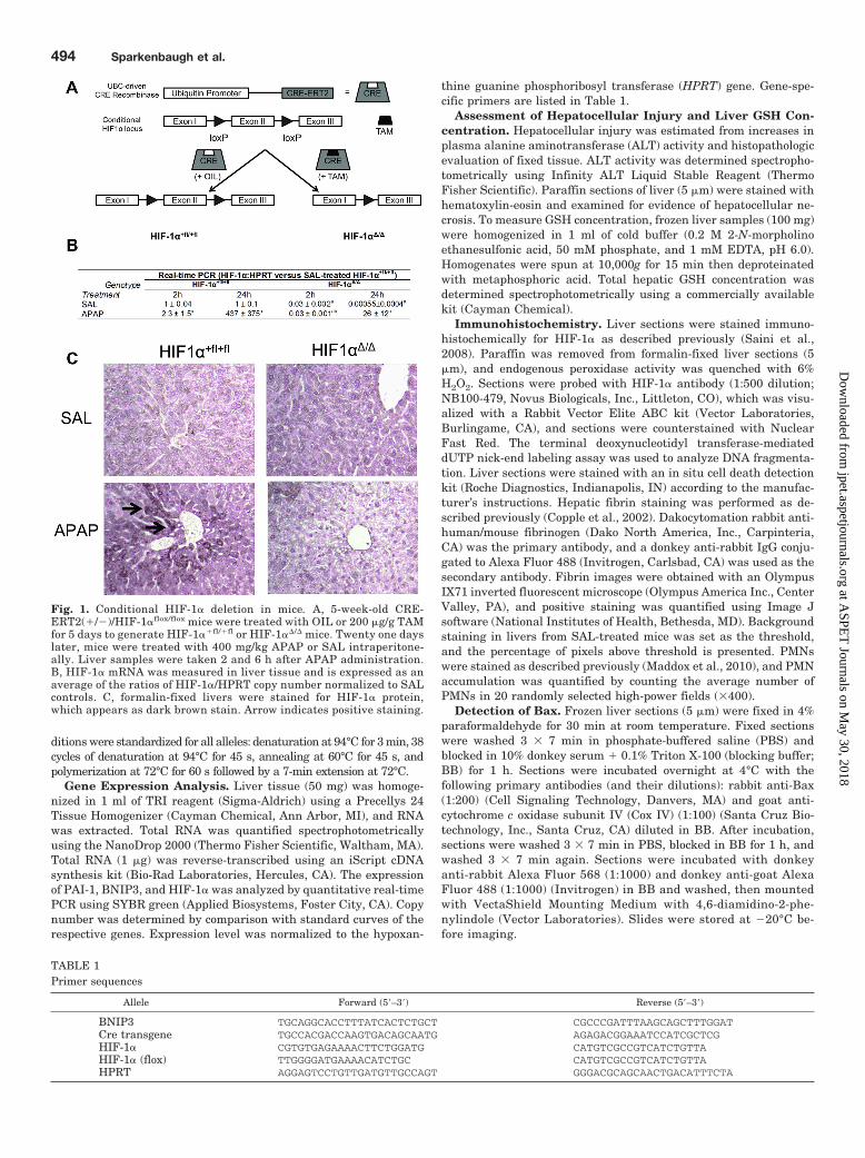
thine guanine phosphoribosyl transferase (HPRT) gene. Gene-spe-cific primers are listed in Table 1.
Assessment of Hepatocellular Injury and Liver GSH Con-centration. Hepatocellular injury was estimated from increases inplasma alanine aminotransferase (ALT) activity and histopathologicevaluation of fixed tissue. ALT activity was determined spectropho-tometrically using Infinity ALT Liquid Stable Reagent (ThermoFisher Scientific). Paraffin sections of liver (5 �m) were stained withhematoxylin-eosin and examined for evidence of hepatocellular ne-crosis. To measure GSH concentration, frozen liver samples (100 mg)were homogenized in 1 ml of cold buffer (0.2 M 2-N-morpholinoethanesulfonic acid, 50 mM phosphate, and 1 mM EDTA, pH 6.0).Homogenates were spun at 10,000g for 15 min then deproteinatedwith metaphosphoric acid. Total hepatic GSH concentration wasdetermined spectrophotometrically using a commercially availablekit (Cayman Chemical).
Immunohistochemistry. Liver sections were stained immuno-histochemically for HIF-1� as described previously (Saini et al.,2008). Paraffin was removed from formalin-fixed liver sections (5�m), and endogenous peroxidase activity was quenched with 6%H2O2. Sections were probed with HIF-1� antibody (1:500 dilution;NB100-479, Novus Biologicals, Inc., Littleton, CO), which was visu-alized with a Rabbit Vector Elite ABC kit (Vector Laboratories,Burlingame, CA), and sections were counterstained with NuclearFast Red. The terminal deoxynucleotidyl transferase-mediateddUTP nick-end labeling assay was used to analyze DNA fragmenta-tion. Liver sections were stained with an in situ cell death detectionkit (Roche Diagnostics, Indianapolis, IN) according to the manufac-turer’s instructions. Hepatic fibrin staining was performed as de-scribed previously (Copple et al., 2002). Dakocytomation rabbit anti-human/mouse fibrinogen (Dako North America, Inc., Carpinteria,CA) was the primary antibody, and a donkey anti-rabbit IgG conju-gated to Alexa Fluor 488 (Invitrogen, Carlsbad, CA) was used as thesecondary antibody. Fibrin images were obtained with an OlympusIX71 inverted fluorescent microscope (Olympus America Inc., CenterValley, PA), and positive staining was quantified using Image Jsoftware (National Institutes of Health, Bethesda, MD). Backgroundstaining in livers from SAL-treated mice was set as the threshold,and the percentage of pixels above threshold is presented. PMNswere stained as described previously (Maddox et al., 2010), and PMNaccumulation was quantified by counting the average number ofPMNs in 20 randomly selected high-power fields (�400).
Detection of Bax. Frozen liver sections (5 �m) were fixed in 4%paraformaldehyde for 30 min at room temperature. Fixed sectionswere washed 3 � 7 min in phosphate-buffered saline (PBS) andblocked in 10% donkey serum � 0.1% Triton X-100 (blocking buffer;BB) for 1 h. Sections were incubated overnight at 4°C with thefollowing primary antibodies (and their dilutions): rabbit anti-Bax(1:200) (Cell Signaling Technology, Danvers, MA) and goat anti-cytochrome c oxidase subunit IV (Cox IV) (1:100) (Santa Cruz Bio-technology, Inc., Santa Cruz, CA) diluted in BB. After incubation,sections were washed 3 � 7 min in PBS, blocked in BB for 1 h, andwashed 3 � 7 min again. Sections were incubated with donkeyanti-rabbit Alexa Fluor 568 (1:1000) and donkey anti-goat AlexaFluor 488 (1:1000) (Invitrogen) in BB and washed, then mountedwith VectaShield Mounting Medium with 4,6-diamidino-2-phe-nylindole (Vector Laboratories). Slides were stored at �20°C be-fore imaging.
TABLE 1Primer sequences
Allele Forward (5�–3�) Reverse (5�–3�)
BNIP3 TGCAGGCACCTTTATCACTCTGCT CGCCCGATTTAAGCAGCTTTGGATCre transgene TGCCACGACCAAGTGACAGCAATG AGAGACGGAAATCCATCGCTCGHIF-1� CGTGTGAGAAAACTTCTGGATG CATGTCGCCGTCATCTGTTAHIF-1� (flox) TTGGGGATGAAAACATCTGC CATGTCGCCGTCATCTGTTAHPRT AGGAGTCCTGTTGATGTTGCCAGT GGGACGCAGCAACTGACATTTCTA
Fig. 1. Conditional HIF-1� deletion in mice. A, 5-week-old CRE-ERT2(�/�)/HIF-1�flox/flox mice were treated with OIL or 200 �g/g TAMfor 5 days to generate HIF-1��fl/�fl or HIF-1��/� mice. Twenty one dayslater, mice were treated with 400 mg/kg APAP or SAL intraperitone-ally. Liver samples were taken 2 and 6 h after APAP administration.B, HIF-1� mRNA was measured in liver tissue and is expressed as anaverage of the ratios of HIF-1�/HPRT copy number normalized to SALcontrols. C, formalin-fixed livers were stained for HIF-1� protein,which appears as dark brown stain. Arrow indicates positive staining.
494 Sparkenbaugh et al.
at ASPE
T Journals on M
ay 30, 2018jpet.aspetjournals.org
Dow
nloaded from

Fluorescent slides were viewed with the Olympus FluoView 1000confocal laser scanning microscope. Images were collected withOlympus FluoView 1000 software, version 2.0. Alexa Fluor 568 was
detected with a 543-nm HeNeG laser with a BA 560-620 emissionfilter, and Alexa Fluor 488 was detected with a 488-nm Ar laser witha BA 505-525 emission filter. Images were scanned with sequentialscan setting for the two lasers. A 60� oil Plan/APO objective (nu-merical aperture 1.42) was used to acquire images. Five fields of viewfrom each liver section were selected at random. Colocalization ofBax and Cox IV pixels was analyzed with Image J software (NationalInstitutes of Health), and data are represented as the percentage ofcolocalized pixels in an image area.
Evaluation of Plasma and Intrahepatic Cytokine Concen-trations. The plasma concentration of active PAI-1 was measuredwith a commercially available enzyme-linked immunosorbent assaykit (Molecular Innovations, Southfield, MI), following the manufac-turer’s instructions. The plasma concentrations of interferon-, IL-1�, IL-2, IL-4, IL-6, IL-10, IL-12 (p70), keratinocyte chemoattractant(KC), macrophage inflammatory protein (MIP)-1�, regulated uponactivation normal T cell expressed and secreted (RANTES), TNF-�,and VEGF were measured with a custom Milliplex MAP kit formouse cytokines (Millipore Corporation, Billerica, MA) using theBio-Plex 200 System (Bio-Rad Laboratories). For determination ofhepatic cytokine concentrations, livers were homogenized in 0.1%Triton X-100 in PBS containing Halt Protease and Phosphataseinhibitors (Thermo Fisher Scientific), and proteins were quantifiedby the bicinchoninic acid assay. Concentrations of KC and RANTESwere determined by enzyme-linked immunosorbent assay (R&D Sys-tems, Minneapolis, MN) and normalized to protein concentrations ofthe samples.
Statistical Analyses. All data are represented as mean S.E.M.Data that were not normally distributed were transformed via Box-cox power transformation. Two- or three-way analysis of variancewas used as appropriate, and multiple comparisons were evaluated
Fig. 2. Effect of HIF-1� deletion on APAP hepatotoxicity. Five-week-oldCRE-ERT2(�/�)/HIF-1�flox/flox mice [labeled Cre (�)/HIF-1�fl/fl] or CRE-ERT2(�/�)/HIF-1�flox/flox mice [labeled Cre (�)/HIF-1�fl/fl] were treated withOIL or 200 �g/g TAM daily for 5 days. Twenty one days later, mice weretreated with APAP or SAL intraperitoneally. Plasma ALT activity wasmeasured 6 h after APAP administration. Data represent means S.E.M. ofn � 3–8 animals per group. a indicates significantly different from respec-tive SAL-treated mice; b indicates significantly different from OIL-treatedmice; c indicates significantly different from HIF-1��fl/�fl mice.
Fig. 3. Time course of APAP-induced liver injury in HIF-1�-deficient mice. HIF-1��fl/�fl or HIF-1��/� mice weretreated with APAP (400 mg/kg) or SAL, and plasma andliver samples were taken 2, 6, and 24 h later. A, liver injurywas assessed from plasma ALT activity; data representmeans S.E.M. of n � 3–8 animals per group. a indicatessignificantly different from SAL-treated mice; b indicatessignificantly different from APAP-treated HIF-1��fl/�fl
mice; c indicates significantly different from the sametreatment at 2 h. B, livers were processed for histology andstained with hematoxylin and eosin. Sections are shownfrom mice with ALT values near the median.
Role of HIF-1� in Acetaminophen Hepatotoxicity 495
at ASPE
T Journals on M
ay 30, 2018jpet.aspetjournals.org
Dow
nloaded from

statistically with appropriate post hoc tests. P � 0.05 was the crite-rion for significance.
ResultsEffect of Conditional Deletion of HIF-1� Gene on
Liver HIF-1� Expression. Conditional HIF-1� knockoutmice were generated by mating HIF-1�flox/flox mice (Ryan et
al., 1998) with transgenic mice that express the Cre-recom-binase transgene (Ruzankina et al., 2007) under the controlof the ubiquitin C promoter, and the HIF-1� gene was inac-tivated upon TAM treatment (Fig. 1A). Mice treated withTAM (HIF-1��/� mice) displayed no obvious phenotypic dif-ferences compared with control animals. There was signifi-cantly less expression of HIF-1� mRNA in HIF-1��/� micecompared with HIF-1��fl/�fl controls (Fig. 1B), demonstrat-ing effective HIF-1� deletion. At 24 h, APAP overdose in-creased expression of HIF-1� mRNA at 24 h by 437-fold inHIF-1��fl/�fl. In HIF-1��/� mice, there was a modest in-crease in HIF-1� mRNA expression (by 26-fold) at 24 h.Expression of HIF-1� protein in livers was evaluated immu-nohistochemically 2 h after SAL or APAP treatment. SAL-treated HIF-1��fl/�fl mice had minimal staining, whereasHIF-1� staining was not observed in HIF-1��/� mice (Fig.1C). APAP overdose increased hepatocellular HIF-1� stain-
Fig. 4. Mitochondrial Bax translocation. Mice were treated with SAL or400 mg/kg APAP, and 6 h later liver samples were taken. A, quantifica-tion of Bax: Cox IV colocalization was performed as described underMaterials and Methods. a indicates significantly different from SAL-treated mice. There were no significant differences between SAL-treatedHIF-1��fl/�fl and HIF-1��/� mice, so they were combined for statisticalpurposes. B, representative �60 confocal fluorescent micrographs of fro-zen liver sections from two to three animals per group. DAPI,4,6-diamidino-2-phenylindole.
Fig. 5. DNA fragmentation after APAP treatment. DNA fragmentationwas evaluated by the terminal deoxynucleotidyl transferase-mediateddUTP nick-end labeling assay in HIF-1��fl/�fl and HIF-1��/� mice treatedwith SAL or 400 mg/kg APAP 6 or 24 h earlier. Magnification: �200.
TABLE 2Hepatic BNIP3 mRNA expression after APAP overdoseHIF-1��fl/�fl and HIF-1��/� mice were treated with SAL or APAP then killed at 2, 6, or 24 h. Real-time PCR was used to analyze liver tissue for expression of BNIP3 mRNA.For each sample, the copy number of BNIP3 was normalized to that of HPRT, then further normalized to SAL-treated HIF-1��fl/�fl mice. Data represent meanBNIP3/HPRT/SAL ratio S.E.M. of n � 3–6 animals.
Mouse
Real-Time PCR (BNIP3/HPRT versus SAL in HIF-1��fl/�fl)
SAL APAP
2 h 6 h 24 h 2 h 6 h 24 h
HIF-1��fl/�fl 1.0 0.02 1.0 0.02 1.0 0.1 0.72 0.13 0.33 0.04 0.4 0.7a
HIF-1��/� 0.78 0.1 0.77 0.1 1.2 0.24 2.1 0.5a,b 0.77 0.26 0.32 0.1a
a Significantly different from SAL-treated mice of the same genotype.b Significantly different from corresponding HIF-1��fl/�fl mice.
496 Sparkenbaugh et al.
at ASPE
T Journals on M
ay 30, 2018jpet.aspetjournals.org
Dow
nloaded from

ing in HIF-1��fl/�fl mice; the staining seemed to be cytoplas-mic in most cells, with some nuclear staining. This supportsprevious evidence that APAP overdose caused HIF-1� accu-mulation in mouse liver (James et al., 2006; Chaudhuri et al.,2011). There was markedly less HIF-1� staining in HIF-1��/� mice treated with APAP, confirming successful deletionof HIF-1�.
HIF-1� Inactivation Protects from Early APAP Hep-atotoxicity. To determine whether TAM treatment could af-fect APAP hepatotoxicity, UBC-Cre-ERT2(�/�)/HIF-1�flox/flox
mice, which do not express a functional Cre recombinase andcannot remove HIF-1�, were treated with OIL or TAM for 5days, and 3 weeks later were treated with SAL or 400 mg/kgAPAP. Both OIL- and TAM-treated mice developed severe liverinjury 6 h after treatment, indicating that TAM alone did not
affect APAP hepatotoxicity (Fig. 2). In contrast, when UBC-Cre-ERT2(�/�)/HIF-1�flox/flox mice, which are capable of TAM-induced Cre recombination, underwent the same treatmentsOIL-treated UBC-Cre-ERT2(�/�)/HIF-1�flox/flox mice (HIF-1�-sufficient) developed severe liver injury 6 h after treatment, butinjury was essentially absent in TAM-treated UBC-Cre-ERT2(�/�)/HIF-1�flox/flox mice (HIF-1�-deficient) (Fig. 2), indi-cating that the acute liver injury depended on HIF-1� signaling.All subsequent experiments were performed in UBC-Cre-ERT2(�/�)/HIF-1�flox/flox mice.
Time Course of APAP Hepatotoxicity. HIF-1��fl/�fl
mice treated with APAP had significantly greater plasmaALT activity at 2 h compared with SAL-treated animals. Theformer developed severe liver injury by 6 h, which continuedto increase through 24 h (Fig. 3A). APAP-treated HIF-1��/�
Fig. 6. Effect of HIF-1� deletion on throm-bin production and fibrin deposition. SALor 400 mg/kg APAP was administeredto HIF-1��fl/�fl and HIF-1��/� mice, andplasma and liver samples were taken 2, 6,and 24 h later. A, plasma TAT dimer wasmeasured as a marker of thrombin gener-ation. Frozen liver samples were stainedimmunohistochemically for fibrin. B, quan-tification of fibrin. a indicates significantlydifferent from SAL-treated mice; b indi-cates significantly different from APAP-treated HIF-1��fl/�fl mice; c indicates sig-nificantly different from the same group at2 and 6 h. C, representative liver sectionsfrom HIF-1��fl/�fl and HIF-1��/� micetreated with APAP.
TABLE 3Hepatic PAI-1 mRNA expression after APAP overdose in HIF-1��fl/�fl and HIF-1��/� miceHIF-1��fl/�fl and HIF-1��/� mice were treated with SAL or APAP then killed after 2, 6, or 24 h. Real-time PCR was used to analyze liver tissue for the expression of PAI-1mRNA. For each sample, the copy number of PAI-1 was normalized to that of HPRT. Data represent mean of the PAI-1/HPRT ratio S.E.M. of n � 3–6.
Mouse
Real-Time PCR (PAI-1/HPRT ratio) �100
SAL APAP
2 h 6 h 24 h 2 h 6 h 24 h
HIF-1��fl/�fl 1 0.5 1.0 0.5 0.03 0.1 16 7a 14 7a 12 4a
HIF-1��/� 0.1 0.03b 0.2 0.03b 0.6 0.2 26 20a 2.9 1b 11 5a
a Significantly different from SAL-treated mice of the same genotype.b Significantly different from corresponding HIF-1��fl/�fl mice.
Role of HIF-1� in Acetaminophen Hepatotoxicity 497
at ASPE
T Journals on M
ay 30, 2018jpet.aspetjournals.org
Dow
nloaded from

mice had complete attenuation of liver injury at 2 and 6 h;however, plasma ALT activity was the same as in HIF-1��fl/�fl
mice at 24 h. Histological analysis confirmed centrilobular hep-atocellular necrosis in HIF-1��fl/�fl mice at 6 and 24 h afterAPAP overdose, and HIF-1��/� mice had no necrosis at 6 h butsignificant lesions at 24 h (Fig. 3B). Hepatic GSH depletion wasused as an indication of APAP bioactivation. HIF-1��fl/�fl andHIF-1��/� mice treated with SAL had 4.4 0.5 and 5.3 0.6�mol GSH/g liver, respectively. After APAP administration,GSH concentration was reduced in HIF-1��fl/�fl and HIF-1��/�
mice to 0.4 0.02 and 1.52 1.1 �mol/g liver, respectively;these values were not significantly different from one another.
Expression of Cell Death Proteins. The contribution ofHIF-1� to production of cell death proteins in APAP over-dose was evaluated. APAP overdose did not affect theexpression of BNIP3 mRNA in liver (Table 2), nor did italter hepatic expression of BNIP3 protein (data notshown). Bax is a proapoptotic protein that translocates tothe mitochondria upon activation and contributes toAPAP-induced hepatocellular necrosis (Bajt et al., 2008a).In the livers of HIF-1��fl/�fl mice, APAP overdose in-creased the colocalization of Bax with Cox IV, a mitochon-drial marker (Fig. 4). In contrast this effect was not ob-served in APAP-treated HIF-1��/� mice. APAP overdosecaused DNA fragmentation in centrilobular hepatocytes inHIF-1��fl/�fl mice at 6 and 24 h; this effect was attenuatedupon HIF-1� deletion (Fig. 5).
HIF-1� Deletion Attenuates Coagulation System Ac-tivation. Thrombin-antithrombin (TAT) concentration reflectsthe activation of thrombin in plasma. In HIF-1��fl/�fl mice,plasma TAT concentration was significantly increased by APAPoverdose 2, 6, and 24 h after treatment, although it was some-what less at 24 h. In HIF-1��/� mice, plasma TAT was elevated2 h after APAP, returned to baseline at 6 h, then increasedsignificantly by 24 h to a concentration greater than the valuein HIF-1��fl/�fl mice (Fig. 6A). A consequence of thrombin ac-tivation in liver is deposition of fibrin, which was assessed byimmunohistochemical staining. In HIF-1��fl/�fl mice, fibrin de-position was detected at 6 h after overdose and increased sig-
nificantly by 24 h. In contrast, there was no fibrin deposition inHIF-1��/� mice detected until 24 h after APAP (Fig. 6B). InHIF-1��fl/�fl mice, fibrin deposition at 6 h seemed to be centri-lobular and sinusoidal after APAP overdose (Fig. 6C).
The fibrinolytic system consists of plasminogen and theplasminogen activators tPA and uPA, which cleave plasmin-ogen to plasmin to dissolve fibrin clots. PAI-1 is the endoge-nous inhibitor of PAs, and elevation of active PAI-1 in plasmasuggests inhibition of fibrinolysis. Hepatic PAI-1 mRNA wasmeasured 2, 6, and 24 h after treatment. In SAL-treatedmice, basal PAI-1 mRNA expression was small (Table 3). InHIF-1��fl/�fl mice treated with APAP, hepatic PAI-1 mRNAwas elevated by more than 10-fold as early as 2 h after APAPand remained so through 24 h. In HIF-1��/� mice, PAI-1mRNA increased to the same level as HIF-1��fl/�fl mice 2 hafter APAP, then decreased to baseline at 6 h only to increaseagain by 24 h. The circulating concentration of active PAI-1was also evaluated at 6 and 24 h. In HIF-1��fl/�fl mice, APAPoverdose increased the appearance of active PAI-1 in plasmaat 6 h, and PAI-1 concentration increased further by 24 h(Fig. 7). This increase in PAI-1 was attenuated in HIF-1��/�
mice at 6 h but was similar to that seen in HIF-1��fl/�fl miceby 24 h.
The Role of HIF-1� in the Inflammatory Response toAPAP. Plasma concentrations of cytokines were measured6 h after APAP exposure. Neither HIF-1� deletion norAPAP overdose affected the plasma concentrations of IL-1�, IL-2, IL-4, TNF�, MIP-1�, or VEGF at this time (Table 4).APAP overdose increased plasma concentrations of IL-6,RANTES, and KC in HIF-1��fl/�fl mice, and these increaseswere significantly attenuated upon HIF-1� deletion (Table4). Plasma concentrations of IL-6, KC, and RANTES wereevaluated 24 h after APAP treatment. APAP overdose in-creased IL-6 (Fig. 8A) and KC (Fig. 8B) in both HIF-1��fl/�fl
and HIF-1��/� mice at 24 h; however, there were nochanges in RANTES (data not shown). Intrahepatic con-centrations of KC and RANTES were also determined.Hepatic RANTES was not altered by APAP (Fig. 8C), buthepatic KC was significantly increased at 6 and 24 h after
Fig. 7. Effect of HIF-1� deletion on PAI-1 production. APAP or SAL(400 mg/kg) was administered to HIF-1��fl/�fl and HIF-1��/� mice, andplasma samples were taken after 2, 6, or 24 h. Active PAI-1 proteinwas measured in plasma, and data represent means S.E.M. of n �3– 8 animals per group. a indicates significantly different from SAL-treated mice; b indicates significantly different from APAP-treatedHIF-1��fl/�fl mice.
TABLE 4Cytokine concentrations in plasma of APAP-treated HIF-1��fl/�fl andHIF-1��/� miceHIF-1��fl/�fl and HIF-1��/� mice were treated with SAL or APAP, and plasma wascollected 6 h after administration and analyzed for cytokine concentrations usingbead array.
Cytokine
Plasma Cytokine
SAL APAP
HIF-1��fl/�fl HIF-1��/� HIF-1��fl/�fl HIF-1��/�
pg/ml
Interferon- 15.9 1.0 17.3 1.4 17.4 1.5 17.6 0.5IL-1� 6.1 0.5 6.2 0.3 6.9 0.6 6.5 0.5IL-2 4.0 0.4 3.8 0.2 5.8 0.9 4.4 0.3IL-4 4.8 0.3 5.0 0.5 4.5 0.4 4.2 0.3IL-6 23.5 3.0 27.8 3.7 231 45.1a 38.2 5.6b
IL-10 10.9 0.4 11.6 0.4 14.0 1.4 11.5 0.2IL-12 (p70) 14.8 0.6 15.4 0.2 14.6 0.5 14.4 0.3KC (mIL-8) 49.8 7.4 40.3 8.0 573 372a 110 25.5b
MIP-1� 11.5 0.5 12.1 0.3 12.4 0.7 12.0 0.3RANTES 28.5 4.0 27.3 1.5 52.9 10.9a 33.2 3.2b
TNF� 17.9 0.4 18.4 0.5 20.0 1.0 18.3 0.4VEGF 19 0.4 20.5 0.6 19.1 0.6 19.5 0.6
a Significantly different from SAL-treated HIF-1��fl/�fl mice.b Significantly different from APAP-treated HIF-1��fl/�fl mice.
498 Sparkenbaugh et al.
at ASPE
T Journals on M
ay 30, 2018jpet.aspetjournals.org
Dow
nloaded from

APAP overdose in HIF-1��fl/�fl mice and by 24 h in HIF-1��/� mice (Fig. 8D). KC is a chemokine important forPMN infiltration, so hepatic PMNs were quantified. Therewere significantly fewer PMNs in the livers of HIF-1��/�
mice 6 and 24 h after APAP administration compared withHIF-1��fl/�fl mice (Fig. 9).
DiscussionHIF-1� deletion protected mice from early APAP-induced
liver injury, but it did not prevent the development of severeliver injury 24 h after overdose (Fig. 3). The protection fromtoxicity at 2 and 6 h was not caused by decreased bioactiva-tion, because the depletion of GSH was similar in HIF-1��fl/�fl
and HIF-1��/� mice. These data suggest that HIF-1� hasdual roles in the pathogenesis of APAP-induced liver injury.HIF-1� seems to have a damaging role in early progression ofinjury, possibly through its contribution to insertion of Baxinto the mitochondria (Fig. 4), hemostasis (Figs. 6 and 7),and/or the inflammatory response (Table 4 and Figs. 8 and 9).The loss of protection at 24 h suggests that HIF-1� has aprotective role at later times, or that its absence delays theonset of liver injury. The former suggestion is consistent witha recently published report indicating that hepatocytes ex-posed to moderate hypoxia were protected from APAP-in-duced cell death (Yan et al., 2010). The protective effect ofhypoxia was attributed to hypoxic preconditioning, becauseHIF-1� can induce the transcription of protective factors,such as heme oxygenase-1 or erythropoietin (Bernhardt etal., 2007). Furthermore, Kato et al. (2011) found that theHIF-1�-regulated gene VEGF is important in liver repairfrom APAP overdose.
APAP overdose increased hepatic HIF-1� protein at 2 h inHIF-1��fl/�fl mice (Fig. 1C), an effect that was attenuated in
HIF-1��/� mice. This is consistent with published reportsthat APAP overdose caused nuclear accumulation of HIF-1�in liver extracts and isolated mouse hepatocytes 1 h aftertreatment, an effect that was maintained through 12 h(James et al., 2006; Chaudhuri et al., 2011). HIF-1� accumu-lation occurred before the development of hypoxia in the liver(Chaudhuri et al., 2011), suggesting that the initial mecha-nism by which HIF-1� is stabilized is independent of hyp-oxia. However, the coagulation system is activated and fibrindeposits appear in the liver beginning 2 h after administra-tion of APAP (Ganey et al., 2007), and tissue hypoxia be-comes apparent between 2 and 4 h (Chaudhuri et al., 2011);accordingly, coagulation-dependent hypoxia could contribute toprolonged stabilization of HIF-1� during the progression of liverinjury.
APAP overdose caused HIF-1�-dependent translocationof Bax to the mitochondrial membrane (Fig. 4). APAPoverdose causes c-Jun NH2-terminal kinase-dependentBax insertion into the mitochondrial membrane beginning1 h after treatment (Saito et al., 2010), and Bax(�/�) micewere protected from APAP hepatotoxicity at 6 h, but not12 h (Bajt et al., 2008a). Bajt et al. hypothesized that Baxcontributes to early mitochondrial permeability transitionformation and release of mitochondrial intermembraneproteins that initiate DNA fragmentation and hepatocel-lular necrosis, but continuous oxidative stress supplantsthis mechanism to cause cell damage at later times. Ourobservation that HIF-1��/� mice had reduced Bax translo-cation (Fig. 4) at 6 h after APAP is consistent with thishypothesis. In addition, APAP-induced DNA fragmenta-tion was attenuated in HIF-1��/� mice compared withHIF-1��fl/�fl animals (Fig. 5). These data suggest thatHIF-1� is necessary for early Bax translocation and DNA
Fig. 8. Hepatic and plasma concentrationof cytokines. Plasma concentrations ofIL-6 and KC were measured at 24 h, liverlysates were prepared, and the concentra-tions of KC and RANTES were deter-mined 6 and 24 h after SAL or APAP.Data represent means S.E.M. of n �3–8 animals per group. A, plasma IL-6concentration at 24 h. B, plasma KC con-centration at 24 h. C, hepatic RANTESconcentration. D, hepatic KC concentra-tion. a indicates significantly differentfrom SAL-treated mice; b indicates signif-icantly different from APAP-treated HIF-1��fl/�fl mice; c indicates significantly dif-ferent from the same treatment at 6 h.
Role of HIF-1� in Acetaminophen Hepatotoxicity 499
at ASPE
T Journals on M
ay 30, 2018jpet.aspetjournals.org
Dow
nloaded from

fragmentation; however, other APAP-induced signalingovercomes this protection by 24 h.
In addition to its role in cell death signaling, HIF-1� mightcontribute to APAP-induced liver injury by modulating thehemostatic system. APAP overdose activates the coagulationsystem and results in sinusoidal fibrin deposition in mice(Ganey et al., 2007), and it is associated with alterations inplasma hemostatic factors in humans (James et al., 2002).Furthermore, reduction in coagulation attenuated liver in-jury 6 h, but not 24 h, after APAP overdose in mice (Ganey etal., 2007), similar to our current finding in HIF-1��/� mice(Fig. 3). In the absence of HIF-1� expression, there wassignificant attenuation of thrombin generation (Fig. 6A) andfibrin deposition (Fig. 6B) at 6 h. By 24 h, thrombin genera-tion in HIF-1��/� mice had exceeded that seen in HIF-1��fl/�fl
mice at the same time, and there was significant sinusoidalfibrin (Fig. 6B). This raises the possibility that the protectionafforded by HIF-1� deletion is mediated by its ability to delaythrombin generation and fibrin deposition, thereby delayingthe development of liver injury. However, it is also possiblethat in the absence of liver injury in HIF-1��/� mice at 2 and6 h there is not a stimulus for activation of thrombin.
HIF-1� also plays a role in fibrinolysis through the regu-lation of PAI-1 expression (Copple et al., 2009). In the present
study, hepatic PAI-1 mRNA and plasma protein were ele-vated at all times measured in APAP-treated HIF-1��fl/�fl
mice (Table 3), consistent with previous findings (Ganey etal., 2007; Bajt et al., 2008b). In contrast, in HIF-1��/� micePAI-1 mRNA was elevated at 2 h, returned to baseline by 6 h,then increased to the same level as HIF-1��fl/�fl mice by 24 h(Table 3). The appearance of active PAI-1 in the plasmafollowed a similar pattern (Fig. 7). This result raises thepossibility that during APAP overdose PAI-1 expression is aconsequence of hepatocellular death and hemostasis, ratherthan caused by a direct regulatory role by HIF-1�; indeedother transcription factors such as egr-1 and HIF-2� contrib-ute to PAI-1 expression (Copple et al., 2009). PAI-1(�/�)mice had enhanced liver necrosis and increased mortalityafter administration of 200 mg/kg APAP compared with con-trol animals, suggesting a protective role for PAI-1; the en-hanced liver injury in PAI-1(�/�) mice was associated withdecreased expression of proliferating cell nuclear antigen andwas therefore attributed to delayed tissue repair (Bajt et al.,2008b).
Appropriate tissue repair is necessary for recovery fromliver injury (Mehendale, 2005), and the HIF-1�-regulatedgene VEGF has been identified as an important mediator oftissue repair after APAP hepatotoxicity (Donahower et al.,2006; Kato et al., 2011). We found no increase in plasmaVEGF at 6 h (Table 4), which is in contrast with previouslypublished reports in which hepatic VEGF protein was in-creased starting 8 h after APAP overdose (Donahower et al.,2006; Kato et al., 2011). VEGF is produced by hepatocytesand acts locally on sinusoidal endothelial cells; therefore, itmight not have reached detectable concentrations in plasma.VEGF plays an important role in hepatocyte regenerationand restoration of liver microvasculature, through activationof repair pathways (Donahower et al., 2006) and angiogene-sis (Kato et al., 2011). Because VEGF is regulated by HIF-1�,it is possible that the progression of liver injury between 6and 24 h in HIF-1��/� mice occurs because of the loss ofregeneration and other repair mechanisms that are initiatedby VEGF.
APAP overdose is associated with increases in inflam-matory cytokines, and the role of HIF-1� in the productionof cytokines is well documented in other conditions. Micewith HIF-1�-deficient monocytes produced less TNF�,IL-6, IL-12, IL-1�, and IL-1� in response to lipopolysac-charide compared with wild-type animals (Peyssonnaux etal., 2007). In human patients, large plasma concentrationsof IL-6, IL-8, and monocyte chemotactic protein-1 corre-lated with the severity of liver injury caused by APAPoverdose (James et al., 2005). Furthermore, APAP over-dose increased plasma concentrations of IL-1�, IL-6, KC,monocyte chemotactic protein-1, MIP-2, and TNF� in mice(Ishida et al., 2002, 2004; Masubuchi et al., 2003). In ourstudy, APAP overdose caused an increase in plasma con-centrations of IL-6, KC, and RANTES at 6 h in HIF-1��fl/
�fl mice, which was attenuated by deletion of HIF-1� (Ta-ble 4). In addition, APAP overdose increased hepaticconcentration of KC 6 and 24 h after treatment in HIF-1��fl/�fl mice and 24 h after treatment in HIF-1��/� mice(Fig. 7). KC is a chemokine important for the recruitmentof PMNs to the liver. HIF-1��/� mice had smaller plasmaconcentrations of KC and RANTES at 6 h (Table 4), which
Fig. 9. Hepatic PMN accumulation after APAP treatment in HIF-1��fl/�fl
and HIF-1��/� mice. A, PMNs were quantified in 20 randomly selectedhigh-power fields (HPF; �400). Data represent means S.E.M. of n �3–8 animals per group. a indicates significantly different from SAL; bindicates significantly different from APAP-treated HIF-1��fl/�fl mice; cindicates significantly different from the same group at 6 h. B, represen-tative sections from HIF-1��fl/�fl and HIF-1��/� mice.
500 Sparkenbaugh et al.
at ASPE
T Journals on M
ay 30, 2018jpet.aspetjournals.org
Dow
nloaded from

was associated with fewer hepatic PMNs 6 and 24 h afterAPAP compared with HIF-1��fl/�fl mice (Fig. 6).
The role of PMNs in APAP-induced liver injury remainscontroversial (Jaeschke, 2008). There is evidence that PMNspromote liver injury (Liu et al., 2006; Jaeschke, 2008) inAPAP overdose; however, more recent evidence suggests thatthey accompany the sterile inflammatory response but do notcontribute to injury (Jaeschke, 2008; Williams et al., 2010).PMNs are necessary for the phagocytosis of necrotic hepato-cytes in APAP hepatotoxicity (Lawson et al., 2000). HIF-1�-deficient monocytes have reduced phagocytic capacity andreduced release of antimicrobial proteins and granule pro-teases such as elastase and cathepsin G (Cramer et al., 2003;Zinkernagel et al., 2007), raising the possibility that HIF-1��/� mice might have reduced ability to phagocytose ne-crotic hepatocytes and thus reduced tissue repair capacity.This might explain why hepatocellular injury seems to re-turn by 24 h.
In summary, deletion of HIF-1� protects mice from theearly progression of APAP-induced liver injury but does notafford lasting protection. At early times, HIF-1� regulatesBax translocation to the mitochondria and consequent DNAfragmentation that results in hepatocellular necrosis. It alsocontributes to regulation of the coagulation and fibrinolyticsystems, as well as the production of inflammatory mediatorsthat can influence the pathogenesis of APAP-induced liverinjury. At later times, HIF-1� deletion does not protect fromsevere liver injury, possibly through regulation of factorsthat support tissue repair and regeneration, such as PMNinfiltration, VEGF, and PAI-1. Our results suggest thatHIF-1� has dual roles in APAP-induced liver injury, promot-ing damage early and conferring protection later during thepathogenesis.
Acknowledgments
We thank Karen Kassel, Nicole Crisp, and Allen Macdonald fortechnical assistance.
Authorship Contributions
Participated in research design: Sparkenbaugh, Saini, LaPres,Luyendyk, Copple, Maddox, Ganey, and Roth.
Conducted experiments: Sparkenbaugh, Luyendyk, and Maddox.Contributed new reagents or analytic tools: Saini, Greenwood, and
LaPres.Performed data analysis: Sparkenbaugh.Wrote or contributed to the writing of the manuscript: Sparken-
baugh, LaPres, Luyendyk, Copple, Ganey, and Roth.
ReferencesBajt ML, Farhood A, Lemasters JJ, and Jaeschke H (2008a) Mitochondrial bax
translocation accelerates DNA fragmentation and cell necrosis in a murine modelof acetaminophen hepatotoxicity. J Pharmacol Exp Ther 324:8–14.
Bajt ML, Yan HM, Farhood A, and Jaeschke H (2008b) Plasminogen activatorinhibitor-1 limits liver injury and facilitates regeneration after acetaminophenoverdose. Toxicol Sci 104:419–427.
Bernhardt WM, Warnecke C, Willam C, Tanaka T, Wiesener MS, and Eckardt KU(2007) Organ protection by hypoxia and hypoxia-inducible factors. Methods Enzy-mol 435:221–245.
Chaudhuri S, McCullough SS, Hennings L, Letzig L, Simpson PM, Hinson JA, andJames LP (2011) Acetaminophen hepatotoxicity and HIF-1� induction in acet-aminophen toxicity in mice occurs without hypoxia. Toxicol Appl Pharmacol 252:211–220.
Copple BL, Banes A, Ganey PE, and Roth RA (2002) Endothelial cell injury andfibrin deposition in rat liver after monocrotaline exposure. Toxicol Sci 65:309–318.
Copple BL, Bustamante JJ, Welch TP, Kim ND, and Moon JO (2009) Hypoxia-inducible factor-dependent production of profibrotic mediators by hypoxic hepato-cytes. Liver Int 29:1010–1021.
Cover C, Liu J, Farhood A, Malle E, Waalkes MP, Bajt ML, and Jaeschke H (2006)
Pathophysiological role of the acute inflammatory response during acetaminophenhepatotoxicity. Toxicol Appl Pharmacol 216:98–107.
Cramer T, Yamanishi Y, Clausen BE, Forster I, Pawlinski R, Mackman N, HaaseVH, Jaenisch R, Corr M, Nizet V, et al. (2003) HIF-1� is essential for myeloidcell-mediated inflammation. Cell 112:645–657.
Cursio R, Miele C, Filippa N, Van Obberghen E, and Gugenheim J (2008) LiverHIF-1� induction precedes apoptosis following normothermic ischemia-reperfusion in rats. Transplant Proc 40:2042–2045.
Donahower B, McCullough SS, Kurten R, Lamps LW, Simpson P, Hinson JA, andJames LP (2006) Vascular endothelial growth factor and hepatocyte regenerationin acetaminophen toxicity. Am J Physiol Gastrointest Liver Physiol 291:G102–G109.
Ganey PE, Luyendyk JP, Newport SW, Eagle TM, Maddox JF, Mackman N, andRoth RA (2007) Role of the coagulation system in acetaminophen-induced hepa-totoxicity in mice. Hepatology 46:1177–1186.
Gorlach A, Diebold I, Schini-Kerth VB, Berchner-Pfannschmidt U, Roth U, BrandesRP, Kietzmann T, and Busse R (2001) Thrombin activates the hypoxia-induciblefactor-1 signaling pathway in vascular smooth muscle cells: role of the p22(phox)-containing NADPH oxidase. Circ Res 89:47–54.
Ishida Y, Kondo T, Ohshima T, Fujiwara H, Iwakura Y, and Mukaida N (2002) Apivotal involvement of IFN- in the pathogenesis of acetaminophen-induced acuteliver injury. FASEB J 16:1227–1236.
Ishida Y, Kondo T, Tsuneyama K, Lu P, Takayasu T, and Mukaida N (2004) Thepathogenic roles of tumor necrosis factor receptor p55 in acetaminophen-inducedliver injury in mice. J Leukoc Biol 75:59–67.
Jaeschke H (2008) Innate immunity and acetaminophen-induced liver injury: why somany controversies? Hepatology 48:699–701.
James LP, Donahower B, Burke AS, McCullough S, and Hinson JA (2006) Inductionof the nuclear factor HIF-1� in acetaminophen toxicity: evidence for oxidativestress. Biochem Biophys Res Commun 343:171–176.
James LP, McCullough SS, Lamps LW, and Hinson JA (2003) Effect of N-acetylcys-teine of acetaminophen toxicity in mice: relationship to reactive nitrogen andcytokine formation. Toxicol Sci 75:458–467.
James LP, Simpson PM, Farrar HC, Kearns GL, Wasserman GS, Blumer JL, ReedMD, Sullivan JE, and Hinson JA (2005) Cytokines and toxicity in acetaminophenoverdose. J Clin Pharmacol 45:1165–1171.
James LP, Wells E, Beard RH, and Farrar HC (2002) Predictors of outcome afteracetaminophen poisoning in children and adolescents. J Pediatr 140:522–526.
Jollow DJ, Mitchell JR, Potter WZ, Davis DC, Gillette JR, and Brodie BB (1973)Acetaminophen-induced hepatic necrosis. II. Role of covalent binding in vivo.J Pharmacol Exp Ther 187:195–202.
Kato T, Ito Y, Hosono K, Suzuki T, Tamaki H, Minamino T, Kato S, Sakagami H,Shibuya M, and Majima M (2011) Vascular endothelial growth factor receptor-1signaling promotes liver repair through restoration of liver microvasculature afteracetaminophen hepatotoxicity. Toxicol Sci 120:218–229.
Klimova T and Chandel NS (2008) Mitochondrial complex III regulates hypoxiaactivation of HIF. Cell Death Differ 15:660–666.
Lawson JA, Farhood A, Hopper RD, Bajt ML, and Jaeschke H (2000) The hepaticinflammatory response after acetaminophen overdose: role of neutrophils. ToxicolSci 54:509–516.
Lee KA, Roth RA, and LaPres JJ (2007) Hypoxia, drug therapy and toxicity. Phar-macol Ther 113:229–246.
Lee WM (2007) Acetaminophen toxicity: changing perceptions on a social/medicalissue. Hepatology 46:966–970.
Li L, Chen SH, Zhang Y, Yu CH, Li SD, and Li YM (2006) Is the hypoxia-induciblefactor-1� mRNA expression activated by ethanol-induced injury, the mechanismunderlying alcoholic liver disease? Hepatobiliary Pancreat Dis Int 5:560–563.
Liu ZX, Han D, Gunawan B, and Kaplowitz N (2006) Neutrophil depletion protectsagainst murine acetaminophen hepatotoxicity. Hepatology 43:1220–1230.
Maddox JF, Amuzie CJ, Li M, Newport SW, Sparkenbaugh E, Cuff CF, Pestka JJ,Cantor GH, Roth RA, and Ganey PE (2010) Bacterial- and viral-induced inflam-mation increases sensitivity to acetaminophen hepatotoxicity. J Toxicol EnvironHealth A 73:58–73.
Masubuchi Y, Bourdi M, Reilly TP, Graf ML, George JW, and Pohl LR (2003) Role ofinterleukin-6 in hepatic heat shock protein expression and protection againstacetaminophen-induced liver disease. Biochem Biophys Res Commun 304:207–212.
Mehendale HM (2005) Tissue repair: an important determinant of final outcome oftoxicant-induced injury. Toxicol Pathol 33:41–51.
Murdoch C, Muthana M, and Lewis CE (2005) Hypoxia regulates macrophagefunctions in inflammation. J Immunol 175:6257–6263.
Peyssonnaux C, Cejudo-Martin P, Doedens A, Zinkernagel AS, Johnson RS, andNizet V (2007) Cutting edge: Essential role of hypoxia inducible factor 1a indevelopment of lipopolysaccharide-induced sepsis. J Immunol 178:7516–7519.
Peyssonnaux C, Datta V, Cramer T, Doedens A, Theodorakis EA, Gallo RL, Hurtado-Ziola N, Nizet V, and Johnson RS (2005) HIF-1� expression regulates the bacte-ricidal capacity of phagocytes. J Clin Invest 115:1806–1815.
Ruzankina Y, Pinzon-Guzman C, Asare A, Ong T, Pontano L, Cotsarelis G, ZediakVP, Velez M, Bhandoola A, and Brown EJ (2007) Deletion of the developmentallyessential gene ATR in adult mice leads to age-related phenotypes and stem cellloss. Cell Stem Cell 1:113–126.
Ryan HE, Lo J, and Johnson RS (1998) HIF-1� is required for solid tumor formationand embryonic vascularization. EMBO J 17:3005–3015.
Saini Y, Harkema JR, and LaPres JJ (2008) HIF-1� is essential for normal intra-uterine differentiation of alveolar epithelium and surfactant production in thenewborn lung of mice. J Biol Chem 283:33650–33657.
Saito C, Lemasters JJ, and Jaeschke H (2010) c-Jun N-terminal kinase modulatesoxidant stress and peroxynitrite formation independent of inducible nitricoxide synthase in acetaminophen hepatotoxicity. Toxicol Appl Pharmacol 246:8 –17.
Role of HIF-1� in Acetaminophen Hepatotoxicity 501
at ASPE
T Journals on M
ay 30, 2018jpet.aspetjournals.org
Dow
nloaded from
Walmsley SR, Cadwallader KA, and Chilvers ER (2005a) The role of HIF-1� inmyeloid cell inflammation. TRENDS Immunol 26:434–439.
Walmsley SR, Print C, Farahi N, Peyssonnaux C, Johnson RS, Cramer T, SobolewskiA, Condliffe AM, Cowburn AS, Johnson N, et al. (2005b) Hypoxia-induced neutro-phil survival is mediated by HIF-1�-dependent NF- B activity. J Exp Med 201:105–115.
Williams CD, Farhood A, and Jaeschke H (2010) Role of caspase-1 and interleu-kin-1� in acetaminophen-induced hepatic inflammation and liver injury. ToxicolAppl Pharmacol 247:169–178.
Yan HM, Ramachandran A, Bajt ML, Lemasters JJ, and Jaeschke H (2010) The
oxygen tension modulates acetaminophen-induced mitochondrial oxidant stressand cell injury in cultured hepatocytes. Toxicol Sci 117:515–523.
Zinkernagel AS, Johnson RS, and Nizet V (2007) Hypoxia inducible factor (HIF) ininnate immunity and infection. J Mol Med 85:1339–1346.
Address correspondence to: Dr. Robert A. Roth, DABT, 221 Food Safetyand Toxicology Center, Michigan State University, East Lansing, MI 48824.E-mail: [email protected]
502 Sparkenbaugh et al.
at ASPE
T Journals on M
ay 30, 2018jpet.aspetjournals.org
Dow
nloaded from