Review Article Acute Respiratory Distress Syndrome: Role ...

Boise State UniversityScholarWorks
College of Arts and Sciences Poster Presentations 2017 Undergraduate Research and ScholarshipConference
4-17-2017
The Role of Development in Kabuki Syndrome: AReviewJoe Christianson
The Role of Development in Kabuki Syndrome: A ReviewJoe Christianson, Julia Oxford Ph.D
Department of Biological Sciences, Boise State University, Boise ID 83725
Mouse model studies have found that the S100A gene cluster is profoundly suppressed in KS mutations, explaining this complexes’ important role in cardiogenesis. Other pathways implicated in pathology include: wnt/ beta-catenin, cardiac stem cell differentiation, cAMP signaling, b-cell development, and cardiac beta-adrenergic signaling [6].
Zebrafish studies simulating KS mutations show that both proteins play a role in early development, especially within craniofacial, brain, and cardiac morphogenesis [14]. Haploisufficiency in either of these genes results hypoplasia of pharyngeal arches, explaining KS craniofacial abnormalities (fig 6). Cardiac development was impaired specifically within cardiac looping involution, resulting in septal defects, and abnormal development of the ventricles and atria. Embryological development of the midbrain also appeared to be altered. Examination of midbrain cells indicated neuronal precursor cells had an impaired ability to differentiate [14].
Neurological Impairment
Patients often present mild intellectual impairment, as well as experience partial seizures. Neurological structures do not seem grossly affected, though hippocampal atrophy and enlargement of the ventricles have been found [2]. It is hypothesized that seizures are the result of cerebral hyperexcitability rather than neurological malformations. Mouse models of heterozygous KMT2D mutation exhibited a decreased dente gyrus mass and lowered hippocampal functioning, supporting prior findings of neurological impairment in KS. Interestingly, researchers of this study found that inhibition of histone deacetylase could mitigate neurological impairment during development in these models [3] .
Immunological Deficiency
KS seems to generate symptoms in patients that are similar to common variable immunodeficiency as a result of either autoimmunity, or decreased serum antibodies. Patients often suffer from chronic upper respiratory infections, and otitis media, which may contribute to hearing loss [13]. A case study of 14 female patients with KMT2D mutations showed that lymphocyte proliferation did not seem to be markedly affected, though differentiation of memory cells was impaired. Cytokine production was also reduced (IL-2, IL-12, IFN gamma), as well as serum Ab IgG, IgG2, and IgA. CD molecules also appeared to be reduced [12]. Patients treated with supplemental antibodies seemed to have reduced occurrence of bacterial infection [12].
Cardiac Abnormalities
Congenital heart defects are found in roughly 40-50% of patients with KS, and are not limited to either of the two mutations. Frequently found malformations are atrial septal defects, ventricular septal defects and aortic coarctation [4]. Surgical intervention is sometimes required to mitigate these defects. KMT2D haploinsufficient mouse models show a general narrowing of the ascending aorta. Those with a complete KMT2D knockout exhibit decreased cardiomyocyte proliferation and inviability, implicating this gene’s role in cardiac formation during development [1].
Diagnosis of KS can be confirmed via genetic testing for mutations in KMT2D or KMD6A gene mutations, though 30% of patients with KMS do not have a mutation in either of these genes. Blood tests have been made available to those who may suspect this mutation runs in their family, including fetal blood tests.
Patients with an unknown mutation are diagnosed based on symptomology, patient history, and thorough clinical evaluation. Symptom based diagnosis of KS involves an individual exhibiting at least 4 of these 5 common symptoms: distinctive facial features (fig. 4), skeletal abnormalities (fig. 1), intellectual disability, dermatoglyphic abnormalities such as persistent fetal finger pads, and postnatal short stature (fig. 2)
Symptoms such as hypotonia can often be the result of hyperinsulinism, which may be underdiagnosed in patients with presenting with a mutation in KMD6A [5].
Kabuki Syndrome (KS) is a rare genetic disorder characterized by multiple developmental abnormalities, and varying degrees of cognitive impairment. Hallmark features of this disorder typically include distinctive facial features, skeletal and dermatoglyphic abnormalities, intellectual disability, and postnatal short stature. These features form the basis for diagnosis, though individuals suffering from KS will present a host of other ailments spanning from cardiovascular and digestive abnormalities, to immunological deficiencies. The majority of cases arise due to de novo mutations, but KS can be inherited through autosomal dominance. Genes identified as suspect in the disease’s manifestation include KMT2D and KMD6A, which are both involved in histone modification. Mutations in these genes account for 75% of those clinically diagnosed with KS. Treatment is centered around the mitigation of symptomology unique to each patient, and early diagnosis is crucial to improving prognosis. Recent findings have shown that neurological and immunological pathology has potential for improvement in animal models, as well as the role of hypoinsulinism in the manifestation of hypotonia and growth retardation. This presentation seeks to review recent data on KS, and to provide information on the role of development in this disease.
I. Abstract
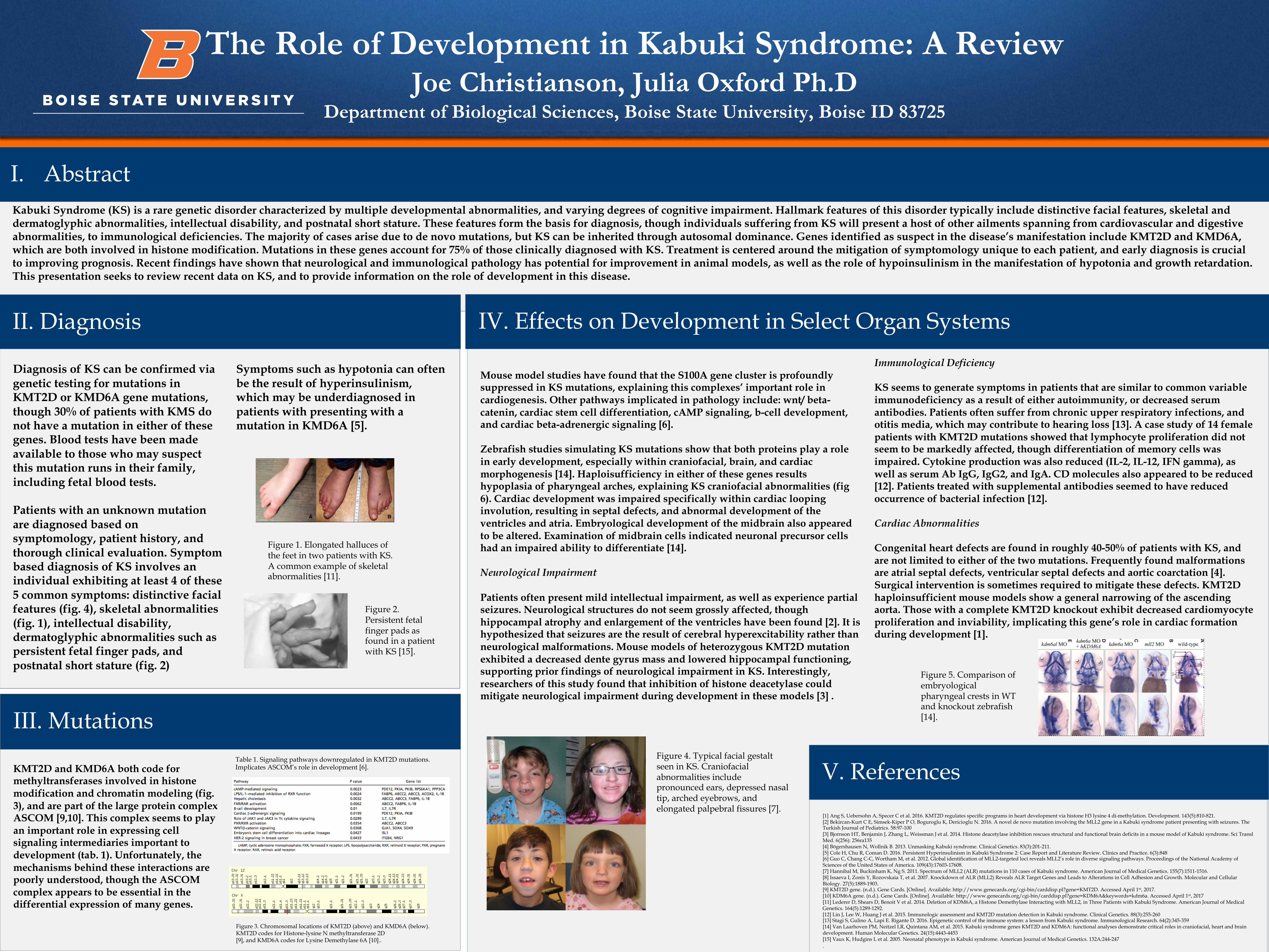
Figure 4. Typical facial gestalt seen in KS. Craniofacial abnormalities include pronounced ears, depressed nasal tip, arched eyebrows, and elongated palpebral fissures [7].
[1] Ang S, Uebersohn A, Specer C et al. 2016. KMT2D regulates specific programs in heart development via histone H3 lysine 4 di-methylation. Development. 143(5):810-821.[2] Bekircan-Kurt C E, Simsek-Kiper P O, Boguroglu K, Dericioglu N. 2016. A novel de novo mutation involving the MLL2 gene in a Kabuki syndrome patient presenting with seizures. The Turkish Journal of Pediatrics. 58:97-100[3] Bjornson HT, Benjamin J, Zhang L, Weissman J et al. 2014. Histone deacetylase inhibition rescues structural and functional brain deficits in a mouse model of Kabuki syndrome. Sci Transl Med. 6(256): 256ra135[4] Bögershausen N, Wollnik B. 2013. Unmasking Kabuki syndrome. Clinical Genetics. 83(3):201-211.[5] Cole H, Chu R, Coman D. 2016. Persistent Hyperinsulinism in Kabuki Syndrome 2: Case Report and Literature Review. Clinics and Practice. 6(3):848[6] Guo C, Chang C-C, Wortham M, et al. 2012. Global identification of MLL2-targeted loci reveals MLL2’s role in diverse signaling pathways. Proceedings of the National Academy of Sciences of the United States of America. 109(43):17603-17608.[7] Hannibal M, Buckinham K, Ng S. 2011. Spectrum of MLL2 (ALR) mutations in 110 cases of Kabuki syndrome. American Journal of Medical Genetics. 155(7):1511-1516.[8] Issaeva I, Zonis Y, Rozovskaia T, et al. 2007. Knockdown of ALR (MLL2) Reveals ALR Target Genes and Leads to Alterations in Cell Adhesion and Growth. Molecular and Cellular Biology. 27(5):1889-1903. [9] KMT2D gene. (n.d.). Gene Cards. [Online]. Available: http://www.genecards.org/cgi-bin/carddisp.pl?gene=KMT2D. Accessed April 1st, 2017.[10] KDM6A gene. (n.d.). Gene Cards. [Online]. Available: http://www.genecards.org/cgi-bin/carddisp.pl?gene=KDM6A&keywords=kdm6a. Accessed April 1st, 2017[11] Lederer D, Shears D, Benoit V et al. 2014. Deletion of KDM6A, a Histone Demethylase Interacting with MLL2, in Three Patients with Kabuki Syndrome. American Journal of Medical Genetics. 164(5):1289-1292.[12] Lin J, Lee W, Huang J et al. 2015. Immunologic assessment and KMT2D mutation detection in Kabuki syndrome. Clinical Genetics. 88(3):255-260 [13] Stagi S, Gulino A, Lapi E. Rigante D. 2016. Epigenetic control of the immune system: a lesson from Kabuki syndrome. Immunological Research. 64(2):345-359[14] Van Laarhoven PM, Neitzel LR, Quintana AM, et al. 2015. Kabuki syndrome genes KMT2D and KDM6A: functional analyses demonstrate critical roles in craniofacial, heart and brain development. Human Molecular Genetics. 24(15):4443-4453[15] Vaux K, Hudgins L et al. 2005. Neonatal phenotype in Kabuki syndrome. American Journal of Medical Genetics. 132A:244-247.
Figure 1. Elongated halluces of the feet in two patients with KS. A common example of skeletal abnormalities [11].
Figure 2. Persistent fetal finger pads as found in a patient with KS [15].
II. Diagnosis IV. Effects on Development in Select Organ Systems
Figure 5. Comparison of embryological pharyngeal crests in WT and knockout zebrafish [14].
V. References
III. Mutations
KMT2D and KMD6A both code for methyltransferases involved in histone modification and chromatin modeling (fig. 3), and are part of the large protein complex ASCOM [9,10]. This complex seems to play an important role in expressing cell signaling intermediaries important to development (tab. 1). Unfortunately, the mechanisms behind these interactions are poorly understood, though the ASCOM complex appears to be essential in the differential expression of many genes.
Table 1. Signaling pathways downregulated in KMT2D mutations. Implicates ASCOM’s role in development [6].
Figure 3. Chromosomal locations of KMT2D (above) and KMD6A (below). KMT2D codes for Histone-lysine N methyltransferase 2D [9], and KMD6A codes for Lysine Demethylase 6A [10]..