The role of cigarette smoke-induced pulmonary vascular ...
15
Song et al. Respir Res (2021) 22:39 https://doi.org/10.1186/s12931-021-01630-1 REVIEW The role of cigarette smoke-induced pulmonary vascular endothelial cell apoptosis in COPD Qing Song, Ping Chen * and Xiang‑Ming Liu Abstract Chronic obstructive pulmonary disease (COPD) is one of the most common chronic respiratory diseases with high morbidity and mortality. It has become the fifth most burdened and the third most deadly disease in the global economy and increases year by year. The prevention and treatment of COPD are urgent. Smoking is the main and most common risk factor for COPD. Cigarette smoke (CS) contains a large number of toxic substances, can cause a series of changes in the trachea, lung tissue, pulmonary blood vessels, and promotes the occurrence and develop‑ ment of COPD. In recent years, the development of epigenetics and molecular biology have provided new guidance for revealing the pathogenesis, diagnosis, and treatment of diseases. The latest research indicates that pulmonary vascular endothelial cell apoptosis initiates and participates in the pathogenesis of COPD. In this review, we summa‑ rize the current research on the epigenetic mechanisms and molecular biology of CS‑induced pulmonary vascular endothelial cell apoptosis in COPD, providing a new research direction for pathogenesis of COPD and a new target for the diagnosis, treatment, and prevention of COPD. Keywords: Chronic obstructive pulmonary disease, Cigarette smoke, Epigenetic, Gene regulation, Molecular biology, Apoptosis, Endothelial cell © The Author(s) 2021. This article is licensed under a Creative Commons Attribution 4.0 International License, which permits use, sharing, adaptation, distribution and reproduction in any medium or format, as long as you give appropriate credit to the original author(s) and the source, provide a link to the Creative Commons licence, and indicate if changes were made. The images or other third party material in this article are included in the article’s Creative Commons licence, unless indicated otherwise in a credit line to the material. If material is not included in the article’s Creative Commons licence and your intended use is not permitted by statutory regulation or exceeds the permitted use, you will need to obtain permission directly from the copyright holder. To view a copy of this licence, visit http://crea‑ tivecommons.org/licenses/by/4.0/. The Creative Commons Public Domain Dedication waiver (http://creativecommons.org/publicdo‑ main/zero/1.0/) applies to the data made available in this article, unless otherwise stated in a credit line to the data. Introduction Chronic obstructive pulmonary disease (COPD) is a chronic respiratory disease caused by a variety of fac- tors. It is characterized by chronic inflammation of the airways, lung tissue, and pulmonary blood vessels. Long- term inflammation causes remodelling of the airway structure and subsequent restriction in respiratory air- flow. e development of restricted respiratory airflow is progressive, and the airflow restriction is irreversible, even after removing the risk factors. Eventually, it seri- ously affects the quality of life of patients, endanger- ing people’s health [1–4]. e latest research data show that the incidence, disability, and mortality of COPD are high, and there is a rising trend year by year. COPD has become a serious worldwide public health problem and one of the major risk factors for death in the global population. e number of patients with COPD is nearly 299.4 million adults in worldwide [5–8]. According to the Global Burden of Disease Study, 3.2 million people died due to COPD in 2017, which represented a more than 23% increase in deaths compared with 1990 [9]. Current research shows that smoking, biofuels, indoor and outdoor air pollution, and industrial dust are the major environmental risk factors for COPD [10]. Ciga- rette smoke (CS) contains many harmful ingredients, which have a stimulating effect on the respiratory tract. Studies have demonstrated that long-term smoking can destroy the structure of the air duct wall, damage the septum of the alveolar wall, and cause interstitial fibrosis [11–14]. In addition, smoking causes increased secretions Open Access *Correspondence: [email protected] Department of Respiratory and Critical Care Medicine, The Second Xiangya Hospital, Research Unit of Respiratory Disease, Diagnosis and Treatment Center of Respiratory Disease, Central South University, 139 Renmin Middle Road, Changsha 410011, Hunan, China
Transcript of The role of cigarette smoke-induced pulmonary vascular ...

Song et al. Respir Res (2021) 22:39 https://doi.org/10.1186/s12931-021-01630-1
REVIEW
The role of cigarette smoke-induced pulmonary vascular endothelial cell apoptosis in COPDQing Song, Ping Chen* and Xiang‑Ming Liu
Abstract
Chronic obstructive pulmonary disease (COPD) is one of the most common chronic respiratory diseases with high morbidity and mortality. It has become the fifth most burdened and the third most deadly disease in the global economy and increases year by year. The prevention and treatment of COPD are urgent. Smoking is the main and most common risk factor for COPD. Cigarette smoke (CS) contains a large number of toxic substances, can cause a series of changes in the trachea, lung tissue, pulmonary blood vessels, and promotes the occurrence and develop‑ment of COPD. In recent years, the development of epigenetics and molecular biology have provided new guidance for revealing the pathogenesis, diagnosis, and treatment of diseases. The latest research indicates that pulmonary vascular endothelial cell apoptosis initiates and participates in the pathogenesis of COPD. In this review, we summa‑rize the current research on the epigenetic mechanisms and molecular biology of CS‑induced pulmonary vascular endothelial cell apoptosis in COPD, providing a new research direction for pathogenesis of COPD and a new target for the diagnosis, treatment, and prevention of COPD.
Keywords: Chronic obstructive pulmonary disease, Cigarette smoke, Epigenetic, Gene regulation, Molecular biology, Apoptosis, Endothelial cell
© The Author(s) 2021. This article is licensed under a Creative Commons Attribution 4.0 International License, which permits use, sharing, adaptation, distribution and reproduction in any medium or format, as long as you give appropriate credit to the original author(s) and the source, provide a link to the Creative Commons licence, and indicate if changes were made. The images or other third party material in this article are included in the article’s Creative Commons licence, unless indicated otherwise in a credit line to the material. If material is not included in the article’s Creative Commons licence and your intended use is not permitted by statutory regulation or exceeds the permitted use, you will need to obtain permission directly from the copyright holder. To view a copy of this licence, visit http://crea‑tivecommons.org/licenses/by/4.0/. The Creative Commons Public Domain Dedication waiver (http://creativecommons.org/publicdo‑main/zero/1.0/) applies to the data made available in this article, unless otherwise stated in a credit line to the data.
IntroductionChronic obstructive pulmonary disease (COPD) is a chronic respiratory disease caused by a variety of fac-tors. It is characterized by chronic inflammation of the airways, lung tissue, and pulmonary blood vessels. Long-term inflammation causes remodelling of the airway structure and subsequent restriction in respiratory air-flow. The development of restricted respiratory airflow is progressive, and the airflow restriction is irreversible, even after removing the risk factors. Eventually, it seri-ously affects the quality of life of patients, endanger-ing people’s health [1–4]. The latest research data show
that the incidence, disability, and mortality of COPD are high, and there is a rising trend year by year. COPD has become a serious worldwide public health problem and one of the major risk factors for death in the global population. The number of patients with COPD is nearly 299.4 million adults in worldwide [5–8]. According to the Global Burden of Disease Study, 3.2 million people died due to COPD in 2017, which represented a more than 23% increase in deaths compared with 1990 [9].
Current research shows that smoking, biofuels, indoor and outdoor air pollution, and industrial dust are the major environmental risk factors for COPD [10]. Ciga-rette smoke (CS) contains many harmful ingredients, which have a stimulating effect on the respiratory tract. Studies have demonstrated that long-term smoking can destroy the structure of the air duct wall, damage the septum of the alveolar wall, and cause interstitial fibrosis [11–14]. In addition, smoking causes increased secretions
Open Access
*Correspondence: [email protected] of Respiratory and Critical Care Medicine, The Second Xiangya Hospital, Research Unit of Respiratory Disease, Diagnosis and Treatment Center of Respiratory Disease, Central South University, 139 Renmin Middle Road, Changsha 410011, Hunan, China

Page 2 of 15Song et al. Respir Res (2021) 22:39
from mucous glands and obstructive bronchiolitis, which aggravates the progression of lung tissue lesions [15, 16]. At the same time, CS can stimulate lung tissue to produce a large amount of reactive oxygen species (ROS), which can lead to an imbalance of the oxidation and antioxidant systems. This finally causes cell dysfunction and induces cell apoptosis [17–19].
Smoking is an established risk factor for COPD. The latest epidemiological data shows that smoking more than 20 packs a year triples the prevalence of COPD in China [20]. In addition to active smoking, passive smok-ing is also related to the occurrence of COPD [21]. Research report shows that the prevalence of patients with COPD who have never smoked is also high [20, 22, 23].
Airway inflammation, oxidative stress, and lung emphysema are the main mechanisms of the onset of COPD [24]. Recent studies have shown that pulmonary vascular endothelial cell apoptosis also initiates and participates in the pathogenesis of COPD [25]. In this review, we summarize the current research on the epige-netic mechanisms and other molecular biology of CSE/CS-induced pulmonary vascular endothelial cell apopto-sis in COPD.
COPD and pulmonary vascular endothelial cell apoptosisApoptosis refers to the physiological or pathological stimulating signals of the cell to the environment, such as DNA damage and oxidative stress. It’s a kind of active and orderly gene control, resulting from environmen-tal changes or mitigation of natural death [26]. The pro-cess of apoptosis is complicated and it is a process that is strictly regulated by multiple genes and molecular signals. It involves a series of changes of molecular sig-nal pathways. This gene-controlled apoptosis is highly conserved among different species. Common apoptotic genes include the Bcl-2 family, caspase family, oncogene C-myc, and tumour suppressor gene P53. [27–29]. Until now, studies have shown there are two main apoptotic pathways: the exogenous or death receptor pathway and the intrinsic or mitochondrial pathway [30]. Apoptosis may be directly or indirectly related to the occurrence and development of many diseases, such as lung cancer, COPD, asthma, atherosclerosis, diabetes, and autoim-mune diseases [31, 32].
A study by Demedts et al. [33] indicated that apopto-sis of lung structural cells may be an important upstream event in the pathogenesis of COPD. Both apoptotic alve-olar epithelial and endothelial cells are increased in the lungs of patients with COPD. These pathological changes cannot be offset by the proliferation of structural cells, and this leads to the destruction of lung tissue and the development of emphysema.
Many studies have confirmed that pulmonary vascu-lar endothelial cell apoptosis initiates and participates in the pathogenesis of COPD. Taraseviciene-Stewart et al. [34] induced the apoptosis of rat pulmonary vas-cular endothelial cells by intraperitoneal injection of cigarette smoke extract (CSE) in 2005 and successfully established a rat emphysema model. Successive stud-ies have found that recombinant human tumour necro-sis factors receptor: Fc fusion protein (rhTNFR: Fc) may interfere with tumour necrosis factor α (TNF-α) and reduce alveolar septal apoptosis in CS-induced rats [35]. In addition, vascular endothelial growth factor (VEGF) is one of the major regulators of endothelial cell sur-vival and is believed to play a role in the pathogenesis of COPD [36]. Guan et al. [37] found that bone marrow mesenchymal stem cells could reduce pulmonary vascu-lar endothelial cell apoptosis and promote cell survival by increasing VEGF expression in CS-induced rats. Moreo-ver, Farkas et al. [38] found that after Smad3 knockout mice were exposed to CS, the expression of VEGF was reduced, which accelerated development of emphysema and COPD. Oral N-acetylcysteine may reduce emphy-sema and CS-induced alveolar septal cell apoptosis by partly increasing VEGF secretion and protein expres-sion [39]. Chen et al. [40] induced the apoptosis of mouse pulmonary vascular endothelial cells with the intraperi-toneal injection of CSE in 2009, also successfully estab-lishing a mouse emphysema model, which provided a powerful guide for future research. With the deepening of research, more and more evidence has shown that epi-genetic and other molecular biological mechanisms play an important role in regulating CSE/CS-induced apopto-sis of pulmonary vascular endothelial cells. At the same time, a new chapter was opened for studying pulmonary vascular endothelial cell apoptosis initiating the develop-ment of COPD (Fig. 1).
Cigarette smoke and epigenetic mechanisms of pulmonary vascular endothelial cell apoptosisEpigenetics refer to heritable changes of gene expression, without changing the nucleotide sequence of genes. More and more studies have shown that epigenetics is involved in the development of lung diseases. Epigenetic mecha-nisms, such as DNA methylation, RNA methylation, histone modification, exosomes (EXs), and non-coding RNA, with regulatory functions have been continuously revealed [41–43]. Studies have shown that long non-coding RNA (lncRNA), microRNA (miRNA) and DNA methylation, through various mechanisms to regulate the transcription of genes and proteins, and activate a series of molecular signal pathways to participate in the process of apoptosis [44–46](Table 1).

Page 3 of 15Song et al. Respir Res (2021) 22:39
DNA methylation and pulmonary vascular endothelial cell apoptosisDNA methylation is the most typical type of chromatin modification. It refers to changes of genetic expression, while without changes of DNA sequence. It is one of the common genetic modifications in epigenetics by adding a methyl group to the 5 ’carbon position of the cytosine of
the genomic CpG dinucleotide through the role of DNA methylation transferase [47, 48]. Research has shown that DNA methylation can regulate gene expression and par-ticipate in cell differentiation and apoptosis by changing DNA stability and structure [49]. Sundar et al. [50] iso-lated DNA from the lung tissue of eight non-smokers, eight current smokers, and eight patients with COPD
Fig. 1 Epigenetic and other molecular biology mechanisms in cigarette smoke‑induced pulmonary vascular endothelial cell apoptosis, which initiates and participates in the development of emphysema and COPD

Page 4 of 15Song et al. Respir Res (2021) 22:39
Tabl
e 1
Ciga
rett
e sm
oke
and
epig
enet
ic m
echa
nism
s of
pul
mon
ary
vasc
ular
end
othe
lial c
ell a
popt
osis
Epig
enet
ics
E gr
oup
C gr
oup
Det
ectin
g pa
ram
eter
Det
ectin
g ap
opto
sis
cells
Com
men
tRe
fere
nce
DN
A m
ethy
latio
nEl
even
CO
PD p
atie
nts
Ten
non‑
COPD
pat
ient
sm
tTFA
, mtT
FA p
rom
oter
met
hyla
‑tio
nH
PVEC
sE
grou
p sh
owin
g ce
ll ap
opto
sis
incr
ease
d, m
tTFA
mRN
A a
nd
prot
ein
expr
essi
on d
ecre
ased
. M
ethy
latio
n ra
te o
f the
mtT
FA
prom
oter
incr
ease
d
Peng
et a
l. [5
3]
Ten
BALB
/c m
ice +
CSE
Ten
BALB
/c m
ice +
PBS
mtT
FA, C
OXI
I, m
tTFA
pro
mot
er
met
hyla
tion
Mou
se p
ulm
onar
y va
scul
ar
endo
thel
ial c
ells
E gr
oup
show
ing
cell
apop
to‑
sis
incr
ease
d. m
tTFA
, CO
X II
mRN
A a
nd p
rote
in e
xpre
ssio
n de
crea
sed.
Met
hyla
tion
rate
of
the
mtT
FA p
rom
oter
incr
ease
d
Zhan
g et
al.
[54]
Ten
COPD
pat
ient
sTe
n no
rmal
sub
ject
sN
otch
1, E
RK, m
tTFA
pro
mot
er
met
hyla
tion
HPM
ECs
E gr
oup
show
ing
cell
apop
to‑
sis,
ERK
mRN
A a
nd p
rote
in
expr
essi
on in
crea
sed.
Not
ch1
mRN
A a
nd p
rote
in e
xpre
ssio
n de
crea
sed.
Met
hyla
tion
rate
of
the
mtT
FA p
rom
oter
incr
ease
d
Zong
et a
l. [5
5]
Ten
BALB
/c m
ice +
CSE
Ten
BALB
/C m
ice +
PBS
Bcl‑2
, Bax
, Bcl
‑2 p
rom
oter
met
hyla
‑tio
nM
ice
pulm
onar
y va
scul
ar e
ndot
he‑
lial c
ells
E gr
oup
show
ing
cell
apop
tosi
s an
d Ba
x m
RNA
and
pro
tein
ex
pres
sion
incr
ease
d. B
cl‑2
m
RNA
and
pro
tein
exp
ress
ion
decr
ease
d. M
ethy
latio
n ra
te o
f th
e Bc
l‑2 p
rom
oter
incr
ease
d
Zeng
et a
l. [5
6]
His
tone
met
hyla
tion
HU
VEC
s + C
SEH
UVE
Cs +
PBS
PRM
T6, H
3R2m
e2a,
H3K
4me3
HU
VEC
sE
grou
p sh
owin
g ce
ll ap
opto
sis
and
H3K
4me3
pro
tein
exp
res‑
sion
incr
ease
d. H
3R2m
e2a
prot
ein
expr
essi
on d
ecre
ased
. PR
MT6
mRN
A a
nd p
rote
in
expr
essi
on d
ecre
ased
Kang
et a
l. [7
2]
miR
NA
HPM
ECs +
CSE
HPM
ECs +
PBS
miR
‑34a
, Not
ch1
HPM
ECs
E gr
oup
show
ing
cell
apop
to‑
sis
and
miR
‑34a
exp
ress
ion
incr
ease
d, N
otch
1 m
RNA
and
pr
otei
n ex
pres
sion
dec
reas
ed
Long
et a
l. [8
2]
HPM
ECs +
CSE
HPM
ECs +
PBS
miR
‑206
, Not
ch3,
VEG
FAH
PMEC
sE
grou
p sh
owin
g ce
ll ap
opto
‑si
s an
d m
iR‑2
06 e
xpre
ssio
n in
crea
sed.
Not
ch3
and
VEG
FA
mRN
A a
nd p
rote
in e
xpre
ssio
n de
crea
sed
Sun
et a
l. [8
3]

Page 5 of 15Song et al. Respir Res (2021) 22:39
Tabl
e 1
(con
tinu
ed)
Epig
enet
ics
E gr
oup
C gr
oup
Det
ectin
g pa
ram
eter
Det
ectin
g ap
opto
sis
cells
Com
men
tRe
fere
nce
lncR
NA
HPV
ECs +
CSE
HPV
ECs +
PBS
lncR
NA
MEG
3, B
ax, c
aspa
se‑3
, Bcl
2H
PVEC
sE
grou
p sh
owin
g ce
ll ap
opto
sis
and
lncR
NA
MEG
3 ex
pres
sion
in
crea
sed.
Bax
and
cas
pase
‑3
expr
essi
on in
crea
sed.
Bcl
‑2
incr
ease
d
Bi e
t al.
[97]
HPM
ECs +
CSE
HPM
ECs +
PBS
lncR
NA
MIR
155H
, miR
NA
‑218
‑5p,
BR
D4
HPM
ECs
E gr
oup
show
ing
cell
apop
tosi
s, ln
cRN
A M
IR15
5H a
nd B
RD4
expr
essi
on in
crea
sedm
iRN
A‑
218‑
5p e
xpre
ssio
n de
crea
sed
Song
et a
l. [9
8]
Exos
omes
RPM
ECs +
CSE
RPM
ECs +
PBS
Exos
omes
RPM
ECs
Exos
omes
indu
ced
by 1
% C
SE s
ig‑
nific
antly
dec
reas
ed th
e ap
opto
‑si
s ra
te o
f end
othe
lial c
ells
Zhao
et a
l. [1
05]

Page 6 of 15Song et al. Respir Res (2021) 22:39
and confirmed the presence of high DNA methylation in smokers and patients with COPD compared with non-smokers. Song et al. [51] isolated bronchial tissue from patients with and without COPD, isolated and cultured goblet cells and promoted their differentiation and found that SAM-pointed domain-containing ETS transcription factor (SPDEF) and forkhead box protein A2 (FOXA2) had abnormal DNA methylation during goblet cell dif-ferentiation. Zinellu et al. [52] studied the methylcytosine levels in the blood of forty-three patients with different degrees of COPD and forty-three control subjects. The results showed that DNA methylation was significantly increased in patients with COPD, especially patients with more severe COPD. These studies have shown that DNA methylation plays a key role in the pathogenesis of COPD.
Peng et al. [53] tested the rate of pulmonary vascular endothelial cell apoptosis in lung tissue of eleven patients with COPD and ten patients with non-COPD squamous cell lung cancer, measured the expression of mitochon-drial transcription factor (mtTFA) mRNA and protein and methylation of the mtTFA promoter. The results showed that the patients with COPD had a higher cell apoptosis rate and lower mtTFA mRNA and protein expression compared with the non-COPD group, which has a negative correlation with pulmonary vascular endothelial cell apoptosis and smoke index. The meth-ylation rate of the mtTFA promoter in the COPD group was significantly increased when compared with the non-COPD group. Zhang et al. [54] found that methylation of the mtTFA promoter and apoptosis rate of pulmonary vascular endothelial cells in the CSE-induced mice group were significantly increased. The mRNA and protein lev-els of both mtTFA and cytochrome c oxidase subunit II (COX II) were significantly decreased, but the group of mice treated with 5-aza-2′-deoxycytidine (AZA, a DNA methyltransferase inhibitor) had restoration of the above changes which suggesting that the removal of DNA methylation by AZA can protect against CSE-induced cell apoptosis. Zong et al. [55] tested the lung tissues of ten patients with COPD and ten normal subjects, respec-tively, and found that Notch1 was mainly expressed in endothelial cells, and was significantly decreased in the endothelial cells of patients with COPD. Furthermore, the results of in vitro cell experiments demonstrated that Notch1 overexpression reduces the CSE-induced apoptosis of human pulmonary microvascular endothe-lial cells (HPMECs), and CSE can significantly activate the extracellular signal-regulated kinase (ERK) signal-ling pathway. Treatment of CSE-induced HPMECs with ERK inhibitors can heavily reduce cell apoptosis and mtTFA methylation. Zeng et al. [56] studied the role and mechanisms of the Bcl protein family in the apoptosis of
emphysema cells by intraperitoneal injection of CSE and AZA into mice, respectively, and found that the apoptosis index was higher than in the control group. The expres-sion of Bcl-2 in CSE-induced mice decreased, but the level of Bcl-2 promoter methylation increased. However, AZA treatment promoted the Bcl-2 promoter demeth-ylation, increased the expression of Bcl-2 and decreased the apoptosis index. These results indicated that the epigenetic mechanism of Bcl-2 promoter methylation is involved in CSE-induced emphysema and lung cell apoptosis.
Histone modification and pulmonary vascular endothelial cell apoptosisHistone modification refers to histone acetylation, ubiq-uitination, phosphorylation, or methylation. Studies have shown that histone modification is involved in the regu-lation of gene expression at the epigenetic level and plays an important role in the development, ageing, differentia-tion, apoptosis, and tumour migration of tissues, organs and cells [57–62]. Sundar et al. [63] performed western blot analysis of targeted histones in lung tissue of CSE-induced mice and patients with COPD who continue to smoke. The results showed that the expression levels of several chromatin-modifying enzymes, including his-tone acetyltransferase, histone methyltransferase, histone domain proteins, and histone kinase were significantly increased. More studies have found that arginine meth-yltransferase-1 participates in the pathogenesis of epi-thelial tract injury in COPD by adding methyl to arginine residues in histones and non-histones to regulate protein modification at post-translational levels [64].
Chronic inflammation of the trachea and bronchi is one of the main characteristics of COPD [65]. Histone modi-fication plays an important role in the chronic inflamma-tion of COPD [66, 67]. Apoptosis of pulmonary vascular endothelial cells is one of the initiating events of COPD. Histone modification is also involved in smoking-induced emphysema and apoptosis [68–70]. He et al. [71] found that the expression of protein arginine methyltransferase 6 (PRMT6) and asymmetric di-methylation of histone H3 arginine 2 (H3R2me2a) were significantly decreased in the lung tissues of patients with COPD who continue to smoke and CSE-induced mice. However, H3R2me2a can prevent the tri-methylation of lysine 4 on histone H3 (H3K4me3) which is located at the transcription start site; the expression of H3K4me3 was significantly increased, and emphysema inflammation, apoptosis, and oxidative stress levels were more severe in CSE-induced mice. Further research found that apoptosis, emphy-sema inflammation, and oxidative stress were mark-edly reduced with overexpression of PRMT6. In other research, it was observed that the apoptosis of human

Page 7 of 15Song et al. Respir Res (2021) 22:39
umbilical vein endothelial cells (HUVECs) increased after CSE exposure and decreased PRMT6 expression. How-ever, a decreased in CSE-induced apoptosis was observed after HUVECs were transfected with a plasmid express-ing PRMT6. Notably, after CSE treatment, the expression of H3K4me3 protein significantly increased in HUVECs, while the expression of H3R2me2a protein decreased significantly in HUVECs. However, the above changes reversed after the transfection of cells with a plasmid expressing PRMT6, suggesting that PRMT6 mediated CSE-induced apoptosis through H3R2me2a in HUVECs [72].
miRNA and pulmonary vascular endothelial cell apoptosismiRNA is a type of non-coding RNA with regula-tory functions and a length of about 22–25 nucleo-tides. miRNA, which can regulate gene expression by incompletely or completely directly binding to mRNA 3′-untranslated region (UTR), also interacts with pro-moters, coding DNA sequence (CDS), and 5′-UTR to participate in gene regulation. It plays an important role in regulating gene expression, organism development, and apoptosis [73–77].
miRNA has been confirmed to be related to COPD and smoking. It plays an important role in the develop-ment of COPD [78, 79]. Conickx et al. [80] exposed mice to air and CS for twenty-four weeks and detected differ-ential expression profiles of miRNAs in mice lung tissue and bronchoalveolar lavage fluid. The results showed that thirty-one miRNAs differentially expressed in lung tissue as well as seventy-eight miRNAs in bronchoal-veolar lavage fluid in the CS exposed group compared with air exposure. Van Pottelberge et al. [81] also found that thirty-four miRNAs were differentially expressed in the sputum supernatants of patients who never smoked and current smokers. Compared with those who had never smoked and had no airflow limitation, the expres-sion levels of eight miRNAs were significantly reduced in patients with COPD who continue to smoke.
In recent years, the role of miRNA in the smoking-induced apoptosis of pulmonary vascular endothelial cells and its related mechanisms also have been stud-ied. Research by Long et al. [82] showed that CSE can induce apoptosis of HPMECs with miR-34a significantly upregulated. The miRNA target gene library was further predicted through a biological information database and Notch1 was determined to be the target of miRNA-34a. At the same time, it was confirmed that miR-34a regu-lates gene expression at post-transcriptional levels by targeting Notch1 mRNA 3′-UTR after luciferase gene determination. Furthermore, studies have confirmed that the expression level of Notch1 in CSE-induced HPMECs is markedly decreased. In vitro cell experiments also
confirmed that miR-34a mimic and Notch1 gene plas-mids were transfected into HPMECs exposed to CSE. Overexpression of miR-34a can significantly increase the apoptosis rate of HPMECs. However, the overexpres-sion of Notch1 has a protective effect on the apoptosis of HPMECs caused by the increased miR-34a and reduced the apoptosis rate. In other research, CSE-induced HPMECs significantly upregulate miR-206 levels. How-ever, the cell apoptosis rate decreased after the miR-206 gene was knocked out. miR-206 participates in the regulation of gene expression by targeting Notch3 and vascular endothelial growth factor A (VEGFA) mRNA 3′-UTR after prediction by the bioinformatics gene database. Then, miR-206 mimic, Notch3 vector plasmid and VEGFA vector plasmid were transfected into CSE-induced HPMECs, respectively. The results showed that overexpression of miR-206 can lead to increased apopto-sis of HPMECs. However, the overexpression of Notch3 and VEGFA can significantly reduce apoptosis [83].
lncRNA and pulmonary vascular endothelial cell apoptosislncRNA is non-protein-coding RNA with a length longer than 200 nucleotides. lncRNA can regulate target gene expression through different mechanisms to participate in cell biological processes, from chromatin regulation to protein degradation [84, 85], such as interacting with miRNAs via ceRNA or directly target miRNAs, by encod-ing proteins or polypeptides and by means of intercellu-lar communication through EXs [86–89].
lncRNA has also has been confirmed to be related to COPD and smoking [90, 91]. Qian et al. [92] identified the differentially expressed lncRNA in smoking and non-smoking patients with COPD through RNA sequencing and bioinformatics analysis and found that ninety-six lncRNA were identified in non-smoking patients, while forty-four lncRNA were identified in smoking patients with COPD. Further research into the lncRNA-miRNA-mRNA interaction network found that let-7c-ADRB1-HLA-DQB1-AS1 interactions may play a key role in the pathogenesis in smoking COPD patients. Let-7c was downregulated in smokers with COPD associated with forced expiratory volume in 1 s. Adrenoceptor beta 1 (ADRB1) is a subtype of the adrenergic receptors which are involved with the management of COPD exacerba-tions and is upregulated in smoking COPD patients [93]. HLA-DQB1-AS1 was a co-expressed lncRNA of ADRB1 and was upregulated in smoking COPD patients. Oth-erwise, there were one hundred and nine lncRNA were also found to be differentially expressed compared to control groups in lung tissue of CS-induced COPD mice models [94]. Studies have shown that lncRNA TUG1 can promote airway remodelling by inhibiting the miR‐145‐5p and DUSP6 axis in a CS-induced COPD mice

Page 8 of 15Song et al. Respir Res (2021) 22:39
model [95]. A study by Zhang et al. [96] found that the lncRNA NEAT1 can inhibit the apoptosis of t-BHP-treated HUVECs by activating the miR-181d-5p and cyc-lin-dependent kinase inhibitor 3 (CDKN3) axis. Bi et al. [97] study indicated that co-cultured human pulmonary vascular endothelial cells (HPVECs) with different con-centrations of CSE (0%, 0.1%, 1% and 10%) significantly promoted cell apoptosis, increased caspase-3 activity, upregulated Bax expression, decreased Bcl-2 expres-sion, and increased expression of the lncRNA MEG3. After the transfection of lncRNA MEG3 with a plasmid, the expression of lncRNA MEG3 was increased, and cell apoptosis further increased. However, knockdown of lncRNA MEG3 showed the opposite effect, decreased cell apoptosis, decreased caspase activity, decreased Bax expression, and upregulated Bcl-2 expression. Also, Song et al. [98] found that the expression of the lncRNA MIR155HG was increased, while miRNA-218-5p was decreased in CSE-induced HPMECs. Subsequently, it was found that miRNA-218-5p was a direct target of MIR155HG. This result was also confirmed in the res-cue experiment, as a miRNA-218-5p inhibitor reduced the inhibition effect of MIR155HG on CSE-induced HPMECs. Further studies showed that miRNA-218-5p directly targeted bromodomain containing 4 (BRD4), and overexpression of miRNA-218-5p reversed cell apoptosis by regulating BRD4. In conclusion, MIR155HG partici-pates in the apoptosis of CSE-induced HPMECs by regu-lating the miRNA-218-5p and BRD4 axis.
Exosomes and pulmonary vascular endothelial cell apoptosisEXs are extracellular vesicles (EVs) with a size of approxi-mately 30–150 nm that produce inward budding origi-nating from the endosomal membrane of the cell upon activation or during apoptosis [99]. It has been demon-strated that EXs play a key role in intercellular commu-nication by carrying biomolecules, including proteins, DNA, miRNA and lncRNA, involved in cell communica-tion, migration, angiogenesis, and proliferation [100].
Some studies have demonstrated that CS can promote the release of EXs in lung tissue cells. Benedikter et al. [101] revealed that CSE exposure could boost the num-ber of EXs secreted by bronchial epithelial cells. In addi-tion, exposure to tobacco smoke extract (TSE) exposure can cause human macrophages to release EVs (includ-ing exosomes and ectosomes), which contribute to the release of matrix metalloproteinase 14 (MMP14) and may contribute to emphysema [102, 103]. Studies have found that MMP14 activity and protein was increased in the airway epithelium of tobacco smoke-exposed mice and decreased MMP14 activity and protein could diminish the mucin 5AC, oligomeric mucus/gel-forming (MUC5AC) transcripts that played significant roles in
the development of COPD [104]. A study by Zhao et al. [105] showed that CS-induced epithelial cell-derived EXs decreased the apoptosis of rat pulmonary microvascular endothelial cells, but the underlying mechanisms remain unclear and need further research.
Cigarette smoke and other molecular biology mechanisms of pulmonary vascular endothelial cell apoptosisThere are still related studies exploring the mechanisms of pulmonary vascular endothelial cell apoptosis in other molecular biology caused by smoking [106]. The metabolism of the three major nutrients of protein, fat and glucose are the basis of the body’s life activities and the basic component of cells. It maintains the stability of cellular structure and participates in the life activities of cells [107]. Studies have shown that glucose production, clearance, oxidation, and glycolysis rates are increased in patients with COPD compared to healthy subjects [108]. In addition, CS exposure has been shown to reduce gly-colysis in type II cells [109]. Similarly, lipid metabolism disorders also exist during acute exacerbations of COPD. Glycerophospholipid and sphingomyelin metabolism are associated with airflow obstruction, decreased lung func-tion, and exacerbation of COPD [110, 111]. Decreased levels of lipoproteins and amino acids were also observed in the serum and urine of patient with COPD [112]. In another study of pulmonary microvascular endothelial cells in CSE-induced mice and patient with COPD, the authors found that the carnitine palmitoyl transferase 1a (Cpt1a) in cells was significantly reduced. In turn, the oxi-dative ability of fatty acids (FAO) and mitochondrial res-piration were decreased, but the apoptosis was increased. Further studies also verified similar results. CSE-induced apoptosis was further increased when pulmonary micro-vascular endothelial cells were treated with Cpt1 inhibi-tor or transfected with Cpt1a siRNA. Treatment with L-carnitine increased the amount of FAO and reduced cell apoptosis by increasing Cpt1a expression [113]. A study by Wang et al. [114] found that the mitochondrial aberrations, fission, oxidative stress, and cell apopto-sis were increased, while mitochondrial respiration and fusion were decreased in CSE-induced rat lung micro-vascular endothelial cells (RLMVECs). However, bar-rier dysfunction and apoptosis decreased in CS-induced RLMVECs after inhibition of mitochondrial fission and anti-oxidant intervention of mitochondria.
There were studies found that in the systemic, CS-induced endothelial dysfunction through the following aspects: firstly, directed toxic effects of CS on endothelial cells; then, promoted the production of auto-antibodies in endothelial cells; next, CS-induced inflammation of vascular; in addition, increased oxidative stress levels with reduced activation of the anti-oxidant pathways in

Page 9 of 15Song et al. Respir Res (2021) 22:39
endothelial cells; finally, CS-induced increased mediators with vasoconstrictor, pro-inflammatory, and remodelling activities and increased endoplasmic reticular stress and the unfolded protein response in endothelial cells [115]. A study by Taraseviciene-Stewart et al. [116] found that intraperitoneal injection of endothelial cells into rats could lead to the generation of anti-endothelial cell anti-bodies, which promoted endothelial cell apoptosis and caused emphysema. However, concomitant injection with the toll-like receptor 4 (TLR4) ligand lipid A into rats could decrease endothelial cell apoptosis and reduce the incidence of emphysema. It was implied that CS induction might lead to the generation of anti-endothe-lial cell antibodies, which promoted vascular endothe-lial cell apoptosis and caused emphysema. However, it needed further research. In addition, a study by Romund-stad et al. [117] found that renal dysfunction was linked to CS-induced lung injury, with an association between emphysema severity and the estimated glomerular fil-tration rate. In addition, patients with COPD who were shown to have more glomerulosclerosis and greater renal arterial and arteriolar sclerosis were linked to vascular endothelial cell injury and apoptosis [118]. It might be that the oxidative stress level was increased which further activated the advanced glycation end product (AGE) and receptor for advanced glycation end products (RAGE) in CS-induced endothelial cells and circulating CS directed toxicity on endothelial cells. Also, the production of anti-endothelial antibodies against endothelial cells. So, it was worthy to further study the connection between lung and kidney endothelial cell injury and apoptosis in patients with COPD with CS [119].
Cell activity is a complex process which is regulated by multiple genes and protein molecules. Cyclooxygenase-2 is a rate-limiting enzyme in the metabolic pathway of cells and can convert arachidonic acid into prostaglan-dins. Studies have demonstrated that CSE can affect the expression of cyclooxygenase-2 in HPVECs, subse-quently affecting the production of prostaglandins. It is worth noting that prostaglandins can inhibit the CSE-induced apoptosis of vascular endothelial cells [120]. P53 is a tumour suppressor gene that encodes the p53 protein involved in the process of cell apoptosis. Macrophage migration inhibitory factor (MIF) is an anti-apoptotic cytokine produced by HPVECs. The expression of MIF was decreased, while P53 was increased when pulmo-nary vascular endothelial cells were exposed to CSE. However, MIF is a negative regulator of p53 expression and can protect the CSE-induced apoptosis of pulmo-nary vascular endothelial cells by combating p53-medi-ated caspase-dependent apoptosis pathways [121]. Another study also found that xanthine oxidoreductase (XOR) is an upstream effector of p53. XOR activity was
significantly increased in the lung tissues of CS-induced mice, promoted the production of ROS, and involved in CS-induced pulmonary vascular endothelial cells apopto-sis through the p53-mediated caspase-dependent apop-tosis pathway [122]. Also, XOR activity was significantly increased in the bronchoalveolar lavage fluid of patients with COPD [123, 124]. Interestingly, Fallica et al. [125] found that MIF, a pleiotropic cytokine, both reduced in mice with CS-induced emphysema and patients with COPD. Further studies have found that MIF, as a determinant factor of ROS production after vascular endothelial cells were exposed to CS, affected the apop-tosis signal-regulating kinase 1 (ASK1) P38 kinase cas-cade, regulated the activity of XOR enzymes produced by ROS and antagonized ASK1-p38-dependent pulmo-nary vascular endothelial cell apoptosis. In general, MIF reduces the CS-induced apoptosis of pulmonary vascular endothelial cells by inhibiting the signal transduction of the ASK1-P38-XOR pathway (Table 2).
In addition, CS-induce pulmonary vascular endothelial cell apoptosis could promote the secretion of transform-ing growth factor-beta 1(TGF-β1) [126, 127]. TGF-β1 is a multi-functional cytokine that regulates angiogenesis, and fibroblasts/myofibroblasts [128]. Moreover, the TGF-β1/Smad2.3 signalling pathway is strongly implicated in endothelial to mesenchymal transition (EndMT) which plays a key role in the pathogenesis of COPD [129–131]. At the same time, accumulated research on EndMT showed that endothelial dysfunction contributes to the pathogenesis of pulmonary hypertension [132] and pul-monary vascular endothelial cell apoptosis can promote the development of pulmonary hypertension, which is a common complication of COPD and is closely related to COPD progression [133]. Studies have shown that the reticular basement membrane (Rbm) had mark-edly increased splitting and hypervascularity, while the lamina propria (LP) was hypovascular in COPD. Inhaled corticosteroid (ICS) therapy increased the density of ves-sels and brought back it to normal levels in the LP, but there was no influence on the Rbm hypervascular, which may suggest that ICS therapy reduces vessel destruction rather than promotes the growth of new vessels. This might be related to vascular endothelial cell apoptosis, but the specific mechanisms need further investigation [134, 135].
ConclusionsCS is one of the main causes of COPD, and it induces pulmonary vascular endothelial cell apoptosis initiates and participates the pathogenesis of COPD. However, the mechanisms of CS-induced apoptosis have not been fully elucidated. Epigenetics has been a hot topic in recent years. Histone modification, miRNA, lncRNA, DNA

Page 10 of 15Song et al. Respir Res (2021) 22:39
Tabl
e 2
Ciga
rett
e sm
oke
and
othe
r mol
ecul
ar b
iolo
gy m
echa
nism
s of
pul
mon
ary
vasc
ular
end
othe
lial c
ell a
popt
osis
E gr
oup
C gr
oup
Det
ectin
g pa
ram
eter
Det
ectin
g ap
opto
sis
cells
Com
men
tRe
fere
nce
Mou
se P
MVE
Cs +
CSE
Mou
se P
MVE
Cs +
PBS
Mito
chon
dria
l res
pira
tion,
FA
O,
oxid
ativ
e ph
osph
oryl
atio
n, C
pt1a
, ce
ram
ide
synt
hesi
s
PMVE
Cs
E gr
oup
show
ing
cell
apop
tosi
s in
crea
sed.
Oxi
dativ
e ph
osph
oryl
a‑tio
n, F
AO
and
Cpt
1a d
ecre
ased
. M
itoch
ondr
ial r
espi
ratio
n de
crea
sed.
Cer
amid
e sy
nthe
sis
incr
ease
d
Gon
g et
al.
[113
]
COPD
pat
ient
s PM
VEC
sH
ealth
y su
bjec
ts P
MVE
Cs
Mito
chon
dria
l res
pira
tion,
FA
O,
oxid
ativ
e ph
osph
oryl
atio
n, C
pt1a
, ce
ram
ide
synt
hesi
s
Lung
tiss
ue c
ells
E gr
oup
show
ing
cell
apop
tosi
s in
crea
sed.
Oxi
dativ
e ph
osph
oryl
a‑tio
n, F
AO
and
Cpt
1a d
ecre
ased
. M
itoch
ondr
ial r
espi
ratio
n de
crea
sed.
Cer
amid
e sy
nthe
sis
incr
ease
d
Rat P
MVE
Cs +
CSE
Rat P
MVE
Cs +
PBS
Mito
chon
dria
l mor
phol
ogy,
oxi
da‑
tive
stre
ss, r
espi
ratio
n; fi
ssio
n an
d fu
sion
PMVE
Cs
E gr
oup
show
ing
cell
apop
tosi
s, m
itoch
ondr
ial a
berr
atio
ns, fi
ssio
n an
d m
itoch
ondr
ial o
xida
tive
stre
ss
incr
ease
d. M
itoch
ondr
ial r
espi
ra‑
tion
and
fusi
on w
as d
ecre
ased
Wan
g et
al.
[114
]
ECV3
04 +
CSE
ECV3
04 +
PBS
COX
IIEC
V304
E gr
oup
show
ing
cell
apop
tosi
s an
d CO
X II
prot
ein
expr
essi
on
incr
ease
d
Shi e
t al.
[122
]
Six
smok
ers
with
CO
PDSe
ven
non‑
smok
ers
with
out C
OPD
an
d se
ven
smok
ers
with
out
COPD
COX
IIVa
scul
ar e
ndot
helia
l cel
ls in
lung
tis
sues
E gr
oup
show
ing
AI o
f med
ium
‑si
zed
vess
els
incr
ease
d. C
OX
II pr
otei
n ex
pres
sion
incr
ease
d
HPA
ECs +
CSE
HPA
ECs +
PBS
P53,
MIF
HPA
ECs
E gr
oup
show
ing
cell
apop
tosi
s in
crea
sed.
P53
and
MIF
mRN
A a
nd
prot
ein
expr
essi
on in
crea
sed
Dam
ico
et a
l. [1
23]
C57
BL/6
mal
e m
ice +
CS
C57
BL/6
mal
e m
ice +
AIR
XOR
prot
ein
and
activ
ity, a
lveo
lar
diam
eter
Non
E gr
oup
show
ing
alve
olar
dia
met
er
enla
rgem
ent.
XOR
prot
ein
expr
es‑
sion
and
XO
R ac
tivity
incr
ease
d
Kim
et a
l. [1
24]
HLM
VEC
s + C
SEH
LMVE
Cs +
PBS
XOR
prot
ein
and
activ
ity, P
53H
LMVE
Cs
E gr
oup
show
ing
cell
apop
tosi
s in
crea
sed
XOR
prot
ein
expr
essi
on
and
XOR
activ
ity in
crea
sed.
P53
m
RNA
and
pro
tein
exp
ress
ion
incr
ease
d
Mif−
/− C
57BL
/6 M
ice +
CS
Mif +
/ +
C57
BL/6
Mic
e +
CS
XOR
activ
ity, P
38 p
rote
in, A
SK1,
ca
spas
e 3/
7 pr
otei
n an
d ac
tivity
Hum
an a
nd ra
t lun
g m
icro
vasc
ular
En
doth
elia
l cel
lsE
grou
p sh
owin
g ce
ll ap
opto
sis
incr
ease
d. P
38 p
rote
in in
crea
sed.
A
SK1
and
casp
ase3
/7 p
rote
in a
nd
activ
ity in
crea
sed
Falli
ca e
t al.
[127
]
Page 11 of 15Song et al. Respir Res (2021) 22:39
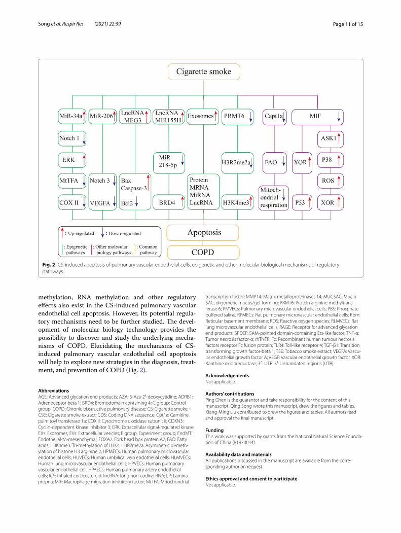
methylation, RNA methylation and other regulatory effects also exist in the CS-induced pulmonary vascular endothelial cell apoptosis. However, its potential regula-tory mechanisms need to be further studied. The devel-opment of molecular biology technology provides the possibility to discover and study the underlying mecha-nisms of COPD. Elucidating the mechanisms of CS-induced pulmonary vascular endothelial cell apoptosis will help to explore new strategies in the diagnosis, treat-ment, and prevention of COPD (Fig. 2).
AbbreviationsAGE: Advanced glycation end products; AZA: 5‑Aza‑2′‑deoxycytidine; ADRB1: Adrenoceptor beta 1; BRD4: Bromodomain containing 4; C group: Control group; COPD: Chronic obstructive pulmonary disease; CS: Cigarette smoke; CSE: Cigarette smoke extract; CDS: Coding DNA sequence; Cpt1a: Carnitine palmitoyl transferase 1a; COX II: Cytochrome c oxidase subunit II; CDKN3: Cyclin‑dependent kinase inhibitor 3; ERK: Extracellular signal‑regulated kinase; EXs: Exosomes; EVs: Extracellular vesicles; E group: Experiment group; EndMT: Endothelial‑to‑mesenchymal; FOXA2: Fork head box protein A2; FAO: Fatty acids; H3K4me3: Tri‑methylation of H3K4; H3R2me2a: Asymmetric di‑meth‑ylation of histone H3 arginine 2; HPMECs: Human pulmonary microvascular endothelial cells; HUVECs: Human umbilical vein endothelial cells; HLMVECs: Human lung microvascular endothelial cells; HPVECs: Human pulmonary vascular endothelial cell; HPAECs: Human pulmonary artery endothelial cells; ICS: Inhaled corticosteroid. lncRNA: long non‑coding RNA; LP: Lamina propria; MIF: Macrophage migration inhibitory factor; MtTFA: Mitochondrial
transcription factor; MMP14: Matrix metalloproteinases 14; MUC5AC: Mucin 5AC, oligomeric mucus/gel‑forming; PRMT6: Protein arginine methyltrans‑ferase 6; PMVECs: Pulmonary microvascular endothelial cells; PBS: Phosphate buffered saline; RPMECs: Rat pulmonary microvascular endothelial cells; Rbm: Reticular basement membrane; ROS: Reactive oxygen species; RLMVECs: Rat lung microvascular endothelial cells; RAGE: Receptor for advanced glycation end products; SPDEF: SAM‑pointed domain‑containing Ets‑like factor; TNF‑α: Tumor necrosis factor‑α; rhTNFR: Fc: Recombinant human tumour necrosis factors receptor Fc fusion protein; TLR4: Toll‑like receptor 4; TGF‑β1: Transition transforming growth factor‑beta 1; TSE: Tobacco smoke extract; VEGFA: Vascu‑lar endothelial growth factor A; VEGF: Vascular endothelial growth factor. XOR: Xanthine oxidoreductase; 3′‑ UTR : 3′‑Untranslated regions (UTR).
AcknowledgementsNot applicable.
Authors’ contributionsPing Chen is the guarantor and take responsibility for the content of this manuscript. Qing Song wrote this manuscript, drew the figures and tables. Xiang‑Ming Liu contributed to drew the figures and tables. All authors read and approval the final manuscript.
FundingThis work was supported by grants from the National Natural Science Founda‑tion of China (81970044).
Availability data and materialsAll publications discussed in the manuscript are available from the corre‑sponding author on request.
Ethics approval and consent to participateNot applicable.
Fig. 2 CS‑induced apoptosis of pulmonary vascular endothelial cells, epigenetic and other molecular biological mechanisms of regulatory pathways

Page 12 of 15Song et al. Respir Res (2021) 22:39
Consent for publicationNot applicable.
Competing interestsThe authors declare that they have no competing interests.
Received: 8 August 2020 Accepted: 20 January 2021
References 1. Zinellu E, Zinellu A, Fois AG, Fois SS, Piras B, Carru C, et al. Reliability and
usefulness of different biomarkers of oxidative stress in chronic obstruc‑tive pulmonary disease. Oxid Med Cell Longev. 2020;2020:4982324.
2. Smith MC, Wrobel JP. Epidemiology and clinical impact of major comorbidities in patients with COPD. Int J Chron Obstruct Pulmon Dis. 2014;9:871–88.
3. Miravitlles M, Ribera A. Understanding the impact of symptoms on the burden of COPD. Respir Res. 2017;18(1):67.
4. Yan F, Gao H, Zhao H, Bhatia M, Zeng Y. Roles of airway smooth muscle dysfunction in chronic obstructive pulmonary disease. J Transl Med. 2018;16(1):262.
5. GBD 2015 Disease and Injury Incidence and Prevalence Collaborators. Global, regional, and national incidence, prevalence, and years lived with disability for 310 diseases and injuries, 1990–2015: a system‑atic analysis for the Global Burden of Disease Study 2015. Lancet. 2016;388:1545–602.
6. GBD 2015 Chronic Respiratory Disease Collaborators. Global, regional, and national deaths, prevalence, disability‑adjusted life years, and years lived with disability for chronic obstructive pulmonary disease and asthma, 1990–2015: a systematic analysis for the Global Burden of Disease Study 2015. Lancet Respir Med. 2017;5:691–706.
7. Adeloye D, Chua S, Lee C, Basquill C, Papana A, Theodoratou E, et al. Global and regional estimates of COPD prevalence: systematic review and meta‑analysis. J Glob Health. 2015;5:020415.
8. GBD Chronic Respiratory Disease Collaborators. Prevalence and attributable health burden of chronic respiratory diseases, 1990–2017: a systematic analysis for the Global Burden of Disease Study 2017. Lancet Respir Med. 2020;8(6):585–96.
9. Li X, Cao X, Guo M, Xie M, Liu X. Trends and risk factors of mortality and disability adjusted life years for chronic respiratory diseases from 1990 to 2017: systematic analysis for the Global Burden of Disease Study 2017. BMJ. 2020;368:m234.
10. Zhu B, Wang Y, Ming J, Chen W, Zhang L. Disease burden of COPD in China: a systematic review. Int J Chron Obstruct Pulmon Dis. 2018;13:1353–64.
11. Hou W, Hu S, Li C, Ma H, Wang Q, Meng G, Guo T, Zhang J. Cigarette smoke induced lung barrier dysfunction, EMT, and tissue remodeling: a possible link between COPD and lung cancer. Biomed Res Int. 2019;2019:2025636.
12. Zong D, Liu X, Li J, Chen P. The role of cigarette smoke‑induced epige‑netic alterations in inflammation. Epigenetics Chromatin. 2019;12(1):65.
13. Baraldo S, Turato G, Saetta M. Pathophysiology of the small airways in chronic obstructive pulmonary disease. Respiration. 2012;84(2):89–97.
14. Leopold PL, O’Mahony MJ, Lian XJ, Tilley AE, Harvey BG, Crystal RG. Smoking is associated with shortened airway cilia. PLoS ONE. 2009;4(12):e8157.
15. Maestrelli P, Saetta M, Mapp CE, Fabbri LM. Remodeling in response to infection and injury. Airway inflammation and hypersecretion of mucus in smoking subjects with chronic obstructive pulmonary disease. Am J Respir Crit Care Med. 2001. https ://doi.org/10.1164/ajrcc m.164.suppl ement _2.21060 67.
16. Van Eeden SF, Hogg JC. Immune‑modulation in chronic obstructive pulmonary disease: current concepts and future strategies. Respiration. 2019. https ://doi.org/10.1159/00050 2261.
17. Domej W, Oettl K, Renner W. Oxidative stress and free radicals in COPD–implications and relevance for treatment. Int J Chron Obstruct Pulmon Dis. 2014;9:1207–24.
18. Yoshida T, Tuder RM. Pathobiology of cigarette smoke‑induced chronic obstructive pulmonary disease. Physiol Rev. 2007;87(3):1047–82.
19. Zinellu E, Zinellu A, Fois AG, Carru C, Pirina P. Circulating biomark‑ers of oxidative stress in chronic obstructive pulmonary disease: a systematic review. Respir Res. 2016;17(1):150.
20. Wang C, Xu J, Yang L, Xu Y, Zhang X, Bai C, Kang J, et al. Prevalence and risk factors of chronic obstructive pulmonary disease in China (the China Pulmonary Health [CPH] study): a national cross‑sectional study. Lancet. 2018;391(10131):1706–17.
21. Yin P, Jiang CQ, Cheng KK, Lam TH, Lam KH, Miller MR, et al. Passive smoking exposure and risk of COPD among adults in China: the Guangzhou Biobank Cohort Study. Lancet. 2007;370(9589):751–7.
22. Mannino DM, Buist AS. Global burden of COPD: risk factors, preva‑lence, and future trends. Lancet. 2007;370(9589):765–73.
23. Hagstad S, Bjerg A, Ekerljung L, Backman H, Lindberg A, Rönmark E, Lundbäck B. Passive smoking exposure is associated with increased risk of COPD in never smokers. Chest. 2014;145(6):1298–324.
24. Barnes PJ, Burney PG, Silverman EK, Celli BR, Vestbo J, Wedzicha JA, et al. Chronic obstructive pulmonary disease. Nat Rev Dis Primers. 2015;1:15076.
25. Zeng H, Kong X, Zhang H, Chen Y, Cai S, Luo H, et al. Inhibiting DNA methylation alleviates cigarette smoke extract‑induced dysregulation of Bcl‑2 and endothelial apoptosis. Tob Induc Dis. 2020;18:51.
26. Elmore S. Apoptosis: a review of programmed cell death. Toxicol Pathol Toxicol Pathol. 2007;35(4):495–516.
27. Schuler M, Green DR. Mechanisms of p53‑dependent apoptosis. Biochem Soc Trans. 2001;29(Pt 6):684–8.
28. Oda E, Ohki R, Murasawa H, Nemoto J, Shibue T, Yamashita T, et al. Noxa, a BH3‑only member of the Bcl‑2 family and candidate media‑tor of p53‑induced apoptosis. Science. 2000;288(5468):1053–8.
29. Green DR, Llambi F. Cell death signaling. Cold Spring Harb Perspect Biol. 2015;7(12):a006080.
30. Igney FH, Krammer PH. Death and anti‑death: tumour resistance to apoptosis. Nat Rev Cancer. 2002;2(4):277–88.
31. Braithwaite AT, Marriott HM, Lawrie A. Divergent roles for TRAIL in lung diseases. Front Med (Lausanne). 2018;5:212.
32. Turkmen K. Inflammation, oxidative stress, apoptosis, and autophagy in diabetes mellitus and diabetic kidney disease: the Four Horsemen of the Apocalypse. Int Urol Nephrol. 2017;49(5):837–44.
33. Demedts IK, Demoor T, Bracke KR, et al. Role of apoptosis in the pathogenesis of COPD and pulmonary emphysema. Respir Res. 2006;7(53):1–10.
34. Taraseviciene‑Stewart L, Scerbavicius R, Choe KH, Moore M, Sullivan A, et al. An animal model of autoimmune emphysema. Am J Respir Crit Care Med. 2005;171:734–42.
35. Zhang C, Cai S, Chen P, Chen JB, Wu J, Wu SJ, Zhou R. Inhibition of tumor necrosis factor‑alpha reduces alveolar septal cell apoptosis in passive smoking rats. Chin Med J. 2008;121(7):597–601.
36. Farid M, Kanaji N, Nakanishi M, Gunji Y, Michalski J, Iwasawa S, et al. Smad3 mediates cigarette smoke extract (CSE) induction of VEGF release by human fetal lung fibroblasts. Toxicol Lett. 2013;220(2):126–34.
37. Guan XJ, Song L, Han FF, Cui ZL, Chen X, Guo XJ, et al. Mesen‑chymal stem cells protect cigarette smoke‑damaged lung and pulmonary function partly via VEGF‑VEGF receptors. J Cell Biochem. 2013;114(2):323–35.
38. Farkas L, Farkas D, Warburton D, Gauldie J, Shi W, Stampfli MR, et al. Cigarette smoke exposure aggravates air space enlargement and alveolar cell apoptosis in Smad3 knockout mice. Am J Physiol Lung Cell Mol Physiol. 2011;301(4):L391‑401.
39. Cai S, Chen P, Zhang C, Chen JB, Wu J. Oral N‑acetylcysteine attenu‑ates pulmonary emphysema and alveolar septal cell apoptosis in smoking‑induced COPD in rats. Respirology. 2009;14(3):354–9.
40. Chen Y, Hanaoka M, Chen P, et al. Protective effect of beraprost sodium, a stablel prostacyclin analogue, in the development of cigarette smoke extract‑induced emphysema. Am J Physiol Lung Cell Mol Physiol. 2009;296(4):648–56.
41. Duru S. Epigenetic and current treatment approaches in chronic obstructive pulmonary disease. Tuberk Toraks. 2016;64(1):47–52.
Page 13 of 15Song et al. Respir Res (2021) 22:39
42. Alashkar Alhamwe B, Alhamdan F, Ruhl A, Potaczek DP, Renz H. The role of epigenetics in allergy and asthma development. Curr Opin Allergy Clin Immunol. 2020;20(1):48–55.
43. Mehta A, Dobersch S, Romero‑Olmedo AJ, Barreto G. Epigenet‑ics in lung cancer diagnosis and therapy. Cancer Metastasis Rev. 2015;34(2):229–41.
44. Liu D, Zhang H, Cong J. H3K27 acetylation‑induced lncRNA EIF3J‑AS1 improved proliferation and impeded apoptosis of colorectal cancer through miR‑3163/YAP1 axis. J Cell Biochem. 2020;121(2):1923–33.
45. Shen Q, Zheng J, Wang X, Hu W, Jiang Y, Jiang Y. lncRNA SNHG5 regu‑lates cell apoptosis and inflammation by miR‑132/PTEN axis in COPD. Biomed Pharmacother. 2020;126:110016.
46. Li F, Zhang C, Zhang G. M6A RNA methylation controls proliferation of human glioma cells by influencing cell apoptosis. Cytogenet Genome Res. 2019;159(3):119–25.
47. Moore LD, Le T, Fan G, Moore LD, et al. DNA methylation and its basic function. Neuropsychopharmacology. 2013;38(1):23–38.
48. Youn HD. Methylation and demethylation of DNA and histones in chromatin: the most complicated epigenetic marker. Exp Mol Med. 2017;49(4):e321.
49. Pan Y, Liu G, Zhou F, Su B, Li Y. DNA methylation profiles in cancer diag‑nosis and therapeutics. Clin Exp Med. 2018;18(1):1–14.
50. Sundar IK, Yin Q, Baier BS, Yan L, Mazur W, Li D, Susiarjo M, Rahman I. DNA methylation profiling in peripheral lung tissues of smokers and patients with COPD. Clin Epigenetics. 2017;9:38.
51. Song J, Heijink IH, Kistemaker LEM, et al. Aberrant DNA methylation and expression of SPDEF and FOXA2 in airway epithelium of patients with COPD. Clin Epigenetics. 2017;9:42.
52. Zinellu A, Sotgiu E, Fois AG, Zinellu E, Sotgia S, Ena S, et al. Blood global DNA methylation is decreased in non‑severe chronic obstructive pul‑monary disease (COPD) patients. Pulm Pharmacol Ther. 2017;46:11–5.
53. Peng H, Yang M, Chen ZY, Chen P, Guan CX, Xiang XD, et al. Expression and methylation of mitochondrial transcription factor a in chronic obstructive pulmonary disease patients with lung cancer. PLoS ONE. 2013;8(12):e82739.
54. Zhang H, Chen P, Zeng H, Zhang Y, Peng H, Chen Y, He Z. Protective effect of demethylation treatment on cigarette smoke extract‑induced mouse emphysema model. J Pharmacol Sci. 2013;123(2):159–66.
55. Zong D, Li J, Cai S, He S, Liu Q, Jiang J, et al. Notch1 regulates endothe‑lial apoptosis via the ERK pathway in chronic obstructive pulmonary disease. Am J Physiol Cell Physiol. 2018;315(3):C330–40.
56. Zeng H, Shi Z, Kong X, Chen Y, Zhang H, Peng H, Luo H, Chen P. Involve‑ment of B‑cell CLL/lymphoma 2 promoter methylation in cigarette smoke extract‑induced emphysema. Exp Biol Med (Maywood). 2016;241(8):808–16.
57. Peleg S, Feller C, Ladurner AG, Imhof A. The metabolic impact on histone acetylation and transcription in ageing. Trends Biochem Sci. 2016;41(8):700–11.
58. Huang Y, Zou Y, Lin L, et al. Effect of BIX‑01294 on proliferation, apop‑tosis, and histone methylation of acute T lymphoblastic leukemia cells. Leuk Res. 2017;62:34–9.
59. Brehove M, Wang T, North J, Luo Y, Dreher SJ, et al. Histone core phosphorylation regulates DNA accessibility. J Biol Chem. 2015;290(37):22612–21.
60. Tushir‑Singh J, Bhatnagar S. In vitro assay to study histone ubiq‑uitination during transcriptional regulation. Methods Mol Biol. 2017;1507:235–44.
61. Yao H, Rahman I. Role of histone deacetylase 2 in epigenetics and cel‑lular senescence: implications in lung inflammaging and COPD. Am J Physiol Lung Cell Mol Physiol. 2012;303(7):L557–66.
62. Kouzarides T. Chromatin modifications and their function. Cell. 2007;128(4):693–705.
63. Sundar IK, Rahman I. Gene expression profiling of epigenetic chromatin modification enzymes and histone marks by cigarette smoke: implica‑tions for COPD and lung cancer. Am J Physiol Lung Cell Mol Physiol. 2016;311(6):L1245–58.
64. Sarker RSJ, Conlon TM, Morrone C, Srivastava B, Konyalilar N, Verleden SE, et al. CARM1 regulates senescence during airway epithelial cell injury in COPD pathogenesis. Am J Physiol Lung Cell Mol Physiol. 2019;317(5):L602–14.
65. Barnes PJ. Cellular and molecular mechanisms of asthma and COPD. Clin Sci (Lond). 2017;131(13):1541–58.
66. Chen TT, Wu SM, Ho SC, Chuang HC, Liu CY, Chan YF, et al. SUV39H1 reduction is implicated in abnormal inflammation in COPD. Sci Rep. 2017;7:46667.
67. Kaur G, Bagam P, Pinkston R, Singh DP, Batra S. Cigarette smoke‑induced inflammation: NLRP10‑mediated mechanisms. Toxicology. 2018;398–399:52–67.
68. Kim SY, Lee JH, Huh JW, Ro JY, Oh YM, Lee SD, et al. Cigarette smoke induces Akt protein degradation by the ubiquitin‑proteasome system. J Biol Chem. 2011;286(37):31932–43.
69. Andresen E, Gunther G, Bullwinkel J, Lange C, Heine H. Increased expression of beta‑defensin 1(DEFB1) in chronic obstructive pulmonary disease. PLoS ONE. 2011;6(7):e21898.
70. Banerjee A, Koziol‑White C, Panettieri R Jr. P38 MAPK inhibitors, IKK2 inhibitors, and TNFα inhibitors in COPD. Curr Opin Pharmacol. 2012;12(3):287–92.
71. He X, Li T, Kang N, Zeng H, Ren S, Zong D, et al. The protective effect of PRMT6 overexpression on cigarette smoke extract‑induced murine emphysema model. Int J Chron Obstruct Pulmon Dis. 2017;12:3245–54.
72. Kang N, Chen P, Chen Y, Zeng H, He X, Zhu Y. PRMT6 mediates CSE induced inflammation and apoptosis. Int Immunopharmacol. 2015;24(1):95–101.
73. O’Brien J, Hayder H, Zayed Y, Peng C. Overview of microRNA Biogenesis, Mechanisms of Actions, and Circulation. Front Endocrinol (Lausanne). 2018;9:402.
74. Yan S, Shi J, Sun D, Lu L. Current insight into the roles of microRNA in vitiligo. Mol Biol Rep. 2020;47(4):3211–9.
75. Qadir MI, Faheem A. miRNA: a diagnostic and therapeutic tool for pancreatic cancer. Crit Rev Eukaryot Gene Expr. 2017;27(3):197–204.
76. Jiang J, Xia Y, Liang Y, Yang M, Zeng W, Zeng X. miR‑190a‑5p participates in the regulation of hypoxia‑induced pulmonary hypertension by tar‑geting KLF15 and can serve as a biomarker of diagnosis and prognosis in chronic obstructive pulmonary disease complicated with pulmonary hypertension. Int J Chron Obstruct Pulmon Dis. 2018;13:3777–90.
77. Atherton LJ, Jorquera PA, Bakre AA, Tripp RA. Determining Immune and miRNA Biomarkers Related to Respiratory Syncytial Virus (RSV) Vaccine Types. Front Immunol. 2019;10:2323.
78. Gu W, Yuan Y, Yang H, Wu H, Wang L, Tang Z, Li Q. Role of miR‑195 in cigarette smoke‑induced chronic obstructive pulmonary disease. Int Immunopharmacol. 2018;55:49–54.
79. Zeng Z, He S, Lu J, Liu C, Li H, Xu C, Xie L, Sun S. microRNA‑21 aggra‑vates chronic obstructive pulmonary disease by promoting autophagy. Exp Lung Res. 2018;44(2):89–97.
80. Conickx G, Avila Cobos F, van den Berge M, et al. microRNA profil‑ing in lung tissue and bronchoalveolar lavage of cigarette smoke‑exposed mice and in COPD patients: a translational approach. Sci Rep. 2017;7(1):12871.
81. Van Pottelberge GR, Mestdagh P, Bracke KR, Faiz A, Timens W, Hiemstra PS, et al. microRNA expression in induced sputum of smokers and patients with chronic obstructive pulmonary disease. Am J Respir Crit Care Med. 2011;183(7):898–906.
82. Long YJ, Liu XP, Chen SS, Zong DD, Chen Y, Chen P. miR‑34a is involved in CSE‑induced apoptosis of human pulmonary microvascular endothelial cells by targeting Notch‑1 receptor protein. Respir Res. 2018;19(1):21.
83. Sun Y, An N, Li J, Xia J, Tian Y, Zhao P, et al. miRNA‑206 regulates human pulmonary microvascular endothelial cell apoptosis via targeting in chronic obstructive pulmonary disease. J Cell Biochem. 2019;120(4):6223–36.
84. Li J, Li Z, Zheng W, Li X, Wang Z, Cui Y, Jiang X. lncRNA‑ATB: An indispen‑sable cancer‑related long noncoding RNA. Cell Prolif. 2017;50(6):e12381.
85. Jathar S, Kumar V, Srivastava J, Tripathi V. Technological developments in lncRNA biology. Adv Exp Med Biol. 2017;1008:283–323.
86. Guo W, Yu Q, Zhang M, Li F, Liu Y, Jiang W, Jiang H, Li H. Long intergenic non‑protein coding RNA 511 promotes the progression of osteosar‑coma cells through sponging microRNA 618 to upregulate the expres‑sion of maelstrom. Aging (Albany NY). 2019;11(15):5351–67.
87. Song B, Ye L, Wu S, Jing Z. Long non‑coding RNA MEG3 regulates CSE‑induced apoptosis and inflammation via regulating miR‑218 in 16HBE cell. Biochem Biophys Res Commun. 2020;521(2):368–74.

Page 14 of 15Song et al. Respir Res (2021) 22:39
88. Tang J, Yu B, Li Y, Zhang W, Alvarez AA, Hu B, et al. TGF‑β‑activated lncRNA LINC00115 is a critical regulator of glioma stem‑like cell tumori‑genicity. EMBO Rep. 2019;20(12):e48170.
89. Chen L, Yang W, Guo Y, Che W, Zheng P, Zeng J, Tong W. Exosomal lncRNA GAS5 regulates the apoptosis of macrophages and vascular endothelial cells in atherosclerosis. PLoS ONE. 2017;12(9):e0185406.
90. De Smet EG, Mestdagh P, Vandesompele J, Brusselle GG, Bracke KR. Non‑coding RNAs in the pathogenesis of COPD. Thorax. 2015;70(8):782–91.
91. Zheng M, Hong W, Gao M, Yi E, Zhang J, Hao B, et al. Long noncod‑ing RNA COPDA1 promotes airway smooth muscle cell proliferation in chronic obstructive pulmonary disease. Am J Respir Cell Mol Biol. 2019;61(5):584–96.
92. Qian Y, Mao ZD, Shi YJ, Liu ZG, Cao Q, Zhang Q. Comprehensive analysis of miRNA‑mRNA‑lncRNA networks in non‑smoking and smoking patients with chronic obstructive pulmonary disease. Cell Physiol Biochem. 2018;50(3):1140–53.
93. Antonelli‑Incalzi R, Pedone C. Respiratory effects of beta‑adrenergic receptor blockers. Curr Med Chem. 2007;14:1121–8.
94. Zhang H, Sun D, Li D, Zheng Z, Xu J, Liang X, et al. Long non‑coding RNA expression patterns in lung tissues of chronic cigarette smoke induced COPD mouse model. Sci Rep. 2019;9(1):7398.
95. Gu W, Yuan Y, Wang L, et al. Long non‑coding RNA TUG1 promotes airway remodeling by suppressing the miR‑145‑5p/DUSP6 axis in cigarette smoke‑induced COPD. J Cell Mol Med. 2019;23(11):7200–9.
96. Zhang M, Wang X, Yao J, Qiu Z. Long non‑coding RNA NEAT1 inhibits oxidative stress‑induced vascular endothelial cell injury by activat‑ing the miR‑181d‑5p/CDKN3 axis. Artif Cells Nanomed Biotechnol. 2019;47(1):3129–37.
97. Bi H, Wang G, Li Z, Zhou L, Zhang M, Ye J, Wang Z. Long noncoding RNA (lncRNA) Maternally Expressed Gene 3 (MEG3) participates in chronic obstructive pulmonary disease through regulating human pulmonary microvascular endothelial cell apoptosis. Med Sci Monit. 2020;26:e920793.
98. Song J, Wang Q, Zong L. LncRNA MIR155HG contributes to smoke‑related chronic obstructive pulmonary disease by targeting miR‑128–5p/BRD4 axis. Biosci Rep. 2020;40(3):BSR20192567.
99. Poulet C, Njock MS, Moermans C, Louis E, Louis R, Malaise M. Int J Mol Sci. 2020;21(10):3580.
100. Kourembanas S. Exosomes: vehicles of intercellular signaling, biomark‑ers, and vectors of cell therapy. Annu Rev Physiol. 2015;77:13–27.
101. Benedikter BJ, Volgers C, Eijck PH, Wouters EFM, Savelkoul PHM, et al. Cigarette smoke extract induced exosome release is mediated by depletion of exofacial thiols and can be inhibited by thiol‑antioxidants. Free Radic Biol Med. 2017;108:334–44.
102. Li M, Yu D, Williams KJ, Liu ML. Tobacco smoke induces the generation of procoagulant microvesicles from human monocytes/macrophages. Arterioscler Thromb Vasc Biol. 2010;30(9):1818–24.
103. O’Farrell HE, Yang IA. Extracellular vesicles in chronic obstructive pulmo‑nary disease (COPD). J Thorac Dis. 2019;11(Suppl 17):S2141–54.
104. Deshmukh HS, McLachlan A, Atkinson JJ, Hardie WD, Korfhagen TR, Dietsch M. Matrix metalloproteinase‑14 mediates a phenotypic shift in the airways to increase mucin production. Am J Respir Crit Care Med. 2009;180(9):834–45.
105. Zhao Y, Yang L, Zhou A, et al. Cigarette smoke‑induced epithelial cell‑derived exosomes regulate the apoptosis of endothelial cells. Chest. 2019;155(4):217A‑217A.
106. Nana‑Sinkam SP, Lee JD, Sotto‑Santiago S, Stearman RS, et al. Prostacyc‑lin prevents pulmonary endothelial cell apoptosis induced by cigarette smoke. Am J Respir Crit Care Med. 2007;175(7):676–85.
107. Goñi FM. The basic structure and dynamics of cell membranes: an update of the Singer‑Nicolson model. Biochim Biophys Acta. 2014;1838(6):1467–76.
108. Li J, Lu Y, Li N, Li P, Wang Z, Ting W, et al. Chemerin: a potential regulator of inflammation and metabolism for chronic obstructive pulmonary disease and pulmonary rehabilitation. Biomed Res Int. 2020;2020:4574509.
109. Agarwal AR, Yin F, Cadenas E. Short‑term cigarette smoke exposure leads to metabolic alterations in lung alveolar cells. Am J Respir Cell Mol Biol. 2014;51(2):284–93.
110. Jiang Z, Knudsen NH, Wang G, Qiu W, Naing ZZC, Bai Y, et al. Genetic control of fatty acid β‑oxidation in chronic obstructive pulmonary disease. Am J Respir Cell Mol Biol. 2017;56(6):738–48.
111. Cruickshank‑Quinn CI, Jacobson S, Hughes G, Powell RL, Petrache I, et al. Metabolomics and transcriptomics pathway approach reveals outcome‑specific perturbations in COPD. Sci Rep. 2018;8(1):17132.
112. Wang L, Tang Y, Liu S, Mao S, Ling Y, Liu D, et al. Metabonomic profiling of serum and urine by (1)H NMR‑based spectroscopy discriminates patients with chronic obstructive pulmonary disease and healthy individuals. PLoS ONE. 2013;8(6):e65675.
113. Gong J, Zhao H, Liu T, Li L, Cheng E, Zhi S, et al. Cigarette smoke reduces fatty acid catabolism, leading to apoptosis in lung endothelial cells: implication for pathogenesis of COPD. Front Pharmacol. 2019;10:941.
114. Wang Z, White A, Wang X, Ko J, Choudhary G, Lange T, et al. Mitochon‑drial fission mediated cigarette smoke‑induced pulmonary endothelial injury. Am J Respir Cell Mol Biol. 2020;63(5):637–51.
115. Polverino F, Celli BR, Owen CA. COPD as an endothelial disorder: endothelial injury linking lesions in the lungs and other organs? (2017 Grover Conference Series). Pulm Circ. 2018;8(1):2045894018758528.
116. Taraseviciene‑Stewart L, Scerbavicius R, Choe KH, Moore M, Sullivan A, Nicolls MR, et al. An animal model of autoimmune emphysema. Am J Respir Crit Care Med. 2005;171(7):734–42.
117. Romundstad S, Naustdal T, Romundstad PR, Sorger H, Langhammer A. COPD and microalbuminuria: a 12‑year follow‑up study. Eur Respir J. 2014;43(4):1042–50.
118. Polverino F, Laucho‑Contreras ME, Petersen H, et al. A pilot study linking endothelial injury in lungs and kidneys in chronic obstructive pulmo‑nary disease. Am J Respir Crit Care Med. 2017;195:1464–76.
119. Voelkel NF. Cigarette Smoke Is an Endothelial Cell Toxin. Am J Respir Crit Care Med. 2018;197(2):274.
120. Lunghi B, De Cunto G, Cavarra E, Fineschi S, Bartalesi B, Lungarella G, Lucattelli M. Smoking p66Shc knocked out mice develop res‑piratory bronchiolitis with fibrosis but not emphysema. PLoS ONE. 2015;10(3):e0119797.
121. Chen H, Liao K, Cui‑Zhao L, Qiang‑Wen F, Feng‑Zeng X, Ping‑Wu F, Liang‑Guo S, Juan‑Chen Y. Cigarette smoke extract induces apoptosis of rat alveolar type II cells via the PLTP/TGF‑β1/Smad2 pathway. Int Immunopharmacol. 2015;28(1):707–14.
122. Shi Z, Chen Y, Pei Y, Long Y, Liu C, Cao J, Chen P. The role of cyclooxyge‑nase‑2 in the protection against apoptosis in vascular endothelial cells induced by cigarette smoking. J Thorac Dis. 2017;9(1):30–41.
123. Damico R, Simms T, Kim BS, Tekeste Z, Amankwan H, et al. P53 mediates cigarette smoke‑induced apoptosis of pulmonary endothelial cells: inhibitory effects of macrophage migration inhibitor factor. Am J Respir Cell Mol Biol. 2011;44(3):323–32.
124. Kim BS, Serebreni L, Hamdan O, Wang L, Parniani A, Sussan T, et al. Xan‑thine oxidoreductase is a critical mediator of cigarette smoke‑induced endothelial cell DNA damage and apoptosis. Free Radic Biol Med. 2013;60:336–46.
125. Pinamonti S, Muzzoli M, Chicca MC, Papi A, Ravenna F, Fabbri LM, et al. Xanthine oxidase activity in bronchoalveolar lavage fluid from patients with chronic obstructive pulmonary disease. Free Radic Biol Med. 1996;21(2):147–55.
126. Pinamonti S, Leis M, Barbieri A, Leoni D, Muzzol M, Sostero S, et al. Detection of xanthine oxidase activity products by EPR and HPLC in bronchoalveolar lavage fluid from patients with chronic obstructive pulmonary disease. Free Radic Biol Med. 1998;25(7):771–9.
127. Fallica J, Varela L, Johnston L, Kim B, Serebreni L, Wang L, et al. Mac‑rophage migration inhibitory factor: a novel inhibitor of apoptosis signal‑regulating kinase 1–p38‑xanthine oxidoreductase‑dependent cigarette smoke‑induced apoptosis. Am J Respir Cell Mol Biol. 2016;54(4):504–14.
128. Kranenburg AR, de Boer WI, Alagappan VK, Sterk PJ, Sharma HS. Enhanced bronchial expression of vascular endothelial growth factor and receptors (Flk‑1 and Flt‑1) in patients with chronic obstructive pulmonary disease. Thorax. 2005;60(2):106–13.
129. Sohal SS. Epithelial and endothelial cell plasticity in chronic obstructive pulmonary disease (COPD). Respir Investig. 2017;55(2):104–13.
130. Mahmood MQ, Reid D, Ward C, Muller HK, Knight DA, Sohal SS, Walters EH. Transforming growth factor (TGF) beta1 and Smad signalling

Page 15 of 15Song et al. Respir Res (2021) 22:39
• fast, convenient online submission
•
thorough peer review by experienced researchers in your field
• rapid publication on acceptance
• support for research data, including large and complex data types
•
gold Open Access which fosters wider collaboration and increased citations
maximum visibility for your research: over 100M website views per year •
At BMC, research is always in progress.
Learn more biomedcentral.com/submissions
Ready to submit your researchReady to submit your research ? Choose BMC and benefit from: ? Choose BMC and benefit from:
pathways: a likely key to EMT‑associated COPD pathogenesis. Respirol‑ogy (Carlton, Vic). 2017;22(1):133–40.
131. Sohal SS. Endothelial to mesenchymal transition (EndMT): an active process in Chronic Obstructive Pulmonary Disease (COPD)? Respir Res. 2016;17:20.
132. Gaikwad AV, Eapen MS, McAlinden KD, Chia C, Larby J, Myers S, et al. Endothelial to mesenchymal transition (EndMT) and vascular remod‑eling in pulmonary hypertension and idiopathic pulmonary fibrosis. Expert Rev Respir Med. 2020;14(10):1027–43.
133. Xue C, Sowden M, Berk BC. Extracellular cyclophilin A, especially acetylated, causes pulmonary hypertension by stimulating endothelial apoptosis, redox stress, and inflammation. Arterioscler Thromb Vasc Biol. 2017;37(6):1138–46.
134. Soltani A, Reid DW, Sohal SS, Wood‑Baker R, Weston S, Muller HK, et al. Basement membrane and vascular remodelling in smokers and chronic obstructive pulmonary disease: a cross‑sectional study. Respir Res. 2010;11(1):105.
135. Soltani A, Walters EH, Reid DW, Shukla SD, Nowrin K, Ward C, et al. Inhaled corticosteroid normalizes some but not all airway vascular remodeling in COPD. Int J Chron Obstruct Pulmon Dis. 2016;11:2359–67.
Publisher’s NoteSpringer Nature remains neutral with regard to jurisdictional claims in pub‑lished maps and institutional affiliations.