The Role of Autoimmunity in the Pathogenesis of Sudden...
10
Review Article The Role of Autoimmunity in the Pathogenesis of Sudden Sensorineural Hearing Loss Guangfei Li, 1 Dan You, 1 Jiaoyao Ma , 1 Wen Li, 1 Huawei Li , 1,2 and Shan Sun 1 1 Key Laboratory of Hearing Medicine of NHFPC, ENT Institute and Otorhinolaryngology Department, Shanghai Engineering Research Centre of Cochlear Implant, Affiliated Eye and ENT Hospital, State Key Laboratory of Medical Neurobiology, Fudan University, Shanghai 200031, China 2 Institutes of Biomedical Sciences and The Institutes of Brain Science and the Collaborative Innovation Center for Brain Science, Fudan University, Shanghai 200032, China Correspondence should be addressed to Huawei Li; [email protected] and Shan Sun; [email protected] Received 21 March 2018; Accepted 10 May 2018; Published 13 June 2018 Academic Editor: Hai Huang Copyright © 2018 Guangfei Li et al. This is an open access article distributed under the Creative Commons Attribution License, which permits unrestricted use, distribution, and reproduction in any medium, provided the original work is properly cited. Sudden sensorineural hearing loss (SSHL) is a clinically common acute symptom in otolaryngology. Although the incidence of SSHL has increased around the world in recent years, the etiology of the disease is still unclear. It has been reported that infections, ototoxic drugs, membrane labyrinth rupture, carcinomas, circulatory system diseases, autoimmune diseases, brain lesions, mental diseases, congenital or inherited diseases, and so on, are all risk factors for SSHL. Here, we discuss the autoimmune mechanisms behind SSHL, which might be induced by type II–IV allergic reactions. We also introduce the main immunosuppressive medications that have been used to treat SSHL, which will help us to identify potential targets for immune therapy. 1. Introduction Sudden sensorineural hearing loss (SSHL) or “idiopathic sudden sensorineural hearing loss” refers to the sudden, unexplained hearing loss of more than 30 dB across all frequencies. The main clinical symptom is hearing loss, sometimes accompanied by tinnitus, ear blockage, dizziness, nausea, and/or vomiting. The pathogenesis of SSHL involves complex systemic or regional symptoms, and there are as yet no effective treatments. Here, we review our current under- standing of SSHL and inner ear structures and cells as the fundamental platform for immune surveillance and respon- siveness in hearing loss, and we present a summary of SSHL that results from immune system dysfunction. We think that immune-modulating medications support the clinical find- ings and suggest some potential targets for therapy in clinics. 2. The Evidence for Autoimmunity in SSHL The inner ear and brain are traditionally viewed as being immune privileged because there is a blood-labyrinthine barrier that acts in a similar manner as the blood-brain barrier, and only a few macrophages are present in these organs [1]. However, a large number of experimental and clinical cases of SSHL have been identified in which SSHL is a symptom associated with other autoimmune diseases or is the primary symptom of spontaneous systemic autoim- mune diseases such as autoimmune hepatitis [2], sympathetic neural hyperalgesia edema syndrome [3], Cogan’s syndrome [4, 5], systemic lupus erythematosus [6, 7], multiple sclerosis [8–10], rheumatoid arthritis [11], nodular polyarteritis [12], Crohn’s disease [13], and so on. Increasing experimental evi- dence suggesting an autoimmune component in the pathol- ogy of SSHL has emerged since 1979 when McCabe first identified 18 patients with autoimmune-associated SSHL who were effectively medicated with glucocorticoid and vincristine [14]. The presence of antibodies against the inner ear 68 kDa antigen and the recovery of hearing after immunosuppressive therapy have further confirmed the immune-mediated mechanism of hearing loss [15–18]. Immunohistochemistry and other techniques have been used to show that immune cells, including lymphocytes, Hindawi Neural Plasticity Volume 2018, Article ID 7691473, 9 pages https://doi.org/10.1155/2018/7691473
Transcript of The Role of Autoimmunity in the Pathogenesis of Sudden...
Review ArticleThe Role of Autoimmunity in the Pathogenesis of SuddenSensorineural Hearing Loss
Guangfei Li,1 Dan You,1 Jiaoyao Ma ,1 Wen Li,1 Huawei Li ,1,2 and Shan Sun 1
1Key Laboratory of Hearing Medicine of NHFPC, ENT Institute and Otorhinolaryngology Department, Shanghai EngineeringResearch Centre of Cochlear Implant, Affiliated Eye and ENT Hospital, State Key Laboratory of Medical Neurobiology,Fudan University, Shanghai 200031, China2Institutes of Biomedical Sciences and The Institutes of Brain Science and the Collaborative Innovation Center for Brain Science,Fudan University, Shanghai 200032, China
Correspondence should be addressed to Huawei Li; [email protected] and Shan Sun; [email protected]
Received 21 March 2018; Accepted 10 May 2018; Published 13 June 2018
Academic Editor: Hai Huang
Copyright © 2018 Guangfei Li et al. This is an open access article distributed under the Creative Commons Attribution License,which permits unrestricted use, distribution, and reproduction in any medium, provided the original work is properly cited.
Sudden sensorineural hearing loss (SSHL) is a clinically common acute symptom in otolaryngology. Although the incidence of SSHLhas increased around the world in recent years, the etiology of the disease is still unclear. It has been reported that infections, ototoxicdrugs, membrane labyrinth rupture, carcinomas, circulatory system diseases, autoimmune diseases, brain lesions, mental diseases,congenital or inherited diseases, and so on, are all risk factors for SSHL. Here, we discuss the autoimmune mechanisms behindSSHL, which might be induced by type II–IV allergic reactions. We also introduce the main immunosuppressive medicationsthat have been used to treat SSHL, which will help us to identify potential targets for immune therapy.
1. Introduction
Sudden sensorineural hearing loss (SSHL) or “idiopathicsudden sensorineural hearing loss” refers to the sudden,unexplained hearing loss of more than 30 dB across allfrequencies. The main clinical symptom is hearing loss,sometimes accompanied by tinnitus, ear blockage, dizziness,nausea, and/or vomiting. The pathogenesis of SSHL involvescomplex systemic or regional symptoms, and there are as yetno effective treatments. Here, we review our current under-standing of SSHL and inner ear structures and cells as thefundamental platform for immune surveillance and respon-siveness in hearing loss, and we present a summary of SSHLthat results from immune system dysfunction. We think thatimmune-modulating medications support the clinical find-ings and suggest some potential targets for therapy in clinics.
2. The Evidence for Autoimmunity in SSHL
The inner ear and brain are traditionally viewed as beingimmune privileged because there is a blood-labyrinthine
barrier that acts in a similar manner as the blood-brainbarrier, and only a few macrophages are present in theseorgans [1]. However, a large number of experimental andclinical cases of SSHL have been identified in which SSHLis a symptom associated with other autoimmune diseases oris the primary symptom of spontaneous systemic autoim-mune diseases such as autoimmune hepatitis [2], sympatheticneural hyperalgesia edema syndrome [3], Cogan’s syndrome[4, 5], systemic lupus erythematosus [6, 7], multiple sclerosis[8–10], rheumatoid arthritis [11], nodular polyarteritis [12],Crohn’s disease [13], and so on. Increasing experimental evi-dence suggesting an autoimmune component in the pathol-ogy of SSHL has emerged since 1979 when McCabe firstidentified 18 patients with autoimmune-associated SSHLwho were effectively medicated with glucocorticoid andvincristine [14]. The presence of antibodies against theinner ear 68 kDa antigen and the recovery of hearing afterimmunosuppressive therapy have further confirmed theimmune-mediated mechanism of hearing loss [15–18].Immunohistochemistry and other techniques have beenused to show that immune cells, including lymphocytes,
HindawiNeural PlasticityVolume 2018, Article ID 7691473, 9 pageshttps://doi.org/10.1155/2018/7691473
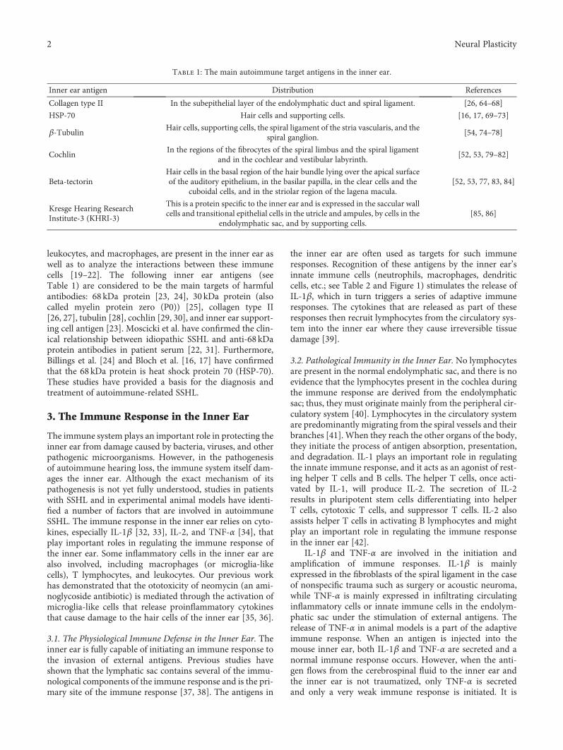
leukocytes, and macrophages, are present in the inner ear aswell as to analyze the interactions between these immunecells [19–22]. The following inner ear antigens (seeTable 1) are considered to be the main targets of harmfulantibodies: 68 kDa protein [23, 24], 30 kDa protein (alsocalled myelin protein zero (P0)) [25], collagen type II[26, 27], tubulin [28], cochlin [29, 30], and inner ear support-ing cell antigen [23]. Moscicki et al. have confirmed the clin-ical relationship between idiopathic SSHL and anti-68 kDaprotein antibodies in patient serum [22, 31]. Furthermore,Billings et al. [24] and Bloch et al. [16, 17] have confirmedthat the 68 kDa protein is heat shock protein 70 (HSP-70).These studies have provided a basis for the diagnosis andtreatment of autoimmune-related SSHL.
3. The Immune Response in the Inner Ear
The immune system plays an important role in protecting theinner ear from damage caused by bacteria, viruses, and otherpathogenic microorganisms. However, in the pathogenesisof autoimmune hearing loss, the immune system itself dam-ages the inner ear. Although the exact mechanism of itspathogenesis is not yet fully understood, studies in patientswith SSHL and in experimental animal models have identi-fied a number of factors that are involved in autoimmuneSSHL. The immune response in the inner ear relies on cyto-kines, especially IL-1β [32, 33], IL-2, and TNF-α [34], thatplay important roles in regulating the immune response ofthe inner ear. Some inflammatory cells in the inner ear arealso involved, including macrophages (or microglia-likecells), T lymphocytes, and leukocytes. Our previous workhas demonstrated that the ototoxicity of neomycin (an ami-noglycoside antibiotic) is mediated through the activation ofmicroglia-like cells that release proinflammatory cytokinesthat cause damage to the hair cells of the inner ear [35, 36].
3.1. The Physiological Immune Defense in the Inner Ear. Theinner ear is fully capable of initiating an immune response tothe invasion of external antigens. Previous studies haveshown that the lymphatic sac contains several of the immu-nological components of the immune response and is the pri-mary site of the immune response [37, 38]. The antigens in
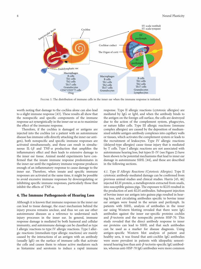
the inner ear are often used as targets for such immuneresponses. Recognition of these antigens by the inner ear’sinnate immune cells (neutrophils, macrophages, dendriticcells, etc.; see Table 2 and Figure 1) stimulates the release ofIL-1β, which in turn triggers a series of adaptive immuneresponses. The cytokines that are released as part of theseresponses then recruit lymphocytes from the circulatory sys-tem into the inner ear where they cause irreversible tissuedamage [39].
3.2. Pathological Immunity in the Inner Ear. No lymphocytesare present in the normal endolymphatic sac, and there is noevidence that the lymphocytes present in the cochlea duringthe immune response are derived from the endolymphaticsac; thus, they must originate mainly from the peripheral cir-culatory system [40]. Lymphocytes in the circulatory systemare predominantly migrating from the spiral vessels and theirbranches [41]. When they reach the other organs of the body,they initiate the process of antigen absorption, presentation,and degradation. IL-1 plays an important role in regulatingthe innate immune response, and it acts as an agonist of rest-ing helper T cells and B cells. The helper T cells, once acti-vated by IL-1, will produce IL-2. The secretion of IL-2results in pluripotent stem cells differentiating into helperT cells, cytotoxic T cells, and suppressor T cells. IL-2 alsoassists helper T cells in activating B lymphocytes and mightplay an important role in regulating the immune responsein the inner ear [42].
IL-1β and TNF-α are involved in the initiation andamplification of immune responses. IL-1β is mainlyexpressed in the fibroblasts of the spiral ligament in the caseof nonspecific trauma such as surgery or acoustic neuroma,while TNF-α is mainly expressed in infiltrating circulatinginflammatory cells or innate immune cells in the endolym-phatic sac under the stimulation of external antigens. Therelease of TNF-α in animal models is a part of the adaptiveimmune response. When an antigen is injected into themouse inner ear, both IL-1β and TNF-α are secreted and anormal immune response occurs. However, when the anti-gen flows from the cerebrospinal fluid to the inner ear andthe inner ear is not traumatized, only TNF-α is secretedand only a very weak immune response is initiated. It is
Table 1: The main autoimmune target antigens in the inner ear.
Inner ear antigen Distribution References
Collagen type II In the subepithelial layer of the endolymphatic duct and spiral ligament. [26, 64–68]
HSP-70 Hair cells and supporting cells. [16, 17, 69–73]
β-TubulinHair cells, supporting cells, the spiral ligament of the stria vascularis, and the
spiral ganglion.[54, 74–78]
CochlinIn the regions of the fibrocytes of the spiral limbus and the spiral ligament
and in the cochlear and vestibular labyrinth.[52, 53, 79–82]
Beta-tectorinHair cells in the basal region of the hair bundle lying over the apical surfaceof the auditory epithelium, in the basilar papilla, in the clear cells and the
cuboidal cells, and in the striolar region of the lagena macula.[52, 53, 77, 83, 84]
Kresge Hearing ResearchInstitute-3 (KHRI-3)
This is a protein specific to the inner ear and is expressed in the saccular wallcells and transitional epithelial cells in the utricle and ampules, by cells in the
endolymphatic sac, and by supporting cells.[85, 86]
2 Neural Plasticity
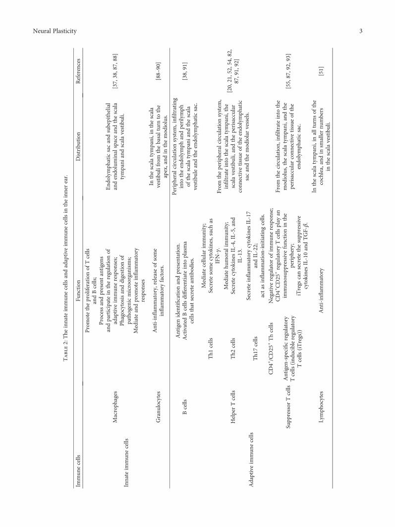
Table2:The
innateim
mun
ecells
andadaptive
immun
ecells
intheinnerear.
Immun
ecells
Function
Distribution
References
Innateim
mun
ecells
Macroph
ages
Promotetheproliferation
ofTcells
andBcells;
Processandpresentantigens
andparticipatein
theregulation
ofadaptive
immun
erespon
ses;
Phagocytosisanddigestionof
pathogenicmicroorganism
s;Mediateandprom
oteinflam
matory
respon
ses
End
olym
phaticsacandsubepithelial
andendo
luminalspaceandthescala
tympani
andscalavestibuli.
[37,38,87,88]
Granu
locytes
Anti-inflam
matory,releaseof
some
inflam
matoryfactors.
Inthescalatympani,inthescala
vestibulifrom
thebasalturnto
the
apex,and
inthemod
iolus.
[88–90]
Adaptiveim
mun
ecells
Bcells
Antigen
identification
andpresentation
.Activated
Bcells
differentiateinto
plasma
cells
that
secreteantibodies.
Peripheralcirculation
system
,infi
ltrating
into
theendo
lymph
andperilymph
ofthescalatympani
andthescala
vestibuleandtheendo
lymph
aticsac.
[38,91]
HelperTcells
Th1
cells
Mediatecellu
larim
mun
ity;
Secretesomecytokines,such
asIFN-γ.
From
theperiph
eralcirculationsystem
,infiltrateinto
thescalatympani,the
scalavestibuli,andtheperisaccular
conn
ective
tissue
oftheendo
lymph
atic
sacandthemod
iolarvessels.
[20,21,52,54,82,
87,91,92]
Th2
cells
Mediatehu
moralim
mun
ity;
SecretecytokinesIL-4,IL-5,and
IL-13.
Th1
7cells
Secreteinflam
matorycytokinesIL-17
andIL-22;
actas
inflam
mation-initiating
cells.
Supp
ressor
Tcells
CD4+/CD25
+Thcells
Negativeregulatorof
immun
erespon
se;
CD4+CD25
+regulatory
Tcells
play
anim
mun
osup
pressive
function
inthe
periph
ery;
iTregs
cansecretethesupp
ressive
cytokinesIL-10andTGF-β.
From
thecirculation,
infiltrateinto
the
mod
iolus,thescalatympani,and
the
perisaccular
conn
ective
tissue
ofthe
endo
lymph
aticsac.
[55,87,92,93]
Antigen-specificregulatory
Tcells
(ind
ucibleregulatory
Tcells
(iTregs))
Lymph
ocytes
Anti-inflam
matory
Inthescalatympani,inallturns
ofthe
cochlea,andin
smallernu
mbers
inthescalavestibuli.
[51]
3Neural Plasticity
worth noting that damage to the cochlea alone can also leadto a slight immune response [43]. These results all show thatthe nonspecific and specific components of the immuneresponse act synergistically in the inner ear so as to maximizethe effect of the immune response.
Therefore, if the cochlea is damaged or antigens areinjected into the cochlea (or a patient with an autoimmunedisease has immune cells directly attacking the inner ear anti-gen), both nonspecific and specific immune responses areactivated simultaneously, and these can result in simulta-neous IL-1β and TNF-α production that amplifies theinflammatory effect and then leads to extensive damage tothe inner ear tissue. Animal model experiments have con-firmed that the innate immune response predominates inthe inner ear until the regulatory immune response producesenough of an inflammatory response to cause damage to theinner ear. Therefore, when innate and specific immuneresponses are activated at the same time, it might be possibleto avoid excessive immune responses by downregulating orinhibiting specific immune responses, particularly those thatinhibit the effects of TNF-α.
4. The Immune Pathogenesis of Hearing Loss
Although it is known that immune responses in the inner earcan lead to tissue damage, the exact mechanism behind theinjury process remains unclear, and thus we can use otherautoimmune diseases as a reference to understand suchinjury processes in the inner ear. In general, immuneresponse damage is mediated by both humoral and cellularimmunity, and autoimmune damage can be classified as typeI allergic reactions to type IV allergic reactions. Type I aller-gic reactions (immediate-type allergic reactions) are mainlycaused by the interactions of an antigen with an antibody(usually IgE) on the surface of immune cells that activatethe cells and causes them to release active mediators suchas histamine and serotonin to induce a rapid immune
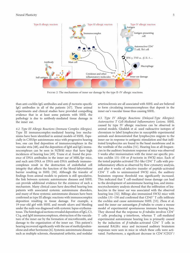
response. Type II allergic reactions (cytotoxic allergies) aremediated by IgG or IgM, and when the antibody binds tothe antigen on the foreign cell surface, the cells are destroyeddue to the action of the complement system, phagocytes,or nature killer cells. Type III allergic reactions (immunecomplex allergies) are caused by the deposition of medium-sized soluble antigen-antibody complexes into capillary wallsor tissues, which activates the complement system or leads tothe recruitment of leukocytes. Type IV allergic reactions(delayed-type allergies) cause tissue injury that is mediatedby T cells. Type I allergic reactions are not associated withautoimmune hearing loss, but types II–IV (see Figure 2) havebeen shown to be potential mechanisms that lead to inner eardamage in autoimmune SSHL [44], and these are describedin the following sections.
4.1. Type II Allergic Reactions (Cytotoxic Allergies). Type IIcytotoxic antibody-mediated damage can be confirmed fromprevious animal studies and clinical studies. Harris [45, 46]injected KLH protein, a metalloprotein extracted from snails,into susceptible guinea pigs. The exposure to KLH resulted inthe production of anti-KLH antibodies. Subsequent injectionof bovine inner ear antigen into guinea pigs resulted in hear-ing loss, and circulating antibodies specific to bovine innerear antigen were found in the serum and perilymph. Inpatients with SSHL, analysis of antibodies in the innerear using Western blotting revealed that there were IgGantibodies against the inner ear-specific proteins cochlinand β-tectorin and the nonspecific protein HSP-70. Thisstudy revealed that the direct antibody response to innerear proteins can lead to SSHL and that such antibodiescan be used as a marker for disease diagnosis. Usingantigen-specific Western blot analysis of patient andhealthy sera, it was found that anti-cochlin IgG antibodieswere more prevalent in patients with idiopathic sensori-neural hearing loss than anti-β-tectorin-specific IgG antibod-ies, whereas anti-HSP-70 IgG antibodies were more common
Cochlea
Endolymphatic sac
Semicircular ducts
EndolymphCochlear cnduct
�e Organ of corti
Macrophages
SV: scala vestibuliST: scala tympani
SVperilymph
STperilymph
iTregCD4+CD25+T cells
Granulocytes�17 cells�2 cells�1 cells
B cells Macrophages
Granulocytes�17 cells�2 cells�1 cells
B cells
GranulocytesLymphocytes
�17 cells�2 cells�1 cells
B cells
Figure 1: The distribution of immune cells in the inner ear when the immune response is initiated.
4 Neural Plasticity
than anti-cochlin IgG antibodies and anti-β-tectorin-specificIgG antibodies in all of the patients [47]. These animalexperiments and clinical studies have provided compellingevidence that in at least some patients with SSHL thepathology is due to antibody-mediated tissue damage inthe inner ear.
4.2. Type III Allergic Reactions (Immune Complex Allergies).Type III immunocomplex-mediated hearing loss mecha-nisms have been identified in animal models of SSHL. Espe-cially in C3H/lpr autoimmune mice with progressive hearingloss, one can find deposition of immunocomplexes in thevascular stria [48], and the deposition of IgM and IgG immu-nocomplexes can be seen in NZB/kl mice that have highincidences of hearing loss [49]. Trune et al. found the pres-ence of DNA antibodies in the inner ear of MRL/lpr mice,and such anti-DNA or DNA-anti-DNA antibody immuno-complexes result in the destruction of endothelial cellintegrity that affects the function of the blood-labyrinthinebarrier resulting in SSHL [50]. Although the transfer offindings from animal models to patients is still speculative,the link between systemic autoimmune diseases and SSHLcan provide additional evidence for the existence of such amechanism. Many clinical cases have described hearing losspatients with associated systemic autoimmune disorders,and many of these systemic autoimmune diseases have beenconfirmed as type III allergic reactions with immunocomplexdeposition resulting in tissue damage. For example, a19-year-old girl with SSHL and mouth ulcers and bleedingunder the nails was diagnosed with systemic lupus erythema-tosus. The histological sections revealed deposition of IgG,C3,C1q, and IgM immunocomplexes, obstruction of the vascula-ture of the inner ear by the formation of microthrombi, anddamage to the organization of the inner ear. This patient’shearing recovered significantly with the use ofmethylprednis-olone and other hormones [6]. Systemic autoimmune diseasessuch as multiple sclerosis, rheumatoid arthritis, and nodular
arteriosclerosis are all associated with SSHL and are believedto form circulating immunocomplexes that deposit in theinner ear’s vascular tissue thus causing SSHL.
4.3. Type IV Allergic Reactions (Delayed-Type Allergies):Autoreactive T Cell-Mediated Inflammatory Lesions. SSHLcaused by type IV allergic reactions can be observed inanimal models. Gloddek et al. used radioactive isotopes ofchromium to label lymphocytes in susceptible experimentalanimals and demonstrated that lymphocytes migrate to theinner ear in response to antigenic stimulation and that infil-trated lymphocytes are found in the basal membrane and inthe vestibule of the cochlea [51]. Hearing loss at all frequen-cies in the auditory brainstem response of mice was observed5 weeks after immunization with the inner ear-specific pro-tein cochlin 131–150 or β-tectorin in SWXJ mice. Each ofthe tested peptides activated Th1-like CD4+ T cells with pro-inflammatory effects as observed by flow cytometry analysis,and after 6 weeks of selective transfer of peptide-activatedCD4+ T cells to unimmunized SWXJ mice, the auditorybrainstem response threshold was significantly increased.This indicated that T cell-mediated tissue damage can leadto the development of autoimmune hearing loss, and immu-nocytochemistry analysis showed that the infiltration of leu-kocytes in the inner ear was associated with the observedhearing loss [52]. Billings also immunized SWXJ mice withcochlin 131–150 and confirmed that CD45+ T cells infiltratethe cochlea and cause autoimmune SSHL [53]. Zhou et al.used the inner ear autoantigen β-tubulin to create a mousemodel of experimental spontaneous immune hearing loss.They showed that the response to β-tubulin involves CD4+
T cells producing γ-interferon, whereas T cell-mediatedexperimental autoimmune hearing loss is primarily causedby the induction of β-tubulin-activated CD4+ T cells inneonatal BALB/c mice and increased auditory brainstemresponses were seen in mice in which these cells were acti-vated. Furthermore, a significant decrease in CD4+/CD25+/
Antibody
Type II allergic reaction Type III allergic reaction Type IV allergic reaction
Self-antigen
Mast cell
Cytokines and otherinflammatory molecules
C1 C4a C5a C3a
Immunocomplex
Macrophage
Inner ear damage
Antigen
Antigen-presentingcell
Autoreactive CD4+
T cell, Th1 cell
IFN-�훾, TNF-�훼, -�훽, IL-3.....
Figure 2: The mechanisms of inner ear damage by the type II–IV allergic reactions.
5Neural Plasticity
Foxp3+ regulatory T cells was observed in mice immunizedwith β-tubulin, which inhibited the proliferation of effectorCD4+/CD25− T cells [54]. Xia et al. used flow cytometry toanalyze the clinical T cell subtypes in 17 patients withautoimmune sensorineural hearing loss, 16 patients withnoise-induced hearing loss, and 100 individuals with normalhearing. There was no significant difference in the T cell sub-types among the three groups, except that the proportion ofCD4+ T cells in the patients with sensorineural hearing lossincreased and the function of CD4+/CD25+ regulatory T cellswas absent [55]. The above experimental animal models andclinical cases have confirmed that autoimmune hearing losscan be caused by cytotoxic T cell-mediated organ-specificautoimmune disorders of the inner ear.
5. Immunosuppressive Therapy for SSHL
Glucocorticoids have remained the main stay of treatmentover the past four decades since McCabe [14] first treatedSSHL with glucocorticoids, and the symptoms of patientswere improved significantly. Owing to the systemic sideeffects of long-term treatment with glucocorticoids, othertherapeutic methods also have been investigated. Ruckensteinet al. [56] and Trune et al. [57] used MRL/lpr mice to showthat prednisolone can protect against hearing loss. In addi-tion, Satoh et al. [58] and Wang et al. [59] used etanercept, aTNF-α antagonist, to treat SSHL and showed that it canreduce inflammation in the inner ear andprevent hearing loss.Clinically, Xenellis et al. [60] have shown that the intratympa-nic injection of steroids is a safe and effectivemethod for SSHLtreatment, andHaynes et al. [61] have shown that intratympa-nic injection of dexamethasone can also improve hearing inSSHL patients when systemic medications fail. Furthermore,Battaglia et al. [62] used a combination therapy of intra-tympanic dexamethasone with high-dose prednisone taperfor SSHL and showed that the patients receiving the com-bination therapy had significant improvements in speech-discrimination score and pure-tone average and recoveredtheir hearing quickly. More recently, azathioprine has beenconfirmed to maintain the hearing threshold, decrease therisk of relapse, and slow down the rate at which patientsrelapse [63].
The evidence to date suggests that autoimmune SSHL ismainly mediated by autoantibodies or T cells or by both. Asautoimmune reactions are increasingly considered to be acause of SSHL, animal models and clinical trials have shownthat autoimmune processes cause damage to the inner earthrough various mechanisms. Humoral immunity and cellu-lar immune-mediated autoimmune damage have both beenshown to play a role in the pathogenesis of autoimmunehearing loss. Although the precise diagnosis of autoimmuneSSHL is still difficult, the response to immunosuppressivetherapy is generally positive for these patients. Therefore,the immune mechanism of SSHL needs further study inorder to identify specific antigens of the inner ear and spe-cific diagnostic markers that can provide a more accurateand timely diagnosis and contribute to a more effectivetreatment plan.
Conflicts of Interest
The authors declare that there is no conflict of interestregarding the publication of this paper.
Authors’ Contributions
Guangfei Li and Dan You contributed equally to this work.
Acknowledgments
Funding was provided by the National Key R&D Programof China (nos. 2017YFA0103900 and 2016YFC0905200),the National Natural Science Foundation of China (nos.81570913 and 81620108005), and the Shanghai PujiangTalents Plan (18PJ1401700).
References
[1] S. K. Juhn, L. P. Rybak, and S. Prado, “Nature of blood-labyrinth barrier in experimental conditions,” The Annals ofOtology, Rhinology, and Laryngology, vol. 90, no. 2, pp. 135–141, 1981.
[2] M. Düzlü, M. Çolak, İ. K. Önal, H. Tutar, R. Karamert, andÇ. Gökdoğan, “Audiological findings in autoimmune hepatitis:hearing loss at high frequencies,”Gazi Medical Journal, vol. 28,no. 4, 2017.
[3] J. H. Check, “Sympathetic neural hyperalgesia edema syn-drome as a cause of autoimmune hearing loss,” Clinical andExperimental Obstetrics & Gynecology, vol. 44, no. 1, pp. 133-134, 2017.
[4] S. Montes, S. Rodríguez-Muguruza, V. Soria, and A. Olivé,“Atypical Cogan’ syndrome associated with sudden deafnessand glucocorticoid response,” Reumatología Clínica, vol. 10,no. 4, pp. 267-268, 2014.
[5] A. Bacciu, E. Pasanisi, F. di Lella, M. Guida, S. Bacciu, andV. Vincenti, “Cochlear implantation in patients with Cogansyndrome: long-term results,” European Archives of Oto-Rhino-Laryngology, vol. 272, no. 11, pp. 3201–3207, 2015.
[6] S. Chawki, J. Aouizerate, S. Trad, J. Prinseau, and T. Hanslik,“Bilateral sudden sensorineural hearing loss as a presentingfeature of systemic lupus erythematosus: case report andbrief review of other published cases,” Medicine, vol. 95,no. 36, article e4345, 2016.
[7] C. A. Bowman, F. H. Linthicum Jr, R. A. Nelson, K. Mikami,and F. Quismorio, “Sensorineural hearing-loss associated withsystemic lupus erythematosus,” Otolaryngology-Head andNeck Surgery, vol. 94, no. 2, pp. 197–204, 1986.
[8] M. Tanaka and K. Tanaka, “Sudden hearing loss as the initialsymptom in Japanese patients with multiple sclerosis and sero-positive neuromyelitis optica spectrum disorders,” Journal ofNeuroimmunology, vol. 298, pp. 16–18, 2016.
[9] M. Tekin, G. O. Acar, O. H. Cam, and F. M. Hanege, “Suddensensorineural hearing loss in a multiple sclerosis case,” North-ern Clinics of Istanbul, vol. 1, no. 2, pp. 109–113, 2014.
[10] M. A. Hellmann, I. Steiner, and R. Mosberg-Galili, “Suddensensorineural hearing loss in multiple sclerosis: clinical courseand possible pathogenesis,” Acta Neurologica Scandinavica,vol. 124, no. 4, pp. 245–249, 2011.
[11] M. A. Melikoglu and K. Senel, “Sudden hearing loss in apatient with rheumatoid arthritis; a case report and review of
6 Neural Plasticity
the literature,” Acta Reumatológica Portuguesa, vol. 38, no. 2,pp. 138-139, 2013.
[12] F. Rubin, N. Tran Khai Hoan, and P. Bonfils, “Sudden bilateralhearing loss revealing polyarteritis nodosa,” European Annalsof Otorhinolaryngology, Head and Neck Diseases, vol. 131,no. 4, pp. 265-266, 2014.
[13] G. Tirelli, P. Tomietto, E. Quatela et al., “Sudden hearing lossand Crohn disease: when Cogan syndrome must be sus-pected,” American Journal of Otolaryngology, vol. 36, no. 4,pp. 590–597, 2015.
[14] B. F. McCabe, “Autoimmune sensorineural hearing loss,” TheAnnals of Otology, Rhinology, and Laryngology, vol. 88, no. 5,pp. 585–589, 1979.
[15] A. Sismanis, C. Wise, and G. Johnson, “Methotrexate man-agement of immune-mediated cochleovestibular disorders,”Otolaryngology and Head and Neck Surgery, vol. 116,no. 2, pp. 146–152, 1997.
[16] D. B. Bloch, J. A. Gutierrez, V. Guerriero Jr, S. D. Rauch, andK. J. Bloch, “Recognition of a dominant epitope in bovineheat-shock protein 70 in inner ear disease,” Laryngoscope,vol. 109, no. 4, pp. 621–625, 1999.
[17] D. B. Bloch, J. E. San Martin, S. D. Rauch, R. A. Moscicki, andK. J. Bloch, “Serum antibodies to heat shock protein 70 in sen-sorineural hearing loss,” Archives of Otolaryngology–Head &Neck Surgery, vol. 121, no. 10, pp. 1167–1171, 1995.
[18] L. Xu, C. R. Pfaltz, andW. Arnold, “Human leukocyte antigensin patients with inner ear diseases of unknown etiology,” ORL,vol. 55, no. 3, pp. 125–134, 1993.
[19] H. Iwai, K. Tomoda, M. Inaba et al., “Evidence of cellularsupplies to the endolymphatic sac from the systemic circu-lation,” Acta Oto-Laryngologica, vol. 115, no. 4, pp. 509–511, 2009.
[20] H. Iwai, M. Inaba, K. Tomoda, S. Ikehara, K. Sugiura, andT. Yamashita, “T cells infiltrating from the systemic circulationproliferate in the endolymphatic sac,” The Annals of Otology,Rhinology, and Laryngology, vol. 108, no. 12, pp. 1146–1150,1999.
[21] B. Gloddek, J. Gloddek, and W. Arnold, “A rat T-cell line thatmediates autoimmune disease of the inner ear in the Lewisrat,” ORL, vol. 61, no. 4, pp. 181–187, 1999.
[22] R. A. Moscicki, J. E. San Martin, C. H. Quintero, S. D. Rauch,Nadol JB Jr, and K. J. Bloch, “Serum antibody to inner ear pro-teins in patients with progressive hearing loss. Correlationwith disease activity and response to corticosteroid treatment,”JAMA, vol. 272, no. 8, pp. 611–616, 1994.
[23] J. Dhingra and M. Mahalingam, “Antibodies to 68 kd antigen-specific for inner ear disease?,” Laryngoscope, vol. 107, no. 3,pp. 405-406, 1997.
[24] P. B. Billings, E. M. Keithley, and J. P. Harris, “Evidence linkingthe 68 kilodalton antigen identified in progressive sensorineu-ral hearing loss patient sera with heat shock protein 70,” TheAnnals of Otology, Rhinology, and Laryngology, vol. 104,no. 3, pp. 181–188, 1995.
[25] G. B. Hughes, B. P. Barna, S. E. Kinney, L. H. Calabrese, andN. J. Nalepa, “Clinical diagnosis of immune inner-ear disease,”Laryngoscope, vol. 98, no. 3, pp. 251–3, 1988.
[26] J. P. Harris, N. K. Woolf, and A. F. Ryan, “A reexaminationof experimental type II collagen autoimmunity: middle andinner ear morphology and function,” The Annals of Otology,Rhinology, and Laryngology, vol. 95, no. 2, pp. 176–180,1986.
[27] K. C. Campbell and J. J. Klemens, “Sudden hearing loss andautoimmune inner ear disease,” Journal of the American Acad-emy of Audiology, vol. 11, no. 7, pp. 361–7, 2000.
[28] R. Hallworth, M. McCoy, and J. Polan-Curtain, “Tubulinexpression in the developing and adult gerbil organ of Corti,”Hearing Research, vol. 139, no. 1-2, pp. 31–41, 2000.
[29] M. Komori, Y. Yamamoto, Y. Yaguchi, T. Ikezono, andH. Kojima, “Cochlin-tomoprotein test and hearing outcomesin surgically treated true idiopathic perilymph fistula,” ActaOto-Laryngologica, vol. 136, no. 9, pp. 901–904, 2016.
[30] P. Baruah, “Cochlin in autoimmune inner ear disease: is thesearch for an inner ear autoantigen over?,” Auris NasusLarynx, vol. 41, no. 6, pp. 499–501, 2014.
[31] M. Gross, R. Eliashar, A. Ben-Yaakov, R. Ulmansky, andJ. Elidan, “Prevalence and clinical significance of anticar-diolipin, anti-β2-glycoprotein-1, and anti-heat shockprotein-70 autoantibodies in sudden sensorineural hearingloss,” Audiology & Neuro-Otology, vol. 13, no. 4, pp. 231–238, 2008.
[32] S. Pathak, E. Goldofsky, E. X. Vivas, V. R. Bonagura, andA. Vambutas, “IL-1β is overexpressed and aberrantly regu-lated in corticosteroid nonresponders with autoimmune innerear disease,” Journal of Immunology, vol. 186, no. 3, pp. 1870–1879, 2011.
[33] S. D. Rauch, “IL-1β inhibition in autoimmune inner eardisease: can you hear me now?,” The Journal of ClinicalInvestigation, vol. 124, no. 9, pp. 3685–3687, 2014.
[34] S. Pathak, C. Stern, and A. Vambutas, “N-Acetylcysteine atten-uates tumor necrosis factor alpha levels in autoimmune innerear disease patients,” Immunologic Research, vol. 63, no. 1–3,pp. 236–245, 2015.
[35] S. Sun, H. Yu, H. Yu et al., “Inhibition of the activation andrecruitment of microglia-like cells protects against neomycin-induced ototoxicity,” Molecular Neurobiology, vol. 51, no. 1,pp. 252–267, 2015.
[36] Z. Wang and H. Li, “Microglia-like cells in rat organ of cortifollowing aminoglycoside ototoxicity,” Neuroreport, vol. 11,no. 7, pp. 1389–1393, 2000.
[37] M. Barbara, G. Attanasio, V. Petrozza, A. Modesti, andR. Filipo, “The endolymphatic sac as the immunocompetentorgan of the inner ear,” Annals of the New York Academy ofSciences, vol. 830, pp. 243–252, 1997.
[38] S. Tomiyama and J. Harris, “The role of the endolymphatic sacin inner ear immunity,” Acta Oto-Laryngologica, vol. 103,no. 3, pp. 182–188, 1987.
[39] S. Hashimoto, P. Billings, J. P. Harris, G. S. Firestein, and E. M.Keithley, “Innate immunity contributes to cochlear adaptiveimmune responses,” Audiology and Neuro-Otology, vol. 10,no. 1, pp. 35–43, 2005.
[40] J. Harris and A. Ryan, “Fundamental immune mechanisms ofthe brain and inner ear,” Otolaryngology-Head and NeckSurgery, vol. 112, no. 6, pp. 639–653, 1995.
[41] J. P. Harris, S. Fukuda, and E. M. Keithley, “Spiral Modiolarvein: Its importance in inner ear inflammation,” Acta Oto-Laryngologica, vol. 110, no. 3-4, pp. 357–364, 1990.
[42] B. Gloddek and J. P. Harris, “Role of lymphokines in theimmune response of the inner ear,” Acta Oto-Laryngologica,vol. 108, no. 1-2, pp. 68–75, 2009.
[43] H. Satoh, G. S. Firestein, P. B. Billings, J. P. Harris, and E. M.Keithley, “Proinflammatory cytokine expression in the endo-lymphatic sac during inner ear inflammation,” Journal of the
7Neural Plasticity
Association for Research in Otolaryngology, vol. 4, no. 2,pp. 139–147, 2003.
[44] Q. Gopen, E. M. Keithley, and J. P. Harris, “Mechanismsunderlying autoimmune inner ear disease,” Drug DiscoveryToday: Disease Mechanisms, vol. 3, no. 1, pp. 137–142, 2006.
[45] J. P. Harris, “Immunology of the inner ear: response of theinner ear to antigen challenge,” Otolaryngology-Head andNeck Surgery, vol. 91, no. 1, pp. 18–23, 1983.
[46] J. P. Harris, “Immunology of the inner ear: evidence of localantibody production,” Annals of Otology, Rhinology & Laryn-gology, vol. 93, no. 2, pp. 157–162, 2016.
[47] A. Naumann, J. M. Hempel, and K. Schorn, “Detection ofhumoral immune response to inner ear proteins in patientswith sensorineural hearing loss,” Laryngo- Rhino- Otologie,vol. 80, no. 5, pp. 237–244, 2001.
[48] D. R. Trune, J. P. Craven, J. I. Morton, and C. Mitchell, “Auto-immune disease and cochlear pathology in the C3H/lpr strainmouse,” Hearing Research, vol. 38, no. 1-2, pp. 57–66, 1989.
[49] H. Nariuchi, M. Sone, C. Tago, T. Kurata, and K. Saito, “Mech-anisms of hearing disturbance in an autoimmune modelmouse NZB/kl,” Acta Oto-Laryngologica. Supplementum,vol. 514, pp. 127–131, 1994.
[50] D. R. Trune, J. B. Kempton, S. H. Hefeneider, and R. M.Bennett, “Inner ear DNA receptors in MRL/lpr autoimmunemice: potential 30 and 70 kDa link between autoimmunedisease and hearing loss,” Hearing Research, vol. 105,no. 1-2, pp. 57–64, 1997.
[51] B. Gloddek, A. F. Ryan, and J. P. Harris, “Homing of lympho-cytes to the inner ear,” Acta Oto-Laryngologica, vol. 111, no. 6,pp. 1051–1059, 2009.
[52] C. A. Solares, A. E. Edling, J. M. Johnson et al., “Murine auto-immune hearing loss mediated by CD4+ T cells specific forinner ear peptides,” Journal of Clinical Investigation, vol. 113,no. 8, pp. 1210–1217, 2004.
[53] P. Billings, “Experimental autoimmune hearing loss,” Journalof Clinical Investigation, vol. 113, no. 8, pp. 1114–1117, 2004.
[54] B. Zhou, M. H. Kermany, J. Glickstein et al., “Murine auto-immune hearing loss mediated by CD4+ T cells specific forβ-tubulin,” Clinical Immunology, vol. 138, no. 2, pp. 222–230, 2011.
[55] M. Xia, H. B. Zhang, F. Liu, H. Y. Yin, and A. T. Xu, “ImpairedCD4+CD25+ regulatory T cell activity in the peripheral bloodof patients with autoimmune sensorineural hearing loss,”European Archives of Oto-Rhino-Laryngology, vol. 265, no. 9,pp. 1027–1033, 2008.
[56] M. J. Ruckenstein, A. Sarwar, L. Hu, H. Shami, and T. N.Marion, “Effects of immunosuppression on the developmentof cochlear disease in the MRL-Faslpr mouse,” Laryngoscope,vol. 109, no. 4, pp. 626–630, 1999.
[57] D. R. Trune, R. J. Wobig, J. B. Kempton, and S. H. Hefeneider,“Steroid treatment in young MRL.MpJ-Faslpr autoimmunemice prevents cochlear dysfunction,” Hearing Research,vol. 137, no. 1-2, pp. 167–173, 1999.
[58] H. Satoh, G. S. Firestein, P. B. Billings, J. P. Harris, and E. M.Keithley, “Tumor necrosis factor-α, an initiator, and etaner-cept, an inhibitor of cochlear inflammation,” Laryngoscope,vol. 112, no. 9, pp. 1627–1634, 2002.
[59] X. Wang, T. Truong, P. B. Billings, J. P. Harris, and E. M.Keithley, “Blockage of immune-mediated inner ear damageby etanercept,” Otology & Neurotology, vol. 24, no. 1, pp. 52–57, 2003.
[60] J. Xenellis, N. Papadimitriou, T. Nikolopoulos et al., “Intra-tympanic steroid treatment in idiopathic sudden sensorineuralhearing loss: a control study,” Otolaryngology and Head andNeck Surgery, vol. 134, no. 6, pp. 940–945, 2016.
[61] B. J. Balough, “Intratympanic dexamethasone for suddensensorineural hearing loss after failure of systemic therapy,”Yearbook of Otolaryngology-Head and Neck Surgery,vol. 2008, pp. 45–47, 2008.
[62] A. Battaglia, R. Burchette, and R. Cueva, “Combination ther-apy (intratympanic dexamethasone + high-dose prednisonetaper) for the treatment of idiopathic sudden sensorineuralhearing loss,” Otology & Neurotology, vol. 29, no. 4, pp. 453–460, 2008.
[63] N. Mata-Castro, J. Gavilanes-Plasencia, R. Ramírez-Camacho, A. García-Fernández, and J. R. García-Berrocal,“Azathioprine reduces the risk of audiometric relapse inimmune-mediated hearing loss,” Acta OtorrinolaringológicaEspañola, 2018.
[64] T. J. Yoo, M. A. Cremer, K. Tomoda, A. S. Townes, J. M. Stuart,and A. H. Kang, “Type II collagen-induced autoimmune sen-sorineural hearing loss and vestibular dysfunction in rats,”Annals of Otology, Rhinology & Laryngology, vol. 92, no. 3,pp. 267–271, 1983.
[65] T. Yoo, Y. Yazawa, K. Tomoda, and R. Floyd, “Type IIcollagen-induced autoimmune endolymphatic hydrops inguinea-pig,” Science, vol. 222, no. 4619, pp. 65–67, 1983.
[66] P. Berger, M. Hillman, M. Tabak, and M. Vollrath, “Thelymphocyte transformation test with type II collagen as adiagnostic tool of autoimmune sensorineural hearing loss,”Laryngoscope, vol. 101, no. 8, pp. 895–899, 1991.
[67] L. F. Bertoli, D. G. Pappas, J. C. Barton, and J. C. Barton,“Serum immunoglobulins in 28 adults with autoimmune sen-sorineural hearing loss: increased prevalence of subnormalimmunoglobulin G1 and immunoglobulin G3,” BMC Immu-nology, vol. 15, no. 1, p. 43, 2014.
[68] T. J. Yoo, K. Tomoda, and A. D. Hernandez, “Type II collageninduced autoimmune inner ear lesions in guinea pigs,” Annalsof Otology, Rhinology & Laryngology, vol. 93, Supplement 5,pp. 3–5, 1984.
[69] P. B. Billings, S. O. Shin, and J. P. Harris, “Assessing the role ofanti-hsp70 in cochlear impairment,” Hearing Research,vol. 126, no. 1-2, pp. 210–212, 1998.
[70] C. Ianuale, G. Cadoni, E. de Feo et al., “A systematic reviewand meta-analysis of the diagnostic accuracy of anti-heatshock protein 70 antibodies for the detection of autoimmunehearing loss,” Otology & Neurotology, vol. 34, no. 2, pp. 214–219, 2013.
[71] K. Yeom, J. Gray, T. S. Nair et al., “Antibodies to HSP-70 innormal donors and autoimmune hearing loss patients,” Laryn-goscope, vol. 113, no. 10, pp. 1770–1776, 2003.
[72] S. Charchat, L. Lavinsky, E. Cohen, C. A. Muhlen, andC. Bonorino, “Use of HSP70 for diagnosis and treatmentof patients of sensorineural autoimmune hearing loss,” CellStress & Chaperones, vol. 5, no. 4, pp. 384–384, 2000.
[73] C. Bonaguri, J. G. Orsoni, L. Zavota et al., “Anti-68 kDaantibodies in autoimmune sensorineural hearing loss - arethese autoantibodies really a diagnostic tool?,” Autoimmunity,vol. 40, no. 1, pp. 73–78, 2007.
[74] T. J. Yoo, X. Ge, O. Sener et al., “Presence of autoantibodiesin the sera of Meniere’s disease,” Annals of Otology, Rhinol-ogy & Laryngology, vol. 110, no. 5, pp. 425–429, 2001.
8 Neural Plasticity
[75] T. J. Yoo, H. Tanaka, S. S. Kwon et al., “β-Tubulin as anautoantigen for autoimmune inner ear disease,” InternationalCongress Series, vol. 1240, pp. 1207–1210, 2003.
[76] T. J. Yoo, X. Du, and S. S. Kwon, “Molecular mechanism ofautoimmune hearing loss,” Acta Oto-Laryngologica, vol. 122,no. 5, pp. 3–9, 2002.
[77] X. Du, T. Yoo, and R. Mora, “Distribution of beta-tubulin inguinea pig inner ear,” ORL, vol. 65, no. 1, pp. 7–16, 2003.
[78] Q. Cai, X. du, B. Zhou, C. Cai, M. H. Kermany, and T. Yoo,“Induction of tolerance by oral administration of beta-tubulin in an animal model of autoimmune inner ear disease,”ORL, vol. 71, no. 3, pp. 135–141, 2009.
[79] N. G. Robertson, L. Lu, S. Heller et al., “Mutations in a novelcochlear gene cause DFNA9, a human nonsyndromic deafnesswith vestibular dysfunction,” Nature Genetics, vol. 20, no. 3,pp. 299–303, 1998.
[80] N. G. Robertson, B. L. Resendes, J. S. Lin et al., “Inner earlocalization of mRNA and protein products of COCH,mutated in the sensorineural deafness and vestibular disorder,DFNA9,” Human Molecular Genetics, vol. 10, no. 22,pp. 2493–2500, 2001.
[81] T. Ikezono, A. Omori, S. Ichinose, R. Pawankar, A. Watanabe,and T. Yagi, “Identification of the protein product of theCoch gene (hereditary deafness gene) as the major compo-nent of bovine inner ear protein,” Biochimica et BiophysicaActa-Molecular Basis of Disease, vol. 1535, no. 3, pp. 258–265, 2001.
[82] M. J. Baek, H.M. Park, J. M. Johnson et al., “Increased frequen-cies of cochlin-specific T cells in patients with autoimmunesensorineural hearing loss,” Journal of Immunology, vol. 177,no. 6, pp. 4203–4210, 2006.
[83] R. Killick, P. K. Legan, C. Malenczak, and G. P. Richardson,“Molecular cloning of chick beta-tectorin, an extracellularmatrix molecule of the inner ear,” Journal of Cell Biology,vol. 129, no. 2, pp. 535–547, 1995.
[84] P. K. Legan, A. Rau, J. N. Keen, and G. P. Richardson, “Themouse tectorins - modular matrix proteins of the inner earhomologous to components of the sperm-egg adhesion sys-tem,” Journal of Biological Chemistry, vol. 272, no. 13,pp. 8791–8801, 1997.
[85] T. S. Nair, D. M. Prieskorn, J. M. Miller, A. Mori, J. Gray, andT. E. Carey, “In vivo binding and hearing loss after intraco-chlear infusion of KHRI-3 antibody,” Hearing Research,vol. 107, no. 1-2, pp. 93–101, 1997.
[86] M. Ptok, T. Nair, T. E. Carey, and R. A. Altschuler, “Distribu-tion of KHRI 3 epitopes in the inner ear,” Hearing Research,vol. 66, no. 2, pp. 245–252, 1993.
[87] M. Takahashi and J. P. Harris, “Analysis of immunocompetentcells following inner ear immunostimulation,” Laryngoscope,vol. 98, no. 10, pp. 1133–1138, 1988.
[88] M. Takahashi and J. P. Harris, “Anatomic distribution andlocalization of immunocompetent cells in normal mouseendolymphatic sac,” Acta Oto-Laryngologica, vol. 106, no. 5-6,pp. 409–416, 1988.
[89] H. F. Schuknecht, “Inner ear pathology in autoimmunedisease,” Progress in Human Auditory and Vestibular Histopa-thology, Kugler Publications, 1997.
[90] H. F. Schuknecht, “Ear pathology in autoimmune disease,”Advances in Oto-Rhino-Laryngology, vol. 46, pp. 50–70,1991.
[91] H. Kawauchi, N. Kaneda, I. Ichimiya, and G. Mogi, “Distribu-tion of immunocompetent cells in the endolymphatic sac,”The Annals of Otology, Rhinology & Laryngology, vol. 101,Supplement 10, pp. 39–47, 1992.
[92] H. Cantor, F. W. Shen, and E. A. Boyse, “Separation of helperT-cells from suppressor T-cells expressing different Lycomponents. II. Activation by antigen: after immunization,antigen-specific suppressor and helper activities are mediatedby distinct T-cell subclasses,” Journal of ExperimentalMedicine, vol. 143, no. 6, pp. 1391–1340, 1976.
[93] A. M. Bilate and J. J. Lafaille, “Induced CD4+Foxp3+ regulatoryT cells in immune tolerance,” Annual Review of Immunology,vol. 30, no. 1, pp. 733–758, 2012.
9Neural Plasticity

Hindawiwww.hindawi.com Volume 2018
Research and TreatmentAutismDepression Research
and TreatmentHindawiwww.hindawi.com Volume 2018
Neurology Research International
Hindawiwww.hindawi.com Volume 2018
Alzheimer’s DiseaseHindawiwww.hindawi.com Volume 2018
International Journal of
Hindawiwww.hindawi.com Volume 2018
BioMed Research International
Hindawiwww.hindawi.com Volume 2018
Research and TreatmentSchizophrenia
Hindawi Publishing Corporation http://www.hindawi.com Volume 2013Hindawiwww.hindawi.com
The Scientific World Journal
Volume 2018Hindawiwww.hindawi.com Volume 2018
Neural PlasticityScienti�caHindawiwww.hindawi.com Volume 2018
Hindawiwww.hindawi.com Volume 2018
Parkinson’s Disease
Sleep DisordersHindawiwww.hindawi.com Volume 2018
Hindawiwww.hindawi.com Volume 2018
Neuroscience Journal
MedicineAdvances in
Hindawiwww.hindawi.com Volume 2018
Hindawiwww.hindawi.com Volume 2018
Psychiatry Journal
Hindawiwww.hindawi.com Volume 2018
Computational and Mathematical Methods in Medicine
Multiple Sclerosis InternationalHindawiwww.hindawi.com Volume 2018
StrokeResearch and TreatmentHindawiwww.hindawi.com Volume 2018
Hindawiwww.hindawi.com Volume 2018
Behavioural Neurology
Hindawiwww.hindawi.com Volume 2018
Case Reports in Neurological Medicine
Submit your manuscripts atwww.hindawi.com


![George Kampessis, Nicholas Maroudias, Papacharalampous ... steroids for sudden... · Over 60 years since its first report by De Kleyn [1], sudden sensorineural hearing loss (SSNHL)](https://static.fdocuments.net/doc/165x107/5c79ad9809d3f2bd0e8b9610/george-kampessis-nicholas-maroudias-papacharalampous-steroids-for-sudden.jpg)