The role of A-kinase anchoring proteins (AKAPs) in the ...
20
The role of A-kinase anchoring proteins (AKAPs) in the pathophysiology of chronic obstructive pulmonary disease (COPD) K.J. Visser 1 ; Prof. Dr. M. Schmidt 2 1 University of Groningen, The Netherlands 2 Department of Molecular Pharmacology, University of Groningen, The Netherlands March 2016
Transcript of The role of A-kinase anchoring proteins (AKAPs) in the ...
The role of A-kinase anchoring proteins (AKAPs) in the
pathophysiology of chronic obstructive pulmonary disease (COPD)
K.J. Visser1; Prof. Dr. M. Schmidt2
1 University of Groningen, The Netherlands
2 Department of Molecular Pharmacology, University of Groningen, The Netherlands
March 2016
Abstract
Chronic obstructive pulmonary disease (COPD) is an increasing global health problem and leading
cause of disability and death. The disease is characterised by chronic inflammation due to increased
levels of cytokines and chemokines as well as alveolar neutrophils, macrophages and T-lymphocytes.
and will result in loss of lung elasticity and mucus hypersecretion. Also, due to exposure to noxious
substances, like cigarette smoke extract, levels of reactive oxygen species (ROS) increase inducing
damage to lipids, proteins and DNA. These responses originate from signal transduction mechanisms
which are controlled by compartmentalization of cAMP-dependent protein kinase (PKA) which are
bound to A-kinase anchoring proteins (AKAPs). The highly conserved members of the AKAP ensures
spatial-temporal control of important signal transduction mechanisms. Dyregulation of AKAPs is
related to a number of disorders like neurodegenerative disease, cardiovascular disease and COPD.
This assay will focus on the role of AKAPs in the respiratory tract and the pathophysiology of COPD.
Abbreviations
AC Adenylyl cyclase
ATP Adenosine triphosphate
AKAP A-kinase anchoring protein
cAMP Cyclic adenosine 3’,5’-monophosphate
COPD Chronic obstructive pulmonary disease
DAG Diacylglycerol
Epac Exchange protein activated by cAMP
ERM Ezrin/Radixin/Moesin
FEV1 Exhaled volume in the first second
FVC Forced vital capacity
GDP Guanosine diphosphate
GOLD Global Initiative for Chronic Obstructive Lung Disease
GTP Guanosine triphosphate
IL-8 Interleukin-8
IP3 Inositol triphosphate
IP-10 Interferon-γ inducible protein
I-TAC Interferon-induced T-cell α-chemo attractant
LABA Long-acting β-agonist
LAMA Long-acting muscarinic antagonist
LTB4 Leukotriene B4
MAPK Mitogen-activated protein kinase
MMP Matrix metalloproteinase
NE Neutrophil-derived elastolytic protease
NF-κB Nuclear factor-kappa B
Nrf2 Nuclear factor-erythroid 2-related factor-2
PDE Phospodiesterase
PKA Protein kinase A
PKC Protein kinase C
PKI Protein kinase inhibitor
ROS Reactive oxygen species
TGF-β Transforming growth factor-β
Index
I. Introduction .............................................................................................................................. 1
Bronchiolitis and emphysema ............................................................................................................. 2
Inflammatory cells ............................................................................................................................... 2
Oxidative stress ................................................................................................................................... 3
II. A-kinase anchoring proteins ...................................................................................................... 5
Cyclic adenosine monophosphate and protein kinase A .................................................................... 5
Regulatory role of A-kinase anchoring proteins .................................................................................. 6
III. AKAPs in the airways ............................................................................................................... 8
AKAP signalling .................................................................................................................................... 8
Contractile function of smooth muscle cells ....................................................................................... 8
Inflammation and remodelling ............................................................................................................ 9
AKAP in relation to disease ................................................................................................................. 9
IV. Outlook ................................................................................................................................. 11
V. References ................................................................................................................................ I
1
I. Introduction
Chronic obstructive pulmonary disease (COPD) is a slowly progressive lung condition and has become
a rising global health problem with a high mobility and mortality rate (Rahman, 2008). Currently, the
disease affects more than 5% of the population and it is the fourth leading cause of death. The World
Health Organization has announced that the disease will become the third leading cause of deaths by
2020 (Barnes, Shapiro and Pauwels, 2003; Mannino, 2006). COPD is associated with pulmonary
obstructive bronchiolitis, emphysema causing loss of lung elasticity, increased mucous secretion,
inflammatory components which ultimately lead to tissue damage and elevated levels of oxidative
stress (Barnes et al., 2003; Fromer and Cooper, 2016). COPD is mainly caused by a prolonged
exposure to noxious gases or particles such as cigarette smoke, the most common cause in well
developed countries. Other risk factors are air pollution (indoor biomass fuels), poor diet and
occupational exposure. COPD can also be caused by a genetic predisposition, like alpha-1 antitrypsin
deficiency. Alph-1 antitrypsin is a protease inhibitor protecting lung tissue by inactivating neutrophil
elastase (Brode et al., 2012). As a result of alpha-1 antitrypsin deficiency some patients start to
develop COPD at a young age (Taraseviciene-Stewart and Voelkel, 2008; Brode et al., 2012). For an
authoritative diagnosis of COPD patients undergo spirometry testing, a standard instrument which is
used for the assessment of the extent of airflow limitation and monitoring the progression of the
disorder. During a spirometry test the FEV1, exhaled volume in one second, and the forced vital
capacity (FVC), maximum volume that can be exhaled after full inhalation, is measured. The ratio of
these two measurements (FEV1/FVC) provides insight into the lung volume of the patient.
COPD is classified into four stages (GOLD I –IV, mild to very severe). This classification is an initiative
of the Global Initiative for Chronic Obstructive Lung Disease (GOLD) started in 2006 en is used
worldwide. Currently there are several approaches for the management of COPD. Most important
are the long-acting β-agonist (LABA) and the long-acting muscarinic antagonist (LAMA), such as
formoterol and salmeterol, used in stage I to stage IV COPD. These bronchodilators reduce
symptoms, such as persistent cough and shortness of breath, and provide relaxation of smooth
muscles in the airways (Carlin, 2016; Fromer and Cooper, 2016). Corticosteroids are used to reduce
exacerbations in COPD stage III en IV but have limited effect on lung function (Barnes,
2005).Phosphodiesterase (PDE) inhibitors are another class of medicine used in the management of
COPD. PDE 4 is an important regulator of cyclic adenosine monophosphate (cAMP) metabolism
during inflammation. Research has shown that roflumilast, a PDE 4 inhibitor, causes reduction in
exacerbations and improvement of FEV1 (Giembycz, 2001; Rennard et al., 2011).
In the next chapters, this paper will focus on the injury and remodelling that occur in the lungs, A-
kinase anchoring proteins (AKAPs) and their involvement in the pathophysiology of COPD.
2
Bronchiolitis and emphysema
Continuous exposure of noxious substances to the lungs causes a persistent inflammatory reaction.
Thereby, cytokines and chemokines are continuously released by the innate and adaptive immune
system. These can stimulate epidermal growth factor receptors nearby which will activate
transcription factors or inflammatory and mucin genes. By this activation there is an increased
release of mucus by goblet cells. Mucus normally ensures clearance of the respiratory tract but
during hypersecretion it has opposite effects. Cilia present in the lungs cannot function under
excessive mucus production as well as mechanism that normally provide mucus degradation. A very
devastating consequence of hypersecretion is the increased degranulation of elastase from
neutrophils. These effect lead to persistent cough and chronic bronchiolitis (Burgel, 2004; Kim and
Criner, 2015).
Persistent inflammation of the airways leads to disruption and damage of the alveolar structure and
will eventually result in emphysema. The activity of protease and anti-protease are of great
importance in the formation of emphysema. Proteases and anti-proteases play a major role in the
intracellular and extracellular regulation of homeostatic, killing bacteria and regeneration of tissue.
However, in case of persistent inflammation the balance between proteases and anti-proteases is
disturbed resulting in an increase of proteases which causes damage (Sharafkhaneh et al., 2008;
Greene and McElvaney, 2009; Abbout, 2012). An important serine protease in this process is
neutrophil-derived elastolytic protease (NE) stored in the granules of neutrophils. NE is released
during inflammation and causes degradation of elastin and collagen, matrix proteins important in the
maintenance of interstitial tissue and airway structure (Nadel, 2000; Zhu et al., 2001; Dunsmore,
2008). Because of the degradation of elastin, the lungs loses their elasticity which reduces lung
capacity and leads to enlargement of the airspace (Starcher, 1986; Taraseviciene-Stewart and
Voelkel, 2008). NE and growth factors causes deposition of collagen and creating fibrosis in this way.
All these processes result in airflow limitation in COPD.
Inflammatory cells
COPD is characterized as an inflammatory disease. Many different inflammatory cells and mediators
derived from the innate and adaptive immune system, are involved in the pathophysiology of the
disorder (Barnes, 2004; Rovina et al., 2013).
Neutrophils are highly involved in the inflammatory process in COPD and form the first defence in
the airways. The cells are present in the bronchioles and parenchyma and are increased in COPD
patients. The amount of neutrophils present is correlated to the progression of the disease and the
decline of lung function (O’Donnell, 2006). Neutrophils secrete inflammatory cytokines and
antimicrobial peptides such as NE, cathepsin G and matrix metalloproteinase (MMP) which
contribute to mucus hypersecretion and causes alveolar destruction resulting in emphysema.
Neutrophils are recruited by chemotactic signals: leukotriene B4 (LTB4), interleukin-8 (IL-8) and CXC
chemokines derived from alveolar macrophages and epithelial cells (Barnes, 2003).
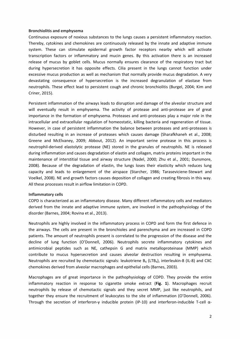
Macrophages are of great importance in the pathophysiology of COPD. They provide the entire
inflammatory reaction in response to cigarette smoke extract (Fig. 1). Macrophages recruit
neutrophils by release of chemotactic signals and they secret MMP, just like neutrophils, and
together they ensure the recruitment of leukocytes to the site of inflammation (O’Donnell, 2006).
Through the secretion of interferon-γ inducible protein (IP-10) and interferon-inducible T-cell α-
3
chemo attractant (I-TAC), CD8+ T cells are attracted which contributes to emphysema. Also, the
amount of macrophages correlates to the damage inflicted on parenchyma (Barnes, 2003).
Figure 1 Macrophages provide inflammatory reaction in response to cigarette smoke. By the release of chemotactic signals such as interleukin-8 (IL-8) and leukotrien B4 (LTB4) macrophages recruit neutrophils to the site of inflammation. Also through the secretion of interferon-γ inducible protein (IP-10) and interferon-inducible T-cell α-chemo attractant (I-TAC) CD8
+ cells are attracted which ultimately contribute to emphysema. Other cells are also recruited in response to signals.
Oxidative stress due to inflammatory response increases reactive oxygen species (ROS) production. This triggers increased transcription of inflammatory genes (Barnes et al., 2003).
By an increased expression of IP-10 by epithelial cells in the respiratory tract, the number of T-
lymphocytes raises, in particular CD8+ compared with CD4+. CD8+ cells cause destruction of lung
parenchyma through the release of perforin and granzymes which leads to apoptosis or necrosis.
Research showed that CD8+ cells are increased in patients with COPD which do not smoke (Chung
and Adcock, 2008; Freeman et al., 2010). This proves that chronic inflammation characteristic for
COPD is continuously stimulated. The moderate elevated levels of CD4+ cells consist of two types of
cells, Th1 and Th17. Interferon γ is secreted by Th1 which ensures migration of leukocytes to the site
of inflammation and promotes NK cells activity. Th17 cell secrete IL-17A and IL-17F that promote
inflammation (Schroder et al., 2003).
The exact function of eosinophils is still unknown. As with asthma, eosinophils are increased in the
airway of patients with COPD. However, it is known that these cells contribute to the production of
reactive oxygen species (ROS) (Barnes, 20003; Chung and Adcock, 2008). The same applies to mast
cells which are also increased in the airways of COPD patient but their function is unknown (Chung
and Adcock, 2008).
Oxidative stress
The lungs are continuously in contact with the external environment. Noxious substances or particles
as well as bacteria and viruses can easily enter the airways which makes the lungs very vulnerable.
Exposure to air pollutants but also the immune response of leukocytes and macrophages to bacteria
en viruses increases oxidative stress in the airways (Langen et al, 2003; Kirkham and Barnes, 2013).
Because of oxidative stress there is an increased attendance of exogenous ROS, free radicals derived
from oxygen like superoxide anion (·O2−), hydroxyl radical (·OH), hydrogen peroxide (H2O2) and
4
singlet oxygen (O2) (Sharma et al., 2012). In addition, there is always endogenous ROS present
derived from mitochondrial respiration. Elevated levels of ROS can cause damage to lipids, proteins
and DNA (van Eeden and Sin, 2008). Because of the constant exposure to endogenous and
exogenous sources of oxidative stress the lungs has expand anti-oxidative defence mechanisms that
neutralize ROS. This anti-oxidative mechanisms consist of enzymatic and non-enzymatic components,
including superoxide dismutase, enzymes of ascorbate-glutathione cycle, guaiacol peroxidise,
carotenoid, etc. (Sharma et al., 2002). In patients with COPD the production of endogenous ROS by
smooth muscle cells and the exogenous ROS are substantially increased. This is mainly the result of
cigarette smoke which causes oxidative stress (Kohen and Nyska, 2002). Cigarette smoke contains a
very high concentration of oxidants and free radicals and includes between 4000 and 4700 different
chemicals (van Eeden and Sin, 2008; Langen et al, 2003).
ROS has a great effect on the respiratory system because it allows the release of cytokines and
chemokines which activate molecular complexes. A well-known complex is nuclear factor-kappa B
(NF-κB), a transcriptional factor. NF-κB actives a number of inflammatory genes which results in an
immune response. In addition, ROS also activates a number of enzymes such as histone
acetyltransferase, which alters histones resulting in an increased transcription of inflammatory
genes, and transforming growth factor-β (TGF-β). TGF-β decreases the action of the anti-oxidative
mechanism of superoxide dismutase whereby ROS can overcome this defence mechanism. Research
has shown that the nuclear factor-erythroid 2-related factor-2 (Nrf2), a transcription factor that
control antioxidant mechanisms, is decreased in patients with COPD and this is likely to be
suppressed by similar mechanisms as TGF-β (Barnes, et al., 2003; Kirkham and Barnes, 2013).
Therefore, oxidative stress plays a major role in the pathophysiology of COPD.
5
II. A-kinase anchoring proteins
Numerous processes take place inside the cell which are crucial for survival such as proliferation,
differentiation, metabolism and gene expression. Many of these processes are driven by extracellular
signals like neurotransmitters and hormones. These signals are passed to intracellular transduction
mechanisms via receptors. These transduction mechanisms are of great importance because the cell
can engender an appropriate physiological response to the changing external environment. This
response is partly achieved by compartmentalization of intracellular effectors by adapting of
anchoring proteins, as will be described in this chapter.
Cyclic adenosine monophosphate and protein kinase A
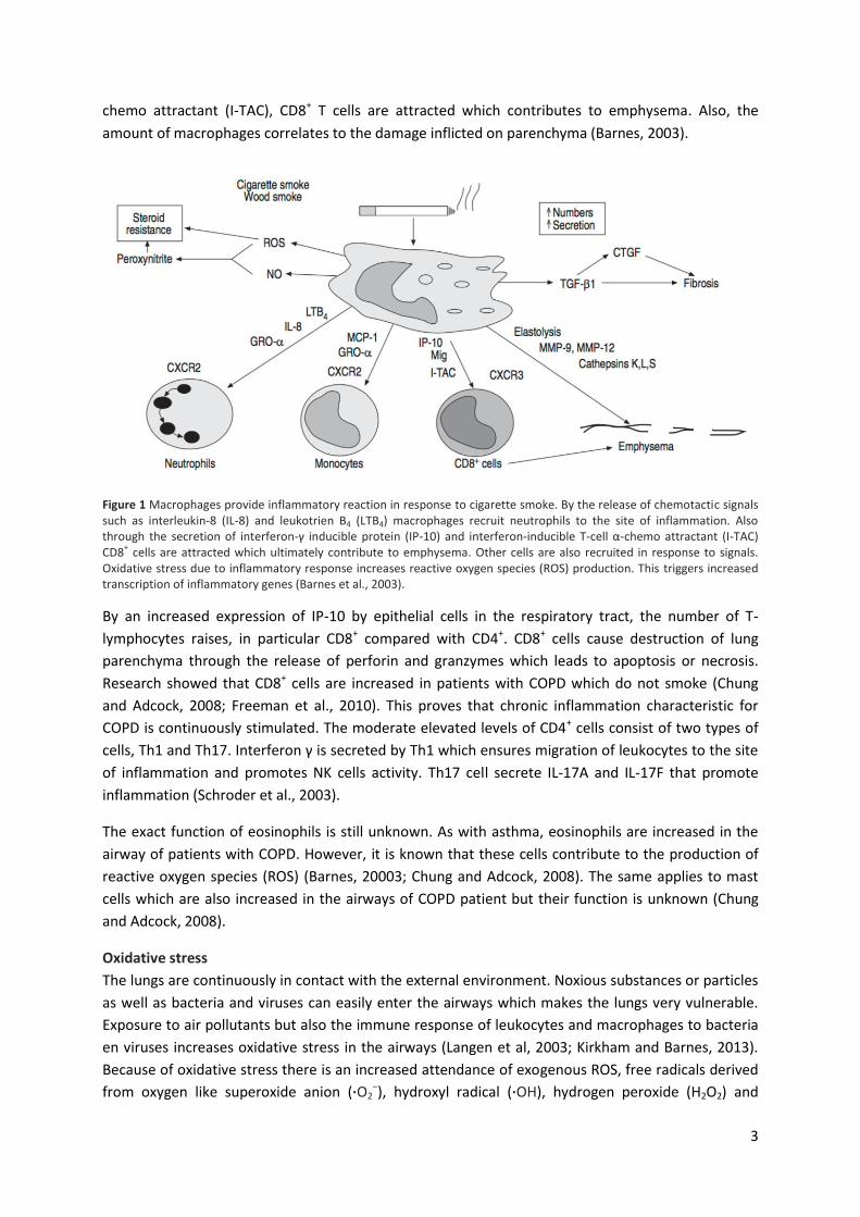
The first discovered and already more than 40 years familiar intracellular signal is the second
messenger cyclic adenosine 3’,5’-monophosphate (cAMP). The cAMP signal transduction
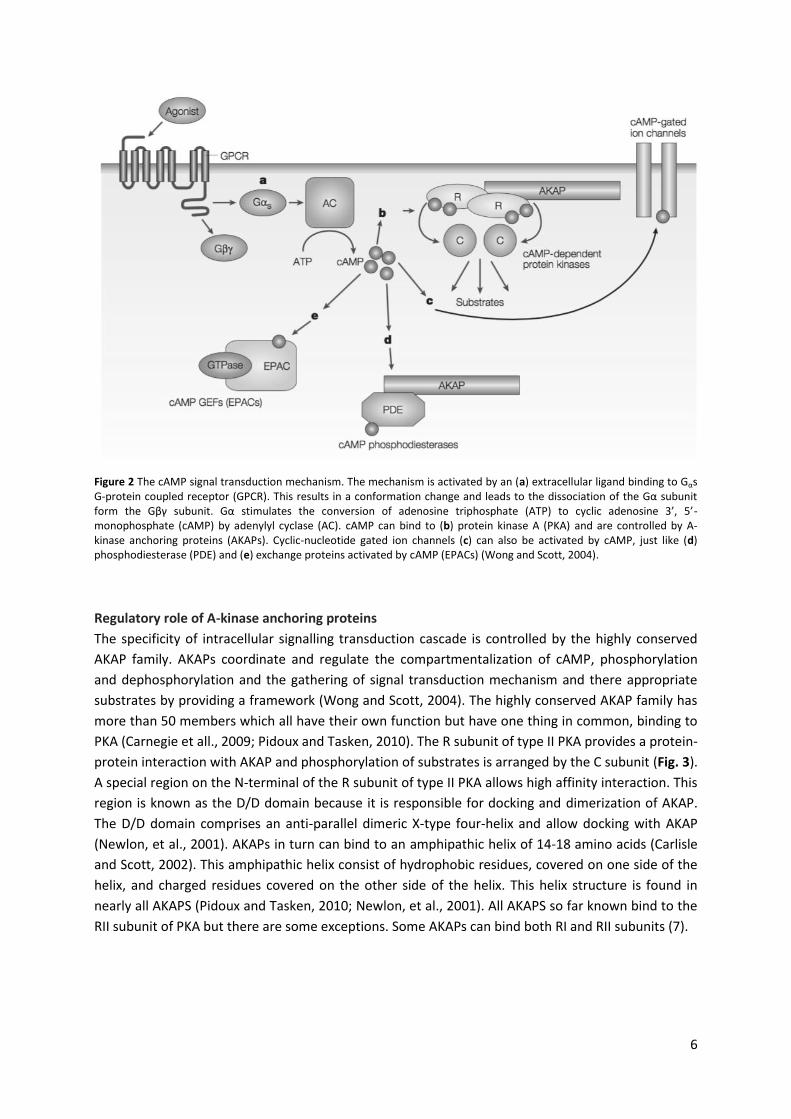
mechanisms is activated by binding of an extracellular ligand to a G protein-coupled receptor (GPCR)
on a target cell causing conformational change (Fig. 2) (Chin eta l., 2002). The intracellular
heterotrimeric G protein (consisting of Gα, Gβ en Gγ subunits) which is attached to the receptor,
exchange guanosine diphosphate (GDP) for guanosine triphosphate (GTP). As a result the G protein is
activated and the Gα subunit dissociates from the Gβγ subunit (Alto and Scott, 2004). The Gα (in this
case Gsα) subunit interact which ensures activation of adenylyl cyclase (AC), an enzyme that catalyse
the turnover of adenosine triphosphate (ATP) to cAMP (Gloerich and Bos, 2010; Perion et al., 2012).
cAMP is regulated by a balance between two types of enzymes, AC and phosphodiesterase (PDE)
which catalyse the conversion of cAMP to 5’-AMP (Sassone-Corsi and Fimia, 2001; Pidoux and
Tasken, 2010).
CAMP achieves its effect by binding to protein kinase A (PKA), but can also bind to exchange protein
activated by cAMP (Epac), cyclic-nucleotide gated ion channels or PDEs, which ensures cAMP
degradation (Sassone-Corsi and Fimia, 2001; Gloerich and Bos, 2010). PKA, also known as cAMP-
dependent protein kinase, is a hetero tetramer composed of two catalytic (C) subunits, encoded by
three genes (Cα, Cβ and Cγ), and two regulatory (R) subunits, encoded by four genes (RIα, RIβ, RIIα
and RIIβ) (Sassone-Corsi and Fimia, 2001; Carnegie et all., 2009). Two types of PKA are known namely
type I (RIα and RIβ dimer), which is mainly cytoplasmic, and type II (RIIα and RIIβ dimer) by which the
sub cellular location is determined by an anchoring protein family (Alto and Scott, 2004; Di
Benedetto et al., 2008; Scott, 1991). Normally, PKA is in an inactive state achieved by the association
the C and R subunit. Activation occurs by dissociation of these two subunits by binding of cAMP to
the R subunit. The activity of PKA is administered by protein kinase inhibitors (PKIs), which
phosphorylate serine and threonine residues and in this way regulating biological processes. The
cAMP signalling transduction cascade via PKA is also controlled by another group of proteins called A-
kinase anchoring proteins (AKAPs). AKAPs are complex but well-organized molecular complexes
which determine the precise spatial-temporal signalling of the transduction mechanism after
activation of GPCR (Pawson, 2003; Rababa’h et all., 2014).
6
Figure 2 The cAMP signal transduction mechanism. The mechanism is activated by an (a) extracellular ligand binding to Gαs G-protein coupled receptor (GPCR). This results in a conformation change and leads to the dissociation of the Gα subunit form the Gβγ subunit. Gα stimulates the conversion of adenosine triphosphate (ATP) to cyclic adenosine 3’, 5’- monophosphate (cAMP) by adenylyl cyclase (AC). cAMP can bind to (b) protein kinase A (PKA) and are controlled by A-kinase anchoring proteins (AKAPs). Cyclic-nucleotide gated ion channels (c) can also be activated by cAMP, just like (d) phosphodiesterase (PDE) and (e) exchange proteins activated by cAMP (EPACs) (Wong and Scott, 2004).
Regulatory role of A-kinase anchoring proteins
The specificity of intracellular signalling transduction cascade is controlled by the highly conserved
AKAP family. AKAPs coordinate and regulate the compartmentalization of cAMP, phosphorylation
and dephosphorylation and the gathering of signal transduction mechanism and there appropriate
substrates by providing a framework (Wong and Scott, 2004). The highly conserved AKAP family has
more than 50 members which all have their own function but have one thing in common, binding to
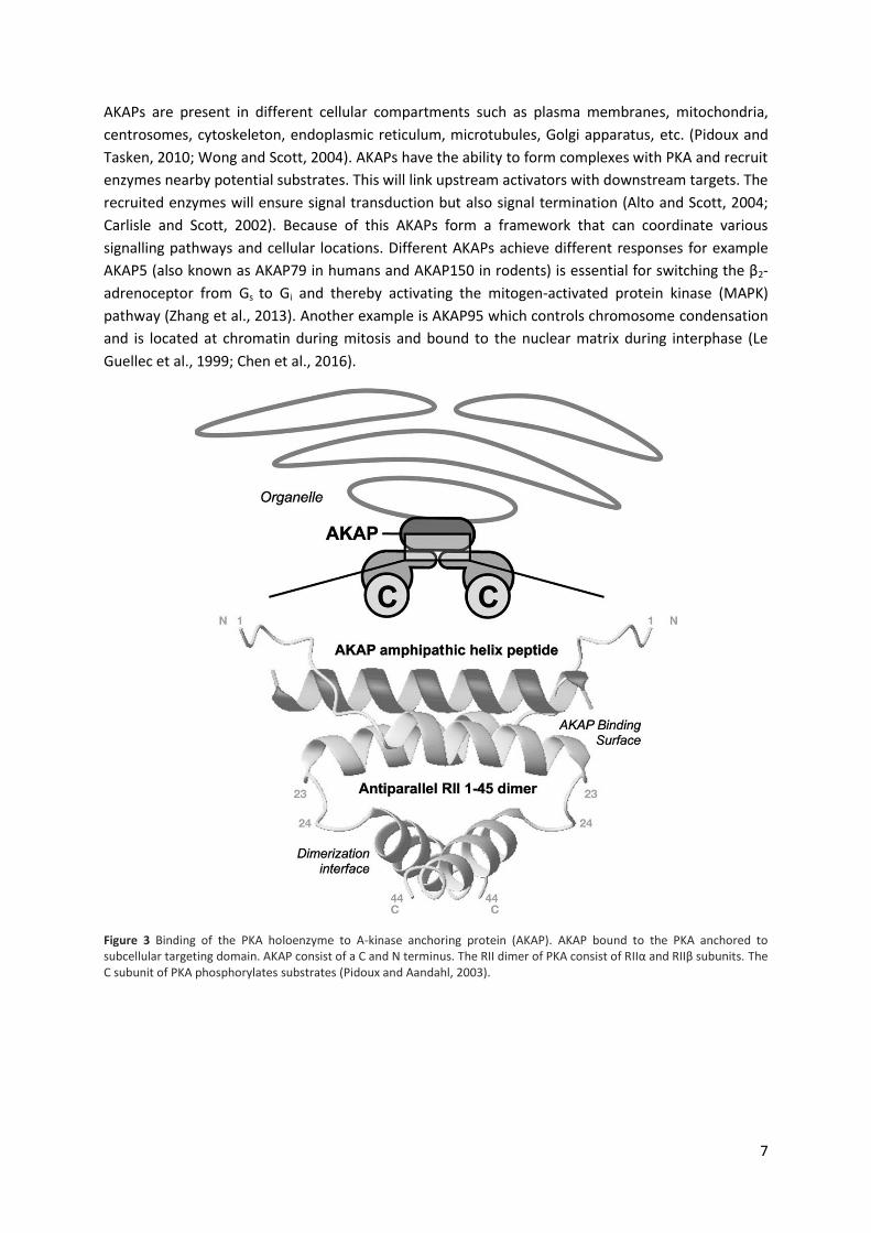
PKA (Carnegie et all., 2009; Pidoux and Tasken, 2010). The R subunit of type II PKA provides a protein-
protein interaction with AKAP and phosphorylation of substrates is arranged by the C subunit (Fig. 3).
A special region on the N-terminal of the R subunit of type II PKA allows high affinity interaction. This
region is known as the D/D domain because it is responsible for docking and dimerization of AKAP.
The D/D domain comprises an anti-parallel dimeric X-type four-helix and allow docking with AKAP
(Newlon, et al., 2001). AKAPs in turn can bind to an amphipathic helix of 14-18 amino acids (Carlisle
and Scott, 2002). This amphipathic helix consist of hydrophobic residues, covered on one side of the
helix, and charged residues covered on the other side of the helix. This helix structure is found in
nearly all AKAPS (Pidoux and Tasken, 2010; Newlon, et al., 2001). All AKAPS so far known bind to the
RII subunit of PKA but there are some exceptions. Some AKAPs can bind both RI and RII subunits (7).
7
AKAPs are present in different cellular compartments such as plasma membranes, mitochondria,
centrosomes, cytoskeleton, endoplasmic reticulum, microtubules, Golgi apparatus, etc. (Pidoux and
Tasken, 2010; Wong and Scott, 2004). AKAPs have the ability to form complexes with PKA and recruit
enzymes nearby potential substrates. This will link upstream activators with downstream targets. The
recruited enzymes will ensure signal transduction but also signal termination (Alto and Scott, 2004;
Carlisle and Scott, 2002). Because of this AKAPs form a framework that can coordinate various
signalling pathways and cellular locations. Different AKAPs achieve different responses for example
AKAP5 (also known as AKAP79 in humans and AKAP150 in rodents) is essential for switching the β2-
adrenoceptor from Gs to Gi and thereby activating the mitogen-activated protein kinase (MAPK)
pathway (Zhang et al., 2013). Another example is AKAP95 which controls chromosome condensation
and is located at chromatin during mitosis and bound to the nuclear matrix during interphase (Le
Guellec et al., 1999; Chen et al., 2016).
Figure 3 Binding of the PKA holoenzyme to A-kinase anchoring protein (AKAP). AKAP bound to the PKA anchored to subcellular targeting domain. AKAP consist of a C and N terminus. The RII dimer of PKA consist of RIIα and RIIβ subunits. The C subunit of PKA phosphorylates substrates (Pidoux and Aandahl, 2003).
8
III. AKAPs in the airways
Several AKAPs are expressed in the airway smooth muscle. The functional role of AKAPs in the
airways and the lung disease COPD is not completely known. Therefore, we review the role and
function of smooth muscle cells such as signalling, contraction, inflammation and remodelling and
the involved AKAPs.
AKAP signalling
Important AKAPs in the signalling of airway smooth muscle cells are AKAP5 (AKAP79/150) and
AKAP12 (Gravin). AKAP5 and AKAP12 share many common features in downstream signalling such as
binding to type II PKA and protein phosphatase-2B. (Tao and Malbon, 2008). But most important is
the interaction with the prototypic GPCR, the β2-adrenoceptor (Fraser et al., 2000; Gao et al., 2011).
When AKAP5 binds to the β2-adrenoceptor, Gs will be activated. AKAP5 however, has the ability to
change the protein Gs to Gi (Poppinga et al., 2014). This process is facilitated by PKA-mediated
phosphorylation of the β2-adrenoceptor. Gs normally ensures the activation of AC but due the switch
Gi activates the MAPK (ERK) pathway. This pathway is involved in cell proliferation, differentiation,
apoptosis, transformation, survival and gene expression (McCubrey et al., 2007; Kim and Choi, 2010).
Also, the pathway is responsible for the activation of cytokine production (Scherle et al., 1998).
AKAP5 has much influence on the β2-adrenoceptor because it provides desensitization and
internalization of the receptor. In this way, AKAP5 can regulate the precise expression of the
receptor. This process occurs through the phosphorylation of the β2-adrenoceptor by G protein-
coupled receptor kinase-2 (GRK2) whereby the βγ subunit, like β-arrestin, is able to bind with high
affinity which result in desensitization. Internalization of the receptor is a slower process and goes via
endocytosis (Chen and Malbon, 2009). After desensitization of the receptor it is essential that
receptor is expressed on the cell membrane again. AKAP12 ensures resensitisation,
dephosphorylation and recycling of the β2-adrenoceptor (Luttrell and Lefkowitz, 2002; Oldenburger
et al., 2012). It is therefore important that AKAP5 and AKAP12 closely collaborate regarding to the
signalling via the β2-adrenoceptor important for downstream interactions such as smooth muscle
relaxation and bronchodilation.
Contractile function of smooth muscle cells
A number of enzymes are important in the contraction of smooth muscle in the respiratory tract.
Contraction occur in response to elevated cytosolic calcium (Ca2+) concentrations. By binding to a
serpentine receptor, phospholipase C is activated. This enzyme ensures catalysis which result in the
production of two second messengers, namely diacylglycerol (DAG) and inositol triphosphate (IP3).
DAG is responsible for the activation of protein kinase C (PKC) which in turn can exert different
biological functions such as receptor desensitization, cell growth, participation in immune responses,
etc. (Choi et al., 2009). IP3 causes release of Ca2+ by binding to specific receptors present on the
sarcoplasmic reticulum. The released Ca2+ binds to the intracellular Ca2+ receptor protein, calmodulin
(CaM) ensures activation of myosin light-chain kinase (MLC kinase) and results in contraction of
smooth muscle (Lincoln, 2007).
9
Smooth muscle relaxation is regulated by MLC phosphatase, together with a reduced level of
cytosolic Ca2+. Rho kinase, a serine/threonine kinase, is of great importance in the regulation of MLC
phosphatase and is activated by RhoA. Rho kinase ensures phosphorylation of the myosin-binding
subunit of MLC phosphatase (Wilson et al., 2001; Webb, 2003). The protein Epac also interact with
this pathway. Epac binds to Rap1 which ensures the inhibition of RhoA and ultimately results in
smooth muscle relaxation (Poppinga et al., 2014).
Ezrin (AKAP78) plays a central role in smooth muscle relaxation. Ezrin is part of the
Ezrin/Radixin/Moesin (ERM) protein family and are cross link actin filaments associated with the
plasma membrane. Rho-regulated Rho-kinase establish phosphorylation of ERM (Matsui et al., 1998).
Because Epac and Ezrin are both involved in this process en also both found in smooth muscle, is it
suggested that inhibition of RhoA by Epac is affected by Ezrin (Poppinga et al., 2014).
Inflammation and remodelling
As described earlier, COPD is characterized as an inflammatory disease. Many chemokines and
cytokines are released in response to cigarettes smoke extract. A cytokine that plays an important
role is IL-8. IL-8 is elevated in the airways of patients with COPD and is release by smooth muscle cells
in response to Gq protein. However, research has shown that PKA and Epac can reduce IL-8 in the
airways. This is achieved through inhibition of ERK signalling which is influenced by a balance
between AKAP5 and AKAP12 and also through the inhibition of NF-κB. Disruption of PKA-AKAP
interaction by Ht31, a protein kinase A anchoring inhibitor, clearly display that PKA and Epac plays an
important role in mediating the anti-inflammatory processes which are under mediation of AKAPs
(Poppinga et al., 2014; Poppinga et al., 2015).
Chronic inflammation, emphysema and mucus hypersecretion are known to contribute to structural
changes in lung tissue and airway remodelling, a major feature seen in patient with COPD (Dournes
and Laurent, 2012; Grzela, 2015). There are a numbers of known pathways which lead to changes in
structures of lung tissues such as the MAPK (ERK) pathway and phosphoinositide 3-kinase (PI3K)
pathway. This pathway regulates cell proliferation and differentiation like MAPK (ERK) (Lui et al.,
2009). PKA and Epac, which are fine-tuned and controlled by the AKAP family, have great influence
on these two pathways and presumably also in airway remodelling. However, the exact mechanism
and role of AKAP family mediated remodelling by PKA and Epac is not clear (Poppinga et al., 2014).
AKAP in relation to disease
AKAPs act as spatial-temporal signalling, such as the compartmentalization of cAMP and associated
transduction mechanisms like cell proliferation, differentiation, gene targeting, etc. It is therefore
essential that these processes are precisely controlled. Disturbance of AKAPs function is associated
with various disorders such as neurodegenerative diseases, cardiovascular disorders, and various
types of cancers as well as lung disease such as COPD (Tröger et al., 2012; Smith et al., 2013).
Exposure of airway smooth muscle cells to cigarette smoke, the main cause of COPD, affects the
expression of several AKAPs. mRNA levels of AKAP9 are decreased in lung tissue. AKAP9 interacts
with E-cadherin. E-cadherin is an important transmembrane glycoprotein and found specific in cell-
cell adhesion on smooth muscle cells (Tunggal et al., 2005; Van Roy and Berx, 2008). Down-
regulation of AKAP9 by cigarette smoke reduces also E-cadherin. This ultimately results in
dysfunction of the epithelial barrier. Also, mRNA levels of AKAP5 and AKAP12 are decreased in

10
response to cigarette smoke and will result ultimately in inflammation (Poppinga et al., 2014;
Oldenburger et al., 2014; Poppinga et al., 2015). Taken together, dysregulation of AKAPs due to
cigarette smoke extract are responsible for the pathophysiology of COPD.
11
IV. Outlook
cAMP is an intracellular messenger which regulates important cellular and biological processes such
as cell proliferation and differentiation, gene transcription, etc.. The specificity of intracellular
signalling transduction mechanisms is controlled by the AKAP family. AKAPs act as spatial-temporal
signalling by compartmentalization of cAMP (McConnachie et al., 2006). Disruption or malfunction of
AKAPs is related to a number of disorders such as heart diseases, immune diseases,
neurodegenerative disease and COPD (Zaccolo, 2011).
It is commonly known that average age of people increases and this will only increase over the years.
With this increasing age, the number of chronic diseases will also raise. Therefore it is crucial to
determine and understand the mechanisms that drives these diseases so it can be managed better.
AKAPs play a major role in the pathophysiology of COPD but also in other chronic disease. Properly
management of diseases by specific drugs, perhaps targeting of AKAPs, is of social importance.
There are many different types of medication for COPD patients such as anticholinergics, β2
agonisten, corticosteroids and combination therapie. However, these drugs are not always effective
(Grimes et al., 2007). AKAPs are involved in different processes and are dysregulated in disease which
makes them potential drug targets. A few experimental drugs are used to disrupt AKAP-PKA
interactions such as peptides derived from PKA-binding domains and FMP-API-1 which are small
disruption molecules. However these drugs lack selectivity (Tröger et al., 2012; Esseltine and Scott,
2013). One way to obtain selectivity for AKAP-PKA interaction might be via nanotechnology. Research
has shown that nanoparticles act more efficiently than molecules and therefore these particles
would be able to perform as effective drug delivery systems. However, for precise effect of
nanosystems, AKAP-PKA interaction and there corresponding interactions in relation to cellular
processes will need to be exactly indentified (Suri et al., 2007). Perhaps in the future, targeting AKAP-
PKA interaction by nanosytems is an effective way for the management of COPD and because
dysregulation occurs not only in COPD, nanosystems can serve as a broad spectrum.
So far medication only focus on disruption of AKAP-PKA interactions. But there is another important
protein present in various biological and cellular processes which is also under spatial-temporal
control of the AKAP family and that is Epac. Therefore, it is important not only to focus on
pharmacological induced disruption of AKAP-PKA interaction but also on AKAP-Epac interactions
(Cheng et al., 2008). In addition, cAMP is also of great interest in the previously mentioned
processes. Increased concentrations of cAMP proved activation of PKA but excessively elevated levels
of cAMP in the cytosol can lead to an increased activion of NF-kB resulting in hyper inflammation
(Monterisi et al., 2012). Therefore it is of interest to pharmacological manage the components,
cAMP, Epac and PKA, that are controlled by the AKAP family and contribute to the pathophysiology
of COPD.
I
V. References Abboud, T. (2012). Pathogenic Mechanisms in
Emphysema: From Protease Anti-Protease
Imbalance to Apoptosis. INTEC*H Open Access
Publisher.
Alto, N. and Scott, J. (2004). The Role of A-Kinase
Anchoring Proteins in cAMP-Mediated Signal
Transduction Pathways. CBB, 40(3S), pp.201-208.
Barnes, P. (2004). Mediators of Chronic
Obstructive Pulmonary Disease. Pharmacological
Reviews, 56(4), pp.515-548.
Barnes, P. (2005). COPD: current therapeutic
interventions and future approaches. European
Respiratory Journal, 25(6), pp.1084-1106.
Barnes, P., Shapiro, S. and Pauwels, R. (2003).
Chronic obstructive pulmonary disease: molecular
and cellular mechanisms. Eur Respir J, 22(4),
pp.672-688.
Brode, S., Ling, S. and Chapman, K. (2012). Alpha-1
antitrypsin deficiency: a commonly overlooked
cause of lung disease. Canadian Medical
Association Journal, 184(12), pp.1365-1371.
Burgel, P. (2004). Roles of epidermal growth factor
receptor activation in epithelial cell repair and
mucin production in airway epithelium. Thorax,
59(11), pp.992-996.
Carlin, B. (2012). COPD and Associated
Comorbidities: A Review of Current Diagnosis and
Treatment. Postgraduate Medicine, 124(4),
pp.225-240.
Carlisle, J., Scott, J. (2002). AKAP mediated signal
transduction. Review of Pharmacology and
Toxicology. 42, pp 235-257.
Carnegie, G., Means, C. and Scott, J. (2009). A-
kinase anchoring proteins: From protein complexes
to physiology and disease. IUBMB Life, 61(4),
pp.394-406.
Chen, M. and Malbon, C. (2009). G-protein-
coupled receptor-associated A-kinase anchoring
proteins AKAP5 and AKAP12: Differential
trafficking and distribution. Cellular Signalling,
21(1), pp.136-142.
Chen, X., Kong, X., Zhuang, W., Teng, B., Yu, X.,
Hua, S., Wang, S., Liang, F., Ma, D., Zhang, S., Zou,
X., Dai, Y., Yang, W. and Zhang, Y. (2016). Dynamic
changes in protein interaction between AKAP95
and Cx43 during cell cycle progression of A549
cells. Sci. Rep., 6, p.21224.
Cheng, X., Ji, Z., Tsalkova, T. and Mei, F. (2008).
Epac and PKA: a tale of two intracellular cAMP
receptors. Acta Biochimica et Biophysica Sinica,
40(7), pp.651-662.
Chin, K., Yang, W., Ravatn, R., Kita, T., Reitman, E.,
Vettori, D., Cvijic, M., Shin, M. And Iacono, L.
(2002). Reinventing the Wheel of Cyclic
AMP. Annals of the New York Academy of Sciences,
968(1), pp.49-64.
Choi, H., Allahdadi, K., Tostes, R. and Webb, R.
(2009). Diacylglycerol Kinase Inhibition and
Vascular Function. Current Enzyme Inhibition, 5(3),
pp.148-152.
Chung, K. and Adcock, I. (2008). Multifaceted
mechanisms in COPD: inflammation, immunity,
and tissue repair and destruction. European
Respiratory Journal, 31(6), pp.1334-1356.
David M Mannino, V. (2006). Changing the burden
of COPD mortality. International Journal of Chronic
Obstructive Pulmonary Disease, 1(3), p.219.
Di Benedetto, G., Zoccarato, A., Lissandron, V.,
Terrin, A., Li, X., Houslay, M., Baillie, G. and
Zaccolo, M. (2008). Protein Kinase A Type I and
Type II Define Distinct Intracellular Signalling
Compartments. Circulation Research, 103(8),
pp.836-844.
Dournes, G. and Laurent, F. (2012). Airway
Remodelling in Asthma and COPD: Findings,
Similarities, and Differences Using Quantitative
CT. Pulmonary Medicine, 2012, pp.1-8.
II
Dunsmore, S. E. (2008). Treatment of COPD: A
matrix perspective. International Journal of Chronic
Obstructive Pulmonary Disease, 3(1), 113–122.
van Eeden, S. and Sin, D. (2008). Chronic
Obstructive Pulmonary Disease: A Chronic Systemic
Inflammatory Disease. Respiration, 75(2), pp.224-
238.
Esseltine, J. and Scott, J. (2013). AKAP signaling
complexes: pointing towards the next generation
of therapeutic targets?. Trends in Pharmacological
Sciences, 34(12), pp.648-655.
Fraser, I., Cong, M., Kim, J., Rollins, E., Daaka, Y.,
Lefkowitz, R. and Scott, J. (2000). Assembly of an A
kinase-anchoring protein–β2-adrenergic receptor
complex facilitates receptor phosphorylation and
signaling. Current Biology, 10(7), pp.409-412.
Freeman, C., Han, M., Martinez, F., Murray, S., Liu,
L., Chensue, S., Polak, T., Sonstein, J., Todt, J.,
Ames, T., Arenberg, D., Meldrum, C., Getty, C.,
McCloskey, L. and Curtis, J. (2010). Cytotoxic
Potential of Lung CD8+ T Cells Increases with
Chronic Obstructive Pulmonary Disease Severity
and with In Vitro Stimulation by IL-18 or IL-15. The
Journal of Immunology, 184(11), pp.6504-6513.
Fromer, L. and Cooper, C. (2008). A review of the
GOLD guidelines for the diagnosis and treatment of
patients with COPD. International Journal of
Clinical Practice, 62(8), pp.1219-1236.
Gao, S., Wang, H. and Malbon, C. (2011). AKAP5
and AKAP12 Form Homo-oligomers.Journal of
Molecular Signaling, 6, p.3.
Giembycz, M. (2001). Cilomilast: a second
generation phosphodiesterase 4 inhibitor for
asthma and chronic obstructive pulmonary
disease. Expert Opinion on Investigational Drugs,
10(7), pp.1361-1379.
Gloerich, M. and Bos, J. (2010). Epac: Defining a
New Mechanism for cAMP Action. Annu. Rev.
Pharmacol. Toxicol., 50(1), pp.355-375.
Greene, C. M., & McElvaney, N. G. (2009).
Proteases and antiproteases in chronic neutrophilic
lung disease – relevance to drug discovery. British
Journal of Pharmacology, 158(4), 1048–1058.
Grimes, G., Manning, J., Patel, P., Marc, R. (2007).
Medication for COPD: A Review of Effectiveness.
Am Fam Physician, 76(8), pp.1141-1148.
Grzela, K., Litwiniuk, M., Zagorska, W. and Grzela,
T. (2015). Airway Remodeling in Chronic
Obstructive Pulmonary Disease and Asthma: the
Role of Matrix Metalloproteinase-9. Arch.
Immunol. Ther. Exp., 64(1), pp.47-55.
Kim, E. and Choi, E. (2010). Pathological roles of
MAPK signaling pathways in human
diseases. Biochimica et Biophysica Acta (BBA) -
Molecular Basis of Disease, 1802(4), pp.396-405.
Kim, V. and Criner, G. (2015). The chronic
bronchitis phenotype in chronic obstructive
pulmonary disease. Current Opinion in Pulmonary
Medicine, 21(2), pp.133-141.
Kirkham, P. and Barnes, P. (2013). Oxidative Stress
in COPD. Chest, 144(1), pp.266-273.
Kohen, R. and Nyska, A. (2002). Oxidation of
Biological Systems: Oxidative Stress Phenomena,
Antioxidants, Redox Reactions, and Methods for
Their Quantification. Toxicologic Pathology, 30(6),
pp.620-650.
Langen, R., Korn, S., Wouters, E. (2003). ROS in the
local and systemic pathogenesis of COPD. Free
Radical Biology and Medicine, 35(3), pp.226-235.
Le Guellec, K., Tasken, K. (1999). The A-Kinase-
Anchoring Protien Akap95 Is a Multivalent Protein
with a Key Role in Chromatin Condensation at
Mitosis. The Journal of Cell Biology, 147 (6),
pp.1167-1180.
Lincoln, T. (2007). Myosin Phosphatase Regulatory
Pathways: Different Functions or Redundant
Functions?. Circulation Research, 100(1), pp.10-12.
Liu, P., Cheng, H., Roberts, T. and Zhao, J. (2009).
Targeting the phosphoinositide 3-kinase pathway
in cancer. Nature Reviews Drug Discovery, 8(8),
pp.627-644.
Luttrell, L., Lefkowitz, R. (2002) The role of β-
arrestins in the termination and transduction of G-
protein-coupled receptor signals. Journal of Cell
Science, 115, pp.455-465.
III
Matsui, T., Maeda, M., Doi, Y., Yonemura, S.,
Amano, M., Kaibuchi, K., Tsukita, S. and Tsukita, S.
(1998). Rho-Kinase Phosphorylates COOH-terminal
Threonines of Ezrin/Radixin/Moesin (ERM) Proteins
and Regulates Their Head-to-Tail Association. J Cell
Biol, 140(3), pp.647-657.
McConnachie, G., Langeberg, L. and Scott, J.
(2006). AKAP signaling complexes: getting to the
heart of the matter. Trends in Molecular Medicine,
12(7), pp.317-323.
McCubrey, J., Steelman, L., Chappell, W., Abrams,
S., Wong, E., Chang, F., Lehmann, B., Terrian, D.,
Milella, M., Tafuri, A., Stivala, F., Libra, M.,
Basecke, J., Evangelisti, C., Martelli, A. and
Franklin, R. (2007). Roles of the Raf/MEK/ERK
pathway in cell growth, malignant transformation
and drug resistance. Biochimica et Biophysica Acta
(BBA) - Molecular Cell Research, 1773(8), pp.1263-
1284.
Monterisi, S., Favia, M., Guerra, L., Cardone, R.,
Marzulli, D., Reshkin, S., Casavola, V. and Zaccolo,
M. (2012). CFTR regulation in human airway
epithelial cells requires integrity of the actin
cytoskeleton and compartmentalized cAMP and
PKA activity. Journal of Cell Science, 125(5),
pp.1106-1117.
Nadel, J. (2000). Role of Neutrophil Elastase in
Hypersecretion During COPD Exacerbations, and
Proposed Therapies. Chest, 117(5), pp.386-389.
Newlon, M., Roy, M., Morikis, D., Carr, D.,
Westphal, R., Scott, J., Jennings, P.(2001). A novel
mechanism of PKA anchoring revealed by solution
structures of anchoring complexes. The EMBO
Journal, 20(7), pp.1651-1662.
O’Donnell, R., Breen, D., Wilson, S., Djukanovic, R.
(2006). Inflammatory cells in the airways in COPD.
Thorax, 61(5), pp.448-454.
Oldenburger, A., Maarsingh, H. and Schmidt, M.
(2012). Multiple Facets of cAMP Signalling and
Physiological Impact: cAMP Compartmentalization
in the Lung. Pharmaceuticals, 5(12), pp.1291-1331.
Oldenburger, A., Poppinga, W., Kos, F., de Bruin,
H., Rijks, W., Heijink, I., Timens, W., Meurs, H.,
Maarsingh, H., Schmidt, M. (2014) A-kinase
anchoring proteins contribute to loss of E-cadherin
and bronchial epithelial barrier by cigarette smoke.
Am J Physiol Lung Cell Mol Physiol, 306, pp.C585-
C597.
Pawson, T. (2003). Assembly of Cell Regulatory
Systems Through Protein Interaction
Domains. Science, 300(5618), pp.445-452.
Perino, A., Ghigo, A., Scott, J. and Hirsch, E.
(2012). Anchoring Proteins as Regulators of
Signaling Pathways. Circulation Research, 111(4),
pp.482-492.
Pidoux, G. and Tasken, K. (2010). Specificity and
spatial dynamics of protein kinase A signaling
organized by A-kinase-anchoring proteins. Journal
of Molecular Endocrinology, 44(5), pp.271-284.
Poppinga, W., Heijink, I., Holtzer, L., Skroblin, P.,
Klussmann, E., Halayko, A., Timens, W.,
Maarsingh, H. and Schmidt, M. (2015). A-kinase-
anchoring proteins coordinate inflammatory
responses to cigarette smoke in airway smooth
muscle. Am J Physiol Lung Cell Mol Physiol, 308(8),
pp.L766-L775.
Poppinga, W., Muñoz-Llancao, P., González-
Billault, C. and Schmidt, M. (2014). A-kinase
anchoring proteins: cAMP compartmentalization in
neurodegenerative and obstructive pulmonary
diseases. British Journal of Pharmacology, 171(24),
pp.5603-5623.
Rababa'h, A., Singh, S., Suryavanshi, S.,
Altarabsheh, S., Deo, S. and McConnell, B. (2014).
Compartmentalization Role of A-Kinase Anchoring
Proteins (AKAPs) in Mediating Protein Kinase A
(PKA) Signaling and Cardiomyocyte
Hypertrophy. IJMS, 16(1), pp.218-229.
Rahman, I. (2008). Review: Antioxidant therapeutic
advances in COPD. Therapeutic Advances in
Respiratory Disease, 2(6), pp.351-374.
IV
Rennard, S., Calverley, P., Goehring, U.,
Bredenbröker, D. and Martinez, F. (2011).
Reduction of exacerbations by the PDE4 inhibitor
roflumilast - the importance of defining different
subsets of patients with COPD. Respiratory
Research, 12(1), pp.18.
Rovina, N., Koutsoukou, A. and Koulouris, N.
(2013). Inflammation and Immune Response in
COPD: Where Do We Stand?. Mediators of
Inflammation, 2013, pp.1-9.
Sassone-Corsi, P. and Fimia, G. (2001). Cyclic AMP
signalling. Journal of Cell Science, 114, pp. 1971-
1972.
Scherle, P., Jones, E., Favata, M., Daulerio, A.,
Covington, M., Nurnberg, S., Magolda, R.,
Trzaskos, J. (1998). Inhibition of MAP Kinase Kinase
Prvents Cytokine and Prostaglandin E2 Production
in Lipopolysaccharide-Stimulated Monocytes. The
journal of Immunology, 161(10), pp.5681-5686.
Schroder, K., Hertzog, P., Ravasi, T., Hume, D.
(2003). Interferon-γ : an overview of signals,
mechanisms and functions. Journal of Leukocyte
Biology, 75(2), pp.163-189.
Scott, J. (1991). Cyclic nucleotide-dependent
protein kinases. Pharmacology & Therapeutics,
50(1), pp.123-145.
Sharafkhaneh, A., Hanania, N. A., & Kim, V.
(2008). Pathogenesis of Emphysema: From the
Bench to the Bedside. Proceedings of the American
Thoracic Society, 5(4), 475–477.
Sharma, P., Jha, A., Dubey, R. and Pessarakli, M.
(2012). Reactive Oxygen Species, Oxidative
Damage, and Antioxidative Defense Mechanism in
Plants under Stressful Conditions. Journal of
Botany, 2012, pp.1-26.
Smith, F., Reichow, S., Esseltine, J., Shi, D.,
Langeberg, L., Scott, J. and Gonen, T. (2013).
Intrinsic disorder within an AKAP-protein kinase A
complex guides local substrate
phosphorylation. eLife, 2.
Starcher, B. (1986). Elastin and the lung. Thorax,
41(8), pp.577-585.
Suri, S., Fenniri, H. and Singh, B. (2007).
Nanotechnology-based drug delivery
systems.Journal of Occupational Medicine and
Toxicology, 2(1), p.16.
Tao, J. and Malbon, C. (2008). G-protein-coupled
receptor-associated A-kinase anchoring proteins
AKAP5 and AKAP12: differential signaling to MAPK
and GPCR recycling. Journal of Molecular Signaling,
3, p.19.
Taraseviciene-Stewart, L. and Voelkel, N. (2008).
Molecular pathogenesis of emphysema. Journal of
Clinical Investigation, 118(2), pp. 394-402.
Tasken, K. and Aandahl, M. (2004). Localized
Effects of cAMP Mediated by Distinct Routes of
Protein Kinase A. Physiological Reviews, 84(1),
pp.137-167.
Tröger, J., Moutty, M., Skroblin, P. and
Klussmann, E. (2012). A-kinase anchoring proteins
as potential drug targets. British Journal of
Pharmacology, 166(2), pp.420-433.
Tunggal, J., Helfrich, I., Schmitz, A., Schwarz, H.,
Günzel, D., Fromm, M., Kemler, R., Krieg, T. and
Niessen, C. (2005). E-cadherin is essential for in
vivo epidermal barrier function by regulating tight
junctions. EMBO J, 24(6), pp.1146-1156.
van Roy, F. and Berx, G. (2008). The cell-cell
adhesion molecule E-cadherin. Cell. Mol. Life Sci.,
65(23), pp.3756-3788.
Webb, R. (2003). Smooth muscle contraction and
relaxation. APS, 27(4), pp.201-206.
Wilson, D., Sutherland, C. and Walsh, M. (2001).
Ca 2+ Activation of Smooth Muscle
Contraction. Journal of Biological Chemistry,
277(3), pp.2186-2192.
Wong. I. and Scott, J. (2004). AKAP signalling
complexes: focal points in space and time. Nature
Reviews Molecular Cell Biology, 5, pp.959-970.
Zaccolo, M. (2011). Spatial control of cAMP
signalling in health and disease. Current Opinion in
Pharmacology, 11(6), pp.649-655.
V
Zhang, M., Patriarchi, T., Stein, I., Qian, H., Matt,
L., Nguyen, M., Xiang, Y., Hell, J. (2013). Andenylyl
Cyclase Anchoring by a Kinase Anchor Protein
AKAP5 (AKAP79/150) Is Important for
Postysynaptic-β-Adrenergic Signalling. The
American Society for Biochemistry and Molecular
Biology, 288(24), pp.17918-17931.
Zhu, Y., Liu, X., Sköld, C., Umino, T., Wang, H.,
Spurzem, J., Kohyama, T., Ertl, R. and Rennard, S.
(2001). Synergistic neutrophil elastase-cytokine
interaction degrades collagen in three-dimensional
culture. American Journal of Physiology - Lung
Cellular and Molecular Physiology, 281(4), pp.868-
878.