
The Ribosome-Bound Protein Pam68 Promotes Insertion of ... · small PSII subunit PsbH. Deletion of...
12
The Ribosome-Bound Protein Pam68 Promotes Insertion of Chlorophyll into the CP47 Subunit of Photosystem II 1[OPEN] Lenka Bu cinská, a,b Éva Kiss, a Peter Koník, a,b Jana Knoppová, a Josef Komenda, a and Roman Sobotka a,b,2 a Laboratory of Photosynthesis, Centre Algatech, Institute of Microbiology, Academy of Sciences, 37981 T rebo n, Czech Republic b Faculty of Science, University of South Bohemia, 37005 Ceské Bud ejovice, Czech Republic ORCID IDs: 0000-0001-8652-5323 (P.K.); 0000-0003-4588-0328 (J.K.); 0000-0002-6359-7604 (J.Kno.); 0000-0001-5909-3879 (R.S.). Photosystem II (PSII) is a large enzyme complex embedded in the thylakoid membrane of oxygenic phototrophs. The biogenesis of PSII requires the assembly of more than 30 subunits, with the assistance of a number of auxiliary proteins. In plants and cyanobacteria, the photosynthesis-affected mutant 68 (Pam68) is important for PSII assembly. However, its mechanisms of action remain unknown. Using a Synechocystis PCC 6803 strain expressing Flag-tagged Pam68, we purified a large protein complex containing ribosomes, SecY translocase, and the chlorophyll-binding PSII inner antenna CP47. Using 2D gel electrophoresis, we identified a pigmented Pam68-CP47 subcomplex and found Pam68 bound to ribosomes. Our results show that Pam68 binds to ribosomes even in the absence of CP47 translation. Furthermore, Pam68 associates with CP47 at an early phase of its biogenesis and promotes the synthesis of this chlorophyll-binding polypeptide until the attachment of the small PSII subunit PsbH. Deletion of both Pam68 and PsbH nearly abolishes the synthesis of CP47, which can be restored by enhancing chlorophyll biosynthesis. These results strongly suggest that ribosome-bound Pam68 stabilizes membrane segments of CP47 and facilitates the insertion of chlorophyll molecules into the translated CP47 polypeptide chain. Photosystem II (PSII) is a large protein‐cofactor complex embedded in the thylakoid membranes of oxygenic phototrophs. The key large structural com- ponents of PSII are the chlorophyll (Chl)-binding pro- teins D1, D2, CP43, and CP47, subjoined with other small and extrinsic subunits (Umena et al., 2011). According to this model, PSII is assembled in a stepwise manner from four preassembled smaller subcomplexes called modules (Komenda et al., 2012). Each module consists of one large Chl-binding subunit (D1, D2, CP43, or CP47) and several low molecular mass mem- brane polypeptides. PSII assembly is initiated through the association of D1 and D2 modules to form an as- sembly intermediate, termed the Reaction Center II (RCII) complex. The CP47 assembly module (CP47m) is then attached to RCII (Boehm et al., 2011), which results in a CP43-less core complex called “RC47” (Boehm et al., 2012). The active, oxygen-evolving PSII is com- pleted by the addition of the CP43 module (Boehm et al., 2011) and attachment of the lumenal extrinsic proteins (Nixon et al., 2010). Biogenesis of PSII is a highly complex process requiring many auxiliary proteins that are not present in the fully assembled complex. A number of these assembly factors have been described (Komenda et al., 2012; Heinz et al., 2016). However, their precise functions remain mostly un- known, and only a few of them have been connected with a specific assembly step (Knoppová et al., 2014; Be cková et al., 2017). The fully assembled PSII contains 35 Chl molecules, most of them bound to the inner PSII antennas CP47 (16) and CP43 (14). According to this model, Chl molecules are integrated directly into synthesized CP47 and CP43, and the insertion of Chl appears to be a prerequisite for the correct folding and stability of these polypep- tides (for review, see Sobotka, 2014). However, little is known about how Chl proteins are produced. PSII Chl- binding subunits are integral membrane proteins most likely cotranslationally inserted into the thylakoid membrane with the assistance of the protein transloca- tion apparatus. This process usually includes the SecYEG translocon, which forms a protein-conducting channel, and an associated insertase/foldase YidC (Sachelaru et al., 2013). Chl synthase is the last enzyme of Chl biosynthesis, and it was recently shown to physically interact with YidC insertase (Chidgey et al., 2014). This interaction suggests that Chl molecules are 1 This work was supported by project 17-08755S of the Grant Agency of the Czech Republic and by the Czech Ministry of Educa- tion (projects CZ 1.05/2.1.00/19.0392 and LO1416). 2 Address correspondence to [email protected]. The author responsible for distribution of materials integral to the findings presented in this article in accordance with the policy de- scribed in the Instructions for Authors (www.plantphysiol.org) is: Roman Sobotka ([email protected]). L.B. constructed the strains and employed most of the biochemical methods under the supervision of R.S.; P.K. performed protein identi- fication by LC-MS/MS; J.K. and J.Kno were responsible for 35 S radio- labeling; E.K. performed various mutant characterizations; R.S., L.B., E.K., and J.K. designed the study and wrote the paper; R.S. supervised the whole study; and all authors discussed the results and commented on the manuscript. [OPEN] Articles can be viewed without a subscription. www.plantphysiol.org/cgi/doi/10.1104/pp.18.00061 Plant Physiology Ò , April 2018, Vol. 176, pp. 2931–2942, www.plantphysiol.org Ó 2018 American Society of Plant Biologists. All Rights Reserved. 2931 www.plantphysiol.org on April 13, 2019 - Published by Downloaded from Copyright © 2018 American Society of Plant Biologists. All rights reserved.
Transcript of The Ribosome-Bound Protein Pam68 Promotes Insertion of ... · small PSII subunit PsbH. Deletion of...

The Ribosome-Bound Protein Pam68 Promotes Insertion ofChlorophyll into the CP47 Subunit of Photosystem II1[OPEN]
Lenka Bu�cinská,a,b Éva Kiss,a Peter Koník,a,b Jana Knoppová,a Josef Komenda,a and Roman Sobotkaa,b,2
aLaboratory of Photosynthesis, Centre Algatech, Institute of Microbiology, Academy of Sciences, 37981 T�rebo�n,Czech RepublicbFaculty of Science, University of South Bohemia, 37005 �Ceské Bud�ejovice, Czech Republic
ORCID IDs: 0000-0001-8652-5323 (P.K.); 0000-0003-4588-0328 (J.K.); 0000-0002-6359-7604 (J.Kno.); 0000-0001-5909-3879 (R.S.).
Photosystem II (PSII) is a large enzyme complex embedded in the thylakoid membrane of oxygenic phototrophs. The biogenesisof PSII requires the assembly of more than 30 subunits, with the assistance of a number of auxiliary proteins. In plants andcyanobacteria, the photosynthesis-affected mutant 68 (Pam68) is important for PSII assembly. However, its mechanisms ofaction remain unknown. Using a Synechocystis PCC 6803 strain expressing Flag-tagged Pam68, we purified a large proteincomplex containing ribosomes, SecY translocase, and the chlorophyll-binding PSII inner antenna CP47. Using 2D gelelectrophoresis, we identified a pigmented Pam68-CP47 subcomplex and found Pam68 bound to ribosomes. Our resultsshow that Pam68 binds to ribosomes even in the absence of CP47 translation. Furthermore, Pam68 associates with CP47 atan early phase of its biogenesis and promotes the synthesis of this chlorophyll-binding polypeptide until the attachment of thesmall PSII subunit PsbH. Deletion of both Pam68 and PsbH nearly abolishes the synthesis of CP47, which can be restored byenhancing chlorophyll biosynthesis. These results strongly suggest that ribosome-bound Pam68 stabilizes membrane segmentsof CP47 and facilitates the insertion of chlorophyll molecules into the translated CP47 polypeptide chain.
Photosystem II (PSII) is a large protein‐cofactorcomplex embedded in the thylakoid membranes ofoxygenic phototrophs. The key large structural com-ponents of PSII are the chlorophyll (Chl)-binding pro-teins D1, D2, CP43, and CP47, subjoined with othersmall and extrinsic subunits (Umena et al., 2011).According to thismodel, PSII is assembled in a stepwisemanner from four preassembled smaller subcomplexescalled modules (Komenda et al., 2012). Each moduleconsists of one large Chl-binding subunit (D1, D2,CP43, or CP47) and several low molecular mass mem-brane polypeptides. PSII assembly is initiated throughthe association of D1 and D2 modules to form an as-sembly intermediate, termed the Reaction Center II(RCII) complex. The CP47 assemblymodule (CP47m) is
then attached to RCII (Boehm et al., 2011), which resultsin a CP43-less core complex called “RC47” (Boehmet al., 2012). The active, oxygen-evolving PSII is com-pleted by the addition of the CP43 module (Boehmet al., 2011) and attachment of the lumenal extrinsicproteins (Nixon et al., 2010). Biogenesis of PSII isa highly complex process requiring many auxiliaryproteins that are not present in the fully assembledcomplex. A number of these assembly factors have beendescribed (Komenda et al., 2012; Heinz et al., 2016).However, their precise functions remain mostly un-known, and only a few of them have been connectedwith a specific assembly step (Knoppová et al., 2014;Be�cková et al., 2017).
The fully assembled PSII contains 35 Chl molecules,most of them bound to the inner PSII antennas CP47 (16)and CP43 (14). According to this model, Chl moleculesare integrated directly into synthesized CP47 and CP43,and the insertion of Chl appears to be a prerequisite forthe correct folding and stability of these polypep-tides (for review, see Sobotka, 2014). However, little isknown about how Chl proteins are produced. PSII Chl-binding subunits are integral membrane proteins mostlikely cotranslationally inserted into the thylakoidmembrane with the assistance of the protein transloca-tion apparatus. This process usually includes theSecYEG translocon, which forms a protein-conductingchannel, and an associated insertase/foldase YidC(Sachelaru et al., 2013). Chl synthase is the last enzyme ofChl biosynthesis, and it was recently shown tophysically interact with YidC insertase (Chidgey et al.,2014). This interaction suggests that Chl molecules are
1 This work was supported by project 17-08755S of the GrantAgency of the Czech Republic and by the Czech Ministry of Educa-tion (projects CZ 1.05/2.1.00/19.0392 and LO1416).
2 Address correspondence to [email protected] author responsible for distribution of materials integral to the
findings presented in this article in accordance with the policy de-scribed in the Instructions for Authors (www.plantphysiol.org) is:Roman Sobotka ([email protected]).
L.B. constructed the strains and employed most of the biochemicalmethods under the supervision of R.S.; P.K. performed protein identi-fication by LC-MS/MS; J.K. and J.Kno were responsible for 35S radio-labeling; E.K. performed various mutant characterizations; R.S., L.B.,E.K., and J.K. designed the study and wrote the paper; R.S. supervisedthe whole study; and all authors discussed the results and commentedon the manuscript.
[OPEN] Articles can be viewed without a subscription.www.plantphysiol.org/cgi/doi/10.1104/pp.18.00061
Plant Physiology�, April 2018, Vol. 176, pp. 2931–2942, www.plantphysiol.org � 2018 American Society of Plant Biologists. All Rights Reserved. 2931 www.plantphysiol.orgon April 13, 2019 - Published by Downloaded from
Copyright © 2018 American Society of Plant Biologists. All rights reserved.

passed directly from Chl synthase to the nascent apo-protein chain in the vicinity of the translocon.
The small PSII subunit PsbHand two assembly factors,hypothetical chloroplast open reading frame 48 (Ycf48)and photosynthesis-affected mutant 68 (Pam68), werefound to be important for the accumulation of CP47m(Komenda, 2005; Rengstl et al., 2013). Here, we identifiedthe cyanobacterial Pam68 protein as a ribosomal factorthat is in contact with the nascent CP47 in the vicinity ofthe SecY translocase. Our data suggest that Pam68 sta-bilizesmembrane segments of CP47duringChl insertion.
RESULTS
Pam68 Associates with the CP47 Protein at an Early Stageof PSII Biogenesis
To identify proteins interacting with Pam68, weconstructed a Synechocystis sp. PCC 6803 strain (here-after Synechocystis) expressing a Flag-tagged Pam68derivative (Pam68.f protein). This protein was purifiedfrom solubilizedmembranes using an anti-Flag gel, andthe obtained elution was analyzed by SDS-PAGE. Theidentities of prominent protein bands were determinedbymass spectrometry (MS; Supplemental Fig. S1A).Weidentified CP47 and ribosomal subunits, which weremissing in the control pull-down, as putative inter-actors (Supplemental Fig. S1A). Consistent with ourprevious reports, Photosystem I (PSI) subunits were theonly substantial contaminants (Knoppová et al., 2014;Be�cková et al., 2017). Furthermore, our control purifi-cation of the Flag-tagged ferrochelatase enzyme (FeCh)showed that the 33Flag-tag does not bind ribosomesubunits nonspecifically (Supplemental Fig. S1B).
Because membrane-bound ribosomes were present inthe Pam68.f elution, we checked for the presence of SecYtranslocase and YidC insertase. Indeed, both these pro-teins coeluted with Pam68.f (Supplemental Fig. S1C).Additionally, our data support the interaction of thelumenal Ycf48 protein with Pam68, as previously sug-gested (Rengstl et al., 2013). Moreover, CP47 was theonly PSII subunit detected in the Pam68.f elution. Re-markably, the PsbH subunit was hardly detectable evenby specific antibodies, despite a high level of CP47 pro-tein in the elution. PsbH is a component of CP47m(Boehm et al., 2011); hence, the absence of PsbH in thePam68.f pull-down indicates that the association of CP47with Pam68 is an early event that occurs before the at-tachment of PsbH to CP47.
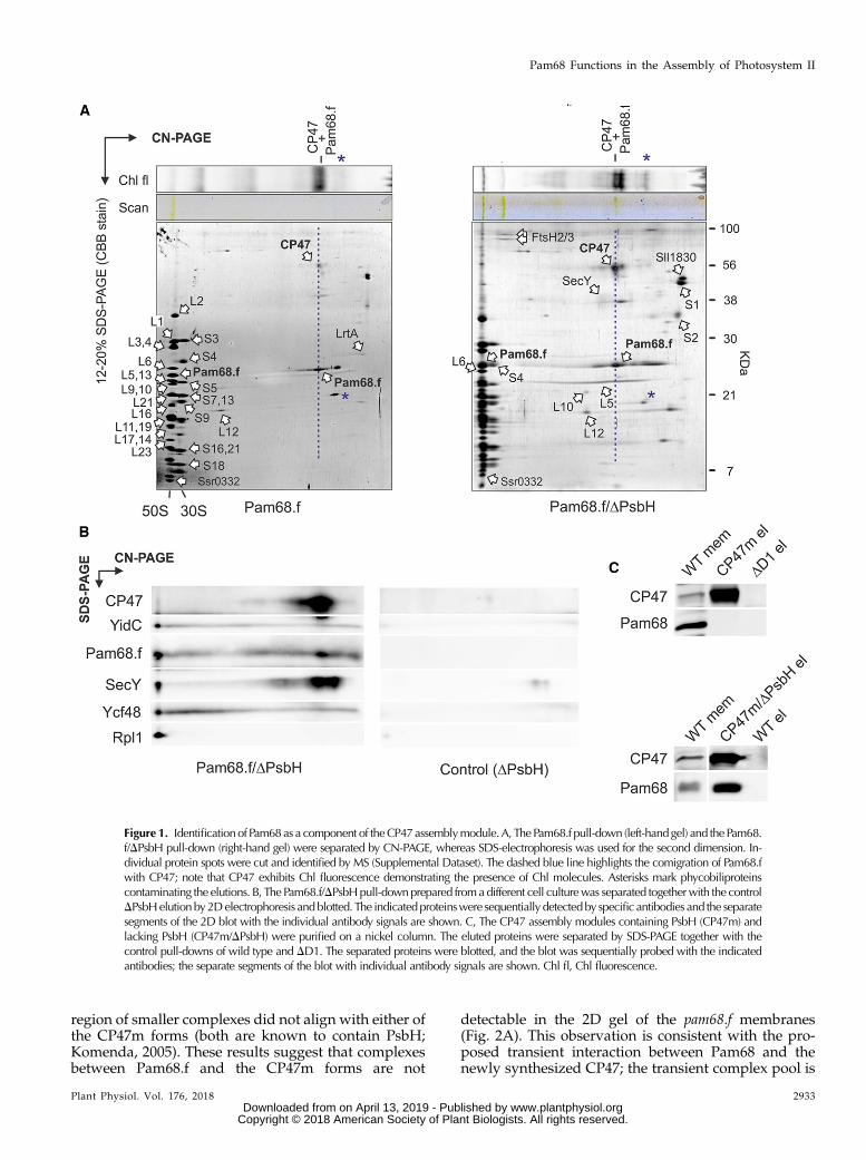
To elucidate whether Pam68.f physically interacts withunassembled CP47 in the absence of PsbH, we purifiedPam68.f from the PsbH-less strain, and both elutions(Pam68.f andPam68.f/DPsbH)were analyzedby2DClear-Native/SDS-PAGE (CN/SDS-PAGE). On the stained gels,we identified large (50S) and small (30S) ribosome subunitsand two fractions of Pam68.f comigrating with 50S andwith CP47, respectively. The Pam68.f-CP47 complexexhibited Chl fluorescence, and its green pigmentationwasvisible on the CN gel (Fig. 1A).
In addition to the ribosome subunits, FtsH proteases,and a smeary band of SecY, the Pam68.f elutions alsocontained two unknown proteins (Sll1830 and Ssr0332).Whereas Sll1830 migrated as a free protein, the smallSsr0332 protein comigrated with the 50S ribosomal sub-unit. Another identified protein was light-repressed pro-tein A (LrtA, Sll0947), which showed sequence similarityto the bacterial pY factor associatedwith stalled ribosomes(Galmozzi et al., 2016). A similar pattern of ribosomalproteins, but with higher levels of LrtA, was also obtainedin the Pam68.f pull-down isolated from theDpsbB (DCP47)mutant background (Supplemental Fig. S2). This resultimplies that Pam68 remains associated with a pool ofmembrane-bound ribosomes even when no CP47 trans-lation occurs in the cell. Notably, the electrophoretic mo-bility of Pam68.f proteins purified from the DpsbH andwild-type backgrounds were slightly different, indicatinga posttranslational modification of Pam68.f upon the psbHdeletion (Fig. 1A). This shift allowed us to distinguish thatthe spot of Pam68.f comigrating with 50S in the Pam68.f/DPsbH pulldown (just above the Rpl6 protein) consists ofonly Pam68.f, with no other (ribosomal) proteins. There isno spot in this position in the Pam68.f elution (Fig. 1A).
To better visualize the pattern of proteins on the 2D gel,the separation of Pam68.f/DPsbH and the control DPsbHpull-downs on 2D CN/SDS-PAGE was followed by im-munoblotting. The immunodetection determined a fractionof YidC, Ycf48, and SecY comigratingwith 50S, as expectedfor the isolated ribosome-translocon apparatus (Fig. 1B).However, the barely visible (SecY) or invisible (YidC,Ycf48)staining of these proteins on the gel indicates that they aresubstantially less abundant than Pam68. Hence, it is un-likely that they connect Pam68.f with ribosomes. CP47wasfound in a spot that had the same mobility as the dissoci-ated Pam68.f, suggesting a mutual complex.
We used an independent approach to verify the in-teraction between the unassembled CP47 and Pam68proteins. We isolated CP47m and a nascent CP47mlacking PsbH (CP47m/DPsbH) via His-tagged CP47from Synechocystis strains accumulating these complexesdue to the absence of the D1 or D1/PsbH PSII subunits,respectively (Boehm et al., 2011; D’Haene et al., 2015).The Pam68 protein was copurified with CP47m/DPsbHbut was not detected in the CP47m elution (Fig. 1C).Therefore, either the binding of PsbH to the CP47-Pam68complex is considerably weaker than to CP47, or Pam68and PsbH share a similar binding side.
N-Terminal Segment of Pam68 Is Required for theInteraction with Ribosomes
To verify that the interaction of Pam68 with ribo-somes is not an artifact of the pull-down assay, solu-bilized membrane complexes from the pam68.f strainwere separated by 2D CN/SDS-PAGE, stained bySYPRO Orange, and blotted onto a polyvinylidenefluoride (PVDF) membrane. Pam68.f comigrated withthe 50S and, unexpectedly, also with the 30S subunit(Fig. 2A). On the other hand, Pam68.f spots in the
2932 Plant Physiol. Vol. 176, 2018
Bu�cinská et al.
www.plantphysiol.orgon April 13, 2019 - Published by Downloaded from Copyright © 2018 American Society of Plant Biologists. All rights reserved.
region of smaller complexes did not align with either ofthe CP47m forms (both are known to contain PsbH;Komenda, 2005). These results suggest that complexesbetween Pam68.f and the CP47m forms are not
detectable in the 2D gel of the pam68.f membranes(Fig. 2A). This observation is consistent with the pro-posed transient interaction between Pam68 and thenewly synthesized CP47; the transient complex pool is
Figure 1. Identificationof Pam68as a component of theCP47assemblymodule.A, ThePam68.f pull-down (left-handgel) and thePam68.f/DPsbH pull-down (right-hand gel) were separated by CN-PAGE, whereas SDS-electrophoresis was used for the second dimension. In-dividual protein spots were cut and identified by MS (Supplemental Dataset). The dashed blue line highlights the comigration of Pam68.fwith CP47; note that CP47 exhibits Chl fluorescence demonstrating the presence of Chl molecules. Asterisks mark phycobiliproteinscontaminating the elutions. B, The Pam68.f/DPsbHpull-downprepared fromadifferent cell culturewas separated togetherwith the controlDPsbHelutionby2Delectrophoresis andblotted. The indicatedproteinswere sequentially detectedby specific antibodies and the separatesegments of the 2D blot with the individual antibody signals are shown. C, The CP47 assembly modules containing PsbH (CP47m) andlacking PsbH (CP47m/DPsbH) were purified on a nickel column. The eluted proteins were separated by SDS-PAGE together with thecontrol pull-downs of wild type and DD1. The separated proteins were blotted, and the blot was sequentially probed with the indicatedantibodies; the separate segments of the blot with individual antibody signals are shown. Chl fl, Chl fluorescence.
Plant Physiol. Vol. 176, 2018 2933
Pam68 Functions in the Assembly of Photosystem II
www.plantphysiol.orgon April 13, 2019 - Published by Downloaded from Copyright © 2018 American Society of Plant Biologists. All rights reserved.

apparently below the detection limit of the immunoblotanalysis.
According to this model, ribosomes can be docked tobacterial membranes via interaction with the largesubunit and the SecYEG translocon, or alternatively,with YidC insertase (Prinz et al., 2000; Seitl et al., 2014).However, the interaction between themembrane-boundribosomes and SecY or YidC in isolated thylakoidswas not preserved in our 2D gel system (Fig. 2A).Therefore, it is unlikely that SecY/YidC facilitatesthe observed association of Pam68.f with ribosomes.
It is likely that Pam68 interacts directly with ribo-somal proteins from both the 50S and 30S subunits.The 30S subunit of the membrane-docked ribosome isclose to the membrane surface (approximately 10 nm;Frauenfeld et al., 2011). Theoretically, the stronglypositively charged N terminus of Pam68.f is longenough (65 amino acids, approximately 20 nm;Supplemental Fig. S3) to reach the 30S subunit. To testthis possibility, we constructed strains expressing var-iants of Pam68.f truncated either up to the V29(t29-pam68.f strain) or the S50 amino acid residues(t50-pam68.f). The t29-Pam68.f protein still comigrated
with ribosomes on the 2D gel (Fig. 2B), but the moretruncated t50-Pam68.f protein was not detectable inany larger complexes, which supports the role of thePam68 N-terminal segment in the interaction withribosomes.
A close relationship between the cyanobacterialPam68 and ribosomes can also be inferred from theexistence of an operon of the pam68 and the rps15genes, which is highly conserved among the cyano-bacterial genomes. According to the STRING database(http://string-db.org/), there are only a few examplesof sequenced cyanobacterial genomes (e.g. Gloeobacterviolaceus) where these two genes are not organizedin tandem. In the Synechocystis genome, the pam68gene is transcribed from the rps15 promoter as a singlemRNA with rps15 (Mitschke et al., 2011). Interest-ingly, the rps15-pam68 mRNA belongs to a smallgroup of ribosomal transcripts that are significantlyup-regulated under stress conditions with the strongestexpression under low temperature (Kopf et al., 2014;Supplemental Fig. S4). Indeed, we found the Pam68protein level to be high during high light or chillingstress (Supplemental Fig. S5).
Figure 2. 2D CN/SDS-PAGE and immunodetection of membrane protein complexes from strains expressing full-length ortruncated Pam68.f. A, Solubilized membrane proteins from the pam68.f strain were separated by 2D CN/SDS-PAGE. The 2D gelwas stained with SYPRO Orange, blotted, and the 2D blot was sequentially probed by the indicated antibodies. Separate seg-ments of the 2D blot with individual antibody signals are shown. The large and small ribosomal subunits are highlighted on thestained gel by red dashed boxes; protein spots belonging to Rpl1 and Rps2 were identified previously (Chidgey et al., 2014). Chlfluorescence was detected after excitation by blue light. CP47m marks two forms of the CP47 assembly module detected in theSynechocystis membrane fraction (Komenda, 2005). B, The same analysis was performed on membranes isolated from strainsexpressing the truncated variants t29-Pam68.f (top panel) and t50-Pam68 (bottom panel). Only a region of the SYPRO Orangestained gel around the Rpl1 protein (SYPRO stain) and separate segments of the 2D blot with signals of anti-Rpl1 and anti-Flagantibodies are shown. Complexes are designated as in (B). Chl fl, Chl fluorescence; L1, Rpl1; PSI[3], trimer of PSI; PSII[1],monomer of PSII; PSII[2], dimer of PSII; S2, Rps2.
2934 Plant Physiol. Vol. 176, 2018
Bu�cinská et al.
www.plantphysiol.orgon April 13, 2019 - Published by Downloaded from Copyright © 2018 American Society of Plant Biologists. All rights reserved.

Enhanced Chl Biosynthesis Rescues the Abolished CP47Synthesis in the DpsbH/Dpam68 Strain
The results described above imply that Pam68 func-tions during the synthesis and/or folding of CP47 beforeit associates with PsbH, which also facilitates CP47 syn-thesis (Komenda, 2005). To test whether PsbH can com-pensate for the absence of Pam68, we characterized theSynechocystisDpam68 andDpsbHmutants and theDpsbH/Dpam68 double mutant. Under moderate light intensities(40 mmol photons m22 s21), Dpam68 grew similarly as thewild-type strain and had a similar Chl content(Supplemental Fig. S6, A and B). The DpsbH mutationaffected both the growth rate and Chl content; neverthe-less, this mutant grew fairly well photoautotrophically(Supplemental Fig. S6,A andB).However, even the singleDpam68 mutant stopped proliferating on plates undermore severe conditions, such as dark-/high-light fluctu-ation or low temperature (Fig. 3A). Moreover, the level ofPsbH was merely affected in the Dpam68 strain and, viceversa, the level of Pam68 in the DpsbH strain remainedcomparable to wild type (Fig. 3B).Unlike the strains containing singlemutations, thedouble
mutant showed extremely slow autotrophic growth (dou-ble time approximately 20 d), accumulated only traces ofChl and died immediately after exposure to mild stressconditions (Fig. 3A; Supplemental Fig. S6). However,photoautotrophy of the DpsbH/Dpam68 strain can be re-stored by the expression of Pam68.f (Fig. 3C), whichprovides evidence that the poor phenotype of the doublemutant is not caused by a position effect, e.g. lower levelsof Rps15. To obtain enough cells of the poor-growingDpsbH/Dpam68 mutant, we first grew all strains withGlc supplementation. Then, we characterized the pheno-type 2 d after removing Glc from the media. As revealedby the CN-PAGE separation of membrane complexes(Fig. 3D), the levels of PSI and PSII were virtually un-changed in the Dpam68 strain, but the DpsbH strain con-tainedmuch less dimeric PSII. In the double mutant, verylittle PSI and only traces of the PSII complexes were de-tectable. Thus, both PsbHandPam68 play distinct roles inthe accumulation of PSII; the parallel elimination of bothof these proteins is nearly fatal for cell viability.For a closer look at the role of PsbH and Pam68 in the
synthesis of PSII wild type, Dpam68, DpsbH, and DpsbH/Dpam68 cells were pulse-labeled and the isolated mem-brane complexes analyzed by 2DCN/SDS-PAGE (Fig. 4).Consistent with the previously published analysis ofDpam68 (Rengstl et al., 2013), this strain showed less la-beled CP47 and CP43 in total, and lacked the labeledunassembled CP47. In addition, we observed severe ac-cumulation of RCIIa and RCII* assembly intermediates,which is a typical feature of cells deficient in the formationof CP47m (Knoppová et al., 2014). The obtained patternforDpsbH differed from theDpam68 strain by having onlyweakly labeled dimeric PSII and also less synthesized D1.A detectable pool of unassembled CP47 was also absentand both RCII complexes accumulated, which impliesthat the rate of CP47m formation limits the process of PSIIassembly. In the DpsbH/Dpam68 strain, the capacity to
synthesize PSII was extremely weak, almost certainlycaused by the lack of CP47m because the intensively la-beled RCII complexes resembled the canonical pattern ofthe DCP47 strain (Fig. 4; Komenda et al., 2004).
Based on the available PSII structure (Umena et al.,2011), theN-terminal segment of PsbH creates a networkof hydrogen bonds with the stromal loops connectingthe first four helixes of CP47 (Supplemental Fig. S7).Therefore, the PsbHprotein couldfix the nascent CP47 in
Figure 3. Characterization of the Synechocystis strains lacking Pam68,PsbH, or both of these proteins. A, Autotrophic growth of the wild-typeand mutant strains on agar plates under various conditions. Growth for5 d under normal light (40 mmol photons m22 s21), low light (10 mmolphotons m22 s21), high light (400 mmol photons m22 s21), fluctuatingdark/high light conditions (5min dark, 5min 400mmol photonsm22 s21),18°C at 40 mmol photons m22 s21, and low nitrogen (0.1 mM NaNO3). B,Levels of PsbH and Pam68 in the Dpam68 and DpsbH strains undernormal light conditions. A comparable amount of Chl was loaded foreach strain. C, Autotrophic growth of the pam68.f/Dpam68/DpsbH strainexpressing the Pam68.f protein under the regulation of the psbAII pro-moter. D, Membranes, isolated from the wild-type and mutant strainsgrown as described in (A), were solubilized and separated by CN-PAGE.D/HL, dark/high light; HL, high light; LL, low light; NL, normal light;PSI[3], trimer of PSI; PSII[1], monomer of PSII; PSII[2], dimer of PSII.
Plant Physiol. Vol. 176, 2018 2935
Pam68 Functions in the Assembly of Photosystem II
www.plantphysiol.orgon April 13, 2019 - Published by Downloaded from Copyright © 2018 American Society of Plant Biologists. All rights reserved.

a position that facilitates prompt insertion of Chl mole-cules. Moreover, the C-terminal region of Pam68 mayplay a similar role. To test the importance of both pro-teins for Chl insertion into CP47, we removed Glc fromthe DpsbH/Dpam68 liquid culture, while supplementingit with 200 nM N-methyl mesoporphyrin IX. This com-pound is a specific inhibitor of the FeCh enzyme and apartial inhibition of FeCh strongly enhances Chlbiosynthesis (Sobotka et al., 2005). Remarkably, theDpsbH/Dpam68 cells treated with the FeCh inhibitorstarted to grow much faster than the control cells with-out the inhibitor (Fig. 5A). The control culture had verylow Chl content on a per-cell basis, whereas the treatedcells progressively built up new Chl-complexes, and in4 d reached approximately 85% of the Chl level whencompared to wild type (Fig. 5B).
Precursors of the Chl biosynthetic pathway differeddramatically between treated and untreated cells.Whereas monovinyl-chlorophyllide was the only de-tectable Chl precursor in the untreated cells, theinhibitor-treated cells contained a spectrum of Chlprecursors typical for wild type (Pilný et al., 2015;Supplemental Fig. S8). Because the earlier precursorsupstream of chlorophyllide were below the detec-tion level in the untreated double mutant, this chlo-rophyllide pool originated almost certainly from Chlrecycling and not from de novo synthesis (Vavilinet al., 2005; Kope�cná et al., 2015). We repeatedthe protein radiolabeling experiment describedabove using DpsbH/Dpam68 cells treated with theFeCh inhibitor. The assembly of PSII was restored(Fig. 5C), suggesting that boosting of the ceased Chl
Figure 4. Synthesis of PSII subunitsin the Dpam68, DpsbH, and DpsbH/Dpam68 mutant strains. Wild-type (A)and the mutant Dpam68 (B), DpsbH (C),and DpsbH/Dpam68 (D) cells grown for2 d without Glc were radiolabeled witha mixture of [35S]Met/Cys using a30-min pulse. Isolated membrane pro-teins were separated by CN-PAGE on a4% to 14% linear gradient gel, whereas12% to 20% SDS-electrophoresis wasused for the second dimension. Thesame amounts of Chl were loadedfor each strain. Note that the DpsbH/Dpam68 strain contains three-times lessChl per cell than DpsbH, meaning thatthe membrane proteins from the doublemutant were overloaded on a per-cellbasis to obtain a detectable radioactivitysignal of the assembled PSII. The 2D gelswere stained with Coomassie Blue, andthe labeled proteins were detected by aphosphoimager (Autorad). Chl fluores-cence emitted by Chl was detected byLAS 4000 (Fuji) after excitation by bluelight. Chl fl, Chl fluorescence; iD1, in-completely processed form of the D1precursor; PSI[3], trimer of PSI; PSII[1],monomer of PSII; PSII[2], dimer ofPSII; RCII*, assembly intermediate (re-action center complex) lacking CP47m(Knoppova et al., 2014); RCIIa, PSII as-sembly intermediate (reaction centercomplex) lacking CP43m (Knoppovaet al., 2014).
2936 Plant Physiol. Vol. 176, 2018
Bu�cinská et al.
www.plantphysiol.orgon April 13, 2019 - Published by Downloaded from Copyright © 2018 American Society of Plant Biologists. All rights reserved.

biosynthesis restores the formation of CP47m in themutant lacking both Pam68 and PsbH.
DISCUSSION
The Pam68 protein was first described in the Ara-bidopsis (Arabidopsis thaliana) pam68-null mutant,which accumulated only approximately 10% of PSII(Armbruster et al., 2010). The function of Pam68 was
originally linked to the synthesis or maturation of theD1 subunit of PSII (Armbruster et al., 2010); however, astrong relationship between Pam68 and CP47 was alsosuggested, based on the low level of Pam68 detected inthe Synechocystis CP47-less strain (Rengstl et al., 2011).Our results agree with a recent study, which demon-strated that the lack of Pam68 in Synechocystis limits thesynthesis of CP47 and CP43 (Rengstl et al., 2013). Giventhat the mechanism of PSII biogenesis is highly con-served, it is likely that the eukaryotic Pam68 is involved
Figure 5. Abolished synthesis of CP47 in theDpsbH/Dpam68mutant is rescued by enhanced Chl biosynthesis. A, TheDpsbH/Dpam68cells grown mixotrophically were harvested and resuspended in a growth medium without Glc. The obtained culture was divided intotwo flasks, with one of them supplementedwith N-methyl-mesoporphyrin IX (Me-MesoP, FeCh inhibitor). The photoautotrophic growthwas then monitored. The inset shows the same growth experiment but with 200 nM Me-MesoP added into the plate. B, Absorbancespectra of mutant cells growing for 4 d in the presence or absence of FeCh inhibitor. Spectra were normalized to light scattering at750 nm. Also shown is the Chl content determined spectroscopically in methanol extract and normalized per OD750 nm. C, DpsbH/Dpam68 cells grown for 2 d photoautotrophically in the presence of 200 nM FeCh inhibitorwere radiolabeledwith amixture of [35S]Met/Cys; incorporation of radioactivity into core PSII subunitswas detected after 2DCN/SDS-PAGE. The sameamounts of Chlwere loadedofeach sample. a.u., absorbance units; d.t., doubling time; iD1, incompletely processed form of the D1 precursor; PSI[3], trimer of PSI;PSII[1],monomer of PSII; PSII[2], dimer of PSII; RCII*, assembly intermediate (reaction center complex) lackingCP47m (Knoppova et al.,2014); RCIIa, PSII assembly intermediate (reaction center complex) lacking CP43m (Knoppova et al., 2014).
Plant Physiol. Vol. 176, 2018 2937
Pam68 Functions in the Assembly of Photosystem II
www.plantphysiol.orgon April 13, 2019 - Published by Downloaded from Copyright © 2018 American Society of Plant Biologists. All rights reserved.
in the synthesis of CP47. Indeed, using a standardmethodology (35S radiolabeling combined with 2D gel-electrophoresis), the synthesis of CP47 in the Arabi-dopsis pam68-null mutant was hardly detectable(Armbruster et al., 2010).
In contrast to Arabidopsis, the inactivation of pam68in Synechocystis had no obvious effect on the PSII levelunder standard growth conditions, although the syn-thesis of CP47 and CP43 was visibly affected in bothorganisms (Rengstl et al., 2013; Fig. 4). However, after3 h of high light treatment (2000mmol photonsm22 s21),the levels of functional PSII in the Dpam68 strain de-creased by approximately 50% (Rengstl et al., 2013). Inaddition, we demonstrated the importance of Pam68under fluctuating light conditions, low temperature,and nitrogen limitation (Fig. 3). These observationsimply that Pam68 is essential once the synthesis of CP47becomes difficult and limits PSII biogenesis.
The PsbH protein is required for sufficient CP47 syn-thesis in plants as well as in cyanobacteria (Komenda,2005; Levey et al., 2014); the Synechocystis DpsbHmutantshows a noticeable growth defect even under nonstress
conditions (Supplemental Fig. S6, A and B). However,the phenotype of this strain is probably quite complex,because PsbH also stabilizes electron transfer processesbetween QA andQB in the PSII complex (Komenda et al.,2002). It is further essential for the association ofphotoprotective high-light-inducible proteins to CP47(Promnares et al., 2006; Fig. 6), and creates an environ-ment for binding of a red Chl molecule in CP47, which isalso supposed to have a protective function (D’Haeneet al., 2015). However, we expect that the impaired CP47synthesis/stability is themajor reason for the slowgrowthof the DpsbH mutant (Supplemental Fig. S6A). This con-clusion is supported by the fact that the growth rate of thisstrain can also be improved by the inhibition of FeCh(Supplemental Fig. S6C), and is consistent with the verypoor phenotype of the DpsbH/Dpam68 double mutant.Therefore, PsbH appears to be more crucial for the bio-genesis than for the functioning of fully assembled PSIIcomplexes. Similarly, the PsbI subunit was found to bemore important for attachment of CP43m to RCII, ratherthan for PSII activity (Dobáková et al., 2007). Other smallPSII subunits may also play roles in assembly.
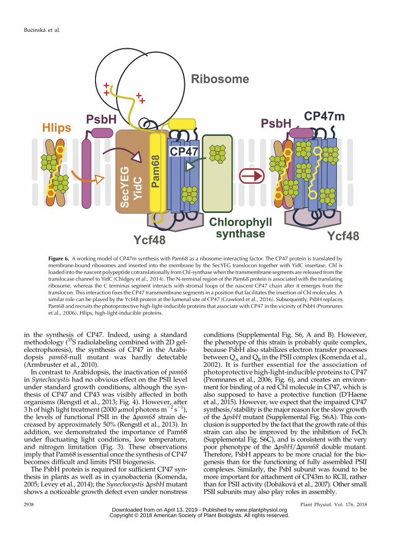
Figure 6. A working model of CP47m synthesis with Pam68 as a ribosome-interacting factor. The CP47 protein is translated bymembrane-bound ribosomes and inserted into the membrane by the SecYEG translocon together with YidC insertase. Chl isloaded into the nascent polypeptide cotranslationally fromChl-synthasewhen the transmembrane segments are released from thetranslocase channel to YidC (Chidgey et al., 2014). The N-terminal region of the Pam68 protein is associated with the translatingribosome, whereas the C terminus segment interacts with stromal loops of the nascent CP47 chain after it emerges from thetranslocon. This interaction fixes the CP47 transmembrane segments in a position that facilitates the insertion of Chl molecules. Asimilar role can be played by the Ycf48 protein at the lumenal site of CP47 (Crawford et al., 2016). Subsequently, PsbH replacesPam68 and recruits the photoprotective high-light-inducible proteins that associate with CP47 in the vicinity of PsbH (Promnareset al., 2006). Hlips, high-light-inducible proteins.
2938 Plant Physiol. Vol. 176, 2018
Bu�cinská et al.
www.plantphysiol.orgon April 13, 2019 - Published by Downloaded from Copyright © 2018 American Society of Plant Biologists. All rights reserved.
We present a working model of CP47m synthesis(Fig. 6). Pam68 is firmly bound to the translating ri-bosome via the N-terminal segment, whereas itsC-terminal end interacts with the stromal loops of thenascent CP47 chain emerging from the translocon. Wespeculate that the coordination of Pam68 (stromalside), YidC (lateral site; Hennon et al., 2015), and Ycf48(lumenal site; Crawford et al., 2016) fixes the CP47helix pairs in a position that is amenable to Chl bind-ing. The Pam68 C terminus contains highly conservedcharged residues (Supplemental Fig. S3) that can forma network of hydrogen bounds resembling the inter-action of the N terminus of PsbH with CP47 (seeSupplemental Fig. S7). The synthesis of CP47 is im-paired in the Dpam68 strain even under nonstressfulconditions (Fig. 4), suggesting that Pam68 perma-nently assists during CP47 synthesis. Because Pam68is particularly critical for the mutant lacking PsbH, it isprobable that both proteins can work similarly aschaperones facilitating the folding of CP47 and/or theloading of Chl into the newly synthesized apo-polypeptide chain.Based on previous data and the results of our radio-
labeling experiment (Rengstl et al., 2013; Fig. 4), Pam68appears to also facilitate the synthesis of the CP43protein and PSI. However, the interaction of Pam68with these proteins is either too weak to detect, or thelower levels of CP43 and PSI in the absence of Pam68 isa secondary phenotype caused by the feeble CP47synthesis. The second possibility is more probable, asthe mutant lacking CP47 has been shown to containconsiderably lower cellular level of Chl in comparisonwith wild type, implying that the level of CP43 and PSIis lower in the absence of CP47 (Be�cková et al., 2017).Although this approach is frequently used, we areaware that arresting particular PSII assembly steps toaccumulate specific assembly intermediates may affectother cellular processes, including the synthesis of Chl-binding proteins. The Flag-tag technology used herehas the advantage of allowing the purification of PSIIassembly intermediates that only exist temporarily inthe cell (such as the Pam68.f-CP47m complex) directlyfrom the wild-type background.The synthesis of CP47 is very sensitive to Chl avail-
ability (Hollingshead et al., 2016), which may explainwhy the lack of Pam68 is not tolerated under stressconditions. The Chl pathway can be temporarilyswitched-off after a shift to stressful conditions(Kopecná et al., 2012) and when the de novo Chlamount decreases, a fine structural stabilization of thenascent CP47 is likely to be particularly important forthe smooth loading of Chls. After addition of the FeChinhibitor, the pool of available Chl increased, and theimpaired CP47 synthesis was rescued (Fig. 5). Thus, ahigh concentration of Chl molecules around the trans-lated CP47 increases the chance that all Chls are inser-ted in time even if the orientation of CP47 is not perfect.A similar effect of FeCh inhibition was reported earlierin the Synechocystis strain harboring a mutated CP47protein (Sobotka et al., 2005).
The observed tight binding of Pam68 to the ribosomeis intriguing. A high level of LrtA protein, which asso-ciates with the 30S ribosomal subunit (Galmozzi et al.,2016), was present in the Pam68.f pull-downs preparedfrom strains lacking CP47 (Supplemental Fig. S2).As the potential function of LrtA is to stabilize stalledribosomal 70S particles (Di Pietro et al., 2013), its ap-pearance in the elution indicates that Pam68 interactswith both the SecY-bound idle ribosomes as well aswith the actively translating ribosomes. In Synechocys-tis, Pam68 is not an abundant membrane protein (it isnot detectable in the stained SDS PAGE gel with sepa-rated cellular membrane proteins), and there is only alimited pool of membrane-bound ribosomes associatedwith Pam68. It is possible that these ribosomes differstructurally from other ribosomes in the cell. The het-erogenic nature of ribosomes is demonstrated by thevariable stoichiometry among core ribosomal subunitsor between the monosome/polysome arrangement ofribosomes according to environmental conditions (Xueand Barna, 2012; Slavov et al., 2015). Similarly, theplastid-encoded Rps15 is not an essential ribosomalsubunit in plants, but under chilling stress, the tobacco(Tobacco nicotiana) Drps15 knockdown showed a drasticreduction in the number of plastid ribosomes(Fleischmann et al., 2011). In Synechocystis, the rps15-pam68 operon as well as the rps18, rps20, and rps25genes are up-regulated under cold stress, whereasmany ribosomal genes are simultaneously down-regulated (Supplemental Fig. S4). This result supportsthe existence of a pool of modified, stress-induced typeof ribosomes in cyanobacteria. It is possible that Pam68has a higher affinity for the stress-induced type of ri-bosomes. Once bound to SecY, the ribosome mightserve as an anchor to localize Pam68 in the vicinity ofthe translocon machinery. Under severe conditionswith limited Chl availability and/or lowered mem-brane fluidity (chilling stress), Pam68 can promptlyassist during the synthesis of CP47.
MATERIALS AND METHODS
Synechocystis Strains and Growth Conditions
All the Synechocystis strains used are summarized in Supplemental Table S1.The Dpam68 strain was kindly provided by Jörg Nickelsen (Ludwig-Maximilians University), and is described in Armbruster et al. (2010). TheDpsbH/Dpam68 double mutant was prepared by transformation of the DpsbHstrain (D’Haene et al., 2015) by genomic DNA isolated from the Dpam68 strain.The Synechocystis strain expressing the Pam68 protein fused with 33Flag at theC terminus (the pam68.f strain) was constructed using pPD-CFLAG plasmid asdescribed in Hollingshead et al. (2012). The pam68.f construct was furthertransformed into the DpsbH and DpsbB cells (Eaton-Rye and Vermaas, 1991) toexpress the Pam68.f protein in these genetic backgrounds. Derivatives ofPam68.f truncated at the amino acid 29 (the t29-pam68.f strain) or 50 (the t50-pam68.f strain) were constructed by PCR amplification of the Synechocystispam68 gene lacking the 39 part (primers are listed in Supplemental Table S2).The obtained PCR products were cloned into a pPD-CFLAG plasmid andtransformed into wild type. To be able to express Pam68.f in theDpam68/DpsbHmutant (already resistant to kanamycin), we replaced the kanamycin-resistancecassette in the pPD-CFLAG plasmid with an erythromycin-resistance cassette,cloned the pam68 gene into this modified construct. and fully segregated thepam68.f/Dpam68/DpsbH strain.
Plant Physiol. Vol. 176, 2018 2939
Pam68 Functions in the Assembly of Photosystem II
www.plantphysiol.orgon April 13, 2019 - Published by Downloaded from Copyright © 2018 American Society of Plant Biologists. All rights reserved.

Unless stated otherwise, strains were grown photoautotrophically in liquidBG-11 medium on a rotary shaker under moderate (normal) light intensities(40 mE m22 s21) at 28°C. For purification of the Pam68.f protein under nativeconditions, 4 L of pam68.f and pam68.f/DpsbH cells were grown in a 10 L flask inBG-11 medium supplemented by 5 mM Glc under normal light conditions andbubbled with air. Strains lacking PSII (DpsbB) were supplemented with 5 mM
Glc and grown under lower light intensities (10 mE m22 s21).
Absorption Spectra and Determination of Chl Content
Absorption spectra of thewhole cellsweremeasured at room temperaturewith aUV-3000 spectrophotometer (Shimadzu). Chl was extracted from cell pellets (2 mL,OD750 = approximately 0.3) with 100% (v/v) methanol, and its concentration wasmeasured spectrophotometrically according to Porra et al. (1989).
Preparation of Cellular Membranes
Cell cultureswere harvested at optical densities of 750 nm=approximately 0.5 to0.7. Cells were pelleted, washed, and resuspended with buffer A (25 mM MES/NaOH, pH 6.5, 10mMCaCl2, 10mMMgCl2, 25% [v/v] glycerol) for the preparationof membranes for 2D electrophoresis and purification of Pam68.f. For nickel-affinitychromatography, the membrane fraction was prepared in 25 mM Na-P buffer, pH7.5, 50 mMNaCl, 10% (v/v) glycerol (buffer B). Cells were broken using glass beads(0.1mmdiameter), and themembrane fractionwas separated from soluble proteinsby centrifugation at high speed (65,0003 g, 20 min).
Isolation of Protein Complexes byAffinity Chromatography
Cellular membranes containing approximately 1 mg/mL Chl were solubi-lized for 1 h with 1% (w/v) b-dodecyl-maltoside at 10°C and centrifuged for20 min at 65,000g to remove cell debris. The Pam68.f complexes were purifiedusing an anti-Flag-M2 agarose column (Sigma-Aldrich). To remove contami-nants, the anti-Flag-resin was washed with 20 resin volumes of buffer A con-taining 0.04% b-dodecyl-maltoside. The Pam68.f complex was eluted with 2.5resin volumes of buffer A containing 150 mg/mL 33Flag peptide (Sigma-Aldrich) and 0.04% b-dodecyl-maltoside. For purification of the His-taggedproteins, solubilized membrane complexes were loaded onto a nickel-affinitychromatography column (Protino Ni–NTA-agarose; Macherey-Nagel). Pro-teins bound to the column were washed with buffer B containing 0.04%b-dodecyl-maltoside and increasing concentrations of imidazole (5, 10, 20, and30 mM); His-tagged proteins were finally eluted with 150 mM imidazole.
Electrophoresis and Immunoblotting
The protein composition of the purified complexes was analyzed by elec-trophoresis in a denaturing 12% to 20% linear gradient polyacrylamide gelcontaining 7 M urea (Dobáková et al., 2009). Proteins were stained either byCoomassie Brilliant Blue or SYPRO Orange stain and subsequently transferredonto a PVDF membrane for immunodetection (see below). For native electro-phoresis, solubilized membrane proteins or isolated complexes were separatedon 4% to 12% CN-PAGE (Wittig et al., 2007). Individual components of proteincomplexes were resolved by incubating the gel strip from the first dimension in2% (w/v) SDS and 1% (w/v) DTT for 30min at room temperature, and proteinswere separated along the second dimension by SDS-PAGE in a denaturing 12%to 20% polyacrylamide gel containing 7 M urea (Dobáková et al., 2009). Proteinswere stained by Coomassie Brilliant Blue or by SYPRO Orange; in the lattercase, they were subsequently transferred onto a PVDF membrane. Membraneswere incubated with specific primary antibodies and then with a secondaryantibody conjugated with horseradish peroxidase (Sigma-Aldrich). The fol-lowing primary antibodies were used in the study: anti-SecY and anti-YidC(Linhartová et al., 2014), anti-CP47, anti-D1 and anti-PsbH (Komenda, 2005),anti-Pam68 (Armbruster et al., 2010), anti-Rpl1 (Agrisera), anti-Flag (Sigma-Aldrich), and anti-Ycf48 (which was raised in rabbit against recombinantSynechocystis Ycf48 and provided by Peter Nixon, Imperial College, London).
Protein Radiolabeling
Forprotein labeling, the cellswere incubatedwith using amixture of [35S]Metand [35S]Cys (Translabel; MP Biochemicals) as described in Dobáková et al.
(2009). After separation of labeled proteins by CN-PAGE in the first dimension,the polyacrylamide gel was scanned for Chl fluorescence and then treated forsecond-dimension separation with 18% SDS-PAGE. The 2D gel was exposed toa Phosphorimager plate (GE Healthcare) overnight and stained by CoomassieBrilliant Blue and scanned by Storm (GE Healthcare).
Protein Identification by LC-MS/MS Analysis
Gel slices were placed in 200 mL of 40% acetonitrile, 200 mM ammoniumbicarbonate and incubated at 37°C for 30 min, after which the solution wasdiscarded. This procedure was performed twice, and the gel was subse-quently dried in a vacuum centrifuge. Ten microliters of 40 mM ammoniumbicarbonate in 9% acetonitrile containing 0.4 mg trypsin (proteomics grade;Sigma-Aldrich) were added to the gel slice and left to soak in the solution at4°C for 45 min. To digest proteins, 20 mL of 9% (v/v) acetonitrile in 40 mM
ammonium bicarbonate was added to the gel and incubated at 37°C over-night. Peptides were purified using ZipTip C18 pipette tips (Millipore). MSanalysis was performed on a NanoAcquity UPLC (Waters) on-line coupled tothe ESI Q-ToF Premier mass spectrometer (Waters). One microliter of thesample was diluted in 3% (v/v) acetonitrile/0.1% (v/v) formic acid, andtryptic peptides were desalted on a Symmetry C18 Trapping column (180 mmi.d., 20 mm length, particle size 5 mm, reverse phase; Waters) with a flow rateof 15 mL/min for 1 min. Trapping was followed by a reverse-phase UHPLCusing the BEH300 C18 analytical column (75 mm i.d. 150 mm length, particlesize 1.7 mm, reverse phase; Waters). The linear gradient elution ranged from97% solvent A (0.1% formic acid) to 40% solvent B (0.1% formic acid in ace-tonitrile) at a flow rate of 0.4 mL/min. Eluted peptides flowed directly into theESI source. Raw data were acquired in the data-independent MSE identitymode (Waters). Precursor ion spectra were acquiredwith a collision energy of5 V and fragment ion spectra with a collision energy of 20 V to 35 V ramp inalternating 1 s scans. Data-dependent analysis mode was used for the secondanalysis; peptide spectra were acquired with a collision energy of 5 V andpeptides with charge states of + 2, +3, and + 4 were selected for MS/MSanalysis. Fragment spectra were collected with a collision energy of 20 V to40 V ramp. In both modes, the acquired spectra were submitted for databasesearch using the PLGS2.3 software (Waters) against Synechocystis proteindatabases from the Cyanobase Web site (http://genome.microbedb.jp/cya-nobase/). Acetyl N-terminal, deamidation N and Q, carbamidomethyl C, andoxidation M were set as variable modifications. Identification of three con-secutive y-ions or b-ions was required for a positive peptide match.
Accession Numbers
Pam68, BAA16881.1; CP47, BAA10458.1; PsbH, BAA17629.1, SecY;BAA17331.1, YidC - BAA18244.1.
Supplemental Data
The following supplemental materials are available.
Supplemental Table S1. A list of Synechocystis strains used in this study.
Supplemental Table S2. A list of primers used to clone the pam68.f gene andits two truncated variants (t29-pam68.f and t50-pam68.f ) into the pPD-CFLAG plasmid (adding of 33Flag tag at the C terminus).
Supplemental Figure S1. Identification of proteins copurified with Pam68.f.
Supplemental Figure S2. 2D CN/SDS-PAGE of the Pam68.f complex puri-fied from the pam68.f/DpsbB strain (A) and the control DCP47 (B).
Supplemental Figure S3. Conservation profile and the prediction of sec-ondary structure of the Synechocystis Pam68 protein.
Supplemental Figure S4. Coexpression of the cyanobacterial pam68-rps15operon with a subset of ribosomal genes.
Supplemental Figure S5. Accumulation of the Pam68 protein under stressconditions.
Supplemental Figure S6. Growth rate and whole cell spectra of the wild-type and mutant strains.
Supplemental Figure S7. Stromal view of the CP47–PsbH complex withindicated hydrogen bonds between the CP47 and the PsbH N terminus.
2940 Plant Physiol. Vol. 176, 2018
Bu�cinská et al.
www.plantphysiol.orgon April 13, 2019 - Published by Downloaded from Copyright © 2018 American Society of Plant Biologists. All rights reserved.

Supplemental Figure S8. Changes in the levels of Chl precursors in theDpsbH/Dpam68 strain after treatment with FeCh inhibitor.
Supplemental Dataset. MS data—numbers of identified trypsin peptidesfor 2D gel protein spots.
Received January 17, 2018; accepted February 7, 2018; published February 20,2018.
LITERATURE CITED
Armbruster U, Zühlke J, Rengstl B, Kreller R, Makarenko E, Rühle T,Schünemann D, Jahns P, Weisshaar B, Nickelsen J, Leister D (2010)The Arabidopsis thylakoid protein PAM68 is required for efficient D1biogenesis and photosystem II assembly. Plant Cell 22: 3439–3460
Be�cková M, Gardian Z, Yu J, Koník P, Nixon PJ, Komenda J (2017) As-sociation of Psb28 and Psb27 proteins with PSII-PSI supercomplexesupon exposure of Synechocystis sp. PCC 6803 to high light. Mol Plant 10:62–72
Boehm M, Romero E, Reisinger V, Yu J, Komenda J, Eichacker LA,Dekker JP, Nixon PJ (2011) Investigating the early stages of photosys-tem II assembly in Synechocystis sp. PCC 6803: isolation of CP47 andCP43 complexes. J Biol Chem 286: 14812–14819
Boehm M, Yu J, Reisinger V, Be�cková M, Eichacker LA, Schlodder E,Komenda J, Nixon PJ (2012) Subunit composition of CP43-less photo-system II complexes of Synechocystis sp. PCC 6803: implications for theassembly and repair of photosystem II. Philos Trans R Soc Lond B BiolSci 367: 3444–3454
Chidgey JW, LinhartováM, Komenda J, Jackson PJ, DickmanMJ, Canniffe DP,Koník P, Pilný J, Hunter CN, Sobotka R (2014) A cyanobacterial chlorophyllsynthase-HliD complex associates with the Ycf39 protein and the YidC/Alb3insertase. Plant Cell 26: 1267–1279
Crawford TS, Eaton-Rye JJ, Summerfield TC (2016) Mutation of Gly195 ofthe ChlH subunit of Mg-chelatase reduces chlorophyll and further dis-rupts PS II assembly in a Ycf48-deficient strain of Synechocystis sp. PCC6803. Front Plant Sci 7: 1060
D’Haene SE, Sobotka R, Bu�cinská L, Dekker JP, Komenda J (2015) In-teraction of the PsbH subunit with a chlorophyll bound to histidine114 of CP47 is responsible for the red 77K fluorescence of PhotosystemII. Biochim Biophys Acta 1847: 1327–1334
Di Pietro F, Brandi A, Dzeladini N, Fabbretti A, Carzaniga T, Piersimoni L,Pon CL, Giuliodori AM (2013) Role of the ribosome-associated proteinPY in the cold-shock response of Escherichia coli. MicrobiologyOpen 2:293–307
Dobáková M, Sobotka R, Tichý M, Komenda J (2009) Psb28 protein isinvolved in the biogenesis of the photosystem II inner antenna CP47(PsbB) in the cyanobacterium Synechocystis sp. PCC 6803. Plant Physiol149: 1076–1086
Dobáková M, Tichý M, Komenda J (2007) Role of the PsbI protein inphotosystem II assembly and repair in the cyanobacterium Synechocystissp. PCC 6803. Plant Physiol 145: 1681–1691
Eaton-Rye JJ, Vermaas WFJ (1991) Oligonucleotide-directed mutagenesisof psbB, the gene encoding CP47, employing a deletion mutant strain ofthe cyanobacterium Synechocystis sp. PCC 6803. Plant Mol Biol 17:1165–1177
Fleischmann TT, Scharff LB, Alkatib S, Hasdorf S, Schöttler MA, Bock R(2011) Nonessential plastid-encoded ribosomal proteins in tobacco: adevelopmental role for plastid translation and implications for reductivegenome evolution. Plant Cell 23: 3137–3155
Frauenfeld J, Gumbart J, Sluis EO, Funes S, Gartmann M, Beatrix B,Mielke T, Berninghausen O, Becker T, Schulten K, Beckmann R (2011)Cryo-EM structure of the ribosome-SecYE complex in the membraneenvironment. Nat Struct Mol Biol 18: 614–621
Galmozzi CV, Florencio FJ, Muro-Pastor MI (2016) The cyanobacterialribosomal-associated protein LrtA Is involved in post-stress survival inSynechocystis sp. PCC 6803. PLoS One 11: e0159346
Heinz S, Liauw P, Nickelsen J, Nowaczyk M (2016) Analysis of photo-system II biogenesis in cyanobacteria. Biochim Biophys Acta 1857:274–287
Hennon SW, Soman R, Zhu L, Dalbey RE (2015) YidC/Alb3/Oxa1 familyof insertases. J Biol Chem 290: 14866–14874
Hollingshead S, Kope�cná J, Armstrong DR, Bu�cinská L, Jackson PJ,Chen GE, Dickman MJ, Williamson MP, Sobotka R, Hunter CN(2016) Synthesis of chlorophyll-binding proteins in a fully segregatedDycf54 strain of the cyanobacterium Synechocystis PCC 6803. FrontPlant Sci 7: 292
Hollingshead S, Kopecná J, Jackson PJ, Canniffe DP, Davison PA,Dickman MJ, Sobotka R, Hunter CN (2012) Conserved chloroplastopen-reading frame ycf54 is required for activity of the magnesiumprotoporphyrin monomethylester oxidative cyclase in Synechocystis PCC6803. J Biol Chem 287: 27823–27833
Knoppová J, Sobotka R, Tichý M, Yu J, Koník P, Halada P, Nixon PJ,Komenda J (2014) Discovery of a chlorophyll binding protein complexinvolved in the early steps of photosystem II assembly in Synechocystis.Plant Cell 26: 1200–1212
Komenda J (2005) Autotrophic cells of the Synechocystis psbH deletionmutant are deficient in synthesis of CP47 and accumulate inactive PS IIcore complexes. Photosynth Res 85: 161–167
Komenda J, Lupínková L, Kopecký J (2002) Absence of the psbH geneproduct destabilizes photosystem II complex and bicarbonate bindingon its acceptor side in Synechocystis PCC 6803. Eur J Biochem 269:610–619
Komenda J, Reisinger V, Müller BC, Dobáková M, Granvogl B, Eichacker LA(2004) Accumulation of the D2 protein is a key regulatory step for assemblyof the photosystem II reaction center complex in Synechocystis PCC 6803.J Biol Chem 279: 48620–48629
Komenda J, Sobotka R, Nixon PJ (2012) Assembling and maintaining thePhotosystem II complex in chloroplasts and cyanobacteria. Curr OpinPlant Biol 15: 245–251
Kopecná J, Komenda J, Bucinská L, Sobotka R (2012) Long-term accli-mation of the cyanobacterium Synechocystis sp. PCC 6803 to high light isaccompanied by an enhanced production of chlorophyll that is prefer-entially channeled to trimeric photosystem I. Plant Physiol 160:2239–2250
Kope�cná J, Pilný J, Krynická V, Tom�cala A, Kis M, Gombos Z,Komenda J, Sobotka R (2015) Lack of phosphatidylglycerol inhibitschlorophyll biosynthesis at multiple sites and limits chlorophyllidereutilization in the cyanobacterium Synechocystis 6803. Plant Physiol169: 1307–1017
Kopf M, Klähn S, Scholz I, Matthiessen JK, Hess WR, Voß B (2014)Comparative analysis of the primary transcriptome of Synechocystissp. PCC 6803. DNA Res 21: 527–539
Levey T, Westhoff P, Meierhoff K (2014) Expression of a nuclear-encodedpsbH gene complements the plastidic RNA processing defect in the PSIImutant hcf107 in Arabidopsis thaliana. Plant J 80: 292–304
Linhartová M, Bu�cinská L, Halada P, Je�cmen T, Setlík J, Komenda J,Sobotka R (2014) Accumulation of the Type IV prepilin triggers deg-radation of SecY and YidC and inhibits synthesis of Photosystem IIproteins in the cyanobacterium Synechocystis PCC 6803. Mol Microbiol93: 1207–1223
Mitschke J, Georg J, Scholz I, Sharma CM, Dienst D, Bantscheff J, Voss B,Steglich C, Wilde A, Vogel J, Hess WR (2011) An experimentally anchoredmap of transcriptional start sites in the model cyanobacterium Synechocystissp. PCC6803. Proc Natl Acad Sci USA 108: 2124–2129
Nixon PJ, Michoux F, Yu J, Boehm M, Komenda J (2010) Recent advancesin understanding the assembly and repair of photosystem II. Ann Bot106: 1–16
Pilný J, Kope�cná J, Noda J, Sobotka R (2015) Detection and quantificationof heme and chlorophyll precursors using a High Performance LiquidChromatography (HPLC) system equipped with two fluorescence de-tectors. Bio Protoc 5: e1390
Porra RJ, Thompson WA, Kriedmann PE (1989) Determination of accurateextinction coefficients and simultaneous equations for assaying chloro-phylls a and b extracted with four different solvents: verification of theconcentration of chlorophyll standards by atomic absorption spectros-copy. Biochim Biophys Acta 975: 384–394
Prinz A, Behrens C, Rapoport TA, Hartmann E, Kalies KU (2000) Evolu-tionarily conserved binding of ribosomes to the translocation channelvia the large ribosomal RNA. EMBO J 19: 1900–1906
Promnares K, Komenda J, Bumba L, Nebesá�rová J, Vácha F, Tichý M(2006) Cyanobacterial small chlorophyll-binding protein ScpD (HliB) islocated on the periphery of photosystem II in the vicinity of PsbH andCP47 subunits. J Biol Chem 281: 32705–32713
Plant Physiol. Vol. 176, 2018 2941
Pam68 Functions in the Assembly of Photosystem II
www.plantphysiol.orgon April 13, 2019 - Published by Downloaded from Copyright © 2018 American Society of Plant Biologists. All rights reserved.

Rengstl B, Knoppová J, Komenda J, Nickelsen J (2013) Characterization ofa Synechocystis double mutant lacking the photosystem II assemblyfactors YCF48 and Sll0933. Planta 237: 471–480
Rengstl B, Oster U, Stengel A, Nickelsen J (2011) An intermediate mem-brane subfraction in cyanobacteria is involved in an assembly networkfor Photosystem II biogenesis. J Biol Chem 286: 21944–21951
Sachelaru I, Petriman NA, Kudva R, Kuhn P, Welte T, Knapp B, Drepper F,Warscheid B, Koch HG (2013) YidC occupies the lateral gate of the SecYEGtranslocon and is sequentially displaced by a nascent membrane protein. JBiol Chem 288: 16295–16307
Seitl I, Wickles S, Beckmann R, Kuhn A, Kiefer D (2014) The C-terminalregions of YidC from Rhodopirellula baltica and Oceanicaulis alexandriibind to ribosomes and partially substitute for SRP receptor function inEscherichia coli. Mol Microbiol 91: 408–421
Slavov N, Semrau S, Airoldi E, Budnik B, van Oudenaarden A (2015) Differentialstoichiometry among core ribosomal proteins. Cell Reports 13: 865–873
Sobotka R (2014) Making proteins green; biosynthesis of chlorophyll-binding proteins in cyanobacteria. Photosynth Res 119: 223–232
Sobotka R, Komenda J, Bumba L, Tichý M (2005) Photosystem II assemblyin CP47 mutant of Synechocystis sp. PCC 6803 is dependent on the levelof chlorophyll precursors regulated by ferrochelatase. J Biol Chem 280:31595–31602
Umena Y, Kawakami K, Shen JR, Kamiya N (2011) Crystal structure ofoxygen-evolving photosystem II at a resolution of 1.9 Å. Nature 473: 55–60
Vavilin D, Brune DC, Vermaas W (2005) 15N-labeling to determine chlo-rophyll synthesis and degradation in Synechocystis sp. PCC 6803 strainslacking one or both photosystems. Biochim Biophys Acta 1708: 91–101
Wittig I, Karas M, Schägger H (2007) High resolution clear native elec-trophoresis for in-gel functional assays and fluorescence studies ofmembrane protein complexes. Mol Cell Proteomics 6: 1215–1225
Xue S, Barna M (2012) Specialized ribosomes: a new frontier in gene reg-ulation and organismal biology. Nat Rev Mol Cell Biol 13: 355–369
2942 Plant Physiol. Vol. 176, 2018
Bu�cinská et al.
www.plantphysiol.orgon April 13, 2019 - Published by Downloaded from Copyright © 2018 American Society of Plant Biologists. All rights reserved.


