The Red Eye
22
The Red Eye WHEN TO TREAT, WHEN TO REFER Dr. Raji Mulukutla, M.D.
Transcript of The Red Eye
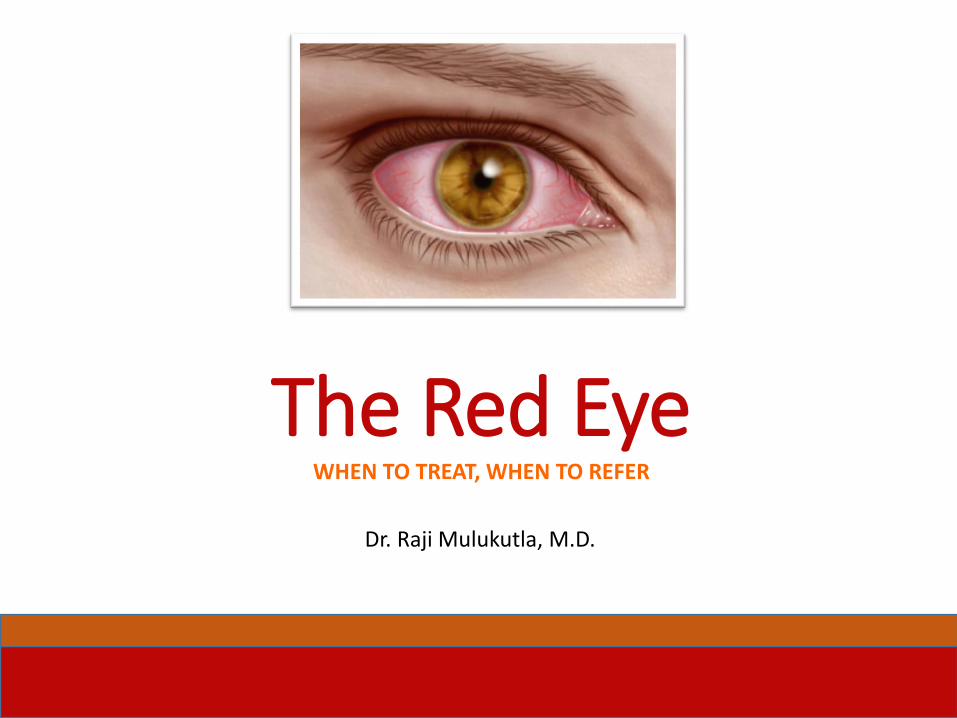
The Red EyeWHEN TO TREAT, WHEN TO REFER
Dr. Raji Mulukutla, M.D.
WHAT IS RED EYE?
• Red eye is a non-specific manifestation of a variety of ocular, orbital, and occasionally systemic diseases that commonly presents to PCP’s and Urgent Care Centers.
• The history and initial presentation can be key to a diagnosis. The complete differential diagnosis is VERY long.
• Always keep in mind that while it is unlikely, it CAN be a sign of a sight threatening, and even a life threatening, condition.

OBJECTIVES:
DIFFERENTIATE BETWEEN BENIGN AND VISION THREATENING CAUSES
MANAGEMENT (Benign)WHEN TO REFER (Vision Threatening)

THE SIMPLEST RULE OF THUMB
TREAT!• Present less than 3 days• Bilateral• No associated severe pain
and/or vision loss
REFER!• Present more than 3 days• Unilateral • Associated pain and/or vision loss• Contact lens • History of trauma
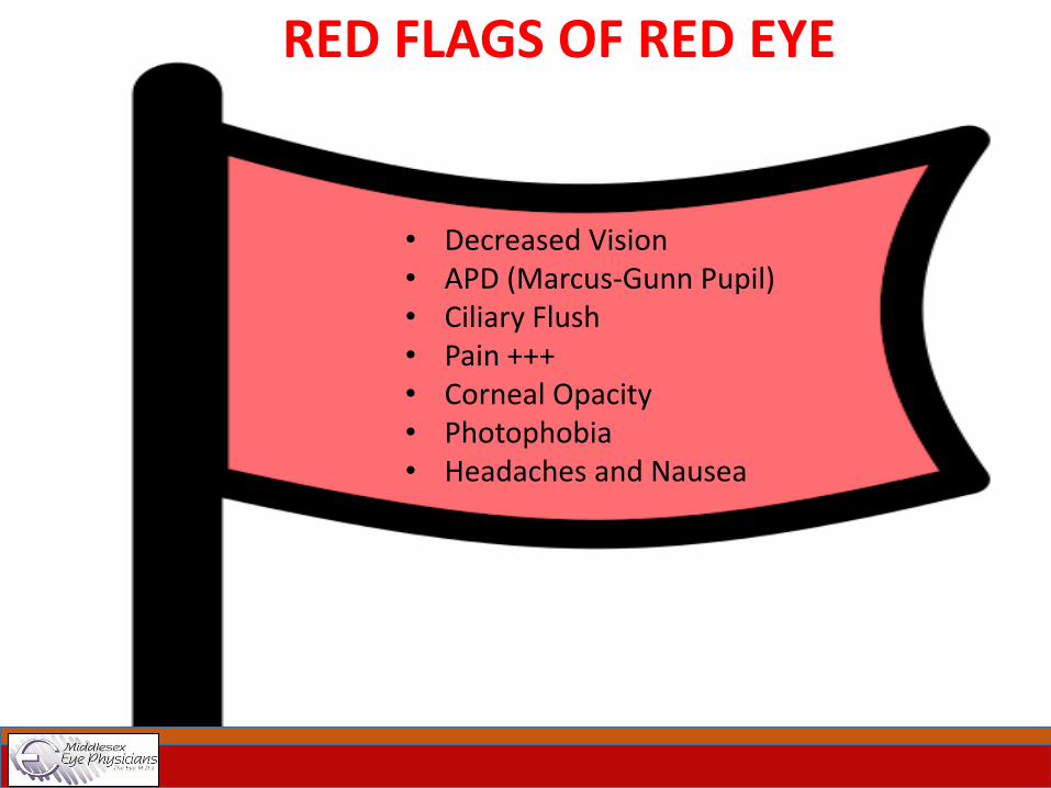
• Decreased Vision• APD (Marcus-Gunn Pupil)• Ciliary Flush• Pain +++• Corneal Opacity• Photophobia• Headaches and Nausea
RED FLAGS OF RED EYE
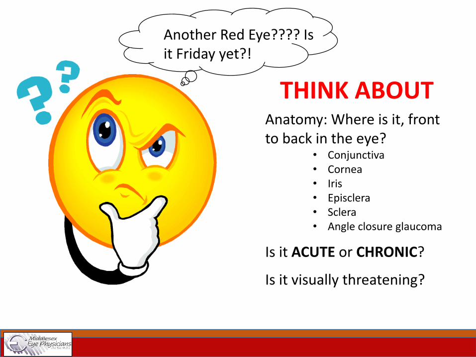
Anatomy: Where is it, front to back in the eye?
• Conjunctiva• Cornea• Iris• Episclera• Sclera• Angle closure glaucoma
Is it ACUTE or CHRONIC?
Is it visually threatening?
THINK ABOUT
Another Red Eye???? Is it Friday yet?!
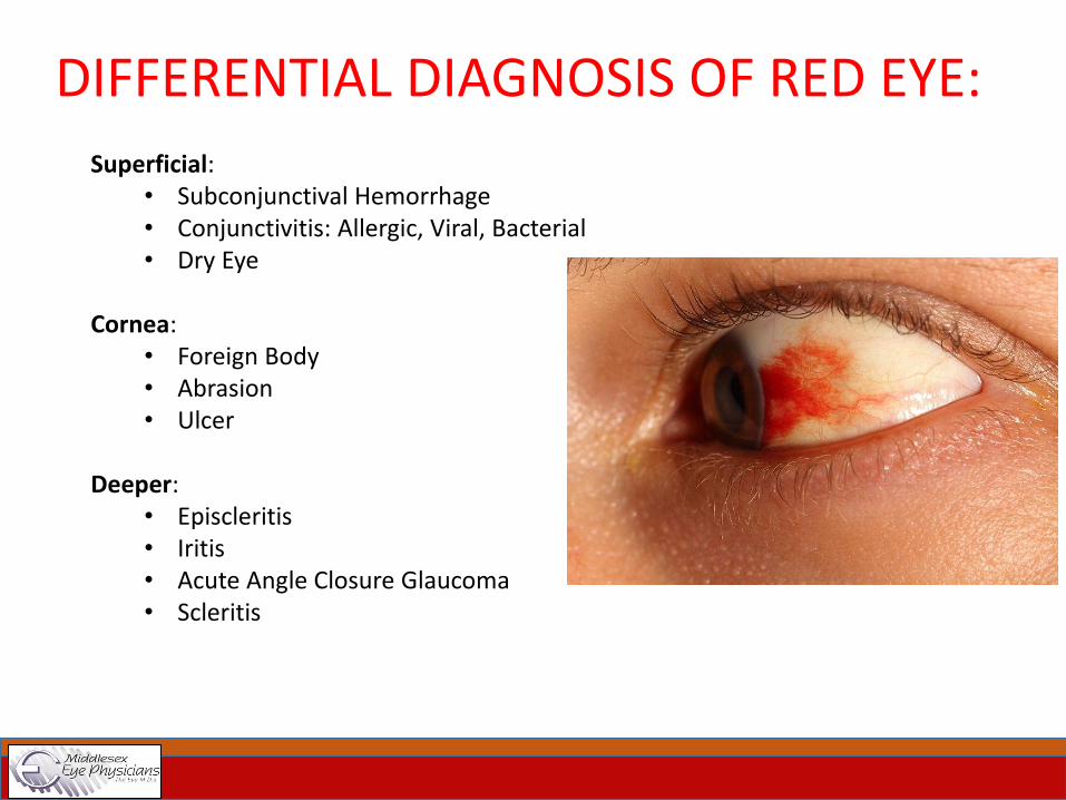
Superficial:• Subconjunctival Hemorrhage• Conjunctivitis: Allergic, Viral, Bacterial• Dry Eye
Cornea:• Foreign Body• Abrasion• Ulcer
Deeper:• Episcleritis• Iritis• Acute Angle Closure Glaucoma• Scleritis
DIFFERENTIAL DIAGNOSIS OF RED EYE:
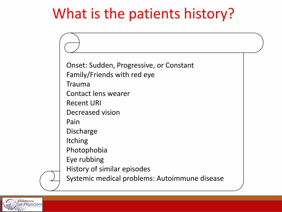
What is the patients history?
Onset: Sudden, Progressive, or ConstantFamily/Friends with red eyeTraumaContact lens wearerRecent URIDecreased vision PainDischargeItchingPhotophobiaEye rubbingHistory of similar episodes Systemic medical problems: Autoimmune disease
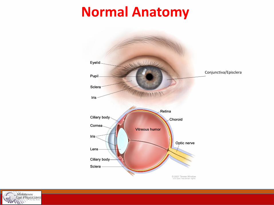
Normal Anatomy
Conjunctiva/Episclera
How Do We Approach Red Eye?
• Acute symptoms need quick attention to rule out vision threatening
complications
• If vision is affected, it can be serious and needs to be managed soon
• If pain is significant, the patient needs to be treated as quickly as
possible
• Minimal or no pain and no vision loss is NOT an acute emergency

SYMPTOMS
• Decreased VA: inflamed cornea, iritis, acute glaucoma• Pain: keratitis, ulcer, iritis, acute glaucoma• Photophobia: iritis• Colored Halos: acute glaucoma• Discharge: conj and/or lid inflammation, corneal ulcer
purulent/mucopurulent = bacterial
watery = viral
scant/white/stringy = allergy or dry eye
• Itching: allergy
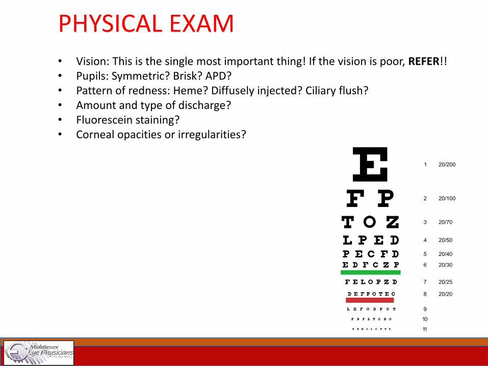
PHYSICAL EXAM• Vision: This is the single most important thing! If the vision is poor, REFER!!• Pupils: Symmetric? Brisk? APD?• Pattern of redness: Heme? Diffusely injected? Ciliary flush?• Amount and type of discharge?• Fluorescein staining?• Corneal opacities or irregularities?
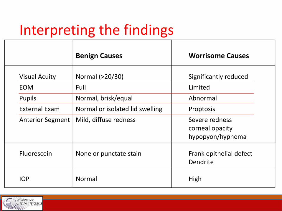
Interpreting the findings
Benign Causes Worrisome Causes
Visual Acuity Normal (>20/30) Significantly reduced
EOM Full Limited
Pupils Normal, brisk/equal Abnormal
External Exam Normal or isolated lid swelling Proptosis
Anterior Segment Mild, diffuse redness Severe rednesscorneal opacityhypopyon/hyphema
Fluorescein None or punctate stain Frank epithelial defectDendrite
IOP Normal High
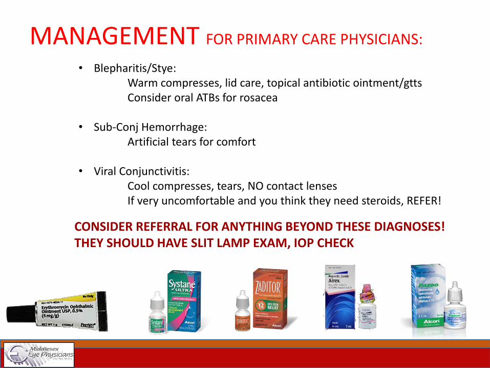
MANAGEMENT FOR PRIMARY CARE PHYSICIANS:
• Blepharitis/Stye: Warm compresses, lid care, topical antibiotic ointment/gttsConsider oral ATBs for rosacea
• Sub-Conj Hemorrhage:Artificial tears for comfort
• Viral Conjunctivitis:Cool compresses, tears, NO contact lensesIf very uncomfortable and you think they need steroids, REFER!
CONSIDER REFERRAL FOR ANYTHING BEYOND THESE DIAGNOSES!THEY SHOULD HAVE SLIT LAMP EXAM, IOP CHECK

IMPORTANT SIDE EFFECTS:
Topical AnestheticsOnly use them to aid in exam and diagnosis- NEVER dispense!
Patients love them and will ask for them!Inhibit epithelial growth and healing, decrease the blink reflexCan lead to dehydration, injury, infection
Topical CorticosteroidsDispense with extreme caution
Can make certain conditions worse - HSVCan mask symptoms - ScleritisOver the long haul, can lead to cataract formation and/orelevated IOP
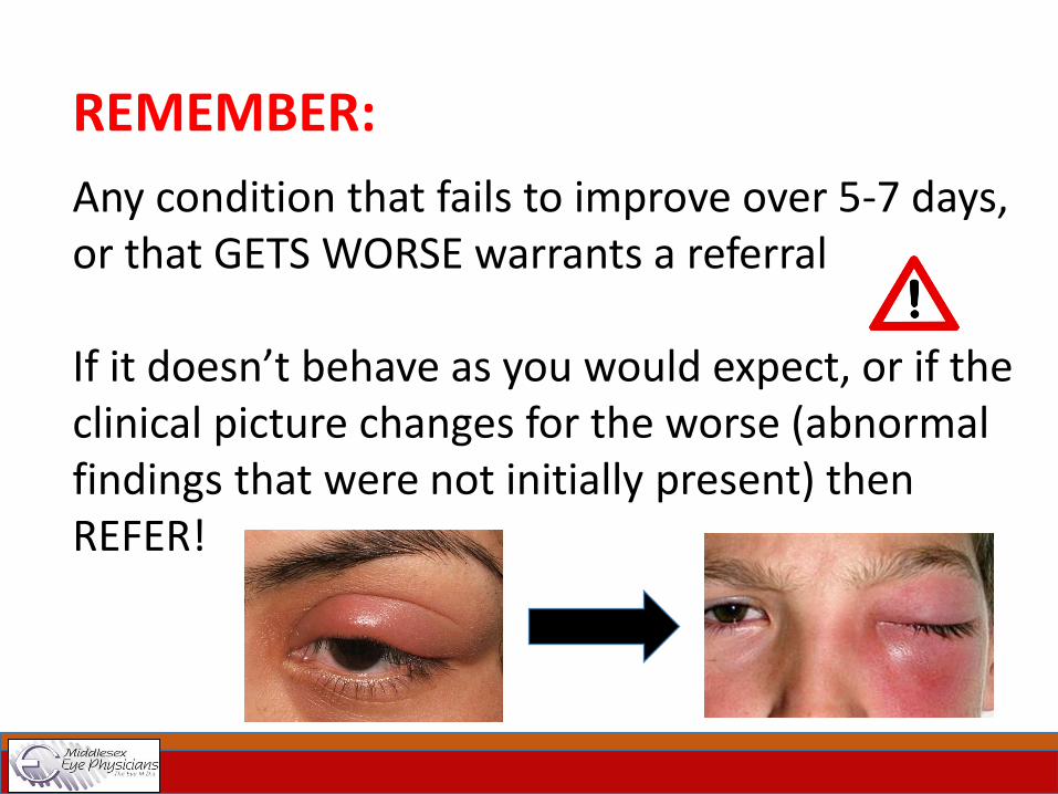
REMEMBER:
Any condition that fails to improve over 5-7 days, or that GETS WORSE warrants a referral
If it doesn’t behave as you would expect, or if the clinical picture changes for the worse (abnormal findings that were not initially present) then REFER!

Case Study• 21 year old college student with 2 day history of redness both eyes,
runny discharge and eyes crusted shut in the morning.
• Started with one eye, but now both eyes are crusty
What do we want to know???
• Cold?• Pink eye contacts? • Contact lens wearer?
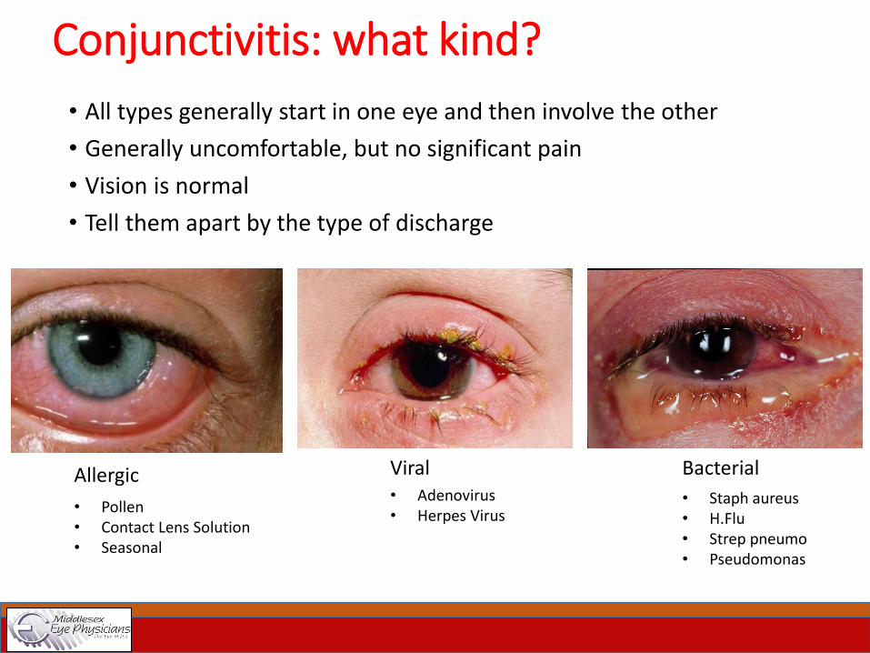
Conjunctivitis: what kind?
• All types generally start in one eye and then involve the other
• Generally uncomfortable, but no significant pain
• Vision is normal
• Tell them apart by the type of discharge
Allergic
• Pollen• Contact Lens Solution• Seasonal
Viral• Adenovirus• Herpes Virus
Bacterial
• Staph aureus• H.Flu• Strep pneumo• Pseudomonas
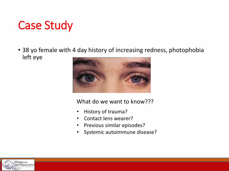
Case Study
• 38 yo female with 4 day history of increasing redness, photophobia left eye
What do we want to know???
• History of trauma? • Contact lens wearer? • Previous similar episodes?• Systemic autoimmune disease?

Case Study• 72 yo male, presents in acute distress
• c/o worsening pain right eye over past 12 hours with blurred vision, halos around lights
• Also c/o nausea and headache
• On exam: Vision is 20/400, cornea appears hazy, and the pupil dilated and sluggish
What should we do???
Take Home MessageR/O sight-threatening disorders in a patient with red eye
Refer if you have any concern about the severity of the disease
Pain and decreased vision is more concerning than isolated red eye
Always look for ocular signs before making a diagnosis, as symptoms are not always enough

Thank You!