
The Question of Virulence among the So-Called Pseudodiphtheria Bacilli
25
The Question of Virulence among the So-Called Pseudodiphtheria Bacilli Author(s): Alice Hamilton Source: The Journal of Infectious Diseases, Vol. 1, No. 4 (Nov. 5, 1904), pp. 690-713 Published by: Oxford University Press Stable URL: http://www.jstor.org/stable/30071790 . Accessed: 20/05/2014 03:34 Your use of the JSTOR archive indicates your acceptance of the Terms & Conditions of Use, available at . http://www.jstor.org/page/info/about/policies/terms.jsp . JSTOR is a not-for-profit service that helps scholars, researchers, and students discover, use, and build upon a wide range of content in a trusted digital archive. We use information technology and tools to increase productivity and facilitate new forms of scholarship. For more information about JSTOR, please contact [email protected]. . Oxford University Press is collaborating with JSTOR to digitize, preserve and extend access to The Journal of Infectious Diseases. http://www.jstor.org This content downloaded from 91.229.248.116 on Tue, 20 May 2014 03:34:37 AM All use subject to JSTOR Terms and Conditions
-
Upload
alice-hamilton -
Category
Documents
-
view
216 -
download
0
Transcript of The Question of Virulence among the So-Called Pseudodiphtheria Bacilli

The Question of Virulence among the So-Called Pseudodiphtheria BacilliAuthor(s): Alice HamiltonSource: The Journal of Infectious Diseases, Vol. 1, No. 4 (Nov. 5, 1904), pp. 690-713Published by: Oxford University PressStable URL: http://www.jstor.org/stable/30071790 .
Accessed: 20/05/2014 03:34
Your use of the JSTOR archive indicates your acceptance of the Terms & Conditions of Use, available at .http://www.jstor.org/page/info/about/policies/terms.jsp
.JSTOR is a not-for-profit service that helps scholars, researchers, and students discover, use, and build upon a wide range ofcontent in a trusted digital archive. We use information technology and tools to increase productivity and facilitate new formsof scholarship. For more information about JSTOR, please contact [email protected].
.
Oxford University Press is collaborating with JSTOR to digitize, preserve and extend access to The Journal ofInfectious Diseases.
http://www.jstor.org
This content downloaded from 91.229.248.116 on Tue, 20 May 2014 03:34:37 AMAll use subject to JSTOR Terms and Conditions

THE QUESTION OF VIRULENCE AMONG THE SO-CALLED PSEUDODIPHTHERIA BACILLI.*
Alice Hamilton.
The apseudodiphtheria bacillus," since the year 1887 when Loeffler and Hoffmann-Wellenhof isolated it, has been the subject of much controversy, and even now it is a matter of dispute whether the organism thus designated is simply an attenuated B.
diphtherise or, as Loeffler thought it, a member of the same group as the latter, but of a separate species. Behring, Ritter, Roux, and Yersin belong to the school which holds that there is no essen- tial difference between the virulent and non-virulent organisms, and recently Wesbrook in this country has appeared as a strong adherent of the same theory. On the other hand, the majority of bacteriologists, among them Loeffler, Hoffmann-Wellenhof, Escherich, Zarniko, Fraenkel, Spronck, Kruse, Fltigge, and Beck, hold that B. diphtheria belongs to a large group of organisms consisting of several distinct species. On the whole, in spite of
Behring's recent utterances, the trend of opinion is in the latter direction.
The earlier investigators thought it a comparatively easy matter to distinguish the pseudodiphtheria bacillus from B. diph- therise, for the former was said to have certain cultural and mor-
phological characteristics quite different from the latter, and also to differ from it in chemical activity and in virulence for animals. Of late, however, each one of these distinguishing characteristics has been called in question, and the distinction between the two
organisms, far from growing plainer, becomes less and less assured, until it seems questionable whether there is any one point which can always be relied upon to distinguish them.
Loeffler -J- and Hoffmann-Wellenhof thought that the appear- * Received for publication August 1,1901.
fit seems unnecessary to publish a bibliography of this subject, in view of the fact that there are already several fairly exhaustive ones appended to recent publications. The reader is referred to Schabad, Jahrb. f. Kinderheilk., 1901, 54, p. 381; LewAndowsky, Centralbl.f. Bakt., 1904, 36, p. 339; and, for English articles, to Graham-Smith, Jour, of Hyg., 1904, 4, p. 258.
690
(From the Memorial Institute for Infectious Diseases, Chicago.)
This content downloaded from 91.229.248.116 on Tue, 20 May 2014 03:34:37 AMAll use subject to JSTOR Terms and Conditions

Virulence among the Pseudodiphtheria Bacilli 691
ance of the growth upon solid media was enough to distinguish the pseudodiphtheria bacillus, but wTe find that, according to the
description given by Loeffler and also by Fraenkel, Barber, and
Sudeck, the growth on agar is scanty, grayish, translucent, while Hoffmann-Wellenhof and the greater number of investigators describe it as abundant, white, moist, and shining. Moreover, it is now generally recognized that B. diphtherise itself may vary so
greatly in its cultural characteristics as to be indistinguishable in this way alone from the allied bacteria. In a recent article
Lewandowsky has endeavored to simplify matters by emphasizing the typical cultural differences between the two forms,, but a care- ful comparison of the results obtained by different observers wTill convince any reader that variations from the usual* type are so
frequent as to render certainty on this ground alone impossible. Hoffmann described the pseudodiphtheria bacillus as shorter and thicker
than the bacillus of true diphtheria, and this statement is concurred in by Escherich and by most observers. Schabad insists that the morphology of the pseudodiphtheria bacillus from a twenty-four-hour serum culture is one of its most distinguishing characteristics. According to Schabad's descrip- tion, the bacillus in question is very different from the Klebs-Loeffler bacillus, not only shorter and thicker, but devoid of granules and bars, and arranged in parallel rows, in contrast to the radiating arrangement of B. diphtherias. On the other hand, Roux and Yersin, Kruse, Fraenkel, Schanz, and Wesbrook insist that it is impossible to distinguish the two by their appearance under the microscope, and Januszewska and Beck give the parallel arrangement of the rods as typical of the true, not the pseudo bacillus. Wesbrook, who has published the results of the study of 608 cases of clinical diphtheria, found that the short, solid bacilli, devoid of granules or bars, corresponding to the description usually given of the pseudodiphtheria bacillus, was often found in the later stages of diphtheria, sometimes in the earlier stages, and, in a few cases, was the only form present throughout the disease. An interesting view of the subject is given by A. P. Ohlmacher in his article " On Some Experimentally Produced Morphologic Variations in B. diphtheriae." Ohl- macher holds that the pseudodiphtheria bacillus is only a modified variety of B. diphtheriae, and that this modification may be brought about by a sojourn in the body of a relatively immune animal. A strain which originally con- sisted of barred and granular forms, changed to the short, solid type after passage through a white rat, but assumed its original character again after the passage through the highly susceptible guinea pig. Conversely, a strain originally composed of the short, solid, plump rods was changed to a typical B. diphtheriae, with greatly heightened virulence, by passage through a guinea pig.
The question as to the value of Neisser's stain is also unsettled. While Schabad, Franke, Auckenthaler, and Beck consider this a very valuable aid
This content downloaded from 91.229.248.116 on Tue, 20 May 2014 03:34:37 AMAll use subject to JSTOR Terms and Conditions

692 Alice Hamilton
to diagnosis, others declare it most unsatisfactory. Butschli demonstrated Neisser granules in several kinds of bacteria. Kurth isolated three strains of indubitable, virulent diphtheria bacilli which had no Neisser granules. Schwoner, Spronck, and Graham-Smith think the method is of little value, and Wesbrook has abandoned it in favor of Loeffler's methylene blue.
The fact, first observed by Zarniko, that the diphtheria bacillus causes an active production of acid in broth cultures, while the pseudodiphtheria bacillus causes either a faintly acid reaction followed quickly by an alkaline, or an alkaline reaction from the outset, has been regarded by some as an important, if not the most important, difference between the two organisms. Escherich has even made the statement that in the presence of an alkaline reaction it is unnecessary to test the pathogenicity of a culture for guinea pigs, for it is an absolute rule that the acid-producers are virulent and the non-acid-producers avirulent?a statement in which Graham-Smith concurs.
Exceptions to this rule, however, are numerous enough to invalidate it as an absolute test. Neisser, who agreed with Escherich as to all diphtheria bacilli forming acid, found an acid-producing, pseudodiphtheria bacillus. Spronck found that not all diphtheria bacilli form acid; Roux and Yersin, that both kinds form first acid and then alkali, the pseudodiphtheria bacillus forming alkali more rapidly than the true diphtheria. Slawyck and Manica- tide came to the same conclusion. Kurth, Peters, Lehmann and Neumann found pseudodiphtheria bacilli which formed as much acid as the true B. diphtheriae. Schabad and Neisser, however, think that by careful titration the two can always be distinguished.
Loeffler considered virulence for guinea pigs the most unvarying charac- teristic of the organism which has received his name, and Escherich con- curred in this. Edema of the subcutaneous tissue, infiltration or hemorrhage at the point of inoculation, pleuritic effusion, hyperemia and enlargement of the adrenals, hyperemia of liver and kidneys, without any change in the spleen, are the typical lesions produced by the injection under the skin of a few drops to one cubic centimeter of a broth culture of the bacillus diph- theriae. Death occurs usually in one to three days. By many bacteriolo- gists all virulent diphtheria-like organisms are considered true diphtheria bacilli; all non-virulent, pseudodiphtheria. That the question is not as simple as this, however, was shown by Roux and Yersin, who succeeded in artificially lowering the virulence of diphtheria bacilli; and by Brieger, Fraenkel, Lubow- ski, Schabad, and Wesbrook, who showed that typical bacilli from cases of virulent diphtheria sometimes prove non-virulent to guinea pigs. Nor is it true that the pseudodiphtheria bacillus is always non-virulent. Spronck, Fraenkel, Zupnik, Gelpke, and others find that a slight degree of virulence for guinea pigs is a property of many strains of the pseudodiphtheria bacillus, and that these strains cannot be distinguished by ordinary means from feebly virulent strains of the true diphtheria bacillus. Some observers even claim a pathogenicity for man in certain cases. Goldscheider isolated pseudodiph- theria bacilli from five cases of angina, with fever and swollen lymphatic glands, and thought these organisms the cause of the symptoms. Warnecke found similar bacilli in pure culture in three cases of otitis media, and in one of these cases he found the same organism in the exudate of meningitis and
This content downloaded from 91.229.248.116 on Tue, 20 May 2014 03:34:37 AMAll use subject to JSTOR Terms and Conditions

Virulence among the Pseudodiphtheria Bacilli 693
in metastatic abscesses in the lung. Kruse and Pasquale cultivated a pseudo- diphtheria bacillus (B. clavatus) from the organs of several cases of Egpptian dysentery, and Sanfelice found one in the pustules of smallpox. In all these cases the organisms were non virulent to guinea pigs.
Soon after the introduction of the agglutination test for the diagnosis of typhoid fever efforts were made to apply the method to the clinical diagnosis of diphtheria, but so far only one experimenter has done so with marked suc- cess. Schwoner succeeded in immunizing a horse in such a way as to produce a serum which agglutinated different strains of diphtheria bacilli in dilu- tions as high as 1:10,000, but not the pseudodiphtheria bacillus. Lubowski, by using non-virulent diphtheria bacilli, obtained a serum which agglutinated twenty-three other strains, but irregularly and not in dilutions over 1:160. Nicholas, Bruno, and Lesieur, after many experiments, declared the agglutina- tion test of no value.
Finally Martini tried to distinguish the true from the pseudo bacilli by the bactericidal action of diphtheria antitoxin on the diphtheria bacillus. He stated that the latter would not grow in such serum, while the pseudo- diphtheria bacillus would; but this has since been disproved. Fraenkel, Spronck, and Landsteiner have shown that the antitoxic serum is not bacteri- cidal to diphtheria bacilli, and that there is no difference between the two organisms in this respect. Graham-Smith endeavors to simplify the question by making three classes of organisms : first, B. diphtherias, which is always pathogenic to guinea pigs and always produces acid in sugar broth; second, the pseudodiphtheria bacillus, never pathogenic and never producing acid ; and, third, a new group, B. diphtheroides. It is not quite clear how Graham- Smith distinguishes this last from the pseudodiphtheria bacillus. Certainly the strains described by him differ not at all from many so-called pseudo- diphtheria bacilli in the literature.
Therefore, according to our present state of knowledge, we must either consider the Klebs-Loeffler bacillus, and those resembling it, as essentially the same organism, and their individual peculiarities as unimportant; or, if this seems not in accordance with our observations and we insist upon recognizing differerent varieties, we must admit that every method of differ- entiating B. diphtheriae from the closely allied forms has failed in the hands of one observer or another, and that we are still waiting for one which will be absolutely decisive.
According to Beck, and to the latest utterances of Loeffler and Spronck, it is impossible to distinguish the true from the pseudo bacillus by means of cultural peculiarities, morphology, presence of Neisser granules, chemical activity, or virulence for animals ; and the only hope for a sure method of differentiation lies in the application of specific sera to neutralize specific toxins. We know that in clinical experience the reaction to the diphtheria antitoxin is often a very valuable aid to diagnosis, for the failure of such a reaction shows that the symptoms in the case were not caused by the specific toxins of the diphtheria bacillus. Experiments to demonstrate the fact that the pseuodiphtheria bacillus does not form these toxins were made by Gluck- mann and by Neisser, who showed that animals treated by repeated injections of pseudodiphtheria bacilli were not thereby rendered immune to B. diphtheriae.
This content downloaded from 91.229.248.116 on Tue, 20 May 2014 03:34:37 AMAll use subject to JSTOR Terms and Conditions

694 Alice Hamilton
The most important and suggestive work in this direction has been done by Spronck. He proved that the xerosis and the pseudodiphtheria bacillus are not attenuated forms of B. diphtheriae, because they do not form the same toxins as the latter. He injected guinea pigs with cultures of xerosis and pseudodiphtheria bacilli which were feebly virulent, causing local edema, loss of weight, loss of appetite, etc.; and the same symptoms appeared in the animals which received a dose of Behring's antitoxin sufficient to protect against a lethal dose of the diphtheria bacillus, as in those which received no antitoxin, showing that the toxins formed by the xerosis and pseudo- diphtheria bacilli are not neutralized by the diphtheria antitoxin. This test, which seems both logical and conclusive; is rejected by Schabad, who still insists that differentiation between the two organisms must rest upon morphological and chemical properties, upon the difference in shape, in Neisser granules, and in acid productions in broth cultures. Spirig also rejecta Spronck's method, but on questionable grounds. Having isolated from the throat of a normal child a bacillus, resembling in appearance and growth the pseudodiphtheria bacillus, he tested it by Spronck's method, and found that, as in the case of Spronck's organisms, the Behring antitoxin did not protect against the effects produced by the bacillus. Nevertheless, Spirig continued to regard the organism as a feebly virulent B. diphtheriae, chiefly on the ground that it was found in the throat of a child exposed to diphtheria in the course of a rather extensive house epidemic of this disease.
The following is an account of the work donenn the Memorial Institute for Infectious Diseases, first by Dr. E. H. Ruediger and later by myself, in the course of which it has appeared probable that the method suggested by Spronck is the most satisfactory one for distinguishing B. diphtheriae from the pseudodiphtheria bacillus, and that by this method it is possible also to prove that the term "pseudodiphtheria" covers not only one bacillus, but a
group of bacilli.
During the months from March to July, 1903, Dr. E. H.
Ruediger succeeded in isolating from the throats of seven fatal cases of scarlatina, which came under his observation in the Memorial Institute for Infectious Diseases, organisms which resembled B. diphtheriae, yet differed from it in several important respects. I quote from Dr. Ruediger's description as follows:*
"Clinically these cases were variously diagnosed as diphtheria, gangrenous tonsilitis, and pharyngitis complicating scarlatina, black scarlet fever, and black diphtheria. The bacteriologic examination in each case made by Dr. Weaver showed "pseudodiphtheria bacilli." Five cases were treated with antidiphtheria serum, but the effects were always bad. All symptoms were greatly aggravated, an experience to which Johannessen called attention
* Trans. Chicago Path. Soc, 1903, 6, 45.
This content downloaded from 91.229.248.116 on Tue, 20 May 2014 03:34:37 AMAll use subject to JSTOR Terms and Conditions
VlKULENCE AMONG THE PSEUDODIPHTHERIA BACILLI 695
years ago. In two of the cases, Nos. 1 and 7, the effects of the antidiphtheria serum were so marked that it was looked upon as having materially shortened life. Case 7 died within eighteen hours after receiving 10 c.c. of serum valued at 4,000 units. The two cases which did not receive the antidiphtheria treatment died within a few hours after admission to the hospital.
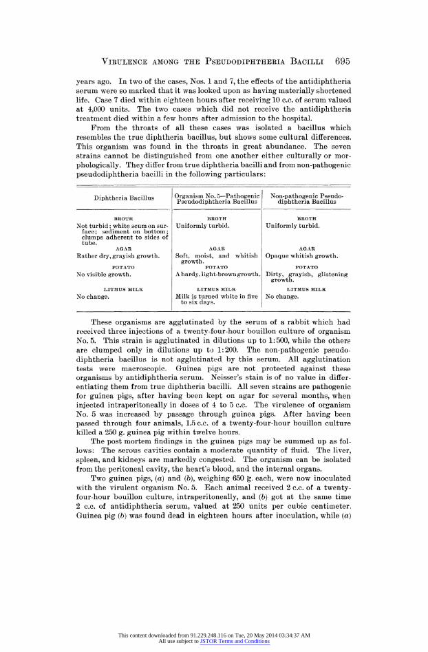
From the throats of all these cases was isolated a bacillus which resembles the true diphtheria bacillus, but shows some cultural differences. This organism was found in the throats in great abundance. The seven strains cannot be distinguished from one another either culturally or mor- phologically. They differ from true diphtheria bacilli and from non-pathogenic pseudodiphtheria bacilli in the following particulars:
BROTH BROTH BROTH Not turbid; white scum on sur- Uniformly turbid. Uniformly turbid,
face; sediment on bottom; clumps adherent to sides of tube.
AGAR AGAR AGAR Rather dry, grayish growth. Soft, moist, and whitish Opaque whitish growth.
growth. POTATO POTATO POTATO
No visible growth. A hardy, light-brown growth. Dirty, grayish, glistening growth.
LITMUS MILK LITMUS MILK LITMUS MILK No change. Milk is turned white in five No change.
to six days.
These organisms are agglutinated by the serum of a rabbit which had received three injections of a twenty-four-hour bouillon culture of organism No. 5. This strain is agglutinated in dilutions up to 1:500, while the others are clumped only in dilutions up to 1:200. The non-pathogenic pseudo- diphtheria bacillus is not agglutinated by this serum. All agglutination tests were macroscopic. Guinea pigs are not protected against these organisms by antidiphtheria serum. Neisser's stain is of no value in differ- entiating them from true diphtheria bacilli. All seven strains are pathogenic for guinea pigs, after having been kept on agar for several months, when injected intraperitoneally in doses of 4 to 5 c.c. The virulence of organism No. 5 was increased by passage through guinea pigs. After having been passed through four animals, 1.5 c.c. of a twenty-four-hour bouillon culture killed a 250 g. guinea pig within twelve hours.
The post mortem findings in the guinea pigs may be summed up as fol- lows: The serous cavities contain a moderate quantity of fluid. The liver, spleen, and kidneys are markedly congested. The organism can be isolated from the peritoneal cavity, the heart's blood, and the internal organs.
Two guinea pigs, (a) and (6), weighing 650 g. each, were now inoculated with the virulent organism No. 5. Each animal received 2 c.c. of a twenty- four-hour bouillon culture, intraperitoneally, and (b) got at the same time 2 c.c. of antidiphtheria serum, valued at 250 units per cubic centimeter. Guinea pig (b) was found dead in eighteen hours after inoculation, while {a)
Diphtheria Bacillus organisation no. 5-hjlojl Pseudodiphtheria Bacillus N(>n:Pathogenic Pseudo-
diphtheria Bacillus
This content downloaded from 91.229.248.116 on Tue, 20 May 2014 03:34:37 AMAll use subject to JSTOR Terms and Conditions

696 Alice Hamilton
was apparently healthy on the second day. Guinea pig (a) was now given 2 c.c. of antidiphtheria serum and died within twelve hours. This antidiph- theria serum protected guinea pigs against three or four times the minimum fatal dose of true diphtheria bacillus, control animals dying in twelve to twenty-four hours.
I now undertook to prepare an antitoxic serum for this organism. A large rabbit was injected at intervals of seven days with 1, 2, and 3 c.c. of a filtered bouillon culture of organism No. 5. A week after the third injection several cubic centimeters of blood were withdrawn from this rabbit, and also from a normal rabbit, allowed to clot, and the sera collected under aseptic precautions. Eight guinea pigs were now injected intraperitoneally with 3 c.c. of a twenty-four-hour bouillon culture of organism No. 5. Immediately after the inoculation, two of these animals were given 2 c.c. of antidiphtheria serum valued at 250 units per cubic centimeter. Two were given 2 c.c. of normal rabbit's serum, and two others 2 c.c. of the immune rabbit's serum. The remaining two animals received no treatment whatever after the inocu- lation. All of these animals died within twenty four hours, except those two which had been treated with the immune rabbit's serum. These two recovered."
A short abstract of this work of Dr. Ruediger's is given in the last article by Graham Smith, who dismisses it with the remark that " the author . . . . appears to use the term ' pseudodiphtheria' bacillus as equivalent to the non-virulent diphtheria bacillus." As the bacillus in question was shown to be decidedly virulent, and was proved to be not a strain of the Bacillus diph- therise?inasmuch as the diphtheria antitoxin failed to protect against it ? it is difficult to see why Graham Smith would have it designated as "a non- virulent diphtheria bacillus."
Dr. Ruediger's work was broken off at this point by his depar- ture for Manila, and at Dr. Hektoen's request I took it up, with a view to discover how often this organism could be found in cases of diphtheroid pharyngitis in scarlet fever. It so happened, however, that the scarlet fever in Chicago this year was of an
exceedingly mild type, and but two cases of pseudomembranous scarlatinal pharyngitis came under observation at the Memorial
Institute, both of which proved to be due to virulent strep- tococcal infection. For lack of this material, I undertook a routine examination of the throats of all the scarlatina patients who had angina of more than usual severity both in this hospital and the Cook County Hospital, making in all thirty-two cases between January 1 and June 1, 1904. Prom twelve of these, organisms resembling more or less B. diphtherise were isolated and studied. Eighteen cases of diphtheria, all but two of them from the Cook County Hospital, were also examined, and eight of
This content downloaded from 91.229.248.116 on Tue, 20 May 2014 03:34:37 AMAll use subject to JSTOR Terms and Conditions

Virulence among the Pseudodiphtheeia Bacilli 697
these proved atypical in one of two ways; either the organism isolated did not correspond to the usual description of B. diphtheriae, or the clinical history showed that the administration of diphtheria antitoxin had not been followed by favorable results. Four cases
diagnosed as diphtheria complicating scarlet fever were examined and pseudodiphtheria bacilli isolated from two; four of diphtheria complicating measles, with pseudodiphtheria bacilli in three; one case diagnosed as scarlet fever and measles combined, and three cases of measles with unusually severe pharyngitis, all of which
yielded pseudodiphtheria bacilli. Finally from one normal throat and from one case of tonsillitis, occurring in the course of acute
inflammatory rheumatism, bacilli of the same kind were isolated. The number of cases of scarlet fever in which the so-called
pseudodiphtheria bacillus was found is small?only eleven out of
thirty-one cases?but I do not think that this represents the real
proportion of positive cases. It is probable that in the earlier
part of the work a faulty technic was responsible for the failure to find pseudodiphtheria bacilli in a larger number of cases. The first cultures were made on Loeffler's blood serum, upon which medium the pseudodiphtheria bacillus usually grows more slowly than the diphtheria bacillus, but when once started grows more
abundantly than the latter. It happened several times that serum cultures twenty-four to forty-eight hours old, from a throat the smears from which had shown the presence of suspicious bacilli, would fail to show anything but cocci, and the cultures would then be rejected. Happening once to re-examine such a culture after seven days, I found it full of slender curved and straight rods with bipolar granules. After that, the first cultures were made on glycerin agar as well as upon serum, and, in case bacilli were not found in either, a loopful of the serum culture was trans- ferred to a tube of broth, and from this tube a loopful to the water of condensation at the bottom of a tube of glycerin agar, and the tube tipped to allow the water to run over the inclined surface of the agar. From such a culture colonies of bacilli could usually be obtained, and, in case of failure, the original serum culture was allowed to stand at room temperature for four or five days, and the procedure repeated. By this means it was found possible
This content downloaded from 91.229.248.116 on Tue, 20 May 2014 03:34:37 AMAll use subject to JSTOR Terms and Conditions

IAGRAM
NO.
1.
1
Tonsilli-
Short,
solid,
-
Same
as in 18
Abundant,
Abundant,
Scanty,
Cloudy,
Pure
culture
from
tis;
acute
wedge-
hrs.
butt*
.
white.
white.
alkaline,
tonsils.
articular
shaped
rods.
later
rheuma-
acid.
tism.
2
Measles
Short
and
me-
-
Do.
\ Grayish
colo-
Abundant,
\ No
growth.
Cloudy,
Slight
and
diph-
dium,
solid
nies.
white.
faintly
loss
of
theria.
and
barred
acid.
weight,
rods.
'
3
Do.
Do.
?
Do.
Slow,
white,
Abundant,
Slow,
scanty,
Cloudy,
Slight
Nos.
2 and
3 were
later
orange.
white,
then
orange.
alkaline.
loss
of
from
the
throats
orange.
weight.
of two
sisters;
no
membrane.
4
Scarlet
Slender,solid,
-
Typical
diph
Abundant,
Abundant,
Abundant,
Cloudy,
Obtained
by
aspi-
fever.
and
long
theria
colorless.
colorless.
colorless.
neutral.
ration
from
granular
bacilli.
swollen
tonsil,
rods.
5
Do.
Short,
solid,
?
Same
as in 18 \ Grayish
colo-
Abundant,
\ No
growth.
Cloudy,
wedge-
hrs.
nies.
white.
acid.
shaped
rods.
'
6
Do.
Short,
thick
-
Do.
\ Grayish
colo-
Scarify,
color-
No
growth.
Cloudy,
Severe
angina;
rods;
bipo-
nies.
less.
alkaline.
many
strepto-
lar
granules.
cocci.
7
Do.
Short,
wedge-
-
Do.
Grayish
colo-
Scanty,
color-
No
growth.
Cloudy,
shaped,
and
nies.
less.
alkaline.
long,
slender
rods.
8
Measles.
Short,
slen-
-
Do.
Moist,
color-
Scanty,
color-
No
growth.
Cloudy,
Severe
angina;
der,
pointed
less
smear.
less.
alkaline.
many
strepto-
1
1 rods.
||
|
|
III
cocci.
No.
Diagnosis
morphology 18 Hrs.
Serum
Morphology, ^-8
Days' Serum
Loeffler's Blood Semm
Glycerin Agar slant
Potato
broth 24 Hrs.
Virulence for
Gui-
nea
p^
Remarks
698 alice hamilton
granules neisser's
This content downloaded from 91.229.248.116 on Tue, 20 May 2014 03:34:37 AMAll use subject to JSTOR Terms and Conditions

9
1 Scarlet
1 Short
and
1
?
| Typical
diph-
1 Grayish
colo-
1 Scanty,
color-
\ No
growth.
1 Clear,
1
1 Tonsilitis;
rash
fever
?
long,
barred
theria
nies.
less.
acid.
atypical,
and
solid,
bacilli,
rods.
10
Diphthe-
Short,
wedge-
-
Short,
granu-
Abundant,
Abundant,
Abundant,
Cloudy,
[ Slight
Antitoxin
had
no
ria.
shaped,
and
lar.
and
creamy,
yellow.
yellow.
acid.
loss
of
effect
in this
swollen
rods.
solid;
long,
later
yellow.
weight.
case,
barred.
Long,
slen-
?
Typical
diph-
Moist,
colo^-
Scanty,
color-
Slow,
scanty,
Cloudy,
Bacillus
isolated
11
Do.
der,
clubbed
theria
less
smear.
less.
white.
acid.
after
membrane
and
spindle,
bacilli.
had
disap-
solid
rods.
peared.
12
Scarlet
Short,
granu-
+
Typical
diph-
\ Grayish
colo-
Abundant,
\ No
growth.
Cloudy,
fever.
larj
and
theria
nies.
white.
faintly
solid
rods.
bacilli.
acid.
13
Do.
Typical
diph-
+
Typical
diph-
Abundant,
Abundant,
Abundant,
Clear,
Obtained
in pure
theria
theria
creamy,
colorless.
yellowish.
acid.
culture
from
bacilli.
bacilli.
liquefying.
aural
discharge
in 3d week.
14
Do.
\ Typical
diph-\
+
Typical
diph-
\ Grayish
colo-
\ Gvayish
colo-
No
growth.
\ Clear,
Slight
Severe
angina;
no
theria
theria
nies.
nies.
acid.
loss
of
membrane.
bacilli.
bacilli.
weight.
15
Diphthe-
Slender,
-
Typical
diph-
Moist,
color-
Abundant,
Abundant,
Cloudy,
Slight
From
same
throat
ria.
barred,
and
theria
less
smear.
gray;
agar
gray,
alkaline,
loss
of
as No.
1 of 2d
solid
rods.
bacilli.
turns
dark
shining.
later
weight.
series.
brown.
acid.
16
Scarlet
Short,
slen-
?
Short,
slen-
Abundant,
Abundant,
Dry,
dark
Cloudy,
|
fever.
der,
solid
der
rods,
liquefying.
green;
agar
brown.
neutral,
1
rods;
few
granules.
turns
purple.
later
small
gran-
acid.
ules.
17
Do.
Short,
solid,
-
Same
as 18
Abundant,
Abundant,
Shining,
Cloudy,
long
barred
hrs.
liquefying,
green;
agar
reddish,
acid.
rods.
turns
purple,
abundant.
virulence among the pseudodiphtheria bacilli 699
This content downloaded from 91.229.248.116 on Tue, 20 May 2014 03:34:37 AMAll use subject to JSTOR Terms and Conditions

700 Alice Hamilton
to isolate pseudodiphtheria bacilli in all cases in which the ori-
ginal smears had shown their presence. Schabad has called attention to this slowly developing and finally abundant growth on serum of the pseudodiphtheria bacillus. Another source of error is the fact, pointed out by Kurth, that these bacilli, when
grown in mixed culture with cocci and other varieties, form much shorter rods than in pure culture. Indeed, they may sometimes look like lance-shaped cocci in the mixed cultures.
At the outset in such an examination one encounters the
question as to what is to be considered a pseudodiphtheria bacil- lus. Usually this term is applied to bacilli which resemble more or less B. diphtherise, but are shorter and thicker than the com- moner types of the latter. Many describe them as short, wedge- shaped rods, often lying in pairs with their bases facing each other (Wesbrook's Type D 2). Probably this is the same form as the one described by Graham-Smith: "darkly stained, oval, with one narrow, unstained septum." By others the typical pseudodiphtheria bacillus is said to be short and thick, and to contain granules whose diameter is smaller than that of the bacil- lus. There are, however, authors who give this name to bacilli which do not in the least resemble the bacillus of diphtheria and could never be mistaken for it?large, thick rods, devoid of bars or granules, either staining solidly or with a clear spot in the center. Such is the pseudodiphtheria bacillus of Spirig and of Schwoner, and such a bacillus is shown in one of the cuts in Schabad's article. These large rods with clear center were encountered
very frequently in the throat cultures examined by me, but they were ignored, for there seemed no grounds for considering them
pseudodiphtheria bacilli, as they do not resemble either morpho- logically or culturally the Klebs-Loeffler bacillus. Only those
organisms were isolated which corresponded to one or the other of the types described and pictured by Wesbrook, namely, granular or barred rods, of varying length or outline, and solid rods, slender, curved or straight, spindle-shaped, clubbed, or wedge- shaped.
Thirty-one such bacilli were isolated, fourteen of which proved pathogenic to guinea pigs, seventeen non-pathogenic. Diagram
This content downloaded from 91.229.248.116 on Tue, 20 May 2014 03:34:37 AMAll use subject to JSTOR Terms and Conditions

Virulence among the Pseudodiphtheria Bacilli 701
1 gives a tabulation of the seventeen non-pathogenic organisms. For brevity's sake, the term "typical diphtheria bacillus" is used to describe a polymorphous bacillus containing granules staining red by Loeffier's methylene blue and presenting the usual variety of forms, including spindles, clubs, and jointed rods. Deviations from the typical pseudodiphtheria bacillus are entered in italics.
Of all these seventeen organisms only two, Nos. 13 and 14, were diagnosed as diphtheria bacilli from the eighteen-hour serum culture. No. 13 was from the aural discharge of a scarlet fever
patient, and was found in pure culture. It differed so much in its cultural characteristics from the Loeffler bacillus, liquefying both gelatin and blood serum, that it could easily be distinguished from the latter.
No. 14 corresponded in morphology and culture with B. diph- theriae, but the clinical history did not point to diphtheria. There was no membrane in the throat, and none of the children in the same ward developed diphtheria. Two successive doses of this
organism?the first as large as 1 per cent, of the body weight, the second 2 per cent.?caused no symptoms in guinea pigs, only a slight loss of weight. It is, of course, possible that this was an attenuated B. diphtheriae.
No. 12 was hardly typical enough to be pronounced an undoubted B. diphtheriae, especially as clinical confirmation was
wanting. The short, solid forms predominated, nevertheless Neisser granules were present, and in its growth on all media
except agar it corresponded to B. diphtheriae. The other fourteen bacilli represented the forms which we are
accustomed to regard as typical of B. pseudodiphtheriae, although, as we have already seen, Wesbrook claims the solid wedges and slender rods as among the commonest types of the true diphtheria bacillus.
There is a great variety in the growth of these different organ- isms on solid media, some growing abundantly, like the typical pseudodiphtheria bacillus, others more like B. diphtheriae. Three of them, Nos. 13, 16, and 17, liquefied serum and gelatin, as did Klein's "B. diphtheroides," and Graham Smith's UB. diphthe- roides liquefaciens." Nos. 15, 16, and 17 differed from all the
This content downloaded from 91.229.248.116 on Tue, 20 May 2014 03:34:37 AMAll use subject to JSTOR Terms and Conditions

702 Alice Hamilton
others in turning agar a deep brownish or purplish color?a
property which both Prochaska and Escherich considered charac- teristic of the pseudodiphtheria bacillus.
Neisser granules were present in Nos. 12, 13, and 14 only. Seven of the seventeen developed in old serum culture typical diphtheria-like forms?a fact which Schabad has also noted of the organisms studied by him.
All were entirely non-pathogenic for guinea pigs, except Nos.
2, 3, 10, 14, and 15, which, in doses of 1.5 to 2 per cent, of the
body weight, caused a transient loss of weight. Apparently Nos.
1, 4, 9, and 13 were pathogenic to man. No. 1 was found in pure culture on the tonsils of a case of acute articular rheumatism with tonsillitis. A similar organism was found by Zarniko in a similar case. This pseudodiphtheria bacillus was typical in morphology and culture, and in absence of virulence for guinea pigs, yet it was apparently the cause of a severe tonsillitis in a woman.
No. 4 was obtained by aspiration of a swollen tonsil which was
supposed to be the seat of suppuration. The few clear drops thus drawn contained a pure culture of a bacillus resembling in many ways B. diphtheriae, but differing from it in the absence of Neis- ser's granules, in growth on potato, and in* absence of virulence for guinea pigs.
No. 9 was found in large numbers, mixed with a few cocci, in the throat of a child who was suffering from a severe tonsillitis with a slight red rash over the body. A positive diagnosis was never made in this case. The child had a history of several such
attacks, but she did not contract scarlet fever after several weeks'
sojourn in the scarlet-fever ward.
Finally, No. 13 was obtained in pure culture from the aural dis-
charge of a convalescent scarlet-fever patient. Seen in stained speci- mens it was apparently a typical B. diphtheriae, but the abundant, moist growth, the liquefaction of serum, and the absence of viru- lence for guinea pigs served to distinguish it from the latter.
Turning now to the sixteen pathogenic organisms, a word of
explanation is needed as to the method employed in determining virulence. Dr. Kuediger had found that his strains of pseudo- diphtheria bacillus killed guinea pigs of 500-600 g. in doses of 5
This content downloaded from 91.229.248.116 on Tue, 20 May 2014 03:34:37 AMAll use subject to JSTOR Terms and Conditions
Virulence among the Pseudodiphtheria Bacilli 703
c.c. of broth culture injected into the peritoneum. One organism, No. 5, he succeeded in rendering more virulent by successive
passages through guinea pigs, but when it reached my hands this increased virulence had already disappeared, and it required 4 c.c. to kill a 500 g. guinea pig. Following his example, I used
intraperitoneal injections instead of subcutaneous, beginning with a dose equal to 1 per cent, of the body weight, and increasing or
diminishing it according to the result. Post-mortem examination of the animals showed that the organisms produced two different
effects, one of which corresponds more to that caused by the intra-
peritoneal injection of diphtheria bacilli, the other to that caused
by the injection of Kuediger's bacillus. The first class caused death in forty-eight hours, rarely sooner, sometimes after four to eight days, in which case the animals became emaciated, and in one case (No. 6) showed typical post-diphtheritic paralysis. Animals
living more than three days usually yielded no cultures at all; those dying more quickly yielded cultures from the site of inocu-
lation, from the peritoneum, and rarely from the blood or organs. The changes found in these animals were usually infiltration or
hemorrhage at the site of inoculation, rarely pleuritic effusion, but usually a clear, reddish, or gelatinous exudate in the peri- toneum. The upper small intestine was collapsed and red, the liver and kidneys somewhat congested, the spleen unchanged, and the adrenals large and dark red.
Animals injected with Ruediger's bacillus, and organisms resembling it, died almost always within eighteen to twenty-four, hours. The body was extraordinarily limp, and there was seldom
any infiltration at the site of inoculation. The peritoneum con- tained a large quantity of clear, reddish fluid, the liver and
kidneys were much congested, the latter often dripping blood on section. The spleen was unchanged, and there was no change in the adrenals. The bacilli could be isolated from the peritoneal and pleural fluids, the blood, urine, and all the organs, and from the subcutaneous tissue in a few cases. This general bacteriemia occurred also when the injection was made into the subcutaneous tissue. According, therefore, to the effect on guinea pigs, we have here two distinct classes of organisms.
This content downloaded from 91.229.248.116 on Tue, 20 May 2014 03:34:37 AMAll use subject to JSTOR Terms and Conditions

DIAGRAM
NO.
2.
Dr.
Scarlet
Regular,
+
Same
as 18
Abundant
on
Cloudy,
Less
than
1#
From
all
or-
?
+
Ruedi-
fever.
short,
and
hrs.
all.
alkaline.
killed
in 24-
gans
and
ger's
long
granu-
48 hrs.
fluids.
No.
1.
tor.
1
Diph-
Short,
slender,
?
Typical
diph-
Like
diph-
Cloudy,
1% killed
in
From
peri-
+
?
theria.
solid
rods.
theria
ba-
theria.
acid.
48 hrs.
toneum;few
cilli.
colonies from
blood.
2
Measles
Short
and
?
Same
as 18
Abundant,
Cloudy,
1% killed
in 4
From
all
or-
+
?
and
scar-
long,
slender,
hrs.,
with
creamy
on
acid.
days;
2% in
gans
and
let
fever,
solid
rods.
short
granu-
serum
and
24 hrs.
fluids;
typ-
lar
and
long-
potato.
ical
diphthe-
barred.
ria
bacilli.
3
Diph-
Short,
solid
?
Typical
diph-
Like
diph-
Cloudy,
1^
killed
in 19
Not
recov-
?
theria.
rods.
theria
ba-
theria.
acid.
days;
with
ered.
cilli.
antitoxin killed
in 5
days.
4
Diph-
Short,
granu-
+
Typical
diph-
Abundant
on
Cloudy,
1\%
killed
in
Fromfluids
?
+
Antitoxin
had
theria.
lar,
and
theria
ba-
agar,
serum,
acid.
48 hrs.
and
organs;
no effect.
solid
rods.
cilli;
and
potato.
typical
diph-
branched.
theria
bacilli.
5
Diph-
Typical
diph-
+
Typical
diph-
Abundant
on
Clear,
1% in 3-11
From
seat
of
?
+
theria.
theria
ba-
theria
ba-
serum
and
acid.
days;
post-
inoculation,
cilli.
cilli.
agar,
none
diphtheritic
on potato.
paralysis.
6
Scarlet
Short,slender,
?
Short
rods,
Abundant,
Clear,
$% killed
in 5
Fromfluids
?
-f
Antitoxin
had
fever
and
solid
rods.
bipolar
yellowonall.
acid.
days;
$% in
and
organs.
no effect.
diph-
granules.
24 hrs.
theria.
7
Do
Short,
solid,
?
Same
as 18
Abundant,
Clear,
H#
killed
in
Fromfluids
?
-f-
Do
and
long
hrs.
with
few
white
on all.
acid.
18-24
hrs.
and
organs,
barred.
granular
Nn
Di^nn^i^
No.
Diagnosis
Morphology, ^^^^^
M?r
8P T>0 a1 v|y,
^J^T Serum
Growth
on
Solid
Media
Broth, 24 Hrs.
Virulence
for
Guinea
Pigs
Bacillus, Recovered
T^Pmark^ Remarks
704 alice hamilton
serum ruedinger
action to
action of anti- action
granules neisser's
This content downloaded from 91.229.248.116 on Tue, 20 May 2014 03:34:37 AMAll use subject to JSTOR Terms and Conditions

8
Normal.
Short,
solid,
?
Same
as 18
Abundant,
Cloudy,
1% in 24 hrs.
Fromfluids
~
+
wedge-
hrs.
creamy
or
alkaline.
and
organs,
shaped
rods.
white
on all.
9
Measles.
Short,
solid,
+
Typical
diph-
Like
diph-
Clear,
1% killed
in
Fromfluids
-
+
long
barred,
theria
ba-
theria
on
acid.
48 hrs.
and
organs,
and
granu-
cilli.
agar
and
lar.
serum; abundant
on
potato.
10
Scarlet
Short
and
-\-
Slender
Purple
on
Clear,
1% killed
in
Fromfluids
?
-\-
Obtained
in
fever.
long
solid,
granular.
potato;
turns
acid.
24 hrs.
and
organs.
pure
culture
few
banned,
agar
purple;
from
aural
and
granu-
liquefies
discharge.
lar.
serum.
11
Diph-
Typical
diph-
+
Typical
diph-
Like
diph-
Clear,
Less
than
1%
Fromfluids
?
+
Antitoxin
had
theria.
theria,
but
theria
ba-
theria.
acid.
killed
in 36-
and
organs.
no effect.
small
gran-
cilli.
48 hrs.
ules.
12
Diph-
Short
and
-
Solid
rods
of
Abundant,
Cloudy,
1 % killed
in
From
peri-
?
?
From
nose
of
theria.
long
solid,
all
shapes.
white
or buff
acid.
18 hrs.
toneum
and
case
four,
long
barred.
on all;
gas
organs.
pure
culture.
in glucose
agar.
13
Measles.
Slender,
regu-
?
Typical
diph-
Abundant,
Cloudy,
11%
killed
in
Fromfluids
?
-
From
same
lar,
solid
theria
ba-
white
or yel-
alkaline.
24 hrs.
and
organs.
throat
as
rods.
cilli.
low
on all
No.
9.
except serum;
gas
m glucose
agar.
14
Measles.
Short,
granu-
-
Same
as 18
Abundant,
Cloudy,
IX killed
in
Fromfluids
?
?
lar,
long
and
hrs.
yellow,trans-
acid.
24 hrs.
and
organs.
short
barred.
lucent
on
agar
and
po-
tato
; gas
in
glucose
agar.
virulence among the pseudodiphtheria bacilli 705
This content downloaded from 91.229.248.116 on Tue, 20 May 2014 03:34:37 AMAll use subject to JSTOR Terms and Conditions

706 Alice Hamilton
Whenever a bacillus had proved virulent to guinea pigs, two further experiments were made with it. A dose of diphtheria antitoxin, sufficient to protect against a lethal dose of diphtheria bacilli, was given to one guinea pig together with the ascertained lethal dose of the organism in question. At the same time a second guinea pig was given a lethal dose of the organism, and also a sufficient quantity of the serum of a rabbit immunized
against Ruediger's bacillus to protect against a lethal dose of the latter. The results of these experiments, as well as the cultural and morphological and chemical characteristics of the fifteen
organisms, are given in Diagram 2. For the sake of brevity, the pathological changes in guinea pigs are designated as
"diphtheria-like," or "like Ruediger;" the cultures on solid media are not given separately, and the description "like diphtheria" signifies grayish colonies on serum, rather scanty, translucent
growth on agar, no visible growth on potato, and a delicate growth in gelatin stab not spreading and not liquefying. Derivatives from the typical B. diphtherise are entered in italics.
It is necessary to add some details to the chief features shown in the diagram:
No. 1 was from a case of typical diphtheria, but the culture, taken on one of the first days of the disease, showed only the short, solid, wedge-shaped rods usually regarded as typical pseudodiphtheria bacilli. It was only in broth cultures that the bacilli developed bipolar granules. This case con- firms the statement of Wesbrook that the short, solid bacillus is sometimes the only form found in cases of clinical diphtheria. At first the growth on solid media was like that of a typical B. diphtherias, but later on it became increasingly luxuriant, creamy, moist, and spreading. No. 15 of the first series was isolated from this same patient.
The virulence of bacillus No. 1 was not great; 1.5 per cent, of the body weight was required to kill a guinea pig. The changes produced were those of experimental- diphtheria; the bacilli recovered were slender, barred and pointed at the ends. Diphtheria antitoxin protected guinea pigs against this bacillus.
No. 2 was from the throat of a girl who, while convalescing from measles, developed a typical scarlatinal rash, with rise of temperature and severe angina. The case was diagnosed as measles complicated with scarlet fever, and antitoxin was not administered. The tonsils and pharynx were swollen deep red, and covered with grayish patches. Cultures from the throat showed large numbers of short and long slender rods, without bars or granules although old cultures showed also short barred and granular forms. The creamy, moist, spreading growth on solid media resembled that of the Kue-
This content downloaded from 91.229.248.116 on Tue, 20 May 2014 03:34:37 AMAll use subject to JSTOR Terms and Conditions

Virulence among the Pseudodiphtheeia Bacilli 707
diger bacillus, and so did the effect upon guinea pigs. Doses of 1 per cent, of the body weight intraperitoneally killed in twenty-four to forty-eight hours, with lesions like those produced by Ruediger's bacillus, and the organism could be recovered from all fluids and organs. At the site of inoculation a hemorrhagic area was usually found, and in one instance, bacilli, identical in appearance (microscopic) with typical diphtheria bacilli, were found. Refer- ence has already been made to Ghlmacher's case, in which the organisms originally isolated were simply solid, short rods, but those recovered after passage through a guinea pig were typical diphtheria bacilli.
No. 2 resembled morphologically, culturally, and in the lesions produced by it the Ruediger bacillus, but differed from it in one essential point: the diphtheria antitoxin protected animals against it, while the Ruediger serum had no protective action. Therefore, in spite of many deviations from the usual type, one is forced to consider it a member of the B. diphtherias group.
No. 3, from a case of diphtheria, resembled No. 1 in that only short, solid rods grew on serum in the first eighteen hours. The cultures resembled macroscopically those of a scantily growing diphtheria bacillus, but in its pathogenesis the organism proved not to be a strain of B. diphtherias. It was injected into two guinea pigs, one of which received also the usual dose of antitoxin, and this animal died in five days, while that which had received no antitoxin lived for nineteen days. Far from protecting, the antitoxin seemed to act unfavorably, hastening the death of the animal. The virulence of this organism was slight and transient, for when an attempt was made later on to test the action of the Ruediger serum, it was found that the bacillus no longer killed guinea pigs. No. 3 is apparently a feeble virulent pseudo- diphtheria bacillus.
The following eight belong to the group of organisms designated by Rue- diger as "virulent pseudodiphtheria bacilli." They differ from each other culturally, morphologically, and in acid production, but have these two impor- tant characteristics in common: they all cause general bacteriemia in guinea pigs, and their effect on these animals is inhibited by Ruediger's serum, but not by diphtheria antitoxin.
No. 4 was from a case of pseudomembranous tonsillitis which was diag- nosed clinically as diphtheria. The membrane on the tonsils and the consti- tutional symptoms were typical, but injections of antitoxin ? 2,000 units administered twice ?had absolutely no effect, either locally or upon the general symptoms. The first serum cultures from the throat were not entirely typical, the rods being somewhat shorter and thicker than the usual forms. The growth on solid media was like that of a typical pseudodiphtheria bacillus; the effect upon guinea pigs was like that of the Ruediger bacillus and was neutralized by the Ruediger serum, but not by diphtheria antitoxin.
No. 4 is therefore a virulent pseudodiphtheria bacillus, and the failure of the diphtheria antitoxin to affect the clinical symptoms would seem to be explained.
No. 5 was in all respects a typical diphtheria bacillus, except that it was not neutralized by diphtheria antitoxin, but was neutralized by the Ruediger serum. Clinically this was a mild case of diphtheria. Antitoxin was admin- istered, but it is impossible to see from the history that any effect followed.
This content downloaded from 91.229.248.116 on Tue, 20 May 2014 03:34:37 AMAll use subject to JSTOR Terms and Conditions

708 Alice Hamilton
Nos. 6 and 7 may be considered together, as, except for slight cultural differences, they resembled each other closely. They were isolated from the throats of a father and son who entered the County Hospital with the diag- nosis of scarlatinal diphtheria. The father said that three other members of his family had had the same disease, one lightly, one much more severely, and the third fatally. In the case of the two in the County Hospital, the throat symptoms consisted in redness and swelling, but no membrane. The man, No. 7, had a high temperature and a vivid, "lobster-colored" rash; the boy had apparently a moderately severe case of scarlatina. Both were given antitoxin with no apparent effect. The organisms were virulent, No. 6 apparently less virulent for a man than No. 7; it was much more so for guinea pigs, proving fatal in doses as small as 0.2 per cent, of the body weight.
Nos. 6 and 7 both caused general infection in guinea pigs, both failed to be neutralized by the diphtheria antitoxin, and both were neutralized by the Ruediger serum.
No. 8 belongs to the same class as the foregoing five in pathogenesis for animals, and in the fact that it was neutralized by Ruediger's serum. Isolated from a normal throat, the organism, in morphology and cultural character- istics, was apparently a typical pseudo-diphtheria bacillus, virulent for guinea pigs, but causing no symptoms in man.
This is an additional proof that the virulence for man of a certain organ- ism cannot be determined with entire certainty by testing it on guinea pigs. In the first series of bacilli not pathogenic for guinea pigs we have seen that four were in all probability pathogenic for man. Here we have an instance of the converse.
No. 9, from a case of measles with unusually severe throat symptoms, is the sixth organism in this series belonging to the class of virulent pseudo- diphtheria bacilli which are neutralized by the Ruediger serum. The appearance under the microscope, of this bacillus, was that of the less usual type of B. diphtherias, but Neisser granules were present. It grew abundantly on potato.
No. 10 was obtained in pure culture from the ear of a scarlet fever con- valescent who developed an otitis media. Culturally it corresponded exactly to the organisms 16 and J 7 of the non-pathogenic series, liquefying serum, causing a browmsh-purple discoloration in agar slants and a growth like a smear of grape-jelly on potato. Morphologically it came nearer to the diphtheria bacillus; the rods were longer, and a few barred forms and Neisser granules were present. The virulence of this bacillus was not tested until fully five months after its isolation, when it was found to cause bac- teriemia in guinea pigs, fatal within twenty-four hours. Diphtheria antitoxin had no effect. Ruediger's serum afforded full protection.
No. 11 also belongs to this class. It was isolated from the throat of a child, who, without appearing very ill, had swollen tonsils, covered with a delicate, veil-like exudate. Smears from the throat showed typical diphtheria bacilli, and 2,000 units of antitoxin were administered. At this time the child was playing about the room, but soon after the injection of antitoxin his temperature rose, he became markedly worse, and died in less than twenty-four hours. Pure cultures from the throat on Loeffler's serum showed
This content downloaded from 91.229.248.116 on Tue, 20 May 2014 03:34:37 AMAll use subject to JSTOR Terms and Conditions

VlKULENCE AMONG THE PSEUDODIPHTHERIA BACILLI 709
short and long, straight and curved, rods with Neisser granules, which, how- ever, were of narrower diameter than the rods. On all culture media the growth resembled that of a diphtheria bacillus. Less than 1 per cent, of the body weight of a broth culture killed guinea pigs in twenty-four to forty- eight hours, producing a general bacteriemia and no change in the adrenals. Diphtheria antitoxin exerted no protective influence ; indeed, the animals injected with it died somewhat earlier than those without (six to eight hours). On the other hand, the Ruediger serum did protect, so that we have here again a virulent pseudodiphtheria bacillus of the same group as Ruediger's.
Five of these eight virulent pseudodiphtheria bacilli were from cases diagnosed as diphtheria and treated with antitoxin. In two of them, Nop. . 4 and 8, no alleviation of the symptoms could be seen, and in one, No. 11, the effect was apparently unfavorable. Nos. 5 and 6 were too mild for any effect to have been noted.
Nos. 12,13, and 14 differ from the other members of the pseudodiphther a group so far described in that they ferment glucose with the formation of gas. This property was discovered accidentally when glucose agar was substi- tuted for the usual glycerin agar. Morphologically they might easily pass for either pseudodiphtheria bacilli or the less usual types of diphtheria bacilli, especially in older serum cultures, when typical granular forms often develop. The cultures on all media except glucose agar might pass for pseudodiphtheria bacilli. They all produced general infection in guinea pigs, but they were not neutralized either by diphtheria antitoxin or by the Ruediger serum. No. 12 was obtained in pure culture from the nose of the patient whose throat yielded No. 4 of this series. No. 13 was found in the same throat as No. 9 of this series. No. 14 was from the throat of a case of measles with severe pharyngitis.
We have, then, twenty-nine organisms which are all, with the exception perhaps of the last three, pseudodiphtheria bacilli; that is, they correspond to some member of this loosely formed
group already described in the literature. They may be divided into at least three separate groups. First, there is the group of those which are non-pathogenic to guinea pigs?a group which
probably should be further subdivided, for some are apparently pathogenic to man, and there are certainly decided differences as far as cultural characteristics go. Yet a basis of sub-classifica- tion would be hard to find. Second, there is a well-defined group of organisms pathogenic to guinea pigs, producing a general bacteriemia, and neutralized by the serum of a rabbit immunized
against one member of the group. Third, the organisms which form gas in glucose media, produce bacteriemia in guinea pigs, and are neutralized neither by diphtheria antitoxin nor by the
pseudodiphtheria serum.
This content downloaded from 91.229.248.116 on Tue, 20 May 2014 03:34:37 AMAll use subject to JSTOR Terms and Conditions
710 Alice Hamilton
The second class is much the most important, for it is apparently the most pathogenic to man, and contains those organisms which are most apt to be mistaken for the diphtheria bacillus.
Including the seven strains isolated by Dr. Ruediger, there have been fifteen organisms belonging to this group obtained
during a little more than a year's time in this laboratory from clinical material which was far from abundant. The cases were scattered through the year and were not connected, except those of the family to which Nos. 6 and 7 belonged.
Five of these fifteen organisms would not have been discovered save as a result of a routine experimental study of the bacteria of a certain number of throats, for there was nothing unusual in the
symptoms of the cases in question. These were, the bacillus from the normal throat, the one from the pharyngitis of measles, the one from the ear in scarlet fever, and the two from the mild cases of supposed diphtheria.
The other ten were from cases in which the clinical symptoms pointed to diphtheria, but in which the diphtheria antitoxin failed to alleviate, if it did not aggravate, the condition present.
A careful search through the literature fails to bring to light instances of diphtheria in which the injection of antitoxin proved ineffectual or deleterious, and in which a thorough bacteriological examination was made with control experiments on animals. Where such instances are mentioned?and even a passing men- tion is rare?it is assumed that they must have been due to "mixed infection," the object apparently being to lay no stress on
anything which might throw doubt on the absolute harmlessness of diphtheria antitoxin. Undoubtedly, many cases of fatal
diphtheria and of diphtheroid pharyngitis in scarlet fever are due to the pyogenic cocci. This has been proved over and over. Yet the experience in this laboratory shows that a certain, and a not insignificant, proportion of these cases is due to invasion by a bacillus morphologically and culturally similar to B. diphtherise, but of an absolutely distinct variety.
It is a question whether in our cases the antitoxin acted more
deleteriously than it often does in perfectly normal persons, when
given for the purpose of immunization. Johannesen has shown
This content downloaded from 91.229.248.116 on Tue, 20 May 2014 03:34:37 AMAll use subject to JSTOR Terms and Conditions

VlEULENCE AMONG THE PSEUDODIPHTHEEIA BACILLI 711
that a large proportion of normal persons react more or less
severely to even small doses of horse serum. Alfoldi* reports a case ending fatally on the fourth day after injection, the child
having been entirely healthy at the time of the injection. In the case of the sick it is, of course, impossible to say how much the
aggravation of the symptoms is due to the course of the disease itself and how much to the administration of the antitoxin. In Nos. 1 and 7 of Dr. Ruediger's cases, and in the one fatal case of
my series?No. 11?there was a strong suspicion that death was hastened by the antitoxin.
It is much to be regretted that no bacteriological examination was made of the blood and organs of these fatal cases. Judging from the effect on guinea pigs, it seems probable that a general bacteriemia would be found in such cases. This, if it proves true, will lead one to ask if it is not possible that some of the instances
reported as "general infection by B. diphtherise" may not belong in this class instead. Such cases have been reported by Councilmen, Wright, and others. Wright succeeded in recovering the organ- isms from the blood and organs of guinea pigs injected with the cultures from some of his cases. I find, however, only one report of experiments to prove the exact nature of the bacillus isolated.
Stephens and Parfitt, who point out this missing link in the
literature, showed that in their cases diphtheria antitoxin afforded
protection to guinea pigs against a lethal dose of the organism. In Howard's famous case of acute ulcerative endocarditis,
without diphtheria, pure cultures of a diphtheria-like organism were isolated from the vegetations on the valves of the heart, and from the spleen and kidneys, but the bacillus proved to be non-
pathogenic for guinea pigs. A rapid method of diagnosis remains to be found between B.
diphtherise and the virulent pseudodiphtheria bacillus?an awkward name which has nothing to recommend it but the lack of a better one. Our results confirm the experiments of Spronck, and show that animal experiments are the only sure means of distinguishing the two; but this method is obviously not adapted to a prompt diag- nosis in cases where a question arises as to the administration of
*Berl. klin. Wchnschr., 1895, p. 285.
This content downloaded from 91.229.248.116 on Tue, 20 May 2014 03:34:37 AMAll use subject to JSTOR Terms and Conditions

712 Alice Hamilton
antitoxin. As we have seen, morphology and presence or absence of Neisser granules are of little value as diagnostic points. The formation of acid or alkali in broth cultures is subject to too
many variations to be depended upon absolutely. Schabad is
perhaps the most enthusiastic advocate of this method of diag- nosis, and his figures show that if the average obtained from a
large number of strains of B. diphtherise is compared with the
average obtained from a large number of pseudodiphtheria bacilli, the contrast is very striking. But single instances of the former
may produce acid very feebly, while the latter may, during the first forty-eight hours?the time when it is most important to make a diagnosis?produce a decided amount of acid. The fol-
lowing results were obtained by titration of nineteen pathogenic bacilli, pseudodiphtheria bacilli of my own series and of Dr.
Ruedigers, Schabad's method having been carefully followed. Schabad gives the amount of acid produced by B. diphtherise,
as follows: Two days, 5 to 22; four days, 0 to 24; seven days, 10 to 21; the numbers signifying cubic centimeters of normal sodium hydroxide solution required to neutralize one liter of broth culture.
The same method was used in the titration of broth cultures of the strains isolated by Dr. Ruediger and the pathogenic strains in my own series, with the following results:
2 Days 4 Days
Dr. Euediger's series | 1 1 7 1.5 2 7 0.5 3 4 2.5 4 2 0.5
? 5 1 1 6 0 1 7 -1 -1
My series pathogenic 1 6 1.5 2 10 3 3 4 5 4 6 7 10 7 6 1 6 8 2 3 9 4 3
10 4 2 11 7 9 12 7 1 13 2 12 14 4 7
This content downloaded from 91.229.248.116 on Tue, 20 May 2014 03:34:37 AMAll use subject to JSTOR Terms and Conditions

VlEULENCE AMONG THE PSEUDODIPHTHEEIA BACILLI 713
These figures show that, although, on the whole, the pseudo- diphtheria bacillus forms less acid in forty-eight hours than B. diph- therise, yet there are strains?no less than nine in the above series? which produce as much as the less vigorous acid-producers among the former. By four days the difference is much more marked, but here, too, there are exceptions, four of the pseudodiphtheria strains still producing large quantities of acid after four days.
In point of accuracy and promptness, then, the production of acid in broth cultures fails as a method of diagnosis between the two organisms, and a more rapid and sure method is demanded. It will be my aim in continuing this study to work toward the dis-
covery of such a method, and also to ascertain the exact nature of the immune serum obtained by injecting rabbits with cultures of these strains of the virulent pseudodiphtheria bacillus.
I take pleasure in acknowledging my indebtedness to Dr.
Hektoen, not only for suggesting this investigation, but for many helpful suggestions in carrying it out.
This content downloaded from 91.229.248.116 on Tue, 20 May 2014 03:34:37 AMAll use subject to JSTOR Terms and Conditions


![BovineTuberculosisandtheEstablishmentofanEradication ...humans to demonstrate that human bacilli possessed a low virulence for cattle [11]. Smith should be credited with being one](https://static.fdocuments.net/doc/165x107/60ef4537ac4303208b21c332/bovinetuberculosisandtheestablishmentofaneradication-humans-to-demonstrate-that.jpg)