The pyridine nucleotide and non-pyridine nucleotide dependence of L-glutamate dehydrogenase in the...
17
Histochemistry53, 117- 133 (1977) Histochemistry by Springer-Verlag 1977 The Pyridine Nucleotide and Non-Pyridine Nucleotide Dependence of L-Glutamate Dehydrogenase in the Histochemical System Helge Andersen and Antonio Contestabile* Laboratory of Cyto- and Histochemistry,Anatomy Department A, University of Copenhagen, Universitetsparken 1, DK-2100 Copenhagen, Denmark Institute of Comparative Anatomy, University of Bologna, Via Belmeloro 8, 1-40126 Bologna, Italy Summary. Under histochemical conditions (fresh frozen sections from liver, kidney and cerebellum of the rat) it was shown that the oxidation of L-glu- tamic acid was carried out by the NAD-dependent L-glutamate dehydrogen- ase (E.C. 1.4.1.2) and/or the NAD- or NADP-dependent L-glutamate dehy- drogenase (E.C. 1.4.1.3) as well as by an enzyme system which is not depen- dent on externally added NAD, NADP, FAD, FMN or CoQ10 for activity. This non-pyridine dependent activity was related to the L-glutamate dehy- drogenases proper, owing to the following: a) the localization of activity noticed corresponds to that obtained with the NAD- or NADP-containing media, b) the incubation time needed for initial formation of red and blue formazans is similar to that with coenzyme-containing media, c) pre-extrac- tion experiments reveal similarity in enzyme diffusion rates, d) the named activity is influenced by the same agents and to the same extent as the activity obtained by the inclusion of NAD or NADP (e.g. dissociation of the dehydrogenase molecule into subunits due to urea, inhibition of activity due to N-ethyl maleimide and 1.10-phenanthroline, activation due to the allosteric effect of ADP and to high substrate concentration, allosteric inhibi- tion caused by GTP and inhibition caused by c~-ketoglutaric acid, no inhib- itory effect of KCN), and e) the named activity was not affected by added PMS (excluding activity due to L-aminoacid oxidase). In the in situ localization of enzyme activity it was found that L-glutamate dehydrogenases E.C. 1.4.1.2 and E.C. 1.4.1.3 co-exist in the cells of kidney and cerebellum, while the L-glutamate dehydrogenase E.C. 1.4.1.3 only was present in liver cells. Finally, it was stated that incubation time should be kept as short as possible in order to avoid "Nothing dehydrogenase" reaction as well as inhibition due to accumulation of c~-ketoglutaric acid. Only "gel" incubation media should be applied. * Recipient of a research grant from the Danish Ministryof Education
-
Upload
helge-andersen -
Category
Documents
-
view
212 -
download
0
Transcript of The pyridine nucleotide and non-pyridine nucleotide dependence of L-glutamate dehydrogenase in the...

Histochemistry 53, 117 - 133 (1977) Histochemistry �9 by Springer-Verlag 1977
The Pyridine Nucleotide and Non-Pyridine Nucleotide Dependence of L-Glutamate Dehydrogenase in the Histochemical System
Helge Andersen and Antonio Contestabile* Laboratory of Cyto- and Histochemistry, Anatomy Department A, University of Copenhagen, Universitetsparken 1, DK-2100 Copenhagen, Denmark Institute of Comparative Anatomy, University of Bologna, Via Belmeloro 8, 1-40126 Bologna, Italy
Summary. Under histochemical conditions (fresh frozen sections from liver, kidney and cerebellum of the rat) it was shown that the oxidation of L-glu- tamic acid was carried out by the NAD-dependent L-glutamate dehydrogen- ase (E.C. 1.4.1.2) and/or the NAD- or NADP-dependent L-glutamate dehy- drogenase (E.C. 1.4.1.3) as well as by an enzyme system which is not depen- dent on externally added NAD, NADP, FAD, FMN or CoQ10 for activity.
This non-pyridine dependent activity was related to the L-glutamate dehy- drogenases proper, owing to the following: a) the localization of activity noticed corresponds to that obtained with the NAD- or NADP-containing media, b) the incubation time needed for initial formation of red and blue formazans is similar to that with coenzyme-containing media, c) pre-extrac- tion experiments reveal similarity in enzyme diffusion rates, d) the named activity is influenced by the same agents and to the same extent as the activity obtained by the inclusion of NAD or NADP (e.g. dissociation of the dehydrogenase molecule into subunits due to urea, inhibition of activity due to N-ethyl maleimide and 1.10-phenanthroline, activation due to the allosteric effect of ADP and to high substrate concentration, allosteric inhibi- tion caused by GTP and inhibition caused by c~-ketoglutaric acid, no inhib- itory effect of KCN), and e) the named activity was not affected by added PMS (excluding activity due to L-aminoacid oxidase).
In the in situ localization of enzyme activity it was found that L-glutamate dehydrogenases E.C. 1.4.1.2 and E.C. 1.4.1.3 co-exist in the cells of kidney and cerebellum, while the L-glutamate dehydrogenase E.C. 1.4.1.3 only was present in liver cells.
Finally, it was stated that incubation time should be kept as short as possible in order to avoid "Nothing dehydrogenase" reaction as well as inhibition due to accumulation of c~-ketoglutaric acid. Only "gel" incubation media should be applied.
* Recipient of a research grant from the Danish Ministry of Education

118 H. Andersen and A. Contestabile
Introduction
The oxidat ion o f L-glutamic acid in biological systems of animal origin is usually carried out by two mitochondria l matrix enzymes: 1. the NAD+-dependen t L-glutamate dehydrogenase (E.C. 1.4.1.2) and 2. the non-specific N A D +- or N A D P + - d e p e n d e n t L-glutamate dehydrogenase (E.C. 1.4.1.3). The great majo- rity o f data concerning these enzymes have been obtained by biochemical in vitro investigations (for review see Smith et al., 1975), while only few (3) papers have dealt with the characterization of the enzymes f rom a histochemical point o f view (e.g. Braun-Falco and Petzold, 1965) concerning the effect of alterations in concentra t ion of substrate, coenzymes, te t razol ium salt, buffer and hydrogen ion; Toledano and Mart inez-Rodr igues (1970) and Mart inez-Rodr igues (1974) concerning the effect of different activators or inhibitors (ADP, ATP, GTP, c~-oxoglutaric acid and oxalacetic acid) on the N A D - d e p e n d e n t enzyme only).
In the present study it is shown that in fresh frozen sections (with and without polyvinyl alcohol (PVA) as a tissue stabilizer) the L-glutamate dehydro- genase does not require the addit ion o f any pyridine nucleotides for activity. The localization of this activity corresponds to that obtained by the use of N A D or N A D P , and the activity is influenced by the same agents (e.g. urea, N-ethyl maleimide, 1.10-phenanthroline, A D P , GTP, e-ketoglutaric acid) as the activity noticed when N A D or N A D P is added, but is no t influenced by the presence of added PMS, F A D , F M N and/or coenzyme Q10.
Fur thermore , it is the aim of the present study to reevaluate some of the most critical steps in the histochemical procedure by the applicat ion of a series of control experiments (concerning diffusion of enzymes and/or reduced interme- diates; "No th ing dehydrogenase" reaction etc.) in order to improve the tech- nique and by that means the reliability of the in situ localization o f enzyme activity.
Materials and Methods
The materials (liver, kidney and cerebellum) were obtained from 3 months old male and female rats of Wistar strain kept under standard laboratory conditions. The animals were sacrified by decapitation and the organs were removed immediately and cut into appropriate blocks (< 5 mm in thickness). These were frozen in a COz expansion cooler with covering device (Slee) and cut in a Pearse-Slee cryostat (type HR) at -18~ to -25~ in 6 microns sections. In some cases corresponding blocks were fixed for 5 min in 0 4 ~ C 1% methanol-free formaldehyde (prepared from paraformaldehyde) buffered to pH 7.2 preceding the freezing procedure. Sections from these blocks were used for N-ethyl maleimide blockage of SH-groups in the double-section incubation method (Andersen and Hoyer, 1974).
Sections were collected on slides and incubation was performed at 37~ in a thermo-regulated water bath incubator to avoid evaporation of incubation media. With the exception of media containing phenazine methosulphate (PMS), the incubation time needed for appearance of both the red and the blue formazans was recorded by the microscope at x 100 magnification (Andersen and Hoyer, 1974; Hoyer and Andersen, 1977). When PMS was included, the incubation was performed in red light. Furthermore, exposure of PMS to daylight or ordinary artificial light was avoided during the preparation of the PMS-containing media (Marzotko et al., 1973). The incubation was interrupted after different intervals (cf. below). The pH-values of the incubation media were tested in a pH-meter with a micro electrode unit (Radiometer).

Pyridine Nucleotide Dependence of L-Glutamate Dehydrogenase Activity 119
Penetration o f Nitro BT and Substantivity o f Formazans
Fresh frozen sections were incubated for 15 min in Nitro BT (0.25 mg/ml) in 0.05 M phosphate buffer at pH 7.2. Excess medium was decanted and the sections were briefly rinsed in the buffer. The tetrazolium salt was then converted to formazan by treating the sections for 10 min with the reducing agent sodium dithionite (10% aqueous solution) or 10% ascorbic acid (adjusted to pH 8,0 by NaOH). The sections were briefly rinsed in distilled water and embedded in glycerol- gelatine.
In ali the experiments Nitro BT from Merck has been used since this sample has proved to be of a good quality (Altman, I976a).
NAD(P) H Tetrazolium Reductase
Due to the fact that the animal L-glutamate dehydrogenases (GDH) characterized to date are 1) the NAD+-dependent L@utamate dehydrogenase (E.C. 1.4.1.2) and 2) the non-specific NAD +- or NADP+-dependent L-glutamate dehydrogenase (E.C. 1.4,1.3) the activity pattern of the two tetrazolium reductases was recorded by using an ordinary aqueous medium as well as a gel medium (containing 22% w/v PVA, grade BO5/140 (Altman, 1971)) and with 1 mg/ml NADH or NADPH as substrate (Andersen and Hoyer, 1974). Furthermore, in the gel medium KCN was added as a terminal block (cf. below). Incubation time was 5-15 rain. Since NADPH can be converted to NADH by present non-specific alkaline phosphomonoesterase (Dixon and Webb, 1964; Leeflang- de Pijper and Hfilsmann, I974), 64 mM /3-glycerophosphate was added to each mI of incubation medium containing NADPH (Hoyer and Andersen, 1977). The diffusion of enzyme as well as the effect of SH-blocking agent was investigated in accordance with a previous study (Andersen and Hoyer, 1974). Finally, due to the use of 1.10 phenanthroline as a possible inhibitor of GDH- activity, the influence of this agent on NAD(P)H tetrazolium reductase activity was investigated (cf. below).
L-Glutamate Dehydrogenases (E.C. 1.4.1.2 and E.C. 1.4.1.3)
The following was used as an ordinary aqueous incubation medium ." sodium L-glutamate (0.1 M) Sigma), Nitro BY (0.5 mg/mi) (Merck), NAD or NADP (0.1 mg/ml (Sigma) KCN (0.66 mg/ml), 0.05 M phosphate buffer (pH 7.2) made up to 1 ml incubation medium. The KCN was added as 0.05 ml of a solution containing 13.2 mg KCN in 1 ml 0.1 M phosphate buffer (pH 7.2) (adjusted with 0.058 ml 3.1 N HC1). As a standard get medium PVA (grade BO5/140) (Wacker) was added [replacing 0.5 ml of the buffer with the same amount of a 50% w/v of PVA in 0.05 M phosphate buffer (pH 7.2)]. Corresponding media were prepared in which PMS (0.003 mg/ml) was added in order to bypass the NAD(P)H tetrazolium reductase. In another series of experiments, incubation media were prepared omitting the coenzymes. In media containing NADP, /%glycerophosphate (64 mM) was always added. The incubation time was 5-15 min for the kidney, 3 20 min for the liver and 20-30 rain for the cerebellum.
Other Tests and Control Experiments
1. "Nothing dehydrogenase" reaction was recorded by incubation of sections in media without the substrate or without substrate and coenzymes.
2. The effect of SH-blocking agent on GDH activity was tested in accordance with Andersen and Hoyer (1974) using preincubation (5 rain) of sections in N-ethyl maleimide followed by incuba- tion in the prdinary aqueous incubation medium.
3. Diffusion of GDH was tested in accordance with a previous study (Andersen and Hoyer, 1974).
4. Rediffusion of reduced intermediates (NAD(P)H and/or PMSH) was recorded by using the double-section incubation method introduced and discussed recently (Andersen and Hoyer, 1974).

120 H. Andersen and A. Contestabile
5. In a biochemical study, Frieden (1962) showed that 3 M urea caused a dissociation of the GDH-molecule into units of molecular weight of approximately 250,000. When the condition leading to the dissociation was removed, the enzymic activity could be partially restored, whereas 6 M urea led to further dissociation into subunits (in the order of 30,000 to 60,000). This effect was irreversible and no activity was restored by removing the condition leading to the dissociation. In accordance with the above-mentioned, we transferred the conditions to the histochemical system by pre-incubation of sections for 10 rain in 3 M or 6 M urea in 0.05 M phosphate buffer (pH 7.2) followed by incubation in the ordinary media containing the same urea concentration or without urea. In another series sections were washed for 5 m~rl in the buffer following the pre-incubation with area, and then incubated in the ordinary media (with and without coenzyme).
6. Due to the fact that zinc is known to be a constituent of the GDH (Frieden, 1962; Smith et al., 1975), the effect of 1.10-phenanthroline (Merck) was investigated. Sections were pre-incubated for 10 min in a saturated solution of 1.10-phenanthroline in 0.05 M phosphate buffer (pH 7.2) and then incubated in the ordinary media with and without the same phenanthroline concentration (Hoyer and Andersen, 1977).
7. The effect of purine nucleotides as activators or inhibitors (Frieden, 1963; Martinez-Rodri- guez, 1974; Di Prisco, 1975) was investigated by adding 1.6 mM adenosin-diphosphate (ADP) (Sigma) or 1.4 mM guanosine 5'-triphosphate (GTP) (Sigma) to the incubation medium with or without the coenzymes NAD or NADP. In another series ADP as well as GTP were added to the incubation media.
8. The effect of Mg + + as an activator (Braun-Falco and Petzoldt, 1965) was tested by adding MgC1 z (5 raM) to the incubation medium with and without the coenzymes.
9. In another series of experiments the effect of adding 9.5 mM or 15 mM of a-ketoglutaric acid (Sigma) (Martinez-Rodriguez, 1974) to the incubation medium with and without the coenzymes was investigated.
10. In a series of experiments 0.1 or 0.2 mg/ml of flavin-adenine-dinucleotide (FAD) (Sigma) or flavinmononucleotide (FMN) (Sigma) was added to the incubation medium omitting the coen- zymes. In a corresponding series (with and without FAD or FMN) sections were incubated on slides previously coated with the carrier-CoQ1o (Sigma) ad modum Wattenberg and Leon (1960).
11, Incubation was carried out at pH 8.0 (0.05 M phosphate buffer) in media with NAD or NADP (for the cerebellum) or without the coenzymes (for the kidney and the cerebellum) and with and without ADP (1.6 mM).
12. In a series of experiments the substrate concentration was raised to 0.2 M of L-glutamate. 13. In another series the concentration of NADP was raised to 0.2 mg/ml incubation medium. 14. Finally, a possible influence of/~-glycerophosphate (64 mM) on GDH activity was investi-
gated by adding the glycerophosphate to the medium containing NAD as the coenzyme. After incubation the sections were briefly washed in distilled water, post-fixed for 15min
at 0-4~ in 1% methanol-free formaldehyde in 0.2 M phosphate buffer at pH 7.2 and finally mounted in glycerol-gelatine.
Results
Penetration of Nitro BT and Formazan Substantivity
W i t h t h e e x c e p t i o n o f t h e n u c l e i a l l t h e s t r u c t u r e s s t u d i e d e x h i b i t h i g h t e t r a z o l i u m
a f f i n i t y a n d f o r m a z a n s u b s t a n t i v i t y w i t h o n l y m i n o r d i f f e r e n c e s b e t w e e n t he
d i f f e r e n t p a r t s o f t h e o r g a n s (e.g. l ow , in w h i t e m a t t e r o f c e r e b e l l u m ) . O f spec ia l
i n t e r e s t f o r t he p r e s e n t s t u d y is t h e u n i f o r m i t y o f al l e p i t h e l i a l s t r u c t u r e s in
t he k idney .
"Nothing dehydrogenase" Reaction
R e m a r k a b l e d i f f e r e n c e s u s u a l l y exis t b e t w e e n d i f f e r e n t a n i m a l s a n d b e t w e e n
t h e o r g a n s s t u d i e d . A c o n s i d e r a b l e r e a c t i o n is n o t i c e d in t h e l i v e r - e s p e c i a l l y
w i t h m e d i u m c o n t a i n i n g N A D , b u t m a y b e s e e n e v e n in m e d i a w i t h o u t the

Pyridine Nucleotide Dependence of !.-Glutamate Dehydrogenase Activity 121
coenzymes. In the kidney the reaction is much lower and again the strongest activity is recorded with NAD-containing medium, whereas without the coen- zyme only a faint red formazan production can be recorded after standard incubation time. The cerebellum shows only faint reaction and no reaction with medium without substrate and coenzyme. For the three organs the distribu- tional pattern of reaction generally corresponds to the pattern for the NAD(P)H tetrazolium reductase (cf. below).
Meanwhile, the incubation time needed for the appearance of the red and blue formazans in sections with the strongest "Nothing dehydrogenase" reaction is prolonged three- to eightfold in comparison with sections incubated in complete media for GDH activity.
NAD(P)H Tetrazolium Reductase
Within 3-10 min of incubation all liver cells show strong activity of the NADH tetrazolium reductase and moderate activity of the NADPH tetrazolium reduc- tase. No difference is noticed between cells of the centrilobular and perilobular areas in the liver lobules.
In the kidney 5-10 min of incubation reveals moderate to weak activity" of NADH tetrazolium reductase in the epithelium of the capsule of Bowman (including the podocytes), and strong activity in the proximal tubule, strongest in the straight portion situated in the medullary rays. The distal tubule shows moderate activity and this is also the case for the tubules in the subcortical area (thick ascending limb of Henle and collecting tubules). Only weak to moderate activity is seen in the collecting tubules and interstitial cells of the medulla.
In comparison, the activity of the NADPH tetrazolium reductase is lower, with the exception of the strong activity in the macula densa. Of special interest for the present paper, no difference is noticed between cells of the convoluted portion of the proximal tubule and cells of the straight portion situated in the medullary rays.
In the cerebellum NADH tetrazolium reductase shows high activity in synap- tic glomeruli of the granular layer and in the cells of Purkinje within 15-20 rain of incubation, while the activity is moderate in the cells and other structural
'components of the molecular layer. The same distribution is noticed for the NADPH tetrazolium reductase, although the activity is lower, except for pro- nounced activity in the perikarya and prolongations of the satellite glial cells situated close to the cells of Purkinje.
Pre-incubation of sections in 0.05 M phosphate buffer (pH 7.2) for 30 rain reveals no diffusion of the NAD(P)H tetrazolium reductase when the activity is compared with the activity of non-preincubated sections. Nor is the activity affected by N-ethyl maleimide or 1.10-phenanthroline.
b-Glutamate Dehydrogenase (E.C. 1.4.1.2 and E.C. 1.4.1.3)
Preincubation of sections in 0.05 M phosphate buffer (pH 7.2) for 10 rain shows that a considerable part of the GDH had diffused into the buffer solution.
122 H. Andersen and A. Contestabile
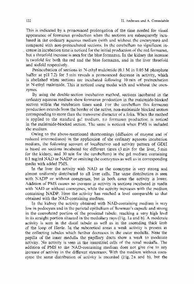
This is indicated by a pronounced prolongation of the time needed for visual appearance of formazan production when the sections are subsequently incu- bated in the ordinary aqueous medium (with and without the coenzymes) and compared with non-preincubated sections. In the cerebellum no significant in- crease in incubation time is noticed for the initial production of the red formazan, but a threefold increase is seen for the blue formazan. In the kidney the increase is twofold for both the red and the blue formazan, and in the liver threefold and sixfold respectively.
Preincubation of sections in N-ethyl maleimide (0.1 M in 0.05 M phosphate buffer at pH 7.2) for 5 rain reveals a pronounced decrease in activity, which is abolished when sections are incubated following 10 rain of preincubation in N-ethyl maleimide. This is noticed using media with and without the coen- zymes.
By using the double-section incubation method, sections incubated in the ordinary aqueous medium show formazan production in the maleimide-blocked section within the incubation times used. For the cerebellum this formazan production extends from the border of the active, non-maleimide blocked section corresponding to more than the transverse diameter of a folia. When the method is applied to the standard gel medium, no formazan production is noticed in the maleimide-blocked section. The same is noticed when PMS is included the medium.
Owing to the above-mentioned shortcomings (diffusion of enzyme and of reduced intermediates) in the application of the ordinary aqueous incubation medium, the following account of localization and activity pattern of GDH is based on sections incubated for different times (3 min for the liver, 5 rain for the kidney, and 30 min for the cerebellum) in the gel medium containing 0.1 mg/ml NAD or NADP or omitting the coenzymes as well as in corresponding media with added PMS.
In the liver the activity with NAD as the coenzyme is very strong and almost uniformly distributed to all liver cells. The same distribution is seen with NADP or without coenzymes, but in both cases the activity is lower. Addition of PMS causes no increase in activity in sections incubated in media with NAD or without coenzymes, while the activity increases with the medium containing NADP. Here the activity has reached a level comparable to that obtained with the NAD-containing medium.
In the kidney the activity obtained with NAD-containing medium is very low in podocytes and in the parietal epithelium of Bowman's capsule and strong in the convoluted portion of the proximal tubule, reaching a very high level in its straight portion situated in the medullary rays (Fig. 1 a and b). A moderate activity is seen in the distal tubule as well as in the ascending thick limb of the loop of Henle. In the subcortical areas a weak activity is present in the collecting tubules which further decreases in the outer medulla. Near the papilla of the inner medulla the papillary ducts show a weak to moderate activity. No activity is seen in the interstitial cells of the renal medulla. The addition of PMS to the NAD-containing medium does not give rise to any increase of activity in the different structures. With the medium without coen- zyme the same distribution of activity is recorded (Fig. 2a and b), but the

Pyridine Nucteotide Dependence of g-Glutamate Dehydrogenase Activity 123
Fig. 1. a Renal cortical activity of GDH with NAD-containing medium Strong activity is seen in the straight portion (arrows) of the proximal tubules situated in the medullary rays. b The same as Figure la following the inclusion of PMS

124 H. Andersen and A. Contestabile
Fig. 2. a Renal cortical activity with medium without N A D or N A D P added. Strong activity in the straight portion (arrows) of the proximal tubules situated in the medullary rays. b The same as Figure 2a with PMS added

Pyridine Nucleotide Dependence of L-Glutamate Dehydrogenase Activity 125
general level of activity is lower. Neither is any increase in activity noticed in the presence of PMS. With the NADP-containing medium the general distri- bution pattern is similar, but the activity is at an intermediate level compared with the previous ones. Meanwhile, no difference in strength of activity is seen between the convoluted portion and the straight portion of the proximal tubules (Fig. 3a). Although the activity of the NADPH tetrazolium reductase is strong in the macula densa area, no difference in the activity of GDH is noticed between this area and the adjacent parts of the distal tubule and the ascending thick limb of the loop of Henle. The addition of PMS gives rise to an increase of activity only in the straight portion of the proximal tubule (Fig. 3 b).
In the cerebellum the activity of G D H using NAD as the coenzyme is moderate to strong in synaptic glomeruli of the granular layer and in the perika- rya and prolongations of the satellite gliaI cells, while lower activity is noticed in the cells of Purkinje (Fig. 4a). In the molecular layer the activity is weak and diffuse although some cells show a more distinct activity in their perikarya. Addition of PMS causes a general increase in activity, most evident in the satellite glial cells adjacent to the cells of Purkinje (Fig. 4b). A similar pattern of activity is noticed when NADP is the coenzyme, but the activity itself is considerably weaker, with the exception of the satellite glial cells. The addition of PMS causes a minor enhancement of activity compared with the NAD- containing medium. Using the medium without added coenzyme, the activity is weak, but the distribution follows that obtained with the coenzyme-containing media. No enhancement in activity is noticed when PMS is a constituent of the medium. Irrespective of the media used, the activity in the cytoplasm of the granular cells is extremely faint or non-existent.
No nuclear activity is observed in any of the three organs regardless of media used.
Effect of Urea. Preincubation with 3 M urea for 10 min and subsequent incuba- tion for 15 rain (kidney), 20 rain (liver), and 30 rain (cerebellum) in medium containing the same concentration of urea causes almost complete inhibition of G D H activity. The activity is partly restored if the incubation after urea pretreatment is carried out in medium without urea as a constituent. In this case one can notice the reappearance of an aliquot of activity which is greater for kidney and cerebellum than for liver. There are no differences in the amounts of activity restored when the sections incubated in coenzyme-containing media are compared with sections incubated in coenzyme-free medium. A 5 rain rinse with the buffer between the urea-preincubation and the incubation in ordinary non-urea containing medium increases slightly the amount of activity restored. When the incubation time needed for the initial visual formation of red and blue formazans in non-urea pretreated sections is compared with the correspond- ing one in urea-preincubated sections the following results are obtained: In non-urea treated sections the red and blue formazan has appeared within 30 sec in the liver and kidney and 1 rain in cerebellar synaptic glomeruli, while in urea-pretreated sections the incubation time increases three- to fourfold for the blue one in the kidney, two- to threefold and eight- to tenfold respectively

126 H. Andersen and A. Contestabile
Fig. 3. a Renal cortical activity of GDH with NADP-containing medium. No difference in activity is noticed between the straight portion (arrows) of the proximal tubules situated in the medullary rays and the convoluted portion situated near the renal corpuscles, b The same as Figure 3a following the inclusion of PMS. Some increase of activity is seen in the straight portion (arrow) of the proximal tubules situated in the medullary rays

Pyridine Nucleotide Dependence of L-Glutamate Dehydrogenase Activity 127
Fig. 4. a Cerebellar cortex with activity of GDH following incubation in NAD-containing medium with 0.2 M of L-glutamic acid. Small arrows indicate the perikarya and prolongations of the satellite glial cells, large arrow head the cell of Purkinje and small arrow heads indicate synaptic glomeruli of the granular layer, b The same as Figure 4a with PMS added. Strong activity in the perikarya and prolongations of the satellite gliai celIs, c Activity following incubation in NAD- containing medium with ADP added and at a pH-level of 8.0. Note the strong activity in the cell of Purkinje
for the cerebel lum, but five- to s ixfold and th i r tyfo ld respect ively for the liver. These effects are no t iced using coenzyme-con ta in ing med ia as well as medium wi thou t the coenzymes.
P re - incuba t ion in 6 M urea and subsequent incuba t ion in med ia with the same concen t ra t ion of urea resul t in d i s appea rance o f all activity. I f the preincu- ba t ion is fo l lowed by incuba t ion in the o rd ina ry , urea-f ree m e d i u m or briefly (5 rain) washed in the buffer between the p r e incuba t i on and o rd ina ry incubat ion, then the act ivi ty res to red is negligible or non-existent .
Effect of l.lO-Phenanthroline. Pre - incuba t ion in a sa tu ra t ed so lu t ion o f phenan- th ro l ine a n d subsequent i ncuba t ion in the o r d i n a r y aqueous m e d i u m sa tu ra ted

128 I-I. Andersen and A. Contestabile
with phenanthroline causes a remarkable decrease in activity of GDH. The decrease is less pronounced if after the pre-incubation procedure the sections are incubated in phenanthroline-free media. In this case the "initial times" for red and blue formazan production increase three- to fourfold in kidney and cerebellum. The effect of phenanthroline is observed using coenzyme-con- taining media as well as media without added coenzyme.
Effect of c~-Ketoglutaric Acid. Addition of 9.5 mM c~-ketoglutaric acid to the incubation medium causes almost complete inhibition of GDH activity in kidney and cerebellum. In liver the same concentration causes only a prononced decrease in activity, while complete inhibition can be obtained raising the concentration to 15 raM. Again, the effect is noticed using coenzyme-containing media as well as media without coenzyme.
Effect of GTP and ADP. The addition of 1.4 mM GTP to the incubation media (with and without the coenzymes) reveals a strong inhibition of enzyme activity. This inhibition could be partly restored if sections are incubated in media containing 1.4 mM GTP as well as 1.6 mM ADP. In particular, this is seen in areas with strong enzyme activity (e.g. the straight portion of the proximal tubule in the kidney). The addition of ADP alone to the incubation media at pH 7.2 does not enhance enzyme activity and in areas with a comparatively low activity such as the cerebellar cortex, a slight decrease in activity seems to occur. If the pH of the ADP-containing media is raised to pH 8.0, the sections from the kidney (only with coenzyme-free medium due to the strong "Nothing dehydrogenase" reaction) show no clear increase in activity. On the other hand, sections from cerebellum show a remarkable increase when the pH of the three ADP-containing media (with and without the coenzymes) is raised, although the increase is more evident in the NAD-containing medium (Fig. 4c). The high pH value itself causes only a slight increase in activity. On the other hand, it causes an enhancement of the "Nothing dehydrogenase" reaction, with the exception of cerebellum where the enhancement is negligible.
Effect of Mg ++. With the composition of the media used in this study, no effect of adding Mg § § is observed.
Effect of FAD, FMN, and Coenzyme Qlo. The addition of FAD or FMN to the NAD- or NADP-free incubation medium causes no change in enzyme activity. The same result is achieved when the sections are incubated on slides coated with coenzyme Q10 in media with or without FAD or FMN added.
The other Experiments. The rise in concentration of NADP (from 0.1 mg/ml to 0.2 mg/ml) fails to have any effect on enzyme activity. Neither is the GDH activity influenced by the addition of ]?-glycerophosphate. The rise in concentra- tion to 0.2 M of L-glutamic acid in the medium causes some increase in activity in the liver and the kidney. With the three different media (with and without the coenzymes) the increase is considerable in the cerebellum, especially with the NAD-containing medium (Fig. 4a).

Pyridine NucIeotide Dependence of 1.-Glutamate Dehydrogenase Activity 129
Discussion and Conclusion
From the present study it may be stated that the L-glutamate dehydrogenase of animal origin possesses the ability to deaminate glutamate in the absence of NAD or N A D P - a t least in the histochemical in situ system. This statement is based on the following: a) the localization of enzyme activity noticed when the NAD or NADP are omitted in the incubation medium corresponds to that obtained when the coenzymes are included; b) the activity appears within the same period of incubation when the incubation time needed for the initial formation of the red and blue formazans is compared with the corresponding times obtained with NAD- or NADP-containing media, although following prolonged incubation time the strength of activity is below the average seen with the NAD-containing medium and below or equal to that noticed for the NADP-containing medium; c) the activity is influenced by the same agents and to the same extent as the activity obtained by the inclusion of NAD or NADP (e.g. the dissociation of the dehydrogenase molecule due to urea, the inhibition of activity due to N-ethyl maleimide and 1.10-phenanthroline, the activation due to ADP and the inhibition caused by GTP or ~-ketoglutaric acid); d) finally, the similarity in diffusion rates shown by the pre-extraction experiments as well as the activation caused by high substrate concentration should be mentioned.
For the following reasons it is unlikely that the activity mentioned above is caused by the L-aminoacid dehydrogenase (usually called L-aminoacid oxidase) (E.C. 1.4.1.5) or by the L-aminoacid oxidase (E.C. 1.4.3.2) demonstrated as a L-aminoacid tetrazolium reductase by Castellano et al. (1969): First, in the methods for L-aminoacid oxidase, PMS is an indispensable requisite (Castellano et al., 1969; Pearse, 1972), while in the present study the activity was unaffected by its presence. Next, the L-glutamate is considered an extremely poor substrate for the L-aminoacid oxidase (Jurtshuk and McManus, 1974; Baldwin, 1967), while in the present study the activity was pronounced using L-glutamate. Fur- thermore, Wohlrab (1965) found that 0.01 M KCN acted as an inhibitor of D-aminoacid oxidase as well as of L-aminoacid oxidase (using L-proline as the substrate) in the rat kidney, while in the present study the same concentration caused no influence on the activity in question.
Finally, it is unlikely that the activity noticed is caused by some aminotrans- ferase (or transaminase) activity which by the aid of the present L-glutamate dehydrogenase should be visualized via the tetrazolium reductases (diaphorases) and reduction of the tetrazolium salt. Such a system was described by Diculescu et al. (1970) and regarded by them as an L-aminoacid tetrazolium reductase (but not as L-aminoacid oxidase). Meanwhile, the visualization of this system was impossible in the absence of NAD, while in the present study the activity was seen without the presence of NAD or NADP. Further, their system required the addition of another amino acid while in the present study only L-glutamic acid was present (the L-glutamic acid (Sigma) is claimed to be of 99 100% in purity).
Since previous studies (Frieden, 1962; Di Prisco, 1975; Smith et al., 1975) have demonstrated that inorganic phosphate has a marked stabilizing effect

130 H. Andersen and A. Contestabile
on crystalline bovine liver mitochondrial L-glutamate dehydrogenase as well as an activating effect at pH above neutrality, a phosphate buffer was used in the present study. However, the pH and ionic strength of the incubation medium have to be taken into consideration, due to the effect on the catalytic and allosteric properties of glutamate dehydrogenase (Di Prisco, 1975). Although a pH 7.6 should be the preferable one, we selected a pH 7.2 in order to minimize the "Nothing dehydrogenase" reaction as far as possible (Andersen, 1965). The low pH value accounts for the observation that the addition of ADP alone to the incubation media has no activating effect on GDH activity and furthermore causes a slight decrease in activity in areas with comparatively low activity (e.g. the cerebellar cortex), and is in agreement with in vitro experiments (Di Prisco, 1975) which show a reversal in effect of ADP at pH values around neutralky. On the other hand, we found that ADP was able to restore partially the inhibition of enzyme activity due to the action of GTP at this pH level. The ionic strength of the buffer may add further to these results although the molarity of the buffer used was low (0.05 M). The same factors may account for the present observation that no activation was obtained by a twofold increase in concentration of coenzyme, which also agree with in vitro experiments (Di Pri- sco, 1975).
It has been claimed (Smith et al., 1975) that the primary effect of the allosteric effectors ADP and GTP attached to two different allosteric sites (Di Prisco, 1975) appears to be upon binding of coenzyme. Nevertheless, in the present study the effect was noticed without the presence of the coenzymes. At present it is difficult to explain this result, which may be due to different condition of the enzyme molecule in the intact tissue section used for the histochemical study compared with the isolated enzyme molecule used for in vitro experiments.
The inhibitory effect of 1.10-phenanthroline noticed in the present study is in accordance with in vitro experiments (Frieden, 1962; Smith et al., 1975). The inhibition has been related to the presence of zinc in the GDH molecule, but possibly zinc is not essential for enzymic activity since it can be removed from the molecule and the zinc-free molecule is inhibited to the same extent by phenanthroline. As claimed by Frieden (1962), the effect of phenanthroline is most likely the disruption of hydrophobic bonds involved in the linkage of the GDH-subunits.
The present results with N-ethyl maleimide as an inhibitor of GDH activity accord well with corresponding in vitro experiments, which result in 72% inacti- vation under standard assay conditions (Smith et al., 1975). The minor difference obtained with the two methods may be related to the different techniques employed.
The results obtained with urea as an agent inducing dissociation of the GDH-molecule into subunits also accord with corresponding in vitro experi- ments (Frieden, 1962) and provide further evidence for the conclusion that the activity noticed with coenzyme-free medium is caused by the L-glutamate dehydrogenase molecule proper.
Although the results from the present study all support the above conclusion, it is difficult at present to explain this property of the GDH molecule. A "Nothing dehydrogenase" reaction as a possible causal factor must be out of the question due to the fact that this reaction was negligible in the absence

Pyridine Nucleotide Dependence of L-Glutamate Dehydrogenase Activity 131
of coenzyme and appeared only after prolonged incubation. Furthermore, the most frequent sources of the "Nothing dehydrogenase" reaction (e.g. preexistent tissue sulphydryl groups, alcohol dehydrogenase (Shaw and Koen, 1965) or glutathion cytochrome c reductase (Stiller and Hempel, 1970)) all require the presence of NAD or NADP. Another possibility could be that the GDH- molecule as a mitochondrial matrix enzyme was able to use other functional components than NAD or NADP. In an attempt to solve this problem the effect of externally added FAD or FMN with and without added carrier-coen- zyme Q~o was studied. As shown, no stimulation of activity was noticed. Finally, the possibility exists that some endogeneous NAD bound in some way in the sections could serve as hydride ion acceptor. However, it is unlikely that endo- geneous NAD could resist extraction from the tissue during preincubational procedures as performed in connection with the urea pretreatment. It is therefore of some interest that a recent study (Jurtshuk and McManus, 1974) has disclosed the presence of a non-pyridine nucleotide-dependent L-glutamate oxidoreductase in Azotobacter Vinelandii which according to the authors does not represent a type of L-aminoacid oxidase. Meanwhile, it is safest to state in agreement with Di Prisco (1975) and Smith et al. (1975) that the regulation of glutamate dehydrogenase activity is extremely complex and much more information concerning the molecule itself is needed.
The control experiments dealing with enzyme diffusion revealed that GDH diffused from the fresh frozen sections leaving incubation in ordinary aqueous media out of the question if a reliable enzyme activity pattern should be obtained. Since mitochondria, although highly variable, are sensitive to thermal injury (for discussion c.f. Pearse, 1968), GDH as a mitochondrial matrix enzyme may escape from the mitochondria during freezing of tissue, passage of the knife through the tissue and subsequent thawing of sections preceding incubation. Although most of the injuries mentioned can be avoided or minimized (by rapid freezing, instantaneous incubation after sectioning, the use of a tissue stabilizer such as PVA during the incubation) some mitochondrial damage [e.g. influenced by ionic strength of incubation medium (Pachenko et al., 1975)] certainly is inevitable and may account for the faint diffuse activity of GDH noticed in the cytoplasm of some cells. Due to this possibility, we have not attempted any description of activity pattern at the intracellular level. On the other hand, a slight damage of mitochondrial membranes may facilitate the penetration of the large di-tetrazolium molecule which is further enhanced by the use of phosphate buffer shown to be a mitochondrial swelling agent (AItman, 1976b). Meanwhile, the use of PVA prevents the diffusion only of the enzyme proper during the incubation, while it allows diffusion of reduced intermediates such as NADH, NADPH or PMSH, leaving the localization of activity unreli- able. As shown in a recent study (Andersen and Hoyer, 1974), this diffusion can be controlled and avoided by using the double-section incubation method and by balancing the concentrations of tetrazolium salt, coenzyme and/or PMS against one another. In the present study this was obtained with the concentra- tions used.
In several papers disagreement exists concerning the degree of efficiency with which GDH utilizes NAD and NADP (Frieden, 1963; Braun-Falco and Petzoldt, 1965; Engel and Dalziel, 1969; Pearse, 1972; Wooton, 1974). In the

132 H. Andersen and A. Contestabile
present study we obtained the strongest activity mainly with media containing NAD. Since animal L-glutamate dehydrogenase exists as the NAD-dependent enzyme (E.C. 1.4.1.2) found in most animal tissue, and as the non-specific NAD- or NADP-dependent enzyme (E.C. 1.4.1.3) found in most livers, it may be concluded from the present study that L-glutamate dehydrogenase (E.C. 1.4.1.3) is present in the cells described in the kidney and the cerebellum, while the contemporary presence of L-glutamate dehydrogenase (E.C. 1.4.1.2) is more diffi- cult to deduce due to the fact that with the exception of cerebellum the "Nothing dehydrogenase" reaction is considerable with NAD-containing media, although far from the activity of GDH obtained by NAD-containing medium. However, the results obtained with PMS-containing media probably add to the evidence of a co-existence of both enzymes in cerebellum and kidney. This conclusion is in disagreement with the study made by Toledano and Martinez (1970) who claimed to find no activity at all in the cerebellum using NADP as the coenzyme.
Previous authors (Braun-Falco and Petzoldt, 1965) have claimed that the enzyme in question (in fresh skin sections) give equally strong activity with NAD and NADP. However, in their study no attention was paid to a possible conversion of NADP to NAD due to the presence of phosphomonoesterases. The use of PMS as well as of fi-glycerophosphate in the present investigation indicates that, at least for the liver, the enzyme utilized the two coenzymes equally, and due to this fact it must be related to L-glutamate dehydrogenase (E.C. 1.4.1.3) only.
The existence of a nuclear glutamate dehydrogenase has for long been a matter of controversy (Smith et al., 1975), due to the fact that it is difficult to obtain non-cytoplasm contaminated nuclei by centrifugation procedures. However, a recent study (Di Prisco and Garofano, 1975) has furnished some evidence for the presence of such an enzyme with properties different from the mitochondrial ones. The present paper adds nothing to the clarification of this problem since the nuclei obviously have no affinity for tetrazolium salts and consequently show no formazan formation. Work dealing with the ability of histones and non-histone nuclear proteins to bind tetrazolium salts is now in progress.
In order to avoid further ingredients in the incubation media, the metal- complexing agent EDTA (ethylendiaminetetraacetate) has been omitted due to the fact that only redistilled water has been used in the experiments and that proteins in the tissue may act as good metal-eomplexing agents (Dixon and Webb, 1964).
Finally, it can be stated that to obtain a reliable activity pattern, the incuba- tion time should be as brief as possible (ascertained by recording the incubation time needed for the initial visual formation of the red and blue formazans) in order to avoid "Nothing dehydrogenase" reaction and diffusion of reduced intermediates as well as a possible inhibitory effect due to accumulation of the reaction product, ~-ketoglutaric acid, which was shown to be a strong inhibitor.
References Altman, F.P.: The use of a new grade of polyvinyl alcohol for stabilizing tissue sections during
histochemical incubations. Histochemie 28, 236-242 (1971) Altman, F.P.: Tetrazolium salts: a consumer's guide. Histochem. J. 8, 471-485 (1976a)

Pyridine Nucleotide Dependence of L-Glutamate Dehydrogenase Activity 133
Altman, F.P.: Tetrazolium salts and formazans. Progr. Histochem. Cytochem. 9, 1 56 (1976b) Andersen, H.: Sulphydryl "Nothing dehydrogenase" activity and lactic dehydrogenase-NADH2
cytochroine c reductase reactions in tissues of the human foetus. Acta Histochem (Jena) 21, 120-134 (1965)
Andersen, H., Hoyer, P.E. : Simplified control experiments in the histochemicai study of coenzyme- linked dehydrogenases. Histochemistry 38, 71 83 (1974)
Baldwin, E.: Dynamic aspects of Biochemistry. 5. ed. London: Cambridge University Press 1967 Braun-Falco, O., Petzoldt, D. : Zur Frage optimaler Reaktionsbedingungen bei der histochemiscben
Darstellung yon Enzymen des Energie-liefernden Stoffwechsels in der Epidermis. I. Dehydrogen- asen. Arch. Klin. exp. Dermatol. 223, 620-633 (1965)
Castellano, M.A., Germino, N.I., De Haro, B., Gerard, G. : Histocbemical demonstration of L-amino acid-tetrazolium reductase. Histochemie 18, 277-280 (I969)
Diculescu, I., Dragomir, C.T., Condrescu-Guidi, M., Popescu, M.: A histochemical method for L-amino acid tetrazolium reductase. Histochemie 21, 9-16 (1970)
Di Prisco, G.: Effect of pH and ionic strength on the catalytic and allosteric properties of native and chemically modified ox liver mitochondrial glutamate dehydrogenase. Arch. Biochem. Biophys. 171, 604-612 (1975)
Di Prisco, G., Garofano, F. : Crystallization and partial characterization of glutamate dehydrogenase from ox liver nuclei. Biochemistry 14, 4673~t679 (1975)
Dixon, M., Webb, E.C.: Enzymes. 2. ed. London: Longmans, Green and Co. Ltd. 1964 Engel, P.C., Dalziel, K. : Kinetic studies of glutamate dehydrogenase with glutamate and norvaline
as substrates. Biochem. J. 115, 621-631 (1969) Frieden, C.: The effect o fpH and other variables on the dissociation of beef liver glutamic dehydro-
genase. J. Biol. Chem. 237, 2396-2400 (1962) Frieden, C. : Glutamate dehydrogenase. V. The relation of enzyme structure to the catalytic function.
J, Biol. Chem. 238, 3286-3299 (1963) Hoyer, P.E,, Andersen, H.: Histochemistry of 3fl-hydroxysteroid dehydrogenase in rat ovary, I.
A methodological study. Histochemistry 51, 167-i93 (1977) Jurtshuk, P., McManus, L. : Non-pyridine nucleotide dependent L-(+)-glutamate oxidoreductase
in Azotobaeter Vinelandii. Biochem. Biophys. Acta 368, 158 172 (1974) Leeflang-de Pijper, A.M., Hfilsmann, W.C. : Pitfalls in histochemical Iocalization studies of NADPH
generating enzymes or enzyme systems in rat small intestine. Histochemistry 39, 143-153 (1974) Martinez-Roderigues, R.: A histochemical study of some aspects of the metabolism of glutamic
acid in the cerebellum of mammals. Acta Histochem. (Jena) 49, 74-82 (1974) Marzotko, D., Warehel, J.B., Warchowiak, R.: Spektrophotometrische Untersuchungen fiber die
Lichtempfindlichkeit yon PMS in w/iBriger L6sung. Acta Histochem. (Jena) 46, 53-59 (1973) Panchenko, L.F., Loktaeva, T.D., Gerasimov, A.M.: Influence of ionic strength of incubation
medium on the release of some enzymes from rat liver mitochondria. Enzyme 19, 337-347 (1975)
Pearse, A.G.E. : Histochemistry, theoretical and applied. 3. ed. vol 1. London: Churchill Livingstone 1968
Pearse, A.G.E. : Histochemistry, theoretical and applied. 3. ed. vol 2. London: Churchill Livingstone 1972
Shaw, C.R., Koen, A.L.: On the identity of "Nothing dehydrogenase". J. Histochem. Cytochem. 13, 431-433 (1965)
Smith, E.L., Austen, B.M., Blumenthal, K.M., Nyc, J.F.: Glutamate dehydrogenases. In: The Enzymes. 3. ed. XI part A, Dehydrogenases I. Ed. Boyer P.D. New York: Academic Press 1975
Stiller von, D., Hempel, E. : Zum Problem des ,,Nothing-dehydrogenase"-Effektes beim topochemi- schen Nachweis dehydrierender Enzymsystemen. Acta Histochem. (Jena) 35, 239 252 (1970)
Toledano, A., Martinez-Roderigues, R. : Etude histochemique et biochimique du cervelet du chat. 1II. glutamo-dehydrogenase. Ann. Histochim. 15, 231-243 (1970)
Wattenberg, L.W., Leong, J.L. : Effects of coenzyme Qlo and menadione on succinic dehydrogenase activity as measured by tetrazolium salt reductions. J. Histochem. Cytochem. 8, 296-303 (1960)
Wohlrab, F. : l~'ber die histochemische ErfaBbarkeit der Aminos/iure-Dehydrogenasen in S~tugetieror- ganem Histochemie 5, 311 325 (1965)
Wootton, J.C. : The coenzyme-binding domains of glutamate dehydrogenases. Nature 252, 542-546 (1974)
Received March 10, 1977