The pupillary light reflex pathway - Dr Luca Ticini, FRSA€¦ · The pupillary light reflex...
8
The pupillary light reflex pathway Cytoarchitectonic probabilistic maps in hemianopic patients E. Papageorgiou, MD L.F. Ticini G. Hardiess, PhD F. Schaeffel, PhD H. Wiethoelter, MD H.A. Mallot, PhD S. Bahlo B. Wilhelm, MD R. Vonthein, PhD U. Schiefer, MD H.-O. Karnath, MD, PhD ABSTRACT Objective: The anatomy of the human pupillary light reflex (PLR) pathway is a matter of debate. The aim of this study was twofold: namely, to investigate the association of a relative afferent pupillary defect (RAPD) in acquired suprageniculate lesions with the location and extent of the cerebral lesions. Further, we suggest a new strategy of lesion analysis by combining established techniques with the stereotaxic probabilistic cytoarchitectonic atlas developed by the Ju ¨lich group. Methods: Twenty-three patients with homonymous visual field defects participated in this study. The RAPD was quantified clinically by two independent examiners with graded neutral density filters (swinging flashlight test). Using MRI in each individual, cerebral regions commonly affected in patients with a RAPD but spared in patients without a RAPD were determined and subse- quently assessed by using cytoarchitectonic probabilistic maps. Results: A RAPD was present in 10/23 patients. Comparison of patients showing a RAPD vs those not showing a RAPD revealed that a region including the course of the optic radiation at its early beginning in the temporal white matter is commonly associated with a RAPD. Conclusions: It was demonstrated that the pupillary light reflex (PLR) depends on the input of supra- geniculate neurons, thus supporting the involvement of a cortical pathway also. The site of integration of cortical signals in relation to the PLR into the pupillomotor pathway may be located supragenicu- lately in the vicinity of the lateral geniculate nucleus. Moreover, the suggested combination of estab- lished lesion analysis techniques with the probabilistic cytoarchitectonic atlas turned out to be a very helpful amelioration of stroke data analyses. Neurology ® 2008;70:956–963 GLOSSARY CG ciliary ganglion; DWI diffusion-weighted imaging; FLAIR fluid-attenuated inversion-recovery; HVFD homony- mous visual field defect; IN intercalated neurons; LGN lateral geniculate nuclei; MNI Montreal Neurological Institute; N.III oculomotor nerve; NEW nucleus Edinger-Westphal; ON optic nerve; OT optic tract; PLR pupillary light reflex; PT pretectal area; RAPD relative afferent pupillary defect; SCN short ciliary nerves. The neural pathway of the pupillary light reflex (PLR) was first described by Wernicke in the 1880s. 1 Afferent fibers from the retina travel in the optic nerve and undergo hemide- cussation at the chiasm before entering the optic tract. In the posterior third of the optic tract, the fibers branch medial to the lateral geniculate nucleus (LGN) and synapse in the ipsilateral pretectal nucleus. Intercalated neurons from each pretectal nucleus then project to both Edinger-Westphal nuclei and parasympathetic fibers from the Edinger- Westphal nuclei innervate the iris pupillary sphincter muscle. A relative afferent pupillary defect (RAPD) is characterized by diminished pupillary constriction on direct illumination with a normal consensual response to illumination of the contralateral eye. It is typically related to lesions within the anterior visual pathways and is almost always present in unilateral or asymmetric bilateral optic nerve disease. A From Centre for Ophthalmology (E.P., F.S., B.W., U.S.), Section Neuropsychology, Center for Neurology (L.F.T., H.-O.K.), and Department of Medical Biometry (R.V.), University of Tuebingen; Department of Zoology (G.H., H.A.M.), Lab of Cognitive Neuroscience, Tuebingen; Department of Neurology (H.W.), Buerger Hospital, Stuttgart; and Bad Urach Rehabilitation Center (S.B.), Germany. Supported by the following grants: European Union (PERACT-Marie Curie Early Stage Research Training, MEST-CT-2004-504321), Bundesministerium fu ¨ r Bildung und Forschung (BMBF-Verbundprojekt “Ra ¨umliche Orientierung” 01GW0641), GRK 778. Disclosure: Dr. Ulrich Schiefer receives royalties from some of the equipment of this study, which do not exceed $10,000/year. Supplemental data at www.neurology.org Address correspondence and reprint requests to Prof. Dr. med. U. Schiefer, Centre for Ophthalmology, Schleichstrasse 12-16, 72076, Tuebingen, Germany Ulrich.Schiefer@uni- tuebingen.de 956 Copyright © 2008 by AAN Enterprises, Inc.
Transcript of The pupillary light reflex pathway - Dr Luca Ticini, FRSA€¦ · The pupillary light reflex...
The pupillary light reflex pathwayCytoarchitectonic probabilistic maps in hemianopic patients
E. Papageorgiou, MDL.F. TiciniG. Hardiess, PhDF. Schaeffel, PhDH. Wiethoelter, MDH.A. Mallot, PhDS. BahloB. Wilhelm, MDR. Vonthein, PhDU. Schiefer, MDH.-O. Karnath, MD,PhD
ABSTRACT
Objective: The anatomy of the human pupillary light reflex (PLR) pathway is a matter of debate.The aim of this study was twofold: namely, to investigate the association of a relative afferentpupillary defect (RAPD) in acquired suprageniculate lesions with the location and extent of thecerebral lesions. Further, we suggest a new strategy of lesion analysis by combining establishedtechniques with the stereotaxic probabilistic cytoarchitectonic atlas developed by the Julichgroup.
Methods: Twenty-three patients with homonymous visual field defects participated in this study.The RAPD was quantified clinically by two independent examiners with graded neutral densityfilters (swinging flashlight test). Using MRI in each individual, cerebral regions commonly affectedin patients with a RAPD but spared in patients without a RAPD were determined and subse-quently assessed by using cytoarchitectonic probabilistic maps.
Results: A RAPD was present in 10/23 patients. Comparison of patients showing a RAPD vsthose not showing a RAPD revealed that a region including the course of the optic radiation at itsearly beginning in the temporal white matter is commonly associated with a RAPD.
Conclusions: It was demonstrated that the pupillary light reflex (PLR) depends on the input of supra-geniculate neurons, thus supporting the involvement of a cortical pathway also. The site of integrationof cortical signals in relation to the PLR into the pupillomotor pathway may be located supragenicu-lately in the vicinity of the lateral geniculate nucleus. Moreover, the suggested combination of estab-lished lesion analysis techniques with the probabilistic cytoarchitectonic atlas turned out to be a veryhelpful amelioration of stroke data analyses. Neurology® 2008;70:956–963
GLOSSARYCG � ciliary ganglion; DWI � diffusion-weighted imaging; FLAIR � fluid-attenuated inversion-recovery; HVFD � homony-mous visual field defect; IN � intercalated neurons; LGN � lateral geniculate nuclei; MNI � Montreal Neurological Institute;N.III � oculomotor nerve; NEW � nucleus Edinger-Westphal; ON � optic nerve; OT � optic tract; PLR � pupillary light reflex;PT � pretectal area; RAPD � relative afferent pupillary defect; SCN � short ciliary nerves.
The neural pathway of the pupillary light reflex (PLR) was first described byWernicke inthe 1880s.1 Afferent fibers from the retina travel in the optic nerve and undergo hemide-cussation at the chiasm before entering the optic tract. In the posterior third of the optictract, the fibers branch medial to the lateral geniculate nucleus (LGN) and synapse in theipsilateral pretectal nucleus. Intercalated neurons from each pretectal nucleus thenproject to both Edinger-Westphal nuclei and parasympathetic fibers from the Edinger-Westphal nuclei innervate the iris pupillary sphincter muscle.
A relative afferent pupillary defect (RAPD) is characterized by diminished pupillaryconstriction on direct illumination with a normal consensual response to illumination ofthe contralateral eye. It is typically related to lesions within the anterior visual pathwaysand is almost always present in unilateral or asymmetric bilateral optic nerve disease. A
From Centre for Ophthalmology (E.P., F.S., B.W., U.S.), Section Neuropsychology, Center for Neurology (L.F.T., H.-O.K.), and Departmentof Medical Biometry (R.V.), University of Tuebingen; Department of Zoology (G.H., H.A.M.), Lab of Cognitive Neuroscience, Tuebingen;Department of Neurology (H.W.), Buerger Hospital, Stuttgart; and Bad Urach Rehabilitation Center (S.B.), Germany.
Supported by the following grants: European Union (PERACT-Marie Curie Early Stage Research Training, MEST-CT-2004-504321),Bundesministerium fur Bildung und Forschung (BMBF-Verbundprojekt “Raumliche Orientierung” 01GW0641), GRK 778.
Disclosure: Dr. Ulrich Schiefer receives royalties from some of the equipment of this study, which do not exceed $10,000/year.
Supplemental data atwww.neurology.org
Address correspondence andreprint requests to Prof. Dr.med. U. Schiefer, Centre forOphthalmology, Schleichstrasse12-16, 72076, Tuebingen,[email protected]
956 Copyright © 2008 by AAN Enterprises, Inc.

RAPD contralateral to the side of the le-sion is also observed in optic tract lesions,which are characterized by incongruenthomonymous visual field defects (HVFDs)and asymmetric bilateral optic disc atro-phy.2 The proposed pathogenesis for thepresence of a contralateral RAPD in an op-tic tract lesion is based on the greater nasalphotoreceptor density, a ratio of crossed touncrossed fibers in the chiasm of 53:47 anda temporal visual field 61% to 71% largerthan the nasal field.3 A tract lesion disruptsfibers from the contralateral nasal retinaand the ipsilateral temporal retina, thusdisproportionally diminishing input fromthe contralateral eye and producing a cor-responding RAPD.4 Furthermore, a RAPDhas also been described in patients withcongenital occipital hemianopia. The sug-gested mechanism is transsynaptic optictract atrophy after intrauterine or perinataldamage to the suprageniculate visual path-way, which presumably affects also the af-ferent pupillary fibers to the pretectal areaof the mesencephalon.5,6 Therefore, the de-tection of a RAPD in acute homonymoushemianopias has been commonly used indifferentiating infrageniculate from supra-geniculate lesions, since neither optic atro-phy nor a RAPD should occur in acquiredaffections of the optic radiation or the vi-sual cortex.3,7-9
However, the presence of a RAPD in ac-quired suprageniculate lesions and the un-derlying anatomic pathway are still amatter of debate, mainly because of nu-merous studies, reporting disturbances ofthe PLR in patients with HVFDs due to le-sions not involving the optic tract. In theearly 1940s and later elaborated investiga-tions by several authors had already shedsome doubt on the validity of Wernicke’smodel of a direct retinal-pretectal connec-tion.1 Furthermore, pupillary hemihypoki-nesia—that is, reduced or absent pupillaryreaction to perimetric stimuli in the blindpart of the visual field—was observed in allkinds of postchiasmal lesions.10-13 Theseearly clinical reports were later confirmedby other groups using modern pupillomet-
ric techniques.14-17 In addition to the formerstudies, a clinically relevant RAPD, as a re-sponse to full-field light stimulation, wasalso reported in suprageniculate lesions, ifthe damaged area is close to the LGN.18
Many theories have been developed to ex-plain these phenomena, the most promi-nent pointing out that the integrity of thepupillomotor response depends upon or isinfluenced also by the occipital cortex.19-22
However, the anatomic evidence is stilllimited to make any clear conclusions.
The aims of this study thus were 1) toassess the presence and magnitude of theRAPD in patients with HVFDs due to cere-brovascular lesions in the posterior andmiddle cerebral artery territories, and 2) toanalyze the association of the pupillaryfindings with the location and extent of thecerebral lesions. Further, the present studysuggests 3) a new strategy of lesion analysisby combining established techniques withthe stereotaxic probabilistic cytoarchitec-tonic atlas developed by the Julichgroup.23,24 In contrast to the reference brainof the Talairach and Tournoux atlas,25 orthe Montreal Neurological Institute (MNI)single subject or group templates,26,27 theseprobabilistic cytoarchitectonic maps providestereotaxic information on the location andvariability of cortical areas in the MNI refer-ence space. They are based on the analysisof the cytoarchitecture in a sample of 10human postmortem brains and already areavailable for various brain regions (http://www.fz-juelich.de/ime/ime_brain_mapping).
METHODS Twenty-three patients with HVFDs (16 menand 7 women), with a mean age of 50.5 years (age range: 21to 74 years, SD 17 years) were enrolled in this study (appen-dix e-1 on theNeurology® Web site at www.neurology.org).Patients were recruited from the Centre for Ophthalmologyat the University of Tubingen (Germany), the UniversityNeurology Clinic of Tubingen, as well as the NeurologyClinic of Buerger Hospital in Stuttgart and the Bad UrachRehabilitation Center. All patients had a homonymous vi-sual field defect, varying from a complete homonymoushemianopia to homonymous paracentral scotomas. Thecause was a unilateral vascular brain lesion in the territoriesof the posterior or middle cerebral arteries, which was docu-mented byMRI. There were 12 patients with right-sided and11 patients with left-sided lesions. Best-corrected monocular(near and distant) visual acuity was at least 16/20. Patientswith unilateral cataract, marked anisocoria, amblyopia,
Neurology 70 March 18, 2008 957

strabismus, ocular motility disorders, optic nerve diseases,glaucoma, advanced diseases of the retina, or funduscopicsigns of bilateral asymmetric optic disc atrophy, which couldindicate an optic tract involvement, were excluded from thisstudy.
The median time since lesion onset and the neuro-ophthalmologic investigation used for the present analysiswas 1 year (range 0.3 to 11.2 years, appendix e-2). TheRAPD was assessed clinically by means of the swingingflashlight test. This test was selected because it represents aneasy-to-apply, clinical examination, which provides reliableresults about visual sensory dysfunction immediately.28 Theaim was to detect the degree of asymmetry between botheyes regarding the pupillary light reflex, which is indepen-dent of the absolute pupillary responses. Therefore only therelationship of responses between both sides (RAPD) wasinvestigated: in a dark room an indirect ophthalmoscope as alight source was held below the level of the line of sight, withits light beam elevated at about a 45 degree angle. Initiallypupil size was assessed under two different ambient lightconditions in order to detect the presence of anisocoria. If noanisocoria was found and the pupils responded well to thelight stimulus, then one eye was illuminated, and after 2 to 3seconds, the light was shifted quickly to the contralateraleye. The process was repeated four or five times. A RAPDwas defined in case of a pathologic swinging flashlight test: ifthe consensual response of the pupil was better than the di-rect, an ipsilateral RAPD was diagnosed, while if the directresponse was better than the consensual, a contralateralRAPD was diagnosed. Using neutral density filters of vary-ing strength, we measured the magnitude of a RAPD, byweakening the light stimulus as it was presented to the bettereye. The filters were separated in 0.3 log unit steps from 0.3to 1.8 log units. A RAPD was defined as any asymmetry of0.3 log units or more, while any asymmetry below 0.3 logunits was defined as a RAPD 0. In order to test the reproduc-ibility and reliability of the results, all pupil examinationswere carried out by two independent examiners, a seniorneuro-ophthalmologist (U.S.) and a resident (E.P.). Bothophthalmic examiners were blinded to the perimetric and theimaging results. Agreement between examiners regardingthe presence and magnitude of a RAPD was 0.74 (SE 0.13),assessed by Cohen’s kappa. All analyses were performed ac-cording to the findings of the senior neuro-ophthalmologist(appendix e-1, Examiner 1).
Mapping and analysis of lesion location was carried outby two experimenters (H.-O.K. and L.F.T.) without knowl-edge of test results and clinical features of the patients. Forlesion delineation, diffusion-weighted imaging (DWI) wasused within the first 48 hours post-stroke and weighted fluid-attenuated inversion-recovery (FLAIR) sequences when im-aging was conducted 48 hours or later after stroke. Twocases with marked perifocal edema or marked hemorrhageleading to a significant shift of anatomic structures were ex-cluded. The median time between stroke and imaging usedfor the present analyses was 4.5 days (range 0 to 200 days,appendix e-2). In one subject (No. 15) the original scans wereof low quality such that new MRI scans were obtained at11.2 years after the brain lesion. Brain imaging had typicallypreceded the neuro-ophthalmologic examination (mediantime between imaging and neuro-ophthalmologic examina-tion 1 year, range 0.3 to 9.9 years, appendix e-2).
In 8 of the 23 patients with HVFDs, MR images wereavailable in digital format. In these cases, the boundary of
the lesion was delineated directly on every single transversalslice of the individual MRI using MRIcro software (http://www.mricro.com).29 Both the scan and lesion shape werethen transformed into stereotaxic space using the spatialnormalization algorithm provided by SPM2 (http://www.fil-.ion.ucl.ac.uk/spm/), using default settings. For determina-tion of the transformation parameters, cost-functionmasking was employed.30 In 15 of the 23 cases MRI datawere not available in digital format. In these cases, MRIcrosoftware was used to manually map the lesion on transversalslices of the T1-template MRI from the MNI (www.bic.mn-i.mcgill.ca/cgi/icbm_view) distributed with MRIcro. Lesionswere mapped onto the slices that correspond toZ-coordinates �40, �32, �24, �16, �8, 0, 8, 16, 24, 32, 40,and 50 mm in MNI coordinates by using the identical or theclosest matching transversal slices of each individual. Sincethe right and left visual pathway can be considered identicalregarding anatomy and function, for the anatomic analysisthe left-sided lesions were mirrored and superimposed on theright side of the brain template.
To identify the anatomic structures that were commonlyaffected in patients showing the disorder (here the RAPD)but were typically spared in patients without the disorder,we here used a new combination of the established lesionsubtraction analysis31 with the stereotaxic probabilistic cyto-architectonic atlas, the latter developed by the Julichgroup.23,24 Subtraction plots directly contrast patients withstroke showing vs not showing the disorder. Since subtrac-tions were made between groups of different sizes propor-tional values were used. Subsequently, the resultingsubtraction image was plotted onto maps of the stereotaxicprobabilistic cytoarchitectonic map of the optic radiation byBurgel and coworkers.32,33 This map illustrates the relativefrequency with which a certain fiber tract of 10 normal hu-man brains was present on a MNI reference brain in a voxel(e.g., a 50% value of a fiber tract in a certain voxel of thereference brain indicates that the fiber tract was present inthat voxel in 5 out of 10 brains). The probabilistic cytoarchi-tectonic map thus serves as a measure of intersubject vari-ability for each voxel of the reference space.
RESULTS A RAPD was present in 10 out of 23patients and was located contralateral to the af-fected hemisphere in all 10 cases. The medianmagnitude was 0.3 log units (six patients) and therange was between 0.3 and 0.9 log units. A RAPDof 0.6 log units was demonstrated in three pa-tients and one patient showed a RAPD of 0.9 logunits. Patients 1, 7, 11, 12, and 17 were re-examined at least three times over a time span of 1year after the brain lesion. The pupillary findingsremained constant in all cases.
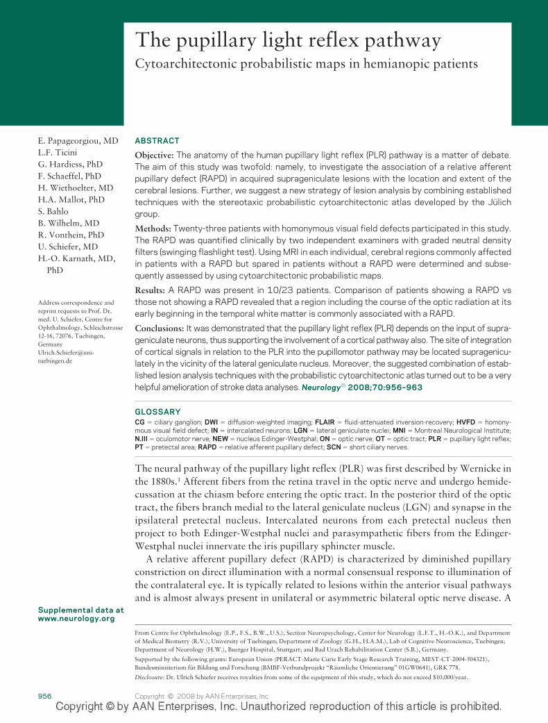
Figure 1A illustrates simple lesion overlayplots for the group of patients with a RAPD andthe group without a RAPD. To identify the ana-tomic structures that were commonly affected inpatients with a RAPD but were typically spared inpatients without a RAPD, we subtracted the su-perimposed lesions of the group without a RAPDfrom the patient group with a RAPD, revealingpercentage overlay plots. Figure 1B illustrates
958 Neurology 70 March 18, 2008
these results. We found the center (orange) of thesubtracted lesion overlap associated with a RAPDin the subcortical temporal white matter, extend-ing further into superior temporal cortex. To an-alyze the anatomic relationship of this center withthe location of the optic radiation, we plotted itonto the recently developed probabilistic cytoar-chitectonic map of the optic radiation by Burgeland coworkers.32,33 Figure 2 illustrates the result.We found the region commonly affected in pa-tients with a RAPD but typically spared in pa-tients without a RAPD primarily at the earlybeginning of the course of the optic radiation inthe temporal white matter. In absolute numbers 6of the 10 patients with a RAPD (60%) had a le-sion in this area, while in only 1 of the 13 patients
without a RAPD (8%) this area was affected. Thedifference was significant (Fisher exact p � 0.019).
The mean percentage of left or right hemi-sphere tissue affected in patients with a RAPDwas 10.6%, while it was 4.5% in patients withouta RAPD. A statistical comparison revealed no sig-nificant difference in the lesion volume betweenboth groups of patients (t � 1.686, df � 11, two-tailed p � 0.12, unpaired t test for unequal vari-ances).
DISCUSSION The PLR has for a long time beenassociated with subcortical projections, and thiswas consistent with some clinical observationswhich suggest that the pupils continue to re-spond, even when the patients are corticallyblind.34,35 Wernicke’s model of a direct retinal–pretectal interaction explained these observationswell and the PLR was thought to be associatedwith a single subcortical neural pathway until theearly 1940s. Since then, numerous studies haveexamined the effect of visual cortical lesions onthe PLR. The results—either the presence of pu-pillary hemihypokinesia in the blind part of thevisual field or a RAPD contralateral to the brainlesion, as a response to full-field light stimula-tion—often contradicted this classic belief andprovided evidence that the PLR is not just a puresubcortical pathway.18,22 However, the exact ana-tomic pathway remained unknown.
Using a new strategy of lesion analysis by com-bining established subtraction techniques31 withthe stereotaxic probabilistic cytoarchitectonic at-las developed by the Julich group,23,24,32,33 ourfindings suggest that a region in the early courseof the optic radiation in the temporal white mat-ter, close to the LGN, seems to be associated withthe presence of the RAPD. This finding is consis-tent with the hypothesis that the connection be-tween visual pathways and pretectal area in thedorsal midbrain is probably closely related to theLGN.4,18,36-38 It has been found that a RAPD insuprageniculate homonymous hemianopsia oc-curred in approximately half of all patients withlesions closer than 10 mm to the LGN or involv-ing it but sparing the optic tract.18 In lesions fur-ther than 18 mm away from the LGN, a RAPDdid not occur at all. The authors concluded thatthe RAPD was probably not caused by a lesion ofthe visual pathway itself, but by a lesion of theintercalated neurons between the visual pathwayand the pupillomotor centers in the pretectal areaof the midbrain. This assumption is strengthenedby several cases of a RAPD with normal visualfunction in lesions close to the LGN or pretectal
Figure 1 Simple lesion overlay plots for the group of patients with relativeafferent pupillary defect (RAPD positive) and the group withoutRAPD (RAPD negative) and subtraction of the superimposed lesionsof the control group (without RAPD) from the overlap image of thegroup with RAPD
(A) Simple lesion overlay plots for the group of patients with RAPD (RAPD positive) and thegroup without RAPD (RAPD negative). Overlapping lesions are color-coded with increasingfrequency from violet (n � 1) to red (n � 10 for RAPD positive; n � 13 for RAPD negative).MNI z-coordinates of each transverse section are given. (B) Subtraction of the superimposedlesions of the control group (without RAPD) from the overlap image of the group with RAPD.The center of the subtracted lesion overlap (orange area) indicates regions damaged at least40% more frequently in patients with RAPD than in patients without RAPD.
Neurology 70 March 18, 2008 959

region as well as by reports of optic tract lesionswithout a RAPD.4,28,37-44 The present findings alsosupport the concept of cortical input into the PLR(figure 3), which may enter the PLR pathway viathe area depicted in figures 1B and 2. Therefore,disturbed processing of signals along this part ofthe geniculostriate pathway can lead to a RAPD.
Another explanation could be that some afferentpupillomotor fibers of infrageniculate origin by-pass the LGN and then travel through this criticalarea to the mesencephalon. Consistent with theaforementioned study, we draw a similar conclu-sion about a suprageniculate effect, although thesite identified in the present study is located moredistantly from the LGN.
Since the description of an impaired PLR in theblind field of subjects with definitely supragenicu-late lesions a number of speculative suggestionshave been offered to explain this finding.10,12-14 Itwas hypothesized that transsynaptic degenerationcould occur, either retrogradely across the genicu-late synapse or into the pretectal area from theneocortical visual system, in line with former ob-servations on cortico-pretectal interaction.10,14,45-48
However, in the case of transsynaptic degenera-tion, one would expect that the RAPD would de-velop slowly, being found not earlier than monthsor years after the occipital lesion, and that in sa-lient instances, optic disc atrophy should be ob-served. In the present study cohort, the RAPD inPatient 17 was demonstrated already a few daysafter the cerebral infarct and remained constantin the follow-up examinations, without any signsof optic disc atrophy. The pupillary findings inPatients 1, 7, 11, and 12 also remained constant inat least three re-examinations over a time span of1 year after the brain lesion. Clinical observationshave also provided evidence that transsynapticdegeneration in adults is still not convincing.49 Inour series, there were no funduscopic signs of par-tial optic atrophy in patients with a RAPD, thusindicating suprageniculate lesions. The time spanbetween lesion and clinical examination exceeded4 months in all cases; therefore the subsequent op-tic atrophy by a potential affection of the optictract should already be visible, since the periodnecessary for retrograde degeneration following apregeniculate lesion is estimated to be about 6weeks.49
Potential models of cortico-pretectal interac-tion, which could be in accordance with our find-ings, have already been described.50 It wasobserved that in patients with retrogeniculatehemianopsia the PLR in the blind hemifield wasreduced but not absent. However, all the otherspecific, “higher” pupil responses to stimulus at-tributes, like stimulus color, structure, or motion,were completely lost. Therefore, it was consid-ered that two or more distinct channels couldserve the PLR: a more primitive “luminance chan-nel,” which connects the retina directly with thepretectal area and responds to diffuse light, and a
Figure 2 Probabilistic cytoarchitectonic map of the optic radiation by Burgeland coworkers25,26
The color bar indicates the absolute frequency of voxels containing the optic radiation from 1(dark blue) individual brain to 10 (red, overlap of all 10 [maximum] brains). The superimposedpink contour represents the center of the subtracted lesion overlap obtained in the presentstudy (see orange area in figure 1B).
Figure 3 Schematic drawing of the pupillarylight reflex pathway (figure modifiedfrom Wilhelm21,52)
Afferent fibers from the retina travel in the optic nerve (ON)and undergo hemidecussation at the optic chiasm before en-tering the optic tract (OT). Close to the lateral geniculate nu-cleus (LGN) the fibers branch from the optic tract and passthrough the brachium of the superior colliculus to reach theipsilateral pretectal nucleus. However, there seems to bemore input from suprageniculate neurons and the occipitalcortex (CI). The exact anatomy of this connection is still un-clear. Our data support that the site of integration of corticalsignals in relation to the PLR into the pupillomotor pathwaymay be located suprageniculately in the early course of theoptic radiation near the LGN (figure 2). Intercalated neurons(IN) from each pretectal nucleus then project to bothEdinger-Westphal nuclei (NEW). Parasympathetic outflowfrom the Edinger-Westphal nuclei travels then with the ocu-lomotor nerve (N.III) to the ciliary ganglion (CG) and via theshort ciliary nerves (SCN) reaches the iris pupillary sphinctermuscle. PT � pretectal area.
960 Neurology 70 March 18, 2008

“pattern channel,” which is mediated supra-geniculately and responds to shifts in structuredstimuli, like isoluminant grating, motion, andisoluminant color stimuli. According to the au-thors, the PLR is primarily mediated by the lumi-nance channel and to a smaller extent by the“weaker,” suprageniculate pattern channel. Thisexplanation could possibly account for the com-paratively small magnitude of the RAPD (pre-dominantly 0.3 logU) in the majority of patientsin the present study. Although these previousstudies refer to pupillary hemihypokinesia, thismodel can also explain the presence of a RAPD insuprageniculate lesions of this critical region nearthe LGN. It is plausible that the critical area in theimmediate vicinity of the LGN (figure 2) containssuprageniculate projections of the so-called “pat-tern channel” to the mesencephalon. A cortico-mesencephalic interaction should thus not besurprising, if one considers the selective loss ofpupil color response in cerebral achromatopsiaand the pupil near response, which must also bemediated by a similar cortical input to theEdinger-Westphal nuclei.34 Recent studies assess-ing the various components of the pupil responsethat have been affected in subjects with damage tothe dorsal midbrain (Parinaud syndrome) havealso demonstrated that there was a small, residualPLR and preserved reactions to pattern and colorstimuli as well as preserved pupillary sleepiness-related oscillations.22 The authors suggested theexistence of a cortical input to the pupillary path-way, since the retinal afferent input to the pretec-tal nuclei had been apparently damaged.22
The present results are still subject to interpre-tation, because our study has some limitations.One could stem from the resolution capacity ofthe imaging methods that are currently available.It cannot be excluded that our patients had addi-tional, subclinical lesions affecting mesencephalicstructures, i.e., the pretectal area or the vicinity ofthe oculomotor nucleus, which could possibly ex-plain the presence of the RAPD. However, mod-ern MRI techniques already provide anatomicinformation from living human beings with veryhigh precision. By using FLAIR sequences, we ex-cluded cases with marked perifocal edema or sig-nificant shift of anatomic structures. However,some tract or midbrain compression from second-ary swelling may have occurred. Under-recognitionof such cases, either due to slight effects or tochanges undetectable with the current methods, is afurther limitation of our study. Additionally, onecould argue that the exclusion of patients with pre-geniculate lesions was done funduscopically by two
examiners (U.S. and E.P.), by detecting signs of bi-lateral asymmetric optic disc atrophy.However, it isvery unlikely that the RAPD in all 10 patients wascaused by an ophthalmoscopically and radiologi-cally invisible pregeniculate or midbrain lesion,since special attention was paid in exactly excludingthese cases. Secondly, it is well-known that lesionsof the optic tract are seldom causes of homonymousvisual field defects. Finally, interindividual variationin the connections between the visual pathway andthe pupillomotor nuclei of the mesencephalonshould be considered when trying to construct ana-tomic models.
Furthermore, the findings are based on theclassification of patients according to a thresholdof a RAPD of 0.3 log units. In a recent study, aRAPD of 0.3 log units was present in 2 of 102healthy subjects.51 Higher RAPDs were not foundin the sample. These results, that were derivedfrom a large number of normal subjects and wereconfirmed by means of modern pupillography,support the use of the threshold of 0.3 log units asa pathologic limit. On the other hand, a certainlimitation of our study derives from a theoreti-cally possible overlap between the groups with/without a RAPD. This may be due to the ratherrough threshold of 0.3 log units used for the divi-sion into groups, which was assessed clinicallywith a certain variability of estimation. However,in order to increase the reliability of results, twoindependent examiners assessed the RAPD in allpatients, resulting in substantial agreement. Theuse of neutral density filters with a transmissionof 0.3 log units further aided in the detection andquantification of a RAPD. Moreover, accordingto the finding that 2 in 102 normal subjects showa RAPD of 0.3 log units, the expected numberamong the same number of normal subjects as pa-tients in the present study would be less than one.Since there are six patients with a RAPD of 0.3 logunits, it cannot be attributed to a normal pupil-lary system in more than one of the six cases andit seems therefore rather unlikely that RAPDs of0.3 log units occurred independently of the brain le-sion.51 The fact that the RAPD in the present studyas well as in previous ones was always located con-tralateral to the brain lesion further supports the be-lief that its presence is not coincidental.18
ACKNOWLEDGMENTThe authors thank Elke Krapp for careful construction of this manu-script as well as Prof. Helmut Wilhelm for providing an original ver-sion of figure 3.
Received April 4, 2007. Accepted in final formNovember 21,2007.
Neurology 70 March 18, 2008 961

REFERENCES1. Wernicke C. Uber hemianopische Pupillenreaktion.
Fortschr Med 1883;1:49–53.2. Savino PJ, Paris M, Schatz NJ, Orr LS, Corbett JJ. Op-
tic tract syndrome: a review of 21 patients. Arch Oph-thalmol 1978;96:656–663.
3. Bell RA, Thompson HS. Relative afferent pupillary de-fect in optic tract hemianopias. Am J Ophthalmol1978;85:538–540.
4. King JT Jr, Galetta SL, Flamm ES. Relative afferentpupillary defect with normal vision in a glial brainstemtumor. Neurology 1991;41:945–946.
5. Tychsen L, Hoyt WF. Occipital lobe dysplasia: magneticresonance findings in two cases of isolated congenitalhemianopia. Arch Ophthalmol 1985;103:680–682.
6. Tychsen L, Hoyt WF. Relative afferent pupillary defectin congenital occipital hemianopia. Am J Ophthalmol1985;100:345–346.
7. Lowenfeld IE. The pupil. Anatomy, Physiology andClinical Applications Vol. 1. Detroit: Wayne State Uni-versity Press; 1993.
8. Newman SA, Miller NR. Optic tract syndrome: neuro-ophthalmologic considerations. Arch Ophthalmol1983;101:1241–1250.
9. Takahashi T, Hohki T, Entani S, Yamashita H, ShibaK. Optic tract syndrome with relative afferent pupil-lary defect. Jpn J Ophthalmol 1991;35:325–330.
10. Alexandridis E, Krastel H, Reuther R. [Disturbances ofthe pupil reflex associated with lesions of the uppervisual pathway (author’s transl).] Albrecht von GraefesArch Klin Exp Ophthalmol 1979;209:199–208.
11. Frydrychowicz G, Harms H. Ergebnisse pupillomo-torischer Untersuchungen bei Gesunden und Kranken.Ber Zusammenkunft Dtsch Ophthalmol Ges 1940;71–79.
12. Harms H. Grundlagen, Methodik und Bedeutung derPupillenperimetrie fur die Physiologie und Pathologiedes Sehorgans. Albrecht von Graefes Arch Ophthal-mologie 1949;149:1–68.
13. Harms H. Hemianopische Pupillenstarre. KlinMonatsbl Augenheilkd 1951;118:133–147.
14. Cibis GW, Campos EC, Aulhorn E. Pupillary hemiaki-nesia in suprageniculate lesions. Arch Ophthalmol1975;93:1322–1327.
15. Hellner KA, Jensen W, Muller-Jensen A. Videopro-cessing pupillographic perimetry in hemianopsia (au-thor’s transl). Klin Monatsbl Augenheilkd 1978;172:731–735.
16. Kardon RH, Kirkali PA, Thompson HS. Automatedpupil perimetry. Ophthalmology 1991;98:485–496.
17. Kardon RH. Pupil perimetry. Ophthalmology 1992;3:565–570.
18. Wilhelm H, Wilhelm B, Petersen D, Schmidt U,Schiefer U. Relative afferent pupillary defects in pa-tients with geniculate and retrogeniculate lesions.Neuro-Ophthalmology 1996;16:219–224.
19. Hamann KU, Hellner KA, Muller-Jensen A, ZschockeS. Videopupillographic and VER investigations in pa-tients with congenital and acquired lesions of the opticradiation. Ophthalmologica 1979;149:348–356.
20. Wilhelm H, Kardon R. The pupillary light reflex path-way. Neuro-Ophthalmology 1997;6:219–224.
21. Wilhelm H. Pupille und retrogenikulare Sehbahn.Ophthalmologe 1996;93:319–324.
22. Wilhelm BJ, Wilhelm H, Moro S, Barbur JL. Pupil re-sponse components: studies in patients with Parinaud’ssyndrome. Brain 2002;125(Pt 10):2296–2307.
23. Amunts K, Zilles K. Advances in cytoarchitectonicmapping of the human cerebral cortex. NeuroimagingClin N Am 2001;11:151–169.
24. Zilles K, Schleicher A, Palomero-Gallagher N, AmuntsK. Quantitative analysis of cyto- and receptor architec-ture of the human brain. In: Mazziotta JC, Toga A,eds. Brain Mapping: The Methods. Amsterdam:Elsevier; 2002:573–602.
25. Talairach J, Tournoux P. Co-Planar Stereotaxic Atlasof the Human Brain. Stuttgart: Thieme; 1988.
26. Collins DL, Neelin P, Peters TM, Evans AC. Auto-matic 3D intersubject registration of MR volumetricdata in standardized Talairach space. J Comput AssistTomogr 1994;18:192–205.
27. Evans AC, Marrett S, Neelin P, et al. Anatomical map-ping of functional activation in stereotactic coordinatespace. Neuroimage 1992;1:43–53.
28. Eliott D, Cunningham ET, Miller NR. Fourth nerveparesis and ipsilateral relative afferent pupillary defectwithout visual sensory disturbance. A sign of contralat-eral dorsal midbrain disease. J Clin Neuroophthalmol1991;11:169–174.
29. Rorden C, Brett M. Stereotaxic display of brain le-sions. Behav Neurol 2000;12:191–200.
30. Brett M, Leff AP, Rorden C, Ashburner J. Spatial nor-malization of brain images with focal lesions using costfunction masking. Neuroimage 2001;14:486–500.
31. Rorden C, Karnath H-O. Using human brain lesions toinfer function: a relic from a past era in the fMRI age?Nat Rev Neurosci 2004;5:813–819.
32. Burgel U, Amunts K, Hoemke L, Gilsbach JM, ZillesK. White matter fiber tracts of the human brain: three-dimensional mapping at microscopic resolution, to-pography and intersubject variability. Neuroimage2006;29:1092–1105.
33. Burgel U, Schormann T, Schleicher A, Zilles K. Map-ping of histologically identified long fiber tracts in hu-man cerebral hemispheres to the MRI volume of areference brain: position and spatial variability of theoptic radiation. Neuroimage 1999;10:489–499.
34. Barbur JL. Learning from the pupil–Studies of basicmechanisms and clinical applications. In: Chalupa LM,Werner JS, eds. The Visual Neurosciences. Cambridge:MIT Press; 2004:641–656.
35. Brindley GS, Gautier-Smith PC, Lewin W. Corticalblindness and the functions of the non-geniculate fibresof the optic tracts. J Neurol Neurosurg Psychiatry1969;32:259–264.
36. Behr C. Hemianopische Pupillenstarre ohne Hemi-anopsie. Z Prakt Augenheilkd 1926;58:398–406.
37. Girkin CA, Perry JD, Miller NR. A relative afferentpupillary defect without any visual sensory deficit.Arch Ophthalmol 1998;116:1544–1545.
38. Johnson RE, Bell RA. Relative afferent pupillary defectin a lesion of the pretectal afferent pupillary pathway.Can J Ophthalmol 1987;22:282–284.
39. Chen CJ, Scheufele M, Sheth M, Torabi A, Hogan N,Frohman EM. Isolated relative afferent pupillary defectsecondary to contralateral midbrain compression.Arch Neurol 2004;61:1451–1453.
962 Neurology 70 March 18, 2008

40. Ellis CJ. Afferent pupillary defect in pineal region tu-mour. J Neurol Neurosurg Psychiatry 1984;47:739–741.
41. Forman S, Behrens MM, Odel JG, Spector RD, Hilal S.Relative afferent pupillary defect with normal visualfunction. Arch Ophthalmol 1990;108:1074–1075.
42. Schiefer U, Dietrich TJ, Wilhelm B, Wilhelm H. Ab-sence of relative afferent pupillary defect and pupillaryhemiakinesia in a child with homonymous hemianopiadue to (retro-)geniculate porencephaly. Br J Ophthal-mol 1998;82:461–462.
43. Staubach F, Pieh C,Maier P, Lagreze WA. Relative affer-ent pupillary defect with normal vision and vertical stra-bismus: implications for pupillary pathway anatomy.Graefes Arch Clin Exp Ophthalmol 2007;245:321–323.
44. Taguchi H, Kashii S, Kikuchi M, Yasuyoshi H, HondaY. Superior oblique paresis with contralateral relativeafferent pupillary defect. Graefes Arch Clin Exp Oph-thalmol 2000;238:927–929.
45. van Buren JM. Trans-synaptic retrograde degenerationin the visual system of primates. J Neurol NeurosurgPsychiatry 1963;26:402–409.
46. Koerner F, Teuber HL. Visual field defects after missileinjuries to the geniculo-striate pathway in man. ExpBrain Res 1973;18:88–113.
47. Sprague JM. Interaction of cortex and superior collicu-lus in mediation of visually guided behavior in the cat.Science 1966;153:1544–1547.
48. Szentagothai J. Die innere Gliederung des Oculomoto-riuskernes. Arch Psychiat 1942;115:127–135.
49. Miller NR, Newman SA. Transsynaptic degeneration.Arch Ophthalmol 1981;99:1654.
50. Barbur JL, Keenleyside MS, Thomson WD. Investiga-tion of central visual processing by means of pupillom-etry. In: Kulikowski JJ, Dickinson CM, Murray TJ,eds. Seeing Contour and Colour. Oxford: PergamonPress; 1987:431–451.
51. Wilhelm H, Peters T, Ludtke H. Relative afferent pu-pillary defect in normal subjects. J Neuroophthalmol2007;27:263–267.
52. Wilhelm H. Neuro-ophthalmology of pupillaryfunction-practical guidelines. J Neurol 1998;245:573–583.
Improve Your Practice and Advocate for YourProfession
The 2008 Annual Meeting practice and advocacy events feature an array of topics that are ofcritical importance to your profession as well as to the care you provide. Activities include anew EHR Chart Challenge on Tuesday, April 15, from 1:00 p.m. to 4:00 p.m., where sixelectronic health record vendors will demonstrate a typical, neurology-specific scenario, al-lowing the audience to make their own product comparisons.
Other events include:
● 10th Annual Guidelines, Practice, and Advocacy Open House
● Practice Colloquium
● BRAINS Colloquium focusing on management and finance issues
● Controversial Issues in Practice session
● Kenneth M. Viste, Jr., MD, Neurology Public Policy Fellowship informational session
All these events are free to attend! For more dates, times, and details, visit www.aan.com/ampractice.
Register On-siteRegister on-site in the McCormick Place West Convention Center beginning at 8:00 a.m. onSaturday, April 12.
Neurology 70 March 18, 2008 963