The pupil and it’s abnormalities
25
The Pupil and it’s abnormalities PRESENTED BY: DR. GLORIA GEORGE MODERATOR: DR. AJAY KAMATH
-
Upload
gloria-george -
Category
Health & Medicine
-
view
892 -
download
0
Transcript of The pupil and it’s abnormalities

The Pupil and it’s abnormalities
PRESENTED BY: DR. GLORIA GEORGE
MODERATOR: DR. AJAY KAMATH
Pupil
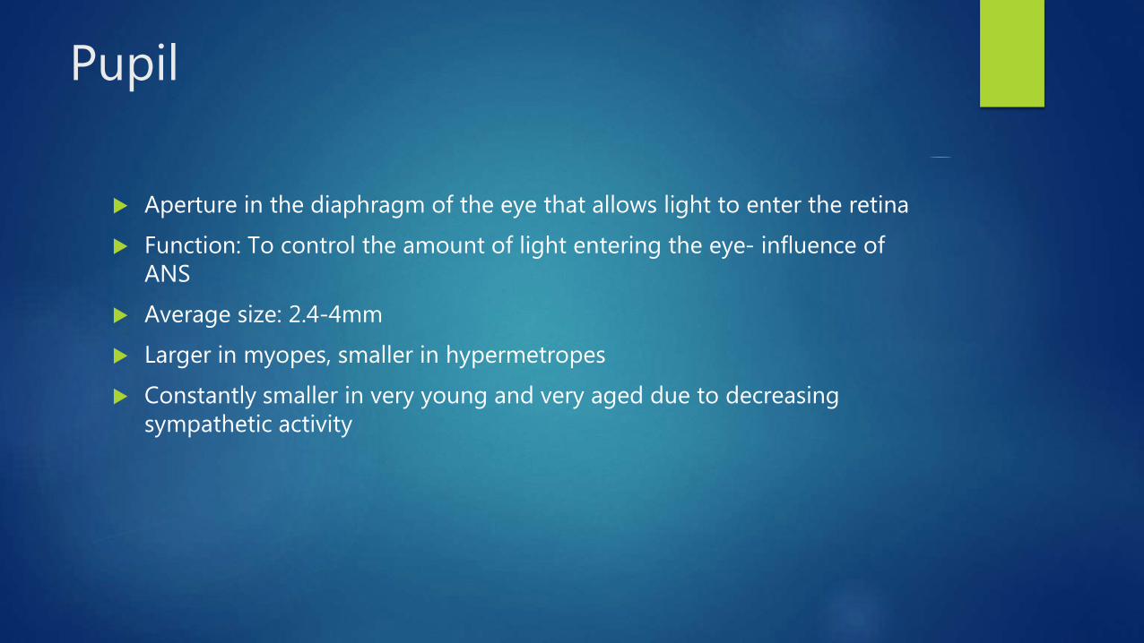
Aperture in the diaphragm of the eye that allows light to enter the retina
Function: To control the amount of light entering the eye- influence of ANS
Average size: 2.4-4mm
Larger in myopes, smaller in hypermetropes
Constantly smaller in very young and very aged due to decreasing sympathetic activity
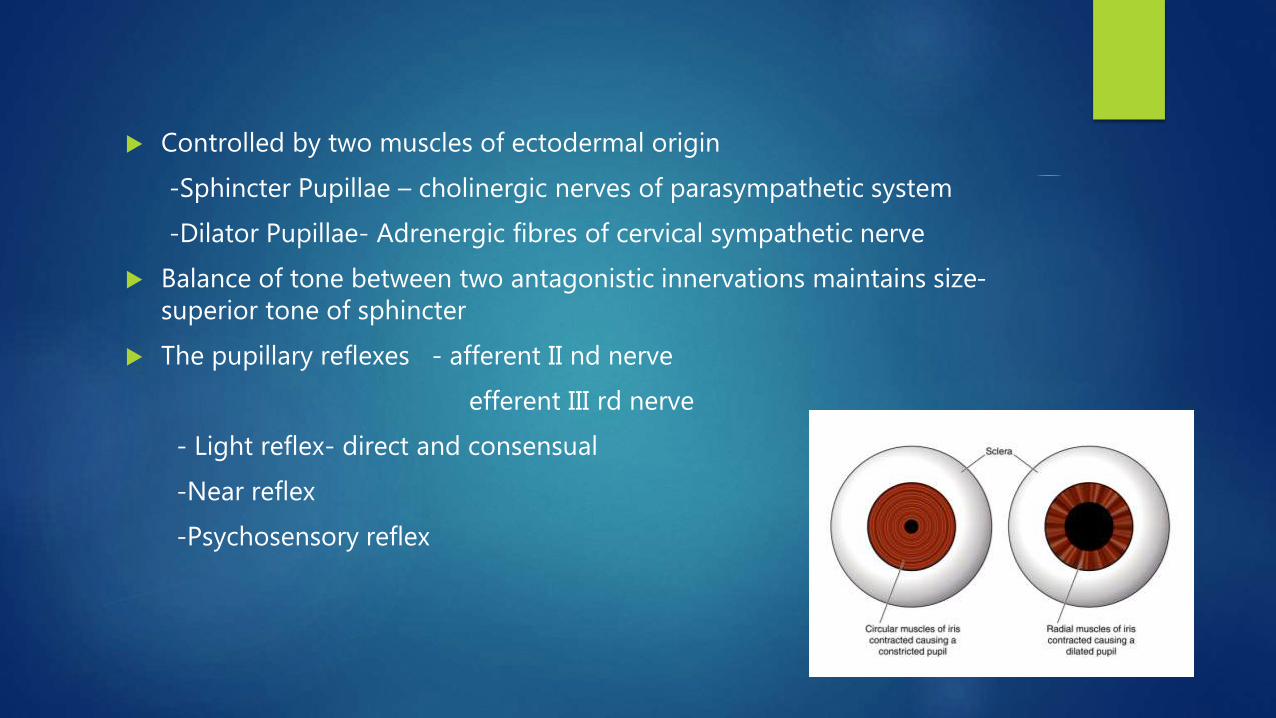
Controlled by two muscles of ectodermal origin
-Sphincter Pupillae – cholinergic nerves of parasympathetic system
-Dilator Pupillae- Adrenergic fibres of cervical sympathetic nerve
Balance of tone between two antagonistic innervations maintains size-superior tone of sphincter
The pupillary reflexes - afferent II nd nerve
efferent III rd nerve
- Light reflex- direct and consensual
-Near reflex
-Psychosensory reflex
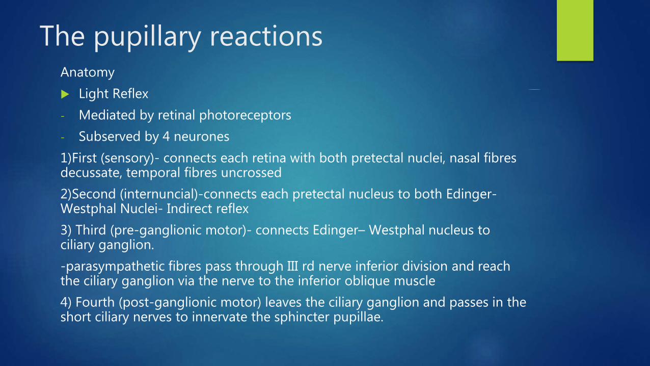
The pupillary reactionsAnatomy
Light Reflex
- Mediated by retinal photoreceptors
- Subserved by 4 neurones
1)First (sensory)- connects each retina with both pretectal nuclei, nasal fibres decussate, temporal fibres uncrossed
2)Second (internuncial)-connects each pretectal nucleus to both Edinger-Westphal Nuclei- Indirect reflex
3) Third (pre-ganglionic motor)- connects Edinger– Westphal nucleus to ciliary ganglion.
-parasympathetic fibres pass through III rd nerve inferior division and reach the ciliary ganglion via the nerve to the inferior oblique muscle
4) Fourth (post-ganglionic motor) leaves the ciliary ganglion and passes in the short ciliary nerves to innervate the sphincter pupillae.
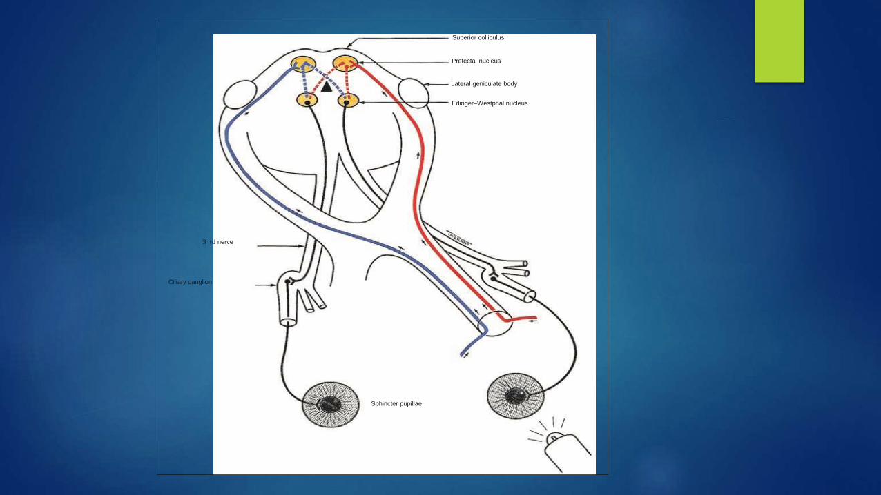
Superior colliculus
Pretectal nucleus
Lateral geniculate body
Edinger–Westphal nucleus
3 rd nerve
Ciliary ganglion
Sphincter pupillae

Near Reflex
- Synkinesis rather than a true reflex
- Accommodation, convergence, miosis on changing gaze from far to near
- Final pathway same as light reflex, centre is ill-defined
Psychosensory reflex

Sympathetic Supply- 3 neurones
1. First (central) starts in posterior hypothalamus and descends, uncrossed
ciliospinal centre of Budge, in the intermediolateral horn of the spinal cord, located between C8 and T2.
2. Second (preganglionic) passes from the ciliospinal centre to the superior cervical ganglion in the neck, closely related to the apical pleura
3. Third (postganglionic) ascends along the internal carotid artery to enter
cavernous sinus ophthalmic division of the trigeminal nerve. The sympathetic fibres reach the ciliary body and the dilator pupillae muscle via the nasociliary nerve and the long ciliary nerves.

Superior cervical ganglion
Ciliospinal centre
of Budge (C8–T2)
Posterior hypothalamus
Abnormal pupillary reactions
Absolute afferent pupillary defect
Complete optic nerve lesion and is characterized by the following:
• The involved eye is completely blind (i.e. no light perception).
• Both pupils are equal in size.
• When the affected eye is stimulated by light neither pupil reacts.
• When the normal eye is stimulated both pupils react normally.
• The near reflex is normal in both eyes.

Relative afferent pupillary defect
-incomplete optic nerve lesion or severe retinal disease
-The clinical features are those of an amaurotic pupil but more subtle
-Thus the pupils respond weakly to stimulation of the diseased eye and briskly to that of the normal eye.
-‘swinging flashlight test’ in which a light source is alternatively switched from one eye to the other and back, thus stimulating each eye in rapid succession
-the dilatation produced by withdrawing the light from the normal eye outweighs the constriction produced by stimulating the abnormal eye.

A
B
C
D
Oculosympathetic Palsy (Horner’s Syndrome)
Mostly unilateral
Mild ptosis (usually 1–2 mm) as a result of weakness of Müller muscle, and miosis due to the unopposed action of the sphincter pupillae with resultant anisocoria.
Normal pupillary reactions to light and near.
Hypochromic heterochromia (iris of different colour – Horner is lighter) may be seen if congenital or long-standing.
Reduced ipsilateral sweating if the lesion is below the superior cervical ganglion, because the sudomotor fibres supplying the skin of the face run along the external carotid artery.

Causes
-Central (1st order neurones):
Brainstem disease (tumour, vascular,demyelination), Syringomyelia , Lateralmedullary (Wallenberg) syndrome, Spinal cord tumour , ,Diabeticautonomic neuropathy
-Preganglionic (second-order neurone)
Pancoast tumour, Carotid and aortic aneurysm and dissection, Necklesions (glands, trauma, postsurgical)
-Postganglionic (third-order neurone)
Cluster headaches (migrainous neuralgia) , Internal carotid arterydissection, Nasopharyngeal tumour, Otitis media , Cavernous sinus mass

Adie’s Pupil
Denervation of the postganglionic supply to the sphincter pupillae and the ciliary muscle, which may follow a viral illness
typically affects young adults and presents as a unilateral condition in 80%
Signs
• Large and regular pupil
• Direct light reflex is absent or sluggish and is associated with vermiform movements of the pupillary border
• Consensual light reflex is absent or sluggish
• The pupil responds slowly to near, following which re-dilatation is also slow.
• Accommodation may manifest similar tonicity
• In long-standing cases the pupil may become small (‘little old Adie’).


In some cases, are diminished deep tendon reflexes (Holmes–Adie syndrome
Pharmacological testing. If 2.5% methacholine or 0.125% pilocarpine is instilled into both eyes, the normal pupil will not constrict, but the abnormal pupil will because of denervation hypersensitivity.
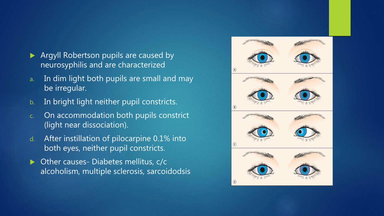
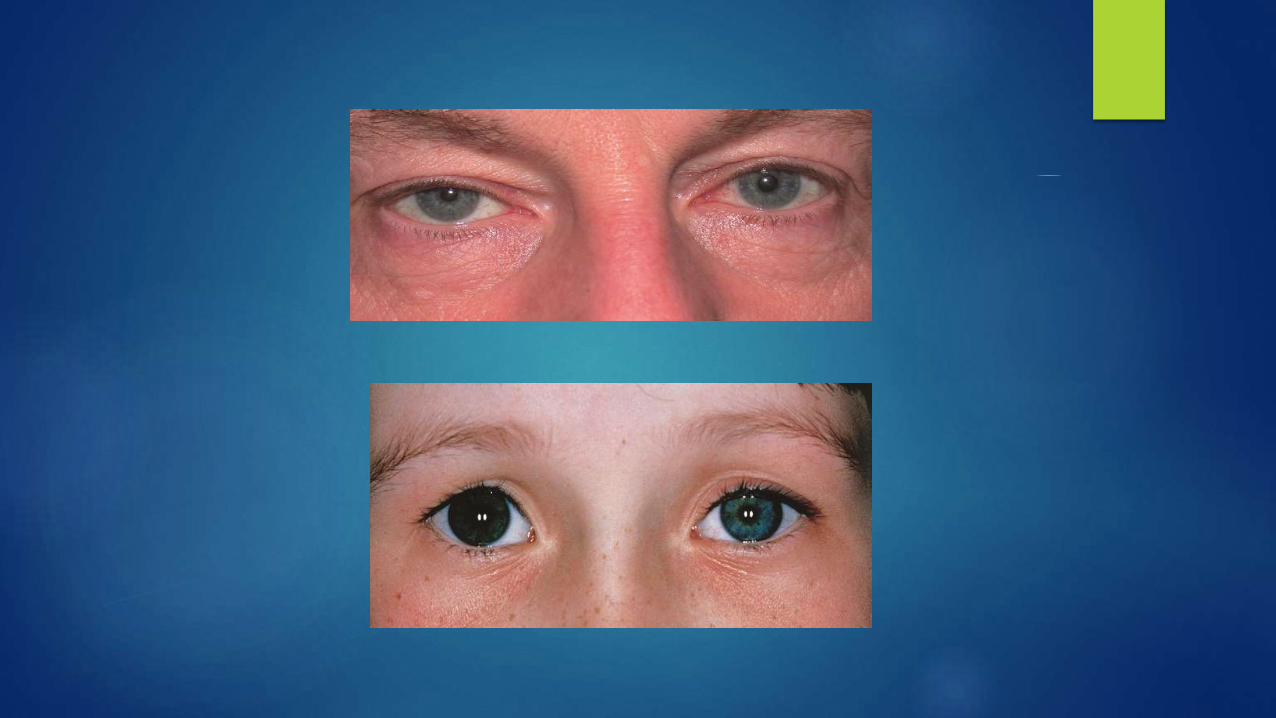
Argyll Robertson pupils are caused by neurosyphilis and are characterized
a. In dim light both pupils are small and may be irregular.
b. In bright light neither pupil constricts.
c. On accommodation both pupils constrict (light near dissociation).
d. After instillation of pilocarpine 0.1% into both eyes, neither pupil constricts.
Other causes- Diabetes mellitus, c/c alcoholism, multiple sclerosis, sarcoidodsis

Tectal (dorsal midbrain) pupils
a. In dim light there is bilateral mydriasiswhich may be asymmetrical.
b. In bright light neither pupil constricts.
c. On accommodation both pupils constrict normally.
d. After instillation of pilocarpine 0.1% to both eyes, neither pupil constricts.

Right episodic mydriasis
a. In dim light the right pupil is larger than the left.
b. In bright light the right pupil does not constrict
c. On accommodation the right pupil does not constrict.
d. Instillation of pilocarpine 0.1% to both eyes fails to constrict either pupil.
e. Instillation of pilocarpine 1% to both eyes induces bilateral miosis.
f. After 24 hours both pupils are equal.

Wernicke hemianopic pupil
• The optic tracts contain both visual and pupillomotor fibres.
The visual fibres terminate in the lateral geniculate body but the pupillary fibres leave the optic tract anterior to the lateral geniculate body, to terminate in the pretectal nuclei.
• An optic tract lesion may therefore give rise to an afferent pupillary conduction defect.
• Characteristically, the pupillary light reflex will be normal when the unaffected hemiretina is stimulated, and absent when the involved hemiretinais stimulated (i.e. light is shone from the hemianopic side).
Difficult to elicit.

Hutchinson pupil
- Seen in comatose patients
- Dilated poorly reactive pupil
- Due to expanding intracranial supratentorial mass causing uncal herniation and IIIrd nerve compression

Assessment of anisocoria
Difference between both pupils >2mm
Dark room test
Miosis – causes
Unilateral Bilateral
- Physiologcal -Physiological senile miosis
- Horner’s syndrome -Pharmacological
- Anterior uveitis -Argyll Robertson pupil
- Long standing Adie’s pupil -Spasm of near reflex
- Pharmacological -Lepromatous miosis
-Congenital microcoria
-Myotonic Dystrophy

Mydriasis- causes
Unilateral Bilateral
-Physiological -Pharmacological
-Pharmacological -Parinaud’s dorsal midbrain syndrome
-Adie’s pupil -Benign periodic mydriasis
-Oculomotor nerve palsy -Brainstem death
-Uncal herniation
-Traumatic sphincter paralysis
-Iris ischemia
-Ocular siderosis

THANK YOU!
THANK YOU!