The Plant Journal 68 Arabidopsis thaliana GEX1 has dual...
13
Arabidopsis thaliana GEX1 has dual functions in gametophyte development and early embryogenesis Monica Alandete-Saez †,‡ , Mily Ron †,§ , Samuel Leiboff and Sheila McCormick * Plant Gene Expression Center and Department of Plant and Microbial Biology, USDA/ARS-UC-Berkeley, 800 Buchanan St., Albany, CA 94710, USA Received 8 April 2011; revised 12 July 2011; accepted 13 July 2011; published online 30 August 2011. * For correspondence (fax +510 559 5678; e-mail [email protected]). † Authors contributed equally to manuscript. ‡ Present address: Public Intellectual Property Resources for Agriculture (PIPRA), University of California at Davis, Davis, CA 95616, USA. § Present address: Department of Plant Biology and Genome Center, University of California at Davis, Davis, CA 95616, USA. SUMMARY GEX1 is a plasma membrane protein that is conserved among plant species, and has previously been shown to be expressed in sperm cells and some sporophytic tissues. Here we show that GEX1 is also expressed in the embryo sac before cellularization, in the egg cell after cellularization, in the zygote/embryo immediately after fertilization and in the pollen vegetative cell. We functionally characterize GEX1 in Arabidopsis thaliana, and show that it is a versatile protein that performs functions during male and female gametophyte development, and during early embryogenesis. gex1-1/+ plants, which synthesize a truncated GEX1 mRNA encoding a protein lacking the predicted cytoplasmic domain, but still targeted to the plasma membrane, had embryos that arrested before the pre-globular stage. gex1-3/+ plants, carrying a null GEX1 allele, had defects during male and female gametophyte development, and during early embryogenesis. Using an antisense GEX1 transgenic line we demonstrate that the predicted GEX1 extracellular domain is sufficient and necessary for GEX1 function during the development of both gametophytes. The predicted cytoplasmic domain is necessary for correct early embryogenesis and mediates homodimer formation at the plasma membrane. We propose that dimerization of GEX1 in the zygote might be an upstream step in a signaling cascade regulating early embryogenesis. Keywords: antisense, embryo sac, pollen, embryo lethal, plasma membrane protein, dimerization, BiFC. INTRODUCTION In flowering plants the mature male gametophyte (pollen grain) contains three cells: a large vegetative cell that forms the pollen tube and two sperm cells enclosed within the vegetative cell cytoplasm (McCormick, 2004). The female gametophyte (embryo sac) develops within the ovule and consists of seven cells of four types: three antipodal cells, two synergid cells, one egg cell and one central cell (Sun- daresan and Alandete-Saez, 2010). The fertilization process begins when the pollen grain germinates on the female stigmatic cells, and ends when the two sperm cells fertilize the egg and the central cell to form the zygote and the primary endosperm cell, respectively. Pollen tubes are guided through female tissues (Pala- nivelu et al., 2003) until they turn towards an available ovule. The two synergid cells within the female gameto- phyte play important roles during pollen tube guidance, as shown in Torenia fournieri (Higashiyama et al., 2001), Zea mays (Okuda et al., 2009), and the Arabidopsis mma1 and maa3 mutants (Shimizu and Okada, 2000). The gametic cells also play a role in pollen tube guidance (von Besser et al., 2006; Chen et al., 2007; Alandete-Saez et al., 2008). The pollen tube enters the micropyle through the filiform apparatus, then releases the two sperm cells within the degenerating synergid cytoplasm (Weterings and Russell, 2004). Pollen tube arrest after entering the micropyle was perturbed by mutations in several genes, including FERO- NIA (Escobar-Restrepo et al., 2007), ANXUR1/2 (Boisson- Dernier et al., 2009), LORELEI (Capron et al., 2008) and AMC (Boisson-Dernier et al., 2008). In most cases, the two sperm cells will complete double fertilization, although single fertilization events may occur after the discharge of two sperm cells (Ron et al., 2010) or after the discharge of only one sperm-like cell (Nowack et al., 2006; Chen et al., 2008; Frank and Johnson, 2009; Aw et al., 2010). Upon double fertilization the development of the embryo and the endosperm is initiated. 620 ª 2011 The Authors The Plant Journal ª 2011 Blackwell Publishing Ltd The Plant Journal (2011) 68, 620–632 doi: 10.1111/j.1365-313X.2011.04713.x
Transcript of The Plant Journal 68 Arabidopsis thaliana GEX1 has dual...

Arabidopsis thaliana GEX1 has dual functions ingametophyte development and early embryogenesis
Monica Alandete-Saez†,‡, Mily Ron†,§, Samuel Leiboff and Sheila McCormick*
Plant Gene Expression Center and Department of Plant and Microbial Biology, USDA/ARS-UC-Berkeley, 800 Buchanan St.,
Albany, CA 94710, USA
Received 8 April 2011; revised 12 July 2011; accepted 13 July 2011; published online 30 August 2011.*For correspondence (fax +510 559 5678; e-mail [email protected]).†Authors contributed equally to manuscript.‡Present address: Public Intellectual Property Resources for Agriculture (PIPRA), University of California at Davis, Davis, CA 95616, USA.§Present address: Department of Plant Biology and Genome Center, University of California at Davis, Davis, CA 95616, USA.
SUMMARY
GEX1 is a plasma membrane protein that is conserved among plant species, and has previously been shown to
be expressed in sperm cells and some sporophytic tissues. Here we show that GEX1 is also expressed in the
embryo sac before cellularization, in the egg cell after cellularization, in the zygote/embryo immediately after
fertilization and in the pollen vegetative cell. We functionally characterize GEX1 in Arabidopsis thaliana, and
show that it is a versatile protein that performs functions during male and female gametophyte development,
and during early embryogenesis. gex1-1/+ plants, which synthesize a truncated GEX1 mRNA encoding a
protein lacking the predicted cytoplasmic domain, but still targeted to the plasma membrane, had embryos
that arrested before the pre-globular stage. gex1-3/+ plants, carrying a null GEX1 allele, had defects during
male and female gametophyte development, and during early embryogenesis. Using an antisense GEX1
transgenic line we demonstrate that the predicted GEX1 extracellular domain is sufficient and necessary for
GEX1 function during the development of both gametophytes. The predicted cytoplasmic domain is necessary
for correct early embryogenesis and mediates homodimer formation at the plasma membrane. We propose
that dimerization of GEX1 in the zygote might be an upstream step in a signaling cascade regulating early
embryogenesis.
Keywords: antisense, embryo sac, pollen, embryo lethal, plasma membrane protein, dimerization, BiFC.
INTRODUCTION
In flowering plants the mature male gametophyte (pollen
grain) contains three cells: a large vegetative cell that forms
the pollen tube and two sperm cells enclosed within the
vegetative cell cytoplasm (McCormick, 2004). The female
gametophyte (embryo sac) develops within the ovule and
consists of seven cells of four types: three antipodal cells,
two synergid cells, one egg cell and one central cell (Sun-
daresan and Alandete-Saez, 2010). The fertilization process
begins when the pollen grain germinates on the female
stigmatic cells, and ends when the two sperm cells fertilize
the egg and the central cell to form the zygote and the
primary endosperm cell, respectively.
Pollen tubes are guided through female tissues (Pala-
nivelu et al., 2003) until they turn towards an available
ovule. The two synergid cells within the female gameto-
phyte play important roles during pollen tube guidance, as
shown in Torenia fournieri (Higashiyama et al., 2001), Zea
mays (Okuda et al., 2009), and the Arabidopsis mma1 and
maa3 mutants (Shimizu and Okada, 2000). The gametic
cells also play a role in pollen tube guidance (von Besser
et al., 2006; Chen et al., 2007; Alandete-Saez et al., 2008).
The pollen tube enters the micropyle through the filiform
apparatus, then releases the two sperm cells within the
degenerating synergid cytoplasm (Weterings and Russell,
2004). Pollen tube arrest after entering the micropyle was
perturbed by mutations in several genes, including FERO-
NIA (Escobar-Restrepo et al., 2007), ANXUR1/2 (Boisson-
Dernier et al., 2009), LORELEI (Capron et al., 2008) and
AMC (Boisson-Dernier et al., 2008). In most cases, the two
sperm cells will complete double fertilization, although
single fertilization events may occur after the discharge of
two sperm cells (Ron et al., 2010) or after the discharge of
only one sperm-like cell (Nowack et al., 2006; Chen et al.,
2008; Frank and Johnson, 2009; Aw et al., 2010). Upon
double fertilization the development of the embryo and the
endosperm is initiated.
620 ª 2011 The AuthorsThe Plant Journal ª 2011 Blackwell Publishing Ltd
The Plant Journal (2011) 68, 620–632 doi: 10.1111/j.1365-313X.2011.04713.x
Embryogenesis is a critical step in the life cycle of seed
plants (Dumas and Rogowsky, 2008). The zygote undergoes
its first unequal cell division to yield an apical cell, which will
develop into the embryo proper, and a basal cell, which will
form the suspensor (Guitton and Berger, 2005). The complex
process of seed development can be conceptually divided
into three phases: the first establishes the pattern of the
embryo by rapid cell division; the second is characterized by
cell expansion and accumulation of reserves; whereas
during the third phase, the seed desiccates (Devic, 2008).
Here we show that Arabidopsis GEX1 is a versatile protein
that functions during the development of the male and
female gametophytes, and during early embryogenesis.
GEX1 is expressed in the embryo sac before cellularization,
in the egg cell after cellularization, in the zygote/embryo
immediately after fertilization and in the male gametophyte,
in both the vegetative cell and in the sperm cells. We
characterized two different GEX1 mutant alleles: gex1-1 and
gex1-3. gex1-1 plants had arrested embryos, whereas gex1-3
plants had defects during male and female gametophyte
development, and during early embryogenesis. We demon-
strate that the predicted GEX1 extracellular domain is
sufficient and necessary for GEX1 function during the
development of male and female gametophytes. Finally,
using bimolecular fluorescence complementation (BiFC), we
show that GEX1 forms homodimers at the plasma mem-
brane through its cytoplasmic domain, and that this cyto-
plasmic domain is necessary for early embryogenesis.
RESULTS
GEX1 in Arabidopsis (At5g55490) is the ortholog of a maize
protein represented by one expressed sequence tag (EST)
among 5000 ESTs from a sperm cell cDNA library (Engel
et al., 2003, 2005). GEX1 has a predicted topology of a signal
peptide, a large extracellular domain (ECD) containing two
coiled-coil domains, three transmembrane domains (TMDs)
and a short cytoplasmic domain (CD), as shown in
Figure 1(a). Consistent with this prediction, a GEX1-eGFP
fusion protein was localized at the plasma membrane upon
transient expression in onion epidermal cells (Engel et al.,
2005), and expression of GEX1 was detected in sperm cells
using a ProGEX1:eGFP reporter line (Engel et al., 2005). The
sequence of GEX1 is conserved among land plants, includ-
ing monocots, eudicots, the moss Physcomitrella and the
lycophyte Selaginella (Figure S1). Plasma membrane pro-
teins that are expressed in gametic cells might play impor-
tant roles in recognition and signaling during gametophyte
development and/or during different steps of the fertilization
process. To determine the role of GEX1 during these stages
we used a reverse genetic approach, and characterized two
T-DNA insertion lines: gex1-1 and gex1-3. The T-DNA
insertions were mapped to two different locations within the
open reading frame (ORF) of GEX1 (Figure 1a), potentially
disrupting different protein domains.
Characterization of mutant phenotypes
To investigate if GEX1 had a role during the fertilization
process, we first determined if the mutant lines had defects
in seed development or seed set. We examined siliques of
gex1-1/+ and gex1-3/+ plants 6–8 days after pollination
(DAP). Both lines showed fewer wild type-like developed
seeds than expected. Superficially, the gex-1 and gex1-3
phenotypes looked similar, but there were differences. The
gex1-1/+ plants showed approximately 25% reduced seed
set (22 � 5% aborted seeds and 3 � 1% undeveloped
ovules; Figure 1c), whereas the gex1-3/+ plants showed 49%
reduced seed set (12 � 2% aborted seeds and 37 � 4%
undeveloped ovules; Figure 1d). We then examined pollen
in open flowers. The gex1-1/+ plants had 100% mature tri-
cellular pollen grains (n = 1399) (Figure S2a,f), but gex1-3/+
plants had 36% aborted pollen (n = 1639) (Figure S2b,g). In
order to ascertain the genetic segregation of each mutant
allele in self-pollinated progeny, we scored the ratio (resis-
tant versus sensitive) of gex1-1/+ and gex1-3/+ plants, using
the linked basta or kanamycin (kan) resistance genes,
respectively. For the gex1-1 allele the segregation ratio was
1.82:1 (1135:622), whereas for the gex1-3 allele the ratio was
1.65:1 (896:620).
These data suggested that each mutant allele partially
affected the transmission of one or both gametophytes,
and/or that the homozygote was embryo lethal. To distin-
guish among these hypotheses, we carried out reciprocal
crosses of gex1-1/+ and gex1-3/+ plants with wild-type
(WT) plants, and scored the transmission efficiency of each
mutation, by selection on basta or kan, respectively.
Table 1 shows that for gex1-1 the transmission of the
T-DNA insertion through the female gametophyte was
complete (approximately 97%), but that transmission
through the male gametophyte was slightly reduced
(approximately 78%). When pollen from these plants was
crossed with WT females, the resulting siliques (6–8 DAP)
had nearly full seed set (93 � 4%, n = 520), therefore
indicating that the male gametophyte defect was not
causing the 25% reduced seed set observed in the
gex1-1/+ plants. In contrast, heterozygous gex1-3/+ plants
showed 41% transmission of the mutant allele through the
female gametophyte, and 47% transmission of the mutant
allele through the male gametophyte (Table 1). When
gex1-3/+ plants were used as pollen donors on WT pistils,
the siliques had nearly full seed set (96 � 4%, n = 312),
indicating, as in gex1-1/+ plants, that the male gameto-
phyte defect was not causing the reduced seed set
observed in the gex1-3/+ plants.
When gex1-3/+ plants were used as females, however, the
siliques had 62 � 8% WT seeds, 36 � 7% undeveloped
ovules and 1 � 1% aborted seeds (n = 211): this data is
consistent with the reduced (41%) transmission of the gex1-
3 allele through the female gametophyte.
Dual functions of GEX1 621
Published 2011. This article is a US Government work and is in the public domain in the USAThe Plant Journal, (2011), 68, 620–632

To determine if all embryo sacs were targeted by pollen
tubes, we used decolorized aniline blue (DAB) to visualize
pollen tubes in the pistils of gex1-1/+ and gex1-3/+ plants.
We collected self-pollinated flowers at 1–2 DAP and stained
the pistils with DAB. In gex1-1/+ plants all ovules were
reached by pollen tubes (Figure 1e), which is consistent with
the full transmission of the gex1-1 allele through the female.
In the gex1-3/+ plants 35 � 6% (n = 615) of the ovules did
not attract pollen tubes (Figure 1f–h), consistent with the
reduced transmission of the gex1-3 allele through the
female. To confirm the female gametophyte defect we
crossed pollen of the ProLAT52:GUS (Twell et al., 1991)
Table 1 Transmission Efficiency in gex1-1/+ and gex 1-3/+
Parental Genotype(Female X Male) BastaR BastaS
TransmissionEfficiency (TE) (%) c2 Test
gex1-1/+ X WT 125 129 97 c2 = 0.063, P = 0.8018WT X gex1-1/+ 165 207 78 c2 = 4.742, P = 0.0294a
gex1-3/+ X WT 73 175 41 c2 = 41.952, P < 0.0001a
WT X gex1-3/+ 74 156 47 c2 = 29.235, P < 0.0001a
Transmission efficiency = Resistant/ Sensitive · 100.aTE is statistically significant from the 100% TE expected for a sporophytic mutation.
(a)
(b)
(c)
(d)
(e)
(g)
(i)
(f)
(h)
(j)
Figure 1. Phenotypic characterization of gex1-1/+ and gex1-3/+ plants.
(a) Top, diagram of genomic locus of GEX1, including exons (black boxes) and introns (lines), and locations of T-DNA insertions in gex1-1 and gex1-3. Bottom,
diagram of the predicted topology of GEX1, including a signal peptide in red and two coiled-coil motifs in grey.
(b) Silique (6–8 days after pollination, DAP) from a wild-type (WT) plant with full seed set.
(c) Silique (6–8 DAP) from a gex1-1/+ plant showing aborted seeds (yellow arrow).
(d) Silique (6–8 DAP) from a gex1-3/+ plant showing aborted seeds (yellow arrow) and undeveloped ovules (white arrow).
(e) Self-crossed pistil (1–2 DAP) of a gex1-1/+ plant stained with DAB; all female gametophytes were reached by a pollen tube.
(f) Self-crossed pistil (1–2 DAP) of a gex1-3/+ plant stained with DAB, showing female gametophytes that were fertilized after the arrival of a pollen tube (white
arrow), and female gametophytes that were not reached by a pollen tube (yellow arrow).
(g and h) Unfertilized ovules showing pollen tube guidance defects of gex1-3/+ plants (yellow arrow points pollen tube).
(i) Two ovules in a pistil (1–2 DAP) from a cross using gex1-3/+ plant as female and the ProLAT52:GUS reporter line as a male. A fertilized ovule (black arrow points to
a blue pollen tube at the micropyle), and a putative gex1-3 ovule (red arrow) that failed to attract a pollen tube towards the micropyle.
(j) Close up of the putative gex1-3 ovule in (i). CD, cytoplasmic domain; ECD, extracellular domain; TMD, transmembrane domain. Scale bars: e, f, 100 lm; g–j,
50 lm.
622 Monica Alandete-Saez et al.
Published 2011. This article is a U.S. Government work and is in the public domain in the USAThe Plant Journal, (2011), 68, 620–632

reporter line with gex1-3/+ plants: 37 � 9% ovules (n = 209)
were not reached by blue pollen tubes (Figure 1i,j) in siliques
at 1–2 DAP. Because defects in female gametophyte devel-
opment can cause a failure in pollen tube attraction (Marton
and Dresselhaus, 2010), we decided to analyze mature
female gametophytes of gex1-3/+ plants to determine if
there were abnormalities in cell polarity and/or in the
position of the nuclei. Pistils of emasculated gex1-3/+ plants
had 63 � 7% (n = 354) WT-like four-celled embryo sacs (FG7
stage) (Sundaresan and Alandete-Saez, 2010), but 36 � 7%
of the embryo sacs had arrested at the functional megaspore
stage (FG1 stage) (Figure 2a,b, respectively). Younger pistils
dissected from closed buds of gex1-3/+ plants also carried
both WT-like FG5 (eight nuclei) and FG6 (seven-celled and
eight nuclei) stage embryo sacs, and embryo sacs that had
arrested at the FG1 stage (Figure 2c,e,d, respectively). All
mature female gametophytes in gex1-1/+ plants showed
correct cell polarity and nuclei position (Figure 2f). These
results suggest that putative gex1-3 female gametophytes
were arrested at the FG1 stage, a phenotype that explains
the reduced ability to attract pollen tubes observed in gex1-
3/+ plants.
gex1-1/gex1-1 and gex1-3/gex1-3 affect early embryo
development
The transmission and seed set results indicated that gex1-1
and gex1-3 mutant phenotypes were not fully penetrant, and
therefore both the male and female gametophytes could
transmit the mutant allele to the next generation. It should
therefore be possible to obtain plants homozygous for these
alleles. We selected seeds from self-pollinated gex1-1/+ and
gex1-3/+ plants on basta or kan, respectively, and genotyped
over 100 seedlings from each line. No homozygotes were
identified for either mutant, suggesting that gex1-1/gex1-1
and gex1-3/gex1-3 are embryo lethal, which was consistent
with the aborted seeds found in siliques of both mutant lines.
To determine the stage of embryogenesis that was
compromised in the putative gex1-1/gex1-1 and gex1-3/
gex1-3 embryos, we dissected and cleared seeds from
siliques at different developmental stages and visualized
them using differential interference contrast (DIC) imaging.
Figure 3(a,b) shows normal embryo development at the
triangular stage (5 DAP), and Figure 3(c,d) shows normal
embryo development at the torpedo stage (6–7 DAP).
Figure 3(e,f) shows abnormal embryo development in a
putative gex1-1/gex1-1 seed from the same silique as
Figure 3(a), whereas Figure 3(g) and 3(h) show seeds with
abnormal embryo development from the same silique as
Figure 3(c). After the analysis of over 100 seeds showing
abnormal embryo development, we observed that these
embryos arrested between the two- and eight-celled embryo
stage, and showed altered cell size in most cases, although
abnormal arrested embryos at later developmental stages
were also found. These seeds were slightly smaller than
other seeds in the same silique, and showed endosperm
nuclear divisions. We saw a similar arrested embryo devel-
opment phenotype in a few seeds of self-pollinated gex1-3/+
plants at 5 DAP (Figure 3i,j). To confirm that the endosperm
development observed in the aborted seeds resulted from a
product of fertilization, and not from autonomous endo-
sperm development, we introgressed the reporter gene line
ProFAC1:GUS, which is expressed in both embryo
and endosperm upon fertilization (Xu et al., 2005), into the
gex1-1/+ background. Figure 3(k) shows a WT developing
seed that has GUS signal in the embryo and endosperm at
2 DAP. Figure 3(L) shows a putative gex1-1/gex1-1 seed with
GUS expression in the arrested embryo and developing
endosperm at 2 DAP. We conclude that the putative gex1-1/
gex1-1 zygote/embryo arrested development soon after
double fertilization, even though the endosperm continued
developing (for at least 6–7 DAP) before the seed finally
aborted.
(a)
(c)
(b)
(e)
(d)
(f)
Figure 2. Female gametophyte development in gex1-1/+ and gex1-3/+ plants.
(a) Mature wild-type embryo sac after cellularization and differentiation (stage
FG7) in emasculated pistils of gex1-3/+ plants.
(b) Arrested embryo sac at FG1 in an ovule from the same pistil as in (a).
(c) Wild-type embryo sac at FG5 in pistils of gex1-3/+ plants.
(d) Arrested embryo sac at FG1 in an ovule from same pistil as (c).
(e) Wild-type embryo sac at FG6 in pistils of gex1-3/+ plants.
(f) Mature wild-type embryo sac after cellularization (stage FG7) in emascu-
lated pistils of gex1-1/+ plants. Ccn, central cell nucleus; Ecn, egg cell nucleus;
FM, functional megaspore; Syn, synergid nucleus. Nuclei are false-colored.
Dual functions of GEX1 623
Published 2011. This article is a US Government work and is in the public domain in the USAThe Plant Journal, (2011), 68, 620–632

We demonstrated that the phenotypes observed in the
two mutant lines were caused by the corresponding T-DNA
insertions by introducing a construct carrying the GEX1 WT
allele driven by its own promoter into the two mutant
backgrounds. In each case, the phenotypes were fully
complemented (Figure S3).
GEX1 is expressed in male and female gametophytes
The phenotypes identified in the insertion lines suggested
a role for GEX1 in the female and male gametophyte, and
in the embryo. To further evaluate GEX1 expression,
we generated the GUS reporter construct ProGEX1:GUS.
A GUS signal was detected in the embryo sac before
cellularization (a very weak signal at the FG1/FG2 stage
and a more visible signal at the FG3/FG4/FG5 stage), in egg
cells of mature female gametophytes and in zygotes/em-
bryos of ovules at 1–2 DAP, but not in embryos older than
2 DAP (Figure 4a–f). This data is consistent with the gene
expression map of Arabidopsis embryo development
(http://www2.bri.nrc.ca/plantembryo) (Xiang et al., 2011),
which reported high expression levels of GEX1 in the
elongated zygote through the octant embryo stage. We
also detected GUS in the vegetative cell, in a few pollen
tubes upon pollen germination, and in root tips
(Figure S4). RT-PCR using RNA from different tissues
(Figure 4g) showed that GEX1 was expressed strongly in
pollen and weakly in unpollinated pistils. These additional
expression data support the role of GEX1 during gameto-
phyte development and early embryogenesis inferred from
the gex1-1 and gex1-3 mutant lines.
(a) (b) (c) (d)
(e) (g) (h)(f)
(i) (j) (k) (l)
Figure 3. Embryo development in gex1-1/+ and gex1-3/+ plants.
(a) Wild-type (WT) embryo development at the heart stage (4 days after pollination, DAP) (arrow).
(b) Higher magnification of (a).
(c) WT embryo development at the torpedo stage (6–7 DAP) (arrow).
(d) Higher magnification of (c).
(e) Abnormal seed development (4 DAP) in gex1-1/+ plants, with endosperm development and an arrested embryo.
(f) Higher magnification of (e) showing the arrested embryo (arrow).
(g) Abnormal seed development (6–7 DAP) in gex1-1/+ plants, with endosperm development and an arrested embryo.
(h) Higher magnification of (g), showing the arrested four-celled embryo (arrow).
(i) Abnormal seed development (4 DAP) in gex1-3/+ plants, showing endosperm development and an arrested embryo.
(j) Higher magnification of (e) showing the arrested two-celled embryo (arrow).
(k) WT seed (2 DAP) fertilized with pollen from the ProFAC1:GUS reporter line, showing GUS signal in the embryo (arrow) and the endosperm.
(l) Putative gex1-1/gex1-1 seed (2 DAP) fertilized with pollen from the ProFAC1:GUS reporter line, showing GUS signal in the arrested embryo and the endosperm.
EMB, embryo; END, endosperm. Scale bar:50 lm.
624 Monica Alandete-Saez et al.
Published 2011. This article is a U.S. Government work and is in the public domain in the USAThe Plant Journal, (2011), 68, 620–632

We performed quantitative real-time PCR (qRT-PCR) to
analyze GEX1 expression levels in the WT and in the mutant
lines. We first extracted RNA from pollen and unpollinated
pistils of gex1-1/+ plants, and observed that GEX1 mRNA
levels were approximately 50% of WT levels in both
(Figure 4h). Because the level of GEX1 expression could be
detected equally in pollen and unpollinated pistils, we used
RNA from unpollinated pistils for further analysis, as they
were easier to collect. The mRNA levels of GEX1 in unpol-
linated pistils of gex1-3/+ plants were approximately 50% of
the levels found in WT plants (Figure 4h). In addition, we
quantified GEX1 expression with a third allele, gex1-2. The
expression of GEX1 in unpollinated pistils, anthers and
leaves, and in pistils at 3 DAP, was higher in gex1-2/+ plants
than in the WT (Figure S5). The T-DNA insertion in gex1-2/+
plants (FLAG line) contained a CaMV35S promoter immedi-
ately before the left border, and we therefore reasoned that
higher expression of GEX1 could be caused by such a
promoter, as has happened for other T-DNA insertion lines
(Williams et al., 2005). We did not characterize gex1-2
further.
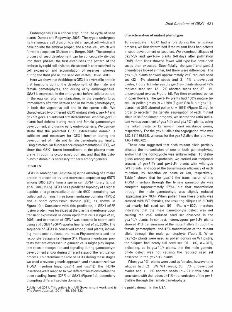
gex1-1/+ plants express a truncated GEX1 protein (gex1-1)
that localizes to the plasma membrane
We thought that the differences observed between the two
alleles might be related to the position of the T-DNA inser-
tion within the open reading frame (ORF) of GEX1, and we
hypothesized that gex1-1/+ plants might synthesize a trun-
cated GEX1 mRNA, up to the T-DNA insertion (Figure 5a),
that might be translated and localized at the plasma mem-
brane. To test this hypothesis we first determined GEX1
mRNA levels using primers located upstream of the T-DNA
insertion in gex1-1/+ plants. qRT-PCR analysis showed that
GEX1 mRNA levels in unpollinated pistils were similar to WT
levels (Figure 5b), suggesting that a partial GEX1 mRNA was
probably transcribed up to the location of the T-DNA inser-
tion; when we used primers located downstream of the
insertion we observed that GEX1 mRNA expression was
decreased to 50% of WT levels (Figures 4h and 5b). In
addition, 3¢ rapid amplification of cDNA ends (3¢RACE) using
RNA from unpollinated pistils of gex1-1/+ plants detected
two bands (Figure 5c), one corresponding to full-length
GEX1 mRNA and the second to a truncated GEX1 mRNA up
to the T-DNA insertion location, and including 51 nucleo-
tides carrying a sequence that is complementary to the left
border of the T-DNA, which would encode 16 amino acids
before a stop codon.
Because this GEX1-1 truncated mRNA was polyadeny-
lated, we tested if the partial mRNA synthesized in gex1-1/+
plants could be translated into a truncated protein lacking
129 amino acids (illustrated in Figure 5a), and if so, to
determine its subcellular localization. We bombarded onion
cells with a construct encoding the putative truncated
version fused to GFP at the C terminus and under the
control of the CaMV35S promoter (Pro35S:gex1-1-eGFP).
Figure 5(d) shows that the WT version of GEX1 is located at
the plasma membrane, as has already been shown by Engel
et al. (2005). We also detected some perinuclear signal, most
likely corresponding to the endoplasmic reticulum (Beraud-
Dufour and Balch, 2002), and probably to the result of excess
fusion protein in the exocytic pathway (Roselli et al., 2004).
Figure 5(f) shows that the truncated mRNA was translated
into a protein that was also targeted to the plasma
(a) (b)
(c) (d)
(g)
(h)
(e)
(f)
Figure 4. Expression in ProGEX1:GUS plants, and GEX1 mRNA level in gex1-
1/+ and gex1-3/+ plants.
(a, b) GUS expression in female gametophytes before the cellularization of
ProGEX1:GUS plants.
(c) GUS signal in the egg cell of ovules dissected from unpollinated pistils.
(d) GUS signal in the zygote/embryo of ovules dissected from siliques 1 day
after pollination (DAP).
(e) Mature pollen grains expressing GUS.
(f) Absence of GUS signal in seed at 2 DAP. The position of the embryo is
shown with a dotted line.
(g) RT-PCR with eight different tissues of Arabidopsis, using GEX1 (upper) and
ACTIN2 (lower) primers.
(h) GEX1 mRNA levels measured using qRT-PCR in pollen and unpollinated
pistils of WT and gex1-1/+ plants; and in unpollinated pistils of WT and gex1-3/
+ plants. Wild-type levels were set to 100%. Scale bar:50 lm. Error bars
represent standard deviation.
Dual functions of GEX1 625
Published 2011. This article is a US Government work and is in the public domain in the USAThe Plant Journal, (2011), 68, 620–632
membrane. The gex1-1-eGFP fusion protein also showed a
weak perinuclear signal.
The truncated gex1-1 protein is equivalent to GEX1 during
gametophyte development but not during early
embryogenesis
Because gex1-1/+ plants did not show pollen tube guidance
defects or embryo sac arrest, nor the pollen abortion ob-
served in gex1-3/+ plants, we hypothesized that the trun-
cated gex1-1 protein (Figure 5a) might be equivalent to
GEX1 during male and female gametophyte development
before double fertilization. If so, silencing the expression
of the gex1-1 truncated mRNA in the male and female
gametophytes in gex1-1/+ plants might phenocopy the
defects observed in gex1-3/+ plants. To test this hypothesis
we generated transgenic lines silencing GEX1 expression
only in the gametophytes by using an antisense construct
driven by the GEX2 promoter that is not active in sporo-
phytic tissues (Engel et al., 2005), nor in the zygote or
embryo, according to the gene expression map of Arabid-
opsis embryo development (http://www2.bri.nrc.ca/plant-
embryo; Xiang et al., 2011), but is expressed both in the
sperm (Engel et al., 2005) and in the egg (Alandete-Saez et
al., 2008).
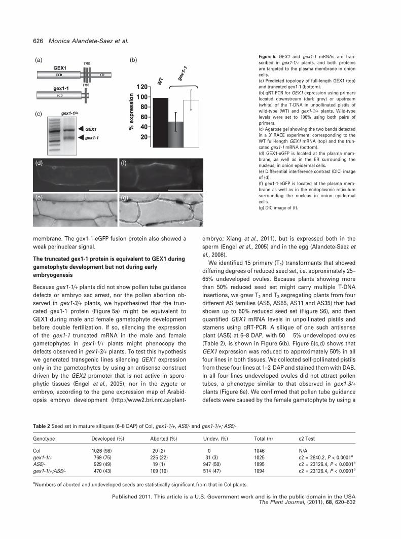
We identified 15 primary (T1) transformants that showed
differing degrees of reduced seed set, i.e. approximately 25–
65% undeveloped ovules. Because plants showing more
than 50% reduced seed set might carry multiple T-DNA
insertions, we grew T2 and T3 segregating plants from four
different AS families (AS5, AS55, AS11 and AS35) that had
shown up to 50% reduced seed set (Figure S6), and then
quantified GEX1 mRNA levels in unpollinated pistils and
stamens using qRT-PCR. A silique of one such antisense
plant (AS5) at 6–8 DAP, with 50 � 5% undeveloped ovules
(Table 2), is shown in Figure 6(b). Figure 6(c,d) shows that
GEX1 expression was reduced to approximately 50% in all
four lines in both tissues. We collected self-pollinated pistils
from these four lines at 1–2 DAP and stained them with DAB.
In all four lines undeveloped ovules did not attract pollen
tubes, a phenotype similar to that observed in gex1-3/+
plants (Figure 6e). We confirmed that pollen tube guidance
defects were caused by the female gametophyte by using a
(a) (b)
(c)
(d)
(e)
(f)
(g)
Figure 5. GEX1 and gex1-1 mRNAs are tran-
scribed in gex1-1/+ plants, and both proteins
are targeted to the plasma membrane in onion
cells.
(a) Predicted topology of full-length GEX1 (top)
and truncated gex1-1 (bottom).
(b) qRT-PCR for GEX1 expression using primers
located downstream (dark grey) or upstream
(white) of the T-DNA in unpollinated pistils of
wild-type (WT) and gex1-1/+ plants. Wild-type
levels were set to 100% using both pairs of
primers.
(c) Agarose gel showing the two bands detected
in a 3¢ RACE experiment, corresponding to the
WT full-length GEX1 mRNA (top) and the trun-
cated gex1-1 mRNA (bottom).
(d) GEX1-eGFP is located at the plasma mem-
brane, as well as in the ER surrounding the
nucleus, in onion epidermal cells.
(e) Differential interference contrast (DIC) image
of (d).
(f) gex1-1-eGFP is located at the plasma mem-
brane as well as in the endoplasmic reticulum
surrounding the nucleus in onion epidermal
cells.
(g) DIC image of (f).
Table 2 Seed set in mature siliques (6–8 DAP) of Col, gex1-1/+, AS5/- and gex1-1/+; AS5/-
Genotype Developed (%) Aborted (%) Undev. (%) Total (n) c2 Test
Col 1026 (98) 20 (2) 0 1046 N/Agex1-1/+ 769 (75) 225 (22) 31 (3) 1025 c2 = 2840.2, P < 0.0001a
AS5/- 929 (49) 19 (1) 947 (50) 1895 c2 = 23126.4, P < 0.0001a
gex1-1/+;AS5/- 470 (43) 109 (10) 514 (47) 1094 c2 = 23126.4, P < 0.0001a
aNumbers of aborted and undeveloped seeds are statistically significant from that in Col plants.
626 Monica Alandete-Saez et al.
Published 2011. This article is a U.S. Government work and is in the public domain in the USAThe Plant Journal, (2011), 68, 620–632
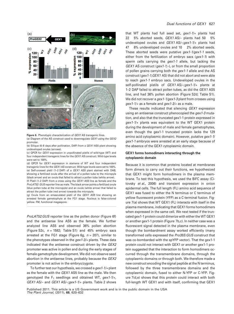
ProLAT52:GUS reporter line as the pollen donor (Figure 6f)
and the antisense line AS5 as the female. We further
analyzed line AS5 and observed 36% pollen abortion
(Figure S2c, n = 1582; Table S1) and 45% embryo sacs
arrested at the FG1 stage (Figure 6g, n = 207), similar to
the phenotypes observed in the gex1-3/+ plants. These data
indicated that the antisense construct driven by the GEX2
promoter was active in pollen and during the early stages of
female gametophyte development. We did not observe seed
abortion in the antisense lines, probably because the GEX2
promoter is not active in the embryo/zygote.
To further test our hypothesis, we crossed a gex1-1/+ plant
as the female with the GEX1 AS5 line as the male. We then
genotyped the F1 seedlings and obtained WT, gex1-1/+,
GEX1 AS/– and GEX1 AS/–;gex1-1/+ plants. Table 2 shows
that WT plants had full seed set, gex1-1/+ plants had
22 � 5% aborted seeds, GEX1 AS/– plants had 50 � 5%
undeveloped ovules and GEX1 AS/–;gex1-1/+ plants had
47 � 6% undeveloped ovules and 10 � 2% aborted seeds.
These aborted seeds were putative gex1-1/gex1-1 seeds,
either from the fertilization of embryo sacs (gex1-1) with
sperm cells carrying the gex1-1 allele, but lacking the
GEX1 AS construct (gex1-1;–), or from the small proportion
of pollen grains carrying both the gex1-1 allele and the AS
construct (gex1-1;GEX1 AS) that did not abort and were able
to reach gex1-1 embryo sacs. Undeveloped ovules in the
self-pollinated pistils of GEX1 AS/–;gex1-1/+ plants at
1–2 DAP failed to attract pollen tubes, as did the GEX1 AS5
line, and had 36% pollen abortion (Figure S2d; Table S1).
We did not recover a gex1-1/gex1-3 plant from crosses using
gex1-1/+ as a female and gex1-3/+ as a male.
These results indicated that silencing GEX1 expression
using an antisense construct phenocopied the gex1-3 muta-
tion, and also that the truncated gex1-1 protein expressed in
gex1-1/+ plants was equivalent to the WT GEX1 protein
during the development of male and female gametophytes,
even though the gex1-1 truncated protein lacks the 129
amino acid cytoplasmic domain). However, putative gex1-1/
gex1-1 embryos were arrested at an early stage because of
the absence of the GEX1 cytoplasmic domain.
GEX1 forms homodimers interacting through the
cytoplasmic domain
Because it is common that proteins located at membranes
form dimers to carry out their functions, we hypothesized
that GEX1 might form homodimers in the plasma mem-
brane. To test this hypothesis, we used the BiFC assay (Ci-
tovsky et al., 2006) and transient expression in onion
epidermal cells. The full length (FL) amino acid sequence of
GEX1 was fused to either the N terminus or C terminus of
yellow fluorescent protein (YFP) as a C-terminal fusion. Fig-
ure 7(a) shows that WT GEX1 (FL) interacts with itself in the
plasma membrane, indicating that GEX1 forms homodimers
when expressed in the same cell. We next tested if the trun-
cated gex1-1 protein could dimerize with either the WT GEX1
or another gex1-1 protein (Figure 7b,c). In neither case was a
fluorescent signal detected in the plasma membrane, even
though the bombardment assay worked efficiently (many
transformed cells expressed the Pro35S:GUS construct that
was co-bombarded with the spYFP vector). That the gex1-1
protein could not interact with GEX1 or another gex1-1 pro-
tein suggested that the interaction to form homodimers oc-
curred through the transmembrane domains, through the
cytoplasmic domains or through both. We therefore made a
new construct encoding the signal peptide at the N terminus,
followed by the three transmembrane domains and the
cytoplasmic domain, fused to either N-YFP or C-YFP. Fig-
ure 7(d,e) shows that this protein could interact with both
full-length WT GEX1 and with itself, confirming that GEX1
(a) (b)
(e) (g)
(c) (d)
(f)
Figure 6. Phenotypic characterization of GEX1 AS transgenic lines.
(a) Diagram of the AS construct used to downregulate GEX1 using the GEX2
promoter.
(b) Silique (6–8 days after pollination, DAP) from a GEX1 AS5 plant showing
undeveloped ovules (arrows).
(c) QPCR for GEX1 expression in unpollinated pistils of wild-type (WT) and
four independent transgenic lines for the GEX1 AS construct. Wild-type levels
were set to 100%.
(d) QPCR for GEX1 expression in stamens of WT and four independent
transgenic lines for the GEX1 AS construct. Wild-type levels were set to 100%.
(e) Self-crossed pistil (1–2 DAP) of a GEX1 AS5 plant stained with DAB,
showing a fertilized ovule after the arrival of a pollen tube to the micropyle
(black arrow) and an ovule that failed to attract a pollen tube (white arrow).
(f) Pistil (1–2 DAP) from a cross using the GEX1 AS5 line as female and the
ProLAT52: GUS reporter line as male. The black arrow points a fertilized ovule
(blue pollen tube at the micropyle) and an ovule (white arrow) that failed to
attract the pollen tube (red arrow) towards the micropyle.
(g) Ovule from an emasculated pistil of the GEX1 AS5 line showing an
arrested female gametophyte at the FG1 stage. Nucleus is false-colored
yellow. FM, functional megaspore.
Dual functions of GEX1 627
Published 2011. This article is a US Government work and is in the public domain in the USAThe Plant Journal, (2011), 68, 620–632
forms homodimers through its transmembrane and cyto-
plasmic domains. We observed perinuclear fluorescence
with all GEX1 protein versions that showed positive interac-
tions in the BiFC assay, which was consistent with the sub-
cellular localization of both the full-length GEX1 and gex1-1
truncated proteins. In addition, we tested whether the
extracellular domain (ECD) of GEX1 could interact with itself
in the opposite orientation, because that would suggest that
GEX1 could form homodimers when in the plasma mem-
brane of two different cells (e.g. egg and sperm cells, before
or during the fusion of their membranes). We used the pre-
dicted ECD of GEX1, without the predicted signal peptide,
fused to either the N terminus or the C terminus of YFP as
C-terminal or N-terminal fusions, but did not detect any
interaction between the extracellular domains.
DISCUSSION
GEX1 was previously shown to be expressed in sperm cells
(Engel et al., 2005; Borges et al., 2008; Slotkin et al., 2009) as
well as in other sporophytic tissues using a GFP reporter
construct (Engel et al., 2005). Here we used a more sensitive
GUS reporter to show that GEX1 is also expressed during
early embryo sac development and in the egg cell, but also
during the first divisions of the zygote/embryo (Figure 4a–d).
We did not detect GUS signal in the sperm cells, possibly
because expression in the vegetative cell masked the sperm
cell signal, or because the promoter used for the GUS
reporter construct (same promoter used in the comple-
mentation constructs) was slightly smaller than that used for
the ProGEX1-eGFP construct (upstream of the ATG at 1606
versus 1808 bp). Using a reverse genetic approach we
showed that GEX1 is a versatile protein of dual function
during gametophyte development and early embryogene-
sis. Our genetic approach was unable to uncover a role for
GEX1 in sperm cells during fertilization, because of its earlier
role during pollen development, although that possibility
cannot be ruled out. The gex1-3 T-DNA insertion, located in
the fourth exon (Figure 1a), disrupts GEX1 mRNA tran-
scription before the sequence encoding its transmembrane
domain and, based on qRT-PCR data, is a null allele. gex1-3/+
plants showed defects in pollen (Figure S2), embryo sac
(Figure 2) and early embryo development (Figure 3).
Although it is well documented that the embryo transcrip-
tome shares a high number of expressed genes with both
(a)
(b)
(c)
(d)
(e)
(f)
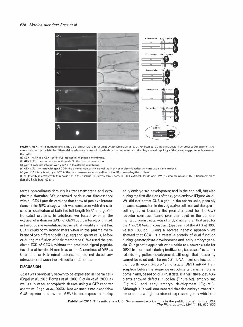
Figure 7. GEX1 forms homodimers in the plasma membrane through its cytoplasmic domain (CD). For each panel, the bimolecular fluorescence complementation
assay is shown on the left, the differential interference contrast image is shown in the center, and the diagram and topology of the interacting proteins is shown on
the right.
(a) GEX1-nCFP and GEX1-cYFP (FL) interact in the plasma membrane.
(b) GEX1 (FL) does not interact with gex1-1 in the plasma membrane.
(c) gex1-1 does not interact with gex1-1 in the plasma membrane.
(d) GEX1 (FL) interacts with gex1-CD in the plasma membrane, as well as in the endoplasmic reticulum surrounding the nucleus.
(e) gex1-CD interacts with gex1-CD in the plasma membrane, as well as in the ER surrounding the nucleus.
(f) nEFP-VirD2 interacts with AtImpa-4cYFP in the nucleus. CD, cytoplasmic domain; ECD, extracellular domain; PM, plasma membrane; TMD, transmembrane
domain. Scale bars:100 lm.
628 Monica Alandete-Saez et al.
Published 2011. This article is a U.S. Government work and is in the public domain in the USAThe Plant Journal, (2011), 68, 620–632
the male and female gametophytes (Haecker et al., 2004;
Berg et al., 2005; Jenik et al., 2007; Spencer et al., 2007; Ba-
yer et al., 2009; Wuest et al., 2010), there is limited infor-
mation linking the expression pattern of a gametophytically
expressed gene with a role during early embryogenesis
(Bayer et al., 2009). The embryo sacs carrying the gex1-3
allele showed arrested development at the FG1 stage (Fig-
ure 2), and did not attract pollen tubes (Figure 1f); most
likely because of the absence of attractant molecules re-
quired for micropylar pollen tube guidance, which are only
secreted when female gametophytes are fully mature (Hig-
ashiyama, 2002; Palanivelu and Preuss, 2006; Dresselhaus
and Marton, 2009). In contrast to mutations in other sperm-
expressed genes (von Besser et al., 2006; Mori et al., 2006;
Nowack et al., 2006; Chen et al., 2008; Ron et al., 2010), gex1-
3 pollen grains did not show defects in pollen tube guidance,
pollen tube reception or fertilization. Instead, gex1-3 pollen
grains aborted, suggesting that GEX1 is essential for correct
pollen maturation, as is the case for other pollen-expressed
genes (Chen and McCormick, 1996; Boavida et al., 2009).
GEX1 is an essential gene in Arabidopsis because both gex1-
3/gex1-3 and gex1-1/gex1-1 embryos arrested at an early
stage. Even though GEX1 is expressed in the male and fe-
male gametic cells, reciprocal crosses indicated that the
embryo arrest phenotype was caused by a recessive muta-
tion and was not maternally or paternally imprinted, as is the
case for the glauce and short suspensor mutants, respec-
tively (Ngo et al., 2007; Bayer et al., 2009).
Half of the gametophytes produced by gex1-1/+ plants
expressed the WT full-length GEX1 mRNA, and the other
half expressed truncated gex1-1 mRNA. We showed that
both transcripts (GEX1 and gex1-1) could be translated into
proteins that were targeted to the plasma membrane
(Figure 5c). gex1-1/+ plants did not have the gametophytic
defects observed in gex1-3/+ plants. Three lines of evidence
support the hypothesis that the predicted extracellular
domain of GEX1, when inserted at the plasma membrane,
is sufficient and necessary for the development of both male
and female gametophytes: (i) gex1-1/+ plants had aborted
seeds because of embryo arrest, and not the gametophytic
defects observed in gex1-3/+ plants (Figure 3); (ii) transgenic
lines carrying a GEX1 antisense construct phenocopied the
gametophytic defects observed in gex1-3 plants, consistent
with the downregulation of GEX1 transcripts (Figure 6); and
(iii) the introduction of the antisense construct in the gex1-1
background phenocopied the gametophytic defects caused
by the gex1-3 mutation, which is consistent with the
downregulation of GEX1 and gex1-1 transcripts (Table 2).
GEX1 formed homodimers in the plasma membrane, and its
predicted cytoplasmic domain (CD) was sufficient and
necessary for GEX1 interaction and function during early
embryogenesis (Figure 7). However, the fully functional
gametophytes found in gex1-1/+ plants indicate that the
role of the predicted extracellular domain (ECD) of GEX1
during gametophyte development does not require GEX1
dimerization. Although we did not obtain evidence that the
extracellular domain of GEX1 can interact with itself when in
opposite orientations, these BiFC experiments cannot rule
out the possibility that GEX1 can form homodimers when in
the plasma membrane of two different cells, because the
interaction might require specific protein folding in the
plasma membrane. Coiled-coil domains are found in diverse
proteins and mediate protein–protein interactions (Suzuki
et al., 2008). The presence of two coiled-coil domains in the
predicted extracellular domain of GEX1 (Figure 1a) support
the idea that the extracellular domain might interact with
itself or with other proteins.
The strong GUS signal observed in embryos at 1–2 DAP
(Figure 4d) and microarray expression data reported in the
gene expression map of Arabidopsis embryo development
(http://www2.bri.nrc.ca/plantembryo; Xiang et al., 2011)
support the speculation that de novo transcription/transla-
tion of GEX1 occurs immediately after karyogamy, instead of
by protein diffusion in the plasma membrane after fusion of
the gametic cells, as is true for other plasma membrane
proteins (Hink et al., 2008). The de novo translated GEX1
would then form dimers in the plasma membrane through
its cytoplasmic domain, and we hypothesize that this
dimerization would be required for an upstream step in a
signaling cascade involved in the establishment and devel-
opment of the early embryo. It would be interesting to
investigate if the homeobox transcription factors WOX2 and
WOX8, which act as complementary cell fate regulators in
the apical and basal lineages during early embryogenesis
(Breuninger et al., 2008), and are co-expressed in both the
egg cell and zygote (Haecker et al., 2004), might be down-
stream players in this signaling cascade.
EXPERIMENTAL PROCEDURES
Primers for cloning and genotyping are listed in Table S2.
Plant materials and growth conditions
Arabidopsis plants (Columbia, Col-0) were grown in a glasshouse asdescribed by Alandete-Saez et al. (2008). Seeds were plated on MSmedium containing 30 lg ml)1 BASTA (gex1-1/+ and GEX1 ASplants) or 50 lg ml)1 kanamycin (gex1-2/+ and gex1-3/+ plants), andthe resulting seedlings were scored for resistance 2 weeks later.
T-DNA line and sequence analyses
The left borders of the T-DNA insertions CS817262 (gex1-1),FLAG_48E09 (gex1-2) and FLAG_081B03 (gex1-3) were amplified byPCR and sequenced to verify the insertion sites. The structure andsubcellular localization of GEX1 were predicted as described inAlandete-Saez et al. (2008).
Cloning and generation of transgenic lines
All constructs were subcloned into pENTR 3C or pENTR/D-TOPOvectors (Invitrogen, http://www.invitrogen.com), confirmed bysequencing and transferred to destination vectors by an LR reactionusing LR Clonase II (Invitrogen). The full-length GEX1 open reading
Dual functions of GEX1 629
Published 2011. This article is a US Government work and is in the public domain in the USAThe Plant Journal, (2011), 68, 620–632

frame was amplified from Arabidopsis pollen cDNA using primersMA7/MA8, and was cloned in an antisense orientation intopB7WG2*-ProGEX2, as in (Alandete-Saez et al., 2008). The GEX1promoter (1.6 kb) was amplified using primers MA9/MA10 andcloned into pBGWFS7 (Karimi et al., 2002) to generate the reporterline ProGEX1:eGFP-GUS. For the complementation construct a 3.8-kb fragment of GEX1, including the promoter and 3¢ untranslatedregion (UTR), was amplified from genomic DNA using primersMA11/MA12 and cloned into a modified pK2GW7 (for gex1-1 com-plementation) or pH2GW7 (for gex1-3 complementation) vectors(Karimi et al., 2002), from which the CaMV35S promoter had beenremoved to generate ProGEX1:GEX1 complementation constructs.
Phenotypic analysis and complementation of gex1-1/+
and gex1-3/+ plants
Phenotypic analyses of seed set and reciprocal crosses were per-formed as described in Alandete-Saez et al. (2008). Decolorizedaniline blue (DAB) staining was performed as described in Moriet al. (2006). GUS expression analyses were performed as describedin Johnson et al. (2004), and seed clearings were performed asdescribed in Pagnussat et al. (2005). To complement the gex1-1mutant, the ProGEX1:GEX1 complementation construct was intro-duced into Col-0 plants using Agrobacterium transformation(Clough and Bent, 1998), then pollen from transgenic T1 lines wasused for crosses with gex1-1/+. Seedlings from these crosses wereselected on kanamycin and were genotyped for the presence of thegex1-1 T-DNA. Five independent ProGEX1:GEX1; gex1-1/+ F1 plantswere taken to the next generation. Seeds were selected on kana-mycin and homozygous gex1-1/gex1-1/GEX1 complementationplants were identified by PCR. To complement the gex1-3 mutant,the ProGEX1:GEX1 complementation construct was introduced intogex1-3/+ plants. T1 lines were selected on hygromycin and geno-typed for the presence of the gex1-3 T-DNA. Positive T1 plants wereallowed to self and T2 seedlings were selected on both hygromycinand kanamycin. Homozygous gex1-3/gex1-3/GEX1 complementa-tion plants were identified by PCR.
RT-PCR and real-time PCR
RNA was extracted from different tissues using an RNeasy plantminiprep kit (Qiagen, http://www.qiagen.com) with on-columnDNAse treatment. cDNA was synthesized using 20-mer oligod(T)and Superscript� III Reverse Transcriptase (Invitrogen). RT-PCR wasperformed using 1 lg of total RNA. Real-time PCR primer pairs weredesigned using PRIMER EXPRESS (Applied Biosystems, http://www.appliedbiosystems.com). IPP2 (At3g02780) was used for nor-malization, as described in Alandete-Saez et al. (2008). Amplifica-tion was performed with a MyIQ Real-Time PCR Detection System(Bio-Rad, http://www.bio-rad.com), with 1.5 lg and 500 ng of totalRNA from unpollinated pistils and anthers, respectively.
Generation of clones for subcellular localization and BiFC
assays
For subcellular localization, the open reading frames of GEX1 or ofgex1-1, without the stop codons, were amplified from cDNA pre-pared from unpollinated pistils and cloned into pB7FWG2 (Karimiet al., 2002) to generate Pro35S:GEX1-eGFP and Pro35S:GEX1-1-eGFP. To generate constructs for BiFC, all fragments were amplifiedfrom cDNA prepared from unpollinated pistils and cloned into eitherpSAT4-nYFP-N1 or pSAT4-cYFP-N1 (Citovsky et al., 2006). The full-length GEX1 sequence was amplified using the primers MA30/MA31 and the truncated gex1-1 was amplified using primers MA30/MA32. The predicted GEX1 ECD without the signal peptide wasamplified using primers MA33/MA34 or MA35/MA36. The cyto-
plasmic domain construct was cloned by overlapping PCR. Thesignal peptide (36 amino acids) was amplified with the primersMA37/MA38 and the sequence encoding the transmembrane andcytoplasmic domains was amplified using the primers MA39/MA40.The amplicons were gel-purified and 0.5 ll of each fragment wasused as template for a second PCR reaction (50 ll) with the primersMA37/MA40 to amplify the full-length overlapping sequence. Theseconstructs were used for particle bombardment in onion epidermalcells. Microcarriers were prepared with 5 lg of plasmid for subcel-lular localization or 2 lg of each construct (i.e. nYFP-fused andcYFP-fused) for BiFC, including 500 ng of the Pro35S:GUS con-struct, mixed and coated onto 1.0-mm gold particles (SeashellTechnology, http://www.seashelltech.com) according to the manu-facturer’s protocol. The particles were bombarded into peeled epi-dermal layers of onion using a PDS-100/He particle delivery system(Bio-Rad). The epidermal layers were incubated in the dark at 22�Cand observed 16–20 h after bombardment using a Zeiss Axiovertmicroscope (Zeiss, http://www.zeiss.com). As negative controls,each protein-nYFP and protein-cYFP fusion construct was bom-barded with the complementary half-YFP vector; both empty vec-tors were also tested.
ACKNOWLEDGEMENTS
We thank Waichoi Shek and Jamison Smith for technical assistance,Dr Leonor Boavida (Gulbenkian Institute, Portugal) for commentson the article, and Dr Stan Gelvin (Purdue University) for providingthe BiFC vectors. This work was supported by the NSF Plant Gen-ome program grant 0211742 and by USDA CRIS 5335-21000-030-00D. MR was partially supported by a Vaadia-BARD PostdoctoralFellowship Award No. FI-391-2006 from BARD, The United States–Israel Binational Agricultural Research and Development Fund.
SUPPORTING INFORMATION
Additional supporting information may be found in the onlineversion of this article:Figure S1. Amino acid sequence alignment of GEX1.Figure S2. Pollen phenotypes of gex1-1/+; gex1-3/+, GEX1AS/– and gex1-1/+;GEX1 AS/– plants.Figure S3. Complementation of gex1-1 and gex1-3 phenotypes.Figure S4. GEX1 expression in plants carrying the ProGEX1:GUSconstruct.Figure S5. Quantification of GEX1 mRNA levels by qRT-PCR inplants carrying the gex1-2 T-DNA insertion.Figure S6. Seed formation in mature siliques of four independentGEX1 AS lines.Table S1. Pollen in open flowers of Col, gex1-1/+, AS5/– andgex1-1/+; AS5/– plants.Table S2. Primers used in this study.Please note: As a service to our authors and readers, this journalprovides supporting information supplied by the authors. Suchmaterials are peer-reviewed and may be re organized for onlinedelivery, but are not copy-edited or typeset. Technical supportissues arising from supporting information (other than missingfiles) should be addressed to the authors.
REFERENCES
Alandete-Saez, M., Ron, M. and McCormick, S. (2008) GEX3, expressed in the
male gametophyte and in the egg cell of Arabidopsis thaliana, is essential
for micropylar pollen tube guidance and plays a role during early
embryogenesis. Mol. Plant, 1, 586–598.
Aw, S., Hamamura, Y., Schnittger, A. and Berger, F. (2010) Sperm entry is
sufficient to trigger division of the central cell but the paternal genome is
required for endosperm development in Arabidopsis. Development, 137,
2683–2690.
630 Monica Alandete-Saez et al.
Published 2011. This article is a U.S. Government work and is in the public domain in the USAThe Plant Journal, (2011), 68, 620–632

Bayer, M., Nawy, T., Giglione, C., Galli, M., Meinnel, T. and Lukowitz, W.
(2009) Paternal control of embryonic patterning in Arabidopsis thaliana.
Science, 323, 1485–1488.
Beraud-Dufour, S. and Balch, W. (2002) A journey through the exocytic
pathway. J. Cell Sci. 115, 1779–1780.
Berg, M., Rogers, R., Muralla, R. and Meinke, D. (2005) Requirement of ami-
noacyl-tRNA synthetases for gametogenesis and embryo development in
Arabidopsis. Plant J. 44, 866–878.
von Besser, K., Frank, A.C., Johnson, M.A. and Preuss, D. (2006) Arabidopsis
HAP2 (GCS1) is a sperm-specific gene required for pollen tube guidance
and fertilization. Development, 133, 4761–4769.
Boavida, L.C., Shuai, B., Yu, H.J., Pagnussat, G.C., Sundaresan, V. and
McCormick, S. (2009) A collection of Ds insertional mutants associated with
defects in male gametophyte development and function in Arabidopsis
thaliana. Genetics, 181, 1369–1385.
Boisson-Dernier, A., Frietsch, S., Kim, T.H., Dizon, M.B. and Schroeder, J.I.
(2008) The peroxin loss-of-function mutation abstinence by mutual consent
disrupts male-female gametophyte recognition. Curr. Biol. 18, 63–68.
Boisson-Dernier, A., Roy, S., Kritsas, K., Grobei, M.A., Jaciubek, M., Schroe-
der, J.I. and Grossniklaus, U. (2009) Disruption of the pollen-expressed
FERONIA homologs ANXUR1 and ANXUR2 triggers pollen tube discharge.
Development, 136, 3279–3288.
Borges, F., Gomes, G., Gardner, R., Moreno, N., McCormick, S., Feijo, J.A. and
Becker, J.D. (2008) Comparative transcriptomics of Arabidopsis sperm
cells. Plant Physiol. 148, 1168–1181.
Breuninger, H., Rikirsch, E., Hermann, M., Ueda, M. and Laux, T. (2008) Dif-
ferential expression of WOX genes mediates apical-basal axis formation in
the Arabidopsis embryo. Dev. Cell, 14, 867–876.
Capron, A., Gourgues, M., Neiva, L.S., et al. (2008) Maternal control of male-
gamete delivery in Arabidopsis involves a putative GPI-anchored protein
encoded by the LORELEI gene. Plant Cell, 20, 3038–3049.
Chen, Y.-C.S. and McCormick, S. (1996) sidecar pollen, an Arabidopsis thali-
ana male gametophyte mutant with aberrant cell divisions during pollen
development. Development, 122, 3243–3253.
Chen, Y.H., Li, H.J., Shi, D.Q., Yuan, L., Liu, J., Sreenivasan, R., Baskar, R.,
Grossniklaus, U. and Yang, W.C. (2007) The central cell plays a critical role
in pollen tube guidance in Arabidopsis. Plant Cell, 19, 3563–3577.
Chen, Z., Tan, J.L., Ingouff, M., Sundaresan, V. and Berger, F. (2008) Chro-
matin assembly factor 1 regulates the cell cycle but not cell fate during male
gametogenesis in Arabidopsis thaliana. Development, 135, 65–73.
Citovsky, V., Lee, L.Y., Vyas, S., Glick, E., Chen, M.H., Vainstein, A., Gafni, Y.,
Gelvin, S.B. and Tzfira, T. (2006) Subcellular localization of interacting
proteins by bimolecular fluorescence complementation in planta. J. Mol.
Biol. 362, 1120–1131.
Clough, S.J. and Bent, A.F. (1998) Floral dip: a simplified method for Agro-
bacterium-mediated transformation of Arabidopsis thaliana. Plant J. 16,
735–743.
Devic, M. (2008) The importance of being essential: EMBRYO-DEFECTIVE
genes in Arabidopsis. C. R. Biol. 331, 726–736.
Dresselhaus, T. and Marton, M.L. (2009) Micropylar pollen tube guidance and
burst: adapted from defense mechanisms? Curr. Opin. Plant Biol. 12, 773–
780.
Dumas, C. and Rogowsky, P. (2008) Fertilization and early seed formation. C.
R. Biol. 331, 715–725.
Engel, M.L., Chaboud, A., Dumas, C. and McCormick, S. (2003) Sperm cells of
Zea mays have a complex complement of mRNAs. Plant J. 34, 697–707.
Engel, M.L., Holmes-Davis, R. and McCormick, S. (2005) Green sperm. Iden-
tification of male gamete promoters in Arabidopsis. Plant Physiol. 138,
2124–2133.
Escobar-Restrepo, J.M., Huck, N., Kessler, S., Gagliardini, V., Gheyselinck, J.,
Yang, W.C. and Grossniklaus, U. (2007) The FERONIA receptor-like kinase
mediates male-female interactions during pollen tube reception. Science,
317, 656–660.
Frank, A.C. and Johnson, M.A. (2009) Expressing the diphtheria toxin A
subunit from the HAP2(GCS1) promoter blocks sperm maturation and
produces single sperm-like cells capable of fertilization. Plant Physiol. 151,
1390–1400.
Guitton, A.E. and Berger, F. (2005) Loss of function of MULTICOPY SUP-
PRESSOR OF IRA 1 produces nonviable parthenogenetic embryos in Ara-
bidopsis. Curr. Biol. 15, 750–754.
Haecker, A., Gross-Hardt, R., Geiges, B., Sarkar, A., Breuninger, H., Herrmann,
M. and Laux, T. (2004) Expression dynamics of WOX genes mark cell fate
decisions during early embryonic patterning in Arabidopsis thaliana.
Development, 131, 657–668.
Higashiyama, T. (2002) The synergid cell: attractor and acceptor of the pollen
tube for double fertilization. J. Plant. Res. 115, 149–160.
Higashiyama, T., Yabe, S., Sasaki, N., Nishimura, Y., Miyagishima, S., Ku-
roiwa, H. and Kuroiwa, T. (2001) Pollen tube attraction by the synergid cell.
Science, 293, 1480–1483.
Hink, M.A., Shah, K., Russinova, E., de Vries, S.C. and Visser, A.J. (2008)
Fluorescence fluctuation analysis of Arabidopsis thaliana somatic
embryogenesis receptor-like kinase and brassinosteroid insensitive 1
receptor oligomerization. Biophys. J. 94, 1052–1062.
Jenik, P.D., Gillmor, C.S. and Lukowitz, W. (2007) Embryonic patterning in
Arabidopsis thaliana. Annu. Rev. Cell. Dev. Biol. 23, 207–236.
Johnson, M.A., von Besser, K., Zhou, Q., Smith, E., Aux, G., Patton, D., Levin,
J.Z. and Preuss, D. (2004) Arabidopsis hapless mutations define essential
gametophytic functions. Genetics, 168, 971–982.
Karimi, M., Inze, D. and Depicker, A. (2002) GATEWAY((TM)) vectors for
Agrobacterium-smediated plant transformation. Trends Plant Sci. 7, 193–
195.
Marton, M.L. and Dresselhaus, T. (2010) Female gametophyte-controlled
pollen tube guidance. Biochem. Soc. Trans. 38, 627–630.
McCormick, S. (2004) Control of male gametophyte development. Plant Cell,
16, S142–S153.
Mori, T., Kuroiwa, H., Higashiyama, T. and Kuroiwa, T. (2006) GENERATIVE
CELL SPECIFIC 1 is essential for angiosperm fertilization. Nat. Cell Biol. 8,
64–71.
Ngo, Q.A., Moore, J.M., Baskar, R., Grossniklaus, U. and Sundaresan, V.
(2007) Arabidopsis GLAUCE promotes fertilization-independent endo-
sperm development and expression of paternally inherited alleles. Devel-
opment, 134, 4107–4117.
Nowack, M.K., Grini, P.E., Jakoby, M.J., Lafos, M., Koncz, C. and Schnittger, A.
(2006) A positive signal from the fertilization of the egg cell sets off endo-
sperm proliferation in angiosperm embryogenesis. Nat. Genet. 38, 63–67.
Okuda, S., Tsutsui, H., Shiina, K. et al. (2009) Defensin-like polypeptide LUREs
are pollen tube attractants secreted from synergid cells. Nature, 458, 357–
361.
Pagnussat, G.C., Yu, H.J., Ngo, Q.A., Rajani, S., Mayalagu, S., Johnson, C.S.,
Capron, A., Xie, L.F., Ye, D. and Sundaresan, V. (2005) Genetic and
molecular identification of genes required for female gametophyte devel-
opment and function in Arabidopsis. Development, 132, 603–614.
Palanivelu, R. and Preuss, D. (2006) Distinct short-range ovule signals attract
or repel Arabidopsis thaliana pollen tubes in vitro. BMC Plant Biol. 6, 7.
Palanivelu, R., Brass, L., Edlund, A.F. and Preuss, D. (2003) Pollen tube growth
and guidance is regulated by POP2, an Arabidopsis gene that controls
GABA levels. Cell, 114, 47–59.
Ron, M., Alandete-Saez, M., Eshed-Williams, L., Fletcher, J.C. and McCor-
mick, S. (2010) Proper regulation of a sperm-specific cis-nat-siRNA is
essential for double fertilization in Arabidopsis. Genes Dev. 24, 1010–1021.
Roselli, S., Moutkine, I., Gribouval, O., Benmerah, A. and Antignac, C. (2004)
Plasma membrane targeting of podocin through the classical exocytic
pathway: effect of NPHS2 mutations. Traffic, 5, 37–44.
Shimizu, K.K. and Okada, K. (2000) Attractive and repulsive interactions be-
tween female and male gametophytes in Arabidopsis pollen tube guid-
ance. Development, 127, 4511–4518.
Slotkin, R.K., Vaughn, M., Borges, F., Tanurdzic, M., Becker, J.D., Feijo, J.A.
and Martienssen, R.A. (2009) Epigenetic reprogramming and small RNA
silencing of transposable elements in pollen. Cell, 136, 461–472.
Spencer, M.W., Casson, S.A. and Lindsey, K. (2007) Transcriptional profiling
of the Arabidopsis embryo. Plant Physiol. 143, 924–940.
Sundaresan, V. and Alandete-Saez, M. (2010) Pattern formation in miniature:
the female gametophyte of flowering plants. Development, 137, 179–189.
Suzuki, K., Mizuno, T. and Tanaka, T. (2008) Regulation of protein-protein
interaction via assembly of coiled-coil domain. Nucleic Acids Symp. Ser.
(Oxf). 52, 461.
Twell, D., Yamaguchi, J., Wing, R.A., Ushiba, J. and McCormick, S. (1991)
Promoter analysis of genes that are coordinately expressed during pollen
development reveals pollen-specific enhancer sequences and shared reg-
ulatory elements. Genes Dev. 5, 496–507.
Dual functions of GEX1 631
Published 2011. This article is a US Government work and is in the public domain in the USAThe Plant Journal, (2011), 68, 620–632

Weterings, K. and Russell, S.D. (2004) Experimental analysis of the fertiliza-
tion process. Plant Cell, 16(Suppl), S107–S118.
Williams, L., Grigg, S.P., Xie, M., Christensen, S. and Fletcher, J.C. (2005)
Regulation of Arabidopsis shoot apical meristem and lateral organ for-
mation by microRNA miR166g and its AtHD-ZIP target genes. Develop-
ment, 132, 3657–3668.
Wuest, S.E., Vijverberg, K., Schmidt, A., Weiss, M., Gheyselinck, J., Lohr, M.,
Wellmer, F., Rahnenfuhrer, J., von Mering, C. and Grossniklaus, U. (2010)
Arabidopsis female gametophyte gene expression map reveals similarities
between plant and animal gametes. Curr. Biol. 20, 506–512.
Xiang, D., Venglat, P., Tibiche, C. et al. (2011) Genome-wide analysis reveals
gene expression and metabolic network dynamics during embryo devel-
opment in Arabidopsis. Plant Physiol. 156, 346–356.
Xu, J., Zhang, H.-Y., Xie, C., Xue, H., Dijkhuis, P. and Liu, C. (2005) EMBRY-
ONIC FACTOR 1 encodes an AMP deaminase and is essential for the zygote
to embryo transition in Arabidopsis. Plant J. 42, 743–756.
632 Monica Alandete-Saez et al.
Published 2011. This article is a U.S. Government work and is in the public domain in the USAThe Plant Journal, (2011), 68, 620–632