The Physiology of the Alimentary Canal of Tyroglyphus...
17
45 The Physiology of the Alimentary Canal of Tyroglyphus farinae BY T. E. HUGHES, M.A. (From the Zoology Department, Birkbeck College, University of London) T HIS investigation into the activities of the gut in Tyroglyphus farinae Linn. was undertaken because so little is known of the physiology of the tyro- glyphid mites. The work was started in 1940 and has been continued as opportunity arose since then. It was hoped that the region of secretion of the digestive enzymes, together with the seat of carbohydrate and protein diges- tion and absorption, might be determined. Since the Malpighian tubes in the Tyroglyphidae (Acaridiae) are reduced or absent, it was hoped to find what region of the gut is responsible for the excretion of nitrogenous waste. An attempt was also made to identify the chief nitrogenous katabolites. METHODS Mites were cultured on food material stained with various indicators; by this means the pH of the gut was determined subject to protein error, and the passage of food was followed. Pig meal and bemax were found to be good culture media for this purpose. The cultures were kept in cells formed by fixing a glass ring on to a slide with gold size; they were covered by another slide held in position by a rubber band. The cells were kept in a dessicator at a relative humidity of 90 per cent. Changes in colour of the indicator could be observed by transparency through the body-wall of the living animal. Cultures were also made on rice starch, potato starch, gluten, and yeast cells to localize carbohydrate and protein digestion. Animals cultured on starch were mounted in a dilute alcoholic solution of iodine. Gluten was used as a protein, because if fed on whole grain the mites attack the germ which is rich in this substance. Fibrin and dried red-blood corpuscles were consistently refused by the animals. Some difficulty was encountered in finding a method of mounting which would render the mites clear without interfering with the colour of the indicator. The method finally adopted was to drop a live mite into glycerine on a slide; on this was placed a coverslip smeared with glycerine jelly. The slide was warmed just sufficiently to melt the jelly; this greatly helped the clearing, and on cooling the weight of the coverslip was generally found to have spread the legs without bursting the body. Mounted in this way, the animal could be examined with an oil-immersion lens. Quarterly Journal Microscopical Science, Vol. 91, part 1, March, 1950.
Transcript of The Physiology of the Alimentary Canal of Tyroglyphus...

45
The Physiology of the Alimentary Canal ofTyroglyphus farinae
BY
T. E. HUGHES, M.A.(From the Zoology Department, Birkbeck College, University of London)
THIS investigation into the activities of the gut in Tyroglyphus farinae Linn.was undertaken because so little is known of the physiology of the tyro-
glyphid mites. The work was started in 1940 and has been continued asopportunity arose since then. It was hoped that the region of secretion of thedigestive enzymes, together with the seat of carbohydrate and protein diges-tion and absorption, might be determined. Since the Malpighian tubes inthe Tyroglyphidae (Acaridiae) are reduced or absent, it was hoped to findwhat region of the gut is responsible for the excretion of nitrogenous waste.An attempt was also made to identify the chief nitrogenous katabolites.
METHODS
Mites were cultured on food material stained with various indicators; bythis means the pH of the gut was determined subject to protein error, and thepassage of food was followed. Pig meal and bemax were found to be goodculture media for this purpose. The cultures were kept in cells formed byfixing a glass ring on to a slide with gold size; they were covered by anotherslide held in position by a rubber band. The cells were kept in a dessicatorat a relative humidity of 90 per cent. Changes in colour of the indicatorcould be observed by transparency through the body-wall of the livinganimal.
Cultures were also made on rice starch, potato starch, gluten, and yeast cellsto localize carbohydrate and protein digestion. Animals cultured on starchwere mounted in a dilute alcoholic solution of iodine. Gluten was used as aprotein, because if fed on whole grain the mites attack the germ which is richin this substance. Fibrin and dried red-blood corpuscles were consistentlyrefused by the animals.
Some difficulty was encountered in finding a method of mounting whichwould render the mites clear without interfering with the colour of theindicator. The method finally adopted was to drop a live mite into glycerineon a slide; on this was placed a coverslip smeared with glycerine jelly. Theslide was warmed just sufficiently to melt the jelly; this greatly helped theclearing, and on cooling the weight of the coverslip was generally found tohave spread the legs without bursting the body. Mounted in this way, theanimal could be examined with an oil-immersion lens.Quarterly Journal Microscopical Science, Vol. 91, part 1, March, 1950.

46 Hughes—The Physiology of
Efforts were made and pursued for several months to dissect out the gut,but without success. As the body-wall of the opisthosoma is punctured,the parenchyma loses fluid and the mite collapses. It appears that mitescultured at a relative humidity of 90 per cent, have a positive internal pres-sure in this part of the body; this is indicated by their turgid and glisteningappearance.
Extracts were made of whole animals collected by standing a beaker ofheavily infected material in a half petri dish of water. The mites leaving theculture collected on the water and could be filtered off and washed; aftergrinding with glass powder, various extracts were made by shaking with theextracting fluid and centrifuging off the solid debris.
Serial sections were cut from paraffin, celloidin, and ester-wax blocks, andstained with Mallory, haematoxylin and eosin, mucicarmine and muci-haematein to investigate the histology of the gut. Some sections were cut afterfixation with a 1 per cent, solution of osmium tetroxide to determine thedistribution of fats. After fixation with Pasted and Leonard's fluid (Lisbon,1936), sections were made and stained with Best's carmine to find the site ofglycogen storage. Bauer's reaction was also successfully employed in thisrespect, as a more specific histochemical test for carbohydrates.
Extracts were made from heavily infected cultures, and specific tests werecarried out for various nitrogenous katabolites. Changes in size, opacity, andbirefringence of the faecal pellet in its passage through the gut were alsoinvestigated. In the following description the term 'dense culture' is employedfor cultures containing more than io4 mites per gramme in all stages of thelife-cycle excluding eggs; young cultures are those containing less than io3
mites per gramme, and here the mites are hard to find. In the former, thewhole medium appears to be in motion when seen under the binocular micro-scope, and ultimately such cultures come to consist of exuvia, faecal pellets,and mites only.
ANATOMY AND FUNCTION
The anatomy of the gut of the Tyroglyphids has been described by Michael(1901) and its homologies discussed by Lonnfors (1930); a general descrip-tion is also given by Vitzthum (1940). The nomenclature used by these andother authors is by no means uniform, and an attempt to correlate it wasthought to be useful.
The arachnid gut like that of all Arthropoda is divisible into three regions,a fore-gut lined with chitin, an endodermal mesenteron, and a chitin-linedhind-gut. It is characteristic of the group that the hind-gut—derived fromthe proctodaeum—is short. The fore-gut becomes differentiated into a buccalcavity and usually a suctorial pharynx of varying complexity, followed by anoesophagus. The mesenteron always develops a system of caeca; and inspiders, pseudoscorpions, pedipalpi, and scorpions, terminates in an enlarge-ment into which the excretory structures discharge, and which connects withthe chitinous hind-gut.
The Alimentary Canal of Tyroglyphus farinae 47
In the Acari the fore-gut, if a chitinous lining is to be taken as any guide,certainly gives rise to the buccal cavity and pharynx. The oesophagus is onlyreported as having a lining of chitin in the prostigmatic Trombidiformes (SigThor, 1904), the Halacaridae (Thomae, 1925), and the Analgesidae (Lonnfors,1930). Other authors have reported the oesophagus, in the forms studied bythem, as having no chitinous lining and consisting of a thin epithelium. Thusthe homology of the oesophagus of the Acari with that of other arachnids,where it is demonstrably part of the fore-gut, is by no means clear. Themesenteron has been ascribed various limits, though the term, when used,has been most frequently restricted to that part which bears the caecal out-growths. The term hind-gut, or Enddarm of German authors, has also beenused in a loose sense to cover the regions of the gut which are not lined withchitin, and which properly should be regarded as part of the mesenteron.
It is proposed here to limit the fore-gut to the buccal cavity, pharnyx, andoesophagus, and to regard the alimentary canal between the oesophagus andthe chitinous region of the rectum as being mesenteron. The term rectum isrestricted to that part of the posterior end of the alimentary canal which islined with chitin; the mesenteron then becomes divisible into an anteriorstomach provided with caeca, an intermediate colon, and finally a post-colon.
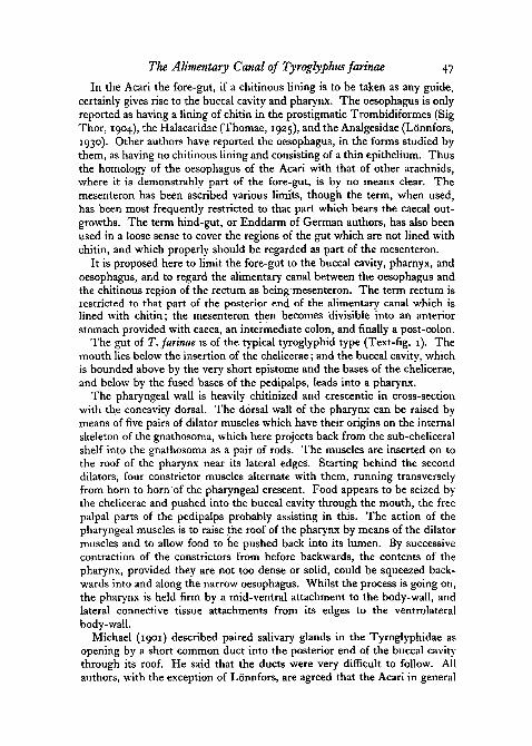
The gut of T. farinae is of the typical tyroglyphid type (Text-fig. 1). Themouth lies below the insertion of the chelicerae; and the buccal cavity, whichis bounded above by the very short epistome and the bases of the chelicerae,and below by the fused bases of the pedipalps, leads into a pharynx.
The pharyngeal wall is heavily chitinized and crescentic in cross-sectionwith the concavity dorsal. The dorsal wall of the pharynx can be raised bymeans of five pairs of dilator muscles which have their origins on the internalskeleton of the gnathosoma, which here projects back from the sub-cheliceralshelf into the gnathosoma as a pair of rods. The muscles are inserted on tothe roof of the pharynx near its lateral edges. Starting behind the seconddilators, four constrictor muscles alternate with them, running transverselyfrom horn to horn of the pharyngeal crescent. Food appears to be seized bythe chelicerae and pushed into the buccal cavity through the mouth, the freepalpal parts of the pedipalps probably assisting in this. The action of thepharyngeal muscles is to raise the roof of the pharynx by means of the dilatormuscles and to allow food to be pushed back into its lumen. By successivecontraction of the constrictors from before backwards, the contents of thepharynx, provided they are not too dense or solid, could be squeezed back-wards into and along the narrow oesophagus. Whilst the process is going on,the pharynx is held firm by a mid-ventral attachment to the body-wall, andlateral connective tissue attachments from its edges to the ventrolateralbody-wall.
Michael (1901) described paired salivary glands in the Tyroglyphidae asopening by a short common duct into the posterior end of the buccal cavitythrough its roof. He said that the ducts were very difficult to follow. Allauthors, with the exception of Lonnfors, are agreed that the Acari in general

48 Hughes—The Physiology of
possess such salivary glands (Vitzthum, 1940). In T. farinae the ducts of theglands open into the posterior angles of the buccal cavity (Text-fig. 2).
In T. farinae the food, whether starch grains from flour or pieces brokenoff whole grain or coarse meal by the chelicerae, is always relatively dry, and
I mm.
TEXT-FIG, I . Dorsal view of Tyroglyphus farinae to show the alimentary canal.
it has to traverse a long pharynx and longer oesophagus. This penetrates thesolid central nervous system, and has walls which are thin and certainlydevoid of muscles. It is improbable that such a passage could be achievedwithout the secretion of some lubricant, leaving aside the question of adigestive function for the saliva. It is difficult to understand, too, how thepharyngeal apparatus could deal with anything other than a fluid or semi-fluid bolus.

The Alimentary Canal of Tyrogfyphus farinae 49
The salivary glands consist of two paired masses of cells on each side ofthe body, each consisting of a few cells only. The cytoplasm of these cellsafter fixation with Leonard and Pasteel's fluid (Lison, 1936) contains smallgranules which stain with eosin; after fixation in alcoholic Bouin the cyto-plasm (Text-fig. 2) contains many small vacuoles giving it a spongy ap-pearance. In addition, each cell contains a large vacuole into which projectsthe nucleus thinly covered with cytoplasm. Each discharges into a commonduct the thin walls of which are composed of flattened cells. Mucihtfemateindoes not stain the general cytoplasm but does colour the small granules;mucicarmine gives a pale staining of the entire gland.
duct of salivary glandmusculature of head
TEXT-FIG. 2. L.S. head.
The pharynx, which supplies the mechanical force to propel the food intothe stomach, passes almost at right angles into the thin-walled oesophagus.As in Glycyphagus domesticus (Hughes and Hughes, 1938) the oesophagusprojects a short distance into the lumen of the stomach, with which it joinsantero-ventrally. The wall of the oesophagus consists of a thin epitheliumwith a very tenuous lining of chitin. The inner lining often shrinks awayfrom the wall in section where the oesophagus is empty. It would appear thatin T. farinae the oesophagus is part of the fore-gut.
The stomach, into which the oesophagus opens, bears post-laterally a pairof blunt caeca. These extend back below and slightly to each side of thesucceeding part of the mesenteron or colon, from which the stomach isseparated by a sphincter. The stomach itself is pear-shaped with the narrowend forwards. Its walls consist of a single layer of cells bounded externallyby a heavy basement membrane. These cells, though all similar, are notidentical (Text-fig. 3); they have granular cytoplasm, and contain manyvacuoles, the nuclei being large and lying near the basement membrane. Noneof them is. a goblet cell, nor are the vacuoles conspicuously near the lumen ofthe gut. The cells of the anterodorsal stomach wall and of the ends of theblunt caeca, though of the same general form, are taller anjd tend to projectinto the lumen; such cells would correspond to the pre- and post-ventricular

50 Hughes—The Physiology of
glands of Lonnfors (1930). In sections fixed in Leonard and Pasteel's fluid,^.cro-chlor-formalin, or Flemming's solution and 'stained with Mallory'striple stain, a layer of deep-blue substance is seen along the free borders ofthe cells, but not in the vacuoles. In sections stained with Masson's acidfuchsin and light green, the contents of the.stomach are green; its wall cellsshow no discrete spherules, only a diffuse staining. The same result is giyenwith mucicarmine and mucihaematein. This would suggest the secretion of a
. -OPS mm.
basement membrane
vacuotes incytoplasm
mucous secretion
TEXT-FIG. 3. Wall of stomach and caeca.
mucus, probably containing a digestive enzyme, by passage through the cellsurface. The taller cells at the tip or anterodorsal region of the caeca, oftencontain red-staining spherules, which seem similar to those recorded in theintestinal wall of Peripatopsis by Manton and Heatley (1937) and regarded bythem as protein reserves. If animals are fed on food stained with indicatorsor vital stains, these cells in particular become filled with small vacuolescoloured with the dye. Lonnfors immersed Analgesidae in aqueous solutionsof vital stains without any very marked coloration, but he does record thatneutral red was taken up by the caeca. T. farinae was fed on coarse meal oron gluten stained with universal indicator, litmus, phenol red, or neutral red.'Universal' indicator was always orange-yellow in the stomach, although itstained the meal red and. the gluten green. Litmus was always red in thestomach and caeca; phenol red was yellow and neutral red remained red.From these results it would appear that the pH.of the stomach and its caecalies between 5-0 and 6-o. Animals fed in this way showed a vacuolization ofthe cells of the stomach wall, particularly at the tips of the caeca and antero-dorsally; this gives the cells concerned the, appearance of bunches of grapes.

The Alimentary Canal of Tyrogfyphus farinae 51
When viewed under an immersion lens it was possible to see small granulesin vibratile motion in each vacuole. It was not found possible to determinethe nature of these granules. There seems little possibility that these vacuolesare not absorption vacuoles, since they occurred whatever dye was used.Although basic dyes, such as neutral red, may cause vacuolization, acidic dyeslike phenol red do not; moreover the fact that a variety of food dyes found inproprietary pudding- and custard-powders also appear in the cells in a similarway, is itself an indication that these cells are actively engaged in absorption.Such vacuolated cells are not confined to the tips of the caeca and antero-dorsal stomach wall, but are most frequent there.
• basement membrane•005 mm.
granularcytoplasm
nucleus
secretion
TEXT-FIG. 4. Wall of colon.
There are no intrinsic muscles in the walls of the stomach and its caeca,but high in its posterior wall and between the caeca, is a sphincter whichleads into the colon. This is spherical in shape and communicates post-ventrally by another aperture, guarded by a sphincter, with the post-colon.The wall of the colon is thinner than that of the stomach and not vacuolated(Text-fig. 4). Its contents are often solid as contrasted with the fluid contentsof the stomach. In animals fed on dyed food, a coloured bolus may be visibleand the walls are always diffusely stained in contrast to the very obviousvacuolization of the stomach walls. During starvation of such animals, dyecan be seen in the colon walls and very diffusely around it some time after ithas entirely disappeared from the stomach and its caeca. 'Universal' indicatoris yellow in the colon, litmus changes from red to purple, phenol red is yellow,and neutral red remains red. The pH therefore rises until it is above 7-0 butbelow 8-o. The contents of the colon may be in either of two conditions:fluid like that of the stomach, or apparently fairly solid and surrounded by athin peritrophic membrane. This, since it appears in the colon, is presumablysecreted by its walls. This peritrophic membrane is colourless in life butstains blue with haematoxylin. Coloration of the colonic wall, the formationof a bolus of greater opacity than the stomach contents that enter it, and whichretains its form in passing the sphincter into the post-colon, indicate that thefunctions of the colon are the absorption of the more fluid part of the materialpassing into it, as well as the production of the peritrophic membrane. Thisprotects its walls and those of the post-colon from the solid bolus which isthus produced. In addition, the walls of the colon may secrete some other

52 Hughes—The Physiology of
substance than the peritrophic membrane, since the pH of the contents riseswhilst in the colon. The peritrophic membrane is mentioned by Vitzthum(1940), but Lonnfors and earlier authors did not apparently see it in thespecies they studied.
In animals taken from a dense culture on wheat germ, where there isextensive fouling of the food with faecal pellets, the contents of the colon arepractically always visible as a pellet. This is much denser than that found inindividuals from young cultures where the food is clean; moreover, withpolarized light, the pellet is birefringent and has a yeljow colour, whereas ifthe food is clean, the contents are rarely birefringent except for easily recog-nizable starch grains. In animals from cultures on dried yeast or gluten, thecolonic pellet closely resembled that of animals from young wheat-germcultures, the pellet being rarely birefringent. On the other hand, in suchanimals and also in animals from dense cultures, the parenchyma of theopisthosoma is usually crowded with birefringent crystals in Brownian move-ment within their vacuoles. It would seem that in animals, the food of whichis contaminated with faecal material, the concentration of this can becomesuch that the absorption of water in the colon is sufficient to precipitate somekatabolite present in crystalline form. In animals kept on food relatively freefrom such contamination, however, the concentration of such material in thecolon is very rarely high enough to cause precipitation, although crystalsaccumulate in the parenchyma if the substrate is richer than usual in protein.The crystals, then, are presumably connected with the protein metabolismof the animal and may well be of an excretory nature. The greater density ofthe colonic contents in the case of dense culture animals is due not only to theprecipitation of crystals, but also to the fact that the animals in such culturesare reduced to eating the more cellular constituents. As any adhering starchyendosperm or embryo has already been consumed there is a greater proportionof unutilizable material from the food itself taken in.
No intrinsic muscles were seen in the region of the gut, nor have they beenrecorded by Michael (1901) in Tyroglyphidae or by Lonnfors (1930) inAnalgesidae, although Vitzthum (1940) speaks in a general way of the gut ofacarids as having a muscle-coat posterior to the stomach. Since Michael,using the same methods, described an extensive musculature on the colonand on the stomach of Oribatidae, it seems reasonable to suppose that thereis considerable variation among the Sarcoptiformes in this respect.
From the colon, the gut contents in the form of a bolus surrounded by aperitrophic membrane, pass into the post-colon. This passage can sometimesbe observed in animals mounted alive in dilute glycerine. It is brought aboutsuddenly by contraction of dorso-ventral muscles of the body coinciding withrelaxation of the sphincter between colon and post-colon. This may coincidealso with the extrusion of a faecal pellet, the anal valves being opened by theirown muscles. However, the post-colon is frequently found empty.
The post-colon, like the colon, is spherical, but ventrally passes by anarrow opening not guarded by a sphincter (Text-fig. 5) into the chitin-lined

The Alimentary Canal of Tyroglyphus farinae 53
rectum. The walls of the post-colon are formed of an epithelium with a deepstriated border apparently composed of long narrow vesicles (Text-fig. 6).The boundaries of the cell bodies are very indistinct and their cytoplasmcontains small vacuoles. These appear coloured in animals fed on dyed foodsand then starved until the gut is empty, the presence of a faecal pellet makingit impossible to see the walls very clearly. In live mounts in glycerine, thestriated border shows as a clear homogeneous zone round the faecal pellet,the cell-bodies as a denser surrounding ring. Nalepa (1884) considered this
Malpighian tubulessphincter betweencolon and post-colon
post-colon
undigested remainof starch grains
lateral caecum
striated border
muscles
late ral caecum
ipithelial cells
chitin•02mm.
'precipitated ghjcogen
~- \ j f —• "->al valveTEXT-FIG. 5. T.S. in the region of the rectum.
region of the gut to be excretory and Michael (1901) was of the same opinion.No excretory granules have been found in its walls or outside the peritrophicmembrane which surrounds the bolus; nor have I ever seen any reference tosuch granules or crystals in the literature dealing with the Tyroglyphidae.Its real function is indicated by the presence of the coloured vacuoles mentionedpreviously, which would suggest a final absorption of water rather than theexcretion of a coloured fluid, since the faecal pellet is a dry, solid structure.The histological structure of the post-colon, which resembles that of thelower ends of the Malpighian tubules of an insect such as Rhodnius(Wigglesworth, 1931), also suggests that its true function is a final absorp-tion of water. This must be an important function of the gut in an animallike T. farinae which lives on food of relatively low water content withoutaccess to fluid water.
Further evidence is obtained by considering the size of the colonic pelletwhen it has been surrounded by a peritrophic membrane and therefore has asharp outline. This enables its diameter to be measured and compared with

54 Hughes—The Physiology of
the diameter of an unvoided faecal pellet in the same animal. Two facts areat once noticeable. In the case of animals taken off clean food, the faecalpellet in the vast majority of cases is smaller than that in the colon, and itsdensity and opacity to transmitted light are greater. In animals from densecultures, however, both faecal and colonic pellets are opaque. Moreover,when the presence of birefringent crystals is investigated, it is found that inanimals from cultures on dried yeast or gluten, the faecal pellet is usually
striated border
vacuole
•005mm. '•:£'•:•. ^ ! : : : : j ; ; : :
nucleus
basement'membrane
TEXT-FIG. 6. Wall of post-colon.
entirely or partially birefringent, and on wheat germ it, too, may show bire-fringent patches. In animals from dense cultures, however, both faecal andcolonic pellets are always strongly birefringent. These facts indicate that thepost-colonic pellet undergoes further drying, which is sufficient to bring outof solution any of the crystalline substance which originally may have beenpresent in solution. The fact that the crystalline substance appears withinthe peritrophic membrane surrounding the pellet, suggests that it was alreadyin this position when the bolus entered the post-colon, so that presumablyexcretion of this substance has already taken place in the colon or possiblyeven in the stomach. No change in pH was detected in this region of the gut,so that the actual method of precipitation of the crystals is different from theprecipitation of uric acid in Rhodnius (Wigglesworth, 1931).
The post-colon opens freely into the rectum which in transverse sectionappears as a bilaterally compressed vertical slit. It is lined with chitin, whichis thickened distally to form the rectal valves and then passes over the edgeof the anal opening into continuity with the exoskeleton. The cellular layerwhich secretes this chitin appears as a syncytium continuous with the epithe-lium of the body-wall. The muscles operating the anal valves are inserted
The Alimentary Canal of Tyroglyphus farinae 55
just inside the anal opening and run from the valves to the ventrolateral body-wall, close to the ventral side of the body. The faecal pellet never appearsto be retained in this region of the body, the sole function of which is to affordit an exit.
Malpighian tubules were first described in T. farinae by Berlese (1897).They are a pair of short, blunt tubules standing out at right angles to the con-striction between colon and post-colon. It appears that they can dischargeany contents they might accumulate only when this sphincter relaxes. Warren(1944) described the paired Malpighian tubules of a species of Urodinychus,and was of the opinion that there was no communication between them andthe gut. Granules which he saw in them and called guanine bodies passedthrough their cytoplasm and that of the gut-wall to reach its lumen. In T.farinae the tubules consist of a few cells only which histologically resemblethose of the post-colon. No granules have been observed in any part ofthem.
Various tests were made on the animal, its faecal pellets, and on extractsfrom cultures in order to ascertain the nature of the crystalline excretory pro-ducts. Water extracts from 50 gm. of dense cultures extracted in 500 c.c. offluid, and from faecal pellets from the- 6-cm. diameter cellophane covers ofculture jars to which some thousands of faecal pellets adhered, extracted in10 c.c. of fluid, both gave negative" results with the urease tests for urea.Weil's test and the picric acid test for creatinine were also negative. Whensimilar extracts made with 2 per cent, sodium carbonate were saturated withammonium chloride and the resultant precipitate subjected to the murexidetest, an orange colour was obtained. Faecal pellets were collected from thecellophane covers and the murexide test applied to them in the cavity of ahollow-ground slide, and again an orange colour resulted. It would appearfrom this that uric acid is not produced as a nitrogenous katabolite. Theorange colour might be due to guanine or xanthine (Cole, 1933).
Animals were taken from cultures on gluten and dense cultures on wheatgerm, all having the parenchyma of the opisthosoma crowded with crystallinebodies. These animals were incubated overnight at 560 C. in various solu-tions and then examined microscopically in a drop of the solution next day.The crystals were found to be soluble in dilute sulphuric acid and in causticpotash, but insoluble in organic solvents or a solution of piperazine. Insolu-bility in piperazine is given by Lison (1936) as a distinguishing test betweenuric acid and guanine.
Faecal pellets treated in the same way lose their birefringence in sulphuricacid and caustic potash, but not in cold dilute hydrochloric acid or piperazine.If a heavy culture is sifted a little at a time through fine silk material, a greydust containing a high proportion of faecal pellets can be obtained. If suchmaterial, or dense cultures themselves, are extracted over a water bath with5 per cent, sulphuric acid, guanine can be precipitated by saturation withammonia. After repeated solution and precipitation by ammonia to free itfrom other substances, the final precipitate was dissolved in a small quantity

56 Hughes—The Physiology of
of hot hydrochloric acid and filtered hot. On cooling, small crystals separatedout which redissolved on heating. A white precipitate could be obtained fromthe solution in hot hydrochloric acid by the addition of 10 per cent, solutionof metaphosphoric acid. This precipitation by metaphosphoric acid was firstused by Wulff (1893) for quantitative extraction of guanine.
Since tests carried out on the granules in the animals, on faecal pellets andon extracts from faecal pellets or dense cultures all give positive results forsuch tests for guanin as can be applied, there seems nothing inherentlyimprobable in the view that this is the chief nitrogenous katabolite andthat uric acid is not produced in a quantity detectable by the meansemployed.
Experiments were also carried out to determine the optimum pH for thedigestion of starches and protein. Tubes were set up in duplicate each con-taining 3 c.c. of a buffer solution in steps of 0-2 pH and 1 c.c. of a filteredstarch solution in saturated sodium chloride. To one series of tubes wasadded 1 c.c. of water to each tube; to the other series 1 c.c. of a water extract(made as described on p. 46) of whole animals was added. After the additionof a drop of toluene, the tubes were plugged with cotton-wool and incubatedat 250 C. for 48 hours. The sugar in each tube was estimated by the JensenHagedorn method, and the difference between any pair of tubes at the samepH taken as a measure of the digestion which had occurred. These experi-ments gave a maximum of sugar of pH 5 -4.
Proteolytic enzymes present in such extracts were tested in similar experi-ments, using 2 c.c. of a 1 per cent, gelatine solution in place of the starchsolution. After 48 hours' incubation the amino-acids present were estimatedby Sorensen's method. The optimum pH was found to be 5-6.
Proteolytic enzymes may be precipitated by safranin (Robertson, 1907) andneutral red (Marston, 1923); the activity of such precipitates was demonstratedby Marston and Holzberg (1913). When such precipitates were preparedfrom extracts of whole animals and used in similar experiments, they had anoptimum pH of 5-6^0-2.
A weak lipase can also be shown to be present in water extracts, by usingan emulsion of olive oil coloured with alkaline phenolphthalein. The salivarysecretion is difficult to investigate, and careful observation of mites on fooddyed with indicators does not suggest that there is any pouring out of thesaliva on to the food.
Storage of food reserves was investigated by using Bauer's method andBest's carmine for glycogen and by osmic fixation for fat. The parenchyma,in which the internal organs lie embedded, contains large quantities ofglycogen (Text-fig. 5), as can be shown by either method. The fluid, whichescapes if the opisthosoma is punctured, also contains glycogen. Fat does notappear to be metabolized to any extent by these animals, since there was noindication of fat droplets in the parenchyma comparable with the amount ofglycogen, deposits of which are very heavy. In sections stained by Bauer'smethod it is possible to see starch grains in stomach, colon, and post-colon in

The Alimentary Canal of Tyroglyphus farinae 57
various stages of digestion. This, together with the detection of starch grainsby birefringence in the colon and post-colon, suggests that much carbo-hydrate material escapes digestion.
DISCUSSION
Lonnfors (1930) called the salivary glands in the Analgesidae and Carpo-glyphus passularum (lactis) studied by him pseudosalivary glands, and con-sidered that they opened above the coxa of leg 1. Grandjean (1937), in hisdescription of the podocephalic canal of various mites based on a study ofwhole mites mounted in lactic acid solution, speaks of the chitinous ducts oftwo glands opening in the region of the pseudostigmatic organ. Grandjeanstates that they are very difficult to see and that he has assumed without proofthat they are homologous with certain ducts present in Otodectes cynotes: healso says in a footnote:—
'II vaut mieux admettre pour le moment, qu'un doubte subsiste parceque le"fil" ne semble pas toujours creux, son apparence etant quelquefois celle d'untendon. Du moins en est il aussi dans les preparations traites a chaud par l'acidelactique. Avant ce traitement, ou un traitement analogue, le fil est a peine dis-cernable. Je n'ai jamais constate en observant des acariens vivants, qu'un tube ouorgane plein d'air aboutisse a la fossette supracoxale.'
The position of the suspected opening above leg 1, in the supracoxal groove,is easily checked in section in T. farinae because of the characteristicallyshaped pseudostigmatic organ which is strongly birefringent in polarizedlight. I have not been able to trace any duct from the salivary glands to thisregion either in transverse, longitudinal, or horizontal sections. Grandjean(1937, Text-fig, IB) figured the possible openings as a straight oblique lineabove and slightly anterior to the pseudostigmatic organ, and a smallercrescentic line just above its base. I have examined T. farinae in 60 per cent,lactic acid solution in lateral view; the straight line is clearly visible, but Ihave been unsuccessful in seeing the smaller crescentic one. In horizontalsections a muscle is seen running ventral to the salivary glands from the regionof convergence of the apodemes of the legs to the lateral body-wall in frontof and above the pseudostigmatic organ. It would appear probable that thestraight line on the exoskeleton'which Grandjean saw is the tendon attach-ment of the muscle.
From personal observation both Bdellonyssus bacoti among the Parasiti-formes and Cheyletus eruditus among the Trombidiformes have glands whichopen anteriorly, although in both these cases the glands assume a greatercomplexity and larger proportion than in T. farinae. This is presumably cor-related with their feeding habits. B. bacoti—which feeds on mammalianblood—must, like other blood-sucking Arthropoda, be able to produce someanticoagulant and laking secretion in fairly large quantities, since the volumeof a meal is large relative to the mite. C. eruditus feeds on the soft parts ofother mites and insects, which are sucked dry leaving the empty exoskeleton.

58 Hughes—The Phys:ology of
. presumably injects into the prey some salivary secretion which carries outa general maceration if not digestion of the viscera and muscles, in order toallow the passage of food along the very narrow oesophagus. It would seemunlikely that the tyroglyphids feeding on relatively dry substances would havelost glands which are present in the two other groups. The possibility of aregurgitation of fluid along the oesophagus from the stomach, in order tofacilitate the passage of food, does not seem very likely. The oesophagealprojection into the stomach is of common occurrence. It appears to form avalvular apparatus for the prevention of such regurgitation. Lonnfors, how-ever, did not find any similar structure in the Analgesidae.
Reuter (1909) divided the alimentary canal of the Acari into four types onthe basis of variation in structure of the stomach. The Sarcoptiformes belongto his type 1 which, he stated, has a round or pyriform stomach provided withone pair of lateral caeca. The primitive condition is taken by Lonnfors (1930)as having three pairs of caeca (type 3 of Reuter), anterior, median, and posterior,such as is now found in the Parasitidae. Lonnfors considers that the sarcopti-form type has been derived by loss of all but the posterior pair of caeca. Theanterior pair has been reduced to the proventicular glands described byMichael (1883) in the Oribatidae, which open into the stomach in a positioncorresponding to the anterior caeca where present. In the Analgesidae whichhe studied, still further reductions were traced by Lonnfors (1930). Hearranged a series of species where first the proventricular glands occur as apair of patches of cells on the anterior wall of the stomach, and finally as asingle median patch above the opening of the oesophagus, this last state beingarrived at by the fusion of the originally paired rudiments. The presence oflarge cells anterodorsally in the stomach wall of T. farinae supports this view.Similar large cells standing out above the general epithelium have beenrecorded in Ixodes sp. (Nordenskiold, 1905), Halarachne (Steding, 1923),Pediculopsis graminum (Reuter, 1909), and in Tyroglyphidae (Michael, 1901,and Nalepa, 1884). In G. domesticus the stomach and its caeca were foundto contain cells whose free ends after forming vesicles become nipped offinto the lumen of the gut (Hughes and Hughes, 1938). Nalepa has sug-gested that such cells form the basis of the excretory system. No such cellshave been found in T. farinae, and the large cells of the stomach and caecashow every sign of being concerned with the absorption of the products ofdigestion rather than with the liberation of any product either digestive orexcretory.
The colon, post-colon, and Malpighian tubules appear to form a functionalunit in the system, in that in the colon and post-colon the solidification of thefaeces takes place and guanine excretory products become recognizable inthe gut contents. In this process the Malpighian tubules appear to playsingularly little part. Vitzthum (1940) says that the Malpighian tubules ofsuch tyroglyphids as possess them are always devoid of contents. The ap-pearance in living animals certainly suggests this in sharp contrast to suchforms as the gamasids where the Malpighian tubules are visible as white lines
The Alimentary Canal of Tyroglyphus farinae 59
through the dorsal body-wall. Similarly the single median excretory tubulein such trombids as C. eruditus makes a definite white line along themiddle of the opisthosoma. In sections of B. bacoti and of C. eruditus, theseexcretory organs are seen to be full of a crystalline substance—stronglybirefringent like that occurring in the faecal pellet of T. farinae. In T.farinae such crystals never occur in the sections of the Malpighian tubuleseven when they are densely packed in the parenchyma. Moreover, in manyrelated forms such as G. domesticus the Malpighian tubules are known tobe absent. It would appear that in T. farinae they are functionless as organsof excretion and that the excretory activity has been taken over by somepart of the gut-wall. It is at least suggestive that in the Trombidiformesthe gut ends blindly, and the single median excretory tubule corresponds inposition and point of opening to the exterior with the posterior part of thealimentary canal. The food-containing part of the gut in these forms, fromits caecal arrangements and what is known of its histology, corresponds to thestomach. It seems at least possible that the colon, post-colon, and rectumhave become separated off to form the median excretory apparatus. Vitzthum(1940) states that the nitrogenous excretion of all mites consists of guanine,but that in the Tyroglyphidae urates may be found as well. Nalepa (1884)considered that non-crystalline urates may be found in the faecal pellets ofT. longior. Leydig (1859) reported finding uric acid in a mite. Guanine hasbeen demonstrated in spiders by Vafropala (1935), in scorpions by Davy (1857)and Marchall (1890). Warren (1944) spoke of guanine bodies in the Mal-pighian tubules of Urodinychus sp. but does not record having applied anyspecific tests to them. In T. farinae guanine seems to be the only considerableexcretory product of nitrogen metabolism, and this is in agreement with thewidespread occurrence of the substance within the Acari and within theArachnida as a class.
ACKNOWLEDGEMENTS
My thanks are due to Professor H. G. Jackson and to Professor AlastairGraham for much helpful advice; and to the Waller Research Fund for lend-ing me a binocular microscope, without which the manipulation of thesemites, during the earlier part of this work, would have been impossible.
SUMMARY
1. The pH of the stomach and its caeca lies between 5-0 and 6-o, that ofthe colon changes to the alkaline side of 7-0 but remains below 8-0. The pHof the post-colon is between 7-0 and 8-o. All these measurements are subjectto protein error.
2. The stomach and its caeca are regions of:
(a) absorption;(b) secretion of a mucus containing a digestive enzyme;(c) digestion of carbohydrate and protein.

60 Hughes—The Physiology of
3. The optimum pH of carbohydrate digestion is 5-4 and of protein
4. The thin-walled colon regulates the pH of the food bolus and is the siteof formation of a peritrophic membrane and some absorption.
5. The post-colon is the site of further absorption of water and solidifica-tion of the faecal pellet.
6. Excretory substances are first recognizable in the colon; they do notappear to be produced by the Malpighian tubules or post-colon.
7. The chief nitrogen katabolite is guanine.
8. There is an accumulation of nitrogenous katabolites in the parenchymaof the body in animals on a high-protein diet.
9. The powers of excretion of nitrogenous waste appear to be poor.
10. The main food reserves are of glycogen in the parenchyma; fat is notstored to any extent.
11. The salivary glands produce a mucus which serves as a lubricant forthe food in its passage through the pharynx and oesophagus.
LIST OF REFERENCESBERLESE, A., 1897. 'Ricerche sugli organi e sulla funzione della digestione negli acari.'
Rivist. Pat. Veg., 5, 129.COLE, S. W., 1933. Practical Physiological Chemistry. Cambridge (Heffer).DAVY, J., 1857. 'On the urinary excretion of fishes with some remarks on this secretion in
others classes of animals.' Trans. Roy. Soc. Edin., 21, 543.GRANDJEAN, F., 1937. 'Sur quelques caracteres des Acaridiae libres.' Bull. Soc. Zool. Fr.,
6a, 388.HOLZBERG, H. L., 1913. 'A new method of isolating trypsin.' Jour. biol. Chem., 14, 335.HUGHES, T. E., and A. M., 1938. 'The internal anatomy and post-embryonic development of
Glyphcyphagus domesticus de Geer.' Proc. Zool. Soc. B, 108, 715.LEYDIG, F., 1859. 'Ueber Haarsack Milben und Kratzmilben.' Arch. Naturgesch., 25, 338.LISON, L., 1936. Histochimie animale. Paris (Gauthier Villars).LONNFORS, F., 1930. 'Beitrage zur Morphologie der Analginen.' Acta Zool. Fenn., 8, r.MANTON, S., and HEATLEY, N., 1937. 'Studies on the Onychophora II.' Phil. Trans. Roy.
Soc. B, 327, 411-MAHCHAL, P., 1890. 'L'acide urique et la fonction r6nale chez les invertebr^s.' Mdm. Soc.
Zool. Fr., 3, 31.MARSTON, H. R., 1923. 'The azine and azonium compounds of the proteolytic enzymes.'
Biochem. Jour., 17, 851.MICHAEL, A. D., 1883. British Oribatidae. London (Ray Soc).
1901. British Tyroglyphidae. London (Ray Soc).NALEPA, A., 1884. 'Die Anatomie der Tyroglyphen.' Sitz. Akad. Wiss. Wien, Abt. 1.NORDENSKIOLD, E., 1905. 'Zur Anatomie und Histologie von Ixodes reduvius.' Zool. Anz.,
28, 478.REUTER, E., 1909. 'Zur Morphologie und Ontogenie der Acariden mit besonderer Beriick-
sichtigung von Pediculopsis graminum.' Acta Soc. Sci. Fenn., 36, 4, 1.ROBERTSON, T., 1907. 'Studies on the chemistry of the ion proteid compounds.' J. biol.
Chem., 2, 317.STEDING, E., 1923. 'Zur Anatomie und Histologie der Halarachne otariae.' Z. wiss. Zool.,
121, 442.THOMAE, H., 1925. 'Beitrage zur Anatomie der Halacariden.' Zool. Jhb. Anat., 47, 155.
The Alimentary Canal of Tyroglyphus farinae 61THOR, S., 1904. 'Recherches sur l'anatomie des acariens prostigmatiques.' Ann. Sci. nat.
(zool.), I9i 1 •VAJROPALA, K., 1935. 'Guanine, the excreta of arachnids.' Nature, 136, 145.VITZTHUM, H. GRAF., 1940. 'Bronns Klassen und Ordnungen des Tierreiches, Acarina.'
Leipzig (Becker & Erler).WARREN, E., 1944. 'Observations on the anatomy and histology of a myrmecophilous mite
and an account of its sporozoan parasites.' Ann. Natal Mus., io, 359.WIGGLESWORTH, V. B'., 1931. 'Excretion in Rhodnius prolixus.' J. exp. Biol., 8, 411.WULFF, C , 1893. 'Beitrage zur Kenntniss der Nucleinbasen.' Zeit. physiol. Chem., 17, 468.