The physiological regulation of glucose flux into muscle ...regulation is dominated by a single step...
9
254 Introduction The Journal of Experimental Biology published a series of articles by Weibel, Taylor, Hoppeler and associates in the 1990s that defined the design of pathways for the utilization of oxygen and substrates (Hoppeler and Weibel, 1998; Roberts et al., 1996; Taylor et al., 1996; Vock et al., 1996a; Vock et al., 1996b; Weber et al., 1996a; Weber et al., 1996b; Weibel et al., 1991; Weibel et al., 1996). In these elegant analyses, the relationships of biological structure to functional limitations were defined by a series of transfer steps. These transfer steps conceptualized the delivery of oxygen from the environment, as well as substrates from storage depots, to working muscle. The concept of symmorphosis, whereby “no more structure is built and maintained than is required to meet functional demand”, was central to building the relationship of structure to functional limitations (Weibel et al., 1991). Here we describe the regulatory factors that operate within the structural framework defined in this classic series of papers, focusing on the functional controllers of glucose influx into skeletal muscle. Blood glucose homeostasis cannot be understood without defining the control of muscle glucose uptake. Indeed, skeletal muscle comprises the bulk of insulin-sensitive tissue, and thus is where glucose uptake is quantitatively the most important. It is also the primary site of glucose uptake during exercise. As exercise and insulin are the primary physiological conditions that stimulate muscle glucose uptake, these conditions act to challenge the systems that control glucose flux into muscle. Here we will highlight studies in which these conditions have been used to perturb the glucoregulatory system. Moreover, defects in flux control that lead to glucose intolerance and insulin resistance resulting from high-fat feeding will be discussed. As in the previous studies summarized by Hoppeler and Weibel (Hoppeler and Weibel, 1998), we will describe the control of glucose flux in terms of the integration of physiological systems, and will emphasize animal models that provide unique insight into metabolic regulation. Muscle glucose uptake defined The physiological regulation of muscle glucose uptake requires that glucose travels from the blood to the interstitium to the intracellular space and then is phosphorylated to glucose 6-phosphate (G6P). The coupling of these processes involved in the influx of glucose is illustrated in Fig. 1. Blood glucose concentration, muscle blood flow and recruitment of capillaries to muscle determine glucose movement from the blood to the interstitium. The plasma membrane glucose transporter content determines glucose transport into the cell. Muscle hexokinase (HK) activity, cellular HK compartmentalization and the concentration of the HK inhibitor, G6P, determine the capacity to phosphorylate glucose. Glucose phosphorylation in muscle is irreversible; therefore, with this reaction, glucose is trapped and the uptake process is complete. These three steps – delivery, transport and phosphorylation – comprise muscle glucose uptake. This is not to say that steps downstream of glucose phosphorylation do not affect glucose uptake. It is just that any downstream step must, by definition, act The Journal of Experimental Biology 214, 254-262 © 2011. Published by The Company of Biologists Ltd doi:10.1242/jeb.048041 The physiological regulation of glucose flux into muscle in vivo David H. Wasserman*, Li Kang, Julio E. Ayala † , Patrick T. Fueger ‡ and Robert S. Lee-Young Department of Molecular Physiology and Biophysics and the Mouse Metabolic Phenotyping Center, Vanderbilt University School of Medicine, Nashville, TN 37232, USA *Author for correspondence ([email protected]) † Present address: Sanford-Burnham Medical Research Institute, 6400 Sanger Road, Orlando, FL 32827, USA ‡ Present address: Herman B. Wells Center for Pediatric Research and Departments of Pediatrics and Cellular & Integrative Physiology, Indiana University School of Medicine, Indianapolis, IN 46202, USA Accepted 17 August 2010 Summary Skeletal muscle glucose uptake increases dramatically in response to physical exercise. Moreover, skeletal muscle comprises the vast majority of insulin-sensitive tissue and is a site of dysregulation in the insulin-resistant state. The biochemical and histological composition of the muscle is well defined in a variety of species. However, the functional consequences of muscle biochemical and histological adaptations to physiological and pathophysiological conditions are not well understood. The physiological regulation of muscle glucose uptake is complex. Sites involved in the regulation of muscle glucose uptake are defined by a three-step process consisting of: (1) delivery of glucose to muscle, (2) transport of glucose into the muscle by GLUT4 and (3) phosphorylation of glucose within the muscle by a hexokinase (HK). Muscle blood flow, capillary recruitment and extracellular matrix characteristics determine glucose movement from the blood to the interstitium. Plasma membrane GLUT4 content determines glucose transport into the cell. Muscle HK activity, cellular HK compartmentalization and the concentration of the HK inhibitor glucose 6-phosphate determine the capacity to phosphorylate glucose. Phosphorylation of glucose is irreversible in muscle; therefore, with this reaction, glucose is trapped and the uptake process is complete. Emphasis has been placed on the role of the glucose transport step for glucose influx into muscle with the past assertion that membrane transport is rate limiting. More recent research definitively shows that the distributed control paradigm more accurately defines the regulation of muscle glucose uptake as each of the three steps that define this process are important sites of flux control. Key words: flux, glucose, in vivo. THE JOURNAL OF EXPERIMENTAL BIOLOGY
Transcript of The physiological regulation of glucose flux into muscle ...regulation is dominated by a single step...

254
IntroductionThe Journal of Experimental Biology published a series of articlesby Weibel, Taylor, Hoppeler and associates in the 1990s thatdefined the design of pathways for the utilization of oxygen andsubstrates (Hoppeler and Weibel, 1998; Roberts et al., 1996; Tayloret al., 1996; Vock et al., 1996a; Vock et al., 1996b; Weber et al.,1996a; Weber et al., 1996b; Weibel et al., 1991; Weibel et al.,1996). In these elegant analyses, the relationships of biologicalstructure to functional limitations were defined by a series oftransfer steps. These transfer steps conceptualized the delivery ofoxygen from the environment, as well as substrates from storagedepots, to working muscle. The concept of symmorphosis, whereby“no more structure is built and maintained than is required to meetfunctional demand”, was central to building the relationship ofstructure to functional limitations (Weibel et al., 1991).
Here we describe the regulatory factors that operate within thestructural framework defined in this classic series of papers,focusing on the functional controllers of glucose influx into skeletalmuscle. Blood glucose homeostasis cannot be understood withoutdefining the control of muscle glucose uptake. Indeed, skeletalmuscle comprises the bulk of insulin-sensitive tissue, and thus iswhere glucose uptake is quantitatively the most important. It is alsothe primary site of glucose uptake during exercise. As exercise andinsulin are the primary physiological conditions that stimulatemuscle glucose uptake, these conditions act to challenge thesystems that control glucose flux into muscle. Here we willhighlight studies in which these conditions have been used to
perturb the glucoregulatory system. Moreover, defects in fluxcontrol that lead to glucose intolerance and insulin resistanceresulting from high-fat feeding will be discussed. As in the previousstudies summarized by Hoppeler and Weibel (Hoppeler andWeibel, 1998), we will describe the control of glucose flux in termsof the integration of physiological systems, and will emphasizeanimal models that provide unique insight into metabolicregulation.
Muscle glucose uptake definedThe physiological regulation of muscle glucose uptake requires thatglucose travels from the blood to the interstitium to the intracellularspace and then is phosphorylated to glucose 6-phosphate (G6P).The coupling of these processes involved in the influx of glucoseis illustrated in Fig.1. Blood glucose concentration, muscle bloodflow and recruitment of capillaries to muscle determine glucosemovement from the blood to the interstitium. The plasmamembrane glucose transporter content determines glucose transportinto the cell. Muscle hexokinase (HK) activity, cellular HKcompartmentalization and the concentration of the HK inhibitor,G6P, determine the capacity to phosphorylate glucose. Glucosephosphorylation in muscle is irreversible; therefore, with thisreaction, glucose is trapped and the uptake process is complete.These three steps – delivery, transport and phosphorylation –comprise muscle glucose uptake. This is not to say that stepsdownstream of glucose phosphorylation do not affect glucoseuptake. It is just that any downstream step must, by definition, act
The Journal of Experimental Biology 214, 254-262© 2011. Published by The Company of Biologists Ltddoi:10.1242/jeb.048041
The physiological regulation of glucose flux into muscle in vivo
David H. Wasserman*, Li Kang, Julio E. Ayala†, Patrick T. Fueger‡ and Robert S. Lee-YoungDepartment of Molecular Physiology and Biophysics and the Mouse Metabolic Phenotyping Center, Vanderbilt University School of
Medicine, Nashville, TN 37232, USA*Author for correspondence ([email protected])
†Present address: Sanford-Burnham Medical Research Institute, 6400 Sanger Road, Orlando, FL 32827, USA‡Present address: Herman B. Wells Center for Pediatric Research and Departments of Pediatrics and Cellular & Integrative Physiology, Indiana
University School of Medicine, Indianapolis, IN 46202, USA
Accepted 17 August 2010
SummarySkeletal muscle glucose uptake increases dramatically in response to physical exercise. Moreover, skeletal muscle comprises thevast majority of insulin-sensitive tissue and is a site of dysregulation in the insulin-resistant state. The biochemical andhistological composition of the muscle is well defined in a variety of species. However, the functional consequences of musclebiochemical and histological adaptations to physiological and pathophysiological conditions are not well understood. Thephysiological regulation of muscle glucose uptake is complex. Sites involved in the regulation of muscle glucose uptake aredefined by a three-step process consisting of: (1) delivery of glucose to muscle, (2) transport of glucose into the muscle by GLUT4and (3) phosphorylation of glucose within the muscle by a hexokinase (HK). Muscle blood flow, capillary recruitment andextracellular matrix characteristics determine glucose movement from the blood to the interstitium. Plasma membrane GLUT4content determines glucose transport into the cell. Muscle HK activity, cellular HK compartmentalization and the concentration ofthe HK inhibitor glucose 6-phosphate determine the capacity to phosphorylate glucose. Phosphorylation of glucose is irreversiblein muscle; therefore, with this reaction, glucose is trapped and the uptake process is complete. Emphasis has been placed on therole of the glucose transport step for glucose influx into muscle with the past assertion that membrane transport is rate limiting.More recent research definitively shows that the distributed control paradigm more accurately defines the regulation of muscleglucose uptake as each of the three steps that define this process are important sites of flux control.
Key words: flux, glucose, in vivo.
THE JOURNAL OF EXPERIMENTAL BIOLOGY

255Glucose uptake in vivo
through glucose delivery, transport or phosphorylation. Forexample, acceleration of glycolysis or glycogen synthesis couldreduce G6P, increase HK activity, increase the capacity for glucosephosphorylation and potentially stimulate muscle glucose uptake.Reciprocally, rapid glycogen breakdown such as that which occurswith exercise could increase the G6P pool, inhibit HK, decrease therate of glucose phosphorylation and, through this mechanism,impede the rate of muscle glucose uptake.
The distributed control paradigm (Fig.1) for muscle glucoseuptake has been challenging to study because intracellular glucose,which is the product of membrane glucose transport and the substratefor glucose phosphorylation, cannot be measured directly. It cantheoretically be calculated indirectly as the difference between totalmuscle glucose and interstitial glucose. Numerous theoretical andmeasurement issues make this calculation untenable. The closecoupling of glucose delivery, transport and phosphorylation and theexistence of glucose compartmentalization and spatial gradientscompelled us to develop new techniques to overcome thesedifficulties associated with defining muscle glucose uptake.
Studying the whole organismThere have been innumerable studies that have attempted to addressthe control of glucose uptake in isolated muscle. These studies haveprovided tremendous insight into the basic cellular mechanismsbehind glucose transport. Isolated muscle preparations, as doesvirtually every experimental model system, have strengths and alsolimitations. Extramyocellular factors involved in the control ofglucose uptake (e.g. glucose delivery to muscle) are necessarilyabsent. Moreover, glucose uptake by isolated muscle preparationsis extremely resistant to insulin (requiring suprapharmacologicalinsulin levels) and contraction (requiring extremely high-intensitycontraction). It is likely that in some instances the rates of glucoseuptake in vitro do not become high enough to test the glucosephosphorylation capacity of muscle. As isolated musclepreparations are relatively simple to execute and far easier tointerpret because they are free of the often complicating variablesof the internal environment of the whole organism, most of theliterature describes studies conducted in vitro. This body of workhas led to the inevitable conclusion that membrane transport is ratelimiting for muscle glucose uptake. One difficulty in studying thewhole organism is that animal models are often stressed duringexperiments. This is particularly true of the mouse, whose smallsize makes the obtainment of blood difficult. The studies from ourlaboratory that are described below were performed using uniquemethods that were specifically designed to avoid stress and werevalidated to be stress-free on the basis of plasma catecholamineconcentrations (Ayala et al., 2006; Berglund et al., 2008). By the
use of these methods, we show that under physiological conditionsthe distributed control paradigm, where regulation of flux isdistributed between multiple steps (Fig.1), better defines muscleglucose uptake than the rate-limiting step paradigm, whereregulation is dominated by a single step (Wasserman, 2009).
Control of muscle glucose influx during exerciseRegulating the supply of glucose to the working muscle
Blood glucose concentration is a key determinant of the rate atwhich muscle can consume glucose. If blood glucoseconcentration falls, the rate of muscle glucose uptake will declineas well. Conversely, an increase in blood glucose concentrationswill cause muscle glucose uptake to increase. Liver release ofglucose is the primary means by which blood glucose is sustainedin the post-absorptive state in the face of constant tissue glucoseusage. Thus, the control of liver glucose output is key to theregulation of muscle glucose uptake. Of course, the gut is key inproviding glucose after a meal, and the ingestion of glucose cansustain blood glucose concentration under circumstances duringwhich the liver rate of glucose release cannot keep pace withtissue glucose utilization.
Glucagon is the primary controller of hepatic glucose productionin the sedentary state (Liljenquist et al., 1977). Exercise is a robustchallenge of the processes involved because of the high rates ofglucose production necessary to maintain blood glucose(Wasserman, 2009). Glucagon secretion from the pancreatic a cellincreases during exercise, whereas insulin secretion from thepancreatic b cell declines. The decline in insulin secretionpotentiates the actions of glucagon (Lavoie et al., 1997; Lins et al.,1983; Wasserman et al., 1989c). Studies in animals (Wasserman etal., 1984; Wasserman et al., 1985; Wasserman et al., 1989b) andhumans (Hirsch et al., 1991; Lavoie et al., 1997; Wolfe et al., 1986)demonstrate that the increase in glucagon is the primary stimulatorof hepatic glucose production during exercise. The powerful effectof glucagon on hepatic glucose production was recentlydemonstrated by Berglund et al. (Berglund et al., 2009). This studyshowed that increasing glucagon in sedentary mice to levels similarto those seen during exercise causes a marked discharge of hepaticenergy stores so that the adenosine monophosphate (AMP) toadenosine triphosphate (ATP) ratio is increased. This increase inthe AMP:ATP ratio, through allosteric mechanisms, facilitates theglucagon-induced breakdown of glycogen and the oxidation of fatin the liver (Wasserman et al., 1989a; Wasserman et al., 1989b).
Some have noted a disassociation between glucagonconcentrations and glucose release from the liver and have used thisto argue that glucagon does not stimulate hepatic glucose outputduring exercise (reviewed by Wasserman, 2009). This argument is
D. H. Wasserman and others
Glucose Glucose6-phosphate
• Blood flow• Capillary recruitment• Spatial barriers• Liver and gut processes that sustain blood glucose
• Transporter number• Transporter activity
• Hexokinase number• Hexokinase compartmentation• Spatial barriers• Regulation by G6P feedback
Fig.1. Distributed control of muscle glucose uptake. Modified fromWasserman and Halseth (Wasserman and Halseth, 1998) andWasserman et al. (Wasserman et al., 1967).
THE JOURNAL OF EXPERIMENTAL BIOLOGY
256
flawed. The reason for this disassociation is that glucagon issecreted into the hepatic portal venous circulation, which traversesthe liver. The liver extracts glucagon, thereby slowing the timecourse and dampening the rise in the hormone in the peripheralcirculation (Coker et al., 1999; Wasserman et al., 1993).
It is unlikely that catecholamines are directly responsible for theincrease in glucose production during exercise, as hepaticdenervation (Wasserman et al., 1990), selective hepatic b- and a-adrenergic receptor blockade (Coker et al., 1997) andadrenalectomy (Moates et al., 1988) have little or no effect in theexercising dog. These findings are consistent with research in otherspecies, including humans (Wasserman, 1995). Other factors suchas interleukin-6 (Febbraio et al., 2004) or an as-yet-undefinedregulator may play a role, perhaps by regulating the endocrinepancreas.
Muscle blood flow, the factor besides glucose concentration thatdetermines blood glucose delivery, is markedly increased withexercise. A hallmark of the physiological response to exercise ismarked hyperemia and an increase in capillary blood flow. Theoverall effect of this phenomenon on glucose influx is that moreglucose is delivered to the working muscle and there is increasedsurface area for exchange of glucose. This hemodynamic effectincreases the delivery not only of glucose but also of all bloodconstituents. Simulating the exercise-induced increase in glucosedelivery in the absence of an increase in glucose transporter type 4(GLUT4) protein translocation to the muscle membrane isinadequate by itself, however, in recreating the exercise-inducedincrease in muscle glucose uptake (Zinker et al., 1993b).
Glucose transport across the plasma membrane of workingmuscle cells
Membrane transport is almost certainly the primary barrier tomuscle glucose uptake in the fasted, sedentary state, as membraneGLUT4 content is low and the membrane is relatively impermeableto glucose. GLUT4 translocation to the muscle membrane isaccelerated by muscle contraction (Etgen et al., 1996; Ploug et al.,1992), and the intracellular signaling mechanism(s) resulting inGLUT4 translocation to the muscle membrane (Funai et al., 2009;Kramer et al., 2007; Kramer et al., 2006; Sakamoto and Goodyear,2002; Witczak et al., 2010) and muscle glucose uptake (Goodyearet al., 1990; Ploug et al., 1984; Richter et al., 1985; Wallberg-Henriksson and Holloszy, 1985; Wasserman et al., 1991;Wasserman et al., 1992) is independent of the actions of insulin.Moreover, the fate of glucose extracted from the blood is differentin response to exercise and insulin (Wasserman et al., 1991; Zinkeret al., 1993a). The working muscle oxidizes glucose, whereasinsulin-stimulated muscle primarily stores glucose.
Glucose phosphorylation within the working muscle cellsThe ability of myocytes to phosphorylate glucose is inhibited byG6P. During exercise, the simultaneous increase in glycogenbreakdown and glucose uptake can potentially lead an increase inthe inhibitory G6P levels. This, combined with exercise hyperemiaand increased glucose transport, predicts a shift in the muscleglucose uptake barrier from transport to phosphorylation. The firsthint that glucose phosphorylation may become a significant barrierto glucose influx was an observation in muscle from exercised rats.It was shown that HK II mRNA, but not GLUT4 mRNA, wasincreased following exercise (O’Doherty et al., 1994) because ofan increase in gene transcription (O’Doherty et al., 1996). Althoughincreased GLUT4 expression has been reported following exercise(Kraniou et al., 2006; Ren et al., 1994), the HK II gene has sincebeen shown to be considerably more responsive (O’Doherty et al.,1994; Pilegaard et al., 2005). The increase in HK II mRNA inresponse to a single bout of exercise only makes sense from anadaptive standpoint if glucose phosphorylation is a barrier tomuscle glucose uptake.
Determining the functional roles of glucose delivery, transport andphosphorylation
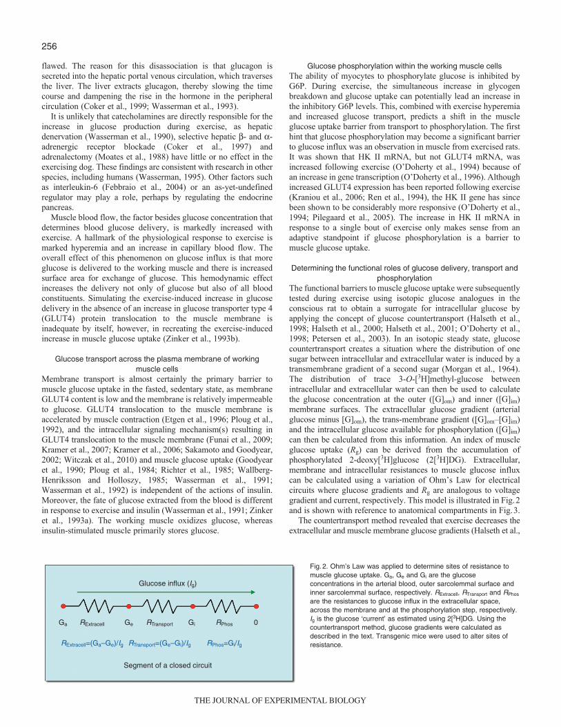
The functional barriers to muscle glucose uptake were subsequentlytested during exercise using isotopic glucose analogues in theconscious rat to obtain a surrogate for intracellular glucose byapplying the concept of glucose countertransport (Halseth et al.,1998; Halseth et al., 2000; Halseth et al., 2001; O’Doherty et al.,1998; Petersen et al., 2003). In an isotopic steady state, glucosecountertransport creates a situation where the distribution of onesugar between intracellular and extracellular water is induced by atransmembrane gradient of a second sugar (Morgan et al., 1964).The distribution of trace 3-O-[3H]methyl-glucose betweenintracellular and extracellular water can then be used to calculatethe glucose concentration at the outer ([G]om) and inner ([G]im)membrane surfaces. The extracellular glucose gradient (arterialglucose minus [G]om), the trans-membrane gradient ([G]om–[G]im)and the intracellular glucose available for phosphorylation ([G]im)can then be calculated from this information. An index of muscleglucose uptake (Rg) can be derived from the accumulation ofphosphorylated 2-deoxy[3H]glucose (2[3H]DG). Extracellular,membrane and intracellular resistances to muscle glucose influxcan be calculated using a variation of Ohm’s Law for electricalcircuits where glucose gradients and Rg are analogous to voltagegradient and current, respectively. This model is illustrated in Fig.2and is shown with reference to anatomical compartments in Fig.3.
The countertransport method revealed that exercise decreases theextracellular and muscle membrane glucose gradients (Halseth et al.,
Glucose influx (Ig)
Segment of a closed circuit
RExtracellGa RTransport RPhos
RExtracell=(Ga–Ge)/Ig RTransport=(Ge–Gi)/Ig RPhos=Gi/Ig
Gi 0Ge
Fig.2. Ohm’s Law was applied to determine sites of resistance tomuscle glucose uptake. Ga, Ge and Gi are the glucoseconcentrations in the arterial blood, outer sarcolemmal surface andinner sarcolemmal surface, respectively. RExtracell, RTransport and RPhos
are the resistances to glucose influx in the extracellular space,across the membrane and at the phosphorylation step, respectively.Ig is the glucose ‘current’ as estimated using 2[3H]DG. Using thecountertransport method, glucose gradients were calculated asdescribed in the text. Transgenic mice were used to alter sites ofresistance.
THE JOURNAL OF EXPERIMENTAL BIOLOGY
257Glucose uptake in vivo
1998), reflecting a shift in control of muscle glucose uptake fromglucose delivery and transport to glucose phosphorylation. Thedecrease in resistance to glucose transport is consistent with thetranslocation of GLUT4 to the plasma membrane. The decrease inresistance to glucose delivery is predictable from the marked exercisehyperemia. The concept that muscle glucose delivery was not a majorbarrier to muscle glucose uptake during exercise (Halseth et al.,1998) is consistent with results obtained with microdialysis(MacLean et al., 1999). The shift in control of muscle glucose influxto glucose phosphorylation during exercise is consistent with theaccumulation of glucose in muscle tissue from exercising humans(Katz et al., 1986; Katz et al., 1991; Richter et al., 1998).
The second approach used to dissect control of glucose flux intoworking muscle was the application of isotopic techniques tomouse models with genetic increases in muscle GLUT4 and HK II.As was the case with the countertransport model, this approach wasalso based on Ohm’s Law, where 2[3H]DG was used to gain anindex of muscle glucose influx. The hypotheses that HK IIoverexpression (deletion of RPhos in Fig.2) would increase thecapacity of muscle to consume glucose, whereas GLUT4overexpression (deletion of RTransport in Fig.2) would have no effectwere tested (Fueger et al., 2004a; Halseth et al., 1999). Miceoverexpressing GLUT4 (GLUT4Tg) and/or HK II (HKTg) werecatheterized and underwent experiments >5days later. Consistentwith predictions of the countertransport approach, HKTg mice hadincreased exercise-stimulated Rg, whereas GLUT4Tg mice did not.A variation of the ‘control coefficient’ concept was applied to thethree steps of muscle glucose uptake and was calculated by theequation derived from control theory (Kacser and Burns, 1995):
CTg �ln(Rg) / �ln(PTg) , (1)
where CTg is the control coefficient for the regulatory site of interestand PTg is calculated from the GLUT4 and HK II expression inGLUT4Tg and HKTg mice relative to their wild-type littermates. In
a closed system, the control coefficients sum to 1.0. A controlcoefficient for glucose delivery is calculated as 1.0 minus the sumof control coefficients for transport and phosphorylation steps. It isassumed that the fold overexpression equals the functional increasein the transgene product. The control coefficient in the fastedsedentary state is highest at the transport step. During exercise, thecontrol coefficient for transport falls to zero, suggesting that themuscle membrane is highly permeable to glucose because ofrecruitment of GLUT4 from intracellular vesicles. The controlcoefficient calculated for delivery also fell to very low levels,suggesting that exercise-induced hyperemia largely removes theglucose delivery barrier. During exercise, the onus of control veryclearly rests with the phosphorylation step. Notably, these resultsare the same as those obtained using the countertransport approach.
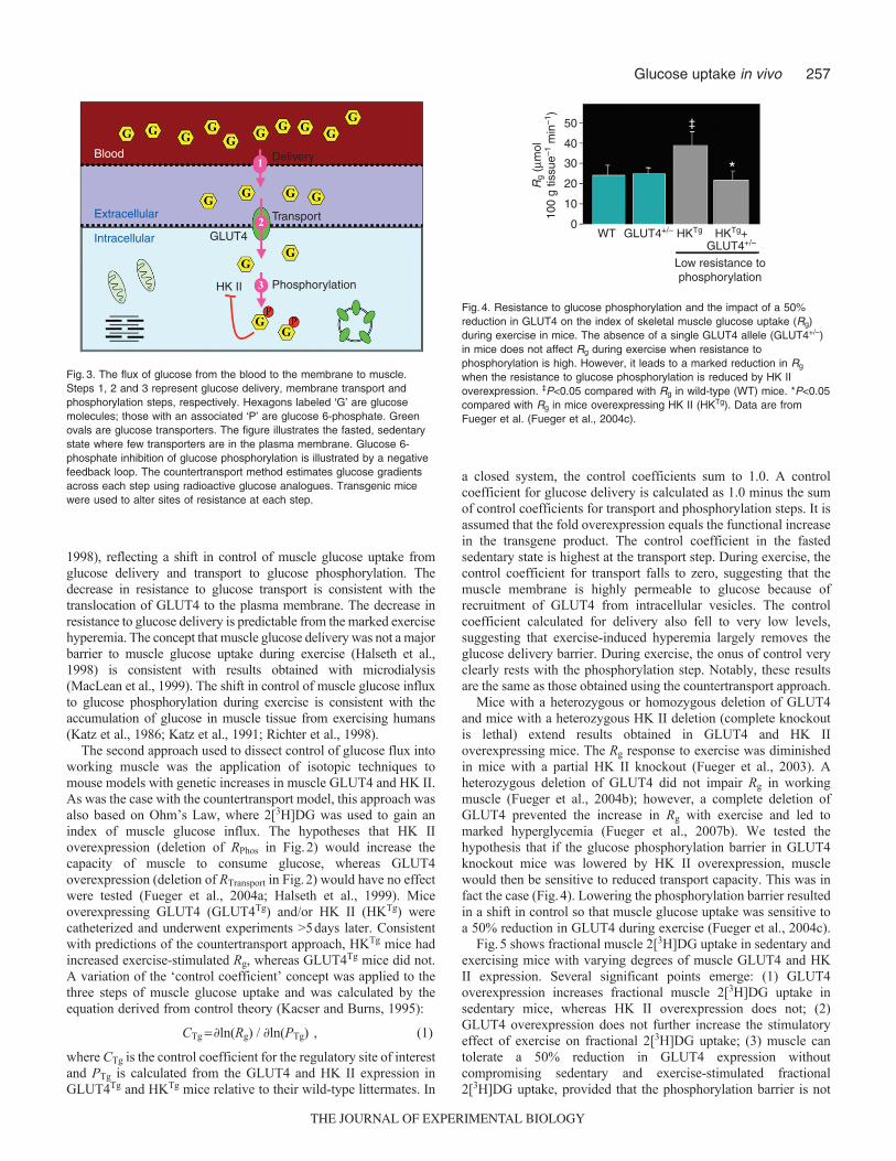
Mice with a heterozygous or homozygous deletion of GLUT4and mice with a heterozygous HK II deletion (complete knockoutis lethal) extend results obtained in GLUT4 and HK IIoverexpressing mice. The Rg response to exercise was diminishedin mice with a partial HK II knockout (Fueger et al., 2003). Aheterozygous deletion of GLUT4 did not impair Rg in workingmuscle (Fueger et al., 2004b); however, a complete deletion ofGLUT4 prevented the increase in Rg with exercise and led tomarked hyperglycemia (Fueger et al., 2007b). We tested thehypothesis that if the glucose phosphorylation barrier in GLUT4knockout mice was lowered by HK II overexpression, musclewould then be sensitive to reduced transport capacity. This was infact the case (Fig.4). Lowering the phosphorylation barrier resultedin a shift in control so that muscle glucose uptake was sensitive toa 50% reduction in GLUT4 during exercise (Fueger et al., 2004c).
Fig.5 shows fractional muscle 2[3H]DG uptake in sedentary andexercising mice with varying degrees of muscle GLUT4 and HKII expression. Several significant points emerge: (1) GLUT4overexpression increases fractional muscle 2[3H]DG uptake insedentary mice, whereas HK II overexpression does not; (2)GLUT4 overexpression does not further increase the stimulatoryeffect of exercise on fractional 2[3H]DG uptake; (3) muscle cantolerate a 50% reduction in GLUT4 expression withoutcompromising sedentary and exercise-stimulated fractional2[3H]DG uptake, provided that the phosphorylation barrier is not
Blood
Extracellular
Intracellular
Delivery
Transport
Phosphorylation
GLUT4
HK II
Fig.3. The flux of glucose from the blood to the membrane to muscle.Steps 1, 2 and 3 represent glucose delivery, membrane transport andphosphorylation steps, respectively. Hexagons labeled ‘G’ are glucosemolecules; those with an associated ‘P’ are glucose 6-phosphate. Greenovals are glucose transporters. The figure illustrates the fasted, sedentarystate where few transporters are in the plasma membrane. Glucose 6-phosphate inhibition of glucose phosphorylation is illustrated by a negativefeedback loop. The countertransport method estimates glucose gradientsacross each step using radioactive glucose analogues. Transgenic micewere used to alter sites of resistance at each step.
0
10
20
30
40
50
Rg
(µm
ol10
0 g
tissu
e–1 m
in–1
)
HKTg
‡
HKTg+GLUT4+/–
WT GLUT4+/–
*
Low resistance tophosphorylation
Fig.4. Resistance to glucose phosphorylation and the impact of a 50%reduction in GLUT4 on the index of skeletal muscle glucose uptake (Rg)during exercise in mice. The absence of a single GLUT4 allele (GLUT4+/–)in mice does not affect Rg during exercise when resistance tophosphorylation is high. However, it leads to a marked reduction in Rg
when the resistance to glucose phosphorylation is reduced by HK IIoverexpression. ‡P<0.05 compared with Rg in wild-type (WT) mice. *P<0.05compared with Rg in mice overexpressing HK II (HKTg). Data are fromFueger et al. (Fueger et al., 2004c).
THE JOURNAL OF EXPERIMENTAL BIOLOGY

258
reduced by HK II overexpression; and (4) HK II overexpressionincreases the maximum velocity (Vmax) of fractional 2[3H]DGuptake during exercise. The shift in control from transport tophosphorylation should not be interpreted as meaning that GLUT4is not important in the regulation of muscle glucose uptake. GLUT4translocation is the fulcrum that distributes the balance of controlbetween the three steps that control muscle glucose uptake. It is thesensitive regulation of GLUT4 translocation that shifts the burdenof control from glucose transport to phosphorylation.
Determining the functional roles of AMPK and NOS signalingIn recent years, the roles of nitric oxide synthase (NOS) (Bradleyet al., 1999; Higaki et al., 2001; Lee-Young et al., 2010; McConellet al., 2006; McConell and Wadley, 2008; Shearer et al., 2004)and AMP-activated protein kinase (AMPK) (Canto et al., 2010;Jensen et al., 2008; Jensen et al., 2009; Lee-Young et al., 2009;Maarbjerg et al., 2009; Mu et al., 2001; Shearer et al., 2004) inthe regulation of glucose uptake by working muscle has receivedconsiderable attention. Studies in vivo show extensive interactionbetween the NOS and AMPK signaling pathways in workingmuscle (Lee-Young et al., 2010; Lee-Young et al., 2009). Wehave assessed how these signaling pathways affect barriers tomuscle glucose uptake in vivo. Mice expressing a dominant-negative mutation of the AMPKa2 subunit within skeletal musclehave an impaired Rg response to exercise when compared withwild-type littermates exercising at the same relative work rate.Surprisingly, however, this decrease appears to have been due notto impaired glucose transport, but rather impaired glucosedelivery (Lee-Young et al., 2009). This result is consistent withthe demonstration that the AMPKa2 mutant mice had a reductionin muscle NOS activity during exercise (Lee-Young et al., 2009),whereas glucose transporter activity was unaffected (Maarbjerget al., 2009). Mice with a global endothelial NOS deletion alsohave a marked decrease in glucose delivery to working muscle,largely due to decreased blood flow (Lee-Young et al., 2010).Remarkably, despite this decrease, glucose influx is greatlyaccelerated, because of the rapid decline in muscle metabolic state(Lee-Young et al., 2010). The ability to adapt to reduced glucose
delivery and sustain muscle glucose uptake reflects the powerfulnature of distributed control of muscle glucose uptake. It isnotable that endothelial NOS deletion results in impairedmitochondrial function. One can speculate that the increasedreliance on glycolysis for energy production (as evidence bymarked increases in plasma lactate levels) reduces G6Pconcentration. It can then be hypothesized that the reduction ofG6P reduces the control coefficient for glucose phosphorylation,permitting high rates of glucose uptake even in the presence oflow glucose delivery rates.
Control of insulin-stimulated muscle glucose influxDetermining the functional roles of glucose delivery, transport and
phosphorylation The use of isotopic analogs was applied to the concept ofcountertransport, as described above, to delineate barriers toinsulin-stimulated muscle glucose influx during hyperinsulinemic,euglycemic clamps (insulin clamps) with physiological doses ofinsulin in unstressed rats (Halseth et al., 1998; Halseth et al., 2000;Halseth et al., 2001; O’Doherty et al., 1998). The countertransportmethod showed that insulin decreases the muscle membraneglucose gradient in a dose-dependent manner, reflecting a shift incontrol of muscle glucose uptake away from glucose transport todelivery and/or phosphorylation. These results were also consistentwith those obtained by muscle interstitial microdialysis (Holmanget al., 1998; Rosdahl et al., 1998).
Examination of insulin-stimulated glucose flux control usingunstressed, transgenic mice during insulin clamps performed athigh physiological insulin levels showed remarkable agreementwith results obtained using the countertransport method in insulin-clamped mice. Consistent with predictions of the countertransportapproach, HKTg mice (deletion of RPhos in Fig.2) had increasedinsulin-stimulated Rg values (Fueger et al., 2004a; Halseth et al.,1999), whereas GLUT4Tg mice (deletion of RTransport in Fig.2) didnot (Fueger et al., 2004b). Control of muscle glucose uptake usingthe three-step process showed that the control coefficient forglucose transport fell to near zero. Thus, as with exercise,physiological insulin stimulation results in increased GLUT4
0
2
4
6Fr
actio
nal 2
[3 H]D
Gup
take
(µl
100
g–1
min
–1)
0 1 2 3
GLUT4 protein (a.u.)
Effect of HK II overexpression
HK II overexpression
Wild-type littermatesExerciseSedentary
Fig.5. Fractional 2-deoxyglucose (2[3H]DG) uptake in the gastrocnemius ofmice expressing 0-, 0.5-, 1.0- and ~3.5-fold wild-type GLUT4 levels duringexercise or in the sedentary state. GLUT4 only affected fractional 2[3H]DGuptake in the sedentary state when it was overexpressed. However, theabsence of GLUT4 caused a marked attenuation of fractional 2[3H]DGuptake during exercise regardless of whether HK II was overexpressed. HKII overexpression had no effect on the response during saline infusion butincreased the exercise. Data points are means ± s.e.m. of 8–11 in vivomouse experiments. Modified from Wasserman (Wasserman, 2009).
0
5
10
15
Rg
(µm
ol10
0 g
tissu
e–1 m
in–1
)
HKTg
‡
HKTg+GLUT4+/–
Low resistance tophosphorylation
WT GLUT4+/–
*
Fig.6. Resistance to glucose phosphorylation and the impact of a 50%reduction in GLUT4 on the index of skeletal muscle glucose uptake (Rg)during the steady-state period of a 4.0mUkg–1min–1 insulin clamp. Theabsence of a single GLUT4 allele (GLUT4+/–) in mice does not affect Rg
during physiological insulin stimulation when resistance to phosphorylationis high. However, it leads to a marked reduction in Rg when the resistanceto glucose phosphorylation is reduced by HK II overexpression. ‡P<0.05compared with Rg in wild-type (WT) mice. *P<0.05 compared with Rg inmice overexpressing HK II (HKTg). Data are from Fueger et al. (Fueger etal., 2004b).
THE JOURNAL OF EXPERIMENTAL BIOLOGY

259Glucose uptake in vivo
translocation and a precipitous fall in the control coefficient fortransport. The difference between insulin stimulation and exerciseis that with insulin, control of muscle glucose uptake is sharedbetween glucose delivery and phosphorylation, whereas duringexercise the onus of control rests with the phosphorylation step.Again, these results are the same as those obtained using thecountertransport approach in rats.
Mice with a heterozygous or homozygous deletion of GLUT4and mice with a heterozygous HK II deletion extended resultsobtained in insulin-clamped GLUT4 and HK II overexpressingmice. A reduction in HK II impairs whole-body insulin sensitivityand heart Rg, but not Rg of the gastrocnemius muscle (Fueger et al.,2007a). As was the case with exercise, a 50% reduction in GLUT4did not impair Rg in insulin-stimulated states (Fueger et al., 2004b).Muscle can tolerate a 50% reduction in GLUT4 during insulinstimulation provided that the barrier to glucose phosphorylation isnot removed by HK II overexpression (Fueger et al., 2004b).However, in the absence of a barrier to glucose phosphorylation,membrane glucose transport becomes a limitation to muscleglucose uptake (Fig.6).
The fractional skeletal muscle 2[3H]DG uptake in saline-infusedand insulin-clamped mice with varying degrees of muscle GLUT4and HK II expression is summarized in Fig.7. The relationships ofthese proteins to muscle 2[3H]DG uptake in insulin-clamped miceis generally similar to the response in exercised mice (Fig.5). It canbe readily seen that: (1) GLUT4, but not HK II, overexpressionincreases fractional muscle 2[3H]DG uptake in saline-infused mice;(2) GLUT4 overexpression does not increase the stimulatory effectof a physiological increase in insulin on fractional 2[3H]DG uptake;(3) muscle can tolerate a 50% reduction in GLUT4 expressionwithout affecting insulin-stimulated fractional 2[3H]DG uptake;and (4) HK II overexpression increases the Vmax of fractional2[3H]DG uptake during an insulin clamp conducted atphysiological hyperinsulinemia. We interpret these findings tomean that the sensitive regulation of GLUT4 translocationincreases glucose membrane permeability to the point where the
onus of control shifts from glucose transport in the saline-infusedstate to glucose phosphorylation (and glucose delivery) underinsulin-clamped conditions.
Barriers to muscle glucose uptake in the diet-induced insulin-resistant state
Insulin resistance can be associated with deficits in muscle bloodflow (Clerk et al., 2007; Duplain et al., 2001; Inyard et al., 2009;Laakso et al., 1992), membrane glucose transport (Han et al., 1995;Liu et al., 1996; Zierath et al., 1997) and intracellular capacity tophosphorylate glucose (Bonadonna et al., 1996; Braithwaite et al.,1995; Pendergrass et al., 1998; Williams et al., 2001). Wedelineated the steps that cause the functional impairment in muscleglucose uptake by applying the countertransport method to rats feda high-fat diet. Results showed that extracellular and intracellularresistances were the two chief causes of the resistance of muscleglucose uptake to insulin. This is not to say that transport is normal.Evidence clearly shows that glucose transport is defective with diet-induced insulin resistance (Liu et al., 1996). The countertransportexperiments show, however, that the primary functional limitationsare in glucose delivery and phosphorylation in this model. Thepathogenesis of insulin resistance is further complicated when oneconsiders that extracellular barriers to glucose delivery are also aptto be barriers to insulin delivery. Impaired insulin delivery willimpact muscle glucose uptake and metabolic regulation in generalif the barrier to insulin delivery is sufficiently large.
Mice of the C57Bl/6J strain develop insulin resistance on a high-fat diet (Surwit et al., 1988) and were used to determine thefunctional deficits that make muscle insulin resistant. Thehypothesis that HK II overexpression could correct insulin-stimulated muscle glucose uptake in mice fed a high-fat diet wastested. In contrast to the marked effect of HK II overexpression oninsulin-stimulated muscle glucose uptake in mice fed chow, therewas no effect in mice fed the high-fat diet (Fueger et al., 2004a)(Fig.8). This suggested that the glucose phosphorylation barrierwas not the functional limitation causing insulin resistance to
0
0.5
1.0
1.5
Frac
tiona
l 2[3 H
]DG
upta
ke (
µl 1
00 g
–1 m
in–1
)
0 1 2 3
GLUT4 protein (a.u.)
Effect of HK II overexpression
HK II overexpression
Wild-type littermatesInsulinSaline
Fig.7. Fractional 2-deoxyglucose (2[3H]DG) uptake in the gastrocnemius ofmice expressing 0-, 0.5-, 1.0- and ~3.5-fold wild-type GLUT4 levels duringthe steady-state period of a 4.0mUkg–1min–1 insulin clamp or during anequal duration saline infusion. HK II content was either normal (WT) or theprotein was overexpressed. GLUT4 only affected fractional 2[3H]DG uptakeduring saline infusion when it was overexpressed. However, the absence ofGLUT4 caused a marked attenuation of fractional 2[3H]DG uptake duringinsulin stimulation regardless of whether HK II was overexpressed. HK IIoverexpression had no effect on the response during saline infusion butincreased the maximal response to insulin. Data points are means ± s.e.m.of 8–11 in vivo mouse experiments. Modified from Wasserman(Wasserman, 2009).
0
5
10
15
Index of skeletalmuscle glucose
uptake(µmol 100 g–1 min–1)
HKTg
Chow fed High fat fed
WT
**,†
SVL
HKTgWT
* *,‡
0
5
10
15
*
*,†Gastroc
* *,‡Insulin-clamped
Saline-infused
Fig.8. Glucose metabolic index measured using 2-deoxyglucose (2[3H]DG)was measured for the gastrocnemius and superficial vastus lateralis (SVL)during the last 30min of a 120-min saline infusion or hyperinsulinemic-euglycemic clamp (4.0mUkg–1min–1) experiment on conscious,unrestrained mice fasted for 5h. Wild-type (WT) or HK II overexpressing(HKTg) mice were fed either a standard diet or a high-fat diet up to the ageof 4months of age and fasted for 5h. Data are means ± s.e.m. for 7–14mice per group. *P<0.05 vs saline condition; †P<0.05 vs WT standard diet;‡P<0.05 vs HKTg standard diet. Data are from Fueger et al. (Fueger et al.,2004b). Figure reproduced from Wasserman (Wasserman, 2009).
THE JOURNAL OF EXPERIMENTAL BIOLOGY

260
muscle glucose uptake. The countertransport data showed thatextracellular resistance was the chief site of resistance to muscleglucose uptake. To further test this finding, we treated mice fed thehigh-fat diet for 3months with the PDE5a inhibitor, sildenafil(Ayala et al., 2007). PDE5a is expressed in vascular smooth muscleand causes breakdown of cyclic guanosine monophosphate(cGMP). cGMP signals relaxation of vascular smooth muscle.Inhibition of PDE5a with sildenafil treatment results in increasedcGMP levels and decreased vascular resistance (reduction inRExtracell in Fig.2). We showed that this compound increasesinsulin-stimulated muscle Rg in mice fed the high-fat diet and it didso without improving muscle insulin signaling (Fig.9). Thissupports the theory that inhibition of PDE5a acts by lowering thebarrier to glucose delivery and, by doing so, decreases insulinresistance. One would predict that sildenafil increases insulindelivery to the muscle as well. The fact that insulin signaling wasnot enhanced would suggest that the improved Rg response was notdue to greater insulin availability. Sildenafil treatment had otherconsequences, including increased energy expenditure anddecreased body weight. So although sildenafil is effective atimproving insulin-stimulated muscle glucose uptake in the insulin-resistant mouse, more work is required before the mechanism ofaction can be fully defined (Ayala et al., 2007).
ConclusionsFor decades, the presumption by researchers was that muscleglucose uptake is rate limited by membrane transport. Thisconclusion was based almost entirely on studies conducted in vitro.In the whole organism, the paradigm for control of glucose fluxinto muscle is much different. This paradigm posits that muscleglucose uptake is under distributed control by processes that controlglucose delivery to, membrane transport into and phosphorylationwithin muscle. This model recognizes that the liver and gut are alsoimportant determinants of muscle glucose uptake as they maintainglucose concentrations and sustain muscle glucose delivery.Moreover, feedback inhibition of HK II by G6P provides a meansby which the distribution of control is transferred to pathwaysdownstream of glucose phosphorylation (i.e. glycogen synthesisand glycolysis). This enables the linkage of energy metabolism/storage with glucose flux. Here we show, using isotopic methodsand transgenic/mutant mice, that each of these three steps can be
formidable barriers to muscle glucose uptake under physiologicalconditions. The therapeutic implication is that one or more of thesesteps should be effective targets for treatment of glucoseintolerance and insulin resistance. Here we showed the importanceof a therapy that targets the glucose delivery step.
AcknowledgementsThe authors’ research described in this review was funded by NIH grants DK54902,DK50277 and DK59637. Deposited in PMC for release after 12 months.
ReferencesAyala, J. E., Bracy, D. P., McGuinness, O. P. and Wasserman, D. H. (2006).
Considerations in the design of hyperinsulinemic-euglycemic clamps in theconscious mouse. Diabetes 55, 390-397.
Ayala, J. E., Bracy, D. P., Julien, B. M., Rottman, J. N., Fueger, P. T. andWasserman, D. H. (2007). Chronic treatment with sildenafil improves energybalance and insulin action in high fat-fed conscious mice. Diabetes 56, 1025-1033.
Berglund, E. D., Li, C. Y., Poffenberger, G., Ayala, J. E., Fueger, P. T., Willis, S.E., Jewell, M. M., Powers, A. C. and Wasserman, D. H. (2008). Glucosemetabolism in vivo in four commonly used inbred mouse strains. Diabetes 57, 1790-1799.
Berglund, E. D., Lee-Young, R. S., Lustig, D. G., Lynes, S. E., Donahue, E. P.,Camacho, R. C., Meredith, M. E., Magnuson, M. A., Charron, M. J. andWasserman, D. H. (2009). Hepatic energy state is regulated by glucagon receptorsignaling in mice. J. Clin. Invest. 119, 2412-2422.
Berria, R., Wang, L., Richardson, D. K., Finlayson, J., Belfort, R., Pratipanawatr,T., De Filippis, E. A., Kashyap, S. and Mandarino, L. J. (2006). Increasedcollagen content in insulin-resistant skeletal muscle. Am. J. Physiol. Endocrinol.Metab. 290, E560-E565.
Bonadonna, R., Prato, S. D., Bonora, E., Saccomani, M., Gulli, G., Natali, A.,Frascerra, S., Pecori, N., Ferrannini, E., Bier, D. et al. (1996). Roles of glucosetransport and glucose phosphorylation in muscle insulin resistance of NIDDM.Diabetes 45, 915-925.
Bradley, S. J., Kingwell, B. A. and McConell, G. K. (1999). Nitric oxide synthaseinhibition reduces leg glucose uptake but not blood flow during dynamic exercise inhumans. Diabetes 48, 1815-1821.
Braithwaite, S. S., Palazuk, B., Colca, J. R., Edwards, C. W. and Hofmann, C.(1995). Reduced expression of hexokinase II in insulin-resistant diabetes. Diabetes44, 43-48.
Canto, C., Jiang, L. Q., Deshmukh, A. S., Mataki, C., Coste, A., Lagouge, M.,Zierath, J. R. and Auwerx, J. (2010). Interdependence of AMPK and SIRT1 formetabolic adaptation to fasting and exercise in skeletal muscle. Cell Metab. 11, 213-219.
Clerk, L. H., Vincent, M. A., Barrett, E. J., Lankford, M. F. and Lindner, J. R.(2007). Skeletal muscle capillary responses to insulin are abnormal in late-stagediabetes and are restored by angiotensin-converting enzyme inhibition. Am. J.Physiol. Endocrinol. Metab. 293, E1804-E1809.
Coker, R. H., Krishna, M. G., Lacy, D. B., Bracy, D. P. and Wasserman, D. H.(1997). Role of hepatic alpha- and beta-adrenergic receptor stimulation on hepaticglucose production during heavy exercise. Am. J. Physiol. 273, E831-E838.
Coker, R. H., Lacy, D. B., Krishna, M. G. and Wasserman, D. H. (1999). Splanchnicglucagon kinetics in exercising alloxan-diabetic dogs. J. Appl. Physiol. 86, 1626-1631.
Duplain, H., Burcelin, R., Sartori, C., Cook, S., Egli, M., Lepori, M., Vollenweider,P., Pedrazzini, T., Nicod, P., Thorens, B. et al. (2001). Insulin resistance,hyperlipidemia, and hypertension in mice lacking endothelial nitric oxide synthase.Circulation 104, 342-345.
Etgen, G. J., Jr, Wilson, C. M., Jensen, J., Cushman, S. W. and Ivy, J. L. (1996).Glucose transport and cell surface GLUT-4 protein in skeletal muscle of the obeseZucker rat. Am. J. Physiol. 271, E294-E301.
Febbraio, M. A., Hiscock, N., Sacchetti, M., Fischer, C. P. and Pedersen, B. K.(2004). Interleukin-6 is a novel factor mediating glucose homeostasis during skeletalmuscle contraction. Diabetes 53, 1643-1648.
Fueger, P. T., Heikkinen, S., Bracy, D. P., Malabanan, C. M., Laakso, M. andWasserman, D. H. (2003). Hexokinase II partial knockout impairs exercise-stimulated muscle glucose uptake in oxidative muscles of mice. Am. J. Physiol. 285,958-963.
Fueger, P. T., Bracy, D. P., Malabanan, C. M., Pencek, R. R., Granner, D. K. andWasserman, D. H. (2004a). Hexokinase II overexpression improves exercise-stimulated but not insulin-stimulated muscle glucose uptake in high-fat-fed C57BL/6Jmice. Diabetes 53, 306-314.
Fueger, P. T., Hess, H. S., Bracy, D. P., Pencek, R. R., Posey, K. A., Charron, M.J. and Wasserman, D. H. (2004b). Regulation of insulin-stimulated muscle glucoseuptake in the conscious mouse: role of glucose transport is dependent on glucosephosphorylation capacity. Endocrinology 145, 4912-4916.
Fueger, P. T., Hess, H. S., Posey, K. A., Bracy, D. P., Pencek, R. R., Charron, M.J. and Wasserman, D. H. (2004c). Control of exercise-stimulated muscle glucoseuptake by GLUT4 is dependent on glucose phosphorylation capacity in theconscious mouse. J. Biol. Chem. 279, 50956-50961.
Fueger, P. T., Lee-Young, R. S., Shearer, J., Bracy, D. P., Heikkinen, S., Laakso,M., Rottman, J. N. and Wasserman, D. H. (2007a). Phosphorylation barriers toskeletal and cardiac muscle glucose uptakes in high-fat fed mice: studies in micewith a 50% reduction of hexokinase II. Diabetes 56, 2476-2484.
Fueger, P. T., Li, C. Y., Ayala, J. E., Shearer, J., Bracy, D. P., Charron, M. J.,Rottman, J. N. and Wasserman, D. H. (2007b). Glucose kinetics and exercisetolerance in mice lacking the GLUT4 glucose transporter. J. Physiol. 582, 801-812.
0
10
Rg
(µm
ol 1
00 g
tiss
ue–1
min
–1)
Soleus
*
Gastrocnemius
*
SVL
*
Sildenafil/L-arginine
Vehicle
50
6070
8090
Fig.9. Hyperinsulinemic-euglycemic clamps on conscious, unrestrainedC57Bl/6J mice fasted for 5h and chronically treated with either vehicle orsildenafil plus arginine subcutaneously for 3months. The the index ofskeletal muscle glucose uptake (Rg) measured using 2[3H]DG is shown forthe soleus, gastrocnemius and superficial vastus lateralis (SVL). Data arethe means ± s.e.m. for 7–8 mice per group. *P<0.05 vs vehicle. Data arefrom Ayala et al. (Ayala et al., 2007). Figure reproduced from Wasserman(Wasserman, 2009).
THE JOURNAL OF EXPERIMENTAL BIOLOGY

261Glucose uptake in vivo
Funai, K., Schweitzer, G. G., Sharma, N., Kanzaki, M. and Cartee, G. D. (2009).Increased AS160 phosphorylation, but not TBC1D1 phosphorylation, with increasedpostexercise insulin sensitivity in rat skeletal muscle. Am. J. Physiol. Endocrinol.Metab. 297, E242-E251.
Goodyear, L. J., Hirshman, M. F., King, P. A., Thompson, C. M., Horton, E. D. andHorton, E. S. (1990). Skeletal muscle plasma membrane glucose transport andglucose transporters after exercise. J. Appl. Physiol. 68, 193-198.
Halseth, A. E., Bracy, D. P. and Wasserman, D. H. (1998). Limitations to exercise-and maximal insulin-stimulated muscle glucose uptake. J. Appl. Physiol. 85, 2305-2313.
Halseth, A., Bracy, D. and Wasserman, D. (1999). Overexpression of hexokinase IIincreases insulin-and exercise-stimulated muscle glucose uptake in vivo. Am. J.Physiol. 276, E70-E77.
Halseth, A. E., Bracy, D. P. and Wasserman, D. H. (2000). Limitations to basal andinsulin-stimulated skeletal muscle glucose uptake in the high-fat-fed rat. Am. J.Physiol. Endocrinol. Metab. 279, E1064-E1071.
Halseth, A. E., Bracy, D. P. and Wasserman, D. H. (2001). Functional limitations toglucose uptake in muscles comprised of different fiber types. Am. J. Physiol.Endocrinol. Metab. 280, E994-E999.
Han, X. X., Handberg, A., Petersen, L. N., Ploug, T. and Galbo, H. (1995). Stabilityof GLUT-1 and GLUT-4 expression in perfused rat muscle stimulated by insulin andexercise. J. Appl. Physiol. 78, 46-52.
Higaki, Y., Hirshman, M. F., Fujii, N. and Goodyear, L. J. (2001). Nitric oxideincreases glucose uptake through a mechanism that is distinct from the insulin andcontraction pathways in rat skeletal muscle. Diabetes 50, 241-247.
Hirsch, I. B., Marker, J. C., Smith, L. J., Spina, R. J., Parvin, C. A., Holloszy, J. O.and Cryer, P. E. (1991). Insulin and glucagon in prevention of hypoglycemia duringexercise in humans. Am. J. Physiol. 260, E695-E704.
Holmang, A., Muller, M., Andersson, O. K. and Lonnroth, P. (1998). Minimalinfluence of blood flow on interstitial glucose and lactate-normal and insulin-resistantmuscle. Am. J. Physiol. 274, E446-E452.
Hoppeler, H. and Weibel, E. R. (1998). Limits for oxygen and substrate transport inmammals. J. Exp. Biol. 201, 1051-1064.
Inyard, A. C., Chong, D. G., Klibanov, A. L. and Barrett, E. J. (2009). Musclecontraction, but not insulin, increases microvascular blood volume in the presence offree fatty acid-induced insulin resistance. Diabetes 58, 2457-2463.
Jensen, T. E., Schjerling, P., Viollet, B., Wojtaszewski, J. F. and Richter, E. A.(2008). AMPK alpha1 activation is required for stimulation of glucose uptake bytwitch contraction, but not by H2O2, in mouse skeletal muscle. PLoS ONE 3, e2102.
Jensen, T. E., Wojtaszewski, J. F. and Richter, E. A. (2009). AMP-activated proteinkinase in contraction regulation of skeletal muscle metabolism: necessary and/orsufficient? Acta Physiol. (Oxf.) 196, 155-174.
Kacser, H. and Burns, J. A. (1995). The control of flux: 21 years on. Biochem. Soc.Trans. 23, 341-366.
Katz, A., Broberg, S., Sahlin, K. and Wahren, J. (1986). Leg glucose uptake duringmaximal dynamic exercise in humans. Am. J. Physiol. 251, E65-E70.
Katz, A., Sahlin, K. and Broberg, S. (1991). Regulation of glucose utilization inhuman skeletal muscle during moderate dynamic exercise. Am. J. Physiol. 260,E411-E415.
Kramer, H. F., Witczak, C. A., Taylor, E. B., Fujii, N., Hirshman, M. F. andGoodyear, L. J. (2006). AS160 regulates insulin- and contraction-stimulated glucoseuptake in mouse skeletal muscle. J. Biol. Chem. 281, 31478-31485.
Kramer, H. F., Taylor, E. B., Witczak, C. A., Fujii, N., Hirshman, M. F. andGoodyear, L. J. (2007). Calmodulin-binding domain of AS160 regulates contraction-but not insulin-stimulated glucose uptake in skeletal muscle. Diabetes 56, 2854-2862.
Kraniou, G. N., Cameron-Smith, D. and Hargreaves, M. (2006). Acute exercise andGLUT4 expression in human skeletal muscle: influence of exercise intensity. J. Appl.Physiol. 101, 934-937.
Laakso, M., Edelman, S. V., Brechtel, G. and Baron, A. D. (1992). Impaired insulin-mediated skeletal muscle blood flow in patients with NIDDM. Diabetes 41, 1076-1083.
Lavoie, C., Ducros, F., Bourque, J., Langelier, H. and Chiasson, J. L. (1997).Glucose metabolism during exercise in man: the role of insulin and glucagon in theregulation of hepatic glucose production and gluconeogenesis. Can. J. Physiol.Pharmacol. 75, 26-35.
Lee-Young, R. S., Griffee, S. R., Lynes, S. E., Bracy, D. P., Ayala, J. E.,McGuinness, O. P. and Wasserman, D. H. (2009). Skeletal muscle AMP-activatedprotein kinase is essential for the metabolic response to exercise in vivo. J. Biol.Chem. 284, 23925-23934.
Lee-Young, R. S., Ayala, J. E., Hunley, C. F., James, F. D., Bracy, D. P., Kang, L.and Wasserman, D. H. (2010). Endothelial nitric oxide synthase is central toskeletal muscle metabolic regulation and enzymatic signaling during exercise in vivo.Am. J. Physiol. Regul. Integr. Comp. Physiol. 298, R1399-R1408.
Liljenquist, J. E., Mueller, G. L., Cherrington, A. D., Keller, U., Chiasson, J. L.,Perry, J. M., Lacy, W. W. and Rabinowitz, D. (1977). Evidence for an importantrole of glucagon in the regulation of hepatic glucose production in normal man. J.Clin. Invest. 59, 369-374.
Lins, P. E., Wajngot, A., Adamson, U., Vranic, M. and Efendic, S. (1983). Minimalincreases in glucagon levels enhance glucose production in man with partialhypoinsulinemia. Diabetes 32, 633-636.
Liu, S., Baracos, V. E., Quinney, H. and Clandinin, M. T. (1996). Dietary fat modifiesexercise-dependent glucose transport in skeletal muscle. J. Appl. Physiol. 80, 1219-1224.
Maarbjerg, S. J., Jørgensen, S. B., Rose, A. J., Jeppesen, J., Jensen, T. E.,Treebak, J. T., Birk, J. B., Schjerling, P., Wojtaszewski, J. F., Richter, E. A.(2009). Genetic impairment of AMPKalpha2 signaling does not reduce muscleglucose uptake during treadmill exercise in mice. Am. J. Physiol. Endocrinol. Metab.297, E924-E934.
MacLean, D. A., Bangsbo, J. and Saltin, B. (1999). Muscle interstitial glucose andlactate levels during dynamic exercise in humans determined by microdialysis. J.Appl. Physiol. 87, 1483-1490.
McConell, G. K. and Wadley, G. D. (2008). Potential role of nitric oxide in contraction-stimulated glucose uptake and mitochondrial biogenesis in skeletal muscle. Clin.Exp. Pharmacol. Physiol. 35, 1488-1492.
McConell, G. K., Huynh, N. N., Lee-Young, R. S., Canny, B. J. and Wadley, G. D.(2006). L-Arginine infusion increases glucose clearance during prolonged exercise inhumans. Am. J. Physiol. Endocrinol. Metab. 290, E60-E66.
Moates, J. M., Lacy, D. B., Goldstein, R. E., Cherrington, A. D. and Wasserman,D. H. (1988). Metabolic role of the exercise-induced increment in epinephrine in thedog. Am. J. Physiol. 255, E428-E436.
Morgan, H. E., Regen, D. M. and Park, C. R. (1964). Identification of a mobile carrier-mediated sugar transport system in muscle. J. Biol. Chem. 239, 369-374.
Mu, J., Brozinick, J. T., Valladares, O., Bucan, M. and Birnbaum, M. J. (2001). Arole of AMP-activated protein kinase in contraction- and hypoxia-regulated glucosetransport in skeletal muscle.Mol.Cell 7, 1085-1094.
O’Doherty, R. M., Bracy, D. P., Osawa, H., Wasserman, D. H. and Granner, D. K.(1994). Rat skeletal muscle hexokinase II mRNA and activity are increased by asingle bout of acute exercise. Am. J. Physiol. 266, E171-E178.
O’Doherty, R. M., Bracy, D. P., Granner, D. K. and Wasserman, D. H. (1996).Transcription of the rat skeletal muscle hexokinase II gene is increased by acuteexercise. J. Appl. Physiol. 81, 789-793.
O’Doherty, R. M., Halseth, A. E., Granner, D. K., Bracy, D. P. and Wasserman, D.H. (1998). Analysis of insulin-stimulated skeletal muscle glucose uptake in consciousrat using isotopic glucose analogs. Am. J. Physiol. 274, E287-E296.
Pendergrass, M., Koval, J., Vogt, C., Yki-Jarvinen, H., Iozzo, P., Pipek, R.,Ardehali, H., Printz, R., Granner, D. K., DeFronzo, R. A. et al. (1998). Insulin-induced hexokinase II expression is reduced in obesity and NIDDM. Diabetes 47,387-394.
Petersen, H. A., Fueger, P. T., Bracy, D. P., Wasserman, D. H. and Halseth, A. E.(2003). Fiber type-specific determinants of Vmax for insulin-stimulated muscleglucose uptake in vivo. Am. J. Physiol. Endocrinol. Metab. 284, E541-E548.
Pilegaard, H., Osada, T., Andersen, L. T., Helge, J. W., Saltin, B. and Neufer, P. D.(2005). Substrate availability and transcriptional regulation of metabolic genes inhuman skeletal muscle during recovery from exercise. Metabolism 54, 1048-1055.
Ploug, T., Galbo, H. and Richter, E. A. (1984). Increased muscle glucose uptakeduring contractions: no need for insulin. Am. J. Physiol. 247, E726-E731.
Ploug, T., Galbo, H., Ohkuwa, T., Tranum-Jensen, J. and Vinten, J. (1992). Kineticsof glucose transport in rat skeletal muscle membrane vesicles: effects of insulin andcontractions. Am. J. Physiol. 262, E700-E711.
Ren, J. M., Semenkovich, C. F., Gulve, E. A., Gao, J. and Holloszy, J. O. (1994).Exercise induces rapid increases in GLUT4 expression, glucose transport capacity,and insulin-stimulated glycogen storage in muscle. J. Biol. Chem. 269, 14396-14401.
Richardson, D. K., Kashyap, S., Bajaj, M., Cusi, K., Mandarino, S. J., Finlayson,J., DeFronzo, R. A., Jenkinson, C. P. and Mandarino, L. J. (2005). Lipid infusiondecreases the expression of nuclear encoded mitochondrial genes and increases theexpression of extracellular matrix genes in human skeletal muscle. J. Biol. Chem.280, 10290-10297.
Richter, E. A., Ploug, T. and Galbo, H. (1985). Increased muscle glucose uptakeafter exercise. No need for insulin during exercise. Diabetes 34, 1041-1048.
Richter, E. A., Jensen, P., Kiens, B. and Kristiansen, S. (1998). Sarcolemmalglucose transport and GLUT-4 translocation during exercise are diminished byendurance training. Am. J. Physiol. 274, E89-E95.
Roberts, T. J., Weber, J. M., Hoppeler, H., Weibel, E. R. and Taylor, C. R. (1996).Design of the oxygen and substrate pathways. II. Defining the upper limits ofcarbohydrate and fat oxidation. J. Exp. Biol. 199, 1651-1658.
Rosdahl, H., Lind, L., Millgard, J., Lithell, H., Ungerstedt, U. and Henriksson, J.(1998). Effect of physiological hyperinsulinemia on blood flow and interstitial glucoseconcentration in human skeletal muscle and adipose tissue studied by microdialysis.Diabetes 47, 1296-1301.
Sakamoto, K. and Goodyear, L. J. (2002). Intracellular signaling in contractingskeletal muscle. J. Appl. Physiol. 93, 369-383.
Shearer, J., Fueger, P. T., Bracy, D. P., Rottman, J. N., Clanton, J. A. andWasserman, D. H. (2004). AMP Kinase-induced skeletal muscle glucose but notLCFA uptake is dependent on nitric oxide. Diabetes 53, 1429-1435.
Surwit, R. S., Kuhn, C. M., Cochrane, C., McCubbin, J. A. and Feinglos, M. N.(1988). Diet-induced type II diabetes in C57BL/6J mice. Diabetes 37, 1163-1167.
Taylor, C. R., Weibel, E. R., Weber, J. M., Vock, R., Hoppeler, H., Roberts, T. J.and Brichon, G. (1996). Design of the oxygen and substrate pathways. I. Modeland strategy to test symmorphosis in a network structure. J. Exp. Biol. 199, 1643-1649.
Vock, R., Hoppeler, H., Claassen, H., Wu, D. X., Billeter, R., Weber, J. M., Taylor,C. R. and Weibel, E. R. (1996a). Design of the oxygen and substrate pathways. VI.structural basis of intracellular substrate supply to mitochondria in muscle cells. J.Exp. Biol. 199, 1689-1697.
Vock, R., Weibel, E. R., Hoppeler, H., Ordway, G., Weber, J. M. and Taylor, C. R.(1996b). Design of the oxygen and substrate pathways. V. Structural basis ofvascular substrate supply to muscle cells. J. Exp. Biol. 199, 1675-1688.
Wallberg-Henriksson, H. and Holloszy, J. O. (1985). Activation of glucose transportin diabetic muscle: responses to contraction and insulin. Am. J. Physiol. 249, C233-C237.
Wasserman, D. H. (1995). Regulation of glucose fluxes during exercise in thepostabsorptive state. Annu. Rev. Physiol. 57, 191-218.
Wasserman, D. H. (2009). Four grams of glucose. Am J. Physiol. Endocrinol. Metab.296, E11-E21.
Wasserman, D. H. and Halseth, A. E. (1998). An overview of muscle glucose uptakeduring exercise. Sites of regulation. Adv. Exp. Med. Biol. 441, 1-16.
Wasserman, D. H., Lickley, H. L. and Vranic, M. (1984). Interactions betweenglucagon and other counterregulatory hormones during normoglycemic andhypoglycemic exercise in dogs. J. Clin. Invest. 74, 1404-1413.
THE JOURNAL OF EXPERIMENTAL BIOLOGY
262
Wasserman, D. H., Lickley, H. L. and Vranic, M. (1985). Important role of glucagonduring exercise in diabetic dogs. J. Appl. Physiol. 59, 1272-1281.
Wasserman, D. H., Spalding, J. A., Bracy, D., Lacy, D. B. and Cherrington, A. D.(1989a). Exercise-induced rise in glucagon and ketogenesis during prolongedmuscular work. Diabetes 38, 799-807.
Wasserman, D. H., Spalding, J. A., Lacy, D. B., Colburn, C. A., Goldstein, R. E.and Cherrington, A. D. (1989b). Glucagon is a primary controller of hepaticglycogenolysis and gluconeogenesis during muscular work. Am. J. Physiol. 257,E108-E117.
Wasserman, D. H., Williams, P. E., Lacy, D. B., Goldstein, R. E. and Cherrington,A. D. (1989c). Exercise-induced fall in insulin and hepatic carbohydrate metabolismduring muscular work. Am. J. Physiol. 256, E500-E509.
Wasserman, D. H., Williams, P. E., Lacy, D. B., Bracy, D. and Cherrington, A. D.(1990). Hepatic nerves are not essential to the increase in hepatic glucoseproduction during muscular work. Am. J. Physiol. 259, E195-E203.
Wasserman, D. H., Geer, R. J., Rice, D. E., Bracy, D., Flakoll, P. J., Brown, L. L.,Hill, J. O. and Abumrad, N. N. (1991). Interaction of exercise and insulin action inhumans. Am. J. Physiol. 260, E37-E45.
Wasserman, D. H., Mohr, T., Kelly, P., Lacy, D. B. and Bracy, D. (1992). The impactof insulin-deficiency on glucose fluxes and muscle glucose metabolism duringexercise. Diabetes 41, 1229-1238.
Wasserman, D. H., Lacy, D. B. and Bracy, D. P. (1993). Relationship betweenarterial and portal vein immunoreactive glucagon during exercise. J. Appl. Physiol.75, 724-729.
Wasserman, K., Van Kessel, A. L. and Burton, G. G. (1967). Interaction ofphysiological mechanisms during exercise. J. Appl. Physiol. 22, 71-85.
Weber, J. M., Brichon, G., Zwingelstein, G., McClelland, G., Saucedo, C., Weibel,E. R. and Taylor, C. R. (1996a). Design of the oxygen and substrate pathways. IV.Partitioning energy provision from fatty acids. J. Exp. Biol. 199, 1667-1674.
Weber, J. M., Roberts, T. J., Vock, R., Weibel, E. R. and Taylor, C. R. (1996b).Design of the oxygen and substrate pathways. III. Partitioning energy provision fromcarbohydrates. J. Exp. Biol. 199, 1659-1666.
Weibel, E. R., Taylor, C. R. and Hoppeler, H. (1991). The concept of symmorphosis:a testable hypothesis of structure-function relationship. Proc. Natl. Acad. Sci. USA88, 10357-10361.
Weibel, E. R., Taylor, C. R., Weber, J. M., Vock, R., Roberts, T. J. and Hoppeler,H. (1996). Design of the oxygen and substrate pathways. VII. Different structurallimits for oxygen and substrate supply to muscle mitochondria. J. Exp. Biol. 199,1699-1709.
Williams, K. V., Price, J. C. and Kelley, D. E. (2001). Interactions of impaired glucosetransport and phosphorylation in skeletal muscle insulin resistance: a dose-responseassessment using positron emission tomography. Diabetes 50, 2069-2079.
Witczak, C. A., Jessen, N., Warro, D. M., Toyoda, T., Fujii, N., Anderson, M. E.,Hirshman, M. F. and Goodyear, L. J. (2010). CaMKII regulates contraction- but notinsulin-induced glucose uptake in mouse skeletal muscle. Am. J. Physiol. Endocrinol.Metab. 298, E1150-E1160.
Wolfe, R. R., Nadel, E. R., Shaw, J. H., Stephenson, L. A. and Wolfe, M. H. (1986).Role of changes in insulin and glucagon in glucose homeostasis in exercise. J. Clin.Invest. 77, 900-907.
Zierath, J. R., Houseknecht, K. L., Gnudi, L. and Kahn, B. B. (1997). High-fatfeeding impairs insulin-stimulated GLUT4 recruitment via an early insulin-signalingdefect. Diabetes 46, 215-223.
Zinker, B. A., Lacy, D. B., Bracy, D., Jacobs, J. and Wasserman, D. H. (1993a).Regulation of glucose uptake and metabolism by working muscle. An in vivoanalysis. Diabetes 42, 956-965.
Zinker, B. A., Lacy, D. B., Bracy, D. P. and Wasserman, D. H. (1993b). Role ofglucose and insulin loads to the exercising limbs in glucose uptake and metabolism.J. Appl. Physiol. 74, 2915-2921.
THE JOURNAL OF EXPERIMENTAL BIOLOGY