Type studies of corticioid Hymenomycetes described by Bresadola
Systematics and Biodiversity 3 (2): 113–157 Issued 23 June 2005
doi:10.1017/S1477200005001623 Printed in the United Kingdom C© The Natural History Museum
The phylogenetic distribution of resupinate forms acrossthe major clades of mushroom-forming fungi(Homobasidiomycetes)Manfred Binder1, David S. Hibbett∗,1, Karl-Henrik Larsson2, Ellen Larsson2, Ewald Langer3 & Gitta Langer31Biology Department, Clark University, 950 Main Street, Worcester, MA 01610, USA2Goteborg University, Botanical Institute, Box 461, 405 30 Goteborg, Sweden3Universitat Kassel, FB 18, Naturwissenschaften, Institut fur Biologie, FG Okologie, Heinrich-Plett-Str. 40,D-34123 Kassel, Germany
submitted January 2004accepted September 2004
*Corresponding author. Email: [email protected]
ContentsAbstract 113
Introduction 114
Material and methods 115Clade names 115Taxon sampling, molecular techniques and alignment 116Phylogenetic analyses 116
Results 117Sequences and alignment 117Analyses of the core dataset 118Two-step heuristic analyses of the full dataset 118Equally weighted PR analyses of the full dataset 118Six-parameter weighted PR analyses of the full dataset 121
Discussion 121Overall phylogenetic resolution 121Relationships of Homobasidiomycetes to heterobasidiomycetes 123Basal Homobasidiomycetes 125Phylogenetic distribution of resupinate forms within the Homobasidiomycetes 127
Conclusions and future directions 152
Acknowledgements 153
References 153
Abstract Phylogenetic relationships of resupinate Homobasidiomycetes (Corticiaceae s. lat. and others) were studiedusing ribosomal DNA (rDNA) sequences from a broad sample of resupinate and nonresupinate taxa. Two datasets wereanalysedusing parsimony, a ‘core’ dataset of 142 species, eachofwhich is representedby four rDNA regions (mitochondrialand nuclear large and small subunits), and a ‘full’ dataset of 656 species, most of which were represented only by nuclearlarge subunit rDNA sequences. Both datasets were analysed using traditional heuristic methods with bootstrapping, andthe full dataset was also analysed with the Parsimony Ratchet, using equal character weights and six-parameter weightedparsimony. Analyses of both datasets supported monophyly of the eight major clades of Homobasidiomycetes recognisedbyHibbett and Thorn, aswell as independent lineages corresponding to theGloeophyllum clade, corticioid clade and Jaapiaargillacea. Analysesof the full dataset resolved twoadditional groups, theathelioid cladeand trechisporoid clade (the lattermay be nested in the polyporoid clade). Thus, there are at least 12 independent clades of Homobasidiomycetes. Higher-level relationships among the major clades are not resolved with confidence. Nevertheless, the euagarics clade, boleteclade, athelioid clade and Jaapia argillacea are consistently resolved as a monophyletic group, whereas the cantharelloidclade, gomphoid-phalloid clade and hymenochaetoid clade are placed at the base of the Homobasidiomycetes, whichis consistent with the preponderance of imperforate parenthesomes in those groups. Resupinate forms occur in each of the
113

114 Manfred Binder et al.
major clades of Homobasidiomycetes, some of which are composed mostly or exclusively of resupinate forms (athelioidclade, corticioid clade, trechisporoid clade, Jaapia). The largest concentrations of resupinate forms occur in the polyporoidclade, russuloid clade and hymenochaetoid clade. The cantharelloid clade also includes many resupinate forms, includingsome that have traditionally been regarded as heterobasidiomycetes (Sebacinaceae, Tulasnellales, Ceratobasidiales). Theeuagarics clade, which is by far the largest clade in the Homobasidiomycetes, has the smallest fraction of resupinatespecies. Results of the present study are compared with recent phylogenetic analyses, and a table summarising thephylogenetic distribution of resupinate taxa is presented, as well as notes on the ecology of resupinate forms and relatedHomobasidiomycetes.
Keywords Corticiaceae, corticioid fungi, heterobasidiomycetes,ParsimonyRatchet, Polyporaceae, systematics, taxonomy,rDNA sequences
IntroductionThe Homobasidiomycetes is a group of Fungi with approxim-ately 16 000 described species (Kirk et al., 2001), includingsuch familiar forms as gilled mushrooms, polypores, coralfungi and gasteromycetes. In addition to these, the Homobasi-diomycetes includes relatively simple resupinate forms thathave flattened, crust-like fruiting bodies. Resupinate Homo-basidiomycetes resemble each other in gross morphology, butthey are diverse in anatomical, ecological, physiological andgenetic attributes, and they have long been regarded as poly-phyletic. Untangling the relationships of this assemblage hasproven to be one of the most difficult challenges of fungalsystematics. The purpose of this study was to use molecularcharacters to provide an overview of the phylogenetic distri-bution of resupinate forms among the Homobasidiomycetes.
In the classical system of Fries (1821), resupinate formswere distributed among the Thelephoraceae, Meruliaceae,Hydnaceae and Polyporaceae, according to their hymenophoreconfigurations. Later, with the application of anatomical char-acters, the diversity of resupinate forms and their relation-ships to non-resupinate taxa started to become apparent(Karsten, 1881; Patouillard, 1900). The early work in tax-onomy of Aphyllophorales was summarised by Donk (1964)in his ‘Conspectus of the families of Aphyllophorales’. Donk’swork marked a major advance toward a phylogenetic classi-fication of the non-gilled/non-gasteroid Homobasidiomycetes,which he divided into 21 families. In 1971, Donk admitted twomore families to the Aphyllophorales.
Resupinate forms occur in 12 families of the Aphyllo-phorales sensu Donk (1971). Approximately 60 genera of resu-pinate forms were included in the Corticiaceae (Donk, 1964).Others were distributed among the Clavariaceae (e.g. Clavuli-cium), Coniophoraceae (Coniophora), Gomphaceae (Ramar-icium), Hericiaceae (Gloeocystidiellum), Hymenochaetaceae(Hymenochaete), Lachnocladiaceae (Scytinostroma), Polypo-raceae (Poria), Punctulariaceae (Punctularia), Stereaceae (Xy-lobolus), Thelephoraceae (Tomentella) and Tulasnellaceae(Tulasnella). Donk considered most of these latter families tobe more or less natural (the Polyporaceae and Clavariaceae be-ing exceptions), and they have remained largely intact in recentclassifications. Donk was clearly unsatisfied with the status ofthe Corticiaceae, however, which he described as “chaotic”,a “big Friesian conglomerate” and an “amorphous mass”
(1964, p. 288; 1971, p. 5–6). The major problems in the sys-tematics of resupinate Homobasidiomycetes still concern therelationships of the members of the Corticiaceae sensu Donk.
Some authors (Eriksson, 1958; Talbot, 1973; Hjortstamet al., 1988a) have employed a broad concept of the Cor-ticiaceae that is based on Donk’s circumscription of the fam-ily, while acknowledging that the group is unnatural. Parmasto(1986) adopted a narrower concept of the Corticiaceae thandid Donk, and divided the group into 11 subfamilies. A rad-ical approach to the taxonomy of resupinate forms, and basi-diomycetes in general, was proposed by Julich (1981), whodistributed the genera of Corticiaceae sensu Donk among ap-proximately 35 families in 16 orders. Julich’s classification waslargely adopted by Ginns & Lefebvre (1993) in their compil-ation of lignicolous corticioid fungi of North America. Othermajor taxonomic treatments of resupinate Homobasidiomy-cetes include those of Julich & Stalpers (1980), Hjortstam(1987), Hjortstam & K.-H. Larsson (1995), Hansen & Knudsen(1997), Hallenberg (1985) and Gilbertson & Ryvarden (1986,1987, poroid forms).
The first major phylogenetic study of resupinate formswas that of Parmasto (1995), who used 86 morphological char-acters to study relationships of 156 genera, representing 1225species of corticioid fungi. The strict consensus tree producedin that study was poorly resolved, indicating that morphologyalone is not useful for estimating phylogenetic relationships inresupinate Homobasidiomycetes. A few resupinate forms star-ted to appear in molecular phylogenetic studies in the 1990s,but the sampling was sparse (Gargas et al., 1995a; Hibbett &Donoghue, 1995; Nakasone, 1996; Hibbett et al., 1997; Brunset al., 1998; Pine et al., 1999; Hallenberg & Parmasto, 1998).The first molecular study with a significant emphasis on re-supinate forms was that of Boidin et al. (1998), who ana-lysed nuclear ribosomal DNA (nuc rDNA) internal transcribedspacer (ITS) sequences in 360 species of Aphyllophorales andother basidiomycetes. The results of Boidin et al. should beviewed with caution because the ITS region is too divergentto be aligned across distantly related clades, and their analysisincluded no measures of branch support. Nevertheless, manyof the terminal groupings in their trees are consistent with cer-tain anatomical characters and have been supported in otherstudies (e.g. the Hericiales).
Hibbett & Thorn (2001) presented a “preliminary phylo-genetic outline” of the Homobasidiomycetes that summarised

Phylogenetic distribution of resupinate forms of mushroom-forming fungi 115
the results of diverse molecular phylogenetic studies. This“outline” divided the Homobasidiomycetes into eight majorclades, which were given informal names (polyporoid clade,euagarics clade, etc.). Hibbett & Thorn indicated that resu-pinate forms occur in all of the major clades, but also notedthat these forms had been undersampled in earlier studies.Recently, there have been several large phylogenetic studiesfocusing on the broad phylogenetic distribution of resupinateforms, including works by Hibbett & Binder (2002), E. Langer(2002), K.-H. Larsson et al. (2004) and Lim (2001; also Kim &Jung, 2000). There have also been several other studies withlarge numbers of resupinate forms that have focused on morerestricted clades, including the russuloid clade (E. Larsson &K.-H. Larsson, 2003), hymenochaetoid clade (Wagner &Fischer, 2001, 2002a) and thelephoroid clade (Koljalg et al.,2000, 2001, 2002).
The present study represents a continuation of the re-search reported by Hibbett & Binder (2002), who studied rela-tionships among 481 species of Homobasidiomycetes, includ-ing 144 resupinate forms. The dataset of Hibbett & Binder(2002) included overlapping sets of sequences from nuclearand mitochondrial (nuc, mt) large and small subunit (lsu, ssu)rDNA regions, with a total aligned length of 3800 bp. Onehundred and seventeen species in the dataset had all four re-gions, 78 species had three regions and 12 had two regions.All taxa were represented by the nuc-lsu rDNA, and 274 taxahad only this region. One hundred and seventy-four nuc-lsurDNA sequences in Hibbett & Binder’s (2002) study werepublished by E. Langer (2002) or Moncalvo et al. (2000). Theintention of Hibbett & Binder’s (2002) sampling regime was toallow the taxa with three or four regions to provide a backbonefor the higher-level relationships (i.e. the major clades sensuHibbett & Thorn, 2001), while using the taxa with only nuc-lsurDNA to provide taxonomic breadth.
The eight major clades proposed by Hibbett & Thorn(2001) were recovered in the study of Hibbett & Binder(2002), although bootstrap support for these clades was gener-ally weak (Hibbett, 2004). Resupinate forms occurred in eachclade, with the major concentrations in the polyporoid, rus-suloid and hymenochaetoid clades. Several additional smallgroups were also resolved: (1) a group of five resupinate spe-cies including Vuilleminia comedens and Dendrocorticium ros-eocarneum, which was labelled the “dendrocorticioid clade”;(2) a group of five species including Sistotremastrum niveocre-meum (as Paullicorticium niveocremeum) and Subulicystidiumlongisporum, which was labelled the “Paullicorticium clade”;(3) a group of three pileate species, including Gloeophyllumsepiarium, Neolentinus lepideus and Heliocybe sulcata, whichwas labelled the “Gloeophyllum clade”; and (4) the resupin-ate species Jaapia argillacea, which was placed as the sis-ter group of the bolete clade plus euagarics clade. Ancestralstate reconstruction on several different trees using parsimonyand maximum likelihood methods suggested that the commonancestor of the Homobasidiomycetes was resupinate, as sug-gested by Parmasto (1986, 1995) and others (Oberwinkler,1985; Ryvarden, 1991). The plesiomorphic form of many ofthe major clades (polyporoid clade, russuloid clade, etc.) wasambiguous, however.
The studies by K.-H. Larsson et al. (2004), E. Langer(2002) and Lim (2001) are also major contributions to the sys-tematics of resupinate Homobasidiomycetes. K.-H. Larssonet al. (2004) analysed nuc-lsu rDNA in 178 species, E. Langer(2002) analysed a combined dataset of nuc-lsu rDNA and sev-eral morphological characters in 220 species, and Lim (2001)used nuc-ssu rDNA to study relationships of 73 Homobasi-diomycetes, including 48 resupinate species. Lim (2001) alsoperformed analyses of ITS sequences in several clades ofHomobasidiomycetes that include resupinate forms. Thephylogenetic trees presented in these studies have many sim-ilarities with those of Hibbett & Binder (2002), but there arealso some discrepancies, which are discussed later.
It is often difficult to reconcile the studies of Hibbett &Binder (2002), K.-H. Larsson et al. (2004), E. Langer (2002)and Lim (2001) because they employ overlapping but non-identical sampling regimes. Adding to the confusion, each ofthese studies employs different names for certain clades. Forexample, the Paullicorticium clade sensu Hibbett & Binder(2002) is called the trechisporoid clade by K.-H. Larsson et al.(2004) or the paullicorticioid and subulicystidioid clades byE. Langer (2002). Similarly, the Dendrocorticium clade sensuHibbett & Binder is called the corticioid clade by K.-H.Larsson et al. (2004) or the laeticorticioid clade by Lim (2001).
The present study draws together a large body of datafrom recent phylogenetic analyses of resupinate Homobasi-diomycetes and adds 158 new sequences from 76 species.The dataset contains 656 OTUs (operational taxonomic units),with multiple representatives of some species. Following thesame general strategy as Hibbett & Binder (2002), some taxaare represented by four rDNA regions but the majority arerepresented only by nuc-lsu rDNA sequences, including al-most all the relevant sequences that were available in GenBank(http://www.ncbi.nlm.nih.gov/Genbank/) as of June 2002. Theoccurrence of missing sequences in the dataset may be a sourceof error, and it certainly increases the computational burden.Even without missing data, a 656-OTU dataset would presentan analytical challenge. This study employed the ParsimonyRatchet (Nixon, 1999), which has been shown to be an effect-ive alternative to traditional heuristic search strategies for largedatasets (e.g. Tehler et al., 2003).
Material and methods
Clade namesThere is a great deal of inconsistency in the use of clade namesin recent phylogenetic studies of Homobasidiomycetes (Kim &Jung, 2000; Hibbett & Thorn, 2001; Lim, 2001; Hibbett &Binder, 2002; E. Langer, 2002; K.-H. Larsson et al., 2004). Thepresent study adopts the terms athelioid clade, trechisporoidclade, corticioid clade and phlebioid clade sensu K.-H. Larssonet al. (2004). Contrary to K.-H. Larsson et al. (2004), however,this study uses the term polyporoid clade in the broad senseof Hibbett & Thorn (2001) and Hibbett & Binder (2002).The restricted group that K.-H. Larsson et al. (2004) calledthe polyporoid clade appears to be equivalent to a clade thatHibbett & Donoghue (1995) called “group 1” in a study of

116 Manfred Binder et al.
polypore phylogeny. This study refers to the group 1 cladeas the “core polyporoid clade”. Other clade names followHibbett & Thorn (2001).
Taxon sampling, molecular techniquesand alignmentThe full dataset includes nuc-ssu, nuc-lsu, mt-ssu and mt-lsu rDNA sequences from 656 isolates, including eight spe-cies of Auriculariales and ten Dacrymycetales, which wereincluded for rooting purposes. One hundred and forty-twoisolates have sequences of all four regions and form the coredataset; 102 isolates have three regions; 18 isolates have tworegions; and 394 isolates have one region. All species arerepresented by nuc-lsu rDNA sequences. Many of the se-quences used in this study are derived from earlier studiesin our laboratory (Hibbett, 1996; Hibbett et al., 1997, 2000;Hibbett & Donoghue, 2001; Binder & Hibbett, 2002; Hibbett &Binder, 2002). The dataset also includes 167 nuc-lsu rDNAsequences from Moncalvo et al. (2002), 82 nuc-lsu rDNA se-quences from E. Langer (2002), 46 nuc-lsu rDNA sequencesfrom Wagner & Fischer (2001, 2002a, b) and 19 nuc-lsurDNA sequences from K.-H. Larsson (2001). Six unpub-lished sequences of Tomentella and Pseudotomentella andthree unpublished sequences of Marchandiomyces were gen-erously provided by Urmas Koljalg and Paula DePriest, re-spectively. One hundred and fifty-eight new sequences weregenerated for this study, including 44 nuc-ssu, 57 nuc-lsu,29 mt-ssu and 28 mt-lsu rDNA sequences. Collection/isolatenumbers and GenBank sequence accession numbers for allmaterials are available as supplementary data. This hasbeen deposited as hard copy in the Biological Data Collec-tion, General Library, The Natural History Museum, London(Email: [email protected]; website: http://www.nhm.ac.uk/library). An electronic version is available on CambridgeJournals Online on: http://www.journals.cup.org/abstractS1477200005001623.
The goal of the taxon sampling scheme was to includerepresentatives of as many independent clades of resupinateforms as possible. Two hundred and fifty-nine resupinate spe-cies in 111 genera were included, which includes 87 generathat are recognised in Hjortstam’s (1987) checklist of 218 cor-ticioid genera. The potential for misidentifications is especiallyworrisome in this study because resupinate taxa are often dif-ficult to identify. To provide a check for identification errors,12 of the resupinate species in the dataset are represented bymultiple isolates. Nineteen isolates are only identified to thegeneric level.
The dataset emphasises resupinate forms, so pileate andgasteroid forms are somewhat under-represented. For example,the euagarics clade contains approximately 63% of the de-scribed species in Homobasidiomycetes (Kirk et al., 2001)but is represented by only 35% of the species in the dataset.In contrast, the hymenochaetoid clade, russuloid clade, can-tharelloid clade and the polyporoid clade are over-represented,owing to the concentrations of resupinate forms in thesegroups.
DNA was extracted from cultured mycelium or driedherbarium specimens using a SDS-NaCl extraction buffer,
with phenol-chloroform extractions and ethanol precipitations(Lee & Taylor, 1990). PCR reactions were performed for twonuclear and two mitochondrial rDNA regions using the primercombinations LR0R-LR5 (nuc-lsu), PNS1-NS41 and NS19b-NS8 (nuc-ssu), ML5-ML6 (mt-lsu) and MS1-MS2 (mt-ssu).The PCR products were cleaned with the GeneClean Kit I(Bio101, La Jolla, California). Sequencing reactions using theABI Prism BigDye Terminator Cycle Sequencing Ready Re-action Kit (Applied Biosystems, Foster City, CA) were per-formed with primers LR0R, LR22, LR3, LR3R, LR5 (nuc-lsu), PNS1, NS19bc, NS19b, NS41, NS51, NS6, NS8 (nuc-ssu), ML5, ML6 (mt-lsu) and MS1, MS2 (mt-ssu) (Vilgalys &Hester, 1990; White et al., 1990; Hibbett, 1996; Moncalvoet al., 2000), and were run on an ABI 377 automated DNAsequencer (Applied Biosystems). Sequences were assembledusing Sequencher 4.1 GeneCodes, Ann Arbor, MI) and weremanually aligned in the editor of PAUP*4.0b510 (Swofford,2003).
Phylogenetic analysesFour sets of phylogenetic analyses were performed: (1) ana-lyses of the core dataset including 142 OTUs (species) with allfour rDNA regions; (2) a two-step heuristic parsimony analysisof the full dataset with all 656 OTUs and all sequences; (3) aParsimony Ratchet (PR) analysis of the full dataset; and (4) aPR analysis of the full dataset using six-parameter weightedparsimony. Analyses 1–3 used equally weighted parsimony.All analyses were performed on Macintosh G4 computers with477 or 500 MHz processors and 512 or 576 MB of RAM, run-ning OS9.
Analyses of the core datasetThe goals of these analyses were to determine whether thereis significant conflict between the nuclear and mitochondrialdata partitions and to resolve the major groups and back-bone phylogeny of the Homobasidiomycetes. Independentbootstrapped parsimony analyses were performed of the mt-rDNA (ssu + lsu) and nuc-rDNA (ssu + lsu) partitions (100replicates, 1 random taxon addition sequence per replicate,MAXTREES = 10000, TBR branch swapping, keeping 1000trees per replicate). Bootstrap consensus trees were createdand taxa with positively conflicting positions in the two datapartitions, each supported by bootstrap values >90%, weredeemed to exhibit significant conflict. Subsequently, the nuc-rDNA and mt-rDNA partitions were combined and a heur-istic search was performed with 1000 random addition se-quences, MAXTREES = 10000, TBR branch swapping, sav-ing 100 trees per replicate. A bootstrap analysis of the com-bined dataset was also performed (1000 replicates, 1 randomtaxon addition sequence per replicate, MAXTREES = 10000,TBR branch swapping, keeping all trees per replicate).
Two-step heuristic analyses of the full datasetA two-step search protocol was employed. In the first step, aheuristic search was performed with 10 random taxon additionsequences (MAXTREES = 10000, TBR branch swapping,

Phylogenetic distribution of resupinate forms of mushroom-forming fungi 117
keeping 10 trees per replicate) were performed. In the secondstep, TBR branch swapping was performed on the shortesttrees found in the first step, keeping all trees up to the limit ofMAXTREES. A bootstrap analysis was also performed, using100 replicates (MAXTREES = 1000, 1 random taxon additionsequence per replicate, keeping 10 trees per replicate).
Equally weighted Parsimony Ratchet (PR) analysesof the full datasetTraditional heuristic searches are hill-climbing procedures andare susceptible to being trapped in local optima. To improvethe chance of finding the global optimum, heuristic searchestypically use many replicate searches, each beginning with aunique starting tree. This approach can be effective, but it istime consuming, especially if each search attempts to recoverall equally most parsimonious trees. PR analysis (Nixon, 1999)is a strategy for finding the most parsimonious tree(s) fromlarge datasets that is designed to address some of the limita-tions of traditional heuristic searches. PR analysis is incorpor-ated in NONA (Goloboff, 1998) and can also be implementedin PAUP* using the companion program PAUPRat (Sikes &Lewis, 2001). The analytical settings of the PR in PAUPRat andNONA differ slightly. This study used PAUPRat and PAUP*to perform PR analyses.
A PR analysis begins like a traditional heuristic search,with a single starting tree that is rearranged by branch swap-ping. Initially, all characters are subject to a uniform weightingregime. Periodically, a randomly selected subset of charactersare reweighted (from two-fold to five-fold in PAUPRat), andbranch swapping proceeds under this perturbed weighting re-gime (starting with the best tree obtained with the originalweights). Following a period of branch swapping under theperturbed weights, the characters are returned to the originalweights, which completes one iteration. The next iteration pro-ceeds using the best tree found under the perturbed weights,which may be shorter, longer or equal in length to the best treeobtained before the data were reweighted.
The branch swapping routines that are performed un-der the original and perturbed character weights in each it-eration are each susceptible to being trapped in local optima(tree ‘islands’), just like standard heuristic analyses. The crit-ical feature of PR analysis is that by periodically perturbingthe character weights, the parsimony surface of treespace isdistorted, which may make it possible (one hopes) to moveaway from a topology that was a local optimum under theoriginal weighting regime. In this way, a PR search wandersthrough treespace, occasionally crossing ‘valleys’ that a tra-ditional heuristic search cannot overcome. PR analyses arefaster than traditional heuristic searches because they do notrequire that multiple starting trees be obtained by taxon ad-dition (or another method) and subsequently refined throughbranch swapping. In addition, PR analysis does not attempt tofind and swap through all the trees in any given island.
PR analyses of the full dataset were performed in batchmode using PAUP* and PAUPRat. Three sets of PR ana-lyses were performed: (1) 20 runs with 200 iterations each(20 × 200) and 15% of the characters randomly reweighted in
each iteration; (2) 20 × 200 iterations with 5% perturbation;and (3) 20 × 200 iterations with 25% perturbation.
Six-parameter weighted PR analyses of the full datasetA set of PR analyses was performed under a six-parameterweighting regime (Stanger-Hall & Cunningham, 1998), whichobtains weights for parsimony analyses based on rates ofnucleotide substitutions estimated with maximum likelihood.Nucleotide transformation rates were estimated in PAUP*under a general time-reversible model, with equal rates of evol-ution for all sites and empirical base frequencies, using a treeand data matrix from Binder & Hibbett (2002) that includes93 species, each with nuc-ssu, nuc-lsu, mt-ssu and mt-lsurDNA. Rate matrices were converted to step-matrices for parsi-mony analysis using an Excel spreadsheet provided by CliffordCunningham (http://www.biology.duke.edu/cunningham/),which takes the natural logarithm of the rates and convertsthem to proportions. Rates and weights for nuc-rDNA andmt-rDNA were estimated separately. For nuc-rDNA, thestep-matrix values were A-C = 3, A-G = 2, A-T = 2, C-G = 2,C-T = 1, G-T = 3; for mt-rDNA, the step-matrix values wereA-C = 2, A-G = 1, A-T = 2, C-G = 3, C-T = 1, G-T = 2.Six-parameter weighted PR analyses were performed withPAUP* and PAUPRat, with ten batches of 200 iterations each,with 15% of the characters reweighted in each iteration.
Results
Sequences and alignmentThe nuc-ssu sequence of Piriformospora indica contained a345 bp group I intron at position 1509 (Gargas et al., 1995b)that was removed prior to alignment. Nuc-ssu rDNA sequencesof Lentinellus spp., Artomyces (Clavicorona) pyxidata andPanellus stypticus have also been shown to contain group Iintrons, but at a different position (Hibbett, 1996); sequencesof these taxa in this dataset have had the intron sequences re-moved. Excluding the P. indica sequence, the nuc-ssu rDNAsequences ranged from 1059 bp (an incomplete sequence) inConiophora puteana to 1790 bp in Physalacria inflata. Thenuc-lsu rDNA sequences ranged from 870 bp in Albatrellusovinus to 972 bp in Scytinostroma renisporum. The nuc-lsurDNA of Antrodia xantha had a 65 bp insertion at position830, which was also removed prior to alignment. No othermajor insertions or deletions were observed in the nuc-rDNA.The mt-ssu rDNA sequences ranged from 418 bp in Cylindro-basidium laeve to 613 bp in Hydnochaete olivacea. The mt-ssurDNA sequences were divided into three blocks (blocks 3, 5,7) to exclude hypervariable regions (Bruns & Szaro, 1992;Hibbett & Donoghue, 1995). The mt-lsu rDNA sequencesranged from 376 bp in Dacryobolus sudans to 680 bp inRepetobasidium mirificium. The 5′ end of the mt-lsu fragmentis highly variable and was trimmed prior to alignment. The totalaligned length of all four regions is 3807 bp, distributed as fol-lows: nuc-ssu = 1859 bp, nuc-lsu = 1103 bp, mt-ssu = 485 bp(block 3 = 137 bp, block 5 = 262 bp, block 7 = 86 bp), and mt-lsu = 360 bp. One hundred and three positions were considered
118 Manfred Binder et al.
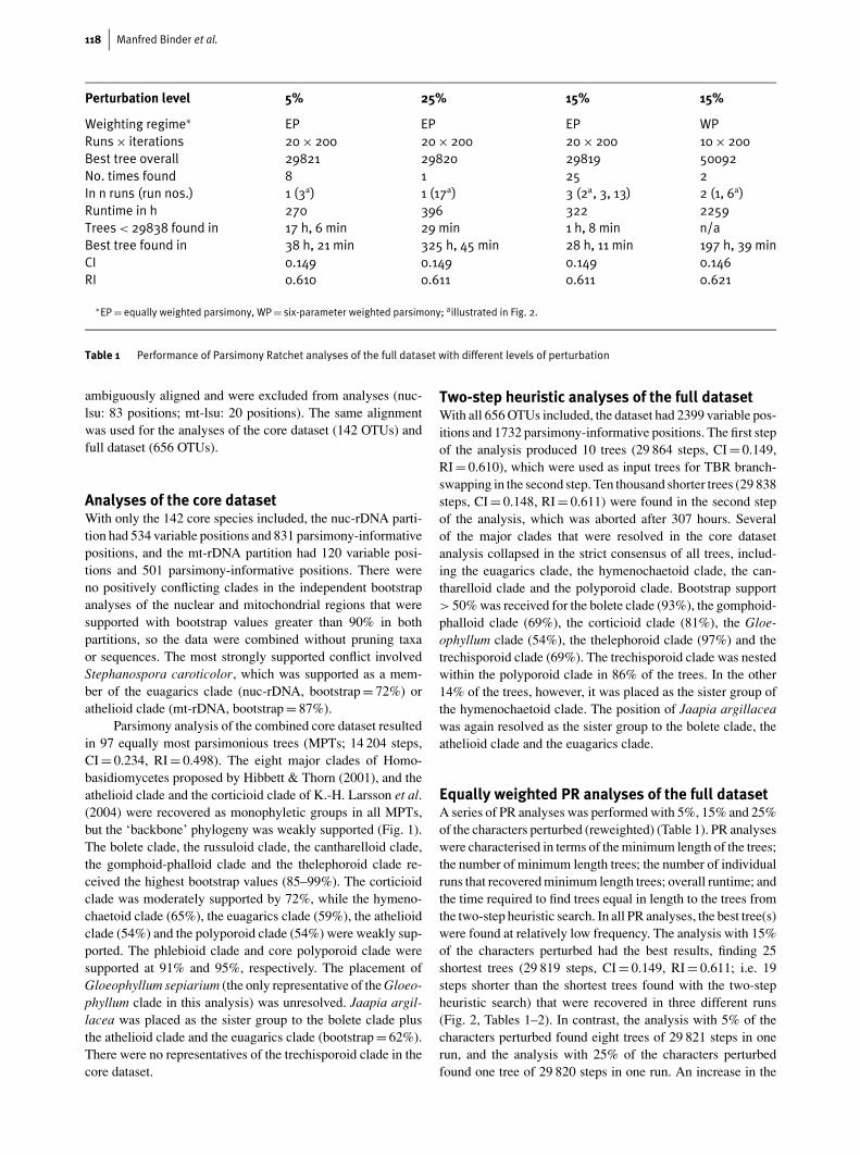
Perturbation level 5% 25% 15% 15%
Weighting regime∗ EP EP EP WPRuns× iterations 20× 200 20× 200 20× 200 10× 200Best tree overall 29821 29820 29819 50092No. times found 8 1 25 2In n runs (run nos.) 1 (3a) 1 (17a) 3 (2a, 3, 13) 2 (1, 6a)Runtime in h 270 396 322 2259Trees< 29838 found in 17 h, 6 min 29 min 1 h, 8 min n/aBest tree found in 38 h, 21 min 325 h, 45 min 28 h, 11 min 197 h, 39 minCI 0.149 0.149 0.149 0.146RI 0.610 0.611 0.611 0.621
∗EP= equally weighted parsimony, WP= six-parameter weighted parsimony; aillustrated in Fig. 2.
Table 1 Performance of Parsimony Ratchet analyses of the full dataset with different levels of perturbation
ambiguously aligned and were excluded from analyses (nuc-lsu: 83 positions; mt-lsu: 20 positions). The same alignmentwas used for the analyses of the core dataset (142 OTUs) andfull dataset (656 OTUs).
Analyses of the core datasetWith only the 142 core species included, the nuc-rDNA parti-tion had 534 variable positions and 831 parsimony-informativepositions, and the mt-rDNA partition had 120 variable posi-tions and 501 parsimony-informative positions. There wereno positively conflicting clades in the independent bootstrapanalyses of the nuclear and mitochondrial regions that weresupported with bootstrap values greater than 90% in bothpartitions, so the data were combined without pruning taxaor sequences. The most strongly supported conflict involvedStephanospora caroticolor, which was supported as a mem-ber of the euagarics clade (nuc-rDNA, bootstrap = 72%) orathelioid clade (mt-rDNA, bootstrap = 87%).
Parsimony analysis of the combined core dataset resultedin 97 equally most parsimonious trees (MPTs; 14 204 steps,CI = 0.234, RI = 0.498). The eight major clades of Homo-basidiomycetes proposed by Hibbett & Thorn (2001), and theathelioid clade and the corticioid clade of K.-H. Larsson et al.(2004) were recovered as monophyletic groups in all MPTs,but the ‘backbone’ phylogeny was weakly supported (Fig. 1).The bolete clade, the russuloid clade, the cantharelloid clade,the gomphoid-phalloid clade and the thelephoroid clade re-ceived the highest bootstrap values (85–99%). The corticioidclade was moderately supported by 72%, while the hymeno-chaetoid clade (65%), the euagarics clade (59%), the athelioidclade (54%) and the polyporoid clade (54%) were weakly sup-ported. The phlebioid clade and core polyporoid clade weresupported at 91% and 95%, respectively. The placement ofGloeophyllum sepiarium (the only representative of the Gloeo-phyllum clade in this analysis) was unresolved. Jaapia argil-lacea was placed as the sister group to the bolete clade plusthe athelioid clade and the euagarics clade (bootstrap = 62%).There were no representatives of the trechisporoid clade in thecore dataset.
Two-step heuristic analyses of the full datasetWith all 656 OTUs included, the dataset had 2399 variable pos-itions and 1732 parsimony-informative positions. The first stepof the analysis produced 10 trees (29 864 steps, CI = 0.149,RI = 0.610), which were used as input trees for TBR branch-swapping in the second step. Ten thousand shorter trees (29 838steps, CI = 0.148, RI = 0.611) were found in the second stepof the analysis, which was aborted after 307 hours. Severalof the major clades that were resolved in the core datasetanalysis collapsed in the strict consensus of all trees, includ-ing the euagarics clade, the hymenochaetoid clade, the can-tharelloid clade and the polyporoid clade. Bootstrap support> 50% was received for the bolete clade (93%), the gomphoid-phalloid clade (69%), the corticioid clade (81%), the Gloe-ophyllum clade (54%), the thelephoroid clade (97%) and thetrechisporoid clade (69%). The trechisporoid clade was nestedwithin the polyporoid clade in 86% of the trees. In the other14% of the trees, however, it was placed as the sister group ofthe hymenochaetoid clade. The position of Jaapia argillaceawas again resolved as the sister group to the bolete clade, theathelioid clade and the euagarics clade.
Equally weighted PR analyses of the full datasetA series of PR analyses was performed with 5%, 15% and 25%of the characters perturbed (reweighted) (Table 1). PR analyseswere characterised in terms of the minimum length of the trees;the number of minimum length trees; the number of individualruns that recovered minimum length trees; overall runtime; andthe time required to find trees equal in length to the trees fromthe two-step heuristic search. In all PR analyses, the best tree(s)were found at relatively low frequency. The analysis with 15%of the characters perturbed had the best results, finding 25shortest trees (29 819 steps, CI = 0.149, RI = 0.611; i.e. 19steps shorter than the shortest trees found with the two-stepheuristic search) that were recovered in three different runs(Fig. 2, Tables 1–2). In contrast, the analysis with 5% of thecharacters perturbed found eight trees of 29 821 steps in onerun, and the analysis with 25% of the characters perturbedfound one tree of 29 820 steps in one run. An increase in the

Phylogenetic distribution of resupinate forms of mushroom-forming fungi 119
Polyporus melanopusPolyporus varius
Polyporus tuberasterDatronia mollis
Polyporus squamosusCryptoporus volvatus
Fomes fomentariusDaedaleopsis confragosa
Polyporus arculariusGanoderma australe
Pycnoporus cinnabarinusPhysalacria inflataWolfiporia cocos
Junghuhnia subundataLenzites betulina
Dentocorticium sulphurellumTyromyces chioneus
Laetiporus sulphureusSparassis spathulata
Fomitopsis pinicolaOligoporus lacteus
Oligoporus leucomallelusOligoporus balsameus
Oligoporus rennyiAmylocystis lapponica
Auriporia aureaDacryobolus sudans
Antrodia carbonicaAbortiporus biennis
Podoscypha petalodesPanus rudisAlbatrellus syringae
Steccherinum fimbriatumMeripilus giganteus
“Athelia epiphylla”?Ceriporia viridansGloeoporus taxicola
Ceriporiopsis subvermisporaPhlebia albomellea
Ceriporia purpurea“Lindtneria trachyspora”
Phlebiopsis giganteaBjerkandera adustaPhanerochaete chrysosporium
Phlebia radiataPulcherricium caeruleum
Candelabrochaete africanaAsterostroma andinumScytinostroma aluta
Peniophora nudaScytinostroma renisporum
Amylostereum laevigatumEchinodontium tinctorium
Russula compactaRussula exalbicans
Clavicorona pyxidataAuriscalpium vulgare
Lentinellus omphalodesDentipellis separans
Laxitextum bicolorHericium coralloides
Bondarzewia berkeleyiBondarzewia montana
Xenasma rimicolaHeterobasidion annosum
Albatrellus skamaniusPolyporoletus sublividus
Acanthophysium cerrusatumStereum hirsutum
Gloeocystidiellum leucoxanthumDendrocorticium roseocarneum
Laetisaria fuciformisGalzinia incrustans
Gloeophyllum sepiariumCyphellopsis anomala
Lachnella villosaHalocyphina villosa
Nia vibrissaFavolaschia intermedia
Schizophyllum communePhysalacria bambusae
Physalacria maipoensisGloiocephala aquatica
Chondrostereum purpureumHenningsomyces candidus
Typhula phacorrhizaInocybe sp.
Stropharia rugosoannulataLaccaria amethystinaLaccaria pumila
Amanita muscariaCortinarius iodes
Agaricus bisporusLycoperdon sp.
Entoloma strictiusPluteus sp.
Limnoperdon incarnatumPleurotus ostreatus
Pleurotus tuberregiumHumidicutis marginata
Hygrophorus sordidusAthelia arachnoidea
“Hyphoderma praetermissum”?Stephanospora caroticolor
Plicaturopsis crispaBoletus satanas
Phylloporus rhodoxanthusParagyrodon sphaerosporus
Calostoma cinnabarinumScleroderma citrinum
Chroogomphus vinicolorGomphidius glutinosus
Suillus cavipesSuillus sinuspaulianus
Rhizopogon subcaerulescensSerpula himantioides
Tapinella atrotomentosaTapinella panuoides
Coniophora aridaConiophora puteana
Jaapia argillaceaSarcodon imbricatus
Thelephora sp.Bankera fuligineo-alba
Phellinus gilvusPhellinus igniarius
Coltricia perennisRepetobasidium mirificium
Resinicium meridionalisHyphodontia alutaria
Gautieria otthiiGomphus floccosus
Ramaria strictaSphaerobolus stellatus
CantharellustubaeformisHydnum repandum
Sistotrema eximumBotryobasidium isabellinumBotryobasidium subcoronatum
Ceratobasidium sp. GEL5602Auricularia auricula-judae
Dacrymyces chrysospermus
50 changes
52
7992
9956
55
72
82
87
10095
52
9987
976554
100
99
99
59100
100
91
54
10010092
82
100
9689
1006668
10090
10098
72 55
55
10080
62
10098
6558
96
100
89
63
51
60
59
51
54 91100
100
90
10099
10094
10069
97
98
96
53
100
100
99
62
62
9887
6586
10095100
58
10085
6785
*
*
*
*
*
*
**
corep
olyp
oro
id cla
de
ph
leb
ioid
clad
e
po
lypo
roid
clad
eru
ssulo
id cla
de
corticioid clade
core
eu
ag
arics cla
de
eu
ag
arics cla
de
athelioid clade
bo
lete
clad
e
Jaapiathelephoroid clade
hymenochaetoid clade
gomphoid-phalloid clade
cantharelloid clade
Gloeophyllum clade
An
trod
iaclade
“residual”polyporoid clade
Figure 1 Phylogenetic relationships of Homobasidiomycetes based on parsimony analysis of the combined core data set with 142 species.One of 97 equally parsimonious trees. Bootstrap values greater than 50% are indicated above branches. Nodes marked withasterisks collapse in the strict consensus tree. Names of resupinate taxa are written in bold type. Species names in quotation marksfollowed by question marks indicate mislabelled isolates.

120 Manfred Binder et al.
29819
29829
29839
29849
29859
29869
29879
29889
29899
29909
29819
29829
29839
29849
29859
29869
29879
29889
29899
29909
29819
29829
29839
29849
29859
29869
29879
29889
29899
29909
50090
50100
50110
50120
50130
50140
50150
50160
5% perturbation
15% perturbation
25% perturbation
15% perturbation, six-parameter weighted
1 25 50 75 100 125 150 175 200
number of iterations
tree length
A
B
C
D
Figure 2 Performance graphs of equally weighted PR analyses with 5%, 15% and 25% perturbation levels (A–C), and one six-parameterweighted PR analysis with 15% perturbation (D). Each graph represents one run, with 200 iterations. Runs shown are those thatfound minimum length trees (for that perturbation level). Arrows indicate the number and the position of the shortest tree(s) found.The dotted line in A–C represents the length of the shortest trees (29 838 steps) obtained with the unperturbed two-step searchapproach.

Phylogenetic distribution of resupinate forms of mushroom-forming fungi 121
Run no. Topology Iteration no.
2a A 150B 151, 153C 152D 169, 170, 171E 162
3 B 170, 178, 186D 169, 172, 173, 174, 177
13 B 125, 126, 127D 69, 71, 73, 119, 120, 121
aIllustrated in Fig. 3.
Table 2 Distribution and topology classes of shortest treesrecovered with the equally weighted PR analysis at 15%perturbation level
number of perturbed characters was correlated with increasedruntimes, which were 270, 322 and 396 hours, with 5%, 15%and 25% of the characters perturbed, respectively.
The progress of the PR was strongly affected by thechoice of perturbation levels (Fig. 2A-C). For example, theanalysis with 5% of the characters perturbed (Table 1, Fig. 2A)advanced slowly, with long ‘plateaus’, up to 20–40 iterationsin duration, in which no progress was made in tree lengths.While the 5% perturbation level yielded the most gradual pro-gress, the 25% perturbation level yielded the most chaoticsearch profiles, with dramatic shifts in tree length between it-erations (Fig. 2). The analysis with 25% perturbation foundtrees equal in length to the trees from the two-step heuristicsearch faster than the analyses with 5% and 15% perturbationlevels (29 minutes, vs. 17 hours, 6 min. and 1 hour, 8 min.,respectively), but never found trees as short as those recoveredby the analysis with 15% perturbation level. The three runswith 15% perturbation that recovered the shortest trees foundthose trees between iterations 150–171 (run no. 2; eight trees),169–186 (run no. 3; eight trees), and 69–127 (run no. 13; ninetrees; Table 2).
In all of the shortest trees, the major clades ofHomobasidiomycetes sensu Hibbett & Thorn (2001) and theathelioid, trechisporoid, corticioid and Gloeophyllum cladeswere resolved as monophyletic (Figs 3–4). Several other ma-jor topological features were shared by all trees (Figs 3–4):(1) the euagarics, bolete and athelioid clades formed a mono-phyletic group in all trees, with Jaapia argillacea as its sistergroup; (2) the trechisporoid clade (K.-H. Larsson et al., 2004)was nested within the polyporoid clade; (3) the cantharelloid,gomphoid-phalloid, and hymenochaetoid clades occupied abasal position; and (4) the Gloeophyllum and corticioid cladeswere sister groups (except in tree G, Fig. 3). None of thesegroupings received strong bootstrap support, however.
The minimum-length trees can be divided into five classesof topologies (A-E; Fig. 3), based on the variable aspects ofthe relationships among major clades. Topologies A, C and Ewere each found only once (i.e. one tree with each of thesetopologies was found), but trees with topology B were found
eight times and trees with topology D were found 14 times(Table 2). Trees with topologies B and D were found in allthree batches that recovered minimum-length trees (Table 2).
Six-parameter weighted PR analysesof the full datasetTwo shortest trees (50 092 steps, CI = 0.146, RI = 0.621) werefound in two different runs (Table 1). Under equally weightedparsimony, these trees were 29 925 and 29 929 steps long(i.e. 106–110 steps longer than the shortest trees obtainedin the equally weighted PR analyses). For comparison, the25 shortest trees obtained in the equally-weighted PR ana-lyses were 50 257–50 306 steps long under the six-parameterweighting regime (i.e. 165–214 steps longer than the shortesttrees obtained in the six-parameter PR analysis).
The six-parameter PR analysis was very time consum-ing. Ten runs with 200 iterations each required 2259 hours ofcomputer time. There are several differences in higher-levelrelationships implied by the two optimal trees. The most strik-ing difference is that in one topology the trechisporoid cladeis nested in the polyporoid clade (as in all shortest trees re-covered with equally weighted PR analysis), whereas in theother topology the trechisporoid clade is placed as the sistergroup of the hymenochaetoid clade (Figs 3–4).
Discussion
Overall phylogenetic resolutionBootstrap support for the major clades of Homobasidiomy-cetes was generally weak in the analysis of the full dataset.Missing sequences, or the presence of certain taxa whose po-sitions are particularly labile (due to homoplasy), may havecontributed to the low bootstrap values. One possible exampleof a ‘destabilising’ taxon is Stephanospora caroticolor, whichwas represented by all four rDNA regions, and was placedin either the euagarics clade or athelioid clade depending onwhether the mt-rDNA or nuc-rDNA was analysed. As the num-ber of taxa sampled increases, the chance of including specieswith aberrant sequences also increases. Therefore, it is notsurprising that there is weak bootstrap support for many ma-jor clades in recent densely sampled phylogenetic studies ofHomobasidiomycetes (e.g. Moncalvo et al., 2000; Hibbett &Binder, 2002; E. Langer, 2002; Moncalvo et al., 2002).
PR analysis was much more effective at finding mini-mum-length trees than the two-step heuristic search strategy.However, the success of the PR was sensitive to the choice ofperturbation levels, and even with the optimal 15% perturba-tion level only 3 out of 20 runs found minimum-length trees,and no more than nine shortest trees were found in any singlerun. In contrast, Nixon (1999, p. 413) reported that “approx-imately three out of four” PR analyses of the 500-species rbcLdataset of Chase et al. (1993) recovered minimum-length trees.Apparently, the full dataset analysed in this study presents amore difficult parsimony landscape than the Chase et al. data-set. The results of this study highlight the importance of doingmultiple PR runs with appropriate perturbation levels and anadequate number of iterations per run.

122 Manfred Binder et al.
e u a g a r i c s
a t h e l i o i d
b o l e t eJ a a p i aa r g i l l a c e a
t h e l e p h o r o i d
c o r t i c i o i d
G l o e o p h y l l u m
r u s s u l o i d
p o l y p o r o i d
h y m e n o c h a e t o i d
g o m p h o i d - p h a l l o i d
c a n t h a r e l l o i d
A u r i c u l a r i a l e s
D a c r y m y c e t a l e s
R e s i n i c i u mm e r i d i o n a l e
e u a g a r i c s
a t h e l i o i d
b o l e t e
J a a p i aa r g i l l a c e a
t h e l e p h o r o i d
c o r t i c i o i d
G l o e o p h y l l u m
r u s s u l o i d
p o l y p o r o i d
c a n t h a r e l l o i d
A u r i c u l a r i a l e s
D a c r y m y c e t a l e s
h y m e n o c h a e t o i d
g o m p h o i d - p h a l l o i d
R e s i n i c i u mm e r i d i o n a l e
e u a g a r i c s
a t h e l i o i d
b o l e t eJ a a p i aa r g i l l a c e a
t h e l e p h o r o i d
c o r t i c i o i d
G l o e o p h y l l u m
r u s s u l o i d
p o l y p o r o i d
h y m e n o c h a e t o i d
g o m p h o i d - p h a l l o i d
c a n t h a r e l l o i d
A u r i c u l a r i a l e s
D a c r y m y c e t a l e s
R e s i n i c i u mm e r i d i o n a l e
e u a g a r i c s
a t h e l i o i d
b o l e t eJ a a p i aa r g i l l a c e a
t h e l e p h o r o i d
c o r t i c i o i d
G l o e o p h y l l u m
r u s s u l o i d
p o l y p o r o i d #
h y m e n o c h a e t o i d
g o m p h o i d - p h a l l o i d
c a n t h a r e l l o i d
A u r i c u l a r i a l e s
D a c r y m y c e t a l e s
R e s i n i c i u mm e r i d i o n a l e
t r e c h i s p o r o i d
A C
D
G H
* * *
**
*
B
E
e u a g a r i c s
a t h e l i o i d
b o l e t e
J a a p i aa r g i l l a c e a
t h e l e p h o r o i d
c o r t i c i o i d
G l o e o p h y l l u m
r u s s u l o i d
p o l y p o r o i d *h y m e n o c h a e t o i d
g o m p h o i d - p h a l l o i d
c a n t h a r e l l o i d
A u r i c u l a r i a l e s
D a c r y m y c e t a l e s
R e s i n i c i u mm e r i d i o n a l i s
F
Figure 3 Simplified topologies of the shortest trees recovered using PR analysis with 15% perturbation. A–E= equally weighted analysesrunning 20× 200 iterations. A= single tree obtained in one run. B= 8 trees obtained in three runs. C= single tree obtained in onerun. D= 14 trees obtained in three runs. E= single tree obtained in one run. F= strict consensus of 25 trees A–E.G–H= six-parameter weighted analyses running 10× 200 iterations. Alternative topologies G= tree one and H= tree two obtainedin two different runs (see Fig. 4. for details). Polyporoid* = the polyporoid clade including the ‘core’ polyporoid clade, thetrechisporoid clade, and the phlebioid clade. Polyporoid#= the polyporoid clade without the trechisporoid clade.

Phylogenetic distribution of resupinate forms of mushroom-forming fungi 123
Six-parameter weighting increased the runtime of PRanalysis approximately seven-fold relative to the equallyweighted PR analysis with 15% perturbation. The increasedruntime may be worthwhile, because character-state weight-ing based on realistic models of molecular evolution can im-prove the accuracy of parsimony analysis (Huelsenbeck, 1995;Cunningham, 1997). The six-parameter trees share many fea-tures of the equally weighted trees, but there are also somedifferences, perhaps the most notable of which is that in one ofthe six-parameter trees (topology G, Fig. 3) the trechisporoidclade is the sister group of the hymenochaetoid clade. Theposition of the trechisporoid clade was also quite labile in theanalyses of Hibbett & Binder (2002), where it was placed inor near the polyporoid clade, hymenochaetoid clade, russuloidclade or Auriculariales.
The differences among the trees produced here and thoseobtained in earlier studies (Binder & Hibbett, 2002; Hibbett &Binder, 2002) indicate that there is considerable uncertaintyabout the higher-level phylogenetic relationships of Homo-basidiomycetes (Fig. 3). Nevertheless, the trees recovered inPR analyses all support the monophyly of the eight majorclades of Homobasidiomycetes sensu Hibbett & Thorn, as wellas the corticioid clade, athelioid clade, Gloeophyllum clade andtrechisporoid clade (which was nested within the polyporoidclade in most trees) (Hibbett & Thorn, 2001; K.-H. Larssonet al., 2004). In this regard, the results of the PR analyses ofthe full dataset are consistent with the results of the core data-set analysis. Other aspects of the higher-level topology sharedby the core and full dataset analyses include the monophylyof the clade that contains the bolete, euagarics, and athelioidclades, and its sister group relationship with Jaapia argillacea,and the basal position of the cantharelloid, gomphoid-phalloid,and hymenochaetoid clades (see below). Thus, it appears thatthe species with multiple regions in the full dataset were ableto provide a ‘backbone’ for the phylogeny, even though 60%of the OTUs were represented only by the nuc-lsu rDNA.
Relationships of Homobasidiomycetesto heterobasidiomycetesThis study sampled representatives of four of the five or-ders of ‘heterobasidiomycetes’ sensu Wells (1994; Wells &Bandoni, 2001), including the Auriculariales, Cerato-basidiales, Dacrymycetales and Tulasnellales but did notsample the Tremellales.
Auriculariales s. str.PR analyses suggest that the Auriculariales s. str. (by whichwe mean Auriculariales excluding Sebacinaceae; see below)is a paraphyletic assemblage of lineages from which theHomobasidiomycetes have been derived (Figs 3–4). Severalother studies have also concluded that the Auriculariales isclosely related to the Homobasidiomycetes, whereas the Dac-rymycetales and Tremellales have a more basal position in theHymenomycetes (Swann & Taylor, 1993, 1995; Gargas et al.,1995a; Begerow et al., 1997; E. Langer, 2002; K.-H. Larssonet al., 2004). Analyses by E. Langer (2002) and Weiß &Oberwinkler (2001) suggest that the Auriculariales s. str.is monophyletic, but with weak bootstrap support, whileHibbett & Binder (2002) recovered trees that showed the group
to be monophyletic or paraphyletic (as in the present study).Thus, it remains ambiguous whether the Auriculariales s. str. ismonophyletic or paraphyletic. Six of the eight isolates of Au-riculariales s. str. included in this study are resupinate (Fig. 4).The pileate forms include Pseudohydnum gelatinosum, whichhas a hydnoid hymenophore, and Auricularia auricula-judae,which has a smooth hymenophore. These two species are ap-parently not closely related (as was also shown by Weiß &Oberwinkler, 2001), which suggests that there have been mul-tiple origins of pileate fruiting bodies within the Auricularialess. str. (Fig. 4).
DacrymycetalesThe Dacrymycetales is strongly supported as monophyletic(bootstrap = 100%, Fig. 4). Nine of the Dacrymycetales inthis study have erect fruiting bodies that are variously coralloid,spathulate, pendulous, or lobate, but one species, Cerinomycesgrandinioides, has a resupinate fruiting body. The tree in Fig. 4suggests that the resupinate fruiting body of C. grandinioides isthe product of reduction, but bootstrap support for the internaltopology of the Dacrymycetales is weak.
Tulasnellales, Ceratobasidiales and SebacinaceaeThe placements of Auriculariales s. str. and Dacrymycetales inthis study are consistent with the traditional division betweenheterobasidiomycetes sensu Wells and Homobasidiomycetes(e.g. Stalpers, in Kirk et al., 2001). However, PR analyses placethe Tulasnellales, Ceratobasidiales and Sebacinaceae (Auricu-lariales s. lat.) in the cantharelloid clade (Fig. 4). These taxainclude forms with highly reduced resupinate to incrustingor coralloid fruiting bodies. Parenthesomes are imperforate inTulasnellales (Moore, 1978; G. Langer, 1994; Wells, 1994) andSebacinaceae (Khan & Kimbrough, 1980), and perforate withlarge pores in Ceratobasidiales (Muller et al., 1998; Wells &Bandoni, 2001). Basidial morphology is quite varied. Thebasidia of Ceratobasidiales are deeply divided by fingerlikesterigmata, but are not septate, whereas those of Tulasnellaleshave inflated epibasidia that develop adventitious septa, andthose of Sebacinaceae are longitudinally septate. Spore re-petition has been demonstrated in all three groups (Wells &Bandoni, 2001). Based on these characters, the Tulasnellales,Ceratobasidiales and Sebacinaceae have been classified as het-erobasidiomycetes (Wells & Bandoni, 2001).
The relationships among heterobasidiomycetes andHomobasidiomycetes suggested by the present study conflictwith the findings of a recent study by Weiß & Oberwinkler(2001), which suggested that: (1) the Auriculariales s. lat.is composed of three independent clades, including Auricu-lariales s. str. (43 species), Sebacinaceae (nine species), anda minor clade including Ceratosebacina calospora and Exidi-opsis gloeophora; (2) the Sebacinaceae is the sister group of allother Hymenomycetes; (3) the Ceratobasidiales (representedby Ceratobasidium pseudocornigerum) and Dacrymycetalesare sister taxa; and (4) the Ceratobasidiales-Dacrymycetalesclade is the sister group of the Homobasidiomycetes. Theseresults were based on a 600 bp region of nuc-lsu rDNA thatwas analysed with neighbour-joining. Taylor et al. (2003) ob-tained similar results, again based on analyses of up to 600 bpof nuc-lsu rDNA.

124 Manfred Binder et al.
bootstrap65-79%80-89%90-100%
50 changes
Cyclomyces fuscus CBS 100.106Cyclomyces tabacinus CBS 311.39Stipitochaete damaecornis DSH 98-006
Hymenochaete adusta TAA 95-37Hymenochaete berteroi CBS 733.86Hymenochaete pseudoadusta TAA 95-38Hymenochaete boidinii CBS 762.91Hymenochaete separabilis CBS 738.86
Hymenochaete rhabarbarina GEL4809Hymenochaete ochromarginata CBS 928.96
Hymenochaete rubiginosa TW 22.9.97Hymenochaete cinnamomea LK 27.9.97
Hymenochaete nanospora CBS 924.96Hymenochaete fuliginosa CBS 933.96Hymenochaete carpatica TW 27.9.97
Hymenochaete separata TAA 95-24Hymenochaete cruenta HB 149/80
Hymenochaete denticulata CBS 780.91Hymenochaete pinnatifida CBS 770.91
Hydnochaete duportii CBS 941.96Hydnochaete japonica CBS 499.76
Hymenochaete acanthophysata CBS 925.96Hymenochaete cervinoidea CBS 736.86
Phellinus igniarius FPL-5599Phellinus lundellii TN 5760
Phylloporia ribis FPL-10677Phellinus laevigatus TN 3260Phellinus conchatus 89-1014
Inonotus hispidus FPL-3597Mensularia hastifera 84-1023a
Hymenochaete corrugata FP-104124-Sp.Pseudochaete tabacina LK 12.10.97
Hydnochaete olivacea CLA 02-003Fomitiporia punctata 85-74
Onnia triquetra TW 411Fuscoporia contigua TW 699Phellinus gilvus FPL-5528
Fuscoporia torulosa Pt 4Fuscoporia ferruginosa 82-930Fuscoporia ferrea 87-8
Coltricia montagnei 96-96Coltricia perennis DSH 93-198
Phellinidium ferrugineofuscum TN 6121Asterodon ferruginosus Dai 3186
Phellopilus nigrolimitatus 85-823Tubulicrinis gracillimus HHB-13180-Sp.
Tubulicrinis subulatus GEL5286Basidioradulum radula FO-23507a
Fibricium rude GEL2121Trichaptum abietinum FPL-8973
Hyphodontia alutacea GEL2937Hyphodontia niemelaei GEL5068
Schizopora radula GEL3798 Hyphodontia serpentiformis GEL3307
Schizopora paradoxa GEL2511Hyphodontia nudiseta GEL5302Hyphodontia aff. breviseta GEL4214Hyphodontia nespori GEL4190
Hyphodontia crustosa GEL5360Hyphodontia sambuci GEL2414
Hyphodontia aspera GEL2135Repetobasidium mirificium FP-133558-Sp.Sphaerobasidium minutum GEL5373
Hyphodontia alutaria ? GEL2071Resinicium bicolor FP-135104-Sp.
Hyphodontia palmae GEL3456Hyphodontia cineracea GEL4875
Hyphodontia pallidula GEL4533Schizopora flavipora GEL3545
Hyphodontia alutaria GEL4553Oxyporus populinus FO35584
Tubulicrinis sp. GEL5046Subulicium sp. GEL4808
Resinicium meridionale FP-150236Trechispora araneosa KHL 8570Trechispora sp. KHL 10715
Trechispora confinis KHL 11064 Trechispora subsphaerospora KHL 8511
Trechispora incisa EH 24/98 Trechispora kavinioides KGN 981002 Trechispora hymenocystis KHL 8795
Trechispora regularis KHL 10881 Trechispora farinacea KHL 8451Trechispora farinacea KHL 8454
Trechispora farinacea KHL 8793Hyphodontia gossypina GEL5042
Subulicystidium longisporum GEL3550 Porpomyces mucidus KHL 8471Porpomyces mucidus KHL 8620Porpomyces mucidus KHL 11062
Tubulicium vermiculare GEL5015Sistotremastrum niveocremeum FO29191
Sistotremastrum niveocremeum EL 96-97Sistotremastrum sp. FO36293b
Anthurus archeri GEL5392Pseudocolus fusiformis DSH 96-033Gastrosporium simplex W 2768
Hysterangium stoloniferum W 3706Geastrum saccatum DSH 96-048Geastrum sessile GEL5319
Sphaerobolus stellatus DSH 96-015Ramaria stricta TENN HDT-5474
Gautieria otthii REG636Gomphus floccosus DSH 94-002
Ramaria formosa M-95Ramaria obtusissima GEL4416
Clavariadelphus ligulus KHL 8560Clavariadelphus pistillaris n/a
Lentaria micheneri RV98/147Ramaricium alboflavescens DAOM 17712
Kavinia himantia FP-101479Cantharellus tubaeformis DSH 93-209Craterellus cornucopioides DSH 96-003Cantharellus cibarius n/a
Hydnum repandum DSH 97-320Hydnum rufescens MB18-6024/1
Hydnum albidum MB11-6024/2Sistotrema brinkmannii GEL3134
Sistotremastrum niveocremeum ? FO36914Multiclavula mucida DSH 96-056
Clavulina cinerea 33Sistotrema eximum RGT420
Sistotrema sernanderi CBS 926.70Botryobasidium subcoronatum GEL4673
Botryobasidium subcoronatum GEL5397Botryobasidium subcoronatum FCUG 1286
Botryobasidium agg. vagum GEL4181Botryobasidium vagum GEL2122Botryobasidium isabellinum GEL2108Botryobasidium isabellinum GEL2109
Botryobasidium sp. GEL4968Botryobasidium sp. GEL5132
Botryobasidium agg. candicans GEL2090 Botryobasidium candicans GEL3083 Ceratobasidium sp. GEL 5602 Uthatobasidium sp. FO30284
Uthatobasidium fusisporum HHB-102155-Sp.Thanatephorus praticola IMI-34886
Tulasnella pruinosa DAOM 17641Tulasnella violea DAOM 222001
Tulasnella sp. GEL4461Tulasnella sp. GEL4745
Piriformospora indica DSM 11827Serendipita vermifera CBS 572.83
Pseudohydnum gelatinosum DSH 97-041Auricularia auricula-judae GJW-855-10
Exidiopsis calcea HHB-15059-Sp.Exidia thuretiana GEL5242Heterochaete sp. GEL4813
Basidiodendron sp. GEL4674Bourdotia sp. GEL4777 Basidiodendron caesiocinereum GEL5361
Calocera cornea FP-102602-Sp.Cerinomyces grandinioides GEL4761
Dacryopinax spathularia GEL5052Dacrymyces sp. GEL5083
Dacrymyces stillatus GEL5264Dacrymyces chrysospermus FPL11353
Ditiola radicata GEL4014Dacryomitra pusilla FO38346
Femsjonia sp. FO28211Guepinia spathularia FO45821
Dacrymycetales
Auriculariales s. str.
cantharelloidclade
gomphoid-phalloidclade
trechisporoidclade
hymenochaetoidclade
Resinicium meridionale
A
Hym
enochaetaceae
100
69
69
Figure 4 For Legend see facing page.

Phylogenetic distribution of resupinate forms of mushroom-forming fungi 125
To compare results of the present study with those ofWeiß & Oberwinkler (2001), the sequences of Sebacinaceae,Ceratobasidium pseudocornigerum, Ceratosebacina calo-spora and other taxa were downloaded, combined with asubset of sequences from the present study, and subjectedto bootstrapped parsimony analyses (Hibbett, unpublished).The sequences of Sebacinaceae from the study of Weiß &Oberwinkler (2001) and Serendipita vermifera from thepresent study were moderately strongly supported as a clade(bootstrap = 89%), confirming that S. vermifera is an appro-priate ‘placeholder’ for the Sebacinaceae, but Ceratobasidiumpseudocornigerum and Ceratosebacina calospora could notbe placed in any clade with confidence (bootstrap < 50 %,Hibbett, unpublished). These results suggest that the Cerato-basidiales as presently delimited could be polyphyletic. In ad-dition, analyses of mt-lsu rDNA by Bruns et al. (1998) sug-gested that Waitea circinata, which is placed in the Cerato-basidiales (Tu et al., 1977; Roberts, 1999), is closely related tothe resupinate homobasidiomycete Piloderma croceum, whichis probably a member of the athelioid clade (see below). Con-flicting results were obtained by DePriest and colleagues (un-published), who performed analyses of ITS and partial nuc-lsu rDNA sequences that suggested that Waitea circinata isin the corticioid clade (see below). The placement of Waiteawill remain unresolved until additional loci and isolates areexamined. Nevertheless, neither of the analyses cited abovesuggest that it is closely related to the cantharelloid clade.
The isolates of Ceratobasidium, Thanatephorus andUthatobasidium included in the present study are stronglysupported as monophyletic and are placed in the cantharel-loid clade in the PR analyses. Bootstrap support for the can-tharelloid clade is weak in the full dataset analyses, but inthe core dataset analysis, Ceratobasidium sp. is nested in thecantharelloid clade, with moderately strong bootstrap support(85%, Figs 1, 4). Taking the results of previous studies intoaccount, the Ceratobasidiales as a whole may be polyphyletic,but Ceratobasidium, Thanatephorus and Uthatobasidium ap-pear to form a monophyletic group within the cantharelloidclade.
Serendipita vermifera is strongly supported as the sis-ter group of the root symbiont Piriformospora indica (Vermaet al., 1998) and the Serendipita–Piriformospora clade isplaced as the sister group of the Tulasnellales, in the cantharel-loid clade (Fig. 4). Monophyly of the Serendipita–Piriformo-spora–Tulasnellales clade is weakly supported (Fig. 4). Nev-ertheless, these results are consistent with the results of mt-lsurDNA analysis by Bruns et al. (1998), which resolved a cladethat includes Tulasnella irregularis and “Sebacina sp.” andplaced it as the sister group of Cantharellus with strong (98%)
Figure 4 Phylogenetic distribution of resupinate forms among the Homobasidiomycetes, based on six-parameter weighted PR analyses of thefull 656-OTU dataset. This phylogram represents topology G (Fig. 3); see figure for branch length scale. Ranges of bootstrap valuesobtained using equally weighted parsimony greater than 65% are indicated with shaded dots on branches (white= 65–79%;grey= 80–89%; black= 90–100%). Exact bootstrap values for major clades are also written along branches, where they are above50%. Species names are followed by strain numbers that were used to generate 25S sequences. Species names in quotation marksfollowed by question marks indicate mislabelled isolates. Names of resupinate taxa are written in bold type. Major clades ofHomobasidiomycetes are indicated with brackets. This is part of the phylogenetic tree, including Dacrymycetales, Auriculariales andbasal clades of Homobasidiomycetes.
bootstrap support (also see Kristiansen et al., 2001). Weiß &Oberwinkler (2001) did not include Tulasnellales in theiranalyses of nuc-lsu rDNA sequences, but they cited unpub-lished analyses of nuc-ssu rDNA sequences, which apparentlyplaced the Tulasnellales near the Auriculariales. In contrast,E. Langer (1998) found strong support (bootstrap = 95%) fora clade including Tulasnella eichleriana and two species ofBotryobasidium, which is a member of the cantharelloid clade(see below), based on mt-ssu rDNA sequences. In addition,Kottke et al. (2003) and Bidartondo et al. (2003) found moder-ately strong (bootstrap = 88–89%) support for a clade includ-ing three species of Tulasnella, several liverwort symbionts,and Multiclavula mucida, which is also a member of the can-tharelloid clade, based on nuc-lsu rDNA sequences. Compar-able results were obtained by Hibbett & Binder (2002) andHibbett & Donoghue (2001). Tulasnellales have highly di-vergent nuclear rDNA sequences (Weiß & Oberwinkler, 2001;Hibbett, unpublished), so it is possible that the results describedby Weiß and Oberwinkler are due to ‘long branch attraction’.
Basal HomobasidiomycetesThe cantharelloid clade, gomphoid-phalloid clade and hy-menochaetoid clade appear to be among the earliest-diverginggroups in the Homobasidiomycetes (Figs 1, 3, 4). In addition,the trechisporoid clade is placed as the sister group of thehymenochaetoid clade in one of the topologies obtained withsix-parameter weighted PR analysis (Figs 3, 4). Bootstrap sup-port for the placements of these clades are weak (Figs 1, 4),but ultrastructural characters of septal pores are consistent withthe view that they occupy basal positions.
Imperforate parenthesomes have been found in thecantharelloid clade (Botryobasidium, Cantharellus, Pirifor-mospora, Sebacina, Tulasnella), gomphoid-phalloid clade(Geastrum, Ramaria), hymenochaetoid clade (Basidioradu-lum, Coltricia, Hymenochaete, Hyphodontia, Schizopora,Trichaptum, etc.), and trechisporoid clade (Hyphodontiagossypina, Subulicystidium longisporum), as well as theAuriculariales and Dacrymycetales (Traquair & McKeen,1978; Moore, 1980; 1985; G. Langer, 1994; Verma et al.,1998; Muller et al., 2000; Hibbett & Thorn, 2001; Wells &Bandoni, 2001; E. Langer, 2002; K.-H. Larsson et al., 2004).Most other Homobasidiomycetes have perforate parenthe-somes (examples are known in the euagarics, polyporoid,bolete, thelephoroid and russuloid clades), which probablyrepresent a derived condition (E. Langer, 1998; Hibbett &Thorn, 2001; E. Langer, 2002). However, imperforate paren-thesomes have been reported in the polyporoid clade (Phanero-chaete sordida) and perforate parenthesomes have been repor-ted in the gomphoid-phalloid clade (Clathrus), cantharelloid

126 Manfred Binder et al.
Sparassis spathulata BR Sparassis brevipes BR
Athelia arachnoidea ? Leptoporus mollis
L indtneria trachyspora?Phlebia centrifuga Bip
Ceriporia purpureaAthelia epiphylla ?Ceriporia viridansByssomerulius sp. Gloeoporus taxicola
Ceriporiopsis subvermispora Cystidiodontia isabellina
Phlebia albomelleaPhlebia nitidula
Ceraceomyces serpens BipCeraceomyces eludensCeraceomyces microsporus
Phlebia lilascensHapalopilus nidulans
Phlebiopsis giganteaPhlebia deflectensPulcherricium caeruleum
Phanerochaete chrysosporium Bip?Phanerochaete sordida
S istotrema musicola ? Bjerkandera adusta Bip
Phlebia acerinaPhlebia rufa Bip
Phlebia lindtneriPhlebia subochracea Bip
Phlebia radiata BipPhlebia tremellosa Bip
Climacodon septentrionalis HHB 13438Climacodon septentrionalis DSH 93-187
Mycoacia aureaPhlebia sp.Phlebia livida
Phlebia subserialisPhlebia chrysocreasPhlebia uda
P eniophora sp. ?Scopuloides hydnoides
Phanerochaete chrysorhizaMycoacia aff. fuscoatra Bip
Gelatoporia pannocincta BipCandelabrochaete africana
Hyphoderma nudicephalumHyphoderma setigerum Bip
Hyphoderma definitumMeripilus giganteus
Physisporinus sanguinolentusHypochnicium eichleriHypochnicium geogenium
Hypochnicium sp.Phlebia bresadolae
Abortiporus biennis Podoscypha petalodes
Hypochnicium polonensePhanerochaete sanguinea
Steccherinum fimbriatum TetCeriporiopsis gilvescens
Albatrellus syringaePhlebia queletii
Panus rudis TetSpongipellis pachyodon Bip
Antrodiella romelliiAntrodiella semisupinaJunghuhnia nitida Tet
Datronia mollis TetPolyporus squamosus Tet
Polyporus melanopusPolyporus tenuiculus
Polyporus tuberaster TetPolyporus varius
Cryptoporus volvatus TetPerenniporia medulla-panis Tet
Fomes fomentarius TetDaedaleopsis confragosaGanoderma australeGloeophyllum trabeum ? Bip BR
Ganoderma lucidum TetGanoderma applanatum
Lentinus tigrinus TetPolyporus arcularius TetGrammothele fuligo
Pycnoporus cinnabarinus TetLenzites betulina Tet
Physalacria inflataWolfiporia cocos BR
Trametes suaveolens TetJunghuhnia subundata
Dendrodontia sp.Dentocorticium sulphurellum
Diplomitoporus lindbladii BipTrametes versicolor
Skeletocutis amorphaTyromyces chioneus
Climacocystis sp. TetOligoporus balsameus Tet BR
Oligoporus lacteus Bip BROligoporus leucomallelus BR
Oligoporus rennyi BR Oligoporus caesius Tet BR
Dacryobolus sudans Tet BRIschnoderma benzoinum Bip
Amylocystis lapponica Tet BRAuriporia aurea BR
Phlebia griseoflavescensOligoporus placentus Bip BR
Antrodia carbonica BRAntrodia xantha BR
Cyphella digitalisdendrotheloid sp.
Grifola frondosa Pycnoporellus fulgens BR
Laetiporus sulphureus Bip BRPhaeolus schweinitzii BR
Parmastomyces transmutans Tet BRFomitopsis pinicola Bip BR
Piptoporus betulinus Bip BRAntrodia serialis BR
Neolentiporus maculatissimus Bip BR
tree 1
Sparassis brevipes ILKKA96-1044 Sparassis spathulata zw-clarku001
Trechispora araneosa Trechispora sp.
Trechispora confinis BipTrechispora subsphaerospora
Trechispora incisa Trechispora kavinioides Trechispora hymenocystis
Trechispora regularis Trechispora farinacea KHL 8451 Trechispora farinacea KHL 8454
Trechispora farinacea KHL8793 Hyphodontia gossypina
Porpomyces mucidus KHL 8471 Porpomyces mucidus KHL 8620 Porpomyces mucidus KHL 11062
Subulicystidium longisporumTubulicium vermiculare
Sistotremastrum niveocremeum Sistotremastrum sp. Sistotremastrum niveocremeum
Bjerkandera adusta DAOM 215869Hapalopilus nidulans KEW211
Phlebiopsis gigantea FCUG 1417Phlebia deflectens FCUG 1568Pulcherricium caeruleum FPL-7658
Phanerochaete chrysosporium FPL-5175Phanerochaete sordida GEL4160
S istotrema musicola ? FPL-8233Leptoporus mollis KEW122L indtneria trachyspora CBS 290.85
Ceriporia purpurea DAOM 21318Athelia arachnoidea ? GEL2529-1
Phlebia centrifuga FCUG 2396Athelia epiphylla ? HHB-8546-Sp.Ceriporia viridans FPL7440Byssomerulius sp. FO22261 Gloeoporus taxicola KEW213
Ceriporiopsis subvermispora FP90031-Sp. Cystidiodontia isabellina GEL4978
Phlebia albomellea CBS 275.92Phlebia nitidula FCUG 2028
Ceraceomyces serpens FP-102285-Sp.Ceraceomyces eludens JS22780Ceraceomyces microsporus KHL8473
Phlebia lilascens FCUG 1801Phlebia acerina FCUG 568Phlebia radiata FPL6140Phlebia rufa FCUG 2397
Phlebia lindtneri FCUG 2413Phlebia subochracea FCUG 1161
Phlebia tremellosa FPL-4294Climacodon septentrionalis HHB-13438Climacodon septentrionalis DSH 93-187
P eniophora sp. ? GEL4884Scopuloides hydnoides GEL3139
Mycoacia aurea GEL5339Phlebia sp. GEL4492Phlebia livida FCUG 2189
Phlebia subserialis FCUG 1434Phlebia chrysocreas FPL-6080Phlebia uda FCUG 2452
Phanerochaete chrysorhiza T-484Mycoacia aff. fuscoatra GEL5166
Gelatoporia pannocincta FCUG 2109 Candelabrochaete africana FP-102987-Sp.
Phanerochaete sanguinea FO25062aSteccherinum fimbriatum FP-102075Ceriporiopsis gilvescens AH 980718
Albatrellus syringae CBS 728.85Phlebia queletii FCUG 722
Panus rudis DSH 92-139Spongipellis pachyodon FO22184h
Antrodiella romellii GEL4231Antrodiella semisupina KEW65Junghuhnia nitida FO24179a
Hypochnicium eichleri GEL3137
Hypochnicium sp. GEL4741Phlebia bresadolae FCUG 1242
Hyphoderma nudicephalum GEL4727Hyphoderma setigerum GEL4001
Hyphoderma definitum GEL2898Meripilus giganteus DSH 93-193Physisporinus sanguinolentus GEL4449Abortiporus biennis KEW210
Podoscypha petalodes DSH 98-001Hypochnicium polonense GEL4428
Ischnoderma benzoinum GEL2914Datronia mollis DAOM 211792
Polyporus squamosus FPL-6846Polyporus melanopus DAOM 212269Polyporus tenuiculus GEL4780
Polyporus tuberaster DAOM 7997BPolyporus varius DSH 93-195
Cryptoporus volvatus DAOM 211791 Perenniporia medulla-panis CBS 457.48
Fomes fomentarius DAOM 129034Daedaleopsis confragosa DSH 93-182Ganoderma australe 0705
Ganoderma lucidum RZGanoderma applanatum GEL4206
Lentinus tigrinus DSH 93-181Polyporus arcularius VT959Grammothele fuligo GEL5391
Pycnoporus cinnabarinus DAOM 72065Lenzites betulina DAOM 180504
Physalacria inflata HHB-13443-Sp.Wolfiporia cocos FPL4198
Trametes suaveolens DAOM 196328Junghuhnia subundata LR-38938
Dendrodontia sp. GEL4767Dentocorticium sulphurellum FPL11801
Diplomitoporus lindbladii KEW212Trametes versicolor DSH 93-197
Skeletocutis amorpha KEW51Tyromyces chioneus KEW141
Climacocystis sp. KEW215Oligoporus balsameus KEW35
Oligoporus lacteus KEW55Oligoporus leucomallelus KEW29
Oligoporus rennyi KEW 57 Oligoporus caesius KHL 11087
Dacryobolus sudans FP-150381Amylocystis lapponica HHB-13400-Sp.
Auriporia aurea FPL-7026 Phlebia griseoflavescens FCUG 1907
Oligoporus placentus CFMR 698Antrodia carbonica DAOM 197828Antrodia xantha KEW43
Cyphella digitalis T-617dendrotheloid sp. GEL4798
Grifola frondosa CBS 480.63 Pycnoporellus fulgens T-325
Laetiporus sulphureus DSH 93-194Phaeolus schweinitzii DSH 93-196
Parmastomyces transmutans L-14910-Sp.Fomitopsis pinicola DAOM 189134
Piptoporus betulinus DSH 93-186Antrodia serialis GEL4465Daedalea quercina DAOM 142475
Neolentiporus maculatissimus Rajchenberg 158
bootstrap65-79%80-89%90-100%
50 changes
BR = brown rotTet = tetrapolarBip = bipolar
Daedalea quercina Bip BR
Hypochnicium geogenium GEL4081
Gloeophyllum trabeum ? CFMR 617-R
A
B
phlebioid clade
phlebioid cladetrechisporoid clade
residual polyp. clade residual polyp. clade
core polyporoid clade core polyporoid clade
Antrodia clade
Antrodia clade
polyporoid clade
polyporoid clade
tree 2
Figure 4 Continued Polyporoid clade. Tree 1 represents topology G, in which the trechisporoid clade is the sister group of thehymenochaetoid clade, and tree 2 represents topology H, in which the trechisporoid clade is nested within thepolyporoid clade (Fig. 2). Mating systems for taxa where this is known are indicated in tree 1 (Tet= tetrapolar,Bip= bipolar). Species that produce a brown rot are also indicated (BR).
clade (Ceratobasidiales, Sistotrema brinkmannii), hymeno-chaetoid clade (Hyphoderma praetermissum) and trech-isporoid clade (Trechispora subsphaerospora) (Eyme &
Parriaud, 1970; E. Langer & Oberwinkler, 1993; G. Langer,1994; Keller, 1997; Wells & Bandoni, 2001). These reports,which should be confirmed, suggest that there has been

Phylogenetic distribution of resupinate forms of mushroom-forming fungi 127
Asterostroma medium CBS 119.50Asterostroma ochroleucum HB 9/89
C oronicium alboglaucum ? GEL5058Asterostroma andinum HHB-8546-Sp.
Scytinostroma aluta CBS 762.81Scytinostroma caudisporum CBS 746.86
Scytinostroma portentosum GEL3225Scytinostroma ochroleucum CBS 767.86
Peniophora nuda FPL4756Dichostereum durum CBS 707.81Dichostereum pallescens CBS 717.81Dichostereum effuscatum CBS 516.80
Vararia insolita CBS 667.81Vararia parmastoi CBS 647.84
Vararia sphaericospora CBS 700.81Scytinostroma eurasiatico-galactinum CBS 666.84
Amphinema byssoides ? HHB-13195-Sp.Scytinostroma renisporum CBS 770.86
Amylostereum chailetii FCUG-2025 Amylostereum laevigatum CBS 623.84
Echinodontium tinctorium DAOM 16666Laurilia sulcata CBS 365.49
Gloeocystidiellum clavuligerum JS16976Lactarius corrugis RV 88/61
Lactarius volemus RV95/150Russula romagnesii JJ60
Russula compacta Duke s.n.Russula mairei RV 89/62
Russula virescens JSH s.nRussula earlei n/a
Russula exalbicans REG MB 95-111Gloeocystidiellum aculeatum n/a
Gloeocystidiellum porosum FCUG 2734Gloeocystidiellum sp. NH13258Gloeocystidiellum sp. NH12972
Lentinellus montanus VT242Lentinellus omphalodes DSH 96-007Lentinellus vulpinus KGN 980825
Auriscalpium vulgare DAOM 128994Gloiodon strigosus GEL5335I
Aleurocystidiellum disciformis NH13003Aleurocystidiellum subcruentatum NH12874
Albatrellus ovinus REG Ao1Albatrellus subrubescens REG As1
Albatrellus cristatus REG Ac1 Albatrellus confluens REG Aco2Albatrellus fletti BG Thesis
Albatrellus skamanius DAOM 220694Polyporoletus sublividus DAOM 221078
Dendrothele candida HHB-3843-Sp.Xenasma rimicola FP-133272-Sp.
Bondarzewia berkeleyi 73BOBondarzewia montana DAOM 415
Heterobasidion annosum RGT 931030/23Cymatoderma caperatum ? HHB-9974-Sp.
Dentipellis separans CBS 538.90Laxitextum bicolor CBS 284.73
Creolophus cirrhatus GEL4351Hericium coralloides DSH 93-189
Hericium erinaceus FO23203Stereum armeniacum GEL4857Stereum hirsutum FPL 8805
Stereum gausapatum GEL4615Stereum rugosum HHB13390
Acanthofungus rimosus Wu9601_1Acanthophysium bisporum T614
Acanthophysium cerrusatum FPL-11527Aleurodiscus lapponicus FP100753Aleurodiscus laurentinus HHB11235Acanthophysium sp. GEL5022
Aleurodiscus botryosus CBS 195.91Stereum annosum FPL-8562
Xylobolus frustulatus FP106073Xylobolus subpileatus FP106735
Acanthophysium lividocaeruleum FP100292Aleurodiscus abietis T330
Gloeocystidiellum leucoxanthum CBS 454.86Aleurodiscus mirabilis Wu9304_105
Aleurodiscus oakesii FP101813Acanthobasidium norvegicum T623Acanthobasidium phragmitis CBS 233.86
Aleurodiscus weirii FP134813Aleurodiscus penicillatus T322
Aleurodiscus amorphus HHB15282Aleurodiscus grantii T541
Dendrocorticium polygonioides FO36469gDendrocorticium roseocarneum FPL1800
Punctularia strigoso-zonata HHB-11897-Sp.Vuilleminia comedens T-583
Marchandiomyces corallinus ATCC200796Marchandiomyces lignicola n/a
Marchandiomyces aurantiacus CBS 718.97Galzinia incrustans HHB-12952-Sp.
Tomentella ferruginea 78 Tomentella stuposa 21
Tomentella coerulea 75 Thelephora sp. DSH 96-010Thelephora palmata 31/38
Thelephora vialis Thv1Hydnellum sp. DSH 96-008
Sarcodon imbricatus REG Sim1Bankera fuligineoalba DAOM 184178
Phellodon tomentosus BG ThesisPseudotomentella mucidula 60 Pseudotomentella nigra 16
Pseudotomentella ochracea EL99-97Gloeophyllum sepiarium DAOM 13786 Neolentinus dactyloides E5252A
Veluticeps berkeleyi RLG-7116-Sp.Gloeophyllum odoratum FO23521
Heliocybe sulcata D.797
bootstrap65-79%80-89%90-100%
50 changes
thelephoroidclade
Gloeophyllumclade
corticioidclade
russuloidclade
/ amylostereaceae
/ bondarzewiaceae/ gloeocystidiellum II
/ russulales
/ gloeocystidiellum I
/ auriscalpiaceae
/ aleurocystidiellum
/ albatrellus
/ bondarzewiaceae
/ hericiaceae
/ stereales
/ peniophorales
C
B
54
97
81
79
84
96
100
73
100
88
Figure 4 Continued Gloeophyllum, thelephoroid, corticioid and russuloid clades. Groups within the russuloid clade correspond to groupsrecognised by E. Larsson and K.-H. Larsson.
extensive homoplasy in parenthesome evolution, possibly in-cluding reversals from perforate to imperforate parenthesomes(K.-H. Larsson et al., 2004). Nevertheless, the occurrence ofimperforate parenthesomes in the Auriculariales s. str. andDacrymycetales, and their preponderance in the cantharelloid,gomphoid-phalloid, hymenochaetoid, and trechisporoidclades suggests that this is the plesiomorphic condition in theHomobasidiomycetes, which is consistent with the topologyinferred with rDNA sequences. The core dataset tree and fiveof the topologies obtained in PR analyses of the full datasetsuggest that the cantharelloid clade is the sister group of theother Homobasidiomycetes, but bootstrap support is weak(Figs 1, 3, 4).
Phylogenetic distribution of resupinate formswithin the Homobasidiomycetes
Resupinate forms occur in every major clade of Homobasi-diomycetes (Hibbett & Binder, 2002; K.-H. Larsson et al.,2004). The following sections and Table 3 provide a clade-by-clade overview of the distribution of resupinate forms, basedon this and other studies. Notes on ecology are also provided.More detailed commentary on the morphology and taxonomyof many of the resupinate forms in this study can be found inE. Larsson (2002), E. Langer (2002), and other works citedbelow. It is not the purpose of this study to infer the his-torical pattern of transformations between resupinate and

128 Manfred Binder et al.
Marasmius capillaris DED4345Physalacria sp. GEL5189
Marasmius delectans DED 89/62Crinipellis campanella DAOM 17785Crinipellis maxima DAOM 196019
Lopharia mirabilis ? FRI 330- TT rechispora farinacea ? HHB 9150
Vararia ochroleuca CBS 465.61Vararia gallica CBS 656.81
Lycoperdon perlatum DSH 96-047 Lycoperdon sp. DSH 96-054
Calvatia gigantea DSH 96-032Lepiota clypeolaria VPI-OKM22029
Leucoagaricus rubrotinctus DUKE-JJ100Leucocoprinus fragilissimus DUKE-JJ84
Agaricus arvensis SAR 93/494Agaricus bisporus SAR 88/411
Agaricus campestris VPI-OKM25665Chlorophyllum molybdites NY-EFM891
Macrolepiota rachodes VPI-OKM19588Leucoagaricus naucinus VPI-OKM15134Leucocoprinus cepaestipes NY-EFM518
Lepiota procera DSH 96-038Macrolepiota procera DUKE-JJ168
Podaxis pistillaris J119Coprinus sterquilinus C123Montagnea arenaria J117
Lepiota cristata DUKE1582Cystolepiota cystidiosa MICH18884
Lepiota acutesquamosa DUKE-JJ177Tulostoma macrocephala Long 10111 FHTulostoma sp. GEL5402
Coprinopsis atramentaria C114Coprinopsis sp.C192Coprinopsis cinerea C13
Coprinopsis kimurae C78Coprinopsis quadrifida RGT 930622/01
Coprinellus bisporus C148Psathyrella candolleana J181
Psathyrella gracilis J130Psathyrella delineata J156
Parasola nudiceps C159Lacrymaria velutina J100
Crepidotus inhonestus MCA638Crepidotus mollis OKM26279
Crepidotus variabilis REG JE 5.3Inocybe cervicolor EL 27-99Inocybe sp. RV7/4
Inocybe geophylla JM96/25Anellaria semiovata SAR s.n.
Panaeolus acuminatus J129Panaeolina foenisecii J152Bolbitius vitellinus SAR 84/100
Conocybe rickenii J183Ripartitella brasiliensis NY-EFM744Hypholoma sublateritium JM96/20Hypholoma subviride JJ69
Stropharia rugosoannulata Hopple D258Pholiota squarrosoides JJ7
Kuehneromyces mutabilis DSM1684Psilocybe silvatica RV5/7/1989Cortinarius stuntzii SAR 85/358
Psilocybe stuntzii VT1263Hebeloma crustuliniforme SAR 87/408Cortinarius bolaris REG MB 96-086Dermocybe marylandensis JM96/24
Rozites caperatus G96-3Cortinarius iodes Moncalvo96/23
Laccaria amethystina DSH s.n.Laccaria pumila DSH s.n.Laccaria bicolor JM96/19
Cortinarius sp. JM96/40Crucibulum laeve REG Crul1
Cyathus striatus REG Cyst1Callistosporium luteoolivaceum RV10/1
Resupinatus dealbatus T-818Resupinatus trichotis GEL4221
Resupinatus sp. VT1520Resupinatus alboniger RV/JMs.n.Arrhenia auriscalpium Lutzoni 930731-3Arrhenia lobata Lutzoni & Lamoure 910824-1
Caulorhiza hygrophoroides DAOM 172075Conchomyces bursaeformis RV95/302
Pleurotus eryngii D643Pleurotus fossulatus D1822Pleurotus populinus D765Pleurotus ostreatus D850 Pleurotus abieticola RHP6551.1Pleurotus pulmonarius D700
Pleurotus calyptratus D1839Pleurotus djamor D1847
Pleurotus cornucopiae D383Pleurotus cystidiosus D420Pleurotus dryinus F91/1116Pleurotus smithii D478
Pleurotus tuberregium DSH 92-155Pleurotus purpureoolivaceus RV95/486
Hohenbuehelia sp. RV95/214 Hohenbuehelia tristis n/a
Chrysomphalina chrysophylla SAR. 7700Chrysomphalina grossula Gulden 417/75
Hygrophorus bakerensis SAR s.n.Hygrophorus sordidus RV94/178
Humidicutis marginata Moncalvo96/32Hygrocybe citrinopallida Lutzoni 930731-1
Athelia arachnoidea DNA815 H yphoderma praetermissum ? L-16187-Sp.
Athelia fibulata GEL 5292Phlebiella sp. GEL4684
Deflexula subsimplex FO41017 Stephanospora caroticolor IOC 137/97
Radulomyces molaris GEL5394Lentaria albovinacea GEL5388Plicaturopsis crispa FP-101310-Sp.
Boletus retipes SARXerocomus chrysenteron TDB-635
Boletus satanas TDB-1000CPhylloporus rhodoxanthus SAR 89/457
Paragyrodon sphaerosporus TDB-420Paxillus filamentosus REG 304
Hydnomerulius pinastri REG 412Calostoma cinnabarina MSC 362913
Scleroderma citrinum REG Sc1Leucogyrophana mollusca REG 447
Suillus cavipes TDB-646Suillus sinuspaulianus DAOM 66995
Suillus luteus JM96/41Chroogomphus vinicolor TDB-1010
Gomphidius glutinosus TDB-953bRhizopogon subcaerulescens F-2882
Coniophora arida REG 373Coniophora olivacea REG 402
Coniophora puteana REG 410Coniophora marmorata REG 411
Leucogyrophana arizonica REG 404 Leucogyrophana olivascens REG 397Leucogyrophana romellii REG 401
Serpula himantioides HHB-17587-Sp. Serpula lacrymans REG 697Serpula incrassata REG 406Leucogyrophana pulverulenta REG 819
Tapinella atrotomentosa REG 310Tapinella panuoides REG 318
Pseudomerulius aureus FP-103859-Sp.Jaapia argillacea REG 425
Clitocybe connata JM90cDendrocollybia racemosa DED5575
Henningsomyces candidus GEL4482Pleurocybella porrigens OKM19644
Phyllotopsis nidulans RV96/1B ulbillomyces farinosus ? FO24378Macrotyphula cf. juncea MIN DM-975Typhula phacorrhiza DSH 96-059
Panellus serotinus DSH 93-218Mycena rutilanthiformis JM96/26Mycena clavicularis RV87/6Mycena flavoalba GEL4649
Panellus stipticus DSH 93-213Resinomycena acadiensis DAOM 169949Mycena haematopoda GEL3777
Favolaschia sp. GEL4781Favolaschia sp. GEL4835
Xeromphalina cauticinalis RV86/11Panellus ringens S. Jacobsson s.n.Pleurotopsis longinqua RV95/473
Chondrostereum purpureum HHB-13334-Sp.Gloeostereum incarnatum 3332Hydropus scabripes DAOM 192847
Baeospora myosura GEL3962Baeospora myriadophylla DAOM 188774
Armillaria tabescens D290Armillariella ostoyae GEL4424
Oudemansiella mucida GEL4363Xerula furfuracea JM96/42Xerula megalospora DAOM 196115
Strobilurus trullisatus DAOM 188775Cyptotrama asprata DAOM 157066
Gloiocephala menieri DAOM 170087Gloiocephala aquatica CIEFAP 50Rhizomarasmius pyrrhocephalus DED4503
Flammulina velutipes SAR s.n.Rhodotus palmatus VT356
Physalacria bambusae CBS 712.83Physalacria maipoensis 2373Inderbitzin
Cylindrobasidium laeve GEL5380Cylindrobasidium laeve HHB-8633-TCylindrobasidium sp. GEL5043
Campanella junghuhnii GEL4720Campanella subdendrophora DAOM 175393
Gerronema strombodes Kuyper 2984Gerronema subclavatum Redhead 5175
Henningsomyces candidus T-156Rectipilus fasciculatus GEL4485
Neonothopanus nambi RVPR27Omphalotus nidiformis T1946.8Lampteromyces japonicus JMlegMURAKAMI
Micromphale perforans RV83/67Marasmius alliaceus GEL4610
Marasmiellus ramealis DED3973Rhodocollybia maculata RV94/175
Collybia dryophila RV83/180Collybia polyphylla RV182.01
Lentinula edodes ATCC 42962Lentinula lateritia DSH 92-143
Clitocybe ramigena RV87/19Hypsizygus ulmarius JM/HW
Clitocybe clavipes JM96/22Limnoperdon incarnatum IFO30398
Clitocybe lateritia Lutzoni 930803-1Limacella glioderma VT(L18)
Pluteus primus JB94/24Pluteus sp. JM96/28
Limacella glischra VPI-GB505Amanita jacksonii TV 96/1
Amanita farinosa RV 96/104Amanita muscaria AR s.n.
Amanita solitariiformis DD 97/12Amanita virosa JM 97/42
Amanita citrina var. grisea HKAS 32506Amanita flavoconia RV 5Aug96
Clitopilus prunulus RV88/109Entoloma odorifer TB 6366
Entoloma strictius Moncalvo 96/10Ossicaulis lignatilis DUKE483Tricholoma giganteum IFO31860
Tricholoma caligatum KMS 452Tricholoma inamoenum REG MB 96-071
Tricholoma atroviolaceum KMS 400Tricholoma vernaticum KMS 246Tricholoma pardinum KMS 278Tricholoma venenatum KMS 393
Tricholoma myomyces JM98/700Tricholoma focale KMS 426
Tricholoma imbricatum KMS 356Tricholoma subaureum KMS 590
Tricholoma intermedium KMS 593Tricholoma portentosum KMS 591
Lyophyllum decastes JM 87/16Termitomyces cylindricus JM/leg.R.S.Hseu.s.n.Podabrella microcarpus PRU3900
Termitomyces heimii JM/leg.S.Muid.s.n.Fistulina antarctica CBS 701.85
Fistulina hepatica DSH 93-183Fistulina pallida CBS 508.63Porodisculus pendulus HHB-13576-Sp.
Auriculariopsis ampla NH-1803-SpainSchizophyllum commune REG Sco1
Lachnella villosa FO25147Dendrothele griseo-cana FP-101995-Sp.Dendrothele acerina GEL5350
Flagelloscypha minutissima CBS 823.88Calathella mangrovei 1-30-01Jones
Favolaschia intermedia L-13421-Sp.Halocyphina villosa IFO32086
Nia vibrissa REG M200Cyphellopsis sp. GEL4873
Cyphellopsis anomala GEL4169Cyphellopsis anomala CBS 151.79Merismodes fasciculata HHB-11894
bootstrap65-79%80-89%90-100%
50 changes
boleteclade
Jaapia
athelioid clade
core euagarics clade
euagarics clade
C93
75
100
85
98
83
100
Figure 4 Continued Jaapia argillacea, bolete clade, athelioid clade and euagarics clade.

Phylogenetic distribution of resupinate forms of mushroom-forming fungi 129
IsolatesbClade
Subclade/species Sequencesa This study Other studies
Athelioid clade‘Amphinema byssoides’ 1,2 −HHB 13195-Sp. A + EL 11-98Athelia arachnoidea 1,2 + 815 B− ‘GEL 2529.1’
2 − ‘GEL 2529.1’Athelia decipiens A + JS 4930‘Athelia epiphylla’ 1,2,3,4 −HHB-8546-sp A + EL 12-98Athelia fibulata 2 + GEL 5292 B−GEL 5292Atheliopsis subinconspicua A + KHL 8490Byssocorticium pulchrum A + KHL 11710Piloderma byssinum A + KHL 8456Piloderma lanatum A + JS 24861Tylospora asterophora A + KHL 8566
Bolete cladeConiophora arida 1,2,3,4 + MB-1823-sp A + KHL 8547
B + AF098375C + SFC 990911-57
Coniophora marmorata 2 + 411Coniophora olivacea 2 + 402Coniophora puteana 1,2,3,4 + FP-102430spHydnomerulius pinastri 2 + 412Leucogyrophana arizonica 2 + 404Leucogyrophana mollusca 2 + 447Leucogyrophana olivascens 2 + 397Leucogyrophana pulverulenta 2 + 819Leucogyrophana romellii 2 + 401 A + KHL 11066Pseudomerulius aureus 1,2,3 + FP-103859sp A +B.Norden
C + SFC 970927-4Serpula himantioides 1,2,3,4 + HHB-17587sp B +GEL 5395Serpula incrassata 2,4 + 406Serpula lacrymans 2 + 697
Cantharelloid cladeBotryobasidium agg. candicans 2 +GEL 2090 B +GEL 2090Botryobasidium agg. vagum 2 +GEL 4181 B +GEL 4181Botryobasidium botryosum A + KHL 11081Botryobasidium candicans 2 +GEL 3083 B +GEL 3083Botryobasidium isabellinum 2 +GEL 2108 B +GEL 2108
1,2,3,4 + GEL 2109 C +GEL 2109Botryobasidium sp. 2 + GEL 4698 B +GEL 4698
2 +GEL 5132 B +GEL 5132Botryobasidium subcoronatum 2 +GEL 4673 B +GEL 4673
2 +GEL 5397 B +GEL 53971,2,3,4 + FCUG 1286 C +GEL 1286
Botryobasidium vagum 2 +GEL 2122Ceratobasidium sp. 1,2,3,4 +GEL 5602Haplotrichum conspersum A + KHL 11063
C + SFC990123-15Membranomyces delectabilis A + KHL 11147Multiclavula mucidac 1,2,3 + DSH 93-056 C +DSH 93-056Piriformospora indicac 1,2,3 +DSM 11827
Table 3 Phylogenetic distribution of resupinate and other selected reduced species among the major clades of Homobasidiomycetes andoutgroups (Auriculariales and Dacrymycetales), as estimated by the present study, K.-H. Larsson et al. (2004), Langer (2002), Lim(2001; nuc-ssu rDNA analyses only) and Kim & Jung (2000)

130 Manfred Binder et al.
IsolatesbClade
Subclade/species Sequencesa This study Other studies
Serendipita vermiferad 2 + CBS 572.83Sistotrema alboluteum A +UK 166Sistotrema brinkmannii 2 +GEL 3134 A +NH 11412/2206
B + FO 31682B +GEL 3134
Sistotrema confluensc A + PV 174Sistotrema coronilla A +NH 7598/785Sistotrema diademiferum C + SFC990521-13Sistotrema eximum 1,2,3,4 + RGT 420Sistotrema sernanderi 2,3 + CBS 926.70‘Sistotrema muscicola’ 1,2,3 − FPL 8233 A + KHL 8794Thanatephorus praticola 1,2,3 + IMI-34886Tulasnella obscura B +GEL 4624Tulasnella pruinosa 2,3,4 + DAOM 17641Tulasnella sp. 2 + GEL 4461 B +GEL 4461
2 +GEL 4745 B +GEL 4745Tulasnella violea 2,3 + DAOM 222001Uthatobasidium fusisporum 1,2,3 + HHB 102155spUthatobasidium sp. 2 + FO 30284 B + FO 30284
Corticioid cladeCorticium roseum A + EL 13-98
C + eSFC 991231-9Dendrocorticium polygonioides 2 + FO 36469g B + FO 36469gDendrocorticium roseocarneum 1,2,3,4 + FPL 1800 A + FPL 1800Dendrothele maculata A +HHB 10621Erythricium laetum A +GB/NH14530Galzinia incrustans 1,2,3,4 + HHB-12952spLaetisaria fuciformis 1,2,3,4 +NJ-2 JacksonMarchandiomyces aurantiacus 2 +DePriestMarchandiomyces corallinus 2 +DePriestMarchandiomyces lignicola 2 +DePriestPunctularia strigoso-zonata 1,2 + HHB-11897sp A + LR 40885Vuilleminia comedens 1,2,3 + T-583 A + EL 1-99
B +GEL 4110C + SFC 990326-21
Vuilleminia macrospora A + EL 21-99
Euagarics cladeAmylocorticium cebennense A + JS 24813Amylocorticium subincarnatum A + AS-95Anomoporia bombycina A +GG u612Anomoporia kamtschatica A + KHL 11072Athelia bombacina C + no dataAuriculariopsis amplac 2,3,4 +NH 1803‘Bulbillomyces farinosus’ 2, + FO 24378 B + FO 24378Calathella mangroveic 1,2,3 + 1-30-01JonesCalyptella campanulac B +Ceraceomyces tessulatus ?KHL 8474Chondrostereum purpureum 1,2,3,4 + HHB-13334sp A + EL 59-97
B +GEL 5348C + SFC 971001-13C + CBS 427.72
Table 3 Continued.

Phylogenetic distribution of resupinate forms of mushroom-forming fungi 131
IsolatesbClade
Subclade/species Sequencesa This study Other studies
Coronicium alboglaucum 2 + GEL 5058 A−NH 4208/377B−GEL 5058
Cylindrobasidium laeve 1,2,3,4 + HHB-8633-T A +Ulvesund2 + GEL 5380 B +GEL 5380
C + SFC990121-8Cylindrobasidium sp. 2 + GEL 5043 B +GEL 5043Cyphellopsis anomalac 1,2,3,4 + CBS 151.79 B +GEL 4169
2 + GEL 4169Cyphellopsis sp.c + GEL 4873 B +GEL 4873Cystostereum murraii C + CBS 257.73Dendrothele acerina 2 + GEL 5350 B +GEL 5350Dendrothele griseocana 2 + FP 101995-sp‘dendrotheloid’ sp. B + GEL 4798Favolaschia intermediac 1,2,3,4 + L-13421-spFlagelloscypha minutissimac 1,2,4 + CBS 823.88Gloeostereum incarnatumc 2 + NH 3332Halocyphina villosac 1,2,3,4 + IFO 32086Henningsomyces candidus2 1,2,3,4 + GEL 4482 B +GEL 4482Hypochniciellum subillaqueatum A + KHL 8493Lachnella villosac 1,2,3,4 + CBS 609.87 B + FO 25147Merismodes fasciulatusc 1,2,3 + HHB-11894Mucronella calva B +GEL 4458Mycoacia copelandii C + SFC990710-6Phlebiella pseudotsugae A +NH 10396/1953Plicaturopsis crispac 1,2,3,4 ?FP 101310-sp B−GEL 4132
C−SFC 990320-8Rectipilus fasciculatus B +GEL 4485Schizophyllum communec 1,2,3,4 + DSH 96-026 B +GEL 4623
Gloeophyllum cladeBoreostereum radiatum C + CBS 417.61Donkioporia expansa C + CBS 299.93Gloeophyllum sepiariumc 1,2,3,4 + DAOM 137861Heliocybe sulcatac 1,2,3 + D. 797Veluticeps berkeleyi 1,2,4 + RLG-7116-sp C + CBS 725.68
Gomphoid-phalloid cladeKavinia alboviridis A + EL 16-98Kavinia himantia 1,2,3,4 + FP-101479sp A + LL-98Kavinia sp. B− FO 25092Ramaricium alboflavescens 1,2,4 + DAOM-17712
Hymenochaetoid cladeAsterodon ferruginosum A + KHL 11176Basidioradulum radula 1,2,3 + FO 23507a A +NH 9453
B +GEL 4107C + no data
Fibricium rude 2 + GEL 2121 B +GEL 2121Hyphoderma guttuliferum A +NH 12012/2438Hyphoderma praetermissum 1,2,3,4 − ‘L-16187-sp.’ A + NH 9536/1708
B +GEL 4845Hyphodontia aff. breviseta 2 + GEL 4214 B +GEL 4214Hyphodontia alienata A + EL14-98Hyphodontia agg. alutaria B +GEL 2034
Table 3 Continued.

132 Manfred Binder et al.
IsolatesbClade
Subclade/species Sequencesa This study Other studies
Hyphodontia alutaria 1,2,3,4 + ‘GEL 2071’ C + ‘GEL 2071’2 + GEL 4553
Hyphodontia alutacea 2 + GEL 2397 B +GEL 2937Hyphodontia aspera 2 + GEL 2135 A + KHL 8530
B +GEL 2135Hyphodontia barbajovis B +GEL 3806Hyphodontia borealis A + JS 26064Hyphodontia breviseta A + JS 17863Hyphodontia cineracea 2 + GEL 4875 B +GEL 4875Hyphodontia crustosa 2 + GEL 5360 B +GEL 5360Hyphodontia nespori 2 + GEL 4190 B +GEL 4190Hyphodontia niemelaei 2 + GEL 5068Hyphodontia nudiseta 2 + GEL 5302 B +GEL 5302Hyphodontia pallidula 2 + GEL 4533 B +GEL 4533Hyphodontia palmae 2 + GEL 4536 B +GEL 3456Hyphodontia quercina A + KHL 11076Hyphodontia sambuci 2 + FO 42008 B +GEL 2414Hyphodontia serpentiformis 2 + GEL 3307 B +GEL 3307Oxyporus populinusc 2 + FO 35584 B + FO 35584Repetobasidium mirificium 1,2,3,4 + FP-133558spResinicium bicolor 1,2,3 + FP-135104sp A +NH 11540/2228
B−GEL 4664C +HHB 10103C + CBS 253.73
Schizopora flavipora 2 + GEL 3545 B +GEL 3545Schizopora paradoxa 1,2,3 + GEL 2511 B +GEL 4188
C−GEL 2511Schizopora radula 2,3 + GEL 3798Sphaerobasidium minutum 2 + GEL 5373 B +GEL 5373Subulicium sp. 2 + GEL 4808 B +GEL 4808Trichaptum abietinumc 1,2,3 + FPL 8973 B +GEL 5237Tubulicrinis gracillimus 1,2 + HHB-13180spTubulicrinis subulatus 2 + GEL 5286 A + KHL 11079
B +GEL 5286Tubulicrinus sp. 2 + GEL 5046 B +GEL 5046
HymenochaetaceaeFomitoporia punctata 2 + 85-74Fuscoporia contigua 2 + TW 699Fuscoporia ferrea 2 + 87-8Fuscoporia ferruginosa 2 + 82-930Hydnochaete olivacea 1,2,3 + CLA 02-003Hymenochaete acanthophysata 2 + CBS 925.96Hymenochaete adusta 2 + TAA 95-37Hymenochaete berteroi 2 + CBS 733.86Hymenochaete boidinii 2 + CBS 726.91Hymenochaete carpatica 2 + TW 27.9.97Hymenochaete cervinoidea 2 + CBS 736.86Hymenochaete cinnamomea 2 + LK 27.9.97 A + EL 6-99Hymenochaete corrugata 1,2,3 + FP-104124spHymenochaete cruenta 2 + HB 149/80Hymenochaete denticulata 2 + CBS 780.91
Table 3 Continued.

Phylogenetic distribution of resupinate forms of mushroom-forming fungi 133
IsolatesbClade
Subclade/species Sequencesa This study Other studies
Hymenochaete duportii 2 + CBS 941.96Hymenochaete fuliginosa 2 + CBS 933.96Hymenochaete japonica 2 + CBS 499.76Hymenochaete nanospora 2 + CBS 924.96Hymenochaete ochromarginata 2 + CBS 928.96Hymenochaete pinnatifida 2 + CBS 770.91Hymenochaete pseudoadusta 2 + TAA 95-38Hymenochaete rhabarbarina 2 + GEL 4809 B +GEL 4809Hymenochaete rubiginosa 2 + TW 22.9.97Hymenochaete separabilis 2 + CBS 738.86Hymenochaete separata 2 + TAA 95-24Hymenochaete sp. 2 A + KHL 11024Mensularia hastifera 2 + 84-1023aPhellinidium ferrugineofuscum 2 + TN 6121Phellinus laevigatus 2 + TN 3260Phellopilus nigrolimitatusc 2 + 85-823Pseudochaete tabacina 2 + FPL 3000
JaapiaJaapia argillacea 1,2,3,4 + Reg 425
Polyporoid cladecore polyporoid cladeDendrodontia sp. 2 + GEL 4767 B +GEL 4767Dentocorticium sulphurellum 1,2,3,4 + FPL 11801 C + FPL 11801Diplomitoporus crustulinus C− CBS 443.48Diplomitoporus lindbladii 1,2 + KEW 212 B +GEL 4653Grammothele fuligo 2 + GEL 5391 B +GEL 5391Junghuhnia subundata 1,2,3,4 + LR-38938Lopharia cinerascens A + EL 63-97
C + CBS 486.62Lopharia mirabilis 1,2,3 − FRI 330-T C + SFC 990623-11??Perenniporia medulla-panis 1,2,3 + CBS 45Wolfiporia cocos 1,2,3,4 + FPL 4198 C + ATCC 13490
phlebioid cladeAnomoporia albolutescens C + CBS 337.63Bjerkandera adustac 1,2,3,4 + DAOM 21586 C +DAOM 21586Byssomerulis corium A + KHL 8593Byssomerulius sp. 2 + FO 22261 B + FO 22261Ceraceomyces eludens 2 + JS22780 A + JS 22780Ceraceomyces microsporus 2 + KHL 8473Ceraceomyces serpens 1,2,3 + FP-102285-sp A + KHL 8478Ceriporia purpurea 1,2,3,4 + DAOM 21318 C +DAOM 21316Ceriporia viridans 1,2,3,4 + FPL 7440 A + KHL 8765
B + FO 24398Ceriporiopsis subvermispora 1,2,3,4 + FP 90031-sp. C− CBS 525.92Climacodon septentrionalec 2,4 + HHB 13438-sp
2 + DSH 93-187Cystidiophora castanea C + SFC 980119-2Cystidiodontia isabellina 2 + GEL 4978 B +GEL 4978Gelatoporia pannocincta 2 + FCUG 2109Gloeoporus taxicolac 1,2,3,4 + KEW 213 A + 98
C + SFC 000111-3C + SFC 950815-16
Table 3 Continued.

134 Manfred Binder et al.
IsolatesbClade
Subclade/species Sequencesa This study Other studies
Irpex lacteus C + ??SFC 951007-39C + IFO 5367
‘Lindtneria trachyspora’ 1,2,3,4 + CBS 290.85Lopharia spadicea C + CBS 474.48Mycoacia aff. fuscoatra 2 + GEL 5166 B +GEL 5166Mycoacia aurea 2 + GEL 5339 A +NH 14434
B +GEL 5339Mycoacia uda B +GEL 3102Mycoaciella bispora A + EL 13-99Oxyporus latemarginatus C + ATCC 9408Phanerochaete chrysorhiza 2 + T-484Phanerochaete chrysosporium 1,2,3,4 + FPL 5175 C + FPL 5175Phanerochaete sordida 2 + GEL 4160 B +GEL 4160
C + SFC 980201-11Phlebia acerina 2 + FCUG 568Phlebia albomellea 1,2,3,4 + CBS 275.92Phlebia centrifuga 2 + FCUG 2396 B + AF 141618Phlebia chrysocreas 1,2,3 + FPL6080 A + KHL 10216Phlebia deflectens 2 + FCUG 1568Phlebia lilascens 2 + FCUG 1801Phlebia lindtneri 2 + FCUG 2413 A +NH 12239/2413Phlebia livida 2 + FCUG 2189Phlebia nitidula 2 + FCUG 2028Phlebia radiata 1,2,3,4 + FPL 6140 A +NH 12118/2423
B + AF 141627B +GEL 5258C + FPL 6140C + ??KCTC 6759
Phlebia rufa 2 + FCUG 2397 A +NH 12094/2397Phleba sp. 2 + GEL 4492 B +GEL 4492Phlebia subochracea 2 + FCUG 1161Phlebia subserialis 2 + FCUG 1434Phlebia tremellosa 2,3 + FPL 4294 A +NH 10162/1813Phlebia uda 2 + FCUG 2452Phlebiopsis gigantea 1,2,3,4 + FP-101815-sp B +GEL 2500Pulcherricium caeruleum 1,2,3,4 + FPL 7658 C + ??IFO 4974
C + FPL 7658Rigidoporus vinctus C + ATCC 32575Scopuloides hydnoides 2 + GEL 3139 B +GEL 3139
B +GEL 3859
Antrodia cladeAntrodia carbonicac 1,2,3,4 + DAOM 197828 C +DAOM 197828Antrodia serialisc 2 + GEL 4465 B +GEL 4465Antrodia xantha 1,2,3 + KEW 43Auriporia aurea 1,2,3,4 + FPL 7026Dacryobolus karstenii C + SFC 971006-13Dacryobolus sudans 1,2,3,4 + FP-150381Parmastomyces transmutans 1,2 + L-14910-spMelanoporia nigra C + CBS 341.63
residual polypores (incertae sedis)Antrodiella americana − CBS 386.51Antrodiella romellii 2 + GEL 4231 B +GEL 4231
Table 3 Continued.

Phylogenetic distribution of resupinate forms of mushroom-forming fungi 135
IsolatesbClade
Subclade/species Sequencesa This study Other studies
Antrodiella semisupinac 2 + KEW 65 B +GEL 4513Candelabrochaete africana 1,2,3,4 + FP-102987-spCeriporiopsis gilvescens 2 + KEW 16Columnocystis abietina C +HHB 12622-spColumnocystis ambigua C + CBS 136.63Cyphella digitalisc 2,3 + Thorn-617‘dendrotheloid’ sp. 2 + GEL 4798Hyphoderma definitum 2 + GEL 2898 B +GEL 2898Hyphoderma incrustatum A + KHL 6685/2029Hyphoderma nemorale A + EM 2793/2324Hyphoderma nudicephalum 2 + GEL 4727 B +GEL 4727Hyphoderma obtusum A + JS 17804Hyphoderma occidentale A + KHL 8469GHyphoderma roseocremeum A +NH 10545/1945Hyphoderma setigerum 2 + GEL 4001 A + KHL 8544/1264
B +GEL 4001Hypochnicium eichleri 2 + GEL 3137 B +GEL 3137Hypochnicium geogenium 2 + GEL 4081 B +GEL 4081Hypochnicium polonense 2 + GEL 4428 B +GEL 4428Hypochnicium sp. 2 + GEL 4741 B +GEL 4741Junghuhnia nitida 2 + FO 24179a B + FO 24179a
C−SFC 940903-7Phanerochaete sanguinea 2 + FO 25062a B + FO 25062aPhlebia bresadolae 2 + FCUG 1242Phlebia griseoflavescens 2 ?FCUG 1907Phlebia queletii 2 + FCUG 722Physisporinus sanguinolentus 2 + GEL 4449 B +GEL 4449Skeletocutis amorphac 2 + KEW 51Skeletocutis subincarnatac B + GEL 3129Steccherinum fimbriatum 1,2,3,4 + FP-102075
Resinicium meridionaleResinicium meridionale 1,2,3,4 + FP-150236
Russuloid cladeAcanthobasidium norvegicum 2 + T-623Acanthobasidium phragmitis 2 + CBS 233.86Acanthofungus rimosus 2 + Wu 9601-1Acanthophysium bisporum 2 + T614Acanthophysium cerrusatum 1,2,3,4 + FPL11572 A +NH 11910/2350f
Acanthophysium lividocaeruleum 2 + FP 100292Acanthophysium sp. 2 + GEL 5022 B +GEL 5022Aleurocystidiellum disciformis 2 + T529Aleurocystidiellum subcruentatum 2 + GEL 5288Aleurodiscus abietis 2 + T-330Aleurodiscus amorphus 2 + HHB 15282 C + no dataAleurodiscus botryosus 1,2,3 + CBS 195.91 C + CBS 195.91Aleurodiscus grantii 2 + T541Aleurodiscus lapponicus 2 + FP-100753-SpAleurodiscus laurentianus 2 + HHB 11235Aleurodiscus mirabilis 2 + Wu 9304Aleurodiscus oakesii 2 + FP 101813Aleurodiscus penicillatus 2 + T-322
Table 3 Continued.

136 Manfred Binder et al.
IsolatesbClade
Subclade/species Sequencesa This study Other studies
Aleurodiscus weirii 2 + FP 134813Amylostereum areolatumc A + NH 8041/1080
B−GEL 5265C + CBS 334.66
Amylostereum chailettii 1,2,3 + FCUG 2025 C + CBS 480.83Amylostereum laevigatum 1,2,3,4 + CBS 623.8Asterostroma andinum 1,2,3,4 + HHB-9023-spAsterostroma laxum A + EL 33-99Asterostroma medium 2 + CBS 119.50Asterostroma musicola A +GB/KHL9573Asterostroma ochroleuca 2 + HB 9/89Dendrothele candida 2 + HHB 3843-spDentipellis separans 1,2,3,4 + CBS 538.90Dichostereum durum 2 + CBS 707.81Dichostereum effuscatum 2 + CBS 516.80 A +GG 930915Dichostereum pallescens 1,2,3 + CBS 717.8Gloeocystidiellum aculeatum 2 + AF265546Gloeocystidiellum clavigerum 2 + JS 16976Gloeocystidiellum leucoxanthum 1,2,3,4 + CBS 454.86 C + CBS 454.86Gloeocystidiellum porosum 2,3 + CBS 510.85Gloeocystidiellum sp. 2 + NH 13258Gloeocystidiellum sp. 2 + NH 12972Gloeocystidiellum subaerisporum A + KHL 8695Gloeodontia discolor A + KHL 10099Gloeohypochnicium analogum A +NH 12140Gloeopeniophorella convolvens A + KHL 10103Gloiothele lactescens A + EL 8-98Lachnocladium sp.c A + KHL 10556Laurilia sulcata 1,2,4 + CBS 365.49Laxitextum bicolor2 1,2,3,4 + CBS 284.73 A +NH 5166/1350
C + CBS 284.73Peniophora cinerea A +NH 9808/1788Peniophora incarnata A +NH 10271/1909Peniophora nuda 1,2,3,4 + FPL 4756 C + FPL 4756‘Peniophora sp.’ 2 −GEL 4884 B−GEL 4884Scytinostroma aluta 1,2,3,4 + CBS 762.81 C + no dataScytinostroma caudisporum 1,2,4 + CBS 746.86Scytinostroma euarasiatico-galactinum 1,2,3 + CBS 666.84Scytinostroma ochroleucum 2 + CBS 767.86Scytinostroma odoratum A + KHL 8546Scytinostroma portentosum 2,3,4 + CBS 503.48 B−GEL 3225Scytinostroma renisporum 1,2,3,4 + CBS 770.86Stereum armeniacumc B + GEL 4857Stereum gausapatumc B + GEL 4615
C + CBS 348.39Stereum hirsutumc 1,2,3,4 + FPL 8805 A +NH 7960/1022
B +GEL 4599C + FPL 8805
Stereum ostreac C + SFC 960921-8Stereum rugosumc 2 + HHB 13390-sp A +NH 11952/2353Stereum subtomentosumc C + ??SFC 990709-12
Table 3 Continued.

Phylogenetic distribution of resupinate forms of mushroom-forming fungi 137
IsolatesbCladeSubclade/species Sequencesa This study Other studies
‘Vararia gallica’ − ?CBS 656.81Vararia insolita 1,2,3 + CBS 667.81Vararia investiens A + 164122‘Vararia ochroleucum’ 2 − CBS 683.81Vararia parmastoi 2,3 + CBS 647.84Vararia sphaericospora 2,3 + CBS 700.81Vesiculomyces citrinus A + EL 53-97Xenasma rimicola 1,2,3,4 + FP-133272-spXylobolus annosumc C + ??CBS 490.76Xylobolus frustulatus 2 + FP 106073Xylobolus subpileatus 2 + FP 106735
Thelephoroid cladeAmaurodon viridis A + 149664Pseudotomentella mucidula 2 + Koljalg 60Pseudotomentella nigra 2 + Koljalg 16Pseudotomentella ochracea 2 + GB, EL99-97 B + AF092847Pseudotomentella tristis A + 159485Tomentella botryoides A + KHL 8453Tomentella caerulea 2 + Koljalg 75Tomentella ferruginea 2 + Koljalg 78Tomentella stuposa 2 + Koljalg 21Tomentella terrestris A + 159557Tomentellopsis echinospora A + KHL 8459
Trechisporoid cladeHyphodontia gossypina 2 ?GEL 5042 B +GEL 5042Porpomyces mucidus 2 + KHL 8471
2 + KHL 86202 + KHL 11062
Sistotremastrum niveocremeum 2 + EL 96-97 A + EL 96-972 + FO 29191g B + FO 36914
B + FO 29191‘Sistotremastrum niveocremeum’ 2 − FO 36914Sistotremastrum sp. 2 + FO 36293b B + FO 36293bSublicystidium longisporum 2 + GEL 3550 B +GEL 5217aSublicystidium sp. A + KHL 10780Trechispora araneosa 2 + KHL 8570 A + KHL 8570Trechispora confinis 2 + KHL 11064 A + KHL 11064
A + KHL 11197Trechispora farinacea 2 + KHL 8451 A + KHL 8793
2 + KHL 84542 + KHL 8793
‘Trechispora farinacea’ 2 −HHB 9150Trechispora hymenocystis 2 + KHL 8795 A + KHL 8795Trechispora incisa 2 + EH 24/98Trechispora kavinioides 2 + KGN 981002 A + PN 1824Trechispora nivea A +G.KristiansenTrechispora regularis 2 + KHL 10881 A + KHL 10881Trechispora sp. 2 + KHL 10715Trechispora subsphaerospora 2 + KHL 8511 A + KHL 8511Tubulicium vermiculare 2 + GEL 5015 B +GEL 5015Tubulicium vermiferum A + KHL 8714
Table 3 Continued.

138 Manfred Binder et al.
IsolatesbCladeSubclade/species Sequencesa This study Other studies
AuricularialesBasidiodendron caesiocinereum 2 + GEL 5361 B +GEL 5361Basidiodendron sp. 2 + GEL 4674 B +GEL 4674Bourdotia sp. 2 + GEL 4777 B +GEL 4777Exidia thuretiana 2 + GEL 5242 B +GEL 5242Exidiopsis calcea 1,2 + HHB-15059-sp A + KHL 11075Heterochaete sp. 2 + GEL 4813 B +GEL 4813
DacrymycetalesCerinomyces crustulinus A + KHL 8688Cerinomyces grandinioides 2 + GEL 4761 B +GEL 4761Paullicorticium ansatum A + KHL 8553
Incertae sedisDeflexula subsimplexc 2 ?FO 41017 B− FO 41017Phlebiella sp. 2 ?GEL 4684 B−GEL 4684Radulomyces confluens A + KHL 8792Radulomyces molaris 2 −GEL 5394 A +ML 0499
B +GEL 5394Radulomyces rickii A + JK 951007
a Key to sequences: 1= nuc-ssu, 2= nuc-lsu, 3=mt-ssu, 4=mt-lsu; numbers in bold type indicate sequences newly reported in this study.b Symbols preceding isolate numbers:+ indicates that species is placed in this clade; – indicates that species was placed in a different clade; the
placement in this table reflects hypothesised correct placement; ? indicates that species is placed in this clade, but there is uncertainty about theplacement or the identification of the isolate; ?? indicates that it is not certain if this isolate was the source of the sequence; names and strain numbers inquotation marks indicate that isolate may be misidentified. Other studies referenced: A= K.-H. Larsson et al. (2004); B= Langer (2002); C = Lim (2001)and Kim & Jung (2000).
c Non-resupinate species.d As Sebacina vermifera.e As Laeticorticium roseocarneum.f As Aleurodiscus cerrusatus.g As Paullicorticium niveocremeum.
erect forms. Readers interested in this subject should refer toHibbett & Binder (2002) and K.-H. Larsson et al. (2004).
This study included 39 genera of resupinate Homobasi-diomycetes that are represented by more than one species(Table 3). Of these, 27 are not resolved as monophyletic(not considering certain taxa where misidentifications arelikely; i.e. ‘Sistotrema muscicola’ and ‘Trechispora farinacea’,see below), which indicates how much work there is to be donein the taxonomy of resupinate Homobasidiomycetes (Fig. 4).There are also many individual isolates whose placementsconflicted with their expected positions based on morphologyor molecular data from other isolates. Some of these results areprobably due to misidentifications, which underscores the im-portance of studying multiple accessions of individual specieswhen working with taxonomically challenging organisms.Other problematical results may be due to the usual vagaries ofmolecular systematics, including PCR contamination and cler-ical error. Because we cannot positively identify the sources oferror in most cases, the problematical sequences are designatedas ‘mislabelled’.
1. Cantharelloid cladeSupport for the monophyly of the cantharelloid clade wasdiscussed previously. The cantharelloid clade includes a seem-ingly heterogeneous assortment of taxa that have been regardedas Homobasidiomycetes or heterobasidiomycetes. Basidialmorphology is remarkably diverse, including not only the vari-ous ‘heterobasidioid’ forms, but also clavate or urniform ho-lobasidia with six or eight sterigmata (e.g. Botryobasidiumsubcoronatum, Sistotrema brinkmannii), and elongate cyl-indric holobasidia with two to four sterigmata (e.g. Clavulinacinerea, Cantharellus cibarius). The topology in Fig. 4 im-plies that holobasidia may be derived within the cantharelloidclade, and therefore may not be homologous with holobasidiain the rest of the Homobasidiomycetes. Admittedly, this hy-pothesis is based on a weakly supported topology within thecantharelloid clade (Fig. 4). Nevertheless, it is consistent withobservations that holobasidia in the cantharelloid clade arestichic (meaning that the axis of the first meiotic division isoriented parallel to the length of the basidium) whereas holob-asidia in the remaining clades of Homobasidiomycetes are

Phylogenetic distribution of resupinate forms of mushroom-forming fungi 139
chiastic (first meiotic spindle is oriented transversely) (Pineet al., 1999; Hibbett & Thorn, 2001).
The cantharelloid clade includes a mixture of resu-pinate and non-resupinate forms. Non-resupinate forms in-clude Cantharellus spp., Craterellus cornucopoides, Hydnumspp., Clavulina cinerea and Multiclavula mucida. Resupin-ate forms occur in six well-supported clades: (1) Tulas-nellales; (2) Piriformospora-Serendipita; (3) Ceratobasidiales;(4) Botryobasidium; (5) Sistotrema eximum and S. sernanderi;and (6) Sistotrema brinkmannii and ‘Sistotremastrum niveo-cremeum’ (Fig. 4). The first three groups have already beendiscussed.
Botryobasidium is represented by eleven sequences fromat least four species, most of which were included in theanalysis of E. Langer (2002). Basidia and basisidiosporesare highly variable in Botryobasidium (G. Langer, 1994;G. Langer et al., 2000). For example, B. subcoronatum has sixsterigmata per basidium and smooth navicular spores, whereasB. isabellinum has four sterigmata and spiny globose spores(G. Langer, 1994). Other Botryobasidium species have as fewas two or as many as eight sterigmata (and in this way resembleSistotrema) and spores that are elliptic, cylindrical, ovoid or‘bananiform’ (G. Langer et al., 2000). Nevertheless, manyBotryobasidium species share anatomical characters, includ-ing a unique rectangular hyphal branching and production ofa Haplotrichum anamorph (G. Langer, 1994). Botryobasidiumis strongly supported as monophyletic (Fig. 4).
The two groups that contain Sistotrema isolates are notresolved as sister taxa. Comparable results were obtained byK.-H. Larsson et al. (2004), who suggested that the basidiawith 6–8 sterigmata have been overemphasised as a genericcharacter. The Sistotrema brinkmannii-‘Sistotremastrum nive-ocremeum’ clade is placed as the sister group of Multiclavulamucida, which suggests the occurrence of a transformationbetween clavarioid and resupinate fruiting body forms. Twopotentially mislabelled sequences involve these groups (Fig. 4,Table 3). The first is the isolate labelled ‘Sistotremastrumniveocremeum’ (FO36914), which is placed as the sister groupof Sistotrema brinkmannii (Fig. 4). Two other isolates ofS. niveocremeum are included in this analysis, as well as oneisolate labelled Sistotremastrum sp., and all three are tightlyclustered in the trechisporoid clade. The second problem isan isolate labelled ‘Sistotrema muscicola’ (FPL8233) that isplaced in the phlebioid clade. K.-H. Larsson et al. (2004) ex-amined a different isolate of S. muscicola and found that it isplaced in the cantharelloid clade, as are the three other speciesof Sistotrema included here.
The composition of the cantharelloid clade in this studyagrees with the findings of K.-H. Larsson et al. (2004) andE. Langer (2002), who sampled many of the same groupsthat were included in this study. Resupinate taxa that K.-H.Larsson et al. (2004) sampled that were not represented in thepresent study include Haplotrichum conspersum, which is ananamorph of Botryobasidium, and Membranomyces delectab-ilis, which was originally classified as a species of Clavuli-cium. Basidia in Clavulicium and Membranomyces have twoto four sterigmata, which (when two-spored) resemble basidia
of the coral fungus Clavulina (Eriksson & Ryvarden, 1973;K.-H. Larsson et al., 2004). Clavulicium has been placedin the Clavulinaceae (Donk, 1964; Parmasto, 1968), butEriksson & Ryvarden (1973) retained it in the Cor-ticiaceae. The analysis of K.-H. Larsson et al. (2004) placedM. delectabilis as the sister group of Clavulina cristata, whichprovides another example of a resupinate-clavarioid transform-ation in the cantharelloid clade. Another noteworthy taxon thatwas included in the analysis of K.-H. Larsson et al. (2004) butnot the present study is Sistotrema confluens, which producespileate-stipitate fruiting bodies with a poroid to hydnoid hy-menophore. The analysis of K.-H. Larsson et al. (2004) placedS. confluens as the sister group of a clade containing Sisto-trema muscicola and Hydnum repandum. Taken together, theresults of K.-H. Larsson et al. (2004) and the present study sug-gest that there have been numerous transformations betweenresupinate and non-resupinate forms in the clade containingSistotrema, Clavulicium, Multiclavula, Clavulina, Hydnumand Cantharellaceae (Fig. 4).
Species in the cantharelloid clade have diverse nu-tritional modes. Botryobasidium is reportedly saprotrophic(G. Langer et al., 2000). The Ceratobasidiales and Tulas-nellales include saprotrophs, orchid symbionts, liverwort sym-bionts and economically important plant pathogens (Stalpers &Andersen, 1996; Roberts, 1999; Hietala et al., 2001; Kristi-ansen et al., 2001; Wells & Bandoni, 2001; Bidartondo et al.,2003; Kottke et al., 2003). Sikaroodi et al. (2001) showed thata lichenicolous (lichen-inhabiting) asexual fungus, which theycalled “marchandiomyces-like”, is closely related to Thanate-phorus praticola and “Rhizoctonia sp.”, and may thereforebe a member of the Ceratobasidiales (other Marchandiomycesspecies are in the corticioid clade; see below). The Cantharel-laceae, Clavulina and Hydnum are well known as ectomycor-rhizal, and recently it has been demonstrated that Sebacin-aceae also form ectomycorrhizae, orchid mycorrhizae, ericoidmycorrhizae and associations with liverworts (Warcup, 1988;Kristiansen et al., 2001; Berch et al., 2002; Selosse et al.,2002; Bidartondo et al., 2003; Kottke et al., 2003; Urbanet al., 2003). Piriformospora indica is a recently discoveredroot symbiont with no known fruiting body that has beenshown to promote the growth of some plant hosts (Varma et al.,1999). It is strongly supported as the sister group of Serendipitavermifera, but it does not form the mantle or hartig net associ-ated with typical ectomycorrhizae. Finally, Multiclavula mu-cida is a basidiolichen (Gargas et al., 1995a; Lutzoni, 1997).Thus, the cantharelloid clade provides an excellent opportunityto study the evolution of symbioses in Homobasidiomycetes,including switches between diverse hosts and apparent shiftsbetween parasitism and mutualism.
2. Gomphoid-phalloid cladeMonophyly of the gomphoid-phalloid clade is stronglysupported in the core dataset analysis (bootstrap = 100%)but only weakly supported in the analysis of the full dataset(bootstrap = 69%). Nevertheless, the gomphoid-phalloid cladeis strongly supported in other phylogenetic studies (Brunset al., 1998; Hibbett et al., 2000; Humpert et al., 2001;

140 Manfred Binder et al.
Binder & Hibbett, 2002; K.-H. Larsson et al., 2004). Thisrelatively small clade contains an amazing diversity of gas-teroid and hymenomycetous fruiting body forms, which havebeen discussed previously (Hibbett et al., 1997; Pine et al.,1999; Humpert et al., 2001). Resupinate taxa in the gomphoid-phalloid clade in the present study include Kavinia himantiaand Ramaricium alboflavescens (Fig. 4). These results agreewith those of Bruns et al. (1998), Humpert et al. (2001) andK.-H. Larsson et al. (2004), who found strong support forthe inclusion of Kavinia alboviridis in the gomphoid-phalloidclade. In contrast, the analysis of E. Langer (2002) did not re-solve the gomphoid-phalloid clade as monophyletic and placedan isolate of ‘Kavinia sp.’ as the sister group of a clade includ-ing Coronicium alboglaucum and Scytinostroma portentosum,with strong support (bootstrap = 96%). Results of the presentstudy suggest that these taxa are actually members of therussuloid clade (see below), suggesting either that Kavinia ispolyphyletic (with one part in the russuloid clade) or the isolateof Kavinia studied by E. Langer (2002) was mislabelled.
Ramaricium has a smooth, corticioid fruiting body,whereas the fruiting body of Kavinia is composed of spinesarising from a loose subiculum (Eriksson & Ryvarden, 1976;Eriksson et al., 1981). Spores are variable in these genera,being either smooth or warted, and cyanophilous or not. Theoccurrence of warted cyanophilous spores as well as greenstaining reactions to iron salts suggest a relationship to Gom-phaceae (Eriksson, 1954; Donk, 1964; Ginns, 1979). The basalposition of Kavinia in Fig. 4 is consistent with the view thatresupinate forms are plesiomorphic in the gomphoid-phalloidclade, but the internal topology of the group is weakly suppor-ted in this study, as was also the case in the analyses of Brunset al. (1998), Humpert et al. (2001) and K.-H. Larsson et al.(2004).
Ginns (1979) and Ginns & Lefebvre (1993) reported thatK. alboviridis and Ramaricium spp. are saprotrophs that areassociated with a white rot and often occur on wood that isdry and suspended off the ground. In contrast, Eriksson &Ryvarden (1976, p. 757) reported that the fruiting bodies ofK. himantia occur on well decayed wood and are “often spread-ing over loose debris and soil”, and Eriksson et al. (1981,p. 1246) reported that in North Europe R. alboochraceum hasbeen collected “only in the basal parts of moss carpets”. Theseobservations suggest that Ramaricium and Kavinia have di-verse ecologies. The fruiting behaviour reported by Erikssonand colleagues is consistent with a mycorrhizal habit (e.g. as inTomentella), although there has been no demonstration (thatwe are aware of) that either Kavinia or Ramaricium formsmycorrhizae.
3. Trechisporoid cladeThe trechisporoid clade was discovered after the ‘overview’ ofHomobasidiomycetes by Hibbett & Thorn (2001). The trech-isporoid clade is here represented by 20 nuc-lsu rDNA se-quences, which originate from the studies of K.-H. Larsson(2001) and E. Langer (2002). In the present study and thatof E. Langer (2002), the group received only moderate sup-port (bootstrap = 69% and 76%, respectively), but in analysesby K.-H. Larsson (2001) and K.-H. Larsson et al. (2004) the
group was strongly supported (bootstrap > 95%). E. Langer(2002) found 100% bootstrap support for two subclades, whichhe called the paullicorticioid and subulicystidioid clades, andK.-H. Larsson (2001) found strong support for the separationof Trechispora and Porpomyces mucidus (bootstrap = 100%).In the present study, the groups identified by K.-H. Larsson(2001) and E. Langer (2002) are interdigitated, with thepaullicorticioid clade sensu E. Langer (which includes onlyS. niveocremeum and ‘Sistotremastrum sp.’) placed as the sis-ter group of the rest of the trechisporoid clade, with strongsupport (Fig. 4).
The higher-level placement of the trechisporoid cladeis very unstable. Depending on the analysis, the trechisporoidclade is placed in or near the polyporoid clade, russuloid clade,hymenochaetoid clade or Auriculariales (K.-H. Larsson, 2001;Hibbett & Binder, 2002; E. Langer, 2002; K.-H. Larsson et al.,2004; Fig. 3). Two species in the trechisporoid clade, Sistotre-mastrum niveocremeum and Trechispora confinis, have beenreported to have bipolar mating systems, which is a relativelyrare condition in Homobasidiomycetes (Boidin & Lanquetin,1984; Nakasone, 1990a). The occurrence of bipolar matingsystems in these species is consistent with the placement ofthe trechisporoid clade in the phlebioid clade (a subgroup of thepolyporoid clade; see below), as suggested by some analyses(Fig. 4, tree 2). Unfortunately, only nuc-lsu rDNA sequencesare available for the trechisporoid clade. Obtaining sequencesof additional genes from this group, as well as more data onseptal pore ultrastructure and mating systems, should be a pri-ority.
The trechisporoid clade is composed primarily of re-supinate species with smooth, poroid or odontioid hymeno-phores, although some taxa in Trechispora become flabelli-form or stipitate (K.-H. Larsson, 2001). Diverse anatomicalcharacters occur in this clade, including hyphal cords andampullate septa (Trechispora, Porpomyces), ampullate septa(Trechispora), rooted lyocystidia (Tubulicium), cystidia orsubicular hyphae with various forms of crystalline ornament-ation (Subulicystidium, Hyphodontia gossypina, Trechisporaspp.) and basidia with six sterigmata (Sistotremastrum) (Keller,1985; G. Langer, 1994; K.-H. Larsson, 1994, 2001; E. Langer,2002; K.-H. Larsson et al., 2004). K.-H. Larsson et al. (2004)stated that there are no obvious anatomical, physiological orecological characters that unite this group. The occurrence ofHyphodontia gossypina in the trechisporoid clade is surprisingbecause most species of Hyphodontia occur in the hymeno-chaetoid clade (see below). Based on cystidial morphology,E. Langer (2002) predicted that several other species of Hypho-dontia will eventually be placed in the trechisporoid clade. Oneisolate in this study labelled ‘Trechispora farinacea’ (HHB9150) is placed in the euagarics clade (Fig. 4, Table 3). Thereare three other isolates of T. farinacea clustered in the trech-isporoid clade, indicating that the isolate in the euagarics cladeis mislabelled.
4. Hymenochaetoid cladeThe hymenochaetoid clade includes the Hymenochaeta-ceae, several groups of resupinate and poroid fungi thathave traditionally been classified in the Corticiaceae and

Phylogenetic distribution of resupinate forms of mushroom-forming fungi 141
Polyporaceae sensu Donk (1964), and possibly certain pileate-stipitate forms that have been classified in the Tricho-lomataceae (Cantharellopsis, Omphalina, Rickenella) andPodoscyphaceae or Corticiacae (Cotylidia) (Reid, 1965;Talbot, 1973; Eriksson & Ryvarden, 1975; Singer, 1986;Hibbett & Thorn, 2001; Moncalvo et al., 2002; Redhead et al.,2002). The hymenochaetoid clade is weakly supported in boththe core dataset analysis (bootstrap = 65%) and the analysisof the full dataset (bootstrap < 50%, Figs 1, 4), and a previousanalysis of nuc-ssu rDNA alone failed to support monophylyof the group (Kim & Jung, 2000). Nevertheless, it receivedmoderate support in the analysis of K.-H. Larsson et al. (2004,bootstrap = 77–86%), and strong support in the four-regionanalyses of Binder & Hibbett (2002, bootstrap = 95–98%),albeit with a much reduced sample of taxa.
The Hymenochaetaceae has long been regarded as a nat-ural group with several unifying features (Oberwinkler, 1977),including the xanthochroic reaction (blackening in KOH), ab-sence of clamp connections, production of a white rot and pres-ence of setae in many species. The close relationship betweenthe Hymenochaetaceae and taxa that lack this combination offeatures is surprising. Nevertheless, almost all the species ofthe hymenochaetoid clade investigated have imperforate par-enthesomes, which is consistent with their grouping based onrDNA sequences (Traquair & McKeen, 1978; Moore, 1980,1985; E. Langer & Oberwinkler, 1993; Muller et al., 2000;Hibbett & Thorn, 2001). One other species of the hymeno-chaetoid clade, Coltricia perennis, was reported to have per-forate parenthesomes (Moore, 1980) but was later shown tohave imperforate parenthesomes (Muller et al., 2000).
The one member of the hymenochaetoid clade thathas been demonstrated to have perforate parenthesomes isHyphoderma praetermissum (Hallenberg, 1990; E. Langer &Oberwinkler, 1993). K.-H. Larsson et al. (2004) showed thatH. praetermissum and H. guttuliferum are in the hymeno-chaetoid clade (however, their analysis also showed that otherHyphoderma spp. are in the polyporoid clade, see below).In contrast, the analysis of E. Langer (2002) suggested thatH. praetermissum is outside of the hymenochaetoid clade andis the sister group of Resinicium bicolor. These results maybe a consequence of the high weight given to parenthesometype in the combined analysis of molecular and morphologicalcharacters by E. Langer (2002). The analysis of K.-H. Larssonet al. (2004) and the present study suggest that Resiniciumis in the hymenochaetoid clade (Fig. 4, Table 3). This studyincluded one isolate labelled ‘H. praetermissum’ (L-16187)that was placed in the athelioid clade; this is almost certainlya mislabelled isolate (Fig. 4, Table 3).
There are numerous resupinate forms within the Hy-menochaetaceae. Most are in Hymenochaete, which is tra-ditionally limited to taxa with a smooth hymenophore.Wagner & Fischer (2002a) showed that Hymenochaete isparaphyletic, and they suggested that Hydnochaete duportiiand H. japonica (resupinate forms with hydnoid hymeno-phores) should be transferred to Hymenochaete, along withStipitochaete damaecornis (pileate-stipitate with a smooth hy-menophore), Cyclomyces fuscus, and C. tabacinus (pileatewith a concentrically lamellate hymenophore). They also
demonstrated that Hymenochaete tabacina is distantly relatedto other species of Hymenochaete, and they erected the se-gregate genus Pseudochaete to accommodate it. Results ofthe present study suggest that the resupinate species Hymeno-chaete corrugata and Hydnochaete olivacea are closely re-lated to P. tabacina, and are therefore candidates for transferto Pseudochaete (Fig. 4). Resupinate fruiting bodies also occurin other genera of Hymenochaetaceae (e.g. Phellinus, Fusco-poria and Asterodon), which indicates there have been nu-merous transformations between pileate and resupinate fruit-ing body forms in the Hymenochaetaceae, as described byWagner & Fischer (2002a, b).
The paraphyletic assemblage of ‘non-Hymenochaeta-ceae’ in the hymenochaetoid clade is dominated by resupinateforms, including Hyphodontia (by far the largest genus, withapproximately 64 species; Kirk et al., 2001), Basidioradulum,Fibricium, Hyphoderma pro parte, Repetobasidium, Schizo-pora, Sphaerobasidium, Subulicium and Tubulicrinis (Fig. 4,Table 3). Hyphodontia and related taxa have been studiedin detail using molecular and morphological approaches byE. Langer (1994, 1998, 2002) and E. Langer & Oberwinkler(1993). Most of the sequences of these taxa in this analysiswere published by E. Langer (1998, 2002). Two sequencesof Hyphodontia alutaria are included in this analysis. Oneisolate (GEL4553) is nested in a clade with H. pallidula andSchizopora flavipora, whereas the other (GEL2071) is groupedwith Resinicium bicolor (FP-135104-Sp.). Both clades receivestrong support (Fig. 4). Hyphodontia alutaria and H. pallidulaare morphologically very similar (Eriksson & Ryvarden,1976), suggesting that isolate GEL2071 is mislabelled.
There is considerable variation in cystidia in these groups,including variation in position (tramal vs. hymenial), shape(tubular, capitate, rooted, etc.), and presence or absence ofcrystalline incrustation (E. Langer, 1994). Cladistic analysesof morphological and molecular characters (E. Langer, 1994,1998, 2002) suggested that Hyphodontia is not monophyleticand that cystidial morphology can provide clues to relation-ships. The groups recognised by E. Langer (2002) are notresolved as monophyletic in this analysis (Fig. 4), suggestingthat there may be more homoplasy in the evolution of anatom-ical features than previously realised.
One noteworthy group in the hymenochaetoid cladeis that containing Repetobasidium mirificum and Sphaero-basidium minutum (the latter represented by a sequence fromE. Langer, 2002). Repetobasidium is distinguished by the pro-duction of ‘repeating’ basidia, which arise from inside the baseof pre-existing spent basidia (Eriksson et al., 1981). The res-ults of the present study support suggestions by Eriksson et al.(1981, 1984) that Sphaerobasidium and Repetobasidium areclosely related, which were based on the shape of the basidiaand the shared presence of capitate cystidia that are encrustedby oily exudates.
Non-resupinate forms in the basal part of the hy-menochaetoid clade in this study include Trichaptum andOxyporus, which have been included in several studies usingdifferent isolates and molecular regions (Hibbett & Donoghue,1995; E. Langer, 2002; K.-H. Larsson et al., 2004; Wagner &Fischer, 2002b). The giant polypore of the Pacific Northwest

142 Manfred Binder et al.
of the USA, Bridgeoporus nobilissimus, has also been shownto be a member of this group based on mt-ssu rDNA sequences(Redberg et al., 2003). Perhaps the most surprising taxa to beplaced in the hymenochaetoid clade are certain minute agar-ics (Omphalina pro parte, Rickenella, Cantharellopsis) andstipitate stereoid forms (Cotylidia). Analyses by Moncalvoet al. (2002) and Redhead et al. (2002) group these taxa withrepresentatives of the hymenochaetoid clade, but with weakbootstrap support (60–68%). Nevertheless, K.-H. Larssonet al. (2004) included a sequence of Rickenella fibula, whichwas also placed in the hymenochaetoid clade, with moderatesupport (bootstrap = 77–86%).
Many members of the hymenochaetoid clade fruit on sub-stantial woody substrates, produce a vigorous white rot, andact as saprotrophs or parasites of woody plants, including tim-ber pathogens (e.g. Phellinus weirii, which causes laminatedroot rot) and the causal agent of the ‘black measles’ grapev-ine disease (Fomitoporia punctata; Larignon & Dubos, 1997).The pileate-stipitate polypore Coltricia perennis fruits on soiland has been reported to form ectomycorrhizae (Danielson,1984). We can only guess at the nutritional mode of manyof the resupinate forms, however, especially those that pro-duce ephemeral fruiting bodies on well-decayed wood (e.g.Repetobasidium mirificum) (Eriksson et al., 1981). Anotherecologically enigmatic member of the hymenochaetoid cladeis Bridgeoporus nobilissimus, which is associated with a brownrot but cannot be cultivated from spores (Burdsall et al., 1996;Redberg et al., 2003). The agaricoid and stipitate stereoidforms are associated with mosses and liverworts, indicatingyet another nutritional mode in this clade (Redhead et al.,2002). Finally, the resupinate forms Hyphoderma praetermis-sum and H. guttuliferum are reported to trap and kill nematodesby means of adhesive stephanocysts (Tzean & Liou, 1993).
5. Polyporoid cladeThe polyporoid clade contains one of the major concentra-tions of resupinate Homobasidiomycetes, including true cor-ticioid forms (those with smooth hymenophores), as well asresupinate polypores that have previously been classified inPoria s. lat. Other taxa in the polyporoid clade include pileatepolypores, agarics (Lentinus, Panus), stipitate stereoid forms(Podoscypha) and the ‘cauliflower fungus’ Sparassis. Mem-bers of the group are ecologically important as wood decayersand timber pathogens. There are no documented mycorrhizalspecies.
The monophyly of the polyporoid clade is controversial.Several single-gene analyses have suggested that the group ispolyphyletic or paraphyletic, including studies based on nuc-lsu rDNA (Hibbett & Vilgalys, 1993; E. Langer, 2002; K.-H.Larsson et al., 2004), nuc-ssu rDNA (Kim & Jung, 2000)and mt-ssu rDNA (Hibbett & Donoghue, 1995). Nevertheless,in the four-region analyses of Binder & Hibbett (2002) andthe present study (Fig. 1) the group has consistently beenresolved as monophyletic. In analyses of the full dataset inthe present study, the polyporoid clade is either monophyleticor paraphyletic. In the latter case, the trechisporoid clade isnested within the polyporoid clade (Figs 3, 4).
Numerous subgroups have been resolved within the poly-poroid clade and have been given informal and Linnaean names
(Hibbett & Donoghue, 1995; Boidin et al., 1998; Kim & Jung,2000; Hibbett & Donoghue, 2001; Lim, 2001; E. Langer, 2002;K.-H. Larsson et al., 2004; de Koker et al., 2003). The poly-poroid clade is here divided into three main groups, the corepolyporoid clade, Antrodia clade and phlebioid clade. Rela-tionships among these groups are not well resolved, and some‘residual’ taxa are not assigned to any group. The followingdiscussion emphasises three suites of characters that have beenimportant in polypore taxonomy: decay mode (white rot vs.brown rot), mating system (bipolar vs. tetrapolar), and hyphalsystem (mono-, di- or trimitic construction).
The core polyporoid clade is equivalent to a clade thatHibbett & Donoghue (1995) recognised based on mt-ssu rDNAsequences, which they called “group 1” (also see Hibbett &Donoghue, 2001; Binder & Hibbett, 2002). It is also equi-valent to the “polyporoid clade” sensu K.-H. Larsson et al.(2004), the Polyporaceae sensu Kim & Jung (2000) and the“Trametes group” of Lim (2001). The clades “polyporoid 14”and “poroid-dendrotheloid 24” of E. Langer (2002) are alsoin this group, as are the Perenniporiales and Trametales sensuBoidin et al. (1998). The core polyporoid clade is stronglysupported in the analysis of the core dataset (bootstrap = 95%,Fig. 1), where it is represented by 16 species, but it is weaklysupported in the analysis of the full dataset, where it is repres-ented by 29 species (Fig. 4).
Most taxa in the core polyporoid clade produce a whiterot, are dimitic or trimitic, and have a tetrapolar mating system(Gilbertson & Ryvarden, 1986, 1987; Hibbett & Donoghue,1995, 2001; Fig. 4). Apparent exceptions include Diplomito-porus lindbladii, which is bipolar, and Wolfiporia cocos, whichproduces a brown rot (Gilbertson & Ryvarden, 1986, 1987).However, the analysis of Kim & Jung (2000) suggested thatWolfiporia cocos is not in the core polyporoid clade, but ratheris closely related to Laetiporus sulphureus and Phaeolus sch-weinitzii (Cantharellus tubaeformis is also in this group in theiranalysis, which is surely an artefact). Wolfiporia cocos, L. sul-phureus and P. schweinitzii are united by the production of abrown rot and the habit of growing as saprotrophs or pathogenson the roots and bases of living trees (Gilbertson & Ryvarden,1986, 1987), which suggests that they may be closely related.The isolate of ‘W. cocos’ in this analysis is strongly supportedas a member of the polyporoid clade (Fig. 1), however, and itmight be mislabelled. Thus, the placement of Wolfiporia cocosneeds to be tested with additional isolates.
In the analysis of the full dataset, Sparassis spathulataand S. brevipes are nested within the core polyporoid clade(Fig. 4). This result contradicts the results of the analysis ofthe core dataset (Fig. 1), which groups Sparassis and Laeti-porus (Fig. 1), as well as a multi-gene analysis (mt-rDNA,nuc-rDNA and RNA polymerase II; Wang et al., 2004), whichgroups Sparassis, Phaeolus and Laetiporus. Sparassis spp.produce a brown rot and form fruiting bodies at the bases ofliving trees, as do Phaeolus and Laetiporus (and Wolfiporia).Therefore, the placement of Sparassis in the analysis of thecore dataset (Fig. 1) is probably correct. Another problematicalresult in the core polyporoid clade concerns the isolate labelled‘Gloeophyllum trabeum’, which is nested with three isolatesof Ganoderma spp. (Fig. 4). Gloeophyllum trabeum has a bi-polar mating system, dimitic construction, brown context, and

Phylogenetic distribution of resupinate forms of mushroom-forming fungi 143
produces a brown rot, all of which justify its placement inGloeophyllum (Gilbertson & Ryvarden, 1986). It is likely thatthe ‘G. trabeum’ isolate included here is actually a Gano-derma that has been mislabelled. Another incongruous taxonin this clade is Physalacria inflata, which produces minute,capitate, monomitic fruiting bodies (Singer, 1986). There areno obvious characters that would support its strongly suppor-ted placement here as the sister group of Wolfiporia cocos(Figs 1, 4), which should be confirmed with additional isolatesand genes.
Resupinate forms in the core polyporoid clade includepolypores (Diplomitoporus lindbladii, Grammothele fuligo,Junghuhnia subundata, Perenniporia medulla-panis and Wolfi-poria cocos) and corticioid forms (Dendrodontia sp. andDentocorticium sulphurellum). Dendrodontia sp. and Dento-corticium sulphurellum are strongly supported as sister taxa(Fig. 4), which is consistent with suggestions that Dendrodon-tia and Dentocorticium are closely related (Boidin & Gilles,1998; Fig. 4). Dentocorticium sulphurellum is dimitic withskeletal hyphae and has dendrohyphidia (Larsen & Gilbertson,1974). Hjortstam & Ryvarden (1980a, b) suggested that it re-sembles Scytinostroma, but that is in the russuloid clade (seebelow).
Non-resupinate forms in the core polyporoid clade in-clude Polyporaceae (e.g. Polyporus spp., Pycnoporus cin-nabarinus, Lenzites betulina, Fomes fomentarius), Ganoder-mataceae, and Lentinus s. str. A clade containing the poly-pores Tyromyces chioneus (pileate) and Skeletocutis amorpha(resupinate to effused-reflexed) is resolved as the sister groupof the core polyporoid clade (Fig. 4). This placement is weaklysupported, but it is consistent with the possession of dimitichyphal construction, tetrapolar mating system, and white rotin both T. chioneus and S. amorpha (Gilbertson & Ryvarden,1987).
The term “Antrodia clade” was introduced by Hibbett &Donoghue (2001) for a group of 14 species that produce abrown rot (except Grifola frondosa, which produces a whiterot) and have bipolar mating systems (as far as is known). TheAntrodia clade contains several groups that have been recog-nised previously, including “group 6” of Hibbett & Donoghue(1995), the Fomitopsidaceae and Laetiporaceae sensu Kim &Jung (2000), the Fomitopsidales and Phaeolales sensu Boidinet al. (1998), the clade “polyporoid 15” of E. Langer (2002),and the “Brown rot group” of Lim (2001). In the present study,the Antrodia clade contains 26 species with weak support inthe analysis of the full dataset. In the analysis of the coredataset, the entire Antrodia clade is again weakly supported(bootstrap = 65%), but the node above Antrodia carbonica(the sister group to the rest of the clade) is strongly supported(bootstrap = 97%; Fig. 1).
At least two species in the Antrodia clade produce awhite rot including Climacocystis sp. and Grifola frondosa(Gilbertson & Ryvarden, 1986). The apparent reversals towhite rot in these taxa suggests that their brown rot precurs-ors may have retained the genes for lignin-degrading enzymes(Hibbett & Donoghue, 2001). The white rot polypore Ischno-derma benzoinum is placed in the Antrodia clade in sometopologies, but in others it is placed among other white rot spe-cies in the ‘residual’ polypores (Fig. 4; see below). The latter
placement suggests a more parsimonious scenario for the evol-ution of decay modes.
Six species in the Antrodia clade are reported to betetrapolar, including Amylocystis lapponica, Climacocystissp., Dacryobolus sudans, Parmastomyces transmutans,Oligoporus balsameus and O. caesius (Gilbertson &Ryvarden, 1986, 1987; Nakasone, 1990a). The mingled distri-bution of bipolar and tetrapolar mating systems in the Antrodiaclade (Fig. 4) suggests that mating loci in this group are sub-ject to rearrangements or ‘self-compatible’ mutations that caninterconvert bipolar and tetrapolar systems (Hibbett & Thorn,2001).
Resupinate forms in the Antrodia clade include the poly-pores Antrodia carbonica, A. xantha, Auriporia aurea, Par-mastomyces transmutans, and the corticioid forms Dacryo-bolus sudans, Phlebia griseoflavescens and an isolate labelled‘dendrotheloid sp.’ from the work of E. Langer (2002). Theplacement of P. griseoflavescens away from other species ofPhlebia in the phlebioid clade is striking, but Eriksson et al.(1981, p. 1122) indicated that it is “not a very typical memberof the genus”. Data on decay type would be useful to evaluateits placement, because other species of Phlebia are associatedwith a white rot (Nakasone, 1990a; Ginns & Lefebvre, 1993).Another potentially problematical taxon in the Antrodia cladeis Cyphella digitalis (type species of the Cyphellaceae). Thereare no obvious characters that support this placement, whichshould be tested. Finally, the analysis of K.-H. Larsson et al.(2004) suggested that the stereoid fungus Lopharia cinerescensis in the core polyporoid clade, whereas the analysis of Kim &Jung (2000) suggested that L. spadicea is in the phlebioidclade. If both results are correct, then Lopharia is poly-phyletic.
The delimitation of the phlebioid clade adopted here de-viates slightly from that of K.-H. Larsson et al. (2004), whointroduced the term. Here, it is based on the results of theanalysis of the core dataset, which recovered a strongly sup-ported clade (bootstrap = 91%) that contains 12 species, in-cluding taxa that Hibbett & Donoghue (1995, 2001) identifiedas “group 5” or the “Phlebia clade”. In the analysis of the fulldataset, the phlebioid clade is a weakly supported group of44 isolates, which is the least inclusive clade that contains all12 species of the phlebioid clade resolved in the analysis ofthe core dataset (Figs 1, 4). The phlebioid clade overlaps withthe Phanerochaetaceae and Steccherinaceae sensu Kim & Jung(2000), the Phanerochaetales and Phlebiales sensu Boidin et al.(1998), clades “phanerochaetoid 19.1” and “phlebioid 19.2” ofE. Langer (2002), the “Irpex group”, “Phanerochaete group”,and “Phlebia group” of Lim (2001), and clades A-D (clade Awas called the “Phanerochaete core group”) of de Koker et al.(2003).
Members of the phlebioid clade are distinguished bythe combination (in most taxa) of a monomitic construction,bipolar mating system and production of a white rot (Hibbett &Donoghue, 2001; K.-H. Larsson et al., 2004). Taxa that havebeen demonstrated to have bipolar mating systems includeBjerkandera adusta, Ceraceomyces serpens, Gelatoporia pan-nocincta, Lopharia spadicea, Phlebia centrifuga, P. radiata,P. rufa, P. subochracea, P. subserialis and P. tremellosa(Domanski, 1972; Gilbertson & Ryvarden, 1986; Nakasone,

144 Manfred Binder et al.
1990a; Ginns & Lefebvre, 1993). However, Phlebia chryso-creas has been listed as “possibly tetrapolar”, and Irpex lacteus,Phanerochaete chrysosporium and P. sordida have been sug-gested to be homothallic (Nakasone, 1990a: 252). Hyphal ana-tomy is also variable in the phlebioid clade; Lopharia spadiceaand Rigidoporus vinctus, which Kim & Jung (2000) showedto be in the phlebioid clade, are both dimitic with skeletalhyphae (Eriksson & Ryvarden, 1976; Gilbertson & Ryvarden,1987).
The phlebioid clade contains many resupinate taxa, in-cluding the large corticioid genera Phanerochaete (63 spp.)and Phlebia (50 spp., Kirk et al., 2001), neither of which is re-solved as monophyletic (Fig. 4). Other corticioid taxa includeByssomerulius sp., Ceraceomyces spp., Gloeoporus taxicola,Mycoacia spp., Phlebiopsis gigantea, Pulcherricium caer-uleum and Scopuloides hydnoides (Fig. 4, Table 3). Erikssonand colleagues (Eriksson & Ryvarden, 1973, 1976; Erikssonet al., 1978, 1981, 1984) commented on similarities amongmany of these genera and Phlebia and Phanerochaete, partic-ularly with regard to hymenial anatomy (with basidia forminga dense palisade).
One potentially problematic isolate in the phlebioidclade is that of Lindtneria trachyspora, which is a resupin-ate form. Lindtneria trachyspora was expected to cluster withthe false truffle Stephanospora caroticolor, but in this analysisS. caroticolor is placed in the athelioid clade (see below;Fig. 4). Lindtneria trachyspora and S. caroticolor share a char-acteristic coarse ornamentation of the spores (Oberwinkler &Horak, 1979; Julich, 1981) and an uncommon chemical com-pound in fungi, 2-chlor-4-nitrophenol (Hellwig: 1999: 110).Moreover, analyses with additional L. trachyspora isolatesand the S. caroticolor sequence from the present study (K.-H.Larsson, unpublished) suggest that L. trachyspora is closely re-lated to S. caroticolor, as well as two species of the resupinategenus Cristinia. All three genera have a cyanophilous gran-ulation in immature basidia and strongly cyanophilous sporewalls. Based on these characters, Eriksson & Ryvarden (1975)suggested that Cristinia and Lindtneria might be related. Thus,it is likely that the isolate of ‘L. trachyspora’ used in this studyis mislabelled.
Other problematical results in the phlebioid clade concernthe isolates labelled Athelia arachnoidea, A. epiphylla, Sisto-trema muscicola and Peniophora sp., which were expected tobe placed in the athelioid, cantharelloid and russuloid clades(see those sections). It is likely that all four are mislabelled.
Resupinate polypores in the phlebioid clade includeCeriporia spp., Ceriporiopsis subvermispora and Gelatoporiapannocincta (Fig. 4). Pileate polypores include Bjerkanderaadusta, Climacodon septentrionale, Hapalopilus nidulans andRigidoporus vinctus (Fig. 4). In addition, Kim & Jung (2000)showed that Oxyporus latemarginatus is in the phlebioid cladeand is closely related to Rigidoporus vinctus. Other studieshave suggested that Oxyporus populinus is in the hymeno-chaetoid clade and is closely related to Bridgeoporus nobilis-simus, which was formerly placed in Rigidoporus (Fig. 4;Hibbett & Donoghue, 1995; Burdsall et al., 1996; Wagner &Fischer, 2002b; Redberg et al., 2003). Collectively, these re-sults suggest that Oxyporus and Rigidoporus s. lat. are poly-
phyletic, with some species in the polyporoid clade and othersin the hymenochaetoid clade.
Twenty-three ‘residual’ species in the polyporoid cladecould not be placed in the core polyporoid clade, Antrodiaclade or phlebioid clade (Fig. 4). Resupinate forms amongthese taxa include the corticioid forms Hyphoderma spp.,Hypochnicium spp., Candelabrochaete africana, Phanero-chaete sanguinea, Phlebia bresadolae, and P. queletii, thehydnoid fungus Spongipellis pachyodon, and the polyporesAntrodiella romellii, Ceriporiopsis gilvescens, Junghuhnianitida and Physisporinus sanguinolentus (Fig. 4). Pileate taxainclude polypores (Abortiporus biennis, Albatrellus syringae,Meripilus giganteus), agarics (Panus rudis) and stipitate ste-reoid forms (Podoscypha petalodes). These taxa overlap withthe Steccherinaceae and Podoscyphaceae sensu Kim & Jung(2000), the Hyphodermatales and Podoscyphales sensu Boidinet al. (1998), and clades “hyphodermoid 20–23”, which formeda paraphyletic assemblage in the analysis of E. Langer (2002).In the present analysis, the residual taxa and phlebioid cladeform a weakly supported monophyletic group (Fig. 4) thatcorresponds to the phlebioid clade sensu K.-H. Larsson et al.(2004).
The Podoscyphaceae of Kim & Jung (2000) is a weaklysupported group (bootstrap = 56%) that includes Cymato-derma caperatum (a stipitate stereoid form), along with Podo-scypha petalodes and Panus rudis. Boidin et al. (1998) alsofound a close relationship between Podoscypha and Cymato-derma, as well as Hypochnicium cystidiatum. An isolate iden-tified as C. caperatum is included in the present analysis, butit is placed in the russuloid clade (Fig. 4). Based on the resultsof Kim & Jung (2000) and Boidin et al. (1998), it is likely thatthe isolate of ‘C. caperatum’ in this study is mislabelled.
With additional data, it is possible that some of the re-sidual taxa will be placed in the phlebioid or core polyporoidclades, but probably not the Antrodia clade, which includesmostly brown rot taxa. For example, Hyphoderma spp., whichare monomitic corticioid forms that have bipolar mating sys-tems, may be correctly placed in the phlebioid clade, as sug-gested by K.-H. Larsson et al. (2004). The same could be saidfor Spongipellis pachyodon, which is also monomitic and bi-polar (Gilbertson & Ryvarden, 1987). In contrast, Junghuhnianitida and Panus rudis are dimitic and have tetrapolar matingsystems (Gilbertson & Ryvarden, 1986; Johnson & Methven,1994, for P. conchatus), and Hypochncium spp. are monomiticwith tetrapolar mating systems (Nakasone, 1990a, data on mat-ing systems for Hypochnicium spp. were not taken from thesame species sampled in the present study). The heterogeneityin anatomical and genetic characters in the residual polyporesand the low bootstrap support for the node uniting them withthe phlebioid clade (Figs 1, 4) are the reasons why these speciesare not classified in the phlebioid clade in this study.
6. Gloeophyllum cladeGloeophyllum sepiarium was placed as an isolated speciesin analyses of homobasidiomycete phylogeny by Hibbett &Donoghue (1995), Hibbett et al. (1997) and Binder & Hibbett(2002), and the recent Dictionary of the Fungi 9th edn. listsGloeophyllum as the sole genus in the Gloeophyllaceae (Kirk

Phylogenetic distribution of resupinate forms of mushroom-forming fungi 145
et al., 2001). Several recent studies have identified close relat-ives of Gloeophyllum, however. Thorn et al. (2000) performedanalyses of nuc-lsu rDNA sequences, which showed thatG. sepiarium is in a clade with Heliocybe sulcata, Neolentinuslepideus, N. kauffmanii and N. dactyloides (bootstrap = 71%).Monophyly of these taxa was confirmed in a combined ana-lysis of nuc-ssu and mt-ssu rDNA sequences by Hibbett &Donoghue (2001; G. sepiarium, N. lepideus, H. sulcata;bootstrap = 97%). Analyses of nuc-ssu rDNA sequences byKim & Jung (2000) suggested that G. sepiarium is closelyrelated to Donkioporia expansa, Boreostereum radiatum andVeluticeps berkeleyi, but with weak (52%) bootstrap support.In addition, the analysis of Kim & Jung (2000) placed Colum-nocystis abietina in the polyporoid clade, which contradictsthe suggestion that Columnocystis and Veluticeps are syn-onyms (Hjortstam & Tellerıa, 1990; Nakasone, 1990b). Lim(2001) performed an analysis of nuc-ssu rDNA sequences thatprovided stronger support (bootstrap = 86%) for the mono-phyly of G. sepiarium, V. berkeleyi and B. radiatum (using thesame sequences as in Kim & Jung, 2001), but the analysis didnot include D. expansa. In the present study, the Gloeophyllumclade includes G. sepiarium, G. odoratum, N. dactyloides andV. berkeleyi (Fig. 4). Bootstrap support is weak (54%) but theresolution of this clade is consistent with the results of thestudies cited previously.
Members of the Gloeophyllum clade have diverse fruit-ing bodies, including pileate-sessile poroid to lamellate forms(Gloeophyllum), pileate-stipitate lentinoid agarics (Heliocybe,Neolentinus), resupinate polypores (Donkioporia) and resu-pinate to effused-reflexed stereoid forms (Boreostereum ra-diatum, Veluticeps berkeleyi). The unifying features of thegroup are ecological and anatomical. All members of theclade are wood decayers and are either dimitic with skeletalhyphae, or trimitic (Redhead & Ginns, 1985; Gilbertson &Ryvarden, 1986; Chamuris, 1988; Nakasone, 1990b).Gilbertson & Ryvarden (1986) commented on the anatomicalsimilarity between Gloeophyllum and Donkioporia.
Decay chemistries are variable in the Gloeophyllumclade. Most members of this group have been shown to pro-duce a brown rot, including Gloeophyllum spp., Heliocybesulcata, Neolentinus spp. and Veluticeps berkeleyi (Martin &Gilbertson, 1973; Redhead & Ginns, 1985; Gilbertson &Ryvarden, 1986; Nakasone, 1990a, b). The exceptions areDonkioporia expansa and Boreostereum radiatum, which arereported to produce a white rot (Gilbertson & Ryvarden, 1986;Nakasone, 1990a). The mode of decay in Boreostereum radi-atum is somewhat ambiguous, however. Substrates associatedwith fruiting bodies have been found to show either brown rotor white rot, and cultural studies for the presence of extracellu-lar oxidases have yielded conflicting results (Chamuris, 1988;Nakasone, 1990a).
Mating systems are also variable in the Gloeophyllumclade. Neolentinus and Gloeophyllum are reported to have bi-polar mating systems (Redhead & Ginns, 1985; Gilbertson &Ryvarden, 1986), whereas Veluticeps has a tetrapolar matingsystem (Martin & Gilbertson, 1973), which is very unusual fora brown-rot fungus (Ryvarden, 1991), and Boreostereum hasbeen presumed to be homothallic (Chamuris, 1988; Nakasone,
1990a). Thus, the Gloeophyllum clade provides an excellentsystem in which to study transformations between differentmating systems and decay modes (as well as fruiting bodyforms) in closely related taxa.
7. Thelephoroid cladeThis clade is equivalent to the order Thelephorales, whichcontains two families: Thelephoraceae, with angular andpigmented spores, and Bankeraceae, with hyaline ornamen-ted spores (Stalpers, 1993). Donk (1964) suggested that theBankeraceae and Thelephoraceae are not closely related, butlater authors have united them (Julich, 1981; Stalpers, 1993;Kirk et al., 2001). Analyses by K.-H. Larsson et al. (2004)and Binder & Hibbett (2002) found moderately strong sup-port for the monophyly of the Thelephoraceae plus Banker-aceae. The present study includes two species of Bankeraceae(Bankera fuligineoalba and Phellodon tomentosus) and tenspecies of Thelephoraceae, which are strongly supported as aclade (bootstrap = 97%, Fig. 4). The Bankeraceae appears tobe nested within the Thelephoraceae, but the basal nodes inthe thelephoroid clade are not strongly resolved (Fig. 4). Theseresults corroborate those of K.-H. Larsson et al. (2004), whoalso studied multiple exemplars of Bankeraceae and Theleph-oraceae.
The thelephoroid clade contains resupinate, clavarioidand pileate forms, with smooth, hydnoid or poroid hymeno-phores. Taxonomy of the resupinate forms has been studiedby Koljalg and colleagues (Larsen, 1968, 1974; Koljalg, 1996;Koljalg et al., 2000, 2001, 2002), using morphological andmolecular approaches. Resupinate taxa in this analysis includeTomentella and Pseudotomentella. The pattern of relationshipsin Fig. 4 suggests that there have been multiple transforma-tions between resupinate and erect forms in the thelephoroidclade. K.-H. Larsson et al. (2004) sampled several resupinateThelephoraceae that were not included in this study, includingTomentellopsis echinospora and Amaurodon viridis.
Non-resupinate Thelephorales fruit on soil and have beenregarded as ectomycorrhizal, whereas resupinate Thelephor-ales typically fruit on wood and have been interpreted as sap-rotrophic (e.g. Stalpers, 1993). However, molecular studies(Bruns et al., 1998; Taylor & Bruns, 1999; Koljalg et al.,2000, 2001, 2002) have demonstrated that many (perhaps all?)resupinate Thelephorales are ectomycorrhizal, often forminga dominant component of the mycorrhizal community.
8. Corticioid cladeThis is a recently discovered clade (Boidin et al., 1998; K.-H. Larsson et al., 2004) that was not included in the over-view of Homobasidiomycetes by Hibbett & Thorn (2001).One species in this group, Dendrocorticium roseocarneum,was included in the analysis of Binder & Hibbett (2002;also see Hibbett & Donoghue, 2001), where it was placed(without bootstrap support) as the sister group of the rest of theHomobasidiomycetes. Other taxa that are probably placed inthe corticioid clade based on this and other studies includeCorticium roseum, Cytidia salcina, Dendrocorticium polygo-nioides, Dendrothele maculata, Duportella tristicula, Eryth-ricium laetum, Galzinia incrustans, Laetisaria fuciformis,

146 Manfred Binder et al.
Limonomyces roseipellis, Marchandiomyces aurantiacus(teleomorph Marchandiobasidium aurantiacum; Diederich etal., 2003), M. corallinus, Punctularia strigoso-zonata, Vuille-minia comedens and V. macrospora (Boidin et al., 1998;Hallenberg & Parmasto, 1998; Lim, 2001; Sikaroodi et al.,2001; Hibbett & Binder, 2002; E. Langer, 2002; K.-H. Larssonet al., 2004; V. Andjic, unpublished; P. DePriest et al., unpub-lished; Table 3). Members of the corticioid clade have beenclassified as the Vuilleminiales (Boidin et al., 1998; E. Langer,2002). K.-H. Larsson et al. (2004) showed that Dendrothelemaculata is a member of the corticioid clade, but they alsocited unpublished analyses that suggest that Dendrothele ishighly polyphyletic. In the present study, D. acerina andD. griseocana are placed in the euagarics clade, D. candida isplaced in the russuloid clade, and an isolate labelled “dendroth-eloid” from the study of E. Langer (2002) was placed in thepolyporoid clade (Fig. 4).
The delimitation of the corticioid clade proposed here(Table 3) conflicts somewhat with the results of Boidin et al.(1998) and P. DePriest et al. (unpublished). The ITS analysisof Boidin et al. (1998) suggested that (1) Erythricium laetumis closely related to Athelia decipiens (athelioid clade, contraK.-H. Larsson et al., 2004) and (2) Duportella tristicula andother Duportella species are nested in Peniophora (russuloidclade, contra Hallenberg & Parmasto, 1998). However, theanalysis of Boidin et al. (1998) did support monophyly of aclade containing Corticium, Dendrocorticium, Punctulariaand Vuilleminia, which is consistent with the present ana-lysis and other studies cited above. Analyses by P. DePriest etal. (unpublished) based on nuclear rDNA sequences sugges-ted that Rhizoctonia zeae and its teleomorph Waitea circinata(Ceratobasidiales) and Tretopileus sphaerophorus (mitosporicfungi) are in the corticioid clade. Waitea circinata is repor-ted to have pinkish white basidiocarps and a probasidial stage(Roberts, 2003), which are also found in other taxa in the cor-ticioid clade (see below). However, a study by Bruns et al.(1998) suggests that Waitea circinata is in the athelioid clade(see below), and evidence from multiple studies that werediscussed previously suggests that other taxa of the Cerato-basidiales are in the cantharelloid clade. At this time, the place-ments of Waitea circinata and Tretopileus sphaerophorus mustbe regarded as unresolved.
The sample of taxa in the corticioid clade in this studylargely overlaps with that in the study of K.-H. Larsson et al.(2004). In both analyses, the group is moderately to stronglysupported (bootstrap = 81% in this study, 93–96% in K.-H.Larsson et al., 2004). The higher-level position of the cor-ticioid clade differs in this study and that of K.-H. Larssonet al. (2004), but in neither analysis is it placed as the sis-ter group of the Homobasidiomycetes (as in the analysis ofBinder & Hibbett, 2002). Diederich et al. (2003) showed thatMarchandiobasidium has perforate parenthesomes, which isconsistent with the view that the corticioid clade is not oneof the basal clades of Homobasidiomycetes (contra Binder &Hibbett, 2002). In additon, Corticium roseum (as Laeticorti-cium roseum) and C. boreoroseum (as Laeticorticium lundellii)were also reported to have perforate parenthesomes (Keller,1997).
There is no obvious synapomorphy for the corticioidclade. Most members of the group are resupinate, but Punc-tularia strigoso-zonata forms effused-reflexed fruiting bodies,Cytidia salicina forms fruiting bodies that are almost cupu-late, and Marchandiomyces spp. are lichen-inhabiting asexualforms that produce sclerotia. Several taxa produce dendro-hyphidia (branched hymenial hairs), including Corticiumroseum, Cytidia salicina, Dendrocorticium polygonioides,D. roseocarneum, Dendrothele maculata, Punctulariastrigoso-zonata and Vuilleminia comedens. In this analysis,the members of the corticioid clade that produce dendrohyph-idia are strongly supported as a monophyletic group (Fig. 4),although that is not the case in the study of K.-H. Larsson et al.(2004). Another feature shared by some taxa in this group isthe production of pink, red or orange pigments in the fruitingbodies, which occurs in Corticium roseum, Cytidia salicina,Erythricium laetum, Galzinia incrustans and Marchandiomy-ces spp. In addition, Laetisaria fuciformis and Limonomycesroseipellis produce characteristic pink-red hyphal masses oninfected grasses, and Punctularia strigoso-zonata is reportedto produce pink mycelial mats in culture (Nakasone, 1990a).The chemical nature of the pigments is not known.
The corticioid clade is ecologically diverse. Most speciesare apparently saprotrophic and are associated with a whiterot, primarily of angiospermous wood (Eriksson & Ryvarden,1975; Eriksson et al., 1981; Chamuris, 1988; Hjortstam et al.,1988b; Nakasone, 1990a; Ginns & Lefebrve, 1993; Wu &Chen, 1993). Several taxa produce fruiting bodies on attachedbranches and standing trunks (e.g. Corticium roseum, Cytidiasalicina, Dendrocorticium roseocarneum, Dendrothele macu-lata, Vuilleminia comedens) and have anatomical features thathave been interpreted as adaptations for xeric habitats, includ-ing the production of a catahymenium and delayed basidialmaturation (Eriksson & Ryvarden; 1975, 1976; Eriksson et al.,1981; Chamuris, 1988; Hjortstam et al., 1988b). These featuresmay allow the fruiting body to remain viable during periodsof drought and rapidly produce basidiospores during brief in-tervals when moisture is available (Hallenberg & Parmasto,1998). Other taxa in the corticioid clade do not inhabit ex-posed substrates. For example, Eriksson & Ryvarden (1975)reported that Erythricium laetum (which was sampled byK.-H. Larsson et al., 2004) occurs under moist conditions ondecayed wood and branches of deciduous trees, dead leavesand wet soil. Similarly, Galzinia incrustans occurs on decayedwood in moist environments (Eriksson & Ryvarden, 1975).
Biotrophic nutrition also occurs in the corticioid clade.Laetisaria fuciformis (which was included in the core data-set analysis, but inadvertently excluded from the other ana-lyses; Fig. 1) is a plant pathogen that causes ‘red thread’ dis-ease of turfgrasses (Stalpers & Loerakker, 1982). Analyses byV. Andjic (unpublished) based on ITS sequences suggest thatL. fuciformis is closely related to Limonomyces roseipellis,which causes a similar ‘pink patch’ disease of turfgrasses. Anunusual ecological habit is found in Marchandiomyces aur-antiacus and M. corallinus, which are parasites of corticolousor saxicolous (rock-inhabiting) lichens (Etayo & Diederich,1996; Sikaroodi et al., 2001; Diederich et al., 2003). Finally,Burt (1926) reported that Erythricium laetum occurs on living

Phylogenetic distribution of resupinate forms of mushroom-forming fungi 147
mosses (as well as wood), which suggests that it may also havethe capacity for biotrophic nutrition.
9. Russuloid cladeThe russuloid clade includes agaricoid forms, polypores, coralfungi, hydnoid fungi and many resupinate taxa. Most mem-bers of this group are saprotrophic, but there are also ecto-mycorrhizal species (Russulaceae, Albatrellus pro parte) andtimber pathogens (Heterobasidion, Echinodontium). Some lig-nicolous species in the russuloid clade form symbiotic asso-ciations with insects, including woodwasps (associated withAmylostereum; Slippers et al., 2001) and bark beetles (associ-ated with Entomocorticium; Hsiau, 1996; Klepzig et al., 2001).Many members of the russuloid clade have spores with amyloidwalls or ornamentations and gloeoplerous hyphae and cystidia.Based on these characters, Donk (1964, 1971) suggested thatthere are relationships among many of the species now placedin this clade, and Oberwinkler (1977) grouped many of themin the order Russulales (also see Stalpers, 1996).
In the present study, the russuloid clade is representedby 85 isolates (82 species). The clade is weakly supportedin the analysis of the full dataset, but strongly supported inthe analysis of the core dataset (bootstrap = 90%, 23 species;Figs 1, 4). Groups within (or equivalent to) the russuloid cladethat have been resolved in other molecular phylogenetic stud-ies with a broad taxonomic focus include the Russulales, Her-iciales, Lachnocladiales and Peniophorales sensu Boidin et al.(1998), the Stereaceae, Hericiaceae, and Amylostereaceaesensu Kim & Jung (2000), the clade “russuloid 12” ofE. Langer (2002) and the “russuloid clade” and “peniophoroidclade” sensu Lim (2001). Several phylogenetic studies havefocused on groups within the russuloid clade, including Aleur-odiscus s. lat. and related taxa (Wu et al., 2001), Stereum andXylobolus (Lim, 2001), Peniophora (Hallenberg & Parmasto,1998), the Gloeocystidiellum porosum-clavuligerum com-plex (Larsson & Hallenberg, 2001), and the Russulaceae(S. L. Miller et al., 2001, 2002). Many mt-lsu rDNA, nuc-lsu rDNA, and ITS sequences of ectomycorrhizal Russulaceaehave been analysed in ecological studies (e.g. Taylor & Bruns,1997; Bruns et al., 1998; Bergemann & Miller, 2002).
By far the most thorough phylogenetic study of the rus-suloid clade as a whole is that of E. Larsson & K.-H. Larsson(2003), who studied relationships among 127 isolates that rep-resent c. 120 species. The dataset emphasised resupinate taxa,many of which have been traditionally classified in Gloeo-cystidiellum s. lat. Based on analyses of nuc-lsu rDNA, 5.8SrDNA, and ITS sequences, E. Larsson & K.-H. Larsson (2003)divided the russuloid clade into 13 major clades, which werelabelled using the notation convention adopted by Moncalvoet al. (2002; e.g. ‘/russulales’). The following discussion isorganised according to the classification of E. Larsson &K.-H. Larsson (2003), which should be consulted for detailedinformation about relevant characters and prior taxonomy.
/stereales. This group contains lignicolous resupinate,discoid and effused-reflexed to pileate taxa that have beenclassified in the Stereaceae s. str. (Stereum, Xylobolus), Aleur-odiscus s. lat. and its segregates (e.g. Acanthophysium), andGloeocystidiellum s. lat. The latter is represented in this study
only by Gloeocystidiellum leucoxanthum, but many otherGloeocystidiellum segregates were included in this group byE. Larsson & K.-H. Larsson (2003; e.g. Boidinia). The /ster-eales is moderately to strongly supported in this analysis(core dataset bootstrap = 100%, full dataset bootstrap = 79%),and was strongly supported by E. Larsson & K.-H. Larsson(2003; bootstrap = 97%), as well as Kim & Jung (2000;bootstrap = 93%).
/hericiaceae. This clade includes resupinate (Dentipellisseparans), effused-reflexed (Laxitextum bicolor) and pileate(Hericium spp.) forms, all with spores that have amyloid ech-inulae (Stalpers, 1996). An isolate labelled as ‘Cymatodermacaperatum’ appears in this clade in the present study, but thatis most likely an artefact, as discussed previously (see above).In other respects, the results of the present study (Figs 1, 4)agree with those of E. Larsson & K.-H. Larsson (2003) for thisclade.
/bondarzewiaceae and /amylostereaceae. There are minordifferences between the results of the present study and thatof E. Larsson & K.-H. Larsson (2003) with respect to thesegroups. The present study recovered a moderately supported(bootstrap = 88%) clade that includes the stereoid, effused-reflexed species Amylostereum chailettii, A. laevigatum andLaurilia sulcata, and the pileate, hydnoid form Echinodon-tium tinctorium (Fig. 4). These taxa all have incrusted cystidia,which is consistent with the view that they are closely related(Eriksson & Ryvarden, 1973; Gilbertson & Ryvarden, 1986;Chamuris, 1988). However, the study of E. Larsson & K.-H.Larsson (2003) grouped Amylostereum spp. with the coralfungus Artomyces (= Clavicorona) pyxidatus in the /amyl-ostereaceae (bootstrap = 73%), and placed L. sulcata andE. tinctorium with the polypores B. berkeleyi and H. an-nosum in the /bondarzewiaceae (bootstrap = 78%). Here,Bondarzewia spp. and H. annosum form a paraphyletic groupfrom which /albatrellus is derived (Fig. 4). In contrast,Bondarzewia and Heterobasidion were strongly supported(bootstrap = 91%) as monophyletic in the analysis of Brunset al. (1998). Both cause white rot in the heartwood, roots,and bases of living trees, and H. annosum is a serious timberpathogen (Gilbertson & Ryvarden, 1986).
/albatrellus. The analysis of the full dataset recovered astrongly supported clade (bootstrap = 96%) that includes thepileate-stipitate polypores Albatrellus pro parte and Polyporo-letus sublividus (A. syringae is in the polyporoid clade, how-ever; Fig. 4). Some species of Albatrellus have amyloid sporesand gloeoplerous hyphae (the latter are also found in P. subliv-idus), which is consistent with their placement in the russuloidclade, as suggested by Stalpers (1992).
The corticioid forms Dendrothele candida and Xenasmarimicola form a paraphyletic group at the base of the /albatrel-lus clade in this study (Fig. 4), but this placement is weaklysupported and is not suggested by any obvious morphologicalcharacters. E. Larsson & K.-H. Larsson (2003) found that asimilar species, Pseudoxenasma verrucisporum (which sharessimilarly ornamented spores and pleurobasidia), is in the russu-loid clade, but could not identify its closest relatives (Erikssonet al., 1981; Hjortstam et al., 1988b; Stalpers, 1996). It is pos-sible that X. rimicola and P. verrucisporum are closely related,

148 Manfred Binder et al.
and it would be desirable to include them in the same analysis.There are no obvious characters that support the placement ofD. candida as a close relative of Albatrellus and Polyporoletus(Fig. 4), although it also has amyloid spores (Lemke, 1964b,as Aleurocorticium candidum).
Another resupinate form that may be related to /albatrel-lus is Byssoporia terrestris, which was sampled by Bruns et al.(1998). Byssoporia terrestris has smooth inamyloid sporesand no gloeoplerous system (Eriksson & Ryvarden, 1973, asByssocorticium terrestre), which is unusual for a member ofthe russuloid clade. Nevertheless, it is reported to be ecto-mycorrhizal, as are Albatrellus ovinus and A. fletti (Kropp &Trappe, 1982; Gilbertson & Ryvarden, 1986; Agerer et al.,1996). Other russuloid species of Albatrellus and P. sublividusmay also be ectomycorrhizal, but this is controversial(Gilbertson & Ryvarden, 1986; Ginns, 1997; Albatrellussyringae in the polyporoid clade is thought to be lignicol-ous). Neither the present analysis or that of E. Larsson &K.-H. Larsson (2003) suggest that the russuloid species ofAlbatrellus are closely related to the Russulaceae (Figs 1, 4).Therefore, the Albatrellus group, including B. terrestre andP. sublividus, probably represents an independent origin of theectomycorrhizal habit in the russuloid clade.
/aleurocystidiellum. The present study finds strong sup-port (bootstrap = 100%) for the monophyly of Aleurocystidiel-lum disciformis and A. subcruentatum, which were segregatedfrom Aleurodiscus sensu lato (Lemke, 1964a), but do not re-solve their closest relatives with confidence (Fig. 4). Theseresults mirror those of E. Larsson & K.-H. Larsson (2003).
/auriscalpiaceae. This weakly suported clade includesagaricoid (Lentinellus spp.) and hydnoid taxa (Auriscalpium,Gloiodon; Fig. 4). E. Larsson & K.-H. Larsson (2003) re-covered a moderately supported clade (bootstrap = 86%) withthe same genera represented by more species and isol-ates than in the present study, plus Dentipratulum bialovie-sense. Gloiodon and Dentipratulum are resupinate or effused-reflexed, whereas the others are pileate. O. K. Miller (1971)found that Lentinellus cochleatus produces a coralloid fruitingbody when cultured at low temperatures, which suggests thatdevelopmental programs in this clade may be quite labile.
/gloeocystidiellum I and /russulales. One of the moststriking findings of E. Larsson & K.-H. Larsson (2003) is thatthe Russulaceae is nested within a clade of resupinate taxa tra-ditionally classified in Gloeocystidiellum s. lat. The same res-ult is obtained in the present study. Here, a clade equivalent to/gloeocystidiellum I (G. porosum and two unidentified isolates)is moderately supported as the sister group of /russulales(Fig. 4). The latter is strongly supported (bootstrap = 100%)and includes Gloeocystidiellum aculeatum, which agrees withthe findings of E. Larsson & K.-H. Larsson (2003), whosampled additional resupinate taxa (Gloeopeniophorella spp.,Boidinia spp.) that form a paraphyletic group in /russulales.It is remarkable that the Russulaceae, with its agaricoid, gas-teroid and pleurotoid forms, is derived from simple corticioidforms. It remains an open question whether the switch to anectomycorrhizal nutritional mode in Russulaceae (includingpleurotoid forms, Henkel et al., 2000) is either a cause orconsequence of the shift from corticioid to pileate forms.
/gloeocystidiellum II. The clade /gloeocystidiellum IIis here represented only by a single isolate of G. clavuli-gerum, whereas E. Larsson & K.-H. Larsson (2003) includedfive isolates representing G. clavuligerum, G. bisporum andG. purpureum. In both studies the closest relatives of /gloeo-cystidiellum II are not resolved with confidence (Fig. 4).
/peniophorales. In the present analysis, the /peniophor-ales clade includes resupinate taxa that have been classi-fied in the Lachnocladiaceae (Asterostroma, Dichostereum,Scytinostroma, Vararia; Reid, 1965; Hallenberg, 1985) andCorticiaceae s. lat. (Peniophora nuda, Amphinema byssoidesand Coronicium alboglaucum; Fig. 4). However, in the ana-lysis of K.-H. Larsson et al. (2004), Amphinema byssoides isplaced in the athelioid clade and C. alboglaucum is placed inthe euagarics clade, suggesting that the positions of these taxahere could be artefacts.
Monophyly of the /peniophorales is weakly supported inthe present study, but it was strongly supported in the analysisof E. Larsson & K.-H. Larsson (2003, bootstrap = 95%). Thelatter study included the same groups that were sampled here(excluding A. byssoides and C. alboglaucum) as well as severalcorticioid taxa representing Gloeocystidiellum s. lat. (Gloeo-cystidiellum irpiscescens, Gloiothele spp. Vesiculomyces cit-rinus), Confertobasidium spp. and Metulodontia nivea. Alsoincluded in their study was an unidentified isolate of Lachno-cladium, which is a group of tropical coralloid fungi that maybe related to the tropical cantharelloid genera Dichantharellusand Dichopleuropus (Reid, 1965; Corner, 1966, 1970). Ex-cept for these last three genera, the /peniophorales containsonly resupinate or effused-reflexed forms. Nevertheless, the/peniophorales is very diverse in anatomical characters, includ-ing species with smooth or ornamented, amyloid or inamyl-oid spores, with or without a gloeoplerous system, and withor without dextrinoid dichohyphidia or asterohyphidia(Hallenberg, 1985; Stalpers, 1996; E. Larsson & K.-H.Larsson, 2003). The latter have been regarded as diagnosticfor the Lachnocladiaceae, which is not resolved as mono-phyletic in this study or that of E. Larsson & K.-H. Larsson(2003).
The higher-level relationships of the Lachnocladiaceaehave been controversial. Donk (1964) classified the generaof the Lachnocladiaceae in two subfamilies of the Hymeno-chaetaceae, the Vararioideae (Vararia and Lachnocla-dium) and Asterostromatoideae (Asterostroma), but placedScytinostroma in the Corticiaceae. He suggested that theAsterostromatoideae could be a link between the Vararioideaeand Hymenochaetoideae (Hymenochaetaceae in the presentsense). This idea may have been based in part on the presencein Asterodon ferruginosum of ‘asterosetae’, which are stellatestructures that resemble the asterohyphidia of Asterostroma(Corner, 1948). Muller et al. (2000) showed that A. ferru-ginosum has imperforate parenthesomes, which is consistentwith its placement in the Hymenochaetaceae. Later, Wagner &Fischer (2001) used nuc-lsu rDNA sequences to study relation-ships of A. ferruginosum and Asterostroma spp., which theyfound to be nested in the Hymenochaetaceae and Lachnocla-diaceae, respectively. This result severed the last possible linkbetween the Lachnocladiaceae and Hymenochaetaceae, and

Phylogenetic distribution of resupinate forms of mushroom-forming fungi 149
supported Oberwinkler’s (1977) suggestion that the Lachno-cladiaceae is related to the Russulales.
10. Bolete clade and JaapiaThe bolete clade (= Boletales) is a major contingent of ecto-mycorrhizal fungi in the Homobasidiomycetes that includesa considerable diversity of fruiting body morphologies. Re-supinate forms among the Boletales are brown-rotting sapro-trophs and parasites with preference for coniferous woods –deciduous trees are less frequently attacked. Some specieslike the dry rot fungi Serpula lacrymans and S. himantioidesdecay timber and cause significant structural damage in build-ings (Jennings & Bravery, 1991). Coniophora puteana andother Coniophora spp. are commonly called ‘cellar fungi’and require higher humidity levels (hence the name wet rot)to colonise and decay wooden structures in basements (seeGinns, 1982, for details). Nilsson & Ginns (1979) demon-strated that the brown-rotters among the Boletales, includingstipitate-pileate forms, show a particular degrading mode bybreaking down pure cellulose in vitro, despite the lack of cel-lulolytic activity which is a typical reaction of brown-rottingfungi when pure cellulose is offered as substrate. Exceptionsin Nilsson & Ginns’ study were Pseudomerulius aureus andTapinella atrotomentosa, which retrieved negative test resultsfor cellulase. The nutritional mode of T. atrotomentosa is stillsomewhat ambiguous. Kropp & Trappe (1982) found that rot-ten logs on which T. atrotomentosa fruits contain abundantconifer roots. They traced the mycelium of a T. atrotomentosafruiting body to nearby western hemlock roots, which werecovered with the same mycelium. A pure culture synthesis ofhemlock seedlings and T. atrotomentosa mycelium was notsuccessful. Kammerer et al. (1985), however, used a differentsystem testing T. atrotomentosa and Jaapia argillacea posit-ive for cellulase, suggesting that both fungi are brown-rotters(so-called ‘Coniophoraceae rot’).
The bolete clade is monophyletic, as shown in variousnuc-lsu rDNA analyses (Jarosch, 2001; Binder & Bresinsky,2002; K.-H. Larsson et al., 2004), and it receives 93–99% boot-strap support in the present study (Figs 1, 4). It is supportedin other studies using different loci, for example, atp6 aminoacid sequences provided bootstrap support of 99% (Kretzer &Bruns, 1999) and mitochondrial large subunit sequences mod-erately supported the bolete clade by 70% (Bruns et al., 1998).The euagarics clade was strongly supported (94%) as the sistergroup of the bolete clade (bootstrap = 100%) using a four re-gion dataset (nuc-ssu, nuc-lsu, mt-ssu, mt-lsu rDNA) includinga 82 species sampling of Homobasidiomycetes (Binder &Hibbett, 2002). The present study sampled 30 Boletales speciesincluding 14 resupinate species mostly drawn from Bresinskyet al. (1999), which are distributed in the genera Coniophora,Leucogyrophana, Pseudomerulius, Serpula (Coniophorineae)and Hydnomerulius (Paxillineae).
The Jaapia clade, consisting of a single species, J. argil-lacea, was discovered in the study of Hibbett & Binder (2002)and it is placed as the sister group of the euagarics clade, boleteclade and athelioid clade (Figs 1, 4). Jaapia has been listedin the Coniophoraceae (e.g. Julich, 1981) based on resupin-ate, cream coloured fruiting bodies having a farinous texture,
light yellow and smooth, fusiform, thick-walled, cyanophilousspores. Hallenberg (1985), however, found the combinationof morphological characters not convincing enough to placeJaapia in the Coniophoraceae and left the genus among thecorticioid fungi. Chemical findings that could assist placingJaapia are lacking as yet, since Besl et al. (1986) did notdetect any pigments in a Jaapia culture including pulvinicacids and derivatives, which are the major pigments of theBoletales. If the placement of Jaapia argillacea in the presentstudy using the same isolate as Kammerer et al. (1985) andBesl et al. (1986) is correct, then this might suggest that re-supinate fruiting bodies, lack of pigments, and saprotrophywith a Coniophoraceae-type rot (or some combination) areplesiomorphic conditions for the euagarics clade, bolete cladeand athelioid clade.
The most comprehensive study on resupinate Boletales isthe study of Jarosch (2001) using multiple isolates of 15 spe-cies in five genera. Jarosch (2001) received 96% (neighbour-joining) bootstrap support for the Coniophorineae, conflict-ing with the results of the present study and the studies ofBresinsky et al. (1999) and Binder & Bresinsky (2002), inwhich the Coniophorineae was not resolved as monophyletic(bootstrap < 50%). The studies of Bruns et al. (1998) andKretzer & Bruns (1999) also suggest that the Coniophorineaeis polyphyletic, but neither study included Leucogyrophanaspp. Besl et al. (1986) analysed the occurrence of pulvinicacids and their derivatives and additional compounds in theConiophoraceae and noticed that the distribution of pigmentsis not only complex, but some unique chemical patterns cor-respond to the pigments found in stipitate-pileate membersof the Boletales. These findings suggested several morpholo-gical transformations from resupinate to stipitate-pileate fruit-ing bodies and that Leucogyrophana sensu Ginns (1978) ispolyphyletic. Based on secondary metabolites, Besl et al.(1986) predicted relationships between Serpula lacrymans andAustropaxillus statuum (syn. Paxillus statuum), Hydnomer-ulius pinastri (syn. Leucogyrophana pinastri) and Paxillusinvolutus, L. mollusca and Hygrophoropsis aurantiaca, andL. olivascens and Tapinella panuoides. Except for the latterhypothesis, all the other relationships assumed by Besl et al.(1986) received strong support in several phylogenetic stud-ies (Bresinsky et al., 1999; Jarosch, 2001; Jarosch & Besl,2001). Recently, Jarosch (2001) showed another remarkablemorphological transformation between Coniophora spp. andtwo southern hemisphere species, ‘Paxillus’ chalybaeus fromNew Caledonia and ‘Paxillus’ gymnopus from Colombia,with paxilloid habit (stipitate-pileate, lamellate hymenophoreand involute margin), nested within the Coniophora clade(bootstrap = 100%).
The present study supports in addition a close relation-ship of Pseudomerulius aureus and Tapinella spp. with 86%,which is controversial to the placement of Tapinella in Jarosch(2001), where it is nested between Coniophora and Leucogyro-phana (bootstrap = 81%). Little is known about the pigmentsof P. aureus (Gill & Steglich, 1987) and microscopical char-acters, except for the identical rhizomorph type of P. aureusand T. panuoides (Agerer, 1999, p. 33), do not indicate itsrelationship to Tapinella. K.-H. Larsson et al. (2004) found
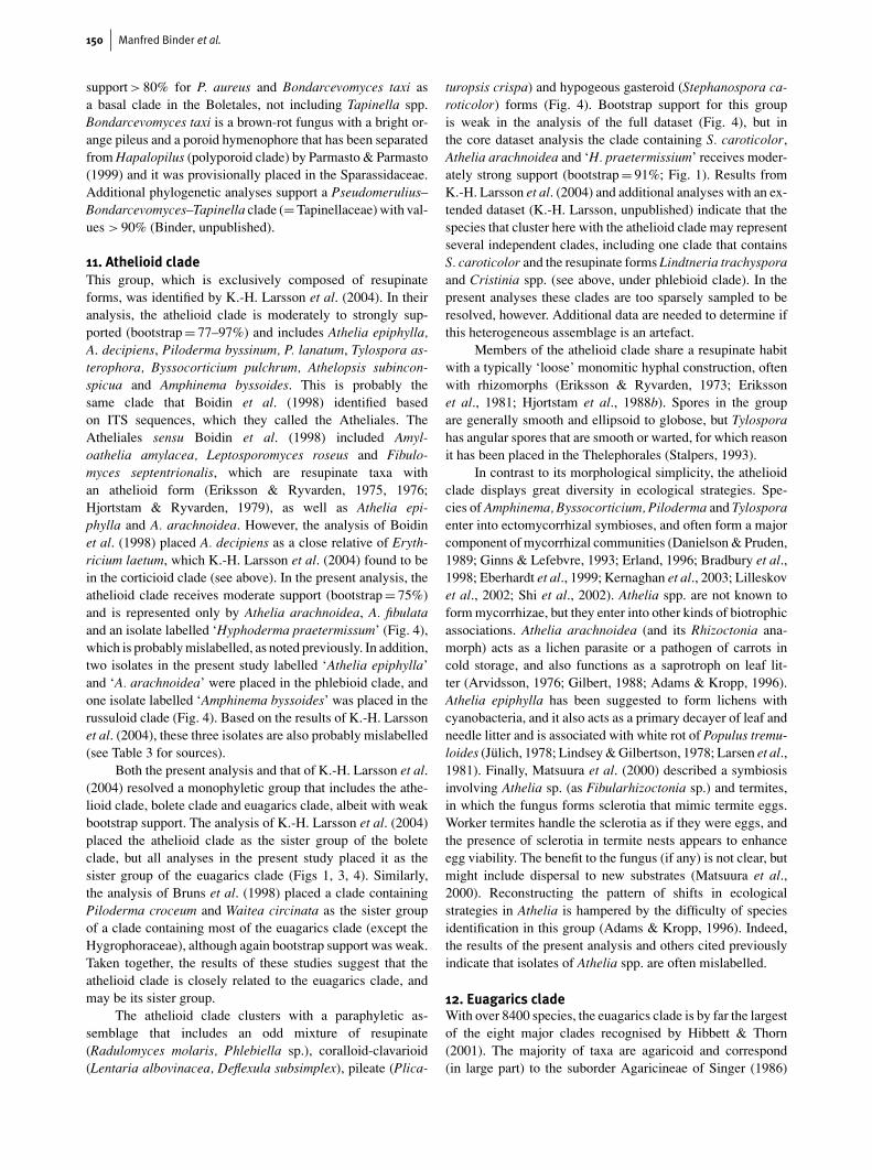
150 Manfred Binder et al.
support > 80% for P. aureus and Bondarcevomyces taxi asa basal clade in the Boletales, not including Tapinella spp.Bondarcevomyces taxi is a brown-rot fungus with a bright or-ange pileus and a poroid hymenophore that has been separatedfrom Hapalopilus (polyporoid clade) by Parmasto & Parmasto(1999) and it was provisionally placed in the Sparassidaceae.Additional phylogenetic analyses support a Pseudomerulius–Bondarcevomyces–Tapinella clade (= Tapinellaceae) with val-ues > 90% (Binder, unpublished).
11. Athelioid cladeThis group, which is exclusively composed of resupinateforms, was identified by K.-H. Larsson et al. (2004). In theiranalysis, the athelioid clade is moderately to strongly sup-ported (bootstrap = 77–97%) and includes Athelia epiphylla,A. decipiens, Piloderma byssinum, P. lanatum, Tylospora as-terophora, Byssocorticium pulchrum, Athelopsis subincon-spicua and Amphinema byssoides. This is probably thesame clade that Boidin et al. (1998) identified basedon ITS sequences, which they called the Atheliales. TheAtheliales sensu Boidin et al. (1998) included Amyl-oathelia amylacea, Leptosporomyces roseus and Fibulo-myces septentrionalis, which are resupinate taxa withan athelioid form (Eriksson & Ryvarden, 1975, 1976;Hjortstam & Ryvarden, 1979), as well as Athelia epi-phylla and A. arachnoidea. However, the analysis of Boidinet al. (1998) placed A. decipiens as a close relative of Eryth-ricium laetum, which K.-H. Larsson et al. (2004) found to bein the corticioid clade (see above). In the present analysis, theathelioid clade receives moderate support (bootstrap = 75%)and is represented only by Athelia arachnoidea, A. fibulataand an isolate labelled ‘Hyphoderma praetermissum’ (Fig. 4),which is probably mislabelled, as noted previously. In addition,two isolates in the present study labelled ‘Athelia epiphylla’and ‘A. arachnoidea’ were placed in the phlebioid clade, andone isolate labelled ‘Amphinema byssoides’ was placed in therussuloid clade (Fig. 4). Based on the results of K.-H. Larssonet al. (2004), these three isolates are also probably mislabelled(see Table 3 for sources).
Both the present analysis and that of K.-H. Larsson et al.(2004) resolved a monophyletic group that includes the athe-lioid clade, bolete clade and euagarics clade, albeit with weakbootstrap support. The analysis of K.-H. Larsson et al. (2004)placed the athelioid clade as the sister group of the boleteclade, but all analyses in the present study placed it as thesister group of the euagarics clade (Figs 1, 3, 4). Similarly,the analysis of Bruns et al. (1998) placed a clade containingPiloderma croceum and Waitea circinata as the sister groupof a clade containing most of the euagarics clade (except theHygrophoraceae), although again bootstrap support was weak.Taken together, the results of these studies suggest that theathelioid clade is closely related to the euagarics clade, andmay be its sister group.
The athelioid clade clusters with a paraphyletic as-semblage that includes an odd mixture of resupinate(Radulomyces molaris, Phlebiella sp.), coralloid-clavarioid(Lentaria albovinacea, Deflexula subsimplex), pileate (Plica-
turopsis crispa) and hypogeous gasteroid (Stephanospora ca-roticolor) forms (Fig. 4). Bootstrap support for this groupis weak in the analysis of the full dataset (Fig. 4), but inthe core dataset analysis the clade containing S. caroticolor,Athelia arachnoidea and ‘H. praetermissium’ receives moder-ately strong support (bootstrap = 91%; Fig. 1). Results fromK.-H. Larsson et al. (2004) and additional analyses with an ex-tended dataset (K.-H. Larsson, unpublished) indicate that thespecies that cluster here with the athelioid clade may representseveral independent clades, including one clade that containsS. caroticolor and the resupinate forms Lindtneria trachysporaand Cristinia spp. (see above, under phlebioid clade). In thepresent analyses these clades are too sparsely sampled to beresolved, however. Additional data are needed to determine ifthis heterogeneous assemblage is an artefact.
Members of the athelioid clade share a resupinate habitwith a typically ‘loose’ monomitic hyphal construction, oftenwith rhizomorphs (Eriksson & Ryvarden, 1973; Erikssonet al., 1981; Hjortstam et al., 1988b). Spores in the groupare generally smooth and ellipsoid to globose, but Tylosporahas angular spores that are smooth or warted, for which reasonit has been placed in the Thelephorales (Stalpers, 1993).
In contrast to its morphological simplicity, the athelioidclade displays great diversity in ecological strategies. Spe-cies of Amphinema, Byssocorticium, Piloderma and Tylosporaenter into ectomycorrhizal symbioses, and often form a majorcomponent of mycorrhizal communities (Danielson & Pruden,1989; Ginns & Lefebvre, 1993; Erland, 1996; Bradbury et al.,1998; Eberhardt et al., 1999; Kernaghan et al., 2003; Lilleskovet al., 2002; Shi et al., 2002). Athelia spp. are not known toform mycorrhizae, but they enter into other kinds of biotrophicassociations. Athelia arachnoidea (and its Rhizoctonia ana-morph) acts as a lichen parasite or a pathogen of carrots incold storage, and also functions as a saprotroph on leaf lit-ter (Arvidsson, 1976; Gilbert, 1988; Adams & Kropp, 1996).Athelia epiphylla has been suggested to form lichens withcyanobacteria, and it also acts as a primary decayer of leaf andneedle litter and is associated with white rot of Populus tremu-loides (Julich, 1978; Lindsey & Gilbertson, 1978; Larsen et al.,1981). Finally, Matsuura et al. (2000) described a symbiosisinvolving Athelia sp. (as Fibularhizoctonia sp.) and termites,in which the fungus forms sclerotia that mimic termite eggs.Worker termites handle the sclerotia as if they were eggs, andthe presence of sclerotia in termite nests appears to enhanceegg viability. The benefit to the fungus (if any) is not clear, butmight include dispersal to new substrates (Matsuura et al.,2000). Reconstructing the pattern of shifts in ecologicalstrategies in Athelia is hampered by the difficulty of speciesidentification in this group (Adams & Kropp, 1996). Indeed,the results of the present analysis and others cited previouslyindicate that isolates of Athelia spp. are often mislabelled.
12. Euagarics cladeWith over 8400 species, the euagarics clade is by far the largestof the eight major clades recognised by Hibbett & Thorn(2001). The majority of taxa are agaricoid and correspond(in large part) to the suborder Agaricineae of Singer (1986)

Phylogenetic distribution of resupinate forms of mushroom-forming fungi 151
and its many gasteroid derivatives. It is now recognised thatthere are also scattered clavarioid forms in the group (Hibbettet al., 1997; Pine et al., 1999; Hibbett & Thorn, 2001; K.-H.Larsson et al., 2004; Moncalvo et al., 2002). The most com-prehensive phylogenetic study of the euagarics clade so faris that of Moncalvo et al. (2002), which included 877 isolatesrepresented by nuc-lsu rDNA sequences. The only species thatapproaches a ‘resupinate’ form in that study is Gloeostereumincarnatum, which produces sessile conchate fruiting bodiesthat may be resupinate at the point of attachment (Petersen &Parmasto, 1993). Several other studies have shown that cer-tain resupinate forms are in the euagarics clade (Kim & Jung,2000; Lim, 2001; E. Langer, 2002; K.-H. Larsson et al., 2004),but the sampling of agaricoid taxa has generally been too lim-ited to address the placements of the resupinate forms on afine scale (but see Langer, 2002, which included 54 speciesfrom the euagarics clade). The present study included a largesample (206 sequences) of non-resupinate forms in the euagar-ics clade, most of which are from the studies of Moncalvoet al. (2000, 2002).
The euagarics clade receives weak bootstrap support inthe analyses of both the core and full datasets (Figs 1, 4).Nevertheless, the general topology, with the Hygrophoraceaeas the sister group of the ‘core euagarics clade’, is consist-ent with the strongly supported results of Binder & Hibbett(2002). One problematical aspect of the results here concernsthe placements of the unclassified taxa that form a paraphyleticgroup at the base of the athelioid clade, including the corticioidforms Phlebiella sp. and Radulomyces molaris, both represen-ted by sequences from the work of E. Langer (2002; also seeHibbett & Binder, 2002; Fig. 4). In the analysis of E. Langer(2002) these taxa were nested in the euagarics clade, althoughtheir closest relatives were not identified with confidence. Sim-ilar results were obtained by K.-H. Larsson et al. (2004), whofound a well supported (bootstrap > 80%) clade containingthree species of Radulomyces, Phlebiella pseudotsugae andCoronicium alboglaucum, which was weakly supported as thesister group of the clavarioid forms Typhula phacorrhiza andMacrotyphula juncea. Taken together, the results of these ana-lyses suggest that Radulomyces, Phlebiella and Coroniciumare nested within or closely related to the euagarics clade. Itwould be valuable to obtain additional sequences of these taxa,which at present are represented only by nuc-lsu rDNA se-quences. Other than their corticioid habit, there are no obviouscharacters that suggest a close relationship among Radulomy-ces, Phlebiella and Coronicium (K.-H. Larsson et al., 2004).
At least four groups of resupinate forms are nested in thecore euagarics clade (Fig. 4). One of these groups is an oddassemblage including two Lachnocladiaceae (Vararia ochro-leucum, V. gallica), Lopharia mirabilis and Trechsipora farin-acea (Fig. 4). In this and other studies (Lim, 2001; K.-H.Larsson et al., 2004), sequences of these genera are placed inthe russuloid clade, polyporoid clade and trechisporoid clade(respectively), suggesting that their placement in the euagaricsclade is erroneous, possibly reflecting misidentifications.
Dendrothele. Two isolates of the polyphyletic corticioidgenus Dendrothele (D. griseocana, D. acerina) are nested in
a moderately supported (bootstrap = 83%) clade that also in-cludes cyphelloid and aquatic Homobasidiomycetes (Fig. 4).This result is consistent with the results of the study ofE. Langer (2002), which was the source of the sequence ofD. acerina and several of the cyphelloid forms. In that ana-lysis, these taxa were grouped in clade “cyphelloid 35”. Aclade including Schizophyllum commune and the cupulate Au-riculariopsis ampla is weakly supported as the sister group ofthe Dendrothele-cyphelloid clade (Fig. 4), which is consistentwith the results of Binder et al. (2001) and Nakasone (1996).
Chondrostereum, Gloeostereum and Cystostereum. Theeffused-reflexed, stereoid fungus Chondrostereum purpureumand Gloeostereum incarnatum are moderately supported(bootstrap = 85%) as a monophyletic group. These results areconsistent with those of Moncalvo et al. (2002) who showedthat G. incarnatum is in the euagarics clade, and Kim & Jung(2000), E. Langer (2002), K.-H. Larsson et al. (2004), andLim (2001), who showed that C. purpureum is in the euagar-ics clade. The studies of Kim & Jung (2000) and Lim (2001)also suggested that the resupinate to effused-reflexed stereoidfungus Cystostereum murraii is in this group.
In contrast to Kim & Jung (2000) and Lim (2001), the ana-lysis of Boidin et al. (1998) suggested that Cystostereum mur-raii is in the phlebioid clade (Phanerochaetales). Cystostereummurraii is dimitic, whereas C. purpureum is monomitic, whichmight seem to support the results of Boidin et al. (1998). Nev-ertheless, both taxa have hyphae in the context with swollen,bladderlike ends. The arrangement of these cells in the twospecies is strikingly similar in the illustrations of Eriksson &Ryvarden (1973, 1975), which supports the conclusions ofKim & Jung (2000) that C. purpureum and C. murraii areclosely related. In C. murraii the vesicles contain oil droplets.The “embedded gloeocystidia” described in G. incarnatum(Petersen & Parmasto, 1993, p. 1214) might be homologous.
Moncalvo et al. (2002) showed that Cheimonophyllumcandidissum, which is a minute pleurotoid agaric, is the sis-ter group of G. incarnatum, and named the resulting cladethe /gloeostereae. The sister group of the /gloeostereae in-cluded the pileate-stipitate agarics Hydropus scabripes, Baeo-spora myosura and B. myriadophylla (Tricholomataceae s.lat.), which were classified as the /baeosporoid clade. Thesister group relationship of /gloeostereae and /baeosporoid isweakly supported in this analysis, which includes many of thesame sequences as in Moncalvo et al. (2002). Nevertheless, ifthis topology is correct, then it suggests a transformation seriesfrom pileate-stipitate agarics (Baeospora spp., H. scabripes),to pleurotoid agarics (C. candidissimum), conchate-partly re-supinate forms with a reduced hymenophore (G. incarnatum),and finally effused-reflexed or fully resupinate stereoid forms(C. purpureum, C. murraii).
Cylindrobasidium. Three isolates of the corticioid genusCylindrobasidium, including two from the study of E. Langer(2002) are strongly supported (bootstrap = 100%) as a mono-phyletic group (Fig. 4). As in the analysis of E. Langer (2002),Cylindrobasidium is nested in a clade that includes the agaricgenera Armillaria and Oudesmansiella (many others are in-cluded in the present study; Fig. 4). The analysis of K.-H.
152 Manfred Binder et al.
Larsson et al. (2004) weakly supported monophyly of Cyl-indrobasidium laeve and Chondrostereum purpureum. If thetaxa that were not sampled by K.-H. Larsson et al. (2004)were pruned from the trees produced in the present study, thenC. laeve and C. purpureum would again be resolved as sistertaxa (Fig. 4).
One problematical result concerns a sequence of the cor-ticioid fungus Bulbillomyces farinosus, which is placed in aclade with the clavarioid forms Typhula phacorrhiza and Mac-rotyphula juncea, the pleurotoid agarics Phyllotopis nidulansand Pleurocybella porrigens, and the cyphelloid Henningso-myces candidus (Fig. 4). This group is equivalent to the clade“collybioid, clavarioid 28” that was resolved in the study ofE. Langer (2002). The monophyly of Bulbillomyces, Typhulaand Macrotyphula is strongly supported (bootstrap = 98%),but there are no characters that would support this place-ment. Bulbillomyces farinosus produces a sclerotial anamorph(Aegerita candida), and in this regard it superficially re-sembles Typhula phacorrhiza, which also produces sclerotia,but the sclerotia differ in size, colour and anatomical features(Remsberg, 1940; Julich, 1974). Analyses with alternative se-quences of Bulbillomyces farinosus derived from two differentcultures and one Aegerita candida isolate suggest that Bulbil-lomyces farinosus is closely related to Hypochnicium spp. inthe residual polypore clade, which is a more explicable po-sition (K.-H. Larsson unpublished, M. Binder & D. Hibbett,unpublished).
Finally, K.-H. Larsson et al. (2004) resolved a weaklysupported clade containing two resupinate polypores (Anomo-poria bombycina and A. kamtschatica) and four corti-cioid fungi (Amylocorticium spp., Ceraceomyces tessulatus,Hypochniciellum subillaqueatum), which was placed as thesister group of the rest of the euagarics clade. None of thesespecies were sampled here, although three different species ofCeraceomyces were included in both the present study andthat of K.-H. Larsson et al. (2004) and found to be in the phle-bioid clade (see above). Analyses with additional sequencesof Ceraceomyces tessulatus and Anomoporia spp., includingA. albolutescens, have upheld the phylogenetic position sug-gested in Larsson et al. (2002) (K.-H. Larsson, unpublished).In contrast, the analysis of Kim & Jung (2000) placedA. albolutescens in the Antrodia clade. This placement wouldbe consistent with the reported production of a brown rot byA. albolutescens (Gilbertson & Ryvarden, 1986), but it is in-consistent with the results of K.-H. Larsson et al. (2004) whichare based on multiple isolates. It is likely that the ‘A. albolutes-cens’ isolate studied by Kim & Jung (2000) is mislabelled.Bootstrap support for the basal nodes of the euagarics cladewas weak in the study of K.-H. Larsson et al. (2004), so itremains unclear whether these last resupinate taxa are actuallymembers of the euagarics clade. Even if they are, the fractionof species that are resupinate in the euagarics clade is muchlower than in other major groups of Homobasidiomycetes(c. 4% in this dataset). One possible explanation for this patternis that the abundance of resupinate forms in groups such as thehymenochaetoid clade, russuloid clade and cantharelloid cladereflects a plesiomorphic condition in these more basal groups(Hibbett & Binder, 2002). Alternatively, the rate of reversals to
resupinate forms (or the rate of speciation of resupinate forms)may be lower in the euagarics clade than in other clades ofHomobasidiomycetes.
Conclusions and future directionsResupinate forms are scattered throughout all of the majorclades of Homobasidiomycetes, as well as heterobasidiomy-cetes. Some of the recently recognised groups of Homobasi-diomycetes, such as the athelioid clade, corticioid clade andtrechisporoid clade (K.-H. Larsson et al., 2004), and the lonetaxon Jaapia argillacea, are composed entirely, or almost en-tirely, of resupinate forms (Fig. 4). The present study analysedone of the larger phylogenetic datasets in fungi to date (but seeMoncalvo et al., 2002; Tehler et al., 2003), but it still includedless than half of the genera of corticioid fungi recognised byHjortstam (1987). As sampling of resupinate taxa continues,it is possible that new major clades will be discovered. Suchdiscoveries could aid analyses of higher-level phylogenetic re-lationships of Homobasidiomycetes by identifying taxa thatbreak up internodes deep in the tree (including those that de-termine the boundary between the Homobasidiomycetes andheterobasidiomycetes), many of which have proven difficult toresolve (Binder & Hibbett, 2002).
Designing a sampling scheme for the remaining resu-pinate taxa will be challenging. For many groups, there arefew anatomical characters to provide clues to higher-level re-lationships, and the monophyly of individual genera is oftenquestionable. For example, Hyphoderma is now understoodto include species in the hymenochaetoid clade and poly-poroid clade. Similarly, species of Veluticeps and Columno-cystis, which were once proposed as generic synonyms, occurin the Gloeophyllum clade and polyporoid clade. These ex-amples are particularly dramatic, but numerous other generaof resupinate fungi have been found to be polyphyletic in thisand other studies cited previously (e.g. Sistotrema, Hyphodon-tia, Schizopora, Phlebia, Phanerochaete, Aleurodiscus, Gloeo-cystidiellum, etc.). Many of the older genera have been splitinto smaller, putatively natural groups, but even some of thesehave been found to be polyphyletic (e.g. Boidinia; E. Larsson &K.-H. Larsson, 2003). Thus, an exemplar-based approach tosampling could lead to significant underestimates of the phylo-genetic diversity of resupinate Homobasidiomycetes.
Ultimately, it will be necessary to construct phylogeny-based classifications that include all the species of resupinateand non-resupinate Homobasidiomycetes. Moreover, it will benecessary to include multiple accessions of individual species,because they can reveal misidentifications (which the presentstudy shows are common), as well as provide insight intobiogeography and intraspecific variation.
To develop comprehensive phylogenetic classificationswill require either simultaneous analyses of very large datasets,or analytical approaches that reconcile overlapping datasets,such as supertree methods (Sanderson et al., 1998). Simultan-eous analyses have certain advantages, not the least of whichis that they permit the estimation of branch lengths, which arenecessary for molecular clock studies and maximum-likeli-hood analyses of character evolution. However, simultaneous

Phylogenetic distribution of resupinate forms of mushroom-forming fungi 153
analyses of large datasets are computationally challenging,especially if model-based methods are employed. Using theParsimony Ratchet, the present study succeeded in analysinga 656-OTU dataset with six-parameter weighted parsimony,but even this large dataset included only about one fifth of the3130 nuc-lsu rDNA sequences of Homobasidiomycetes thatare available in GenBank as of this writing.
Given the limitations of current computer hardware andalgorithms, a rigorous simultaneous analysis of all the avail-able homobasidiomycete sequences would be very difficult.To develop detailed phylogenetic hypotheses within individualclades will require more focused efforts, as exemplified by thestudies of E. Larsson & K.-H. Larsson (2003) in the russuloidclade and Moncalvo et al. (2002) in the euagarics clade. Atthe same time, analyses of multigene datasets of exemplarsfrom the major groups will be needed to estimate higher-levelrelationships. In the present study and that of Binder & Hibbett(2002), a dataset with four mitochondrial and nuclear rDNAregions was used for this purpose. It should be a priority tosequence these same regions in exemplars of the major cladesthat have so far been studied only with single genes, such asthe athelioid clade and trechisporoid clade. Of course, not allnodes will be resolved with rDNA alone (e.g. the polyporoidclade; Binder & Hibbett, 2002), so exploration of protein-coding loci will also be necessary to resolve the phylogeneticrelationships of resupinate Homobasidiomycetes.
AcknowledgementsThe authors are indebted to their colleagues and the institutions whoprovided cultures, specimens, DNA samples, sequence data, and ac-cess to unpublished results, including Vera Andjic, Helmut Besl, PaulaDePriest, Margit Jarosch, Urmas Koljalg, Phillip Franken, the USDAForest Products Laboratory, Canadian Collection of Fungal Culturesand Centraalbureau voor Schimmelcultures. This research was sup-ported by National Science Foundation awards DEB-9903835 (toDSH) and DEB-0128925 (to DSH and MB).
ReferencesADAMS, G.A. & KROPP, B.R. 1996. Athelia arachnoidea, the sexual
state of Rhizoctonia carotae, a pathogen of carrot in cold storage.Mycologia 88, 459–472.
AGERER, R. 1999. Never change a functionally successful principle:the evolution of Boletales s. l. (Hymenomycetes, Basidiomycota)as seen from below ground features. Sendtnera 6, 5–91.
AGERER, R., KLOSTERMEYER, D., STEGLICH, W., FRANZ, F. &ACKER, G. 1996. Ectomycorrhizae of Albatrellus ovinus (Scuti-geraceae) on Norway spruce with some remarks on the systematicposition of the family. Mycotaxon 59, 289–307.
ARVIDSSON, L. 1976. Athelia epiphylla (Berk.) Jul., and its influenceon epiphytic cryptogams in urban areas. Goteborgs SvampklubArsskrift 1976, 4–10.
BEGEROW, D., BAUER, R. & OBERWINKLER, F. 1997. Phylogeneticstudies on nuclear large subunit ribosomal DNA sequences of smutfungi and related taxa. Canadian Journal of Botany 75, 2045–2056.
BERCH, S.M., ALLEN, T.R. & BERBEE, M.L. 2002. Community struc-ture and phylogeny of ericoid mycorrhizal fungi. Plant and Soil244, 55–66.
BERGEMANN, S.E. & MILLER, S.L. 2002. Size, distribution, and per-sistence of genets in local populations of the late-stage ectomy-corrhizal basidiomycete, Russula brevipes. New Phytologist 156,313–320.
BESL, H., BRESINSKY, A. & KAMMERER, A. 1986. Chemosystematikder Coniophoraceae. Zeitschrift fur Mykologie 52, 277–286.
BIDARTONDO, M.I., BRUNS, T.D., WEIß, M., SERGIO, C. & READ, D.J.2003. Specialized cheating of the ectomycorrhizal symbiosis byan epiparasitic liverwort. Proceedings of the Royal Society London(Series B) 270, 835–842.
BINDER, M. & BRESINSKY, A. 2002. Derivation of a polymorphiclineage of Gasteromycetes from boletoid ancestors. Mycologia94, 85–98.
BINDER, M. & HIBBETT, D.S. 2002. Higher-level phylogenetic re-lationships of Homobasidiomycetes (mushroom-forming fungi)inferred from four rDNA regions. Molecular Phylogenetics andEvolution 22, 76–90.
BINDER, M., HIBBETT, D.S. & MOLITORIS, H.-P. 2001. Phylogeneticrelationships of the marine gasteromycete Nia vibrissa. Mycologia93, 679–688.
BOIDIN, J. & GILLES, G. 1998. Contribution a l’etude des genresDendrocorticum, Dendrodontia et Dentocorticium (Basidiomy-cotina). Cryptogamie, Mycologie 19, 181–202.
BOIDIN, J. & LANQUETIN, P. 1984. Repertoire des donnees utilespour effectuer les teste d’intercompatibilite chez BasidiomycetesIII. Aphyllophorales non porees. Cryptogamie, Mycologie 5, 193–245.
BOIDIN, J., MUGNIER, J. & CANALES, R. 1998. Taxonomie moleculairedes Aphyllophorales. Mycotaxon 66, 445–491.
BRADBURY, S.M., DANIELSON, R.M. & VISSER, S. 1998. Ectomycor-rhizas of regenerating stands of lodgepole pine (Pinus contorta).Canadian Journal of Botany 76, 218–227.
BRESINSKY, A., JAROSCH, M., FISCHER, M., SCHONBERGER, I. &WITTMANN-BRESINSKY, B. 1999. Polyphyletic relationshipswithin Paxillus s. l. (Basidiomycetes, Boletales): separation ofa southern hemisphere genus. Plant Biology 1, 327–333.
BRUNS, T.D. & SZARO, T.M. 1992. Rate and mode differences betweennuclear and mitochondrial small-subunit rRNA genes in mush-rooms. Molecular Biology and Evolution 9, 836–855.
BRUNS, T.D., SZARO, T.M., GARDES, M., CULLINGS, K.W., PAN, J.J.,TAYLOR, D.L., HORTON, D.R., KRETZER, A., GARBELOTTO, M. &LI, Y. 1998. A sequence database for the identification of ecto-mycorrhizal basidiomycetes by phylogenetic analysis. MolecularEcology 7, 257–272.
BURDSALL, H.H. JR, VOLK, T.J. & AMMIRATI, J.F. JR. 1996. Bridgeo-porus, a new genus to accommodate Oxyporus nobilissimus (Basi-diomycotina, Polyporaceae). Mycotaxon 60, 387–395.
BURT, E.A. 1926. The Thelephoraceae of North America XV. Annalsof the Missouri Botanical Garden 13, 173–354.
CHAMURIS, G.P. 1988. The non-stipitate stereoid fungi in the north-eastern United States and adjacent Canada. Mycologia Memoirs14, 1–247.
CHASE, M. ET AL. 1993. Phylogenetics of seed plants: an analysis ofnucleotide sequences from the plastid gene rbcL. Annals of theMissouri Botanical Garden 80, 528–580.
CORNER, E.J.H. 1948. Asterodon, a clue to the morphology of fungusfruit-bodies: with notes on Asterostroma and Asterostromella.Transactions of the British Mycological Society 31, 234–241.
CORNER, E.J.H. 1966. A Monograph of Cantharelloid Fungi. OxfordUniversity Press, London.
CORNER, E.J.H. 1970. Supplement to ‘A monograph of Clavaria andallied genera’. Nova Hedwigia Beihefte 33, 1–299.
CUNNINGHAM, C.W. 1997. Is congruence between data partitions areliable predictor of phylogenetic accuracy? Empirically testingan iterative procedure for choosing among phylogenetic methods.Systematic Biology 46, 464–478.
DANIELSON, R.M. 1984. Ectomycorrhizal associations in jack pinestands in northeastern Alberta. Canadian Journal of Botany 62,932–939.
DANIELSON, R.M. & PRUDEN, M. 1989. The ectomycorrhizal statusof urban spruce. Mycologia 81, 335–341.
DE KOKER, T.H., NAKASONE, K.K., HAARHOF, J., BURDSALL,H.H. JR & JANSE, B.J.H. 2003. Phylogenetic relationships of the

154 Manfred Binder et al.
genus Phanerochaete inferred from the internal transcribed spacerregion. Mycological Research 107, 1032–1034.
DIEDERICH, P., SCHULTEIS, B. & BLACKWELL, M. 2003. Marchandio-basidium aurantiacum gen. sp. nov., the teleomorph of Marchan-diomyces aurantiacus (Basidiomycota, Ceratobasidiales). Myco-logical Research 107, 523–527.
DOMANSKI, S. 1972. Fungi Polyporaceae I (resupinatae) Mucro-poronaceae I (resupinatae). National Center for Scientific, Tech-nical and Economic Information, Warsaw.
DONK, M.A. 1964. A conspectus of the families of Aphyllophorales.Persoonia 3, 199–324.
DONK, M.A. 1971. Progress in the study of the classification of thehigher basidiomycetes. In: PETERSEN, R.H., Ed., Evolution in theHigher Basidiomycetes. University of Tennessee Press, Knoxville,pp. 3–25.
EBERHARDT, U., WALTER, L. & KOTTKE, I. 1999. Molecular andmorphological discrimination between Tylospora fibrillosa andTylospora asterophora mycorrhizae. Canadian Journal of Botany77, 11–21.
ERIKSSON, J. 1954. Ramaricium n. gen., a corticioid member of theRamaria group. Svensk Botanisk Tidskrift 48, 188–198.
ERIKSSON, J. 1958. Studies in the Heterobasidiomycetes and Homo-basidiomycetes – Aphyllophorales of Muddus National Park inNorth Sweden. Symbolae Botanicae Upsalienses 16, 1–172.
ERIKSSON, J., HJORTSTAM, K. & RYVARDEN, L. 1978. The Cor-ticiaceae of North Europe. Vol. 5, Mycoaciella-Phanerochaete.Fungiflora, Oslo.
ERIKSSON, J., HJORTSTAM, K. & RYVARDEN, L. 1981. The Cor-ticiaceae of North Europe. Vol. 6, Phlebia-Sarcodontia. Fungiflora,Oslo.
ERIKSSON, J., HJORTSTAM, K. & RYVARDEN, L. 1984. The Cor-ticiaceae of North Europe. Vol. 7, Schizopora-Suillosporium.Fungiflora, Oslo.
ERIKSSON, J. & RYVARDEN, L. 1973. The Corticiaceae of NorthEurope. Vol. 2, Aleurodiscus-Confertobasidium. Fungiflora, Oslo.
ERIKSSON, J. & RYVARDEN, L. 1975. The Corticiaceae of NorthEurope. Vol. 3, Coronicium-Hyphoderma. Fungiflora, Oslo.
ERIKSSON, J. & RYVARDEN, L. 1976. The Corticiaceae of NorthEurope. Vol. 4, Hyphodermella-Mycoacia. Fungiflora, Oslo.
ERLAND, S. 1996. Abundance of Tylospora fibrillosa ectomycorrhizasin a South Swedish spruce forest measured by RFLP analysis ofthe PCR-amplified rDNA ITS region. Mycological Research 99,1425–1428.
ETAYO, J. & DIEDERICH, P. 1996. Lichenicolous fungi from the westernPyrenees, France and Spain. II. More Deuteromycetes. Mycotaxon60, 415–428.
EYME, J. & PARRIAUD, H. 1970. Au sujet de l’infrastructure des hyphesde Clathrus cancellatus Tournefort, champignon gasteromycete.Compte Rendu de l’Academie des Sciences Paris (Serie D) 270,1890–1892.
FRIES, E.M. 1821. Systema Mycologicum, sistens fungorum ordines,genera et species huc usque cognitas. Vol. I. Berling, Lund.
GARGAS, A., DEPRIEST, P.T., GRUBE, M. & TEHLER, A. 1995a. Mul-tiple origins of lichen symbioses in fungi suggested by ssu rDNAphylogeny. Science 268, 1492–1495.
GARGAS, A., DEPRIEST, P.T. & TAYLOR, J.W. 1995b. Positions of mul-tiple insertions in SSU rDNA of lichen-forming fungi. MolecularBiology and Evolution 12, 208–218.
GILBERT, O.L. 1988. Studies on the destruction of Lecanora coniza-eoides by the lichenicolous fungus Athelia arachnoidea. Licheno-logist 20, 183–190.
GILBERTSON, R.L. & RYVARDEN, L. 1986. North American Polypores.Vol. 1. Fungiflora, Oslo.
GILBERTSON, R.L. & RYVARDEN, L. 1987. North American Polypores.Vol. 2. Fungiflora, Oslo.
GILL, M. & STEGLICH, W. 1987. Pigments of fungi (Macromycetes).Progress in the Chemistry of Organic Natural Products 51, 1–317.
GINNS, J. 1978. Leucogyrophana (Aphyllophorales): Identification ofspecies. Canadian Journal of Botany 56, 1953–1973.
GINNS, J. 1979. The genus Ramaricium (Gomphaceae). BotaniskaNotiser 132, 93–102.
GINNS, J. 1982. A monograph of the genus Coniophora (Aphyllo-phorales, Basidiomycetes). Opera Botanica 61, 1–61.
GINNS, J. 1997. The taxonomy and distribution of rare or uncom-mon species of Albatrellus in western North America. CanadianJournal of Botany 75, 261–273.
GINNS, J. & LEFEBVRE, M.N.L. 1993. Lignicolous corticioid fungi(Basidiomycota) of North America: systematics, distribution, andecology. Mycologia Memoirs 19, 1–247.
GOLOBOFF, P. 1998. NONA. Computer program and software. Pub-lished by the author. INSUE Fundacion e Instituto Miguel LilloMiguel Lillo 205, 4000 S.M. de Tucuman, Argentina. http://www.cladistics.com/aboutNona.htm
HALLENBERG, N. 1985. The Lachnocladiaceae and Coniophoraceaeof North Europe. Fungiflora, Oslo.
HALLENBERG, N. 1990. Ultrastructure of stephanocysts and basi-diospores in Hyphoderma praetermissum. Mycological Research94, 1090–1095.
HALLENBERG, N. & PARMASTO, E. 1998. Phylogenetic studies inspecies of Corticiaceae growing on branches. Mycologia 90, 640–654.
HANSEN, L. & KNUDSEN, H., EDS., 1997. Nordic macromycetes, 3.Heterobasidioid, aphyllophoroid and gastromycetoid basidiomy-cetes. Nordsvamp, Copenhagen.
HELLWIG, V. 1999. Isolierung, Strukturaufklarung und chemotaxono-mische Untersuchung von Sekundarmetaboliten aus Pilzen. Ph.D.Thesis, Universitat Munchen, Fakultat Chemie und Pharmazie.
HENKEL, T.W., AIME, M.C. & MILLER, S.L. 2000. Systematics ofpleurotoid Russulaceae from Guyana and Japan, with notes ontheir ectomycorrhizal status. Mycologia 92, 1119–1132.
HIBBETT, D.S. 1996. Phylogenetic evidence for horizontal trans-mission of Group 1 introns in the nuclear ribosomal DNA ofmushroom-forming fungi. Molecular Biology and Evolution 13,903–917.
HIBBETT, D.S. 2004. Trends in morphological evolution in Homo-basidiomycetes inferred using maximum likelihood: a comparisonof binary and multistate approaches. Systematic Biology 53, 889–903.
HIBBETT, D.S. & BINDER, M. 2002. Evolution of complex fruitingbody morphologies in Homobasidiomycetes. Proceedings of theRoyal Society London (Series B) 269, 1963–1969.
HIBBETT, D.S. & DONOGHUE, M.J. 1995. Progress toward a phylo-genetic classification of the Polyporaceae through parsimony ana-lyses of ribosomal DNA sequences. Canadian Journal of Botany73 (Supplement 1), S853–S861.
HIBBETT, D.S. & DONOGHUE, M.J. 2001. Analysis of character cor-relations among wood decay mechanism, mating systems, andsubstrate ranges in Homobasidiomycetes. Systematic Biology 50,215–242.
HIBBETT, D.S., GILBERT, L.-B. & DONOGHUE, M.J. 2000. Evolution-ary instability of ectomycorrhizal symbioses in basidiomycetes.Nature 407, 506–508.
HIBBETT, D.S., PINE, E.M., LANGER, E., LANGER, G. & DONOGHUE,M.J. 1997. Evolution of gilled mushrooms and puffballs inferredfrom ribosomal DNA sequences. Proceedings of the NationalAcademy of Sciences USA 94, 12002–12006.
HIBBETT, D.S. & THORN, R.G. 2001. Basidiomycota: Homobasidio-mycetes. In: MCLAUGHLIN, D.J., MCLAUGHLIN, E.G. & LEMKE,P.A., EDS., The Mycota VII, Systematics and Evolution. Part B.Springer Verlag, Berlin, pp. 121–168.
HIBBETT, D.S. & VILGALYS, R. 1993. Phylogenetic relationships ofLentinus (Basidiomycotina) inferred from molecular and morpho-logical characters. Systematic Botany 18, 409–433.
HIETALA, A.M., VAHALA, J. & HANTULA, J. 2001. Molecular evidencesuggests that Ceratobasidium bicorne has an anamorph known asa conifer pathogen. Mycological Research 105, 555–562.
HJORTSTAM, K. 1987. A check-list to genera and species of corticioidfungi (Hymenomycetes). Windahlia 17, 55–85.

Phylogenetic distribution of resupinate forms of mushroom-forming fungi 155
HJORTSTAM, K. & LARSSON, K.-H. 1995. Annotated check-listto genera and species of corticioid fungi (Aphyllophorales,Basidiomycotina) with special regards to tropical and subtropicalareas. Windahlia 21, 1–75.
HJORTSTAM, K., LARSSON, K.-H. & RYVARDEN, L. 1988a. The Cor-ticiaceae of North Europe. Vol. 1. Introduction and keys. Fungi-flora, Oslo.
HJORTSTAM, K., LARSSON, K.-H., RYVARDEN, L. & ERIKSSON, J.1988b. The Corticiaceae of North Europe. Vol. 8. Phlebiella,Thanatephorus-Ypsilonidium. Fungiflora, Oslo.
HJORTSTAM, K. & RYVARDEN, L. 1979. Notes on Corticiaceae (Basi-diomycetes) IV. Mycotaxon 10, 201–209.
HJORTSTAM, K. & RYVARDEN, L. 1980a. Studies in tropical Cor-ticiaceae (Basidiomycetes) I. Mycotaxon 10, 269–287.
HJORTSTAM, K. & RYVARDEN, L. 1980b. Studies in tropical Cor-ticiaceae (Basidiomycetes) II. Mycotaxon 12, 168–184.
HJORTSTAM, K. & TELLERIA, M.T. 1990. Columnocystis, a synonymof Veluticeps. Mycotaxon 57, 53–56.
HSIAU, P. 1996. The taxonomy and phylogeny of the mycangial fungifrom Dendroctonus brevicomis and D. frontalis (Coleoptera: Sco-lytidae). Ph.D. thesis, Iowa State University, Ames, Iowa.
HUELSENBECK, J.P. 1995. Performance of phylogenetic methods insimulation. Systematic Biology 44, 17–63.
HUMPERT, A.J., MUENCH, E.L., GIACHINI, A.J., CASTELLANO,M.A. & SPATAFORA, J.W. 2001. Molecular phylogenetics ofRamaria and related genera: evidence from nuclear large subunitand mitochondrial small subunit rDNA sequences. Mycologia 93,465–477.
JAROSCH, M. 2001. Zur molekularen Systematik der Boletales: Conio-phorineae, Paxillineae und Suillineae. Bibliotheca Mycologica191, 1–158.
JAROSCH, M. & BESL, H. 2001. Leucogyrophana, a polyphyletic genusof the order Boletales (Basidiomycetes). Plant Biology 3, 443–448.
JENNINGS, D.H. & BRAVERY, A.F., EDS., 1991. Serpula lacrymans:Fundamental biology and control strategies. John Wiley & Sons,New York.
JOHNSON, J.E. & METHVEN, A.S. 1994. Panus conchatus: Culturalcharacters and mating data. Mycologia 86, 146–150.
JULICH, W. 1974. The genera of the Hyphodermoideae (Corticiaceae).Persoonia 8, 59–97.
JULICH, W. 1978. A new lichenized Athelia from Florida. Persoonia12, 149–151.
JULICH, W. 1981. Higher taxa of Basidiomycetes. Cramer, Lehre.JULICH, W. & STALPERS, J.A. 1980. The resupinate non-poroid Aphyl-
lophorales of the temperate northern hemisphere. Verhandelin-gen der Koninklijke Nederlandse Akademie van Wetenschappen,Afd Natuurkunde, Tweede Reeks. North-Holland Publishing Com-pany, Amsterdam.
KAMMERER, A., BESL, H. & BRESINSKY, A. 1985. Omphalotaceaefam. nov. und Paxillaceae, ein chemotaxonomischer Vergleichzweier Pilzfamilien der Boletales. Plant Systematics and Evol-ution 150, 101–117.
KARSTEN, P.A. 1881. Enumeratio Boletinarum et PolyporearumFennicarum, Systemate novo dispositarum. Revue Mycologie(Toulouse) 3, 1–19.
KELLER, J. 1985. Les cystides cristalliferes des Aphyllophorales.Mycologia Helvetica 1, 277–305.
KELLER, J. 1997. Atlas des Basidiomycetes. Union des SocietesSuisses de Mycologie, Neuchatel.
KERNAGHAN, G., SIGLER, L. & KHASA, D. 2003. Mycorrhizal and rootendophytic fungi of containerized Picea glauca seedlings assessedby rDNA sequence analysis. Microbial Ecology 45, 128–136.
KHAN, S.R. & KIMBROUGH, J.W. 1980. Septal ultrastructure in somegenera of the Tremellaceae. Canadian Journal of Botany 58, 55–60.
KIM, S.Y. & JUNG, H.S. 2000. Phylogenetic relationships of the Aphyl-lophorales inferred from sequence analysis of nuclear small sub-unit ribosomal DNA. Journal of Microbiology 38, 122–131.
KIRK, P.M., CANNON, P.F., DAVID, J.C. & STALPERS, J.A. 2001.Ainsworth and Bisby’s Dictionary of the Fungi, 9th edn.Cambridge University Press, Cambridge.
KLEPZIG, K.D., MOSER, J.C., LOMBARDERO, F.J., HOFSTETTER,R.W. & AYRES, M.P. 2001. Symbiosis and competition: complexinteractions among beetles, fungi, and mites. Symbiosis 30, 83–96.
KOLJALG, U. 1996. Tomentella (Basidiomycota) and related genera intemperate Eurasia. Synopsis Fungorum 9, 1–213.
KOLJALG, U., DAHLBERG, A., TAYLOR, A.F., LARSSON, E.,HALLENBERG, N., STENLID, J., LARSSON, K.-H., FRANSSON, P.M.,KAREN, O. & JONSSON, L. 2000. Diversity and abundance ofresupinate thelephoroid fungi as ectomycorrhizal symbionts inSwedish boreal forests. Molecular Ecology 9, 1985–1996.
KOLJALG, U., ERZSEBET, J., BOKA, K. & AGERER, R. 2001. Three ec-tomycorrhiza with cystidia formed by different Tomentella speciesas revealed by rDNA ITS sequences and anatomical characterist-ics. Folia Cryptogamica Estonica 38, 27–39.
KOLJALG, U., TAMMI, H., TIMONEN, S., AGERER, R. & SEN, R. 2002.ITS rDNA sequence-based phylogenetic analysis of Tomentellop-sis species from boreal and temperate forests, and the identificationof pink-type ectomycorrhizas. Mycological Progress 1, 81–92.
KOTTKE, I., BEITER, A., WEIß, M., HAUG, I., OBERWINKLER, F. &NEBEL, M. 2003. Heterobasidiomycetes form symbiotic associ-ations with hepatics: Jungermanniales have sebacinoid mycobi-onts while Aneura pinguis (Metzgeriales) is associated with aTulasnella species. Mycological Research 107, 957–968.
KRETZER, A.M. & BRUNS, T.D. 1999. Use of atp6 in fungal phylo-genetics: an example from the Boletales. Molecular Phylogeneticsand Evolution 13, 483–492.
KRISTIANSEN, K.A., TAYLOR, D.L., KJØLLER, R., RASMUSSEN,H.N. & ROSENDAHL, S. 2001. Identification of mycorrhizalfungi from single pelotons of Dactylorhiza majalis (Orchidaceae)using single-strand conformation polymorphism and mitochon-drial ribosomal large subunit DNA sequences. Molecular Ecology10, 2089–2093.
KROPP, B.R. & TRAPPE, J.M. 1982. Ectomycorrhizal fungi of Tsugaheterophylla. Mycologia 74, 479–488.
LANGER, E. 1994. Die Gattung Hyphodontia John Eriksson. Biblio-theca Mycologica 154, 1–298.
LANGER, E. 1998. Evolution of Hyphodontia (Corticiaceae, Basidio-mycetes) and related Aphyllophorales inferred from ribosomalDNA sequences. Folia Cryptogamica Estonica 33, 57–62.
LANGER, E. 2002. Phylogeny of non-gilled and gilled basidiomycetes:DNA sequence inference, ultrastructure and comparative mor-phology. Habilitationsschrift, Universitat Tubingen, Tubingen,Germany.
LANGER, E. & OBERWINKLER, F. 1993. Corticioid Basidiomycetes. I.Morphology and ultrastructure. Windahlia 20, 1–28.
LANGER, G. 1994. Die Gattung Botryobasidium Donk (Corticiaceae,Basidiomycetes). Bibliotheca Mycologica 158, 1–459.
LANGER, G., LANGER, E. & CHEN, C.-J. 2000. Botryobasidium mu-saisporum sp. nov. collected in Taiwan. Mycological Research104, 510–512.
LARIGNON, P. & DUBOS, B. 1997. Fungi associated with esca diseasein grapevine. Europe an Journal of Plant Pathology 103, 147–157.
LARSEN, M.J. 1968. Tomentelloid fungi of North America. State Uni-versity College of Forestry at Syracuse University, Technical Pub-lication No. 93, 1–157.
LARSEN, M.J. 1974. A contribution to the taxonomy of the genusTomentella. Mycologia Memoirs 4, 1–145.
LARSEN, M.J. & GILBERTSON, R.L. 1974. Dendrocorticium andDentocorticium, gen. nov. (Aphyllophorales, Corticiaceae) assegregates from Laeticorticium. Norwegian Journal of Botany 21,223–226.
LARSEN, M.J., JURGENSEN, M.F. & HARVEY, A.E. 1981. Athelia epi-phylla associated with colonization of subalpine fir foliage underpsychrophilic conditions. Mycologia 73, 1195–1202.
LARSSON, E. 2002. Phylogeny of corticioid fungi with russuloid char-acteristics. Ph.D. Thesis, Goteborg University, Goteborg, Sweden.

156 Manfred Binder et al.
LARSSON, E. & HALLENBERG, N. 2001. Species delimitation in theGloeocystidiellum porosum-clavuligerum complex inferred fromcompatibility studies and nuclear rDNA sequence data. Mycologia93, 907–914.
LARSSON, E. & LARSSON, K.-H. 2003. Phylogenetic relationships ofrussuloid basidiomycetes with emphasis on aphyllophoralean taxa.Mycologia 95, 1037–1065.
LARSSON, K.-H. 1994. Poroid species in Trechispora and the use ofcalcium oxalate crystals for species identification. MycologicalResearch 98, 1153–1172.
LARSSON, K.-H. 2001. The position of Poria mucida inferred fromnuclear ribosomal DNA sequences. Harvard Papers in Botany 6,131–138.
LARSSON, K.-H., LARSSON, E. & KOLJALG, U. 2004. High phylogen-etic diversity among corticioid homobasidomycetes. MycologicalResearch 108, 983–1002.
LEE, S.B. & TAYLOR, J.W. 1990. Isolation of DNA from fungal my-celia and single cells. In: INNIS, M.A., GELFAND, D.H., SNINSKY,J.J. & WHITE, T.J., EDS., PCR protocols, a guide to methods andapplications. Academic Press, San Diego, pp. 282–287.
LEMKE, P.A. 1964a. The genus Aleurodiscus (sensu lato) in NorthAmerica. Canadian Journal of Botany 42, 213–282.
LEMKE, P.A. 1964b. The genus Aleurodiscus (sensu lato) in NorthAmerica. Canadian Journal of Botany 42, 723–768.
LILLESKOV, E.A., FAHEY, T.J., HORTON, T.R. & LOVETT, G.M. 2002.Belowground ectomycorrhizal fungal community change over anitrogen deposition gradient in Alaska. Ecology 83, 104–115.
LIM, Y.W. 2001. Systematic study of corticioid fungi based on mo-lecular sequence analyses. Ph.D. Thesis, School of BiologicalSciences, Seoul National University, Korea.
LINDSEY, J.P. & GILBERTSON, R.L. 1978. Basidiomycetes that decayaspen in North America. Bibliotheca Mycologica 63, 1–406.
LUTZONI, F.M. 1997. Phylogeny of lichen- and non-lichen-formingomphalinoid mushrooms and the utility of testing for combinabil-ity among multiple datasets. Systematic Biology 46, 373–406.
MARTIN, K.J. & GILBERTSON, R.L. 1973. The mating system andsome other cultural aspects of Veluticeps berkeleyi. Mycologia 65,548–557.
MATSUURA, K., TANAKA, C. & NISHIDA, T. 2000. Symbiosis of atermite and a sclerotium-forming fungus: sclerotia mimic termiteeggs. Ecological Research 15, 405–414.
MILLER, O.K. JR. 1971. The relationship of cultural characters to thetaxonomy of the agarics. In: PETERSEN, R.H., ED., Evolution in theHigher Basidiomycetes. University of Tennessee Press, Knoxville,pp. 197–215.
MILLER, S.L., AIME, M.C. & HENKEL, T.W. 2002. Russulaceae fromthe Pakaraima Mountains of Guyana. I. New species of pleurotoidLactarius. Mycologia 94, 545–553.
MILLER, S.L., MCCLEAN, T.M., WALKER, J. & BUYCK, B. 2001.A molecular phylogeny of the Russulaceae including agaricoid,gasteroid and pleurotoid taxa. Mycologia 93, 344–354.
MONCALVO, J.-M., LUTZONI, F.M., REHNER, S.A., JOHNSON, J. &VILGALYS, R. 2000. Phylogenetic relationships of agaric fungibased on nuclear large subunit ribosomal DNA sequences.Systematic Biology 49, 278–305.
MONCALVO, J.-M., VILGALYS, R., REDHEAD, S.A., JOHNSON, J.E.,JAMES, T.Y., AIME, M.C., HOFSTETTER, V., VERDUIN, S.J.W.,LARSSON, E., BARONI, T.J., THORN, R.G., JACOBSSON, S.,CLEMENCON, H. & MILLER, O.K. JR. 2002. One hundred andseventeen clades of euagarics. Molecular Phylogenetics and Evol-ution 23, 357–400.
MOORE, R.T. 1978. Taxonomic significance of septal ultrastructurewith particular references to the jelly fungi. Mycologia 70, 1007–1024.
MOORE, R.T. 1980. Taxonomic significance of septal ultrastructurein the genus Onnia Karsten (Polyporineae/Hymenochaetaceae).Botaniska Notiser 133, 169–175.
MOORE, R.T. 1985. The challenge of the dolipore septum. In: MOORE,D., CASSELTON, L.A., WOOD, D.A. & FRANKLAND, J.C., EDS.,
Developmental Biology of the Higher Fungi. Cambridge Univer-sity Press, Cambridge, pp. 175–212.
MULLER, W.H., STALPERS, J.A., VAN AELST, A.C., VAN DER KRIFT,T.P. & BOEKHOUT, T. 1998. Field emission gun-scanning electronmicroscopy of septal pore caps of selected species in the Rhizoc-tonia s.l. complex. Mycologia 90, 170–179.
MULLER, W.H., STALPERS, J.A., VAN AELST, A.C., DE JONG, M.D.M.,VAN DER KRIFT, T.P. & BOEKHOUT, T. 2000. The taxonomic pos-ition of Asterodon, Asterostroma and Coltricia inferred from theseptal pore cap ultrastructure. Mycological Research 104, 1485–1492.
NAKASONE, K.K. 1990a. Cultural studies and identification of wood-inhabiting Corticiaceae and selected Hymenomycetes from NorthAmerica. Mycologia Memoirs 15, 1–412.
NAKASONE, K.K. 1990b. Taxonomic study of Veluticeps (Aphyllo-phorales). Mycologia 82, 622–641.
NAKASONE, K.K. 1996. Morphological and molecular studies onAuriculariopsis albomellea and Phlebia albida and a reassessmentof A. ampla. Mycologia 88, 762–775.
NILSSON, T. & GINNS, J. 1979. Cellulolytic activity and the taxonomicposition of selected brown-rot fungi. Mycologia 71, 170–177.
NIXON, K.C. 1999. The Parsimony Ratchet, a new method for rapidparsimony analysis. Cladistics 15, 407–414.
OBERWINKLER, F. 1977. Das neue System der Basidiomyceten. In:FREY, H., HURKA, H. & OBERWINKLER, F., EDS., Beitrage zurBiologie der niederen Pflanzen. G. Fischer, Stuttgart, pp. 59–105.
OBERWINKLER, F. 1985. Anmerkungen zur Evolution und System-atik der Basidiomyceten. Botanische Jahrbucher fur Systematik,Pflanzengeschichte und Pflanzengeographie 107, 541–580.
OBERWINKLER, F. & HORAK, E. 1979. Stephanosporaceae, eine neueFamilie der Basidiomycetes mit aphyllophoralen und gastroidenFruchtkorpern. Plant Systematics and Evolution 131, 157–164.
PARMASTO, E. 1968. Conspectus systematis corticearum. Tartu.PARMASTO, E. 1986. On the origin of Hymenomycetes (What are
corticioid fungi?). Windahlia 16, 3–19.PARMASTO, E. 1995. Corticioid fungi: a cladistic study of a para-
phyletic group. Canadian Journal of Botany 73 (Supplement 1),S843–S852.
PARMASTO, E. & PARMASTO, I. 1999. Bondarcevomyces, a new genusof polypores (Hymenomycetes, Basidiomycota). Mycotaxon 70,219–225.
PATOUILLARD, N.T. 1900. Essai taxonomique sur les familles et lesgenres des Hymenomycetes. Declume, Lons-le-Saunier, pp. 1–184.
PETERSEN, R.H. & PARMASTO, E. 1993. A redescription of Gloeo-stereum incarnatum. Mycological Research 97, 1213–1216.
PINE, E.M., HIBBETT, D.S. & DONOGHUE, M.J. 1999. Evolutionaryrelationships of cantharelloid and clavarioid fungi. Mycologia 91,944–963.
REDBERG, G.L., RODRIGUEZ, R., AMMIRATI, J. & HIBBETT, D.S. 2003.Phylogenetic relationships of Bridgeoporus nobilissimus inferredfrom rDNA sequences. Mycologia 95, 836–845.
REDHEAD, S.A. & GINNS, J.H. 1985. A reappraisal of agaric generaassociated with brown rots of wood. Transactions of the Mycolo-gical Society of Japan 26, 349–381.
REDHEAD, S.A., MONCALVO, J.-M., VILGALYS, R. & LUTZONI, F.2002. Phylogeny of agarics: partial systematics solutions for bryo-philous omphalinoid agarics outside of the Agaricales (euagarics).Mycotaxon 82, 151–168.
REID, D.A. 1965. A monograph of the stipitate stereoid fungi. NovaHedwigia Beihefte 18, 1–382.
REMSBERG, R. 1940. Studies in the genus Typhula. Mycologia 32,52–96.
ROBERTS, P. 1999. Rhizoctonia-forming fungi: A taxonomic guide.Royal Botanic Gardens, Kew.
ROBERTS, P. 2003. New British records. 234 Waitea circinata Warcup& P.H.B. Talbot. Mycologist 17, 63.
RYVARDEN, L. 1991. Genera of polypores: nomenclature and tax-onomy. Synopsis Fungorum 5, 1–363.

Phylogenetic distribution of resupinate forms of mushroom-forming fungi 157
SANDERSON, M., PURVIS, A. & HENZE, C. 1998. Phylogenetic super-trees: assembling the trees of life. Trends in Ecology and Evolution13, 105–109.
SELOSSE, M.A., BAUER, R. & MOYERSOEN, B. 2002. Basal hymeno-mycetes belonging to Sebacinaceae are ectomycorrhizal on tem-perate deciduous trees. New Phytologist 155, 183–195.
SHI, L.B., GUTTENBERGER, M., KOTTKE, I. & HAMPP, R. 2002. Theeffect of drought on mycorrhizas of beech (Fagus sylvatica L.):changes in community structure, and the content of carbohydratesand nitrogen storage bodies of the fungi. Mycorrhiza 12, 303–311.
SIKAROODI, M., LAWREY, J.D., HAWKSWORTH, D.L. & DEPRIEST, P.T.2001. The phylogenetic position of selected lichenicolous fungi:Hobsonia, Illosporium, and Marchandiomyces. Mycological Re-search 105, 453–460.
SIKES, D.S. & LEWIS, P.O. 2001. Beta Software, version 1. PAUPRat:PAUP* implementation of the Parsimony Ratchet. Distributedby the authors. Department of Ecology and Evolutionary Biology,University of Connecticut, Storrs, USA. http://viceroy.eeb.uconn.edu/paupratweb/pauprat.htm.
SINGER, R. 1986. The Agaricales in Modern Taxonomy, 4th edn.Koeltz, Koenigstein.
SLIPPERS, B., WINGFIELD, M.J., COUTINHO, T.A. & WINGFIELD, B.D.2001. Population structure and possible origin of Amylostereumareolatum in South Africa. Plant Pathology 50, 206–210.
STALPERS, J.A. 1992. Albatrellus and the Hericiaceae. Persoonia 14,537–541.
STALPERS, J.A. 1993. The Aphyllophoraceous fungi I: keys to thespecies of the Thelephorales. Studies in Mycology 35, 1–168.
STALPERS, J.A. 1996. The Aphyllophoraceous fungi II: keys to thespecies of the Hericiales. Studies in Mycology 40, 1–185.
STALPERS, J.A. & ANDERSEN, T.F. 1996. A synopsis of the tax-onomy of teleomorphs connected with Rhizoctonia s.l. In: SNEH,B., JABAJI-HARE, S., NEATE, S. & DIJST, G., EDS., RhizoctoniaSpecies Taxonomy, Molecular Biology, Ecology, Pathology andDisease Control. Kluwer Academic, Dordrecht, pp. 49–63.
STALPERS, J.A. & LOERAKKER, W.M. 1982. Laetisaria and Limono-myces species (Corticiaceae) causing pink diseases in turf grasses.Canadian Journal of Botany 60, 529–537.
STANGER-HALL, K. & CUNNINGHAM, C.W. 1998. Support for a mono-phyletic Lemuriformes: overcoming incongruence between datapartitions. Molecular Biology and Evolution 15, 1572–1577.
SWANN, E.C. & TAYLOR, J.W. 1993. Higher taxa of basidiomycetes:an 18S rRNA gene perspective. Mycologia 85, 923–936.
SWANN, E.C. & TAYLOR, J.W. 1995. Phylogenetic perspectives onbasidiomycete systematics: Evidence from the 18S rRNA gene.Canadian Journal of Botany 73 (Supplement 1), S862–S868.
SWOFFORD, D.L. 2003. PAUP*: Phylogenetic Analysis Using Parsi-mony (*and other methods). Version 4.0b510. Sinauer Associates,Sunderland, Massachusetts.
TALBOT, P.H.B. 1973. Aphyllophorales I: General characterist-ics; Thelephoroid and cupuloid families. In: AINSWORTH, G.C.,SPARROW, F.K. & SUSSMAN, A.S., EDS., The Fungi, an AdvancedTreatise, Vol. IV B. Academic Press, New York, pp. 327–350.
TAYLOR, D.L. & BRUNS, T.D. 1997. Independent, specialized inva-sions of ectomycorrhizal mutualism by two nonphotosyntheticorchids. Proceedings of the National Academy of Sciences USA94, 4510–4515.
TAYLOR, D.L. & BRUNS, T.D. 1999. Community structure of ecto-mycorrhizal fungi in a Pinus muricata forest: minimal overlapbetween the mature forest and resistant propagule communities.Molecular Ecology 8, 1837–1850.
TAYLOR, D.L., BRUNS, T.D., SZARO, T.M. & HODGES, S.A. 2003. Di-vergence in mycorrhizal specialization within Hexalectris spicata(Orchidaceae), a nonphotosynthetic desert orchid. AmericanJournal of Botany 90, 1168–1179.
TEHLER, A., LITTLE, D.P. & FARRIS, S.J. 2003. The full-length phylo-genetic tree from 1551 ribosomal sequences of chitinous fungi.Fungi. Mycological Research 107, 901–916.
THORN, R.G., MONCALVO, J.-M., REDDY, C.A. & VILGALYS, R. 2000.Phylogenetic analyses and the distribution of nematophagy supporta monophyletic Pleurotaceae within the polyphyletic pleurotoid-lentinoid fungi. Mycologia 92, 241–252.
TRAQUAIR, J.A. & MCKEEN, W.E. 1978. Ultrastructure of the doliporeseptum in Hirschioporus paragamenus (Polyporaceae). CanadianJournal of Microbiology 24, 767–771.
TU, C.C., KIMBROUGH, J.W. & ALDRICH, H.C. 1977. Cytology andultrastructure of Thanatephorus cucumeris and related taxa of theRhizoctonia complex. Canadian Journal of Botany 55, 2419–2436.
TZEAN, S.S. & LIOU, J.Y. 1993. Nematophagous resupinate basidio-mycetous fungi. Phytopathology 83, 1015–1020.
URBAN, A., WEIß, M. & BAUER, R. 2003. Ectomycorrhizas involvingsebacinoid mycobionts. Mycological Research 107, 3–14.
VARMA, A., VERMA, S., SUDHA, SAHAY, N., BUTEHORN., B. &FRANKEN, P. 1999. Piriformospora indica, a cultivable plant-growth-promoting root endophyte. Applied and EnvironmentalMicrobiology 65, 2741–2744.
VERMA, S., VARMA, A., REXER, K.-H., HASSEL, A., KOST, G.,SARBHOY, A., BISEN, P., BUTEHORN, B. & FRANKEN, P. 1998.Piriformospora indica, gen. et sp. nov., a new root-colonizingfungus. Mycologia 90, 896–903.
VILGALYS, R. & HESTER, M. 1990. Rapid genetic identification andmapping of enzymatically amplified ribosomal DNA from severalCryptococcus species. Journal of Bacteriology 172, 4238–4246.
WAGNER, T. & FISCHER, M. 2001. Natural groups and a revised sys-tem for the European poroid Hymenochaetales (Basidiomycota)supported by nLSU rDNA sequence data. Mycological Research105, 773–782.
WAGNER, T. & FISCHER, M. 2002a. Classification and phylogeneticrelationships of Hymenochaete and allied genera of the Hymeno-chaetales, inferred from rDNA sequence data and nuclear beha-viour of vegetative mycelium. Mycological Progress 1, 93–104.
WAGNER, T. & FISCHER, M. 2002b. Proceedings towards a naturalclassification of the worldwide taxa Phellinus s.l. and Inonotuss.l., and phylogenetic relationships of allied genera. Mycologia94, 998–1016.
WANG, Z., BINDER, M., DAI, Y. & HIBBETT, D.S. 2004. Phylogeneticrelationships of Sparassis inferred from nuclear and mitochondrialribosomal DNA and RNA polymerase sequences. Mycologia 96,1013–1027.
WARCUP, J.H. 1988. Mycorrhizal associations of isolates of Sebacinavermifera. New Phytologist 110, 227–231.
WEIß, M. & OBERWINKLER, F. 2001. Phylogenetic relationships inAuriculariales and related groups – hypotheses derived from nuc-lear ribosomal DNA sequences. Mycological Research 105, 403–415.
WELLS, K. 1994. Jelly fungi, then and now! Mycologia 86, 18–48.WELLS, K. & BANDONI, R.J. 2001. Heterobasidiomycetes. In:
MCLAUGHLIN, D.J., MCLAUGHLIN, E.G. & LEMKE, P.A., EDS., TheMycota VII. Systematics and Evolution. Part B. Springer-Verlag,Berlin, pp. 85–120.
WHITE, T.J., BRUNS, T.D., LEE, S. & TAYLOR, J. 1990. Amplificationand direct sequencing of fungal ribosomal RNA genes for phylo-genetics. In: INNIS, M.A., GELFAND, D.H., SNINSKY, J.J. & WHITE,T.J., EDS., PCR Protocols, a Guide to Methods and Applications.Academic Press, San Diego, pp. 315–322.
WU, S.-H. & CHEN, Z.-C. 1993. The genus Duportella Pat.(Corticiaceae s.l., Basidiomycotina) in Taiwan. Bulletin of the Na-tional Museum of Natural Science 4, 101–112.
WU, S.-H., HIBBETT, D.S. & BINDER, M. 2001. Phylogenetic analysesof Aleurodiscus s.l. and allied genera. Mycologia 93, 720–731.