
The pharynx and the parapharyngeal spaces.
35
János Hanics M.D., Ph.D. The pharynx and the parapharyngeal spaces.
Transcript of The pharynx and the parapharyngeal spaces.
János Hanics M.D., Ph.D.
The pharynx and the
parapharyngeal spaces.

Location and connections of the pharynx
Oral cavity proper:
Isthmus of fauces
Pharynx (cavity)
Nasal cavity (Choana)
Larynx (Laryngeal inlet)
Esophagus (C6)

Pharynx
It has 3 levels:
C6
Choana
Isthmnus of fauces
Laryngeal inlet
esophagus
3 openings represent the anterior wall of the
pharynx and it is continuous with the esophagus

Pharynx - wall1) Tunica mucosa
2) Tunica fibrosa
3) Tunica muscularis (striated
muscles)
4) Tunica adventitia
Origin is on the base of the skull:
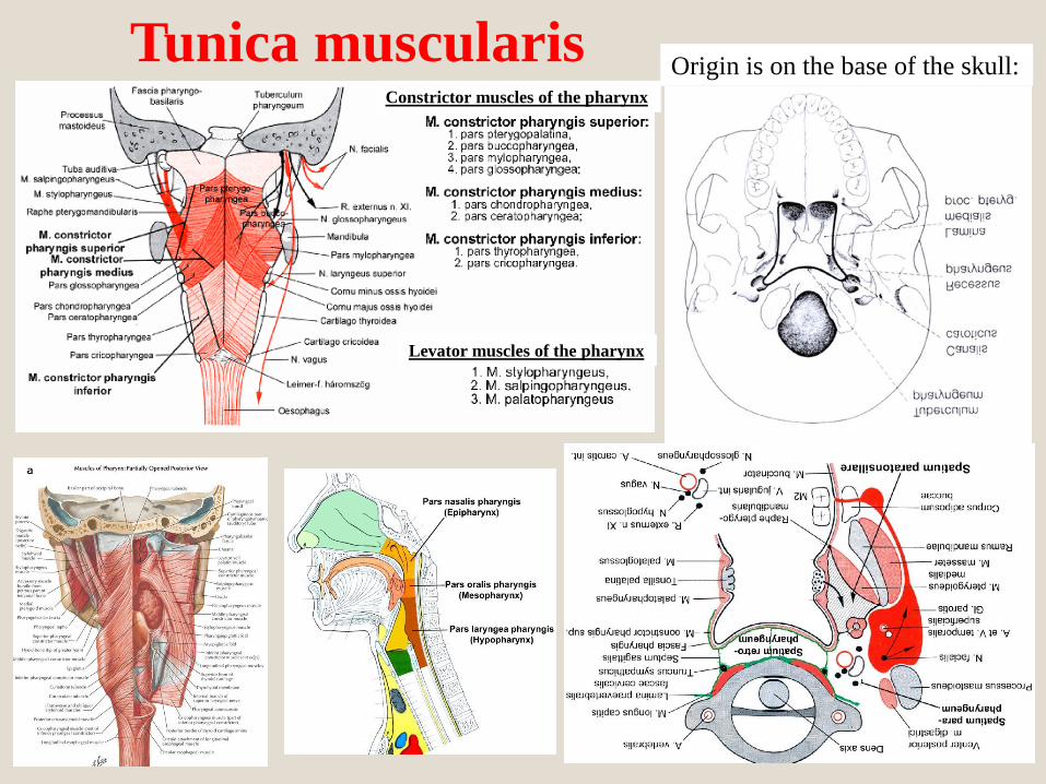
Tunica muscularisOrigin is on the base of the skull:
Constrictor muscles of the pharynx
Levator muscles of the pharynx

Muscles of the pharynxLevator muscles of the pharynxConstrictor muscles of the pharynx
(3 groups)
n. IX
n. X
Raphe pharyngis
Constrictor muscles of the pharynx
Levator muscles of the pharynx
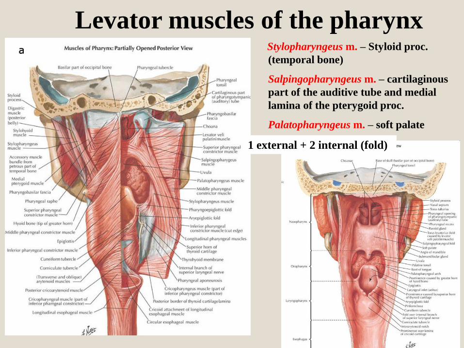
Levator muscles of the pharynxStylopharyngeus m. – Styloid proc.
(temporal bone)
Salpingopharyngeus m. – cartilaginous
part of the auditive tube and medial
lamina of the pterygoid proc.
Palatopharyngeus m. – soft palate
1 external + 2 internal (fold)

Arteries of the head and neckVertebral aa.
ICA – intracranial division
ECA:
Au – post. auricular a.
Oc – occipital a.
StyM – stylomastoid a.
Te – temporal superfic. a.
Ma – maxillary a.
Fa – facial a.
Pha – asc. pharyngeal a.
Ly – lingual a.
Thy – sup. thyroid a.
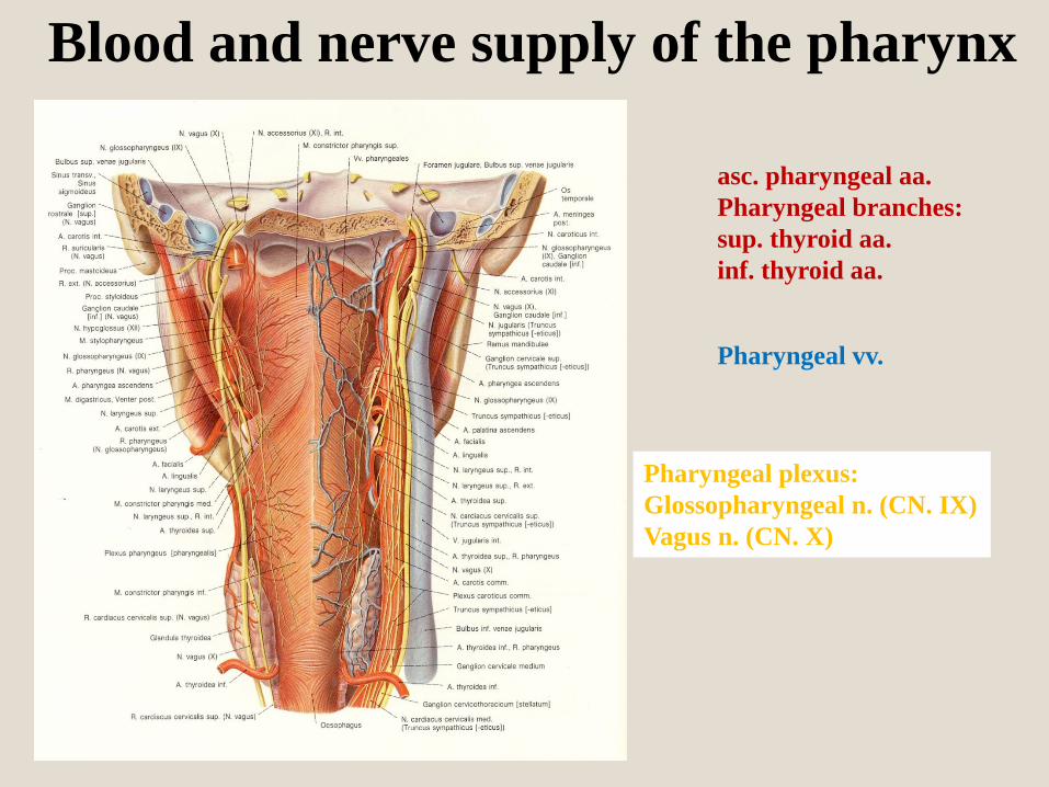
Blood and nerve supply of the pharynx
asc. pharyngeal aa.
Pharyngeal branches:
sup. thyroid aa.
inf. thyroid aa.
Pharyngeal vv.
Pharyngeal plexus:
Glossopharyngeal n. (CN. IX)
Vagus n. (CN. X)

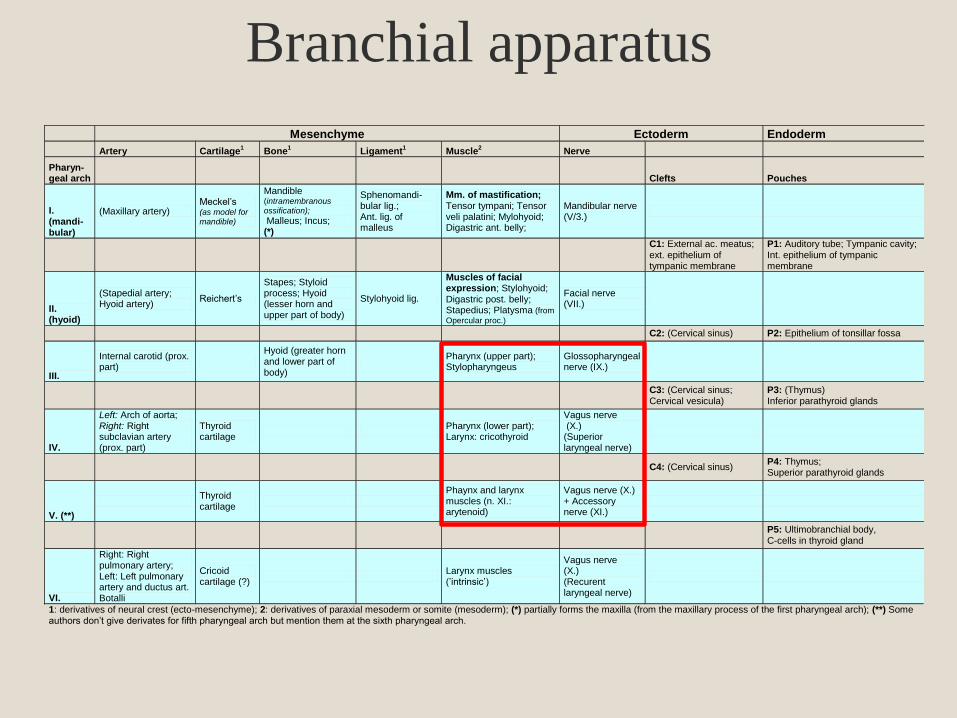
Mesenchyme Ectoderm Endoderm
Artery Cartilage1
Bone1
Ligament1
Muscle2
Nerve
Pharyn- geal arch Clefts Pouches
I. (mandi-bular)
(Maxillary artery) Meckel’s (as model for mandible)
Mandible (intramembranous ossification);
Malleus; Incus; (*)
Sphenomandi-bular lig.; Ant. lig. of malleus
Mm. of mastification; Tensor tympani; Tensor veli palatini; Mylohyoid; Digastric ant. belly;
Mandibular nerve (V/3.)
C1: External ac. meatus; ext. epithelium of tympanic membrane
P1: Auditory tube; Tympanic cavity; Int. epithelium of tympanic membrane
II. (hyoid)
(Stapedial artery; Hyoid artery)
Reichert’s
Stapes; Styloid process; Hyoid (lesser horn and upper part of body)
Stylohyoid lig.
Muscles of facial expression; Stylohyoid; Digastric post. belly; Stapedius; Platysma (from
Opercular proc.)
Facial nerve (VII.)
C2: (Cervical sinus) P2: Epithelium of tonsillar fossa
III.
Internal carotid (prox. part)
Hyoid (greater horn and lower part of body)
Pharynx (upper part); Stylopharyngeus
Glossopharyngeal nerve (IX.)
C3: (Cervical sinus; Cervical vesicula)
P3: (Thymus) Inferior parathyroid glands
IV.
Left: Arch of aorta; Right: Right subclavian artery (prox. part)
Thyroid cartilage
Pharynx (lower part); Larynx: cricothyroid
Vagus nerve (X.) (Superior laryngeal nerve)
C4: (Cervical sinus)
P4: Thymus; Superior parathyroid glands
V. (**)
Thyroid cartilage
Phaynx and larynx muscles (n. XI.: arytenoid)
Vagus nerve (X.) + Accessory nerve (XI.)
P5: Ultimobranchial body, C-cells in thyroid gland
VI.
Right: Right pulmonary artery; Left: Left pulmonary artery and ductus art. Botalli
Cricoid cartilage (?)
Larynx muscles (’intrinsic’)
Vagus nerve (X.) (Recurent laryngeal nerve)
1: derivatives of neural crest (ecto-mesenchyme); 2: derivatives of paraxial mesoderm or somite (mesoderm); (*) partially forms the maxilla (from the maxillary process of the first pharyngeal arch); (**) Some authors don’t give derivates for fifth pharyngeal arch but mention them at the sixth pharyngeal arch.
Branchial apparatus
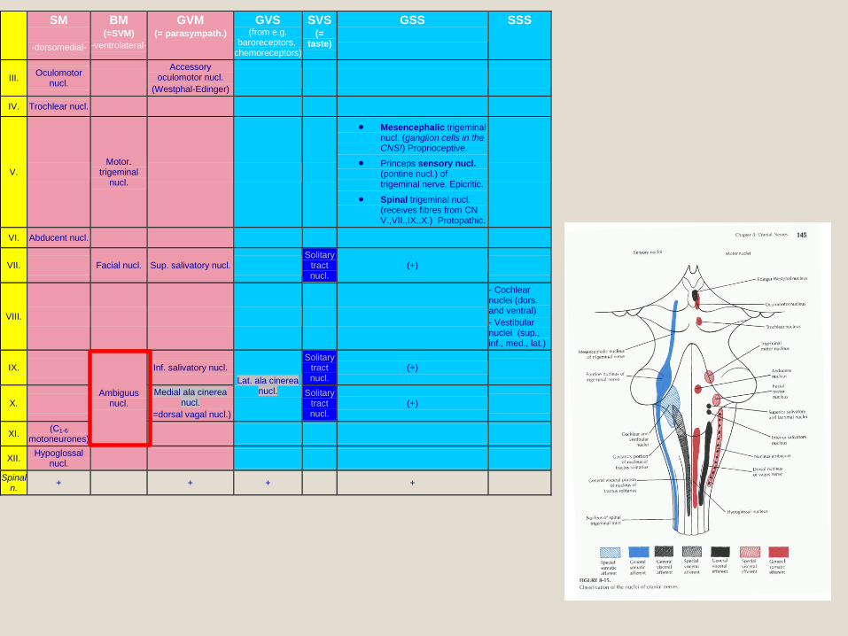
SM
-dorsomedial-
BM (=SVM)
-ventrolateral-
GVM (= parasympath.)
GVS (from e.g.
baroreceptors, chemoreceptors)
SVS (=
taste)
GSS
SSS
III. Oculomotor nucl.
Accessory oculomotor nucl.
(Westphal-Edinger)
IV. Trochlear nucl.
V. Motor.
trigeminal nucl.
Mesencephalic trigeminal nucl. (ganglion cells in the CNS!) Proprioceptive.
Princeps sensory nucl. (pontine nucl.) of trigeminal nerve. Epicritic.
Spinal trigeminal nucl. (receives fibres from CN V.,VII.,IX.,X.) Protopathic.
VI. Abducent nucl.
VII. Facial nucl. Sup. salivatory nucl. Solitary
tract nucl.
(+)
VIII.
- Cochlear nuclei (dors. and ventral) - Vestibular nuclei (sup., inf., med., lat.)
IX.
Ambiguus nucl.
Inf. salivatory nucl. Lat. ala cinerea
nucl.
Solitary tract nucl.
(+)
X. Medial ala cinerea
nucl. (=dorsal vagal nucl.)
Solitary tract nucl.
(+)
XI. (C1-6 motoneurones)
XII. Hypoglossal nucl.
Spinal n. + + + +
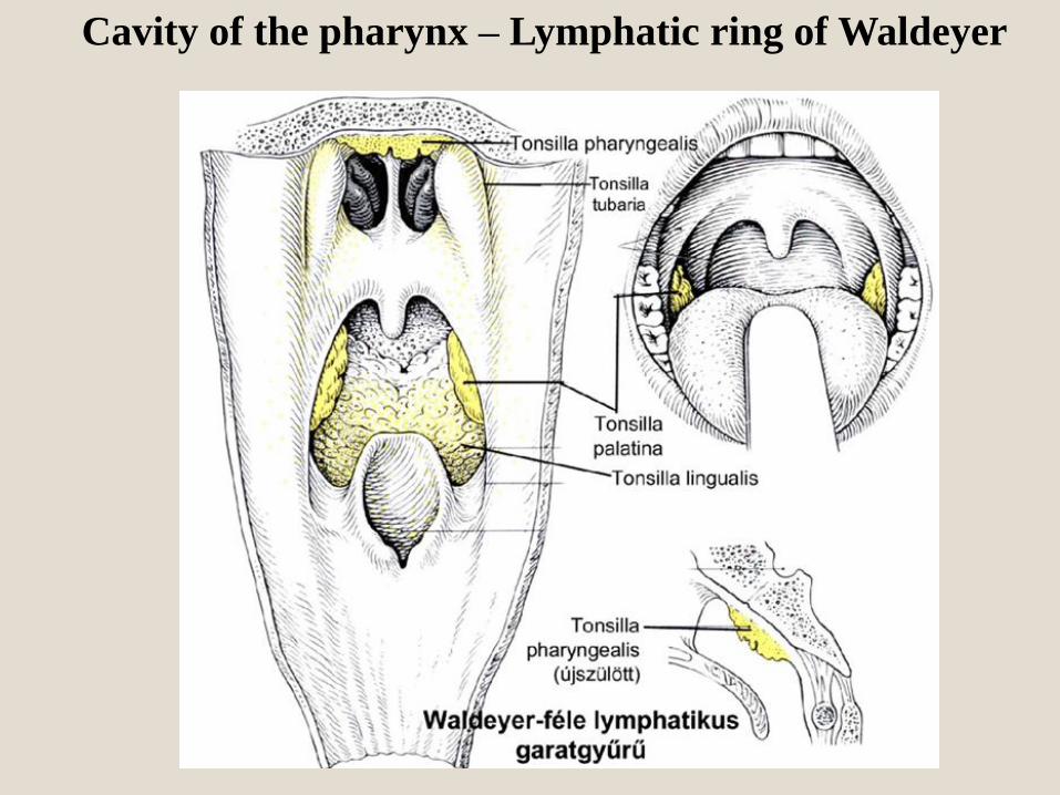
Cavity of the pharynx – Lymphatic ring of Waldeyer
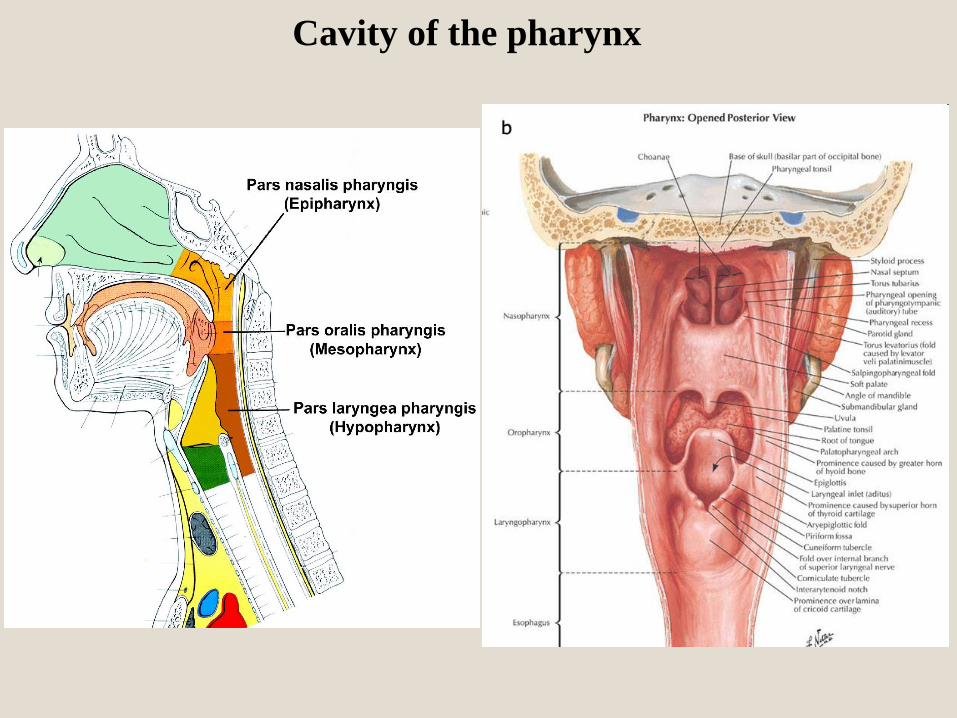
Cavity of the pharynx
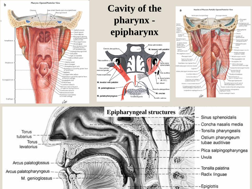
Epipharyngeal structures
Cavity of the
pharynx -
epipharynx

Cavity of the pharynx - mesopharynx
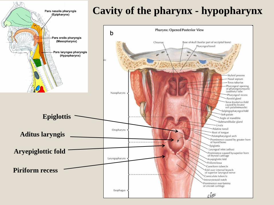
Epiglottis
Aditus laryngis
Aryepiglottic fold
Piriform recess
Cavity of the pharynx - hypopharynx

Pharyngeal spaces
(spatium)
Pharynx
Esophagus
Retropharyngeal
space
Posterior
mediastinum
!!!

Spaces
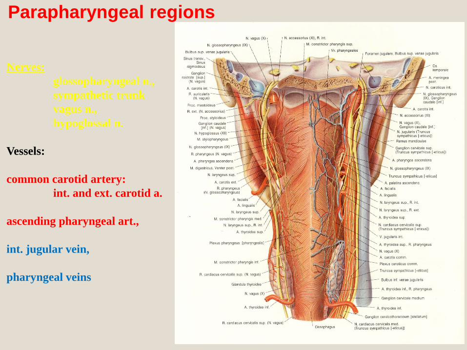
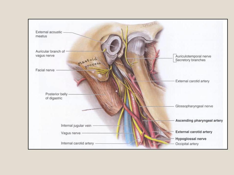
Parapharyngeal regions
Nerves:
glossopharyngeal n.,
sympathetic trunk
vagus n.,
hypoglossal n.
Vessels:
common carotid artery:
int. and ext. carotid a.
ascending pharyngeal art.,
int. jugular vein,
pharyngeal veins
1. R. meningeus
posterior
2. R. auricularis n.
vagi
3. Rr.
pharyngei
PLEXUS
PHARYNGE
US

Cranio-cervical part
sup. (jugulare) ggl.
inf. (nodosum) ggl.

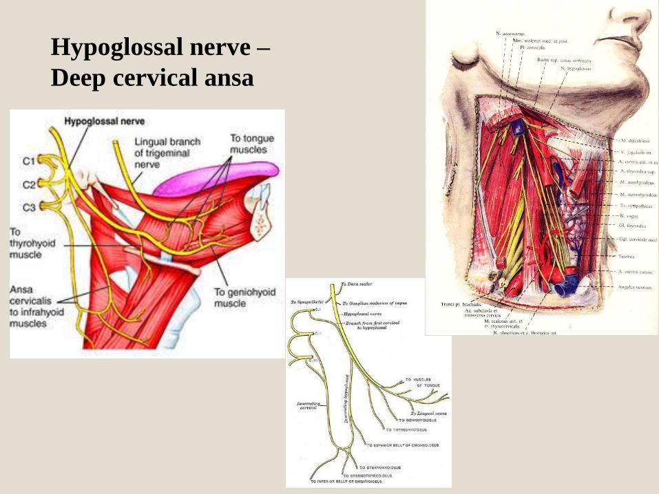
Hypoglossal nerve –
Deep cervical ansa


Sup. laryngeal nerve
r. externus ( cricothyroid m.)
r. internus
Recurrent and inf. laryngeal nerve

Cranio-cervical part of the vagus
Sup. Laryngeal N.(BM,GVM,GVS,SVS,GSS)
Pharyngeal plexus(BM,GVM,GVS,GSS)
Sup. cardiac br.(GVM,GVS)
Recurrent laryngeal N.(BM,GVM,
GVS)


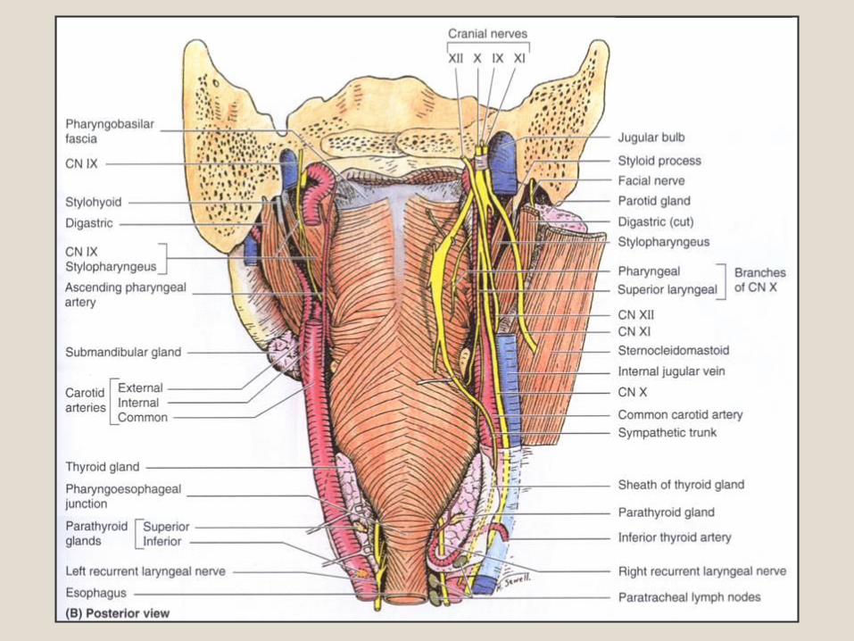
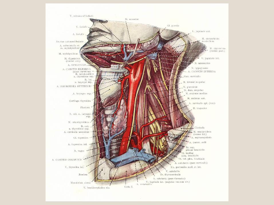
Pharyngeal topography


Cervical fascia (3 lamina)

Thank you for your kind attention!!!


