THE PATHOLOGY OF SARCOIDOSIS - pmj.bmj.com · regional ileitis show histological appearances in ......
6
248 THE PATHOLOGY OF SARCOIDOSIS By A. D. 'HOMSON, M.A., M.D., M.R.C.P. Bland-Sutton Institute of Pathology, Middlesex Hospital, l,ondon Introduction Elsewhere in this issue, possible aetiological fac- tors, the diagnosis, the clinical manifestations and the treatment of sarcoidosis are discussed. The purpose of this article is to review the pathology and place it in its proper perspective. Sarcoidosis can be regarded as a clinico-patho- logical syndrome which may have divers causes and certainly presents with protean clinical mani- festations. To the morbid anatomist there is but one basic histological appearance underlying all these clinical variations, as the cellular features are similar in every affected tissue. There is, however, much confusion over the use of the terms ' Sarcoidosis' and 'Sarcoid.' It appears that clinicians use the term ' Sarcoidosis ' to denote the generalized disease, whereas pathologists, who only see the localized lesion on biopsy, frequently prefer the word ' Sarcoid.' In fact the multi- plicity of these localized sarcoids produce the clinical manifestations of generalized sarcoidosis. There is a further source of confusion in that localized sarcoid reactions are frequently encoun- tered in routine histological examinations and yet these patients do not appear to be suffering from generalized sarcoidosis. Sources of Material The accumulated material for this study has reached the laboratory from two sources-biopsy and autopsy. From the relatively benign nature of the disease, autopsy material is scarce, although it is possible that by the time the disease has reached the late stage of non-specific fibrosis some examples coming to post-mortem examinations remain undetected. This may he so, for instance, in the terminal stages of pulmonary heart disease, when active sarcoid tissue can no longer be found in the lungs. The biopsy material has been submitted from a variety of tissues, particularly lymph nodes, skin, liver, nasal mucosa, tonsil, conjunctiva, spleen, palate, lachrymal gland, parotid gland, and bron- chial mucosa. Skin biopsies from Kviem tests and other inoculation sites have also been examined for confirmatory evidence of sarcoidosis. Finally, serial skin biopsies have been studied, both before and after treatment, to review the activity of the disease and to assess the influence of different forms of therapy. Incidence Sarcoidosis occurs in all parts of the world but there is no agreement as to the frequency of its occurrence. Thus Robb-Smith (I952) reported a rate of o.66 patients per Ioo,ooo of the general population in Oxfordshire, while in young soldiers, receiving routine medical examinations, the incidence increased to 13 per Ioo,ooo in the Swiss Army (Sch6nholzer, I947) and 17.8I per 100,000 Negroes in the United States Army (Michael et al., 1950). Age Sarcoidosis has been reported as occurring in all age groups but the disease most frequently presents between the ages of 20 to 40. In the Middlesex Hospital series, 66 per cent. of the patients were in this age group. Sex IThe sex incidence of the many published series does not give an accurate assessment of the sex distribution, as many of the investigations have been carried out on Army recruits with, therefore, a high male preponderance. At the Middlesex Hospital the first 200 cases of the series contained 86 males and I 4 females. Sites Sarcoidosis is often a widely disseminated disease and, on microscopy, almost any tissue of the body may be involved. The disease, however, shows a predilection for certain organs and these, as assessed during life, are listed below. Sites of Clinical Involvement of 200 Patients Lungs .. .. 91 Spleen ... 13 Skin . . 50 Bones . .. Iymph nodes .. 37 Parotid .. 6 Eyes .. .. 25 C.N.S.. .. 4 copyright. on 11 November 2018 by guest. Protected by http://pmj.bmj.com/ Postgrad Med J: first published as 10.1136/pgmj.34.391.248 on 1 May 1958. Downloaded from
Transcript of THE PATHOLOGY OF SARCOIDOSIS - pmj.bmj.com · regional ileitis show histological appearances in ......

248
THE PATHOLOGY OF SARCOIDOSISBy A. D. 'HOMSON, M.A., M.D., M.R.C.P.
Bland-Sutton Institute of Pathology, Middlesex Hospital, l,ondon
IntroductionElsewhere in this issue, possible aetiological fac-
tors, the diagnosis, the clinical manifestations andthe treatment of sarcoidosis are discussed. Thepurpose of this article is to review the pathologyand place it in its proper perspective.
Sarcoidosis can be regarded as a clinico-patho-logical syndrome which may have divers causesand certainly presents with protean clinical mani-festations. To the morbid anatomist there is butone basic histological appearance underlying allthese clinical variations, as the cellular featuresare similar in every affected tissue. There is,however, much confusion over the use of theterms ' Sarcoidosis' and 'Sarcoid.' It appearsthat clinicians use the term ' Sarcoidosis ' to denotethe generalized disease, whereas pathologists, whoonly see the localized lesion on biopsy, frequentlyprefer the word ' Sarcoid.' In fact the multi-plicity of these localized sarcoids produce theclinical manifestations of generalized sarcoidosis.There is a further source of confusion in thatlocalized sarcoid reactions are frequently encoun-tered in routine histological examinations and yetthese patients do not appear to be suffering fromgeneralized sarcoidosis.
Sources of MaterialThe accumulated material for this study has
reached the laboratory from two sources-biopsyand autopsy. From the relatively benign natureof the disease, autopsy material is scarce, althoughit is possible that by the time the disease hasreached the late stage of non-specific fibrosis someexamples coming to post-mortem examinationsremain undetected. This may he so, for instance,in the terminal stages of pulmonary heart disease,when active sarcoid tissue can no longer be foundin the lungs.The biopsy material has been submitted from a
variety of tissues, particularly lymph nodes, skin,liver, nasal mucosa, tonsil, conjunctiva, spleen,palate, lachrymal gland, parotid gland, and bron-chial mucosa. Skin biopsies from Kviem tests andother inoculation sites have also been examined forconfirmatory evidence of sarcoidosis. Finally,
serial skin biopsies have been studied, both beforeand after treatment, to review the activity of thedisease and to assess the influence of differentforms of therapy.Incidence
Sarcoidosis occurs in all parts of the world butthere is no agreement as to the frequency of itsoccurrence. Thus Robb-Smith (I952) reporteda rate of o.66 patients per Ioo,ooo of the generalpopulation in Oxfordshire, while in youngsoldiers, receiving routine medical examinations,the incidence increased to 13 per Ioo,ooo in theSwiss Army (Sch6nholzer, I947) and 17.8I per100,000 Negroes in the United States Army(Michael et al., 1950).Age
Sarcoidosis has been reported as occurring in allage groups but the disease most frequentlypresents between the ages of 20 to 40. In theMiddlesex Hospital series, 66 per cent. of thepatients were in this age group.Sex
IThe sex incidence of the many published seriesdoes not give an accurate assessment of the sexdistribution, as many of the investigations havebeen carried out on Army recruits with, therefore,a high male preponderance. At the MiddlesexHospital the first 200 cases of the series contained86 males and I 4 females.
SitesSarcoidosis is often a widely disseminated
disease and, on microscopy, almost any tissue ofthe body may be involved. The disease, however,shows a predilection for certain organs and these,as assessed during life, are listed below.
Sites of Clinical Involvement of 200 Patients
Lungs .. .. 91 Spleen... 13Skin . . 50 Bones . ..Iymph nodes .. 37 Parotid .. 6Eyes .. .. 25 C.N.S.. .. 4
copyright. on 11 N
ovember 2018 by guest. P
rotected byhttp://pm
j.bmj.com
/P
ostgrad Med J: first published as 10.1136/pgm
j.34.391.248 on 1 May 1958. D
ownloaded from

May 1958 'HOMSON: The Pathology o' Sarcoidosis 249
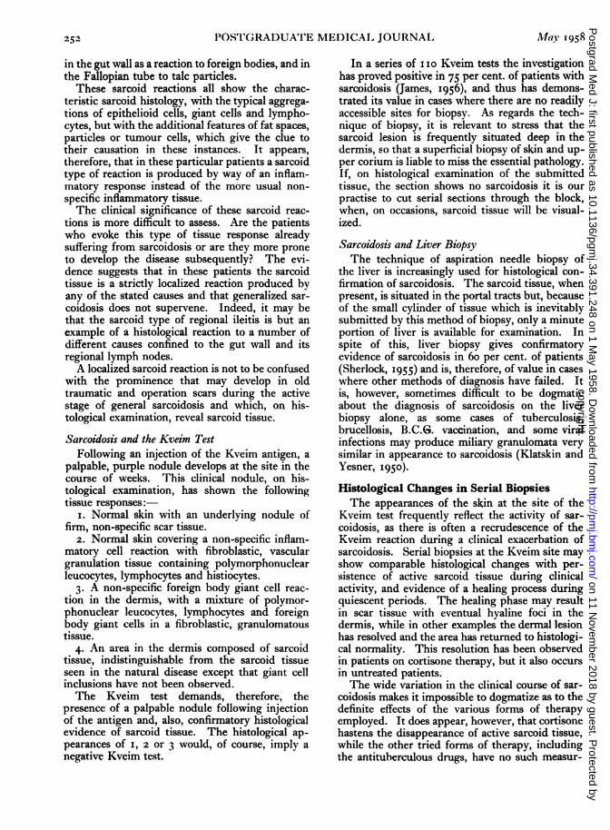
It is, of course, impossible to know the exactincidence of involvement of the various tissuesduring life as, at autopsy on patients dying of thedisease, sarcoidosis is usually present in manyorgans which showed no evidence of clinicalderangement. Similarly, the technique of blindbiopsy of various organs such as scalene nodes andliver has drawn attention to sarcoid involvementin tissues which would otherwise have remainedundetected. Thus biopsy of the liver revealssarcoid tissue in 6o per cent. of cases (Sherlock,1955). To the pathologist, therefore, the fre-quency of involvement cannot be assessed on sub-mitted pathological material alone, as this isdependant so much upon the diagnostic enthusiasmof the clinician and the accessibility of the organ.
Other organs not already listed may be involved,-and sarcoid tissue has been found in the heart,skeletal muscle, kidneys and the genital tract inthis series. The importance of myocardial in-volvement is that it may result in sudden death,while renal sarcoidosis may be associated withnephrocalcinosis.The true incidence of intestinal sarcoidosis is
unknown as it is entirely dependant upon thecriteria used for its inclusion with this disease.Thus Hadfield (1939) regards regional ileitis as aform of sarcoidosis while others (e.g. Cowdell,1954) deny this. Certainly some examples ofregional ileitis show histological appearances inthe gut wall and in the mesenteric lymph nodesindistinguishable from sarcoid, and on histologicalgrounds it is possible that sarcoidosis producesone type of regional ileitis. However, Longcopeand Freiman (1952) report on the infrequency ofintestinal involvement in generalized sarcoidosisand Watson et al. (1945), after an extensive searchof the literature, could only accept six cases, withtwo of their own, as true examples of intestinalsarcoidosis.
In spite of the histological similarity of somecases of regional ileitis and sarcoidosis there doesappear to be a clinical difference, as I have not asyet seen a case of regional ileitis with a sarcoidhistology progress to generalized sarcoidosis.These differences can be tabulated:-
SIarcoidosis Regional lBeitis1 ,1ings -
Liver -Lymph nodes -- +Gut Rare Invariable
MacroscopicalSarcoidosis in its earliest recognizable form
shows multiple, minute, pale, pin-points ofmaterial studded in the area of involved tissue.These foci enlarge as the disease progresses, to
4', .'.4.
4~ . "4"':, . "~
'r* '*'"!F..
4' ..4~r~" ~',,,4~~'~~. ~'-'..'/':
'.4',
'4.-.4
~~
F'(;. i.-Showing the circular aggregations of epithelioidcells in sarcoidosis replacing a lymph node. x 85.
form readily visible nodules of pale tissue, oftenwith scalloped margins. These areas may becomeconfluent in some organs and at a later stage thediseased tissue may become firmer than normal,due to progressive replacement by fibrous tissue.
MicroscopicalThe histological appearances of sarcoidosis re-
main characteristically similar in all the bodytissues, so that a description of one such area ofinvolvement is exactly mirrored in any othertissue. The essential histological features are thefocal collections of epithelioid cells, usually ar-ranged in spherical fashion and surrounded by avariable, but frequently scanty, rim of lympho-cytes (Fig. i). These epithelioid cells have palestaining, vesicular nuclei, with pink staining cyto-plasm and clearly demarcated cell boundaries.Additional histological features may be present;there are frequently giant cells of either theforeign body or Langhans' type; a variety of in-clusions may be visible within these giant cellsbut they are in no way pathognomonic of sar-coidosis; foci of necrosis may also be present inthe epithelioid cell aggregations. This necrosis isnever a conspicuous feature, and when present it is
copyright. on 11 N
ovember 2018 by guest. P
rotected byhttp://pm
j.bmj.com
/P
ostgrad Med J: first published as 10.1136/pgm
j.34.391.248 on 1 May 1958. D
ownloaded from

250 POS'TGRADUATE MEDICALf JOURNAL May 1958
confined to the centres of the epithelioid foci andis of the fibrinoid type.The fate of the Sarcoidosis HistologyThe fate of the cellular reaction of sarcoidosis is
relevant to the eventual outcome of the disease. Itis certain on clinical and post-mortem evidencethat the process can resolve with disappearance ofthe cells and a return of the tissues to normal.This statement is based on the frequency ofclinical regression in previously diseased areas, thereturn to normal of the dermis on repeated biopsyexaminations of dermal sarcoidosis and the occa-sional reports of the post-mortem findings ofnormal internal organs which had previous histo-logical evidence of sarcoidosis during life. Whenresolution does not occur the area of sarcoidosismay persist for long periods as an active cellularlesion or may eventually fibrose when the diseasedarea becomes replaced by hyaline acellular fibroustissue.Thus sarcoid tissue at any site may persist as
active sarcoidosis, resolve, or be converted intohyaline collagen.Prognosis
Sarcoidosis can be regarded as a generalizeddisease with an excellent prognosis in the majorityof patients in that disappearance of active sarcoidtissue is the rule. There are, however, five im-portant exceptions to this favourable outcome:-
i. Pulmonary fibrosis-cor pulmonale.2. Uveal tract fibrosis-eye complications and
blindness.3. Central nervous system and pituitary gland
involvement.4. Hypercalcaemia and nephrocalcinosis.5. Cardiac involvement.
Causes of DeathDeath is rarely due to sarcoidosis directly, except
when the heart, the central nervous system or thekidneys are extensively involved. In the majorityof patients the immediate cause of death is due tosome totally unrelated pathology. Thus:-
Ricker and Clark (I949), 22 autopsies, threedeaths due to sarcoidosis.Longcope and Freiman (1952), 12 autopsies,
five deaths due to sarcoidosis.Cowdell (I954), 17 deaths, eight deaths due to
sarcoidosis.l)anbolt (1947) recorded the autopsy findings
on Boeck's original case of skin sarcoidosis and, atdeath, 44 years after the appearance of the disease,no evidence of sarcoidosis could be demonstratedanywhere in the body.The commonest cause of death in patients with
sarcoidosis is pulmonary fibrosis, and in Leitner's
t._.l,r. . , l 'o:%!A ~' ,: ~ '..
~ ~ ~.~.K ~"J',.d.o
.~'W,~,'~,..,~: ·T,
q4.g· ..
A
- -'$I1v
..!.:
iS ·~~;~.~l~
· :·
"'~..~:~ )"
FIG. 2.-Showing fibrosis of the lung tissue due tosarcoidosis in which there are many giant cellswith inclusions. Note the ' honeycomb ' lung andthe vascular thickening. 85.
(I949) series of 72 autopsies, 26 died in heartfailure. Pulmonary sarcoidosis resulting in pul-monary fibrosis is, therefore, one of the fatalsequelae of the disease and this fibrosis may takethe form of 'honeycomb' lung as seen in Fig. 2(Heppleston, I956). In most of the publishedseries, tuberculosis is present at the termination ofmany of the patients, and Leitner (I949) recorded12 cases of active tuberculosis in 72 autopsies andRiley (I950) reported six with tuberculosis in 13fatal cases.
Therefore death from from sarcoidosis occurson the rare occasions when the heart is primarilyinvolved by the disease or, more frequently, whensecondarily implicated due to extensive pulmonaryfibrosis. Death may also be due to central nervoussystem involvement or, rarely, from extensive sar-coidosis in the kidneys. In the absence of these,the cause of death is due to coincidental disease ofany type with tuberculosis as a fairly commonlyrecorded termination.
Tuberculin ReactionGreat stress is frequently made of the failure of
sarcoidosis patients to react to intradermal injec-
copyright. on 11 N
ovember 2018 by guest. P
rotected byhttp://pm
j.bmj.com
/P
ostgrad Med J: first published as 10.1136/pgm
j.34.391.248 on 1 May 1958. D
ownloaded from

May I958 THOMSON: The Pathology of Sarcoidosis 251
tions of I: Ioo old tuberculin. Indeed, 60 percent. of the patients in the present series wereMantoux negative, but the presence of a positivereaction does not negate the diagnosis. The factthat by using an oily depot of tuberculin many ofthe Mantoux negative patients showed evidence ofa positive reaction reduces the significancenormally attached to the anergic state of thesepeople (James and Pepys, I956).Laboratory InvestigationsBlood CountThe changes in the blood in sarcoidosis are in-
constant and do not show specifically diagnosticfeatures. Frequently the blood count is normalalthough the sedimentation rate may be raisedespecially when erythema nodosum is present(James et al., i956). The white cell count may benormal or leucopenic while, less commonly, thereis a monocytosis or an eosinophilia (Bruschi andHowe, 1950).Serum ProteinsThere is a hyperglobulinaemia in about*25 per
cent. of the patients, but this finding is present inmany other diseases and is of little diagnosticvalue. Its main importance is as confirmatoryevidence of activity in an already established caseof sarcoidosis.
Serum CalciumA rare finding in sarcoidosis is the association of
a raised serum calcium with calcinuria andnephrocalcinosis. This abnormality is due to ahypersensitivity to the action of vitamin ' I) '(Anderson et al., 1954), but it only occurs in abouti per cent. of sarcoidosis cases (James, 1956).Alkaline PhosphataseThe serum alkaline phosphatase is raised in a
minority of patients with sarcoidosis. This isregarded by some as indicating active liver in-volvement while others suggest that it is due tobone resorption by sarcoid tissue in the bonemarrow.
Histological DiagnosisIt is important to stress that during the early and
active stages of sarcoidosis it is possible to findsome accessible tissue involved by the disease in ahigh proportion of patients. In the present seriesthe following tissues have given histological con-firmation of sarcoidosis:-Lymph nodes TonsilSkin EyeLiver Bronchial biopsyLung SpleenNose Palate
Post-mortem Lachrymal glandKviem test Parotid glandOf interest is our failure to obtain histological
evidence of sarcoidosis on blind biopsy of theconjunctiva as reported by Crick et al., (I955).Histological Differential Diagnosis
Although in typical cases of sarcoidosis thehistological appearances are characteristic, thereare certain other diseases and lesions in whichsimilar histological features may occur and someof these diagnostic hazards are discussed below.
Sarcoidosis and TuberculosisThe histological features of some examples of
tuberculosis may closely simulate those of sar-coidosis and on occasions it is difficult to segregatethe two processes on histological appearancesalone. Essentially, sarcoid tissue resembles atuberculous granuloma without the caseation ofthe latter, but closer inspection reveals otherpoints of detail which may be helpful in decidingwhether a tuberculous or a sarcoid pathology isoperative.
I. The sarcoidosis foci are more uniformivspherical in shape and size.
2. The sarcoidosis foci tend to remain discretewith a well-defined margin.
3. The epithelioid cells in sarcoidosis have adistinct cell boundary and a more uniform ar-rangement.
4. Lymphocytes are scanty and may he absentin sarcoidosis.
5. The giant cells of sarcoidosis are inconstantand, when present, tend to be of both Ianghans'and foreign body in type.
6. Necrosis in sarcoidosis is not common but,when present, is of the ' fibrinoid' type and isconfined to a small area in the centre of theepithelioid cell aggregations.
7. Giant cell inclusions of the 'asteroid' typeare rarely (if ever) seen in tuberculous lesions.
8. Ziehl-Neelsen staining is always negative insarcoidosis.
9. Absence of calcification in the sarcoidosistissue.
Sarcoidosis and Sarcoid ReactionIt is important to stress the frequency with
which apparently localized sarcoid reactions arefound in various tissues of the body. I have seensarcoid reactions in the dermis in response to sandand glass particles, around sites of injection of waxand oil, and in areas of fat necrosis. Sarcoid tissue'may be found in lymph nodes associated with oildroplets, around carbon particles, and adjacent toHodgkin's tissue and carcinoma cells. Sarcoid re-actions are also found, amongst many other organs,
copyright. on 11 N
ovember 2018 by guest. P
rotected byhttp://pm
j.bmj.com
/P
ostgrad Med J: first published as 10.1136/pgm
j.34.391.248 on 1 May 1958. D
ownloaded from
252 IOSTGRADUATE MEDICAI JOURNAL May 195g
in the gut wall as a reaction to foreign bodies, and inthe Fallopian tube to talc particles.
These sarcoid reactions all show the charac-teristic sarcoid histology, with the typical aggrega-tions of epithelioid cells, giant cells and lympho-cytes, but with the additional features of fat spaces,particles or tumour cells, which give the clue totheir causation in these instances. It appears,therefore, that in these particular patients a sarcoidtype of reaction is produced by way of an inflam-matory response instead of the more usual non-specific inflammatory tissue.The clinical significance of these sarcoid reac-
tions is more difficult to assess. Are the patientswho evoke this type of tissue response alreadysuffering from sarcoidosis or are they more proneto develop the disease subsequently? The evi-dence suggests that in these patients the sarcoidtissue is a strictly localized reaction produced byany of the stated causes and that generalized sar-coidosis does not supervene. Indeed, it may bethat the sarcoid type of regional ileitis is but anexample of a histological reaction to a number ofdifferent causes confined to the gut wall and itsregional lymph nodes.A localized sarcoid reaction is not to be confused
with the prominence that may develop in oldtraumatic and operation scars during the activestage of general sarcoidosis and which, on his-tological examination, reveal sarcoid tissue.
Sarcoidosis and the Kveim TestFollowing an injection of the Kveim antigen, a
palpable, purple nodule develops at the site in thecourse of weeks. This clinical nodule, on his-tological examination, has shown the followingtissue responses:
I. Normal skin with an underlying nodule offirm, non-specific scar tissue.
2. Normal skin covering a non-specific inflam-matory cell reaction with fibroblastic, vasculargranulation tissue containing polymorphonuclearleucocytes, lymphocytes and histiocytes.
3. A non-specific foreign body giant cell reac-tion in the dermis, with a mixture of polymor-phonuclear leucocytes, lymphocytes and foreignbody giant cells in a fibroblastic, granulomatoustissue.
4. An area in the dermis composed of sarcoidtissue, indistinguishable from the sarcoid tissueseen in the natural disease except that giant cellinclusions have not been observed.The Kveim test demands, therefore, the
presence of a palpable nodule following injectionof the antigen and, also, confirmatory histologicalevidence of sarcoid tissue. The histological ap-pearances of I, 2 or 3 would, of course, imply anegative Kveim test.
In a series of I o Kveim tests the investigationhas proved positive in 75 per cent. of patients withsarcoidosis (James, 1956), and thus has demons-trated its value in cases where there are no readilyaccessible sites for biopsy. As regards the tech-nique of biopsy, it is relevant to stress that thesarcoid lesion is frequently situated deep in thedermis, so that a superficial biopsy of skin and up-per corium is liable to miss the essential pathology.If, on histological examination of the submittedtissue, the section shows no sarcoidosis it is ourpractise to cut serial sections through the block,when, on occasions, sarcoid tissue will be visual-ized.
Sarcoidosis and Liver BiopsyThe technique of aspiration needle biopsy of
the liver is increasingly used for histological con-firmation of sarcoidosis. The sarcoid tissue, whenpresent, is situated in the portal tracts but, becauseof the small cylinder of tissue which is inevitablysubmitted by this method of biopsy, only a minuteportion of liver is available for examination. Inspite of this, liver biopsy gives confirmatoryevidence of sarcoidosis in 60 per cent. of patients(Sherlock, 1955) and is, therefore, of value in caseswhere other methods of diagnosis have failed. Itis, however, sometimes difficult to be dogmaticabout the diagnosis of sarcoidosis on the liverbiopsy alone, as some cases of tuberculosis,brucellosis, B.C.G. vaccination, and some viralinfections may produce miliary granulomata verysimilar in appearance to sarcoidosis (Klatskin andYesner, 1950).Histological Changes in Serial BiopsiesThe appearances of the skin at the site of the
Kveim test frequently reflect the activity of sar-coidosis, as there is often a recrudescence of theKveim reaction during a clinical exacerbation ofsarcoidosis. Serial biopsies at the Kveim site mayshow comparable histological changes with per-sistence of active sarcoid tissue during clinicalactivity, and evidence of a healing process duringquiescent periods. The healing phase may resultin scar tissue with eventual hyaline foci in thedermis, while in other examples the dermal lesionhas resolved and the area has returned to histologi-cal normality. This resolution has been observedin patients on cortisone therapy, but it also occursin untreated patients.The wide variation in the clinical course of sar-
coidosis makes it impossible to dogmatize as to thedefinite effects of the various forms of therapyemployed. It does appear, however, that cortisonehastens the disappearance of active sarcoid tissue,while the other tried forms of therapy, includingthe antituberculous drugs, have no such measur-
copyright. on 11 N
ovember 2018 by guest. P
rotected byhttp://pm
j.bmj.com
/P
ostgrad Med J: first published as 10.1136/pgm
j.34.391.248 on 1 May 1958. D
ownloaded from

May I 1958 TIHONISON: The Pathology of Sarcoidosis 253
LLOYD-LUKE --
Books that enshrine profound tholuhtOPHTHALMOLOGY PRACTICAL FLUID BALANCE
A Textbook for Diploma Students OBSTETRIC PROBLEMS IN SURGICAL PRACTICEby P. D. TREVOR-ROPER by IAN DONALD (2nd edit.)
M.B., B.Chir.(Camb.), F.R.C.S., by L. P. LE QUESNED.O.M.S.(Eng.) M.B.E., M.D.(Lond.), F.R.C.O.G. D.M(Oxon), F.R.C.S.(Eng.)(1955) 75s. net (1955) 47s. 6d. net (1957) 20s. net
GENERAL PATHOLOGY MEDICAL ETHICS(2nd edit.) A Guide to Students POSTURAL DRAINAGE
dited band Practitioners by E. WINIFRED THACKEREdited by SIR HOWARD FLOREY
DFRCPFRS Edited b/ MAURICE DAVIDSON M.C.S.P.. , . ., . . . D.M.(Oxon.), F.R.C.P.(Lond.) (1956) 8s. 6d. net(1958) 84s. net (1957) 20s. net
RECENT TRENDS THE RESPIRATORY MUSCLESIN CHRONIC BRONCHITIS AND THE MECHANICS OF ANAESTHETIC ACCIDENTSEdited by NEVILLE C. OSWALD BREATHING by V. KEATING
T.D., M.D.(Cantab.), F.R.C.P.(Lond.) by E. J. MORAN CAMPBELL M.B., B.Ch., D.A., F.F.A.R.C.S.(Eng.)M.D., Ph.D.(Lond.), M.R.C.P.(Lond.) (1956) 25s. net(1958) 30s. net (1958) 20s. net
Dsscriptive catalogue available on request
LLOYD-LUKE (MEDICAL BOOKS) LTD., 49 Newman Street, W.I
able effect in excess of the natural processes seenin spontaneous remissions. Thus in io patientswho were subjected to serial biopsies from skinlesions, nasal mucosa, or Kveim sites, no histo-logical evidence of healing during a course of anti-tuberculous chemotherapy could be demon-strated.
In summary, therefore, sarcoidosis is a general-ized disease involving especially the lungs, liver,skin and lymph nodes, and the diagnosis is madeon the clinical picture confirmed by a compatible,histological appearance. No pathological tests arepathognomonic of the disease except the histologyof the affected tissue, which is the sole method ofdiagnostic confirmation. The area of sarcoidosismay persist in the active cellular phase, resolve orfibrose, and the fate of the patient is dependentboth on the outcome of this histological lesion aswell as the sites of involvement. Death is rarelydue to active sarcoidosis, although the late sequelaeof fibrosis, especially in the lungs, may be fatal.The histology of the Kveim test gives useful con-firmatory evidence of sarcoidosis in those caseswhere ino accessible tissutc is available for biopsy,and the Kvcimi site formlns a cnllvcnient area for
examination to assess both the activity of thedisease and the efficacy of treatment.
BIBLIOGRAPHYANDERSON, J., DENT, C. E., HARPER, C., and PHILPO1',
(. R. (1954), Lancet, ii, 720.BRUSCHI, MI., and HOWE, J. S. (1950), Blood, 5, 478.CO\VDELL, R. H. (1954), Quart. J. Med., 23, 29.CRICK, R., HOYLE, C., and MATHER, G;. (1955), Brit. medl. J.,
ii, I 80.DANBOLT, N. (1947), Schweeiz. med. WIschr., 77, 1149.HADFIELD, G. (1939), Lancet, ii, 773.HEPPLESTON, A. G. (1956), Thorax, 11, 77.JAMES, 1). G. (1956), Brit. med. '., ii, 900.JAMES, D. G., and PEPYS, J. (1956), Lancet, i, 602.JAM1ES, D. G., THOMSON, A. D., and WILLCOX, A. (1956),
Ibid., ii, 218.KLATSKIN, (., and YESNER, R. (1950), Vale 7. Biol. MIed., 23,
207.,EITNER, S. J. (1949), Dei- Morbus B2snier-Boeck-Schlau-
mnann,' Basal (Schwabe).LO()NGCOPE, \V. T., and FREIMAN, D. (;. (1952), Medicine,
31, 1.MICHAEL, M., COLE, R. MI., BEESON, P. B., and OLSEN,
B. J. (1950), Amler. Rev. Tuberc., 62, 4.03.RICKER, W., and CLARK, /I. (1949), Amer.J. clin. Path., 19, 725.RILEY, E. A. (1950), Amer. Rev. Tuberc., 62, 231.ROBB-SMITH, A. H. T. (I952), ' Modern Practice in Tubercu-
losis,' Vol. 2, p. 265, London (Butterworth).SCIH() NHOLZER, G. (1947), S'chweiz. med. II'schr., 77, 585.SIIERI,OCK, S. (1955), Diseases of the Liver and Biliary Tract,'
Oxford (Blackwell).\\AS'ION, C. J., RIGLER, L. G., WANGENSTEEN, (). H aiii
McCARTHY, J. S. (T945), Gastroenterology, 4, 30.
copyright. on 11 N
ovember 2018 by guest. P
rotected byhttp://pm
j.bmj.com
/P
ostgrad Med J: first published as 10.1136/pgm
j.34.391.248 on 1 May 1958. D
ownloaded from