The p160 RhoA-Binding Kinase ROKα Is a Member of a Kinase
15
MOLECULAR AND CELLULAR BIOLOGY, Oct. 1996, p. 5313–5327 Vol. 16, No. 10 0270-7306/96/$04.0010 Copyright q 1996, American Society for Microbiology The p160 RhoA-Binding Kinase ROKa Is a Member of a Kinase Family and Is Involved in the Reorganization of the Cytoskeleton THOMAS LEUNG, 1 XIANG-QUN CHEN, 1 EDWARD MANSER, 1 AND LOUIS LIM 1,2 * Glaxo-IMCB Group, Institute of Molecular & Cell Biology, National University of Singapore, Singapore 119260, Singapore, 1 and Institute of Neurology, London WC1N 1PJ, United Kingdom 2 Received 27 March 1996/Returned for modification 14 May 1996/Accepted 4 July 1996 The GTPase RhoA has been implicated in various cellular activities, including the formation of stress fibers, motility, and cytokinesis. We recently reported on a p150 serine/threonine kinase (termed ROKa) binding RhoA only in its active GTP-bound state and on its cDNA; introduction of RhoA into HeLa cells resulted in translocation of the cytoplasmic kinase to plasma membranes, consistent with ROKa being a target for RhoA (T. Leung, E. Manser, L. Tan, and L. Lim, J. Biol. Chem. 256:29051–29054, 1995). Reanalysis of the cDNA revealed that ROKa contains an additional N-terminal region. We also isolated another cDNA which encoded a protein (ROKb) with 90% identity to ROKa in the kinase domain. Both ROKa and ROKb, which had a molecular mass of 160 kDa, contained a highly conserved cysteine/histidine-rich domain located within a putative pleckstrin homology domain. The kinases bound RhoA, RhoB, and RhoC but not Rac1 and Cdc42. The Rho-binding domain comprises about 30 amino acids. Mutations within this domain caused partial or complete loss of Rho binding. The morphological effects of ROKa were investigated by microinjecting HeLa cells with DNA constructs encoding various forms of ROKa. Full-length ROKa promoted formation of stress fibers and focal adhesion complexes, consistent with its being an effector of RhoA. ROKa truncated at the C terminus promoted this formation and also extensive condensation of actin microfilaments and nuclear disruption. The proteins exhibited protein kinase activity which was required for stress fiber formation; the kinase-dead ROKaK112A and N-terminally truncated mutants showed no such promotion. The latter mutant instead induced disassembly of stress fibers and focal adhesion complexes, accompanied by cell spreading. These effects were mediated by the C-terminal region containing Rho-binding, cysteine/histidine-rich, and pleckstrin homology domains. Thus, the multidomained ROKa appears to be involved in reorganization of the cytoskeleton, with the N and C termini acting as positive and negative regulators, respectively, of the kinase domain whose activity is crucial for formation of stress fibers and focal adhesion complexes. In mammals, the Ras-related Rho subfamily includes RhoA, -B, and -C, Rac1 and -2, and Cdc42, which play pivotal roles in cytoskeletal control and cell morphology. RhoA has been im- plicated in stress fiber formation (33, 34), whereas Rac1 (36) and Cdc42 (15, 29) are involved in lamellipodial and filopodial formation, respectively. Fibroblasts injected with these GTPases display a set of distinctive morphological changes, suggestive of probable hierarchy in the order Cdc42, Rac, and Rho. These changes demand a high level of flexibility in the dynamic reorganization of actin microfilaments in cells. RhoA, -B, and -C have about 85% identity and appear to have differ- ent cellular localizations (1). Although their exact roles in cells have not been clearly defined, these GTPases have been im- plicated in a variety of cellular activities either directly by overexpression of wild-type and mutant forms or indirectly by inhibiting their functions with toxins such as the Clostridium botulinum C3 transferase. RhoA has been shown to affect morphology (30), cell motility (39), cytokinesis (14), tumor progression (31, 32), and apoptosis (12). RhoB, which exhibits a cell cycle-dependent expression pattern, has an effect on cell transformation (42). The morphological effect of RhoA on actin-containing cytoskeletal structures (36, 38) appears to be cell type specific. Thus, although in most cells, including fibro- blasts, the major effect of RhoA is formation of stress fibers, membrane ruffling has also been observed in other cell types (28). Biochemically, the targets of the Rho GTPases which initi- ate these phenotypic changes remain largely uncharacterized. The use of specific kinase inhibitors has shown that the path- ways leading to formation of stress fibers and of focal adhesion complexes are distinct; they also implicate the possible involve- ment of kinases, especially downstream tyrosine kinases, in the formation of focal adhesions (35). RhoA is also involved in activating phosphatidylinositol 3-kinase in Swiss 3T3 cells and in human platelet cytosolic extracts (43) and possibly in the activation of phosphatidylinositol 5-kinase (6). In rat liver membranes, RhoA also has a role in the activation of mem- brane-associated phospholipase D, although direct interaction between the two has not been demonstrated (3). More re- cently, RhoA has been implicated in the serum-responsive factor-linked signalling pathway (10), while Cdc42 and Rac have been linked to the mitogen-activated protein kinase c- JNK signalling pathway (7, 25). These nuclear events and the identification of kinase targets for Cdc42 and Rac (21, 22) support the notion that the GTPases are participants in dis- tinctive kinase cascades. We have recently isolated a serine/ threonine kinase, ROKa (Rho-associated kinase), which re- sponds to activated RhoA by translocating from the cytoplasm to the plasma membranes (18). We now report that the cDNA for ROKa encodes an additional N-terminal region compared with that previously reported (18) and that ROKa is a member * Corresponding author. Mailing address: Glaxo-IMCB Group, Institute of Molecular & Cell Biology, National University of Singapore, Kent Ridge, Singapore 119260, Singapore. Phone: (65) 772-6167. Fax: (65) 774-0742. 5313 on April 4, 2019 by guest http://mcb.asm.org/ Downloaded from
Transcript of The p160 RhoA-Binding Kinase ROKα Is a Member of a Kinase

MOLECULAR AND CELLULAR BIOLOGY, Oct. 1996, p. 5313–5327 Vol. 16, No. 100270-7306/96/$04.0010Copyright q 1996, American Society for Microbiology
The p160 RhoA-Binding Kinase ROKa Is a Member of aKinase Family and Is Involved in the Reorganization of
the CytoskeletonTHOMAS LEUNG,1 XIANG-QUN CHEN,1 EDWARD MANSER,1 AND LOUIS LIM1,2*
Glaxo-IMCB Group, Institute of Molecular & Cell Biology, National University of Singapore, Singapore 119260,Singapore,1 and Institute of Neurology, London WC1N 1PJ, United Kingdom2
Received 27 March 1996/Returned for modification 14 May 1996/Accepted 4 July 1996
The GTPase RhoA has been implicated in various cellular activities, including the formation of stress fibers,motility, and cytokinesis. We recently reported on a p150 serine/threonine kinase (termed ROKa) bindingRhoA only in its active GTP-bound state and on its cDNA; introduction of RhoA into HeLa cells resulted intranslocation of the cytoplasmic kinase to plasma membranes, consistent with ROKa being a target for RhoA(T. Leung, E. Manser, L. Tan, and L. Lim, J. Biol. Chem. 256:29051–29054, 1995). Reanalysis of the cDNArevealed that ROKa contains an additional N-terminal region. We also isolated another cDNA which encodeda protein (ROKb) with 90% identity to ROKa in the kinase domain. Both ROKa and ROKb, which had amolecular mass of 160 kDa, contained a highly conserved cysteine/histidine-rich domain located within aputative pleckstrin homology domain. The kinases bound RhoA, RhoB, and RhoC but not Rac1 and Cdc42. TheRho-binding domain comprises about 30 amino acids. Mutations within this domain caused partial orcomplete loss of Rho binding. The morphological effects of ROKa were investigated by microinjecting HeLacells with DNA constructs encoding various forms of ROKa. Full-length ROKa promoted formation of stressfibers and focal adhesion complexes, consistent with its being an effector of RhoA. ROKa truncated at the Cterminus promoted this formation and also extensive condensation of actin microfilaments and nucleardisruption. The proteins exhibited protein kinase activity which was required for stress fiber formation; thekinase-dead ROKaK112A and N-terminally truncated mutants showed no such promotion. The latter mutantinstead induced disassembly of stress fibers and focal adhesion complexes, accompanied by cell spreading.These effects were mediated by the C-terminal region containing Rho-binding, cysteine/histidine-rich, andpleckstrin homology domains. Thus, the multidomained ROKa appears to be involved in reorganization of thecytoskeleton, with the N and C termini acting as positive and negative regulators, respectively, of the kinasedomain whose activity is crucial for formation of stress fibers and focal adhesion complexes.
In mammals, the Ras-related Rho subfamily includes RhoA,-B, and -C, Rac1 and -2, and Cdc42, which play pivotal roles incytoskeletal control and cell morphology. RhoA has been im-plicated in stress fiber formation (33, 34), whereas Rac1 (36)and Cdc42 (15, 29) are involved in lamellipodial and filopodialformation, respectively. Fibroblasts injected with theseGTPases display a set of distinctive morphological changes,suggestive of probable hierarchy in the order Cdc42, Rac, andRho. These changes demand a high level of flexibility in thedynamic reorganization of actin microfilaments in cells. RhoA,-B, and -C have about 85% identity and appear to have differ-ent cellular localizations (1). Although their exact roles in cellshave not been clearly defined, these GTPases have been im-plicated in a variety of cellular activities either directly byoverexpression of wild-type and mutant forms or indirectly byinhibiting their functions with toxins such as the Clostridiumbotulinum C3 transferase. RhoA has been shown to affectmorphology (30), cell motility (39), cytokinesis (14), tumorprogression (31, 32), and apoptosis (12). RhoB, which exhibitsa cell cycle-dependent expression pattern, has an effect on celltransformation (42). The morphological effect of RhoA onactin-containing cytoskeletal structures (36, 38) appears to becell type specific. Thus, although in most cells, including fibro-
blasts, the major effect of RhoA is formation of stress fibers,membrane ruffling has also been observed in other cell types(28).Biochemically, the targets of the Rho GTPases which initi-
ate these phenotypic changes remain largely uncharacterized.The use of specific kinase inhibitors has shown that the path-ways leading to formation of stress fibers and of focal adhesioncomplexes are distinct; they also implicate the possible involve-ment of kinases, especially downstream tyrosine kinases, in theformation of focal adhesions (35). RhoA is also involved inactivating phosphatidylinositol 3-kinase in Swiss 3T3 cells andin human platelet cytosolic extracts (43) and possibly in theactivation of phosphatidylinositol 5-kinase (6). In rat livermembranes, RhoA also has a role in the activation of mem-brane-associated phospholipase D, although direct interactionbetween the two has not been demonstrated (3). More re-cently, RhoA has been implicated in the serum-responsivefactor-linked signalling pathway (10), while Cdc42 and Rachave been linked to the mitogen-activated protein kinase c-JNK signalling pathway (7, 25). These nuclear events and theidentification of kinase targets for Cdc42 and Rac (21, 22)support the notion that the GTPases are participants in dis-tinctive kinase cascades. We have recently isolated a serine/threonine kinase, ROKa (Rho-associated kinase), which re-sponds to activated RhoA by translocating from the cytoplasmto the plasma membranes (18). We now report that the cDNAfor ROKa encodes an additional N-terminal region comparedwith that previously reported (18) and that ROKa is a member
* Corresponding author. Mailing address: Glaxo-IMCB Group,Institute of Molecular & Cell Biology, National University of Singapore,Kent Ridge, Singapore 119260, Singapore. Phone: (65) 772-6167. Fax:(65) 774-0742.
5313
on April 4, 2019 by guest
http://mcb.asm
.org/D
ownloaded from

FIG. 1. Analysis of ROKa and ROKb sequences. (A) Predicted amino acid sequence of ROKa and ROKb. The presumed translation product is indicated insingle-letter code. The kinase, RhoA-binding, and putative PH (containing the CRD) domains are in boldface in order of appearance, N to C terminal. The peptidesequence encoded by the initial cDNA sequence of ROKb obtained by expression screening is underlined. (B) ROKa and ROKb are members of a kinase family.Multiple alignment of ROKb and ROKa with other related kinases was performed with the Clustal method (DNASTAR). The kinases include human myotonicdystrophy kinase (DMK; accession number L08835), Drosophila warts gene product (Wts; accession number L39837), Neurospora cot-1 (accession number P38679), andrat protein kinase Ca (PKCa; accession number P28867). (C) Alignment of PH domains of ROKa and ROKb with other PH domains from consensus sequences(boldface capital letters) of most commonly occurring amino acids as defined by Musacchio et al. (27). Note the presence of the CRD within the PH domains of theROKs. (D) Alignment of the CRDs of ROKb and ROKa with the CRDs of PKCa, Raf-1, and n-chimaerin (N-chim). The invariant residues present in most CRDsof this class are in boldface. (E) Diagrammatic representation of the linear sequences of ROKb and ROKa. The percent identities of different regions, including theRho-binding domain (BD), are also indicated.
5314 LEUNG ET AL. MOL. CELL. BIOL.
on April 4, 2019 by guest
http://mcb.asm
.org/D
ownloaded from

FIG. 1—Continued.
VOL. 16, 1996 Rho-BINDING KINASES AND CYTOSKELETON ORGANIZATION 5315
on April 4, 2019 by guest
http://mcb.asm
.org/D
ownloaded from

of a family of related kinases binding Rho GTPase. Microin-jection of an expression vector encoding ROKa results in theformation of stress fibers and focal adhesion complexes inHeLa cells. This effect required both the kinase domain andthe N-terminal region and was dependent on kinase activity.Deletion of the N-terminal region resulted in the truncatedROKa being kinase inactive and inducing loss of stress fibersand focal adhesion complexes, accompanied by cell spreading.These effects were dependent on the RhoA binding and PH(pleckstrin homology)/CRD (cysteine/histidine-rich domain)region. Our results indicate that ROKa is a Rho-target kinasewhich plays a major role in the restructuring of the cytoskele-ton.
MATERIALS AND METHODS
Screening of cDNA libraries from rat brain and liver to obtain ROKb cDNA.Further expression screening of a rat brain cDNA library (Stratagene) by using[g-32P]GTP-labeled RhoA yielded an additional cDNA clone which was differentfrom our previously isolated ROKa (18). This cDNA fragment (nucleotides [nt]2203 to 3190), which included the RhoA-binding domain (see Fig. 1 and 3), wasused as a probe to screen 500,000 phages from the rat brain cDNA library.Twelve clones were obtained, and overlapping restriction fragments were sub-cloned into pBluescript for sequencing in both directions, using a Sequenase kit(U.S. Biochemical). The 59 ends of these clones contained divergent sequences(which lacked DNA encoding the first ;70 amino acids [aa]), possibly as a resultof cloning artifacts or differential splicing events in brain tissue. Upon screeninga rat liver cDNA library which should contain high levels of ROKb cDNA, aspredicted from the Northern (RNA) analysis (see Fig. 2B), we obtained fouroverlapping clones encoding full-length ROKb. The 59 ends of two of theseclones showed significant homology to the 59 end of ROKa; this enabled us topinpoint the upstream initiation codon for both ROKa and ROKb cDNAs.Protein analysis. Rat brain tissues were obtained by homogenization in ex-
traction buffer containing 50 mM Tris-HCl (pH 8.0), 5 mM MgCl2, 0.1% TritonX-100, 1 mM phenylmethylsulfonyl fluoride, and 1 mg each of aprotinin, pep-statin, and leupeptin per ml. Soluble fractions (150 mg) after centrifugation at100,000 3 g for 30 min at 48C were separated on sodium dodecyl sulfate(SDS)–9% polyacrylamide gels and transferred to nitrocellulose filters for[g-32P]GTP-RhoA binding assays as described previously (22). Western blot(immunoblot) analyses were performed with polyclonal antibodies raised againstthe conserved kinase domain of ROKa (aa 112 to 393) and the p21-bindingdomain of ROKb (aa 670 to 1027). Specific immunoreactivity was detected byusing an ECL kit (Amersham International, Amersham, England).RNA analysis. Total RNA was obtained by the guanidinium isothiocyanate
method and separated on a 1% formaldehyde agarose gel as described previously(16). Poly(A)1 RNA from various rat tissues was purchased from Clontech.After transfer to Hybond filter (Amersham), blots were hybridized with either32P-labeled ROKa (nt 407 to 1252) or ROKb (nt 402 to 1266). Blots wereexposed to X-ray films and developed after 24 h of exposure. As a control, thesame blot was hybridized to a 32P-labeled b-actin cDNA probe (Clontech) andexposed for 4 h.Expression and purification of fusion proteins for analysis. Glutathione S-
transferase (GST) fusion proteins of RhoA, RhoAV14, Rac1, and Cdc42Hs wereobtained as previously described (20). GST-RhoAV14 was further cleaved withthrombin and used at 0.5 mg/ml for microinjection. Human RhoB and RhoC weregenerated from published sequences (5) by PCR and subcloned in frame intopGEX-2TK vector (Pharmacia) for expression. For expressing the binding do-mains of the kinases, a HhaI-HindIII fragment of ROKb (nt 2950 to 3190;encoding aa 947 to 1027) and a HincII-HindIII fragment of ROKa (nt 2498 to3257; encoding aa 809 to 1062) were used to subclone in frame into the pMALvector (New England Biolabs) for expression. GST-aPAK was obtained as pre-viously described (19). For mutagenesis, ROKa was used as the template for tworounds of PCR using Taq polymerase (19). Mutants were subcloned in frame intopMAL vector and fully sequenced. The following primer pairs were used formutants M1 to M3: for M1, 59CGCAGTCAGCAGCTGCTTC39 and 59GCAACACTCAAGACTCAAG39; for M2, 59GAGTCTCGAGTGTTCGCTCAG39and 59TAGCTGTGAATAAGTTGG39; and for M3, 59TAGTCACAGCTTGAGTC39 and 59CGTTGGCAGAGATCATG39. Wild-type and mutant proteinswere purified by recommended procedures (New England Biolabs), and 1 mg ofthese proteins was separated on an SDS–10% polyacrylamide gel for staining andtransfer to nitrocellulose filter for binding assays. For determining the bindingspecificity of the recombinant proteins, fusion proteins (1 mg) with bindingdomains of ROKa, ROKb, and aPAK were dotted onto nitrocellulose filters.Filters were blotted with renaturation buffer for 2 h before assaying for bindingwith various p21s prelabeled with [g-32P]GTP (22). After three washes withwashing buffer, pieces of filters containing the dotted protein were excised forscintillation counting.Construction of mammalian vectors, cell culture, microinjection, and confocal
microscopy. For investigating the effect of overexpression of ROK on mamma-
lian cells, ROKa was expressed under the control of the cytomegalovirus en-hancer/promoter in pXJ40 vector (40) which has been modified to include theinfluenza virus hemagglutinin (HA) peptide recognized by monoclonal antibody(MAb) 12CA5 (Boehringer Mannheim) and a polylinker site. The full-lengthROKa construct was made in two steps. First, the sequence encoding the kinasedomain (nt 71 to 1556) was obtained by PCR (using primers 59CGGATCCAAAATGCCCGGCGCCCC39 and 59CTCTTTCTAGCTGTCTT39). The 1.5-kbPCR product was digested with BamHI and cloned into the BamHI-SmaI-cutpXJ40 vector. The second stage involved the cloning of the rest of the 39sequence to produce a full-length construct (aa 1 to 1379). Likewise, the N-terminally truncated constructs were derived from a subclone of a PCR product(using primers 59CAGGATCCATGAAAGCAGAAGACTATG39 and 59CTCTTTCTAGCTGTCTT39 to give ROKa78-498). The p21 binding-deficient mutantwas obtained by replacing the wild-type SpeI-Nde region (18) with a mutatedregion containing the M3 double mutation (NK to TT, aa 1027 and 1028).Deletion mutants were obtained by cloning of restriction fragments from full-length (aa 1 to 1379) and N-terminally truncated (aa 78 to 1379) constructs:BamHI-EcoRV (aa 1 to 543), BamHI-PvuII (aa 78 to 398), BamHI-SpeI (aa 78to 970), BamHI-XmnI (aa 1 to 1109 and 78 to 1109), BamHI-BglI (aa 1 to 1271and 78 to 1271), SpeI-SphI (aa 971 to 1379), and XmnI-SphI (aa 1110 to 1379).All constructs were checked by sequencing, and the production of the correct-size proteins was confirmed by in vitro transcription and translation assay byusing a Promega kit and by transient transfection into COS-7 cells. DNAs forRhoAN19 and Rac1N17 were obtained by PCR mutagenesis and subcloned intopXJ40 vector. C. botulinum C3 exoenzyme was purchased from UBI Biochem-ical.For microinjection, HeLa cells maintained in minimal essential medium in the
presence or absence of 10% fetal bovine serum (FBS) were used. Subconfluentcells plated on coverslips for 48 h were microinjected with the different constructs(50 ng/ml), using an Eppendorf micromanipulator. Two hours after injection,cells were fixed with 4% paraformaldehyde and double stained with tetra-methylrhodamine isothiocyanate-phalloidin (1 mg/ml; Sigma) and MAb 12CA5(anti-HA)-fluorescein isothiocyanate (FITC) (Boehringer Mannheim) or withantivinculin (Sigma) or anti-a-tubulin (Sigma) with FITC-conjugated anti-mouseantibody (Sigma) and rhodamine-conjugated anti-HA (Boehringer Mannheim)essentially as previously described (18). For coinjection experiments, HeLa cellswere first injected with C3 toxin (0.2 mg/ml) into the cytoplasm followed byinjection of a plasmid construct into the cell nucleus. RhoAV14 (0.5 mg/ml) wasinjected either with mouse immunoglobulin G (0.5 mg/ml) or in combination withC3 toxin. Cells were fixed and double stained with FITC-conjugated anti-mouseantibody and phalloidin after 30 min of incubation. Stained cells were analyzedwith an MRC 600 confocal imager.COS-7 cell transfection, immunoprecipitation, and kinase assay. COS-7 cells
grown in Dulbecco modified Eagle medium with 10% FBS were transfected withvarious HA-tagged DNA constructs (1 mg/ml) with Lipofectamine (Gibco/BRL)according to recommended protocol. At 16 h after transfection, cells were ex-tracted with cell lysis buffer containing 25 mM N-2-hydroxyethylpiperazine-N9-2-ethanesulfonic acid (HEPES; pH 7.3), 0.3 M NaCl, 1.5 mM MgCl2, 0.2 mMEDTA, 20 mM sodium b-glycerophosphate, 1 mM sodium vanadate, 0.5% Tri-ton X-100, and 5% glycerol. For analysis of expression, extracts (100 mg ofprotein) were separated on an SDS–12% polyacrylamide gel, transferred tonitrocellulose, and probed with rabbit antibodies against ROKa. For immuno-precipitation studies, extracts (400 mg) were incubated with 5 ml of MAb 12CA5(Boehringer Mannheim) for 2 h before collection on 25 ml of gammaBindSepharose (Pharmacia). After a wash with 200 ml of lysis buffer and 400 ml ofphosphate-buffered saline (with 0.1% Triton X-100), a kinase reaction was car-ried out in buffer containing 50 mM HEPES (pH 7.3), 10 mM MgCl2, 2 mMMnCl2, 1 mM dithiothreitol, and 0.05% Triton X-100 with 10 mM [g-33P]ATPand 5 mg of myelin basic protein (MBP). After 30 min, the reaction was stoppedby adding 100 ml of SDS sample buffer. After boiling for 5 min, a fraction (20 ml)of this preparation was run on an SDS–12% polyacrylamide gel and transferredto nitrocellulose filter for autoradiography and probing with a rabbit anti-ROKantiserum.Nucleotide sequence accession numbers. The cDNA sequences of ROKa and
ROKb are deposited with GenBank under accession numbers U38481 andU61266, respectively.
RESULTS
ROKa is a member of a kinase family. The amino acidsequence of the Rho-binding kinase ROKa that we reportedearlier (18) was derived from a cDNA which appeared to startat the first methionine upstream of the kinase domain. Follow-ing the observations by Kaibuchi and Narumiya (13a) thatcDNAs encoding ROKa-related kinases contained additionalamino acid residues at the N terminus, we reanalyzed ourROKa cDNAs. This analysis revealed ROKa cDNA to encodean additional N-terminal 77 amino acid residues. The revisedsequence of ROKa containing 1,379 amino acid residues is
5316 LEUNG ET AL. MOL. CELL. BIOL.
on April 4, 2019 by guest
http://mcb.asm
.org/D
ownloaded from

shown in Fig. 1A. Expression screening of a rat brain cDNAlibrary using RhoA labeled with [g-32P]GTP also yielded asequence related to ROKa as well as several RhoA-bindingfragments containing overlapping sequences of ROKa. This1-kb partial cDNA sequence, which encompassed the RhoA-binding site, was then used as a probe in the isolation ofoverlapping cDNAs encoding the full-length sequence of therelated kinase (see Materials and Methods). The cDNA forthis new ROK encoded a protein containing 1,369 amino acidresidues with a molecular mass of 160 kDa (Fig. 1A). This newRho-binding kinase, termed ROKb, has 64% overall identitywith ROKa. ROKa and ROKb contain serine/threonine ki-nase domains with 90% identity. The region immediately fol-lowing the kinase domain appears to assume an a-helical coil-coiled structure (data not shown). The kinase domains ofROKa and ROKb have substantial homology with the myo-tonic dystrophy kinase (4, 8) and its related family members,including the warts gene product of Drosophila melanogaster(13) and the fungal cot-1 gene product (41) (Fig. 1B). Anotherhighly conserved region is the PH domain (27) (Fig. 1C), whichinterestingly contains a CRD (2) toward the C terminus (Fig.1D). Conservation of the different domains in ROKa andROKb is shown in Fig. 1E.Antisera raised against the ROKa kinase domain recognized
ubiquitous bands of ;160-kDa proteins which generally cor-responded to the pattern of RhoA-binding proteins (Fig. 2A).Both methods showed the presence of proteins of differentsizes in the various tissues, consistent with kinase heterogene-ity. Antibodies raised against the Rho-binding domain ofROKb recognized the same population of proteins as the an-tibodies against ROKa kinase domain (data not shown) andwere therefore unable to distinguish between the differentp160 variants. The cDNAs of ROKa and ROKb were thenused to probe Northern blots containing RNAs from a varietyof tissues (Fig. 2B). Using the ROKb probe, we detected a7-kb mRNA which was present in all tissues, although at higherlevels in the liver, lung, and testis. In the brain, two majorbands were found, suggesting that they were products of eitheralternative splicing or different genes. The ROKa probe de-tected a major 9-kb mRNA which was enriched in the brainand muscle.Specificity of p21 binding. Our earlier data indicated that
ROKa binds specifically to RhoA and that binding was depen-
dent on the functional state of RhoA (i.e., only the GTP-boundform). RhoA, -B, and -C were all found to bind to the relevantdomains of both ROKa and ROKb. Rac1 and Cdc42Hs werenot bound (Fig. 3A). The latter GTPases were bound to theaPAK-binding domain, shown for comparison. Although theoverall identity between the binding domains of ROKa andROKb was only 60% (Fig. 1E), their alignment revealed ahighly conserved stretch of 30 aa, with 20 aa being identical(Fig. 3B). To examine its significance to RhoA binding, muta-tions were made within this region. The mutants showed loss ofRhoA binding, either in great part or totally (Fig. 3C), indi-cating that this short region is absolutely essential for bindingRhoA.Expression of ROKa promotes formation of stress fibers
and focal adhesion complexes. RhoA, either introduced bymicroinjection or activated by serum growth factors such aslysophosphatidic acid in quiescent fibroblasts, can induce for-mation of stress fibers and focal adhesion complexes (34).Microinjection of RhoAV14 into HeLa cells also resulted in
FIG. 2. Expression of ROKs in rat tissues. (A) RhoA binding and Westernblotting. Total soluble proteins (150 mg) from rat tissues were separated on a 9%polyacrylamide gel for probing with [g-32P]GTP-RhoA (upper panel) or poly-clonal antibodies raised against ROKa kinase domain (lower panel). Lane 1,brain; lane 2, liver; lane 3, heart; lane 4, kidney; lane 5, lung; lane 6, spleen; lane7, testis. (B) Northern analysis. The 32P-labeled cDNAs of ROKb and ROKawere used to probe Northern blots containing poly(A)1 RNA from rat tissues(Clontech). Lane 1, brain; lane 2, spleen; lane 3, lung; lane 4, liver; lane 5,skeletal muscle; lane 6, kidney; lane 7, testis. b-Actin cDNA was used as acontrol.
FIG. 3. Specificity of ROK binding. (A) Fusion proteins (1 mg) containingthe binding domains of ROKa, ROKb, and aPAK were dotted onto a nitrocel-lulose membrane for probing with [g-32P]GTP-p21. After exposure to X-ray film,radioactive spots were excised for scintillation counting. Results are the meansfrom three determinations. (B) Alignment of the Rho-binding domains of ROKaand ROKb. The positions of the mutations in mutants M1, M2, and M3 are alsoindicated. (C) Loss of RhoA binding upon mutation of the binding domain.Fusion proteins of the wild type (Wt) and mutants M1 to M3 (1 mg) weresubjected to a [g-32P]GTP-RhoA binding assay. After washing, the filter wasexposed to Hyperfilm for 4 h. For staining, 5 mg of the fusion protein was used.Mr, molecular weight markers.
VOL. 16, 1996 Rho-BINDING KINASES AND CYTOSKELETON ORGANIZATION 5317
on April 4, 2019 by guest
http://mcb.asm
.org/D
ownloaded from
increased formation of stress fibers; this did not occur in thepresence of coinjected C3 toxin (Fig. 4C, e and f). To testwhether ROKa had any role in mediating the effects of Rho,we microinjected HeLa cells grown in serum-free medium with
plasmid DNA encoding HA-tagged (i) full-length ROKa (con-taining 1,379 aa), (ii) ROKaK112A mutated in the kinasedomain, or (iii) C-terminally truncated ROKa1-1271. The ex-pression of the appropriate-size protein from these and other
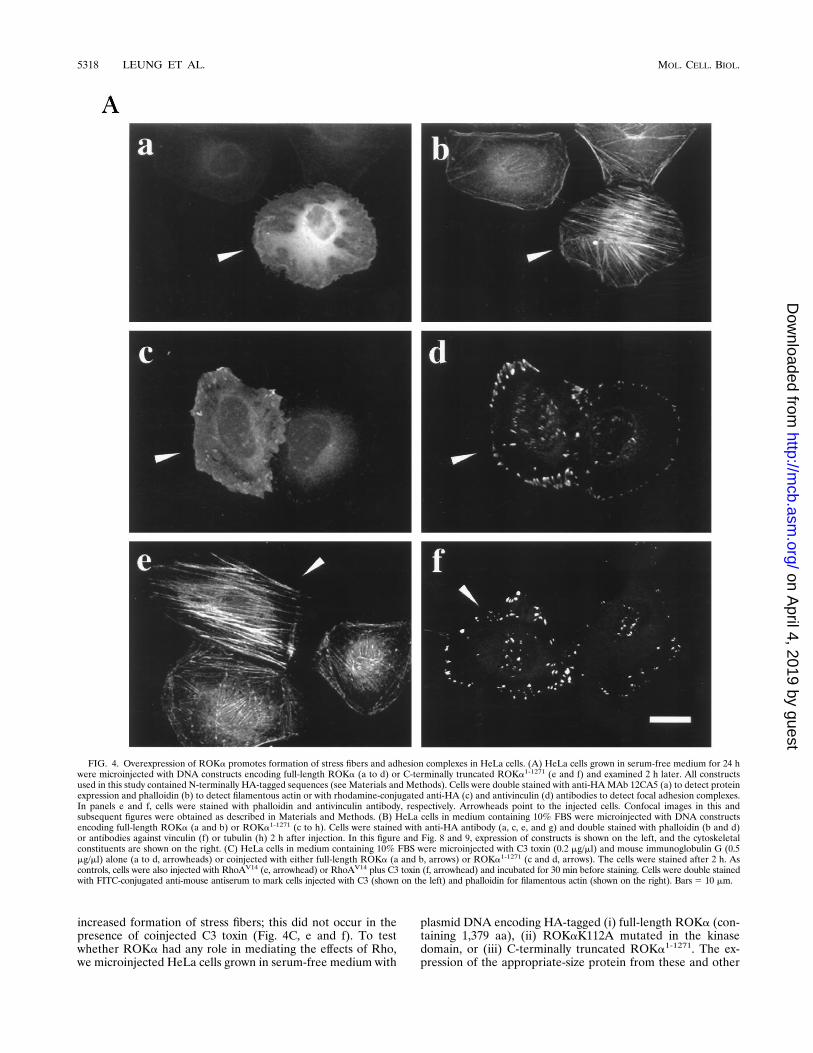
FIG. 4. Overexpression of ROKa promotes formation of stress fibers and adhesion complexes in HeLa cells. (A) HeLa cells grown in serum-free medium for 24 hwere microinjected with DNA constructs encoding full-length ROKa (a to d) or C-terminally truncated ROKa1-1271 (e and f) and examined 2 h later. All constructsused in this study contained N-terminally HA-tagged sequences (see Materials and Methods). Cells were double stained with anti-HAMAb 12CA5 (a) to detect proteinexpression and phalloidin (b) to detect filamentous actin or with rhodamine-conjugated anti-HA (c) and antivinculin (d) antibodies to detect focal adhesion complexes.In panels e and f, cells were stained with phalloidin and antivinculin antibody, respectively. Arrowheads point to the injected cells. Confocal images in this andsubsequent figures were obtained as described in Materials and Methods. (B) HeLa cells in medium containing 10% FBS were microinjected with DNA constructsencoding full-length ROKa (a and b) or ROKa1-1271 (c to h). Cells were stained with anti-HA antibody (a, c, e, and g) and double stained with phalloidin (b and d)or antibodies against vinculin (f) or tubulin (h) 2 h after injection. In this figure and Fig. 8 and 9, expression of constructs is shown on the left, and the cytoskeletalconstituents are shown on the right. (C) HeLa cells in medium containing 10% FBS were microinjected with C3 toxin (0.2 mg/ml) and mouse immunoglobulin G (0.5mg/ml) alone (a to d, arrowheads) or coinjected with either full-length ROKa (a and b, arrows) or ROKa1-1271 (c and d, arrows). The cells were stained after 2 h. Ascontrols, cells were also injected with RhoAV14 (e, arrowhead) or RhoAV14 plus C3 toxin (f, arrowhead) and incubated for 30 min before staining. Cells were double stainedwith FITC-conjugated anti-mouse antiserum to mark cells injected with C3 (shown on the left) and phalloidin for filamentous actin (shown on the right). Bars5 10 mm.
5318 LEUNG ET AL. MOL. CELL. BIOL.
on April 4, 2019 by guest
http://mcb.asm
.org/D
ownloaded from

FIG. 4—Continued.
VOL. 16, 1996 Rho-BINDING KINASES AND CYTOSKELETON ORGANIZATION 5319
on April 4, 2019 by guest
http://mcb.asm
.org/D
ownloaded from

constructs used in this study was confirmed by in vitro tran-scription-translation assay and by analysis of COS-7 cells trans-fected with these same constructs (see Fig. 9B). Within 1 to 2 hof the injection, stress fibers had formed in cells injected withDNA encoding ROKa (Fig. 4A, a and b), accompanied byincreases in vinculin staining (Fig. 4A, c and d), indicative ofincreased focal adhesion complexes. These effects were notseen in cells injected with cDNA encoding kinase-dead mutantROKaK112A (Fig. 5). Injection of DNA encoding C-termi-nally truncated ROKa1-1271 resulted in more extensive forma-tion of stress fibers (Fig. 4A, e) and focal adhesion complexes(Fig. 4A, f).The amount of stress fibers in HeLa cells growing in medium
containing 10% FBS (which already contain some stress fibers)could also be increased upon injection of DNA encoding full-
length ROKa (Fig. 4B, a and b). Injection of DNA encodingmutant ROK1-1271 into these cells again resulted in much moreextensive formation of stress fibers. These fibers then appearedto coalesce, leading to a gross condensation of actin filamentswithin the center of the cell, with some filaments still visiblyattached to the cell periphery, and also nuclear disorganization(Fig. 4B, c and d). There were also increased concentrations offocal adhesion complexes (Fig. 4B, e and f). Microtubules werenot affected (Fig. 4B, g and h). The effects of both ROKa andROKa1-1271 on stress fiber formation occurred even in thepresence of C3 toxin (Fig. 4C, a to d), indicating that theseproteins acted downstream of Rho.To identify the specific domain(s) responsible for the for-
mation of stress fibers and focal adhesion complexes, we testedvarious mutants of ROKa for their effects on serum-starved
FIG. 4—Continued.
5320 LEUNG ET AL. MOL. CELL. BIOL.
on April 4, 2019 by guest
http://mcb.asm
.org/D
ownloaded from

cells (Fig. 5). Formation of these cellular structures could bepromoted by a short fragment only of ROKa containing theN-terminal portion of kinase domain (i.e., ROKa1-543). Boththe intact kinase domain and the proximal N-terminal regionwere required, since ROKaK112A and the N-terminally trun-cated mutant ROKa78-1379 were ineffective in this regard. Un-der these conditions of overexpression, the RhoA-binding do-main was not essential for formation of stress fibers, with themutants deficient in Rho binding or lacking the binding do-main being effective. The C-terminally truncated mutants wereall observed to promote formation of more stress fibers andfocal adhesion complexes than the full-length ROKa.The kinase activities of these proteins were analyzed by
assaying immunoprecipitated HA-tagged ROKa and mutantsfrom extracts of COS-7 cells transfected with the appropriateDNA constructs. Wild-type (full-length) ROKa exhibited au-tophosphorylating and kinase activities toward MBP as waspreviously observed with native ROKa (18). Coexpression withRhoAV14 resulted in only a slight increase in ROKa activ-ity (Fig. 6). The ROKaK112A mutant exhibited very littlekinase activity, as did the N-terminally truncated mutantROKa78-1379. The N-terminus appears to be essential for ki-nase activity. On the other hand, the C-terminally truncatedROKa1-1271 and ROKa1-543 displayed markedly increased ki-nase activity against MBP. The levels of kinase activities of thevarious proteins thus appeared to correlate with their morpho-logical effects (Fig. 5).The N-terminally truncated ROKa78-1379 induces loss of stress
fibers and focal adhesion complexes. Rather than promotingtheir formation, we found that the truncated ROKa78-1379 in-duced disappearance of both stress fibers (Fig. 7a and b) andfocal adhesion complexes (Fig. 7e and f) upon injection of itsDNA. DNA encoding the kinase domain without the N termi-nus, ROK78-398, was ineffective, with the stress fibers (Fig. 7cand d) and focal adhesion complexes (Fig. 7g and h) remainingunaffected. The effect of ROKa78-1379 was specific to actinmicrofilaments, as microtubules were not affected (Fig. 7i and j).Loss of stress fibers is accompanied by rapid cell spreading.
The loss of stress fibers and focal complexes induced by over-expression of ROKa78-1379 was accompanied by a rapid in-crease in cell size, probably as a result of cell flattening andspreading (Fig. 8a and b). These effects were not confined toHeLa cells, as Swiss 3T3 fibroblasts were similarly affected
FIG. 5. Analysis of ROKa domains involved in formation of stress fibers and focal adhesion complexes. HeLa cells in serum-free medium were microinjected withDNA constructs encoding ROKa and various mutants. Numbers refer to the N-terminal and C-terminal amino acid residues of the products encoded by the constructs,synthesized as described in Materials and Methods. XX denotes the NK-to-TT mutations (aa 1027 and 1028) in the Rho-binding domain (see Fig. 3C). X marks theposition of the K-to-A (aa 112) mutation in the kinase domain. The morphology of the injected cells was analyzed as described for Fig. 4. The effects of themicroinjection on formation of stress fibers and focal adhesion complexes are shown on the right, with the relative intensity denoted by the number of plus signs.
FIG. 6. Protein kinase activities of expressed ROKa and mutants. HA-tagged proteins were immunoprecipitated from COS-7 cells transfected withvarious HA-tagged DNA constructs. Kinase activity and Western blotting anal-yses were performed as described in Materials and Methods, using [g-33P]ATPand a rabbit anti-ROK antibody, respectively. The arrowhead indicates theposition of the immunoprecipitated protein. MBP was used as a kinase substrate.In the lower panel, MBP phosphorylation by each construct was determined witha Molecular Dynamics PhosphorImager, and the results are presented as meansand standard errors from at least three separate experiments. The activity ofwild-type (W/T) ROKa was taken as 1. Sizes are indicated in kilodaltons.
VOL. 16, 1996 Rho-BINDING KINASES AND CYTOSKELETON ORGANIZATION 5321
on April 4, 2019 by guest
http://mcb.asm
.org/D
ownloaded from

FIG. 7. Effects of overexpression of N-terminally truncated ROKa78-1379 on stress fibers, focal adhesion complexes, and tubulin. HeLa cells in 10% FBS weremicroinjected with DNA encoding ROKa78-1379 (a, b, e, f, i, and j) and with DNA encoding only kinase domain ROKa78-398 (c, d, g, and h) and examined 2 h later.Protein expression was detected with either MAb 12CA5 (a and c) or rhodamine-conjugated anti-HA antibodies (e, g, and i). Cells were double stained with phalloidin(b and d), antivinculin antibody (f and h), and antitubulin antibody (j). Bar 5 10 mm.
5322
on April 4, 2019 by guest
http://mcb.asm
.org/D
ownloaded from

FIG. 8. Overexpression of N-terminally truncated ROKa78-1379 causes cell spreading. HeLa cells in 10% FBS were injected with DNA constructs encoding ROKa78-1379
(a to d), ROKa78-1379 coinjected with Rac1N17 cDNA (e to f), C3 transferase and GST (g and h), or RhoAN19 (i and j). Expression of ROKa78-1379 and RhoAN19 was detectedwith anti-HA MAb 12CA5 (a, c, e, and i). In panel g, rabbit anti-GST–FITC-conjugated anti-rabbit secondary antibody was used. Cells were fixed 2 h (a, b, e, f, i, and j), 4 h(c and d), or 30 min (cells injected with C3 transferase [g and h]) after injection. Fixed cells were double stained with phalloidin (b, d, f, h, and j). Bar5 10 mm.
5323
on April 4, 2019 by guest
http://mcb.asm
.org/D
ownloaded from

(result not shown). Prolonged overexpression (3 to 4 h) ortransient transfection (results not shown) led to the collapsingof plasma membranes onto the nucleus, resulting in a roundedcell body with irregular branches (Fig. 8c and d). These cellswere still viable, as assessed by trypan blue exclusion (data notshown). Because Cdc42Hs, Rac1, and RhoA may operate withina suggested hierarchy (15, 29), the ROKa78-1379 mutant, by inter-fering with normal RhoA function (i.e., formation of stress fi-bers), could conceivably enhance the function of Rac1, perhapsthe GTPase responsible for the cell spreading. This possibility wastested by coinjecting DNAs for truncated ROKa78-1379 and forthe dominant negative Rac1N17 mutant, which blocks the effectsof endogenous Rac1 (36). ROKa78-1379 still induced cell spread-ing (Fig. 8e and f), showing that the effect is independent of Rac1.The direct inhibition of Rho by C3 transferase (Fig. 8g and h) andthe putative dominant negative RhoAN19 mutant (Fig. 8i and j)resulted in stress fiber loss, although only C3 induced some cellspreading.
The PH/CRD motif is essential for loss of stress fibers, lossof focal adhesion complexes, and cell spreading. To determinewhich particular region of ROKa was involved in inducing lossof the cellular structures and the cell spreading effect, severaldeletion and point mutation constructs of ROKa were tested.The deletion constructs and a summary of their effects areshown in Fig. 9A. Appropriate-size polypeptides were ex-pressed upon transient transfection of the constructs intoCOS-7 cells, as detected by antibodies against the HA tag (Fig.9B) or ROKa (data not shown). Microinjection of these con-structs into HeLa cells showed that kinase-dead ROKaK112Ainduced losses of stress fibers and focal adhesion complexes(Fig. 9C, a and b) as well as the N-terminally truncatedROKa78-1379. No such effect occurred if the C terminus wastruncated within the PH domain (Fig. 9A, e.g., ROKa78-1271).A short fragment containing the C-terminal Rho-binding andPH/CRD motif, ROKa971-1379, was extremely effective in in-ducing the losses (Fig. 9C, c and d) and associated cell spread-
FIG. 9. The PH/CRD motif is responsible for disassembly of stress fibers and focal adhesion complexes and for cell spreading. (A) HeLa cells in 10% FBS weremicroinjected with DNA constructs encoding ROKa and its mutants. Morphology was analyzed 2 h later as described for Fig. 6. Number designations are as in Fig.5. Synthesis of the various DNA constructs is described in Materials and Methods. The effects on stress fibers and on cell spreading are summarized on the right, withthe relative intensity denoted by increasing the number of plus signs. 6 indicates that cell spreading is found in about 50% of the injected cells. (B) Protein expressionfrom various DNA constructs. Plasmid DNA (1 mg) corresponding to constructs shown in panel A was used for transient transfection of COS-7 cells with Lipofectamine(Gibco/BRL). At 16 h after transfection, cells were harvested in lysis buffer and 100-mg aliquots of the soluble extracts were separated on SDS–12% polyacrylamidegels. After transfer onto nitrocellulose, the blot was immunostained with MAb 12CA5. (C) Effects of kinase-dead mutant and C-terminal fragments of ROKa. HeLacells microinjected with DNA encoding ROKaK112A (a and b), ROKa971-1379 (c and d), and ROKa1110-1379 (e and f) were stained with either phalloidin (a, c, ande) or antivinculin antibody (b, d, and f) 2 h later. Arrowheads point to the injected cells. Bar 5 10 mm.
5324 LEUNG ET AL. MOL. CELL. BIOL.
on April 4, 2019 by guest
http://mcb.asm
.org/D
ownloaded from

ing (Fig. 9A). ROKa1110-1379, containing only the PH/CRDmotif, also induced some loss of the structures (Fig. 9B, e andf), but this was accompanied by cell spreading in only about50% of the injected cells (Fig. 9A).
DISCUSSION
Our earlier work uncovered the presence of ubiquitous pro-teins of ;150-kDa interacting with RhoA (22). One of theseproteins was subsequently isolated and characterized as beinga novel serine/threonine kinase termed ROKa (18). From ourimmunological and mRNA analyses (Fig. 2), we find that the;150-kDa kinases exist as a heterogenous population, withdiverse constituents being present in different tissues, consis-tent with their being expressed from a number of related genes.The identities and relationship of two of these kinases (ROKa
and ROKb) have now been established through isolation oftheir cDNAs (Fig. 1). Alignment of the ROKa and ROKbprotein sequences allowed us to pinpoint a short stretch of 20to 30 aa in both kinases responsible for the interaction withRhoA, -B, and -C. The binding of all three Rho family mem-bers is not unexpected, as these p21s have between 85 and 90%sequence identity. Mutational analysis of some critical residuesshowed this region to be absolutely essential for specific Rhointeraction. This region bears no resemblance to the previouslyreported p21-binding domain of PAK65 and related proteins(22), which specifically interacts with Rac1 and Cdc42 but notRhoA.These Rho-binding kinases belong to a family of related
serine/threonine kinases which includes the myotonic dystro-phy kinase, implicated in the etiology of neuromuscular degen-
FIG. 9—Continued.
VOL. 16, 1996 Rho-BINDING KINASES AND CYTOSKELETON ORGANIZATION 5325
on April 4, 2019 by guest
http://mcb.asm
.org/D
ownloaded from

eration (4, 8) probably as a result of its involvement in ionchannel function (26), the fungal cot-1-encoded kinase re-quired for controlling filamentous growth (41), and the re-cently reported Drosophila anti-oncogene warts gene product,which is essential for controlling cell growth and morphologyduring development (13). We have also isolated several ;180-kDa proteins containing related kinase domains which do notbind Rho but instead bind Cdc42 and Rac1 (17). In ROKa andROKb, there are other highly conserved regions, including theCRD, which lies within the region containing the PH domain,apart from the kinase domain. This family of multidomainedROKs has also been characterized by others (11, 23) whosefindings that stimulation of kinase activity toward MBP byRhoA is less than fivefold broadly concur with our results (Fig.6). This level of GTPase stimulation is very much less than thatobserved with PAK (19, 22).Kinase activity appears to be essential for ROKa to promote
the formation of stress fibers and focal adhesion complexes.There was a direct relationship between the level of kinaseactivity and the extent of the morphological effects; mutantslacking kinase activity caused no stress fibers, while mutantsexhibiting increased kinase activities promoted more stressfibers than wild-type ROKa (Fig. 5 and 6). However, the hy-peractive mutants, e.g., ROKa1-1271, appeared to exert unreg-ulated effects on actin microfilaments, with large-scale forma-tion of stress fibers leading to condensation centrally of actinfibers and also nuclear disorganization. Both the extreme N-and C-terminal regions are necessary for the proper regulationof ROKa kinase and morphological activities, with the N- andC-terminal regions apparently assuming positive and negativeregulatory roles, respectively. Thus, N-terminal deletion resultsin the loss of kinase and stress fiber-forming activities, whileC-terminal deletion leads to enhanced kinase activity andgreater effects on stress fibers.In our studies involving the expression of ROKa from in-
troduced DNA, the binding domain did not appear to be es-sential for ROKa to promote formation of stress fibers andfocal adhesion complexes. It is possible that overexpression ofexogenous ROKa, which generated an active kinase, had alsooverridden its need to bind RhoA to function appropriately.Kinase activity is also not substantially increased upon coex-pression of activated RhoAV14, which is consistent with RhoAnot markedly stimulating native ROK activity in vitro (18).Since introduction of RhoAV14 leads to translocation of solu-ble ROKa to peripheral membranes (18), perhaps in cellsexpressing normal levels of ROK and Rho, activated Rho isrequired to direct the kinase to its appropriate destination. Inepithelial cells as in fibroblasts, RhoA promotes stress fiberformation which is inhibited by C3 toxin. Nevertheless, priorinjection of C3 did not prevent ROKa from promoting stressfibers. However, the kinase-dead ROKaK112A mutant causedloss of stress fibers (present in HeLa cells in 10% FBS) (Fig.9A and C), behaving as a putative dominant negative inhibitorof endogenous ROK (see below). These results taken togetherindicate that ROKa acts downstream of Rho in the Rho-dependent pathway of stress fiber formation. The exact role ofthe Rho-binding domain remains to be established.The ROKamutants lacking kinase activity induced morpho-
logical changes opposite those promoted by intact ROKa, suchas the disassembly of stress fibers and focal adhesion com-plexes. One plausible explanation is that these mutants inter-fered with the endogenous ROKs responsible for formation ofpreexisting stress fibers. This could occur through the mutantsacting either as dominant negative mutants (by binding to andblocking the downstream targets of ROKs, e.g., kinase-deadmutant) or as direct inhibitors of the ROKs themselves (e.g.,
the C-terminal mutants which could act as negative regulatorsof kinase activity; see above). The C-terminal fragment con-taining only the PH domain and CRD (both of which have thepotential to interact with membranes [2, 9]) induced disassem-bly. The inclusion of the Rho-binding domain potentiated thiseffect, perhaps by facilitating the translocation of the inhibitoryfragment to peripheral membrane sites (18) where endogenousROKa operate.The assembly and disassembly of focal adhesion complexes
and concomitant cell spreading have been implicated in migra-tory activities of squamous cell carcinoma cells (24). Treatmentof these cells with hepatocyte growth factor/scatter factor re-sulted in a biphasic morphological response, with the initial cellspreading being followed by transformation into a spindle-shape phenotype whose motility was sharply increased within1.5 to 2 h of treatment. This response resembles that elicitedwith the inhibitory fragments of ROKa, and it would be ofinterest to determine if inhibition of ROKs which are ubiqui-tous precedes cell spreading and whether cell spreading of thesquamous cells and that induced in HeLa cells are relatedevents. In addition to ROKa, other kinases such as the tyrosinekinases downstream of Rho (35) participate in the formationof stress fibers and focal adhesion complexes. Some of themorphological effects of other Rho family members are ex-erted through nuclear events (7, 10, 25) which are mediated bykinase cascades. Thus, in the yeast pheromone mating path-way, Cdc42 participates in the mitogen-activated protein ki-nase pathway (37, 44). In mammalian cells, Rac1 and Cdc42are linked to the c-JNK/mitogen-activated protein kinase cas-cade (7, 25), while RhoA appears to be involved in a separatenuclear event leading to the activation of a serum-responsivefactor-linked pathway (10). The specific interaction of ROKswith RhoA, -B, and -C only in their GTP-bound forms and theability of ROKa to promote stress fiber formation are consis-tent with these kinases being target and effector proteins. Thepresence of multiple domains in ROKs capable of interactingwith other signalling and cytoskeletal components (and someperhaps having discrete functions) clearly endows them withthe ability to mediate the various roles of the Rho GTPases incellular functions as diverse as apoptosis (12) and transforma-tion (32).
ACKNOWLEDGMENTS
We thank Lydia Tan, Ivan Tan, and Ivy Ho for expert technicalassistance; Ben Li for oligonucleotide synthesis; and Francis Leong forphotographic reproduction.This work was supported by the Glaxo Singapore Research Fund.
REFERENCES1. Adamson, P., H. F. Paterson, and A. Hall. 1992. Intracellular localization ofthe p21rho proteins. J. Cell Biol. 119:617–627.
2. Ahmed, S., R. Kozma, J. Lee, C. Monfries, N. Harden, and L. Lim. 1991. Thecysteine-rich domain of human proteins, neuronal chimaerin, protein kinaseC and diacylglycerol kinase binds zinc. Biochem. J. 280:233–241.
3. Bowman, E. P., D. J. Uhlinger, and J. D. Lambeth. 1993. Neutrophil phos-pholipase D is activated by a membrane-associated Rho family small mo-lecular weight GTP-binding protein. J. Biol. Chem. 268:21509–21512.
4. Brook, J. D., M. E. McCurrach, H. G. Harley, A. J. Buckler, D. Church, H.Aburatani, K. Hunter, V. P. Stanton, J.-P. Thirion, T. Hudson, R. Sohn, B.Zemelman, R. G. Snell, S. A. Rundle, S. Crow, J. Davies, P. Shelbourne, J.Buxton, C. Jones, V. Juvonen, K. Johnson, P. S. Harper, D. J. Shaw, andD. E. Housman. 1992. Molecular basis of myotonic dystrophy: expansion ofa trinucleotide (CTG) repeat at the 39 end of a transcript encoding a proteinkinase family member. Cell 68:799–808.
5. Chardin, P., P. Madaule, and A. Tavitian. 1988. Coding sequence of humanrho cDNAs clone 6 and clone 9. Nucleic Acids Res. 16:2717.
6. Chong, L. D., A. Traynor-Kaplan, G. M. Bokoch, and M. A. Schwartz. 1994.The small GTP-binding protein Rho regulates a phosphatidylinositol 4-phos-phate 5-kinase in mammalian cells. Cell 79:507–513.
7. Coso, O. A., M. Cheariello, J.-C. Yu, H. Teramoto, P. Crespo, N. Xu, T. Miki,
5326 LEUNG ET AL. MOL. CELL. BIOL.
on April 4, 2019 by guest
http://mcb.asm
.org/D
ownloaded from

and J. S. Gutkind. 1995. The small GTP-binding protein Rac1 and Cdc42regulate the activity of the JNK/SAKP signaling pathway. Cell 81:1137–1146.
8. Fu, Y.-H., A. Pizzuti, R. G. Fenwick, Jr., J. King, S. Rajnarayan, P. W.Dunne, J. Dubel, G. A. Nasser, T. Ashizawa, P. de Jong, B. Wieringa, R.Korneluk, M. B. Perryman, H. F. Epstein, and C. T. Caskey. 1992. Anunstable triplet repeat in a gene related to myotonic muscular dystrophy.Science 255:1256–1258.
9. Harlan, J. E., H. S. Yoon, P. J. Hajduk, and S. W. Fesik. 1995. Structuralcharacterization of the interaction between a pleckstrin homology domainand phosphatidylinositol 4,5-bisphosphate. Biochemistry 34:9859–9864.
10. Hill, C. S., J. Wynne, and R. Treisman. 1995. The Rho family GTPasesRhoA, Rac1, and Cdc42Hs regulate transcriptional activation by SRF. Cell81:1159–1170.
11. Ishizaki, T., M. Maekawa, K. Fujisawa, K. Okawa, A. Iwamatsu, A. Fujita,N. Watanabe, Y. Saito, A. Kakizuka, N. Morii, and S. Narumiya. 1996. Thesmall GTP-binding protein Rho binds to and activates a 160 kDa Ser/Treprotein kinase homologous to myotonic dystrophy kinase. EMBO J. 15:1885–1893.
12. Jimenez, B., M. Arends, P. Esteve, R. Perona, R. Sanchez, S. R. Cahal, A.Wyllie, and J. C. Lacal. 1995. Induction of apoptosis in NIH3T3 cells afterserum deprivation by overexpression of rho-p21, a GTPase protein of the rassuperfamily. Oncogene 10:811–816.
13. Justice, R. W., O. Zilian, D. F. Woods, M. Noll, and P. J. Bryant. 1995. Thedrosophila tumour suppressor gene warts encodes a homolog of humanmyotonic dystrophy kinase and is required for the control of cell shape andproliferation. Genes Dev. 9:534–546.
13a.Kaibuchi, K., and S. Narumiya. Personal communication.14. Kishi, K., T. Sasaki, S. Kuroda, T. Itoh, and Y. Takai. 1993. Regulation of
cytoplasmic division of Xenopus embryo by rhop21 and its inhibitory GDP/GTP exchange protein (rho GDI). J. Cell Biol. 120:1187–1195.
15. Kozma, R., S. Ahmed, A. Best, and L. Lim. 1995. The Ras-related proteinCdc42Hs and bradykinin promote formation of peripheral actin microspikesand filopodia in Swiss 3T3 fibroblasts. Mol. Cell. Biol. 15:1942–1952.
16. Leung, T., B.-E. How, E. Manser, and L. Lim. 1993. Germ cell b-chimaerin,a new GTPase-activating protein for p21rac, is specifically expressed duringthe acrosomal assembly stage in rat testis. J. Biol. Chem. 268:3813–3816.
17. Leung, T., E. Manser, and L. Lim. Unpublished data.18. Leung, T., E. Manser, L. Tan, and L. Lim. 1995. A novel serine/threonine
kinase binding the ras-related RhoA GTPase which translocates the kinaseto peripheral membranes. J. Biol. Chem. 256:29051–29054.
19. Manser, E., C. Chong, Z.-S. Zhao, T. Leung, G. Michael, C. Hall, and L.Lim. 1995. Molecular cloning of a new member of the p21-Cdc42/Rac-activated kinase (PAK) family. J. Biol. Chem. 270:25070–25078.
20. Manser, E., T. Leung, C. Monfries, M. Teo, C. Hall, and L. Lim. 1992.Diversity and versatility of GTPase activating proteins for the p21rho sub-family of ras G proteins detected by a novel overlay assay. J. Biol. Chem.267:16025–16028.
21. Manser, E., T. Leung, H. Salihuddin, L. Tan, and L. Lim. 1993. A non-receptor tyrosine kinase that inhibits the GTPase activity of p21cdc42. Na-ture (London) 363:364–367.
22. Manser, E., T. Leung, H. Salihuddin, Z.-S. Zhao, and L. Lim. 1994. A brainserine/threonine protein kinase activated by Cdc42 and Rac1. Nature (Lon-don) 367:40–46.
23. Matsui, T., M. Amano, T. Yamamoto, K. Chihara, M. Nakafuku, M. Ito, T.Nakano, K. Okawa, A. Iwamatsu, and K. Kaibuchi. 1996. Rho-associatedkinase, a novel serine/threonine kinase, as a putative target for the smallGTP binding protein Rho. EMBO J. 15:2208–2216.
24. Matsumoto, K., K. Matsumoto, T. Nakamura, and R. H. Kramer. 1994.Hepatocyte growth factor/scatter factor induces tyrosine phosphorylation offocal adhesion kinase (p125FAK) and promotes migration and invasion byoral squamous cell carcinoma cells. J. Biol. Chem. 269:31807–31813.
25. Minden, A., A. Lin, F.-X. Claret, A. Abo, and M. Karin. 1995. Selectiveactivation of the JNK signaling cascade and c-Jun transcriptional activity bythe small GTPases Rac and Cdc42Hs. Cell 81:1147–1157.
26. Mounsey, J. P., P. Xu, J. E. John, L. T. Horne, J. Gilbert, A. D. Roses, andJ. R. Moorman. 1995. Modulation of skeletal muscle sodium channels byhuman myotonin protein kinase. J. Clin. Invest. 95:2379–2384.
27. Musacchio, A., T. Gibson, P. Rice, J. Thompson, and M. Saraste. 1993. ThePH domain: a common piece in the structural patchwork of signalling pro-teins. Trends Biochem. Sci. 19:343–348.
28. Nishiyama, T., T. Sasaki, K. Takaishi, M. Kato, H. Yaku, K. Araki, Y.Matsuura, and Y. Takai. 1994. rac p21 is involved in insulin-induced mem-brane ruffling and rho is involved in hepatocyte growth factor- and 12-O-tetradecanoylphorbol-13-acetate-induced membrane ruffling in KB cells.Mol. Cell. Biol. 14:2447–2456.
29. Nobes, C. D., and A. Hall. 1995. Rho, Rac, and Cdc42 GTPases regulate theassembly of multimolecular focal complexes associated with actin stressfibers, lamellipodia, and filopodia. Cell 81:53–62.
30. Paterson, H. F., A. J. Self, M. D. Garrett, I. Just, K. Atkories, and A. Hall.1990. Microinjection of recombinant p21rho induces rapid change in cellmorphology. J. Cell Biol. 111:1001–1007.
31. Perona, R., P. Esteve, B. Jimenez, R. P. Ballestero, S. R. Cajal, and J. C.Lacal. 1993. Tumorigenic activity of rho genes from Aplysia california. On-cogene 8:1285–1292.
32. Prendergast, G. C., R. Khosravi-Far, P. A. Solske, H. Kurzawa, P. F. Leb-owitz, and C. J. Der. 1995. Critical role of Rho in cell transformation byoncogenic Ras. Oncogene 10:2289–2296.
33. Ridley, A. J. 1995. Rho-related proteins: actin cytoskeleton and cell cycle.Curr. Opin. Genet. Dev. 5:24–30.
34. Ridley, A. J., and A. Hall. 1992. The small GTP-binding protein rho regulatesthe assembly of focal adhesion and actin stress fibres in response to growthfactors. Cell 70:389–399.
35. Ridley, A. J., and A. Hall. 1994. Signal transduction pathways regulatingrho-mediated stress fiber formation: requirement for a tyrosine kinase.EMBO J. 13:2600–2610.
36. Ridley, A. J., H. F. Paterson, C. L. Johnston, D. Diekmann, and A. Hall.1992. The small GTP-binding protein rac regulates growth factor inducedmembrane ruffling. Cell 70:401–410.
37. Simon, M.-N., C. de Virgillo, B. Souza, J. R. Pringle, A. Abo, and S. I. Reed.1995. Role for the Rho-family GTPase Cdc42 in yeast mating pheromonesignal pathway. Nature (London) 376:702–705.
38. Takai, Y., T. Sasaki, K. Tanaka, and H. Nakanishi. 1995. Rho as a regulatorof the cytoskeleton. Trends Biochem. Sci. 20:227–231.
39. Takaishi, K., A. Kikuchi, S. Kuroda, K. Kotani, T. Sasaki, and Y. Takai.1993. Involvement of rhop21 and its inhibitory GDP/GTP exchange protein(rhoGDI) in cell motility. Mol. Cell. Biol. 13:72–79.
40. Xiao, J. H., I. Davidson, H. Matthes, J.-M. Garnier, and P. Chambon. 1991.Cloning, expression and transcriptional properties of the human enhancerfactor TEF-1. Cell 65:551–568.
41. Yarden, O., M. Plamann, D. J. Ebbole, and C. Yanofsky. 1992. cot-1, a generequired for hyphal elongation in Neurospora crassa, encodes a proteinkinase. EMBO J. 11:2159–2166.
42. Zalcman, G., V. Glosson, G. Linares-Cruz, F. Lerebours, N. Hovore, A.Taritain, and B. Olofsson. 1995. Regulation of ras-related RhoB proteinexpression during the cell cycle. Oncogene 10:1935–1945.
43. Zhang, J., W. G. King, S. Dillon, A. Hall, L. Feig, and S. E. Rittenhouse.1993. Activation of platelet phosphatidylinositide 3-kinase requires the smallGTP-binding protein Rho. J. Biol. Chem. 268:22251–22254.
44. Zhao, Z.-S., T. Leung, E. Manser, and L. Lim. 1995. Pheromone signallingin Saccharomyces cerevisiae requires the small GTP-binding protein Cdc42pand its activator CDC24. Mol. Cell. Biol. 15:5246–5257.
VOL. 16, 1996 Rho-BINDING KINASES AND CYTOSKELETON ORGANIZATION 5327
on April 4, 2019 by guest
http://mcb.asm
.org/D
ownloaded from