THE OUTWANDERING OF CELLS IN TISSUE CULTURES...
18
266 THE OUTWANDERING OF CELLS IN TISSUE CULTURES OF NERVES UNDERGOING WALLERIAN DEGENERATION BY M. ABERCROMBIE* Department of Anatomy, University of Birmingham AND M. L. JOHNSON Department of Zoology, University of Birmingham (Received 24 August 1 <}•]?•) (With One Text-figure) INTRODUCTION When a nerve is sectioned the whole peripheral stump undergoes Wallerian degeneration, and a small part of the central stump near the cut also shows degenerative changes. While axons and myelin degenerate during this process the Schwann (or sheath) cells proliferate and in the peripheral stump become arranged in the well-known 'bands of Biingner'. The endoneurial fibroblasts also proliferate. A study of the outwandering from explants of degenerating nerves in tissue culture should throw light on the changes in Schwann cells and fibroblasts, the physiology of which is little understood. It may also be of practical importance in nerve surgery, for it seems that outward migration of the Schwann cells from the cut surfaces of the nerve, which will be reflected by their outwandering in vitro, plays an important part in ensuring adequate healing (Young, 1942). Several workers have grown Schwann cells in vitro, notably Ingebrigtsen (1916), Chlopin (1939) and Murray, Stout & Bradley (1940). Of these only Ingebrigtsen was concerned with the changes in the cells during Wallerian degeneration. He studied the peripheral stump during the first 19 days of degeneration, using as a criterion of activity the percentage of explants which showed any outwandering. Our work may be considered as an expansion of Ingebrigtsen's suggestive beginning. We have investigated regions of both stumps, degenerated for periods of up to a year, and we have used for estimating activity a method which is more easily interpreted than that of Ingebrigtsen. The present paper is a general survey of quantitative differences of cell outwandering in vitro between various regions of the severed nerve and between nerves at different times after they have been severed. We are much indebted to Mr J. Z. Young for suggesting that we should undertake this investigation, and for criticizing the manuscript. TECHNIQUE The explants were taken from the sciatic nerve of adult rabbits. In most of the experi- ments the peroneal branch was used; in a few, the tibial branch. There was no significant difference of behaviour of the two branches. At an initial operation a piece of varying • Beit Memorial Research Fellow.
Transcript of THE OUTWANDERING OF CELLS IN TISSUE CULTURES...
266
THE OUTWANDERING OF CELLS IN TISSUECULTURES OF NERVES UNDERGOING
WALLERIAN DEGENERATION
BY M. ABERCROMBIE*Department of Anatomy, University of Birmingham
AND
M. L. JOHNSONDepartment of Zoology, University of Birmingham
(Received 24 August 1 <}•]?•)
(With One Text-figure)
INTRODUCTION
When a nerve is sectioned the whole peripheral stump undergoes Wallerian degeneration,and a small part of the central stump near the cut also shows degenerative changes. Whileaxons and myelin degenerate during this process the Schwann (or sheath) cells proliferateand in the peripheral stump become arranged in the well-known 'bands of Biingner'.The endoneurial fibroblasts also proliferate. A study of the outwandering from explantsof degenerating nerves in tissue culture should throw light on the changes in Schwanncells and fibroblasts, the physiology of which is little understood. It may also be ofpractical importance in nerve surgery, for it seems that outward migration of the Schwanncells from the cut surfaces of the nerve, which will be reflected by their outwanderingin vitro, plays an important part in ensuring adequate healing (Young, 1942).
Several workers have grown Schwann cells in vitro, notably Ingebrigtsen (1916),Chlopin (1939) and Murray, Stout & Bradley (1940). Of these only Ingebrigtsen wasconcerned with the changes in the cells during Wallerian degeneration. He studied theperipheral stump during the first 19 days of degeneration, using as a criterion of activitythe percentage of explants which showed any outwandering. Our work may be consideredas an expansion of Ingebrigtsen's suggestive beginning. We have investigated regionsof both stumps, degenerated for periods of up to a year, and we have used for estimatingactivity a method which is more easily interpreted than that of Ingebrigtsen. The presentpaper is a general survey of quantitative differences of cell outwandering in vitro betweenvarious regions of the severed nerve and between nerves at different times after theyhave been severed. We are much indebted to Mr J. Z. Young for suggesting that weshould undertake this investigation, and for criticizing the manuscript.
TECHNIQUE
The explants were taken from the sciatic nerve of adult rabbits. In most of the experi-ments the peroneal branch was used; in a few, the tibial branch. There was no significantdifference of behaviour of the two branches. At an initial operation a piece of varying
• Beit Memorial Research Fellow.
Wallerian 'degeneration 267
•ngth was cut from the nerye with sharp scissors in the thigh region at about the levelof the third trochanter of the femur. After a varying interval of time we made a secondoperation and removed lengths of the central and peripheral stumps of the nerve forculture. In each experiment we cultured a number of explants from each of severaldifferent parts of the stumps of the severed nerve, as listed below.
In most experiments our only provision to prevent reinnervation of the peripheralstump was to make the gap between stumps large; but when nerves were left to degeneratea long time the possibility that reinnervation had occurred was investigated by treatinghistological sections by Bodian's method for staining of axons. In none of the peripheralstumps used for this paper were more than a very few isolated axons found.
In some experiments the central stump was killed at the initial operation to ensure thatit did not reinnervate the peripheral stump. This was done by injecting the central stumpwith a 1 % aqueous solution of crystal violet (a method of axon destruction used byGuttmann & Medawar, 1942). The method was not entirely suitable for our purpose,because we found that the proximal part of the peripheral stump was sometimes a littledamaged.
The ordinary hanging-drop method of tissue culture was used. Two explants wereplaced on each cover-slip. The medium consisted of fowl plasma and chick embryoextract. Through most of the series of experiments we made the extract from a driedembryo brei (see Peacock & Shukoff, 1940) kindly prepared for.us by Dr E. Chain of thePathology Department, Oxford University. We used this at a concentration representing20 % of pure embryo juice, the diluent being Pannet and Compton's saline. The driedpreparation was reasonably constant in action during the time it was in use. It was alreadyseveral months old when we made most of our experiments, and had lost some of itsoriginal power. In a confirmatory set of experiments we used fresh embryo extract, whichwas considerably more stimulating. The epineurium was removed from the nerve beforeculture. The nerve was then divided into explants about 1 mm. long, consisting of thewhole or half of the cross-section. Cultures were kept at 380 C, and in most experimentswere cultured 4 days, in some cases 3-7 days, before fixing. Conditions of culture werekept as constant as possible throughout the series of experiments, and the different timesof degeneration we used were well randomized with respect to dates at which we madethe experiments. In many experiments normal and degenerated nerve, or nerves ofdifferent periods of degeneration, were cultured simultaneously.
Cultures were fixed in 4 % formaldehyde and stained in Ehrlich's haematoxylin.Wandering cells, fibroblasts and Schwann cells occurred in the zone of outwandering.The wandering cells were sporadic in occurrence, and we do not report on them here.Fibroblasts were sometimes very numerous, and the amount of their outwandering wasestimated by eye, using an arbitrary scale with 4 divisions, the index 4 representing thehighest number. The amount of Schwann cell outwandering was obtained by .countingthe Schwann nuclei in the zone of outwandering. This could be fairly accurately donebecause the number of nuclei in most cultures was small and it seldom exceeded 1000.Since mitosis of Schwann cells in the zone of outwandering is very rare the nuclear countcorresponds closely to the number of nuclei which have wandered out. The length inprofile of the transverse cut surface of the explant was measured, and the Schwann cellactivity of a given region of nerve was then expressed as the mean number of Schwannnuclei which have wandered out per mm. of this measurement (usually during 4 days
268 M. ABERCROMBIE and M. L. JOHNSON
in vitro). Such a standardized measure of activity, even though imperfect, is an ad van tagsince there was considerable variation of the size of the nerves, but it was not worthapplying to the rough estimates of fibroblast activity. It may be added that the nuclearcount reflects approximately the distance the Schwann cells have migrated into the clot.
Note on identification of Schwann cells
The Schwann cell in vitro has been described by Ingebrigtsen (1916), Chlopin (1939)and Murray et al. (1940). It is unnecessary to add anything to these descriptions. At alater date we intend to publish a detailed account of the cytology of the Schwann cellin vitro. It is not difficult to distinguish the majority of Schwann cells from fibroblasts;but difficulties sometimes arise when separating very elongated fibroblasts, such as mayoccur in the substance of the clot, from the more richly cytoplasmic Schwann cells.A series of intermediate forms can be traced. So the decision is sometimes arbitrary;but these doubtful cells are very few in number compared with the clearly identifiablecells. Since it may have introduced a systematic error into our results, a more seriousdifficulty is that a new type of cell appears in the cultures from long-degenerated stumps.Although typical Schwann cells and typical fibroblasts still occur, many of the cells areof extraordinarily ramifying and straggling form. They have characteristics of, and gradeinto, both typical Schwann cells and typical fibroblasts. We believe them to be Schwanncells and have counted them as such; but if we are mistaken, our counts of Schwann cellsfrom I39 days onward are over-estimated by about 50 %.
THE ACTIVITY OF NORMAL NERVE
In the conditions of culture used in these experiments, normal (i.e. not predegenerated)nerve is very inactive. Nineteen experiments with normal nerve were made, and of thesethirteen showed no outwandering whatever. Of the 213 explants cultured in these experi-ments, 191 (90 %) were blank after 4-6 days in vitro; the remaining twenty-two explants,which were ccmfined to six of the experiments, realized between them a total of thirtySchwann nuclei and a few fibroblasts. Half of this total came from explants taken atdifferent times from the right and left nerves, of one particular rabbit. The mean numberof Schwann cells per mm. of explant was about o-i and of fibrsblasts about 1, for allnormal nerve cultures.
One of the active nerves was sectioned and examined histologically. A group ofdegenerated fibres was found in it. The activity shown by some normal nerves can perhapsbe accounted for by the occasional occurrence of a few degenerated fibres.
In view of the sporadic occurrence of activity in normal nerve, we made, when necessary,cultures of normal nerve from the same rabbit simultaneously with those from previouslycut nerves. These controls were always blank. For purposes of comparison with pre-viously cut nerves, normal nerve can therefore be considered inactive.
THE PERIPHERAL STUMP
Several different regions of the peripheral stump were cultured, the nerve having alwaysbeen cut about half-way down the .thigh. The regions used were as follows:
Bulb, the terminal swelling derived from the hernia produced by the retraction of theperineurium and epineurium from the nerve fibres at the cut surface.
WaUerian degeneration 269
Traumatic, a region 5 mm. long immediately peripheral to the bulb.Standard, the region stretching from the traumatic region to the biceps blood vessels.Knee, the peroneal nerve at knee level.Shank, anterior tibial branch of peroneal in. shank.Schwannoma region, the connective tissue immediately central to the bulb, into which
cells migrate from the bulb forming eventually a tumour-like mass.
SCHWANN CELL ACTIVITY IN PERIPHERAL STUMP
(a) Traumatic and standard regions
Most of our data are derived from the traumatic and standard regions, and in the earlystages of the work these two regions were not distinguished. So we shall first considerthe traumatic and standard regions jointly.
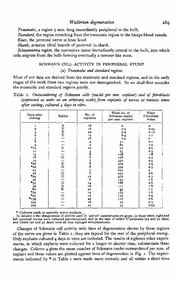
Table 1. Outwandering of Schwann cells {nuclei per mm. explant) and of fibroblasts{expressed as units on an arbitrary scale) from explants of nerves at various timesafter cutting, cultured 4 days in vitro.
Days aftercutting
i
2
34569
• i oII
\l19
• 2 0• 2 0
2325
•2529
•353960
• 7 2•97
•139344402
Rabbit
XYXY———Z
G—ZO—G0—OKK—O———
No. ofexplants
1616241 4
1642
12
t678876
12
6
It2 0
14122 21625
Mean no. ofSchwann nucleiper mm. explant
0
0 20-96 40 9
51657153
15819835°4623922472554701 2 5188i l l
9,4159130116
5035
MeanFibroblast
index
00-050 1 7I - I0
o-i1-2
0 72 22-90-52-42 333-62 7i-6o-6i -6I - I
i-60 4o-60 30-23
• Cultures made in specially active medium. .In column 2 the designations of rabbits used in ' paired' experiments are giv^n; in these cases, right and
left peroneal nerves were cultured, simultaneously and in the case of rabbit O peroneals (25 and 97 days)and tibials (20 and 35 days) were all four cultmed simultaneously.
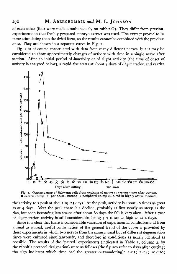
Changes of Schwann cell activity with time of degeneration shown by these regionsof the nerve are given in Table 1; they are typical for the rest of the peripheral stump.Only explants cultured 4 days in vitro are included. The results of eighteen other experi-ments, in which explants were cultured for a longer or shorter time, substantiate thesechanges. Column 4 gives the mean number of Schwann nuclei outwandered per mm. ofexplant and these values are plotted against time of degeneration in Fig. 1. The experi-ments indicated by • in Table 1 were made more recently and all within a short time
270 M. ABERCROMBIE and M. L. JOHNSON
of each other (four were made simultaneously on rabbit O). They differ from previouexperiments in that freshly prepared embryo extract was used. The extract proved to bemore stimulating than the dried form, so the results cannot be combined with the previousones. They are shown in a separate curve in Fig. 1.
Fig. 1 is of course constructed with data from many different nerves, but it may beconsidered to show approximately changes of activity with time in a single nerve aftersection. After an initial period of inactivity or of slight activity (the time of onset ofactivity is analysed below), a rapid rise starts at about 4 days of degeneration and carries
450
400
&350
300
250
o.
au
T3
i2 200
1150
I 1 0 0
50
00 10 20 30 40 50 60 70 80 90 100 110 120 130 140 f 3~40 350 360 370 380 3 0 400
Days after cutting 200 days
Fig. 1. Outwandering of Schwann cells from explants of nerves at various times after cutting.• central stump; O peripheral stump; © peripheral stump cultured in highly active medium.
the activity to a peak at about 19-25 days. At the peak, activity is about 40 times as greatas at 4 days. After the peak there is a decline,, probably at first nearly as steep as therise, but soon becoming less steep; after about 60 days the fall is very slow. After 1 yearof degeneration activity is still considerable, being 5-7 times as high as at 4 days.
Since it is clear that there is considerable variation of experimental conditions and fromanimal to animal, useful confirmation of the general trend of the curve is provided bythose experiments in which two nerves from the same animal but of different degenerationtimes were cultured simultaneously, and therefore in conditions as nearly identical aspossible. The results of the 'paired' experiments (indicated in Table 1, column 2, bythe rabbit's protocol designation) were as follows (the figures refer to days after cutting;the sign indicates which time had the greater outwandering): i < 3 ; 2<4; io<2o;
Wallerian degeneration 271
*6<25; 39>6o. In one case (rabbit 0) peroneals and tibials were simultaneously cul-tured; of the peroneals, 25 >97, of the tibials, 2 0 3 5 ; while comparing a tibial with aperoneal, 20 < 25 and 35 > 97. The difference in activity between the members of a pairwas in three instances statistically significant; between 10 and 20 days of degeneration(mean activity and standard error 71 + 20 and 462 + 34 respectively); between 20 and35 days of degeneration (392 ± 67 and 188 + 26 respectively); and between 25 and 97 daysof degeneration (470 + 57 and 130 + 12 respectively).
Comparison of traumatic and standard regions. It is known that the region of the peri-pheral stump immediately adjacent to the cut undergoes precocious degeneration (Cajal,1928). The effect of this on Schwann cell activity was investigated in twenty-two experi-ments comprising 130 traumatic and 166 standard explants.
In each experiment explants from the two regions of a single nerve were culturedsimultaneously, and their activity compared. We found that the mean activity of thetraumatic region explants was greater than that of the standard region explants in seven-teen of the experiments (owing to the small numbers of explants counted, however, thedifference was only rarely statistically significant in an individual experiment). In fourother experiments the mean activities were the same. In the remaining experiment,at 1 day of degeneration, we found no activity of either region. In the early stages ofdegeneration (2-4 days), the mean activity of the traumatic region explants is very muchhigher (4-20 times) than that of the corresponding standard region. With further degenera-tion it is a small but rather constant amount higher, averaging 154+ 16 % of that of thestandard region, a significant difference. It is clear that the raised activity of the trau-matic region persists for 2-3 months after the initial cut; and possibly, though our dataare insufficient, for a year.
We found indications that a traumatic effect was also produced by recutting a nervewhich had already degenerated. Two experiments of this kind were made. A nerve of15 days' degeneration was recut and cultured after a further 18 days, when the new trau-matic region was 510 % of the standard region; similarly a nerve recut after 30 days'regeneration and cultured after a further 18 days had a new traumatic region 145 %of its standard region.
Onset of activity. The fact that the activity of the traumatic region is so markedly inadvance of that of the standard region during the early stages of degeneration calls formore detailed investigation of the time of onset of activity in the two regions after theinitial cut.
After 4 days in vitro both standard and traumatic regions of one day of degenerationwere entirely inactive. At 2 days of degeneration the standard region was inactive, butthe traumatic region had produced an average of 0-5 Schwann nucleus per explant. At3 days of degeneration the standard region produced an average of 0-2 Schwann nucleusper explant, the traumatic 3-5. Thereafter the activity of both increased, the traumaticregion maintaining its lead, but proportionately a much reduced one.
A longer cultivation of the apparently inactive nerves was undertaken. Even after8 days in vitro both regions of the stump at 1 day of degeneration are quite inactive. Butwhile no activity was detectable in the 2-day degenerated standard region after 4 daysin vitro, slight activity was shown after 7 days in vitro (mostly in the form of filamentsof Schwann cell cytoplasm, without nuclei). Strictly parallel .experiments done withnormal undegenerated nerve showed complete inactivity after 7 days in vitro. It can
272 M. ABERCROMBIE and M. L. JOHNSON
therefore be inferred that the traumatic region becomes active at a slightly earlier timaof degeneration than the standard region (probably late on the first day), and that thestandard region first acquires activity on the second day of degeneration.
(b) Bulb
The terminal bulb of the peripheral stump was not used to derive the curve of Schwanncell activity shown in Fig. 1 owing to its variability and the frequency with which theSchwann cells are obscured by fibroblasts. In spite of considerable variability the changesof the Schwann cell activity of the bulb with time of degeneration repeat in general formthe course of activity shown in Fig. 1; activity starts on the second day, and from thefourth day rises fast to a peak at 15-25 days after cutting, thereafter declining. Duringthese changes, but excluding the very long degeneration times of 344 and 402 days, thebulb is approximately as active as the traumatic region (though it varies between 50 and400 % of this region); it is therefore (in twelve out of fourteen comparable experiments)more active than the standard region. However, in the two experiments with nerves ofvery long degeneration (344 and 402 days) the activity of the bulb was about one-fifthor less of that of the rest of the peripheral stump. The bulb was studied in 25 experi-ments, comprising 90 explants.1
(c) Knee and shank
The standard, traumatic and bulb regions discussed above are all parts of the sciaticnerve in the thigh. It seemed possible that activity might differ in the more peripheralparts of the nerve. In several experiments in which the nerve was cut in the usual place,pieces were taken for culture from the peroneal nerve at knee level (16 experiments,100 explants) and from the anterior tibial branch of the peroneal half-way down theshank (9 experiments, 52 explants).
In both regions the changes of activity with time of degeneration are approximatelythe same as those of the thigh region, though a good deal more irregular. There is thesame rise to a peak, at 20-23 days' degeneration, and the same fall after the peak, at firstrapid, then slow. The general level of activity proved to be higher than that of thestandard region of the same nerve in nine out of eleven experiments on the knee regionand in all eight experiments on the shank where comparison is possible. When theactivities of the knee and shank regions were expressed as percentages of those of thestandard region, much the highest values were obtained in the experiments at theshortest degeneration times. These were for the knee region 640% at 5 days and 500%at 7 days: for the shank 500% at 5 days and 760% at 10 days. At longer degenerationtimes the percentages decreased. They varied for the knee region (13-402 days) between30 and 200%, the mean and its standard error being 120+ 19%; and for the shankregion (21-97 days) between 112 and- 260%, the mean and its standard error being210 + 26%. In corhparable experiments the activities of the shank and knee regionswere not significantly different from each other.
It should be noted that the shank region, separated by about 8 cm. from the cut inthe nerve, cannot be affected by the initial operation except in so far as Wallerian de-generation is concerned.
(d) Schwarmoma region
It is known that Schwann cells migrate out from the cut surface of the peripheralstump (i.e. the bulb) in vivo, and infiltrate the neighbouring connective tissue, forming
Wallerian degeneration 273
a tumour-like mass (Nageotte, 1913) m the later, stages of degeneration. We culturedthe tissue from immediately beyond the tip of the bulb after various times of degenera-tion to test the activity of these emigrant Schwann cells. Twenty-two such experimentscomprising 105 explants were made.
Explants from the schwannoma region are of variable activity, because it is not homo-geneous nervous tissue, and some of the explants turn out to be connective tissue only.For this reason detailed quantitative treatment is not useful. But the general trend ofactivity with time of degeneration is clear. No Schwann cells appeared from explantstaken from nerves of 6 days' degeneration or less (five experiments). At 7 and 11 daysof degeneration only very few Schwann cells were observed (two experiments). Butfrom 13 to 25 days of degeneration (seven experiments) there were many explants whichproduced abundant Schwann cells, fully as many as the bulb or traumatic region. Withlonger degeneration (60-139 days, six experiments) activity is less, far below that of theperipheral stump proper, e.g. at 139 days the schwannoma region had an activity 20%of that of the bulb. Finally, in the experiments at 344 and 402 days of degeneration,when a massive tumour-like schwannoma had formed, the explants were almost com-pletely inactive. At 344 days the mean number of Schwann nuclei (twelve explants)was 1 % of that of the peripheral stump proper; at 402 days no Schwann cells out-wandered (five explants). In these experiments the bulb had also only very slightactivity.
It is notable that when activity is high Schwann cells often grow from the schwannomaexplants in the form of isolated massive trunks, which dissociate into separate cells asoutwandering proceeds. This is presumably because the schwannoma contains thick,cords of Schwann cells.
Latency and rate of outwandering
As we might expect, the latency (i.e. the period between the setting up of the culturesand the first appearance of outwandering) varies inversely, with the amount of activityof the explant. (Schwann cells wander out before or at the same time as fibroblasts inall regions except the bulb and schwannoma.) Changes of latency are not, however, theonly cause of changes of activity with time of degeneration. There are also changes ofthe rate of outwandering when the period of latency is over. The outwandering per mm.of explant per day of the period, of outwandering changes in a way generally similar to theactivity for 4 days in vitro (Table 1, column 4) so far considered. In particular, there isa fall of rate similar to the fall of activity after a peak at 19-25 days of degeneration.In fact, latency is negatively correlated with rate of outwandering and changes in bothtogether lead to the observed changes of amount of activity.
But the relationship between latency and rate of outwandering is not constant at alltimes of degeneration. We have compared explants of the same rate of outwanderingbut from nerves of different degeneration times. (Explants with a period of outwanderingof 2 or 3 days' duration were used; they had been in vitro 3-6 days, according to theirperiod of latency.) We found that, in the later (post-peak) stages of degeneration, latencyis, for the same rate of outwandering, greater than in early degeneration. For a rate ofbetween 20 and 60 Schwann nuclei per mm. per day, nerves of 7-16 days of degenerationhave a mean latency of 1*3 ±0-13 days (nineteen explants) and of 33-402 days of de-generation a mean latency of 1-710-07 days (fifty-seven explants), the difference being
274 M. ABERCROMBIE and M. L. JOHNSON
significant. Similarly, for a rate of outwandering of between 60 and 90 Schwann nucleiper mm. per day, nerves of 23-30 days of degeneration have a mean latency of 1 • 1 + 0-04(thirteen explants) and of 33-139 days' degeneration a mean latency of 17 ±0-14 (fifteenexplants), the difference being significant. Thus both the pre-peak and the peak explantshave a shorter latency for the same rate of outwandering than the post-peak explants.Our data do not conclusively show that there is- a progressive increase of latency duringthe post-peak decline of activity. With a rate of outwandering of 20—60 Schwann nucleiper mm. per day, explants from nerves of 33-60 days of degeneration have a meanlatency of I -6±O-IO (twenty-nine explants) and those from nerves of 139-402 days ofdegeneration a mean latency of 1 -8 ±0-074 (twenty-eight explants). The difference ishardly significant (P= 0-05).
FIBROBLAST ACTIVITY IN THE PERIPHERAL STUMP
The fibroblasts are often too numerous to count, and the fact that they are intermixedwith Schwann cells makes estimation By area of outwandering inaccurate. Only anapproximate analysis of their outwandering can therefore be made, using an arbitraryscale of 4 indices, the index 4 representing the highest number of cells. The fibroblastactivity of a nerve is expressed as the mean fibroblast index per explant.
The changes in the mean index of fibroblast activity with time of degeneration areshown in Table 1, column 5. Column 4 shows the Schwann cell activity of the sameexplants. The general trends of the Schwann cell and fibroblast figures are closelysimilar, though the fibroblast activity is a good deal more variable both between explantsand between experiments. At different degeneration times the proportion of fibroblaststo Schwann cells shows no significant changes. With respect to the time of degenerationat which activity begins there is no discernible difference between fibroblasts andSchwann cells.
When traumatic and standard regions are considered separately we find that thechanges of their fibroblast activity follow substantially those shown for the two regionsjointly in Table 1. The difference between fibroblast activities of the traumatic andstandard regions parallels the difference between their Schwann cell activities. At2-4 days of degeneration the fibroblast activity of the traumatic region is considerablyhigher (7-50 times) than that of the normal region, while at later stages it is only abouttwice as high.
In the bulb the activity of fibroblasts does not follow that of the Schwann cells. Thereis a rapid rise of fibroblast activity in the early stages of degeneration. In the experimentsat 4 days of tlegeneration fibroblast activity was already half the maximum reached.Thereafter there is a slow rise accompanying the rapid rise in Schwann cell activity.In the post-peak period of Schwann cell activity there is no sharp fall in fibroblastactivity which is well maintained for at least 4 months of degeneration. Cultures after344 and 402 days of degeneration were, however, practically inactive in fibroblasts, asthey were in Schwann cells. In five experiments for which satisfactory comparable dataare available, the bulb is twice as active in fibroblasts as the traumatic region, and4 times as active as the standard region. Thus the ratio of fibroblasts to Schwann cellsis about i|—2 times as big as in the traumatic or normal regions.
The fibroblasts of the knee region are like those of the standard and traumatic regionsin that they follow the Schwann cells in their changes of activity with time of degenera-
Wallerian degeneration 275
tion; but they differ in that (in eight out of ten experiments) they form a relativelyhigher proportion of the outwandering—roughly i\ times as many fibroblasts perSchwann cell as in the traumatic and standard regions. The absolute level of fibroblastactivity is about twice that of the standard region.
Data for the shank region are not sufficient to show anything but that the usual curveof activity with time of degeneration is followed, and that the proportion of fibroblaststo Schwann cells and the absolute level of fibroblast activity are probably higher thanin the standard region.
Fibroblast outwandering in the schwannoma is variable like Schwann cell activity.Its changes with time of degeneration are rather similar to those of the bulb. At 4, 5,and 6 days after cutting a few explants already showed considerable fibroblast (and noSchwann cell) activity. Activity probably increased further up to about 20 days. Thesubsequent decline was much more marked than in the bulb, and activity was poor inexperiments at 2 months after cutting. Almost complete inactivity was reached at 344and 402 days after cutting. The activity we obtained from explants of the peripheralbulb suggests that many of the fibroblasts of the schwannoma will have wandered therefrom the cut surface of the stump.
THE CENTRAL STUMP
The following regions of the central stump were cultured:Bulb, which has the same position and origin as the bulb of the peripheral stump.Distal region, extending for 3-4 mm. above the bulb.Proximal region, central to the distal region, separated from it by $—2 cm.Neuroma region, the connective tissue immediately distal to the bulb, into which cells
and axons migrate from the bulb, forming eventually a tumour-like mass, correspondingto the schwannoma of the peripheral stump.
SCHWANN CELL ACTIVITY IN THE CENTRAL STUMP
In the early stages after cutting, activity of the Schwann cells in the central stump isconfined to the region immediately near the cut end. This part, consisting of the bulband distal regions, is so small that the data have been grouped for comparison with theperipheral stump. The changes of activity with time after cutting are shown in Table 2and Fig. 1. The explants which supplied these data were all 4 days in vitro with the ex-ception of those at 344 and 402 days of degeneration, which were longer (their valuesat 4 days were approximately half those given).
Changes of activity with the course of degeneration are not nearly so marked as inthe peripharal stump. Activity is first found at 2 days of degeneration. After 4 days ofdegeneration it has already reached about half its maximum. There does not appear tobe a sharply defined peak of activity as in the peripheral stump, but rather a plateau ofraised activity. A series of 'paired' experiments in which nerves of the same rabbit,cut at different times previously, were simultaneously cultured, together with some othercultures kept longer than 4 days in vitro and therefore not included in Table 2 andFig. 1, suggest that the highest part of the plateau may be early, between 5 and 10 daysafter cutting. The results of the 'paired' experiments were as follows (the figures referto days after cutting, the sign indicates which time had the greater outwandering);

276 M. ABERCRCMBIE and M. L,. JOHNSON
I < 3 , 2<4, 7> i3 , i6>25, 23 > 400, and 3>344. Of the seven experiments in whichthe explants were kept in vitro for more than 4 days the two highest mean activitiesoccurred at 6 and 7 days after cutting. But although the position of the highest pointin the activity curve is uncertain (and it is quite possible that different nerves may differwidely in this), it is clear that the maximum activity of the central stump is less than10% of the maximum activity reached by the peripheral stump. The data indicate thatwith increasing time after cutting there sooner or later sets in a very slow decline inactivity. A year after cutting, activity is still present to the extent of about 10% of themaximum.
When the activity of the central stump is compared with the activity of the corre-sponding region, cultured at the same time, of the peripheral stump (i.e. the peripheralbulb and the traumatic regions) of the same nerve a clear change with time after cuttingis apparent. At 2 days after cutting in one experiment the central stump mean was
Table 2. Outzoandering of Schwann cells from explants of terminal 5 mm. of
central stumps at various times after cutting of nerve
Days aftercutting
12345
1315
No. ofexplants
88886
1212
Mean no. ofSchwann nucleiper mm. explant
0
3'501
10-528
20'41-9
Days aftercutting
16232535
139344402
No. ofexplantB
118
128
121212
Mean no. ofSchwann nucleiper mm. explant
I O O
130
l\12°'54'5
900% of the peripheral stump mean, in a second experiment 250%. At 3 days aftercutting it was 143 % in one experiment, 5 % in another. At 4 days after cutting it was186% in one, 95 % in another. At 5 days after cutting it was 23 %. Thereafter it neverexceeded 12 % of the peripheral stump. Activity is therefore much greater in the centralstump than in the peripheral stump in the early stages after cutting. We have not shownthat it starts earlier. Likewise the peripheral bulb and traumatic regions are much moreactive in this stage of degeneration than the standard region; the central stump istherefore far more active than the standard region of the peripheral stump. But whenthe peripheral stump starts its rapid rise at 4 days of degeneration, it quickly overtakesthe central stump, and is thereafter more active: at its peak at 19-25 days, it is 20-100times more active than the central stump; and after a year, about 10 times.
The central bulb and the stump immediately above it separately show the same be-haviour as that described for them jointly. They differ in that the bulb usually has thehigher activity. In thirteen out of nineteen experiments the bulb is ahead, averaging inall about twice the activity of the stump proper.
We found that the proximal region, more central than the distal region so far dis-cussed, was inactive in the early stages after cutting but that later it acquires someactivity. This region was cultured in 18 experiments comprising 125 explants. Of nineexperiments, covering 1-19 days after cutting, only one showed a trace of activity: thisconsisted in the appearance of a few cytoplasmic processes. Of nine experiments,covering 23-402 days after cutting, only one shows no activity, and this was cultured
Wallerian degeneration 277
|3 days after cutting. All experiments made after 23 days show activity in this regionof the central stump. Normal (undegenerated) nerves were cultured simultaneouslywith four of the eight positive cultures. All normal explants were entirely inactive.
The activity of this proximal region of the stump is very small, however, and has avery long latent period. Adequate comparison with the more distal part of the centralstump is not usually possible, because the latter was fixed after about 4 days in vitro,and at this time the former was still blank. In three experiments, at 23, 344 and 402 daysof degeneration, in which all regions of the central stump were fixed at the same time,the proximal central stump averaged 35 % of the distal.
The neuroma region was cultured in nine experiments comprising sixty-four explants.In experiments at 15, 23, 23, 35, 53 and 139 days after cutting, the neuroma regionshowed always a moderate degree of Schwann cell activity, sometimes more, sometimesless than the central bulb. Activity is far less than that of the schwannoma region;actual figures are available only for one experiment, at 139 days, and in this there were10 times as many Schwann cells in the schwannoma region. The other experimentsshowed a similar order of difference. Three massive neuromata, taken from nerves cut298, 344 and 402 days previously, were quite inactive, showing that there is a declinein activity with long degeneration, as in the schwannoma.
FIBROBLAST ACTIVITY IN THE CENTRAL STUMP
In the central bulb the activity of fibroblasts at different times after cutting of thenerve is very similar to that of the peripheral bulb. The highest activity we found wasat 4 days after cutting, and up to 2 months after cutting little decline was apparent. Incentral bulb9 cultured a long time after cutting (139, 344 and 402 days) however nofibroblasts outwandered. In the central bulb there is, compared with the peripheralbulb, higher fibroblast activity, just as there is higher Schwann cell activity, betweenthe second and fifth days after cutting. But after 5 days of degeneration the centralbulb has, in nine out of ten experiments, a slightly lower fibroblast activity, averaging80% of that of the peripheral bulb. Later than 5 days after cutting, the ratio of fibro-blasts to Schwann cells is much higher in the central than in the peripheral bulb, forthe fibroblasts in the central bulb are only slightly less active thair those in theperipheral bulb, while the Schwann cells are far less active. Up to 5 days after cuttingthe ratio is about the same in the two bulbs.
The active terminal part of the central stump (distal region) just above the bulb showsexactly the same trends as the central bulb. Fibroblast activity is very low or absentbeyond 3 months after cutting. When compared with the corresponding region of theperipheral stump (traumatic region) its activity is found to be equal, or slightly higherin the first few days after cutting, but subsequently always lower, averaging 30% ofthat of the peripheral traumatic region.
The slight Schwann cell activity which appears late after cutting in the proximal partof the central stump is not associated with any fibroblast activity.
Our data on fibroblast activity of the neuroma are too incomplete for comparativepurposes, but a decline to zero is fairly well established about a year after the cut.Between 15 and 53 days after cutting there were always considerable numbers of fibro-blasts in the outwanderings (five experiments), at 139 days there were very few, and at298, 344 and 402 days there were no fibroblasts, as there were no Schwann cells.
278 M. ABERCROMBIE and M. L. JOHNSON
CONCLUSIONS AND DISCUSSION
The nature of activation
We have found that when explants from the stumps of a severed nerve are culturedin vitro, the number of Schwann cells and fibroblasts which wander out varies accordingto the region of the stumps from which the explants are taken and the length of timewhich has elapsed since the original section was made. Normal (i.e. not previouslysevered) nerve is usually completely inactive; so, we found, was nerve taken 1 day afterit had been cut, whatever the region cultured. On the second day after section a veryslight amount of activity is apparent in both peripheral and central stumps, and up tothe fourth day corresponding regions of the two stumps show a similar, usually small,increase in activity. With longer periods between section of the nerve and culture ofthe stumps further changes of activity o«cur, but differently in the two stumps. In theperipheral stump (none or very little reinnervation having occurred) all regions tested,including parts 8 cm. away from the original cut, show a very rapid rise in Schwanncell and fibroblast activity, beginning at the 4th-5th day (except for the bulb fibroblasts,which show an earlier rise) and reaching a high peak at about the 2Oth-25th day aftersection. This initial rise of activity is in agreement with the results of Ingebrigtsen (1916)who, investigating the percentage of explants showing any activity, found that this indexincreased from 17% on the 5th day after section to 82% on the 19th day. He found nooutwandering before the 5th day. Ingebrigtsen probably used a medium of far lessgrowth-promoting power than ours. He did not experiment on nerves which had under-gone longer degeneration, nor on central stumps. In peripheral stumps taken morethan 25 days since the nerve was cut we found a fall in activity with time since section,rapid at first but soon becoming slower. At 2 months after cutting the level of activityis back to that of the ioth-i2th day after cutting (one-third of the peak activity) whileduring the next 10 months it falls another 50%, to the same activity as that of roughlythe 7th day after cutting.
In the central stump on the contrary, activity is at first confined to the few mm. atthe cut end, a id shows no rapid changes with time since cutting. After an initial risea maximum is reached probably between the 5th and 10th days, but at a level which,except in the case of bulb fibroblasts, is negligible compared with that of the peripheralstump (equivalent perhaps to the peripheral stump at the 5th day); activity then slowlyfalls, but is still not zero after one year. After 23 days since section and up to the longesttime tested (402 days) we found that the central stump 1-2 cm. proximal to the cut,previously inactive, developed a very slight activity.
The spatial distribution of the activity corresponds to that of the degeneration of thenerve fibres, which as is well known starts in the whole length of the peripheral stumpand in the tip (1-2 cm.) of the central stump soon after the nerve is severed (see Cajal,1928). It must be assumed that activity is the direct result of this degeneration. Theoccurrence of a very small amount of activity occasionally in undegenerated nerve andconsistently in the more central parts of the central stump during the later stages aftercutting, may be independent of degeneration. But w£ believe that the occasional activityof normal (undegenerated) nerve is no more than can be accounted for by the occurrenceof a few degenerate fibres (Duncan, 1930) and in one instance we verified the presence
Wallerian degeneration 279
fcf degenerate fibres in an active normal nerve. As for the proximal part of the centralStump, it is said (see Spielmeyer, 1929) that when no reinnervation occurs a slow de-generation or atrophy of individual fibres sets in, which would perhaps account for theslight activity which we found to occur after the 23rd day after cutting. Degenerationof Wallerian type is probably rare here, the changes being rather a diminution of fibresize, including the elimination of part of the myelin.
It is notable that in the peripheral stump (except in the bulb) where the changes ofactivity are pronounced, both Schwann cells and fibroblasts change in substantially thesame way with time of degeneration (see Table 1). Both are presumably affected,directly or indirectly by the same changing stimulus. It is a reasonable hypothesis thatthis stimulus is a chemical one which is present during the early stages of Walleriandegeneration, contemporaneously in fact with the destruction of the major part of thenerve fibre. This occurs between the 4th and 20th days of degeneration, when the riseof activity is maximal. The stimulus presumably ceases to be present from about the25th day onwards, and the result is a sharp fall in activity, not to zero, but, for reasonsas yet unclear, to a moderate and slowly decreasing leve . The persistence of this lowlevel of activity is peculiar to the atmosphere of the nerve stump. The cells which out-wander from the cut end of the peripheral stump in vivo, and eventually form theschwannoma, show the same rise to a peak of activity during the early stages of de-generation; but the subsequent fall is more rapid and activity is zero after a year ofdegeneration. The almost complete absence of outwandering in these schwannomata oflong standing is doubtless connected with the fact that they are extremely fibrous, withfew cells, as sections of the explanted material showed. Holmes & Young (1942) suggestthat in such schwannomata the Schwann cells which originally formed part of them mayhave atrophied.
Since the terminal 1-2 mm. of the central stump undergoes a degeneration which isquite similar to the Wallerian degeneration of the peripheral stump, it might be expectedto show the same changes of activity with time since cutting as the peripheral stump.That it does not do so can probably be ascribed to the presence of growing axons, whichis the most important difference between central and peripheral stumps. But althoughthe growing axons perhaps prevent the activity in the central stump from reaching a highlevel, they do not reduce it to zero even a year after cutting. The persistence of activityin the central stump recalls that in the peripheral stump, although it is at a much lowerlevel; and as in the schwannoma, the cells which have wandered out from the cut endof the stump to form the neuroma do not maintain their activity after long degeneration.
The \ cm. of the peripheral stump immediately adjacent to the cut has a higheractivity than the next | cm. during at least the first 97 days of degeneration. It is knownthat degenerative changes begin earlier in the first few mm. of the peripheral stump,and in the corresponding region of the central stump, than in the rest of the peripheralstump. Correspondingly, activity is precociously high in these regions (particularly so,for an unknown reason, in the central stump); and, as a result, during the rise in activityin the peripheral stump, the first \ cm. is more active than the second. The fact, however,that activity remains higher in the first \ cm. at and after the peak of activity, insteadof undergoing a precocious decline, must mean that the stimulus given to the cells inthis region is not only precocious but also greater. This is also indicated by the factthat stimulation was obtained in the neighbourhood of the wound when an already
JEB . 19, 3 l 8
280 M. ABERCROMBIE and M. L. JOHNSON
degenerated nerve was recut (though we have only done two experiments to test this)There appears to be therefore a distinct and additional traumatic stimulus, similalperhaps to that which activates fibroblasts when connective tissue is wounded, which issuperimposed on the Wallerian activation near the cut end of the peripheral stump. Itmust be assumed, since the trauma affects the ends of the two stumps equally, that thecondition is the same in the central stump.
The traumatic activation affects fibroblast and Schwann cells equally in the peripheralstump just below the terminal bulb. But in the bulb itself the number of fibroblasts isincreased proportionately to the number of Schwann cells, especially in the early stagesof degeneration (2nd-5th days). This is also true of the central bulb, where the numberof fibroblasts is almost the same as in the peripheral bulb, although the number ofSchwann cells (after the first 3 days of degeneration) is far smaller. The simplest ex-planation is that there is an early invasion of fibroblasts, stimulated by the wound, fromthe perineurium or other adjacent connective tissue, through the cut surface of thestumps into the bulbs. Later the direction of the migration is of course reversed, andcells leave the bulbs to form Jhe neuroma and schwannoma. The schwannoma, like thebulb, shows a high fibroblast activity in the first few (4-6) days of degeneration, withno Schwann cell activity until after the 6th day. Whether these are the fibroblasts whichinvade the bulb is unknown.
We found that after degeneration of a year, the peripheral bulb had lost far more ofits activity than had the rest of the peripheral stump. This is no doubt connected withthe extensive fibrosis of the Schwann bundles which occurs in late degeneration closeto the lesion (see Holmes & Young, 1942). The fibrosis in its turn may be related tothe intense fibroblast activity characteristic of the bulb region in the earlier stages ofdegeneration. The loss of activity in the schwannoma is perhaps to be similarly explained.
Significance of activity
Clearly the changes in activity which the Schwann cells and fibroblasts show in vitrowith increasing time of degeneration is a result of changes in the physiology of thenerve, which result from degeneration. We do not yet know anything further aboutthese physiological changes, and it is not possible to correlate them clearly with theknown changes of these cells during degeneration in vivo. Some correlation of mitoticactivity and outwandering would be expected, since they have important features incommon (pseudopodial activity, response to same stimuli in tissue culture). The literaturesuggests that the temporal correspondence in the changes of the two activities is notclosej mitosis starting in the Schwann cells and probably in the endoneurial fibroblastsat the 4th day of degeneration, reaching its maximum at about the 6th-o,th days andceasing after the i^th-zoth day (Cajal, 1928). Roughly speaking, however, the rise tothe peak of outwandering activity coincides with the total duration of mitotic activity.It is probable that some migration of the Schwann cells takes place in the degeneratingnerve in vivo, for instance during the formation of the Bungner bands (Holmes &Young, 1942).
However, it is highly probable that the changes in activity found in vitro will bedirectly reflected in one aspect of the behaviour of the cells in vivo: the outwanderingfrom the cut surface of the nerve stumps into the surrounding connective tissue. Thechanges with time of this outwandering in vivo during the building up of the neuroma
Wallerian degeneration 281
• id schwannoma cannot be directly inferred from our experiments. But if a degeneratedperipheral stump is recut, the new in vivo outwandering from the cut surface will, it ishighly probable, vary according to the curve of activity with degeneration time whichwe have obtained in vitro. The actual amount of outwandering will not necessarilycorrespond, owing to the difference of medium in which the cells grow; and the pro-portionate changes may not be exactly the same, owing to possible differential effectsof the media; but it is unlikely that the general trend will differ significantly. Holmesand Young (1942), by measuring the length of the schwannoma produced from a cutnerve in vivo, after various degeneration times, have in fact obtained direct evidenceon this point which is in general concordance with our conclusions.
Does the activity shown during the time spent in vitro represent the physiologicalstate of the nerve at the moment of explanation: or do the processes of Wallerian de-generation proceed in vitro as they would in vivo, correspondingly increasing the activityof the cells? It is clear that if activation does proceed in vitro, it does not do so at thein vivo rate. Normal, i.e. undegenerated nerve (with a few exceptions) and nerve onthe first day of degeneration, do not develop activity even though kept in vitro quitelong enough (8 days and more) to do do if they changed at the rate they do in vivo.Deterioration of the medium is not the cause of this, since subculturing during thisperiod also failed to elicit any outwandering. An experiment in which explants of anearly stage of degeneration were subcultured after 4 days in vitro, and then grownanother 4 days, showed that there was a slight rise in activity during the second period,but only a fraction of what would have occurred in vivo. We conclude therefore that,at least in the early stages of degeneration, activity in vitro represents fairly closely thephysiological state of the nerve at the time of explanation.
Practical bearing
It has been pointed out that Schwann cell activity in vitro may be expected to reflectthe Schwann cell outwandering from the cut end of the nerve in vivo. In the repair ofnerves by suture or graft a successful junction is probably formed by a vigorous out-wandering of Schwann cells from the peripheral stump or from the graft (Young, 1942;Holmes & Young, 1942). We find that, except for a short initial period, the longer anerve is left before surgical repair is undertaken, the less active the Schwann cells ofthe peripheral stump are likely to be in forming a junction. In the rabbit the optimumtime for repair from the point of view of Schwann cell outwandering is not later than25 days after the initial lesion. Further, our results suggest that immediate suture wouldnot be as favourable as suture delayed for a few (say 10-20) days, in order to allow thedevelopment of a fairly high Schwann cell activity at the time of suture. In this waythere would be less likelihood that the Schwann cell junction will be hindered by theprior development in the suture-line of serious fibrosis. Such fibrosis is a likely conse-quence of the numerous active fibroblasts which we found in the bulb and nearbyconnective tissue from 4 days after the nerve was cut. The same argument indicatesthe use of predegenerated grafts, provided predegeneration is short (10-20 days).
These conclusions are in entire agreement with those of Holmes & Young (1942),who found from experimental suture that long delay after the initial lesion before surgicalinterference is inimical to good repair, but that immediate suture is not so effective assomewhat delayed suture. Further, Sanders & Young (1942), comparing autografts of
18-2
282 M. ABERCROMBIE and M. L. JOHNSON
fresh nerve with autografts predegenerated 6-9, 14-16 or 25-28 days, found adelay of the growing axons at the junction of graft and central stump with predegeneratedthan with fresh grafts; a difference which they regard as suggestive though not statis-tically significant.
It may prove possible to apply our results with reasonable assurance to the humanwithout experimental analysis by a study of the histological correlations of the activitycurve we have obtained in the rabbit.
SUMMARY
1. The technique of tissue culture has been applied to a study of the physiologicalchanges undergone by the cells of a severed nerve. The sciatic nerve of adult rabbitswas cut in the middle of the thigh and pieces of the central and peripheral stump wereexplanted at varying times after the original cut. The ' activity' of a part of a nerve isexpressed as the amount of outwandering of the Schwann cells and fibroblasts after4 days in vitro.
2. In general, except in the terminal bulbs (derived from the herniated ends of thecut nerve) Schwann cell and fibroblast activity changes in a similar way.
3. In the conditions of our experiments normal (undegenerated) nerve shows activityonly very rarely. Such activity 3s does sometimes occur can be explained by the presenceof a few degenerate fibres.
4. In the peripheral stump Schwann cell activity begins on the 2nd day after cuttingand from the 4th day rises rapidly to a peak at the io,th-25th day. It then falls quicklyup to about the 60th day and afterwards more slowly. Activity is still appreciable morethan a year after cutting. These changes of activity with time of degeneration are shownby thigh, knee and shank regions of the peripheral stump. The knee region, and theshank (which is 8 cm. distal to the initial cut) are more active than the thigh region,especially in the early days of degeneration.
5. In the central stump activity is at first confined to a few mm. immediately adjacentto the cut. From the 2nd to the 4th day after cutting the central stump is more activethan the peripheral stump, but thereafter it is much less active. Its maximum activity,never, except for the bulb fibroblasts, more than 10% of the maximum activity of theperipheral stump, is reached probably between 5 and 10 days after cutting, after whichit falls slowly. Activity is still appreciable more than a year after the cut was made.The more proximal part of the central stump, at first inactive, begins to show slightSchwann cell activity after 23 days and is still active after more than a year.
6. The \ cm. of the peripheral stump near the cut, including the bulb, is at first moreactive than the adjacent more distal region. But after degeneration of a year or more theperipheral bulb becomes on the contrary less active than the rest of the peripheral stump.
7. There is a particularly high fibroblast activity in the terminal bulbs of both peri-pheral and central stumps during the first 2-4 months, probably as a result of an earlyinvasion from the neighbouring connective tissue. Relative to Schwann cell activity'the bulb fibroblasts are most active during the 2nd-5th days of degeneration.
8. The schwannoma and neuroma in general show the same changes of activity asthe bulbs from which they are formed. Almost no activity was found after degenerationof about a year. In the schwannoma no Schwann cells appear until the 6th day ofdegeneration, though fibroblasts are very active before this.
Wallerian degeneration 283
9. It is concluded that activation of the Schwann cells and fibroblasts is due to(a) degeneration of the nerve fibres, (b) in the region close to the cut, a traumatic effectof cutting the nerve, superimposed on (a).
10. Since Schwann cells probably play an important part in forming the junctionwhen severed nerves are repaired this work indicates the optimum time from this pointof view for making grafts or sutures. Our experiments indicate that the Schwann cellswill be more active in joining together two nerve stumps if the nerve or nerve graft isleft a few (say 10-20) days to degenerate before making the repair; and that (in therabbit) the optimum time for suture is passed 25 days after the nerve is cut.
The expenses of this research were defrayed by a grant from the Rockefeller Founda-tion to Professor Lancelot Hogben for research work in the Department of Zoology inthe University of Birmingham.
REFERENCESCAJAL, S. R. (r928). Degeneration and Regeneration of the Nervous System. London.CHLOPIN, N. G. (i939)- C.R. Acad. Sd. U.R.S.S. 23, 175.DUNCAN, D. (1930). J. Comp. Neurol. 51, 197.GUTTMANN, L. & MEDAWAR, P. B.' (1942). Brit. J. Surg. (in the Press).HOLMES, W. & YOUNO, J. Z. (1943). J. Anat., Land, (in the Press).INGEBRIOTSEN, R. (1916). J. Exp. Med. 23, 251.MURRAY, M. R., STOUT, A. P. & BRADLEY, C. F. (1940). Amer. J. Pathol. 16, 41.NAGEOTTE, J..(i9i3). C.R. Soc. Biol., Paris, 75, 186.PEACOCK, P. R. 8c SHUKOFF, R. J. (1940). Nature, Land., 146, 30.SANDERS F. K. & YOUNO, J. Z. (1943). J. Anat., Lond., 76, 143.SPEELMEYHR, W. (1929). Handbuch der normalen u. pathologischen Physiologie, 9, 285. Berlin.YOUNO, J. Z. (.1942). Physiol. Rev. (in the Press).