The Osseotite Implant
8
The Osseotite ® Implant Documented Success
Transcript of The Osseotite Implant
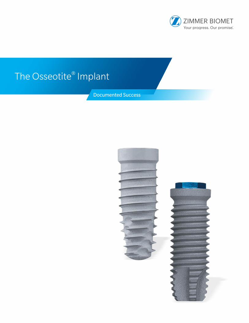
The Osseotite® Implant
Documented Success

Proven Clinical Success
The Osseotite Surface has more than 10 years of documentation from numerous global multi-center clinical studies1-6 and meta-analyses.7-8 Clinical studies on the Osseotite Surface continue to document the benefits of increased contact osteogenesis, especially in poor-quality bone.6
The Osseotite Implant features an acid-etched surface designed to facilitate osseointegration.
The Osseotite Surface • Facilitates The Osseointegration Process• Demonstrates High Contact of Implant with New Bone• Human Histology with Demonstrated High Bone-To-Implant Contact9
• Five-Year Study10 Showed No Increased Risk of Peri-implantitis vs. a Zimmer Biomet Hybrid Implant
Comprehensive Clinical Research• One of the Most Well-Researched Dental Implant Surfaces on the Market Today• Numerous Studies Report 98% Cumulative Success Rates6
Full Osseotite Surface
Osseotite Surface at 20,000x magnification
The Osseotite Implant Overview
Image courtesy of Jun Y. Park, The Bone Interface Group.

Enhanced microscopy image of the Osseotite surface showing platelet activation.
The Osseotite Surface Features Are Precisely Sized To Entangle The Fibrin Strands
Blood Clotting And Implant Attachment
A blood clot attaches to an implant when its fibrin strands become
intertwined in an implant’s micro-surface features. The strength of the
clot/implant attachment depends on how tightly the fibrin strands
are entangled in the surface. Fibrin strands are typically sub-micron in
diameter. Therefore, for the strongest bond, the implant surface features
should create a maze of slightly larger spaces that can tightly capture
the fibrin strands. Characterized by a 1 to 3 micron peak-to-peak surface
created by a unique acid-etch process, the Osseotite Surface features are
precisely sized to entangle the fibrin strands of the blood clot.
Platelet Aggregation
Platelet Activation Up-Regulates Healing Response
Osteogenic cell migration will occur through the blood clot and can be
expected to be influenced by the release of cytokines and other growth
factors from activated cellular components of the blood clot. In a study of
red blood cell (RBC) and platelet interactions with implant surfaces, the
amount of RBC agglomeration on the Osseotite Surface was 54% greater
than as seen on a smooth-machined surface.11
In addition, platelet adhesion onto the Osseotite Surface was enhanced by
110% in comparison to a smooth-machined surface.11 RBC agglomeration
is known to enhance blood clot permeability, which can lead to enhanced
wound healing. Increased platelet activity can also lead to enhanced wound
healing by the release of cytokines and growth factors.12 Taken together,
both platelet adhesion and RBC agglomeration can therefore result in
increased bone formation on the Osseotite Surface.
The Osseotite Surface And The Healing Process

Smooth - Healing Existing Machined Bone BoneImplant
Distance Osteogenesis –A gradual process of bone healing inward from the edge of the osteotomy toward the implant. Bone does not grow directly on the implant surface.
Clot Attachment Increases Contact Osteogenesis
Contact Osteogenesis Facilitates Bone Healing
Bone heals around an implant through two
distinct and overlapping phenomena: distance
osteogenesis and contact osteogenesis. The
rate and extent of healing around an implant is
dependent on the degree of contact osteogenesis
that occurs at the implant surface. The migration
of osteogenic cells through the clot matrix causes
contraction of the fibrin strands in the clot matrix,
which can detach the strands from smooth-
machined implant surfaces, disrupting or stopping
contact osteogenesis and osteoconduction.13
Osseotite Healing Existing
Implant Bone Bone
Contact Osteogenesis –The direct migration of bone-building cells through the clot matrix to the implant surface. Bone is quickly formed directly on the implant surface.
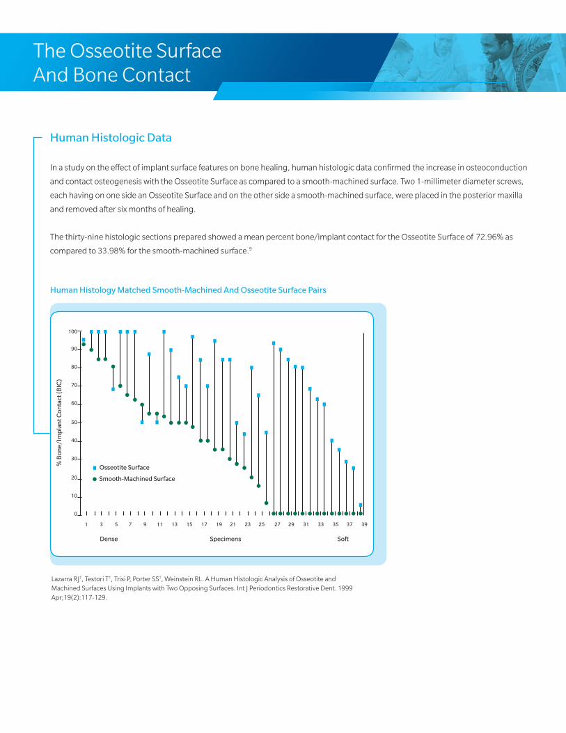
Human Histologic Data
In a study on the effect of implant surface features on bone healing, human histologic data confirmed the increase in osteoconduction
and contact osteogenesis with the Osseotite Surface as compared to a smooth-machined surface. Two 1-millimeter diameter screws,
each having on one side an Osseotite Surface and on the other side a smooth-machined surface, were placed in the posterior maxilla
and removed after six months of healing.
The thirty-nine histologic sections prepared showed a mean percent bone/implant contact for the Osseotite Surface of 72.96% as
compared to 33.98% for the smooth-machined surface.9
Human Histology Matched Smooth-Machined And Osseotite Surface Pairs
1 3 5 7 9 11 13 15 17 19 21 23 25 27 29 31 33 35 37 39
100
90
80
70
60
50
40
30
20
10
0
Osseotite Surface
Smooth-Machined Surface
Dense Specimens Soft
% B
one/
Imp
lan
t Con
tact
(BIC
)
Lazarra RJ†, Testori T†, Trisi P, Porter SS†, Weinstein RL. A Human Histologic Analysis of Osseotite and Machined Surfaces Using Implants with Two Opposing Surfaces. Int J Periodontics Restorative Dent. 1999 Apr;19(2):117-129.
The Osseotite Surface And Bone Contact

A Five-Year Study
A five-year prospective, multicenter, randomized-controlled study of the incidence
of peri-implantitis for hybrid-DAE and fully-DAE implants.10
Considerations for potential benefits of extending the DAE surface to the seating
surface led to this prospective randomized-controlled study designed to assess the
risk and incidence of peri-implantitis for fully-DAE-surfaced implants (Full Osseotite/FOSS).
Study implants, fully-DAE-surfaced “test” implants and hybrid-DAE “control” implants, were placed in a single-stage approach
with the seating surface level with the crestal margin of the alveolar bone. The implants were allowed to heal for two months
and were then provisionalized. Final restorations were placed at six months and patients were followed for five years at annual
intervals. Follow-up evaluations included Sulcus Bleeding Index scores (SBI), probing for suppuration, assessments for mobility
and periapical radiographs to identify radiolucencies and crestal bone levels.
One hundred twelve patients were enrolled and 165 test and 139
control implants were placed supporting 127 prostheses. No substantial
differences in mucosal health outcomes between test and control groups
were observed throughout the five year follow-up. For both groups, the
bleeding-on-probing scores were no different. There was one case of
peri-implantitis reported over the five years of observation and this was
for a hybrid implant.
Radiographic analysis of crestal bone regression demonstrated that the
mean change from baseline (provisionalization) is less for test implants
in comparison to control implants (P<.01). The results of this five-year
study showed no increased risk in adverse soft-tissue outcomes or peri-
implantitis for fully-DAE-surfaced implants versus the control implants in
this study.
Control Implant:
hybrid-DAE
Test Implant:
fully-DAE
Full Osseotite Surface
Full Osseotite ImplantsAnd Peri-Implantitis
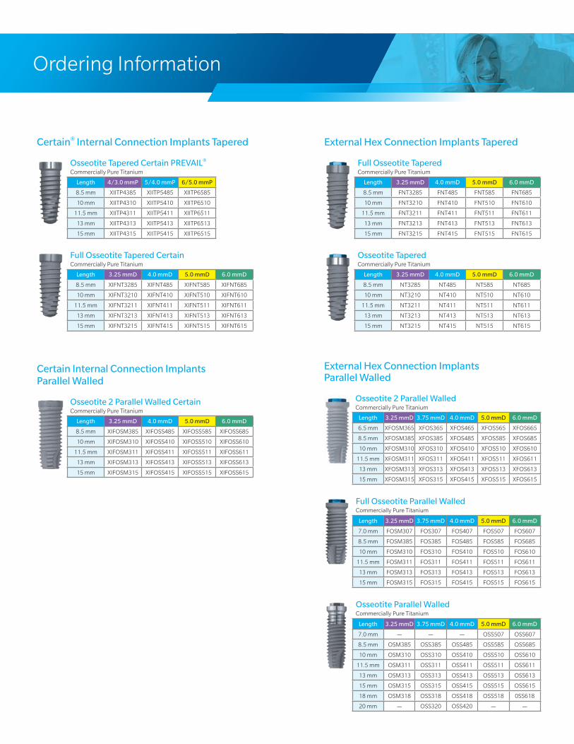
External Hex Connection Implants Tapered
Full Osseotite TaperedCommercially Pure Titanium
Length 3.25 mmD 4.0 mmD 5.0 mmD 6.0 mmD
8.5 mm FNT3285 FNT485 FNT585 FNT685
10 mm FNT3210 FNT410 FNT510 FNT610
11.5 mm FNT3211 FNT411 FNT511 FNT611
13 mm FNT3213 FNT413 FNT513 FNT613
15 mm FNT3215 FNT415 FNT515 FNT615
Osseotite TaperedCommercially Pure Titanium
Length 3.25 mmD 4.0 mmD 5.0 mmD 6.0 mmD
8.5 mm NT3285 NT485 NT585 NT685
10 mm NT3210 NT410 NT510 NT610
11.5 mm NT3211 NT411 NT511 NT611
13 mm NT3213 NT413 NT513 NT613
15 mm NT3215 NT415 NT515 NT615
Certain® Internal Connection Implants Tapered
Osseotite Tapered Certain PREVAIL®
Commercially Pure Titanium
Length 4/3.0 mmP 5/4.0 mmP 6/5.0 mmP
8.5 mm XIITP4385 XIITP5485 XIITP6585
10 mm XIITP4310 XIITP5410 XIITP6510
11.5 mm XIITP4311 XIITP5411 XIITP6511
13 mm XIITP4313 XIITP5413 XIITP6513
15 mm XIITP4315 XIITP5415 XIITP6515
Full Osseotite Tapered CertainCommercially Pure Titanium
Length 3.25 mmD 4.0 mmD 5.0 mmD 6.0 mmD
8.5 mm XIFNT3285 XIFNT485 XIFNT585 XIFNT685
10 mm XIFNT3210 XIFNT410 XIFNT510 XIFNT610
11.5 mm XIFNT3211 XIFNT411 XIFNT511 XIFNT611
13 mm XIFNT3213 XIFNT413 XIFNT513 XIFNT613
15 mm XIFNT3215 XIFNT415 XIFNT515 XIFNT615
Certain Internal Connection ImplantsParallel Walled
Osseotite 2 Parallel Walled CertainCommercially Pure Titanium
Length 3.25 mmD 4.0 mmD 5.0 mmD 6.0 mmD
8.5 mm XIFOSM385 XIFOSS485 XIFOSS585 XIFOSS685
10 mm XIFOSM310 XIFOSS410 XIFOSS510 XIFOSS610
11.5 mm XIFOSM311 XIFOSS411 XIFOSS511 XIFOSS611
13 mm XIFOSM313 XIFOSS413 XIFOSS513 XIFOSS613
15 mm XIFOSM315 XIFOSS415 XIFOSS515 XIFOSS615
External Hex Connection ImplantsParallel Walled
Osseotite 2 Parallel WalledCommercially Pure Titanium
Length 3.25 mmD 3.75 mmD 4.0 mmD 5.0 mmD 6.0 mmD
6.5 mm XFOSM365 XFOS365 XFOS465 XFOS565 XFOS665
8.5 mm XFOSM385 XFOS385 XFOS485 XFOS585 XFOS685
10 mm XFOSM310 XFOS310 XFOS410 XFOS510 XFOS610
11.5 mm XFOSM311 XFOS311 XFOS411 XFOS511 XFOS611
13 mm XFOSM313 XFOS313 XFOS413 XFOS513 XFOS613
15 mm XFOSM315 XFOS315 XFOS415 XFOS515 XFOS615
Full Osseotite Parallel WalledCommercially Pure Titanium
Length 3.25 mmD 3.75 mmD 4.0 mmD 5.0 mmD 6.0 mmD
7.0 mm FOSM307 FOS307 FOS407 FOS507 FOS607
8.5 mm FOSM385 FOS385 FOS485 FOS585 FOS685
10 mm FOSM310 FOS310 FOS410 FOS510 FOS610
11.5 mm FOSM311 FOS311 FOS411 FOS511 FOS611
13 mm FOSM313 FOS313 FOS413 FOS513 FOS613
15 mm FOSM315 FOS315 FOS415 FOS515 FOS615
Osseotite Parallel WalledCommercially Pure Titanium
Length 3.25 mmD 3.75 mmD 4.0 mmD 5.0 mmD 6.0 mmD
7.0 mm — — — OSS507 OSS607
8.5 mm OSM385 OSS385 OSS485 OSS585 OSS685
10 mm OSM310 OSS310 OSS410 OSS510 OSS610
11.5 mm OSM311 OSS311 OSS411 OSS511 OSS611
13 mm OSM313 OSS313 OSS413 OSS513 OSS613
15 mm OSM315 OSS315 OSS415 OSS515 OSS615
18 mm OSM318 OSS318 OSS418 OSS518 0SS618
20 mm — OSS320 OSS420 — —
Ordering Information
Contact us at 1-800-342-5454 or visit
zimmerbiometdental.com
Unless otherwise indicated, as referenced herein, all trademarks are the property of Zimmer Biomet; and all products are manufactured by one or more of the dental subsidiaries of Zimmer Biomet Holdings, Inc. and marketed and distributed by Zimmer Biomet Dental and its authorized marketing partners. For additional product information, please refer to the individual product labeling or instructions for use. Product clearance and availability may be limited to certain countries/regions. This material is intended for clinicians only and does not comprise medical advice or recommendations. Distribution to any other recipient is prohibited. This material may not be copied or reprinted without the express written consent of Zimmer Biomet Dental. ZB0067 REV A 10/18 ©2018 Zimmer Biomet. All rights reserved.
Zimmer Biomet Dental
Global Headquarters
4555 Riverside Drive
Palm Beach Gardens, FL 33410
Tel: +1-561-776-6700
Fax: +1-561-776-1272
References:1. Sullivan DY, Sherwood RL, Porter SS. Long-Term Performance of Osseotite Implants: A Six-Year Clinical Follow-up. Compendium Contin Edu Dent. 2001
Apr;22(4):326-334.2. Davarpanah M, Martinez H, Etienne D, Zabalegui I, Mattout P, Chiche F†, Michel J. A prospective multi-center evaluation of 1,538 3i implants: 1 to 5-year data. Int J
Oral Maxillofac Implants. 2002 Nov-Dec;17(6):820-828.3. Feldman S, Boitel N, Weng D, Kohles SS, Stach RM†. Five-Year Survival Distributions of Short-Length (10mm or less) Machined-Surfaced and Osseotite Implants.
Clin Implant Dent Relat Res. 2004;6(1):16-23.4. Sullivan D, Vincenzi G, Feldman S. Early Loading of Osseotite Implants 2 Months After Placement in the Maxilla and Mandible: A 5-year Report. Int J Oral Maxillofac
Implants 2005 Nov-Dec;20(6):905-912. 5. Stach RM†, Kohles SS. A Meta-Analysis Examining the Clinical Survivability of Machined-Surfaced and Osseotite Implants in Poor-Quality Bone. Implant Dent.
2003;12(1):87-96.6. Testori T†, Wiseman L, Woolfe S, Porter SS†. A Prospective Multicenter Clinical Study of the Osseotite Implant: Four-Year Interim Report. Int J Oral Maxillofac
Implants. 2001 Mar-Apr;16(2):193-200.7. Gaucher H, Bentley K, Roy S, Head T, Blomfield J, Blondeau F, NicholsonL, Chehade A, Tardif N, Emery R†. A Multi-Centre Study of Osseotite Implants Supporting
Mandibular Restorations: A 3-Year Report. J Can Dent Assoc (Tor). 2001 Oct;67(9):528-533.8. Testori T†, Fabbro MD, Feldman S, Vincenzi G, Sullivan D, Rossi R, Anitua E, Bianchi F, Francetti L, Weinstein RL. A Multicenter Prospective Evaluation of 2-months
Loaded Osseotite Implants Placed in the Posterior Jaws: 3-year Follow-up Results. Clin Oral Implants Res. 2002 Apr;13(2):154-161.9. Lazarra RJ†, Testori T†, Trisi P, Porter SS†, Weinstein RL. A Human Histologic Analysis of Osseotite and Machined Surfaces Using Implants with Two Opposing
Surfaces. Int J Periodontics Restorative Dent. 1999 Apr;19(2):117-129.10. Zetterqvist L, Feldman S, Rotter B, Vincenzi G, Wennström JL, Chierico A†, Stach RM†, Kenealy JN†. A Prospective, Multicenter, Randomized-Controlled Five-Year
Study of Hybrid and Fully-etched Implants for the Incidence of Peri-implantitis. J Periodontol. 2010 Apr;81(4):493-501.11. Park JY, Davies JE†. Red Blood Cell and Platelet Interactions with Titanium Implant Surfaces. Clin Oral Implants Res. 2000 Dec;11(6):530-539. 12. Gemmell CH, Park JY. Initial Blood Interactions with Endosseous Implant Materials. International bone engineering workshop; Bone engineering; 1999; Toronto,
Canada. Chapter 9 in Bone Engineering (ed. Davies JE†); Em Squared Inc. 2000 108-117pp.13. Davies JE†. Mechanisms of Endosseous Integration. Int J Prosthodont. 1998 Sep-Oct;11(5):391-401.
† Clinicians have or had a financial relationship with Zimmer Biomet Dental resulting from speaking engagements, consulting engagements and other retained services.