The ornithopod dinosaur Rhabdodon from the Late Cretaceous of ...
198
HAL Id: tel-00841228 https://tel.archives-ouvertes.fr/tel-00841228 Submitted on 4 Jul 2013 HAL is a multi-disciplinary open access archive for the deposit and dissemination of sci- entific research documents, whether they are pub- lished or not. The documents may come from teaching and research institutions in France or abroad, or from public or private research centers. L’archive ouverte pluridisciplinaire HAL, est destinée au dépôt et à la diffusion de documents scientifiques de niveau recherche, publiés ou non, émanant des établissements d’enseignement et de recherche français ou étrangers, des laboratoires publics ou privés. The ornithopod dinosaur Rhabdodon from the Late Cretaceous of France: anatomy, systematics and paleobiology Phornphen Chanthasit To cite this version: Phornphen Chanthasit. The ornithopod dinosaur Rhabdodon from the Late Cretaceous of France : anatomy, systematics and paleobiology. Paleontology. Université Claude Bernard - Lyon I, 2010. English. <NNT : 2010LYO10098>. <tel-00841228>
Transcript of The ornithopod dinosaur Rhabdodon from the Late Cretaceous of ...

HAL Id: tel-00841228https://tel.archives-ouvertes.fr/tel-00841228
Submitted on 4 Jul 2013
HAL is a multi-disciplinary open accessarchive for the deposit and dissemination of sci-entific research documents, whether they are pub-lished or not. The documents may come fromteaching and research institutions in France orabroad, or from public or private research centers.
L’archive ouverte pluridisciplinaire HAL, estdestinée au dépôt et à la diffusion de documentsscientifiques de niveau recherche, publiés ou non,émanant des établissements d’enseignement et derecherche français ou étrangers, des laboratoirespublics ou privés.
The ornithopod dinosaur Rhabdodon from the LateCretaceous of France : anatomy, systematics and
paleobiologyPhornphen Chanthasit
To cite this version:Phornphen Chanthasit. The ornithopod dinosaur Rhabdodon from the Late Cretaceous of France :anatomy, systematics and paleobiology. Paleontology. Université Claude Bernard - Lyon I, 2010.English. <NNT : 2010LYO10098>. <tel-00841228>

N° d’ordre : 98-2010 Année 2010
THESE DE L‘UNIVERSITE DE LYON
Délivrée par
L’UNIVERSITE CLAUDE BERNARD LYON 1
ECOLE DOCTORALE
DIPLOME DE DOCTORAT
(arrêté du 7 août 2006)
soutenue publiquement le 30 juin 2010
par
Mlle Phornphen CHANTHASIT
The ornithopod dinosaur Rhabdodon from the Late Cretaceous of France:
anatomy, systematics and paleobiology.
Le dinosaure ornithopode Rhabdodon dans le Crétacé supérieur de France:
anatomie, systématique et paléobiologie.
Directeur de thèse: M. Eric BUFFETAUT
Co-directeur de thèse: M. Jean-Michel MAZIN
JURY:
M. Jean LE LOEUFF Musée des Dinosaures d’Espéraza Rapporteur
M. Pascal GODEFROIT Institut royal des Sciences naturelles de Belgique Rapporteur
M. Eric BUFFETAUT Ecole Normale Supérieure, Paris Directeur
M. Jean-Michel MAZIN Université Claude Bernard Lyon 1 Co-directeur
M. Christophe LECUYER Université Claude Bernard Lyon 1 Examinateur
M. Julien CLAUDE Université Montpellier 2 Examinateur

1
Acknowledgements
Firstly, I gratefully acknowledge the support from the Development and Promotion
of Science and Technology Talents Project (DPST) conducted jointly by the Royal Thai
Government Agencies and the Institute for the Promotion of Teaching Science and
Technology (IPST), who gave me a great opportunity and a scholarship covering the entire
duration of my studies in France.
I would like to express my gratitude to Eric Buffetaut, my supervisor, for his
confidence and help since my Master’s degree and for giving the opportunity to attend to
this thesis topic under his direction. Without his support, suggestions and geniality, this
work would never have been done.
I wish to acknowledge also Jean-Michel Mazin, my co-supervisor, for his support
and his kindly concern during my study in Université Claude Bernard Lyon1.
I am grateful to all jury members, Jean Le Lœuff, Pascal Godefroit, Julien Claude
and Christophe Lécuyer for their time, their critics and suggestions that will help greatly to
improve this manuscript and my future work.
I thank the following people and institutes for allowing me access to specimens:
Abel Prieur, University Claude Bernard Lyon1; ACAP and Musée de Cruzy, Mairie de
Quarante; Jean Le Lœuff, Musée des dinosaures d’Espéraza; Gilles Cheylan, Muséum
d'Histoire Naturelle d’Aix-en-Provence; Anne Médard-Blondel, Muséum d’Histoire
Naturelle de Marseille; Annie and Patrick Méchin; Muséum national d'Histoire Naturelle
Paris; Angela Milner and Sandra Chapman, Natural History Museum London; Pascal
Godefroit, Institut royal des Sciences naturelles de Belgique.
I would like to pay special tribute to all the people who have taken part in the
discovery of the ornithopod dinosaur, Rhabdodon, without them this animal would never
have been known. I am also indebted to the Association Cuturelle, Archéologique et
Paléontologique de l’Ouest Biterrois at Cruzy (ACAP), because of their heart, their time,
their cooperation and hard work in the field, thousands of vertebrate remains have been
unearthed.
Merci Titi, Didier, Bernard, Eric, Haiyan de m’avoir appris à fouiller avec le couteau
à huître et de m’avoir emmené à Massecaps où j’ai commencé mon métier de paléontologue.

2
Merci Gérard, José, Stéphane, Jacques, Michèle, Michel, Pierre, Gilles, Jean, Jérémy, Julien,
P.Khan, Romain, Jean-Marc, Delphine, Scarlett, Azzura, Clément, Franck, Vincent et tous
les gens qui ont passé au chantier des fouilles pour vos gentillesses, les superbes ambiances
pendant les fouilles, et également pour le cours de langue française familiale que je ne peux
pas apprendre ailleurs. Je voudrais dire merci à tous les Cruzyates pour vos exceptionnels et
chaleureux accueils et vos aides pendent toutes ces années et particulièrement merci
Fernande, Odile, Catherine et Charles de me faire découvrir la vraie cuisine française!!
Grâce à ces gens, j’ai passé un moment formidable pendant les vacances de Pâques et en
juillet chaque année. Merci du fond du cœur!
Special thanks are also due to Jean Le Lœuff, and Dinosauria for the support and
giving the opportunity to professionals and amateurs, French and foreigners, to participate in
the excavations at the exceptional Late Cretaceous vertebrate site, Bellevue. I thank also the
personnel of the association and the Musée des dinosaures d’Espéraza, Christel, Arnaud,
Jean Pierre, Corinne, Muriel, Marie, for their help during my work in the collections of the
museum. Thanks enormously to all volunteers for their hard work, their patience in the
difficult work conditions in the field and for their friendship during our stay in the “loft” at
Espéraza. Thanks to Romain Liard and Yvain Leclerc, chiefs of the excavation teams for
taking care of all crews both in the loft and in the field.
I am grateful to Patrick and Annie Méchin for their kindness and a warm welcome to
their place and their care all along my study of specimens in their collection at Vitrolles.
Merci infiniment!
I would like thank Yves Dutour, Thierry Tortosa, Eric Turini, Séverine Berton and
other excavation crews for the exceptional discovery of the partial articulated skeleton of
Rhabdodon in 2007. It came out just at the right time!
I thank also François Escuillié for allowing me to study the specimens from Vitrolles
when they were borrowed in Gannat and merci papa Roland and Ludo for the advice on the
restoration of specimens and the help during my two months in Gannat.
I would like to acknowledge all colleagues in the laboratory, in particular Anne-
Marie Bodergat who have consistently supported and encouraged me all these years and the
personnel in UMR 5125, particularly, Nathalie Pierre who was in charge of the field work
mission procedures and Dominique Barbe who kindly helped in the reprography of this
manuscript.

3
I am also indebted to my close friend, Pitaksit Ditbanjong, for his help and advice on
the graphic work in this thesis and for furnishing constantly the support and encouragements
all along this study.
I thank Eric and Haiyan again for giving me the warmness and the familiarity which
makes me feel getting closer to my home even though I am actually 9000 km far away.
My thanks must go to all my Thai and French friends, Romain Amiot who is always
available to share the opinions and always ready to help since my first day in the Université
Claude Bernard Lyon1; Jor-ra-ké yak Jérémy, petit ours, David, Guillaume, Kellie, Olivier,
Loïc, Yun, les ch’tis (Sit, Nico, les deux Aurel, Violette) etc. for your kindness and all the
encouragements. Thank Pong, Pingpong, P. Anne, P.Hinghoy, P.Ying, Antoine and P.Khan
for your avaibility and cares and for very good Thai food. ขอบคุณมากคะ
Finally in order to acknowledge, this thesis is dedicated to my parents for their love,
endless support and encouragements.
เพ่ือขอบพระคุณสําหรับความรักและกําลังใจท่ีมใีหหนูมาตลอด 28 ป วิทยานพินธเลมน้ีอุทิศแก พอและแม บคุคลท่ีไดเลี้ยงดูหนูมา จนมีโอกาสไดมาเรียนตางประเทศ และเปดโลกทัศน เรียนรูสิง่ใหมๆ ที่ไมสามารถหาซ้ือได

4
5
Table of Contents
Acknowledgements………………………………………….…………………………… 1
Chapter 1: Introduction…………………………………………………………………. 7
Chapter 2: Overview of the taxonomic history of Rhabdodon………………………… 13
Chapter 3: The discovery and geological setting of the main Rhabdodon localities
in France………………………………………………………………………. 21
3.1 Department of Var…………………………………………………….. 22
3.2 Department of Bouches-du-Rhône……………………………………. 23
3.3 Department of Hérault………………………………………………… 26
3.4 Department of Aude………………………………………….............. 28
Chapter 4: Osteology of Rhabdodon from the Late Cretaceous of southern France
4.1 Material and Methods…………………………………………………. 35
4.2 Description
4.2.1 Skull and mandible…………...……………………………... 36
4.2.2 Dentition…………………………………………………….. 55
4.2.3 Postcranial Skeleton………………………………................. 60
4.3 New partial articulated skeleton of Rhabdodon priscus (Dinosauria:
Ornithopoda) from the Late Cretaceous of Vitrolles-Couperigne,
Bouches-du-Rhône, southern France………………… ………….…… 96

6
Chapter 5: Systematics
5.1 Discussion of the morphological variation in Rhabdodon and in
Rhabdodontidae……………………………………………………... 131
5.2 Phylogenetic relationships of Rhabdodon…………………………… 140
5.3 Conclusion……………………………………………………….. …. 143
Chapter 6: Discussion
6.1 Geographic and temporal distribution of Rhabdodon …..................... 147
6.2 Paleobiogeographic implications……………..……………................. 148
6.3 Feeding……………………………………………………………...... 150
6.4 Posture and locomotion……………………………………………… 151
Chapter 7: Conclusions……………………………………….……………………….... 155
Appendix I: Rhabdodontid Locality List………………………………………...…...….. 159
Appendix II: The anatomical measurements of the Rhabdodon elements
described in this work…………………………………………..……...….. 165
Appendix III: ……………………………………………………………………………. 179
Appendix IV:……………………………………………………...……………..….…… 185

7
Chapter 1:
Introduction
Ornithopoda was a very diverse herbivorous dinosaur group which was widespread
on various continents through the Early Jurassic - Late Cretaceous time span (Norman et al.
2004). Rhabdodon is a medium-sized ornithopod dinosaur from the Late Cretaceous of
Europe.
Rhabdodon was first known from dentary fragments with teeth and some postcranial
elements discovered in the mid-nineteenth century at la Nerthe, in the Bouches-du-Rhône
region of southern France. Because of the characteristic fluted teeth, Matheron erected a
generic name for this new Iguanodon-like dinosaur and described it as a new species,
Rhabdodon priscum (later amended to Rhabdodon priscus) on the basis of the remains from
la Nerthe (Matheron, 1869a, 1869b).
Since that time, abundant additional remains attributed to Rhabdodon have been
regularly reported not only from France but also from other European countries (Spain,
Romania, Austria and Hungary) (Bunzel, 1871; Seeley, 1881; Nopcsa, 1900 and 1902;
Pereda-Suberbiola and Sanz, 1999; Weishampel et al., 2003; Sachs and Hornung, 2006;
Godefroit et al., 2009; Ösi, 2004). It cannot be denied that Rhabdodon was one of the
dinosaur taxa that dominated terrestrial faunas during Late Cretaceous times in Europe.
In particular in France, several Late Cretaceous continental deposits have yielded
numerous dinosaur remains including Rhabdodon. Rhabdodon localities are distributed in
different regions in southern France, Var, Bouches-du-Rhône, Gard, Hérault, Aude and
Ariège (Gervais, 1877; Lapparent, 1947; Buffetaut and Le Lœuff, 1991; Le Lœuff, 1991;
Buffetaut et al., 1996 and 1999; Pincemaille, 1997; Sigé et al., 1997; Garcia et al., 1999;
Pincemaille-Quillévéré 2002; Allain and Pereda-Suberbiola, 2003). Besides Rhabdodon
priscus which represents most of French remains, another species, Rhabdodon septimanicus,
was subsequently erected by Buffetaut and Le Lœuff (1991) on the basis of a characteristic
dentary from a Late Cretaceous locality at Montouliers (Hérault). As R. priscus and R.
septimanicus were found in the same formation of Grés à Reptiles, the validity of the latter
species was doubted and considered as a junior synonym of R. priscus by some authors. In

8
addition, the morphological variation of Rhabdodon from Saint-Chinian was also mentioned
by Buffetaut (2005). These issues have been in debate and raise the question:
How many species of Rhabdodon existed in Southern France?
The question of the variability of Rhabdodon has remained unanswered for a long
time because of the lack of well-preserved material to affirm the validity of R. septimanicus
and because a great number of Rhabdodon specimens discovered in France have never been
studied in detail.
In the late twentieth century, systematic excavations have been conducted in many
Late Cretaceous localities in southern France, such as Cruzy, Campagne-sur-Aude,
Vitrolles, Aix-en-Provence and Fox-Amphoux. The recent discoveries provide very
abundant vertebrate remains which allow a better understanding of the diversity of Late
Cretaceous faunas in southern France. Rhabdodon is one of the most found taxa in the
course of those excavations. A great number of Rhabdodon specimens are housed in many
institutes and private collections in France, however the majority of them have never been
studied. Therefore, the primary objectives of this study are as follows:
- to accomplish the osteological description of undescribed material and observe
the morphological variation among this material,
- to answer the question: How many species of Rhabdodon existed in Southern
France based on the new material and
- to better understand the palaeobiology of Rhabdodon and achieve a
reconstruction of this animal.
The debate about the morphological variability of Rhabdodon is not restricted only
to France but the diversity of Rhabdodon has been also suggested from different localities in
other countries in Europe. Many species were proposed for this European taxon and were
diversely allied to hypsilophodontids, camptosaurids, and iguanodontids. An overview of the
taxonomic history of this taxon will be provided further in this work. Recently, Weishampel
et al. (2003) erected Zalmoxes as a new genus on the basis of cranial and postcranial
material from Transylvania in Romania and they suggested two species, Zalmoxes robustus
and Zalmoxes shqiperorum as members of this genus. In addition, Weishampel et al. (2003)
have placed Zalmoxes in a new family, Rhabdodontidae, which includes Rhabdodon as
sister-taxon. It therefore seems that now there are two genera representing non-hadrosaurid

9
ornithopods in the Late Cretaceous of Europe. On the basis of this previous work on this
dinosaur group, the supplementary objectives of this thesis are the following:
- to anatomically compare Rhabdodon with other related taxa, especially
Zalmoxes, in order to reevaluate the diversity of this dinosaur group and
- to understand the systematics and the phylogenetic relationships of Rhabdodon
with other taxa.
Following the objectives of this study, the principal parts of this thesis will consist
of:
- An overview of the taxonomic history of Rhabdodon,
- Recent discoveries and the geographical and geological setting of main localities
in southern France,
- The osteological description of Rhabdodon material from those localities,
- The systematics and the phylogenetic relationships of Rhabdodon and related
taxa and
- A discussion about the distribution, paleobiogeographical implications and
palaeobiology of Rhabdodon.

10
References
Allain, R., Pereda-Suberbiola, X. 2003. Dinosaurs of France. Comptes Rendus Palevol 2,
27- 44.
Buffetaut, E. 2005. Late Cretaceous vertebrates from the Saint-Chinian area (southern
France): a review of previous research and an update on recent finds. Acta
Palaeontologica Romaniae 5, 39-48.
Buffetaut, E., Le Lœuff, J. 1991. Une nouvelle espèce de Rhabdodon (Dinosauria,
Ornithischia) du Crétacé supérieur de l’Hérault (Sud de la France). Compte rendu de
l’Académie des Sciences de Paris, Série II 312, 943-948.
Buffetaut, E., Costa, G., Le Lœuff, J., Martin, M., Rage, J.-C., Valentin, X., Tong, H. 1996.
An Early Campanian vertebrate fauna from the Villeveyrac Basin (Hérault, Southern
France). Neues Jahrbuch für Geologie und Paläontologie Monatshefte, 1-16.
Buffetaut, E., Le Lœuff, J., Tong, H., Duffaud, S., Cavin, L., Garcia, G., Ward, D.,
l’Association culturelle, archéologique et paléontologique de Cruzy. 1999. Un
nouveau gisement de Vertébrés du Crétacé supérieur à Cruzy (Hérault, Sud de la
France). Comptes Rendus de l’Académie des Sciences de Paris, Sciences de la Terre
et des planètes 328, 203-208.
Bunzel, E. 1871. Die Reptilfauna der Gosau-Formation in der Neuen Welt bei Wiener- Neustadt. Abhandlungen der kaiserlich-königlichen Geologischen Reichsanstalt 5, 1–18.
Garcia, G., Pincemaille, M., Vianey-Liaud, M., Marandat, B., Lorenz, E., Cheylan, G.,
Cappetta, H., Michaux, J., Sudre, J. 1999. Découverte du premier squelette presque
complet de Rhabdodon priscus (Dinosauria, Ornithopoda) du Maastrichtien inférieur
de Provence. Comptes Rendus de l’Académie des Sciences de Paris 328, 415–421.
Gervais P. 1877. De la structure des coquilles calcaires des oeufs et des caract res que l’on
peut en tirer. Comptes Rendus de l’Académie des Sciences de Paris 84, 159-165.
Godefroit, P., Codrea, V., Weishampel, D.B. 2009. Osteology of Zalmoxes shqiperorum
(Dinosauria, Ornithopod), based on new specimens from the Upper Cretaceous of
N<laY-Vad (Romania). Geodiversitas 31(3), 525-553.

11
Lapparent, A.F. 1947. Les dinosauriens du Crétacé supérieur du Midi de la France.
Mémoires de la Société géologique de France 56, 1-54.
Le Lœuff, J. 1991. Les vertébrés maastrichtiens du Mas d’Azil (Ariège, France): étude
préliminaire de la collection Pouech. Revue de Paléobiologie 10(1), 61-67.
Matheron, P. 1869a. Notice sur les reptiles fossiles des dépôts fluvio-lacustres crétacés du
bassin à lignite de Fuveau. Mémoires de l’Académie impériale des Sciences, Belles-
Lettres et Arts de Marseille, 1-39.
Matheron, P. 1869b. Notice sur les reptiles fossils des dépôts fluvio-lacustres crétacé du
basin à lignite de Fuveau. Bulletin de la Société Géologique de France 26(2), 781–
795.
Nopcsa, F. 1900. Dinosaurierreste aus Siebenbürgen (Schädel von Limnosaurus
transsylvanicus nov. gen. et spec.). Denkschriften der kaiserlichen Akademie der
Wissenschaften Wien. Mathematisch-Naturwissenschaftliche Klasse 68, 555–591.
Nopcsa, F. 1902. Dinosaurierreste aus Siebenbürgen II. (Schädelreste von Mochlodon).
Denkschriften der kaiserlichen Akademie der Wissenschaften Wien. Mathematisch-
Naturwissenschaftliche Klasse 72, 149–175.
Norman, D.B., Sues, H-D., Witmer, L.M., Coria, R.A. 2004. Basal Ornithopoda. In:
Weishampel, D.B., Dodson, P., Osmolska, H. (Eds.) The Dinosauria (2nd Edition).
Berkeley: University of California Press. pp. 393–412.
Ösi, A. 2004. The first dinosaur remains from the Upper Creataceous of Hungary
(Csehbànxa Formation, Bakony Mts). Geobios 37, 749–753.
Pereda-Suberbiola, X., Sanz, J.L. 1999. The ornithopod dinosaur Rhabdodon from the
Upper Cretaceous of Laño (Iberian Peninsula). Estudios del Museo de Ciencias
Naturales de Alva 14 (Número especial 1), 257–272.
Pincemaille, M. 1997. Un ornithopode du Crétacé supérieur de Vitrolles (Bouches-du-
Rhône) Rhabdodon priscus. Mémoire de DEA, Université Montpellier II,
Montpellier, Unpublished.

12
Pincemaille-Quillévéré, M. 2002. Description d’un squelette partiel de Rhabdodon priscus
(Euornithopoda) du Crétacé supérieur de Vitrolles (Bouches du Rhône, France).
Oryctos 4, 39-70.
Sachs, S., Hornung. J. J. 2006. Juvenile ornithopod (Dinosauria: Rhabdodontidae) remains
from the Upper Cretaceous (lower Campanian, Gosau Group) of Muthmannsdorf
(Lower Austria). Géobios 39, 415-425.
Seeley, H.G. 1881. The reptile fauna of the Gosau Formation preserved in the Geological Museum of the University of Vienna. Quarterly Journal of the Geological Society London 37, 620–707.
Sige, B., Buscalioni, A.D., Duffaud, S., Gayet, M., Orth, B., Rage, J.-C., Sanz, J.L. 1997.
Etat des données sur le gisement Crétacé supérieur continental de Champ-Garimond
(Gard, Sud de la France). Münchner Geoswissenschaften Abhandlungen 34, 111-
130.
Weishampel, D.B., Jainu, C-M, Csiki Z., Norman, D.B. 2003. Osteology and Phylogeny of
Zalmoxes (N.G.), an unusual Euornithopod Dinosaur from the latest Cretaceous of
Romania. Journal of the Systematic Paleontology 1(2), 65-123.

13
Chapter 2:
Overview of the taxonomic history of Rhabdodon
The discovery of the first late Cretaceous dinosaur of Europe was an unexpected
consequence of the work of tunneling the railway (Paris-Lyon-Marseille) at la Nerthe in the
Bouches-du-Rhône region in France, for which Philippe Matheron, a geologist, was
responsible in 1844. Matheron (1846) described some remains of a reptile which included
some teeth. These teeth reminded him of a tooth, figured by Cuvier (1824), which was a
tooth of the Iguanodon found by Gideon Mantell in Great Britain. Matheron perceptively
observed the resemblance and differences between the teeth. Finally in 1869, on the basis of
the unique characteris of the fluted teeth, Matheron proposed a generic name for this
dinosaur, Rhabdodon.
« ….les restes d’un grand reptile terrestre nouveau qui avait les plus grands rapports avec
l’iguanodon et auquel, à raison de ses dents cannelées, je propose de donner le nom
générique de Rabdodon1. »
(Matheron, 1869a: p.32)
The new gigantic reptile remains had been recovered from lacustrine marls in the
upper part of the Rognacian stage (an equivalent of the Maastrichtian). These remains
included fragments of vertebrae, humerus, femur, tibia and dentary fragments with teeth. He
described these elements and assigned them to a new species, Rhabdodon priscum (amended
to Rhabdodon priscus) which was the first dinosaur from Provence. He also regarded this
dinosaur as a close relative of Iguanodon.
1 According to Brinkmann (1986), when Rhabdodon Matheron, 1869 was established two different spellings
were used by the author: Rabdodon in the text of Matheron (1869a), Rhabdodon in the text of Matheron
(1869b) and at the top of the tables in Matheron (1869a). Gervais (1877), Gaudry (1890, p. 222) and Zittel
(1890, p. 763) were the first authors to quote this genus, and they adopted Rhabdodon as the correct original
spelling. All later workers, including Matheron (1892) have adhered to this.

14
Almost 80 years after the first description of Rhabdodon, additional French material
was reported again by a French paleontologist, l’abbé Albert-François de Lapparent in his
memoir about the dinosaurs from the Upper Cretaceous of southern France (Lapparent,
1947). Lapparent described various dinosaur remains including an ornithopod referred to
Rhabdodon priscus, Matheron 1869 and he also considered Rhabdodon as related to
Iguanodon but notably smaller in size. These Rhabdodon specimens consist not only of
cranial elements which include a quadrate, fragments of dentary and maxilla and some teeth
but also a great number of postcranial elements. Because of his work, Rhabdodon from
several Campanian-Maastrichtian localities in Provence and Languedoc became much better
known.
As early as the discovery of Rhabdodon in southern France, Bunzel (1871) described
a new vertebrate fauna from a coal mine at Muthmannsdorf in Niederösterreich (Eastern
Austria) and erected some new taxa including an ornithopod dinosaur, Iguanodon suessi.
Later, Seeley (1881) noticed that Iguanodon suessi had significantly diagnostic differences
to Iguanodon Mantell, 1825 so that he erected the new genus Mochlodon for this species.
This genus Mochlodon was also considered as including ornithopod material from the HaYeg
basin of Transylvania. Some of this material from Romania was referred to Mochlodon
suessi and some referred to a new species M. robustus (Nopcsa 1900, 1902). Firstly, Nopsca
(1901) regarded Mochlodon as a hypsilophodontid but later (Nopsca 1902, 1904) he
suggested it was closely related to Camptosaurus from the Late Jurassic of America. In
1915, Nopsca considered the similarities between Mochlodon and Rhabdodon as sexual
dimorphism and he referred Rhabdodon to Camptosauridae.
Until later researches on European dinosaurs in the 1980s, Mochlodon had been used
for all material referred to Rhabdodon and Mochlodon by Weishampel and Weishampel
(1983), Weishampel (1984), Norman (1984a, b), Milner and Norman (1984) for the reason
that Rhabdodon Matheron, 1869 was in conflict with an earlier named living snake,
Rhabdodon Fleischmann, 1831. Mochlodon was regarded as a hypsilophodontid or
dryosaurid ornithopods in these works; however, it was suggested as a member of
Iguanodontia by Sereno (1986) and Carroll (1988). The conflict of the generic name was
solved when the conservation of Rhabdodon Matheron, 1869 was accepted by the
International Commission on Zoological Nomenclature which suppressed the senior
homonym Rhabdodon Fleischmann, 1831 (Brinkmann, 1986; ICZN 1988) and Brinkmann
(1986) determined the synonymy of Rhabdodon and Mochlodon as in the favor of
Rhabdodon. The later work of Brinkmann (1988) suggested Rhabdodon as a

15
hypsilophodontid ornithopod, nevertheless in more recent studies by Norman and
Weishampel (1990) and Weishampel (1990) Rhabdodon was regarded as an euornithopod
(sensu Weishampel, 1990).
In the last two decades, paleontological excavations in southern France have been
carried out systematically and have resulted in many great fossil vertebrate discoveries. A
new species, Rhabdodon septimanicus, was suggested on the basis of the dentary of a
juvenile individual discovered in a late Cretaceous locality at Montouliers (Hérault, France)
(Buffetaut and Le Lœuff, 1991). The validity of R. septimanicus was questioned differences
with R. priscus being ascribed to individual differences and/or sexual dimorphism (Allain
and Pereda Suberbiola, 2003). Later, this species was accepted as a result of the systematic
reassessment of Rhabdodontidae by Weishampel et al. (2003).
Besides Montouliers, in the Saint-Chinain region, there are a number of Late
Cretaceous vertebrate sites, for instance at Combebelle, Plo-Saint-Pons and Cruzy, which
have provided significant Rhabdodon remains (Buffetaut et al., 1999; Buffetaut, 2005a, b).
Furthermore, Rhabdodon material was also discovered at an Early Campanian locality in the
Villeveyrac basin. The Late Cretaceous vertebrate fauna of Villeveyrac has actually been
known since 1872 by Bleicher’s work. Some of the vertebrae mentioned by Bleicher (1872)
were revised by Gervais (1877) and interpreted as belonging to Rhabdodon. According to a
systematic revision of old collections and recent excavations, Buffetaut et al. (1996)
described the Rhabdodon of Villeveyrac and suggested that this material could be referred to
cf. Rhabdodon priscus.
Rhabdodon was mostly based on isolated material, so that this dinosaur remained
poorly understood for more than a century, until in 1995, the first partial articulated skeleton
of an ornithopod dinosaur finally was found in the lower Maastrichian of Couperigne near
Vitrolles. It was identified and described as Rhabdodon priscus (Garcia et al., 1999;
Pincemaille-Quillevéré, 2002). Although some of these remains are not quite well preserved
and most of them are deformed, Pincemaille-Quillevéré (2002) suggested that Rhabdodon is
a member of Iguanodontia and closely related to Tenontosaurus from the Early Cretaceous
of North America (see also Pincemaille-Quillevéré et al. 2006).
Besides the remains of Rhabdodon from France, Austria and Romania, Ösi (2004)
reported some teeth of a herbivorous dinosaur recovered from an Upper Cretaceous
(Santonian) locality at Iharkút in Hungary. These remains are referred to Rhabdodontidae
indet. In addition, Rhabdodon sp. is present in many localities in Spain, primarily from the
Upper Cretaceous of Laño quarry (Pereda-Suberbiola and Sanz, 1999).

16
In the most recent taxonomic reevaluation of Rhabdodon, Weishampel et al. (2003)
carried out a cladistic analysis as part of the revision of the material from the HaYeg basin,
Transylvania. They defined the Romanian material as a new genus, Zalmoxes (sister-group
of Rhabdodon), including Z. robustus (Nopcsa, 1900, 1902) (type species) and Z.
shqiperorum Weishampel, Jianu, Csiki and Norman, 2003, within a new family,
Rhabdodontidae.
These non-hadrosaurid ornithopods are among the most common dinosaurs known
from the Late Cretaceous of Europe (in France, Spain, Austria, Romania and Hungary). As
mentioned above, Rhabdodon has a long and complicated taxonomy history. Because of its
morphological variability between different localities, many species have been proposed for
this European taxon and it was diversely allied to hypsilophodontids, camptosaurids,
iguanodontids and to the recently erected group of rhabdodontids. Solving the problem of
the taxonomic diversity of the Rhabdodontidae therefore is one of the objectives of my
work, and the phylogenetic relationships of Rhabdodon will be reevaluated in a following
chapter of this dissertation.

17
References
Allain, R., Pereda Suberbiola, X. 2003. Dinosaurs of France. Comptes Rendus Palevol 2,
27–44.
Bleicher, G. 1872. Etudes de géologie pratique dans les environs de Montpellier. Revue des
Sciences Naturelles Montpellier 1, 63–73.
Brinkmann, W. 1986. Rhabdodon Matheron, 1869 (Reptilia, Ornithischia): Proposed
conservation by suppression of Rhabdodon Fleischmann, 1831 (Reptilia, Serpentes).
Case 2536. Bulletin of Zoological Nomenclature 43, 269-272.
Buffetaut, E., Le Lœuff, J. 1991. Une nouvelle espèce de Rhabdodon (Dinosauria
Ornithischia) du Crétacé de l’Hérault (Sud de la France). C.R. Acad. Sci. Paris. Sér.2
312, 943-948.
Buffetaut, E., Costa, G., Le Lœuff, J., Martin, M., Rage, J.C, Valentin, X., Tong, H. 1996
An Early Campanian vertebrate fauna from the Villeveyrac Basin (Hérault, Southern
France). Neues Jahrbuch für Geologie und Paläontologie 1, 1-16.
Buffetaut, E., Le Lœuff, J., Tong, H., Duffaud, S., Cavin, L., Garcia, G., Ward, D.,
Association culturelle, archéologique et paléontologique de Cruzy. 1999. Un
nouveau gisement de vertébrés du Crétacé supérieur à Cruzy (Hérault, Sud de la
France). Comptes Rendus de l’Académie des Sciences de Paris, II 328, 203-208.
Buffetaut, E. 2005a. Sur les chemins des dinosaures. Editions Aurian, pp.1-88.
Buffetaut, E. 2005b. Late Cretaceous vertebrates from the Saint-Chinian area (southern
France): a review of previous research and an update on recent finds. Acta
Palaeontologica Romaniae 5, 39-48.
Bunzel, E. 1871. Die Reptilfauna der Gosau-Formation in der Neuen Welt bei Wiener-
Neustadt. Abhandlungen der kaiserlich-königlichen Geologischen Reichsanstalt 5,
1–18.
Cuvier G. 1824. Recherches sur les Ossements fossiles. Nelle édition. Dufour and
d’Ocagne, Paris 5(2), pl. XXI, fig. 32.
Fleischmann, F. L. 1831. Dalmatiae nova serpentum genera. Heyder, Erlangen 35 pp.

18
Garcia, G., Pincemaille, M., Vianey-Liaud, M., Marandat, B., Lorenz, E., Cheylan, G.,
Cappetta, H., Michaux, J., Sudre, J. 1999. Découverte du premier squelette presque
complet de Rhabdodon priscus (Dinosauria, Ornithopoda) du Maastrichtien inférieur
de Provence. Comptes Rendus de l’Académie des Sciences de Paris 328, 415–421.
Gaudry, A. 1890. Les enchainements du monde animal dans les temps geologiques: Fossiles
secondaires. Savy, Paris. 323 pp.
Gervais P. 1877. De la structure des coquilles calcaires des oeufs et des caract res que l’on
peut en tirer. Comptes Rendus de l’Académie des Sciences de Paris 84, 159-165.
ICZN. 1988. Opinion 1483. Rhabdodon Matheron, 1869 (Reptilia, Ornithischia): Conserved.
Bulletin of Zoological Nomenclature 45, 85-86.
Lapparent, A. F. de. 1947. Les dinosauriens du Crétacé supérieur du Midi de la France.
Mémoires de la Société géologique de France 56, 1-54.
Matheron, P. 1846. Sur les terrains traversés par le souterrain de la Nerthe, près Marseille.
Bulletin de la Société géologique de France 4(2), 261-269.
Matheron, P. 1869a. Notice sur les reptiles fossils des dépôts fluvio-lacustres crétacé du
basin à lignite de Fuveau. Mémoires de l’Académie impériale des Sciences, Belles-
Lettres et Arts de Marseille, 1-39.
Matheron, P. 1869b. Notice sur les reptiles fossils des dépôts fluvio-lacustres crétacé du
basin à lignite de Fuveau. Bulletin de la Société Géologique de France 26(2), 781–
795.
Mantell, G. 1825. Notice on the Iguanodon, a newly discovened fossil reptile, from the
sandstone of Tilgate Forest, in Sussex. Philosophical Transactions of the Royal
Society of London 115, 179–186.
Milner, A. R., Norman, D. B. 1984. The biogeography of advanced ornithopod dinosaurs
(Archosauria: Ornithischia)—a cladistic-vicariance model. In Reif, W.-E., Westphal
F. (Eds.), Third Symposium on Mesozoic Terrestrial Ecosystems, Short Papers.
Attempto Verlag, Tübingen, 145-150.

19
Nopcsa, F. 1900. Dinosaurierreste aus Siebenbürgen (Schädel von Limnosaurus
transsylvanicus nov. gen. et spec.). Denkschriften der kaiserlichen Akademie der
Wissenschaften Wien. Mathematisch-Naturwissenschaftliche Klasse 68, 555–591.
Nopcsa, F. 1902. Dinosaurierreste aus Siebenbürgen II. (Schädelreste von Mochlodon).
Denkschriften der kaiserlichen Akademie der Wissenschaften Wien. Mathematisch-
Naturwissenschaftliche Klasse 72, 149–175.
Nopcsa, F. 1904. Dinosaurierreste aus Siebenbürgen III. (Weitere Schädelreste von
Mochlodon). Denkschriften der kaiserlichen Akademie der Wissenschaften Wien.
Mathematisch-Naturwissenschaftliche Klasse 74, 229–263.
Nopcsa, F. 1915. Die Dinosaurierreste der siebenbürgischen Landesteile Ungarns.
Mitteilungen aus dem Jahrbuch der königlich ungarischen geologischen
Reichsanstalt. Budapest 23, 3–24.
Norman, D.B. 1984a. On the cranial morphology and evolution of ornithopod dinosaurs.
Symposium of the Zoological Society of London 52, 521-547.
Norman, D. B. 1984b. A systematic reappraisal of the reptile order Ornithioschia. In Reif
Wolf, E., Westphal, F. (Eds.), Third Symposium on Mesozoic Terrestrial
Ecosystems; Short Papers. Tübingen Univ. Press, Tübingen, Federal Republic of
Germany, 157-162.
Norman, D.B., Weishampel, D.B. 1990. Iguanodontidae and related ornithopods. In:
Weishampel, D.B., Dodson, P., Osmolska, H. (Eds.), The Dinosauria. University of
California Press, Berkeley, 510–533.
Ösi, A. 2004. The first dinosaur remains from the Upper Creataceous of Hungary
(Csehbànxa Formation, Bakony Mts). Geobios 37, 749–753.
Pereda-Suberbiola, X., Sanz, J.L. 1999. The ornithopod dinosaur Rhabdodon from the
Upper Cretaceous of Laño (Iberian Peninsula). Estudios del Museo de Ciencias
Naturales de Alva 14 (Número especial 1), 257–272.
Pincemaille-Quillévéré, M. 2002. Description d’un squelette partiel de Rhabdodon priscus
(Euornithopoda) du Crétacé supérieur de Vitrolles (Bouches du Rhône, France).
ORYTOS 4, 39-70.

20
Pincemaille-Quillévéré, M., Buffetaut, E., Quillévéré, F. 2006. Osteological description of
the braincase of Rhabdodon (Dinosauria, Euornithopoda) and phylogenetic
implications. Bulletin de la Société Géologique de France 177, 97-104.
Seeley, H.G. 1881. The reptile fauna of the Gosau Formation preserved in the Geological
Museum of the University of Vienna. Quarterly Journal of the Geological Society
London 37, 620–707.
Sereno, P.C. 1986. Phylogeny of the bird-hipped dinosaurs. National Geographic Research
2, 234–256.
Weishampel, D.B. 1984. Evolution of Jaw Mechanisms in Ornithopod Dinosaurs. Advances
in Anatomy Embryology and Cell Biology, 87, 1-109.
Weishampel, D.B., 1990. Ornithopoda. In: Weishampel, D.B., Dodson, P., Osmolska, H.
(Eds.), The Dinosauria. University of California Press, Berkely, 484–485.
Weishampel, D. B., Weishampel, J. B. 1983. Annotated localities of ornithopod dinosaurs:
implications to Mesozoic paleobiogeography. The Mosasaur 1, 43-87.
Weishampel, D.B., Jainu, C-M, Csiki Z., Norman, D.B. 2003. Osteology and Phylogeny of
Zalmoxes (N.G.), an unusual Euornithopod Dinosaur from the latest Cretaceous of
Romania. Journal of the Systematic Paleontology 1(2), 65-123.
Zittel, K. A. 1890. Handbuch der Palaeontologie . I. Abth. Palaeozoologie, III. Band:
Vertebrata (Pisces, Amphibia, Reptilia, Aves). Oldenburg. Munchen und Leipzig,
1887-1890. XII + 900 pp.

21
Chapter 3:
The discovery and geological setting of the main
Rhabdodon localities in France
Rhabdodon is represented in most of the Late Cretaceous (Campanian-Maastrichtian)
vertebrate localities in southern France. These non-marine deposits are widely distributed in
the Var region of Provence in south-eastern France, through the Aix-en-Provence basin in
Bouches-du-Rhône, in the Gard region, then westward to Saint-Chinian south of the
Montagne Noire in Hérault, and to the Aude valley near the foothills of the Pyrenees, with
the westernmost occurence in Ariège (Figure 3.1)(see also Appendix I: Rhabdodontid
locality list).
In this chapter, I shall focus only on the discovery and the geological setting of the
main localities which yield abundant Rhabdodon remains and where excavations are still
being undertaken. In addition, the unpublished material which is described in this thesis was
recovered from these main localities in different departments of France as follows:
Figure 3.1 The distribution of Rhabdodon localities in different departments of France: Var(1), Bouches-du-Rhône (2-3), Gard (4), Hérault (5-6), Aude (7) and Ariège (8-9) respectively from East to West. 1 : Fox-Amphoux; 2 : Trets-La Boucharde, Roques-Hautes, Rousset; 3 : Vitrolles-Courperigne, La Nerthe; 4 : Champ-Garimond; 5 : Villeveyrac; 6 : Cruzy, Montouliers, Quarante; 7 : Campagne-sur-Aude, Rennes-le-Château; 8 : Dreuilhe; 9 : Le Mas d’Azil

22
3.1 Department of Var
Main Locality: Fox-Amphoux
3.1.1 The discovery
Fox-Amphoux basin is located in the northern part of Var (Provence) in south-
eastern France. Fox-Amphoux is one of the Upper Cretaceous localities which are rich in
dinosaur remains. The vertebrate localities of Fox-Amphoux were firstly excavated by
Lapparent in 1939 at Métisson (Lapparent, 1947) (Figure 3.2). Paleontological excavations
at Fox-Amphoux were carried out again in 1979 by Broin et al. (1980) through a program of
research on the continental Mesozoic ecosystems. Afterward, many vertebrate discoveries
were made at sites near Fox-Amphoux by both professionals and amateurs. Despite the fact
that a majority of vertebrate remains, including dinosaurs, from this region are in private
collections (notably the Méchin collection), studies of the specimens have been allowed and
proved the biodiversity of the Late Cretaceous fauna from Fox-Amphoux in several
scientific works (Buffetaut et al. 1988, 1995, 2000 and 2006; Tong et al. 1998; Chanthasit
and Buffetaut 2009). Moreover, because of the kindness of Patrick and Annie Méchin, I had
the opportunity to study their collection and many complete Rhabdodon specimens from that
collection are consequently described in this work.
Figure 3.2 Geographical locations of different localities that yielded Rhabdodon remains and geological boundary of the Grès à Reptiles Formation near Fox-Amphoux. (Modified from BRGM, geologic map Notice Tavernes 1:50,000)

23
3.1.2 Geological setting
Rhabdodon has been found at many sites in the vicinity of Fox Amphoux village
such as la Bastide Neuve, Métisson, Basségat and Mourrefrey (Figure 3.2). At Bastide
Neuve, the fossiliferous beds are composed of yellowish sandy clays corresponding to
fluvial deposits. At other localities, such as Métisson, the bones are found in sandstones.
This non-marine formation is situated in the southern part of the Montmeyan syncline and
belongs to the Grès à Reptile Formation which is considered as the lower part of the
Rognacian, or equivalent in age to the early Maastrichtian based on magnetostratigraphy
(Westphal and Durand, 1990). Furthermore, the association of herbivorous dinosaurs
represented mostly by titanosaurs and Rhabdodon characterizes the late Campanian to early
Maastrichtian in southern France (Le Lœuff et al., 1994), whereas late Maastrichtian
assemblages are dominated by hadrosaurid dinosaurs, which have not been reported from
Fox Amphoux. Besides Rhabdodon, it has yielded an abundant and diverse vertebrate
assemblage (Buffetaut et al., 2006), including turtles, crocodilians, pterosaurs, dinosaurs
(ankylosaurs, titanosaurs, dromaeosaurs), and flightless birds.
3.2 Department of Bouches-du-Rhône
Main localities: Vitrolles-Courperigne and along the A8 highway.
3.2.1 The discovery
- Vitrolles-Couperigne
Dinosaur remains from Bouches-du-Rhône have been reported since the nineteenth
century (Matheron, 1869). A systematic revision was done by Lapparent (1947) and his
work indicated that vertebrate fossils, including dinosaurs, are widely distributed in this part
of Provence. Since then, the Late Cretaceous vertebrate sites of this region attracted the
interest of many paleontologists; however, in most instances only fragmentary or
unarticulated Rhabdodon remains were found. Then, in 1993, the first almost complete
Rhabdodon skeleton was found at the Vitrolles-Couperigne locality which is situated south
of Vitrolles near the locality of Couperigne, a few kilometers from Marseille Provence
Airport (Figure 3.3). The systematical excavations were carried out in 1994 by a team from
the Muséum d’Histoire Naturelle d’Aix-en-Provence and the Institut des Sciences de

24
l’Evolution de Montpellier (Garcia et al., 1999; Pincemaille-Quillevéré, 2002). Then in
2007, following a project of SNCF to fill the whole area to meet the same level as the
railway and create a settling basin, the Muséum d'Histoire Naturelle d'Aix-en-Provence
contacted and asked permission from the town of Vitrolles, SNCF and the region to organize
an excavation before this area became inaccessible. The museum finally reached its
objective in September 2007. Although this new attempt yielded nothing interesting at the
first site, surprisingly fifty meters from where the first skeleton was found a second partial
articulated skeleton referred to Rhabdodon was exposed. The elements of the latter skeleton
are less scattered and more articulated and they were preserved in better condition
(Chanthasit et al., in review).
- A8 Highway
Following the campaigns of exploration that took place in late 2004 and early 2005,
about 10 potential sites were identified throughout the area affected by the enlargement of
A8 highway (Figure 3.3). The recently excavated vertebrate fossils sites are located along
the A8 highway about 15 kilometers east from Aix-en-Provence between Châteauneuf-le-
Rouge and Saint Maximin-la-Sainte-Baume. These paleontological excavations were finally
started in 2006 as a project of the partnership between the ESCOTA (réseau autoroutes
Estérel, Côte d'Azur, Provence et Alpes), the Community of the Pays d'Aix (CPA) and the
Muséum d’histoire naturelle d’Aix-en-Provence. Systematic excavations are financed by
ESCOTA and CPA organized the excavations with the scientific monitoring of Muséum
d’histoire naturelle d’Aix-en-Provence. At the end of 2006 some of those sites proved
exceptional both in quantity and quality of specimens. After an interruption due to lack of
funding, this project was renewed again in January 2010 and vertebrate remains are
continually unearthed from these remarkable localities.
3.2.2 Geological setting
- Vitrolles-Couperigne
Vitrolles is situated in a syncline of the Aix-en-Provence basin which yielded
numerous dinosaur bones and eggshell sites. The Vitrolles-Couperigne site consists of a
succession of marls and limestones which are situated fifty meters below the Rognacian
limestone (see Garcia et al., 1999, Fig.1). The Rhabdodon skeleton was found in a grey

25
Figure 3.3 The geographical and geological location of Vitrolles-Couperigne locality (on the left) and A8 sites (on the top right). (Modified from BRGM, geologic map Notice Martigues 1:50,000)
sandy marl bed with carbonate cementation, the lithology of which indicates a floodplain
environment. There are also some theropod and crocodile teeth and a few indeterminable
large bones which probably belong to a sauropod from the same level as Rhabdodon. Above
this facies there is an alternation of limestones, from which dinosaur eggshell fragments,
gastropods and charophytes have been also recovered by screen washing. A few meters
below the Rhabdodon level, microfossils and a crocodile tooth were recovered from grey
marly limestone and some crocodile vertebrae and other reptiles remains were found in
yellowish marl situated in the lower bed. On the basis of the presence of typical plants and
malacofauna, Garcia et al. (1999) suggested an Early Rognacian (Early Maastrichtian) age
for the site, which is also supported by stratigraphic correlation.
- A8 Highway
The excavated sites along the A8 highway which have yielded Rhabdodon remains
include Pourrières, Le Jas neuf, and La Cairanne (Figure 3.3). The lithology of these sites is
various as reported by Dutour and Berton (2007). Pourrières and Le Jas-Neuf sites are
composed of red clay superimposed with sandstone. The majority of bones were discovered
from the well cemented fine to medium grained sandstone. Although the rock is very hard,
the fossils are very well preserved. The sandstone beds correspond to former meandering
stream deposits during successive floods with more or less violent current. The vertebrate
assemblage includes teeth of dinosaurs, crocodiles and a shark, and titanosaur remains

26
which probably belong to a single individual. At La Cairanne site, the fossils were recovered
from the red sandy clays interbedded between sandstone beds. Some particular areas showed
exceptional concentrations of fossils. 25 theropod teeth, numerous tendon fragments and an
incomplete sacrum of Rhabdodon were found in an area of 2 m2 with a depth of 40 cm. This
fossiliferous pocket could be interpreted as a depression in which the elements were
deposited and were covered immediately by the sediments. The age of this site has not yet
been confirmed but the faunal assemblage suggests a Campano-Maastrichtian ecosystem as
present in other localities in southern France.
3.3 Department of Hérault
Main Locality: Cruzy (Massecaps)
3.3.1 The discovery
The village of Cruzy is situated a few kilometers south of Saint-Chinian (Hérault),
30 kilometers west of Béziers. The Late Cretaceous vertebrate remains of Saint-Chinian
were first mentioned by Paul Gervais in 1877. Almost twenty years later, abundant fossil
bones were eventually discovered near Saint-Chinian by Miquel (1897). These remains were
subsequently described by Depéret (1899, 1900a, b), Nopcsa (1929) and Lapparent (1947).
Rhabdodon was initially reported from Quarante and Montouliers by Lapparent (1947,
1954). Later the discovery of a dentary at Montouliers indicated a greater faunal diversity
since this lower jaw belonged to a new species of Rhabdodon, Rhabdodon septimanicus
(Buffetaut and Le Lœuff, 1991). The Late Cretaceous vertebrates of Saint-Chinian attracted
attention again mainly because of members the Association Culturelle, Archéologique et
Paléontologique de l’Ouest Biterrois (ACAP) who actively explored the Late Cretaceous
continental deposits of this region. Consequently a significant vertebrate fossil site was
discovered at Massecaps near Cruzy in 1996. Since then systematic palaeontological
excavations have been carried out at several localities near Cruzy and Villespassans, by the
collaboration of ACAP and the Centre National de la Recherche Scientifique (CNRS). The
Rhabdodon material described in this work was found in the course of these excavations at
Massecaps, Montplo, Combebelle, Plo Saint Pons and Sainte-Foy (Figure 3.4). Massecaps
seems to be the most productive site for vertebrate fossils and it has been excavated until
today.

27
Figure 3.4 The geographical and geological location of Massecaps and sites around Cruzy which have yielded Rhabdodon remains. (Modified from BRGM, geologic map Notice Béziers 1:50,000)
3.3.2 Geological setting
At Massecaps, the vertebrate remains are mostly scattered through varicolored clays
which sometimes contain iron oxide nodules, and are cut by unfossiliferous sandy channels.
Vertebrate remains were occasionally found in oxidized coarse-grained deposits as shown
by black gravel accumulations. It would appear to be a floodplain deposit, containing bones
in various states of preservation (Buffetaut et al., 1999). The fossil vertebrate assemblage of
the Massecaps locality includes fishes, amphibians, turtles, crocodilians, pterosaurs,
dinosaurs (ankylosaurs, sauropods and theropods), birds and mammals (Buffetaut, 2005).
The dominant terrestrial faunal elements in this locality are the herbivorous dinosaurs
(Saltasauridae and Rhabdodon) that are also found in many other Late Campanian and Early
Maastrichtian localities in southern France. Besides the macrofauna, the Massecaps site also
contains microvertebrate remains.
28
3.4 Department of Aude
Main locality: Campagne-sur-Aude (Bellevue)
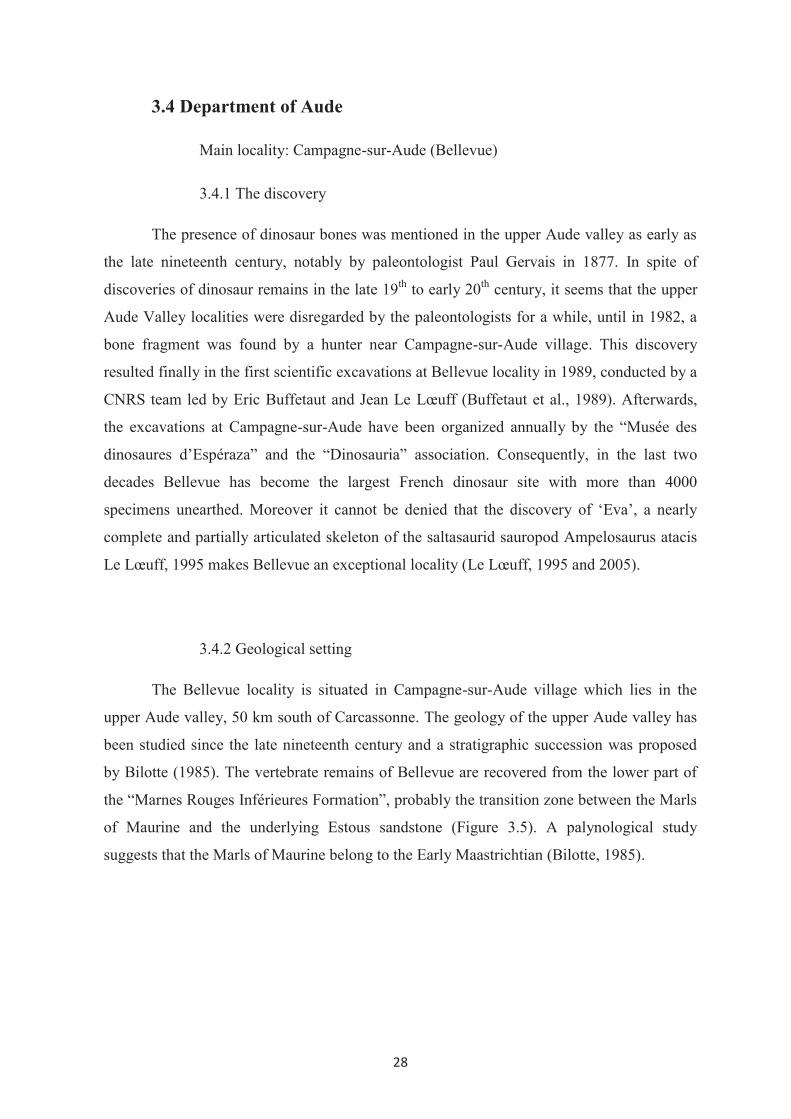
3.4.1 The discovery
The presence of dinosaur bones was mentioned in the upper Aude valley as early as
the late nineteenth century, notably by paleontologist Paul Gervais in 1877. In spite of
discoveries of dinosaur remains in the late 19th to early 20th century, it seems that the upper
Aude Valley localities were disregarded by the paleontologists for a while, until in 1982, a
bone fragment was found by a hunter near Campagne-sur-Aude village. This discovery
resulted finally in the first scientific excavations at Bellevue locality in 1989, conducted by a
CNRS team led by Eric Buffetaut and Jean Le Lœuff (Buffetaut et al., 1989). Afterwards,
the excavations at Campagne-sur-Aude have been organized annually by the “Musée des
dinosaures d’Espéraza” and the “Dinosauria” association. Consequently, in the last two
decades Bellevue has become the largest French dinosaur site with more than 4000
specimens unearthed. Moreover it cannot be denied that the discovery of ‘Eva’, a nearly
complete and partially articulated skeleton of the saltasaurid sauropod Ampelosaurus atacis
Le Lœuff, 1995 makes Bellevue an exceptional locality (Le Lœuff, 1995 and 2005).
3.4.2 Geological setting
The Bellevue locality is situated in Campagne-sur-Aude village which lies in the
upper Aude valley, 50 km south of Carcassonne. The geology of the upper Aude valley has
been studied since the late nineteenth century and a stratigraphic succession was proposed
by Bilotte (1985). The vertebrate remains of Bellevue are recovered from the lower part of
the “Marnes Rouges Inférieures Formation”, probably the transition zone between the Marls
of Maurine and the underlying Estous sandstone (Figure 3.5). A palynological study
suggests that the Marls of Maurine belong to the Early Maastrichtian (Bilotte, 1985).

29
Figure 3.5 The geographical and geological location of Bellevue locality and geological
boundary of Early Maastrichtian in the Aude Valley. (Modified from BRGM, geologic map
Notice Quillan 1:50,000)
The sedimentary strata of this site are composed of purplish red sandy clay
irregularly interbedded with sandstone lenses. The rust-colored sediments are related to
oxidation and indicate an ancient floodplain. The presence of interbedded coarse-grained
deposits and fresh water faunal remains suggest a fluvial environment. According to the
facies assemblage, the site is interpreted as presumably the overbank deposits of a
meandering stream (Leclerc, 2007). As in other continental Late Cretaceous localities in
southern France, the vertebrate assemblage of Bellevue includes bony fish, turtles,
crocodiles, pterosaurs and dinosaurs, among which Rhabdodon is one of the most common
taxa in this site.

30
References
Bilotte, M. 1985. Le Crétacé supérieur des plates formes est-pyrénéennes. Strata série II 5,
1 – 438.
Buffetaut, E. 2005. Late Cretaceous vertebrates from the Saint-Chinian area (southern
France): a review of previous research and an update on recent finds. Acta
Palaeontologica Romaniae 5, 39-48.
Buffetaut, E., Le Lœuff, J. 1991. Une nouvelle espèce de Rhabdodon (Dinosauria,
Ornithischia) du Crétacé supérieur de l’Hérault (Sud de la France). Compte rendu de
l’Académie des Sciences de Paris, Série II 312, 943-948
Buffetaut, E., Mechin, P., Mechin-Salessy, A. 1988. Un dinosaure théropode d’affinités
gondwaniennes dans le Crétacé supérieur de Provence. Compte rendu de
l’Académie des Sciences de Paris Série II 306, 153-158.
Buffetaut, E., Clottes, P., Cuny, G., Ducrocq, S., Le Lœuff, J., Martin, M., Powell, J. E.,
Raynaud, C., Tong H. 1989. Les gisements de dinosaures maastrichtiens de la haute
vallée de l’Aude (France) : Premiers résultats des fouilles de 1989. Compte rendu de
l’Académie des Sciences de Paris 309, 1723 – 1727.
Buffetaut E., Le Lœuff J., Mechin P., Mechin-Salessy, A. 1995. A large French
Cretaceous bird, Nature 377, p.110.
Buffetaut, E., Le Lœuff, J., Tong, H., Duffaud, S., Cavin, L., Garcia, G., Ward, D.,
Association culturelle, archéologique et paléontologique de Cruzy. 1999. Un
nouveau gisement de vertébrés du Crétacé supérieur à Cruzy (Hérault, Sud de la
France). Comptes Rendus de l’Académie des Sciences de Paris, II 328, 203-208.
Buffetaut, E., Mechin, P., Mechin-Salessy, A. 2000. An archaic bird (Enantiornithes) from
the Upper Cretaceous of Provence (Southern France). Comptes Rendus de
l'Académie des Sciences de Paris 331, 557-561.

31
Buffetaut E., Mechin P., Mechin-Salessy A. 2006. An azhdarchid pterosaur from the Upper
Cretaceous of Provence (southern France). In: Z. CZIKI, Ed., Mesozoic and
Cenozoic vertebrates and paleoenvironments. Tributes the career of Professor Dan
Grigorescu. – Editura Ars Docendi, Bucuresti, 95-100.
Broin, F., Buffetaut, E., Cappetta, H., Kerourio, P., Koeniger, J. C., Russell, D. E., Secretan,
S., Sigogneau-Russell, D., Taquet, P., Wenz, S. 1980. Nouvelles découvertes de
vertébrés maestrichtiens dans le gisement de Fox-Amphoux (Var), in 8e Réunion
Annuelle des Sciences de la Terre, Marseille, p. 68.
Chanthasit, P., Buffetaut, E. 2009. New data on the Dromaeosauridae (Dinosauria:
Theropoda) from the Late Cretaceous of southern France. Bulletin de la Société
géologique de France 180(2), 145-154.
Depéret C. 1899. Aperçu sur la géologie du chaînon de Saint-Chinian. Bulletin de la Société
géologique de France 27,686-709
Depéret C. 1900a. Sur les dinosauriens des étages de Rognac et de Vitrolles au pied de la
Montagne Noire. Comptes Rendus de l'Academie des Sciences Paris 130, 637-639
Depéret C. 1900b. Sur des restes de dinosauriens du Crétacé supérieur de la région de Saint-
Chinian, Bulletin de la Société géologique de France 28, 107-108
Dutour, Y., Berton, S. 2007. Fouilles paléontologiques préventives de l’autoroute A8 entre
Châteauneuf-le-Rouge et Saint-Maximin, Résultats partiels. Unpublished report, 1-
28.
Garcia, G., Pincemaille, M., Vianey-Liaud, M., Marandat, B., Lorenz, E., Cheylan, G.,
Cappetta, H., Michaux, J., Sudre, J. 1999. Découverte du premier squelette presque
complet de Rhabdodon priscus (Dinosauria, Ornithopoda) du Maastrichtien inférieur
de Provence. Comptes Rendus de l’Académie des Sciences de Paris 328, 415–421.
Gervais P. 1877. De la structure des coquilles calcaires des œufs et des caractères qu’on peut
en tirer. Comptes Rendus de l’Académie des Sciences de Paris 84, 159-l65.
Lapparent, A.F. 1947. Les dinosauriens du Crétacé supérieur du Midi de la France.
Mémoires de la Société géologique de France 56, 1-54.

32
Lapparent A.F. 1954. Nouvelle révision des gisements a Dinosauriens de la région de
Saint-Chinian (Hérault). Bulletin de la Société géologique de France 4, 409-413
Le Lœuff, J., Buffetaut E., Martin, M. 1994. The last stages of dinosaur faunal history in
Europe: a succession of Maastrichtian dinosaur assemblages from the Corbieres
(southern France). Geological Magazine, Cambridge 131(5), 625-630.
Le Lœuff, J. 1995. Ampelosaurus atacis (nov. gen., nov. sp.), un nouveau Titanosauridae
(Dinosauria, Sauropoda) du Crétacé supérieur de la Haute Vallée de l’Aude (France).
Comptes Rendus de l’Academie des Sciences Paris, series Iia 321, 693-699.
Le Lœuff, J. 2005. Osteology of Ampelosaurus atacis (Titanosauria) from Southern France.
In: Tidwell, V., Carpenter, K. (Eds.). Thunder-Lizards: The Sauropodomorph
Dinosaurs. Bloomington: Indiana University Press, 115–137.
Leclerc, Y. 2007. Taphonomie d’un gisement a dinosaures du Crétacé supérieur de la Haute
Vallée de l’Aude. Rapport de TER du Master 1 de l’Université Paris-Sud. 1-34.
(unpublished)
Matheron, P. 1869. Notice sur les reptiles fossils des dépôts fluvio-lacustres crétacé du basin
à lignite de Fuveau. Mémoires de l’Académie impériale des Sciences, Belles-Lettres
et Arts de Marseille, 1-39.
Miquel J. 1897. Note sur la géologie des terrains secondaires et tertiaires du département de
l’Hérault, Bulletin de la Société d’Etudes des Sciences Naturelles de Béziers 19,31-
74.
Nopcsa F. 1929. Dinosaurierreste aus Siebenbtirgen. Geológica Hungarica. ser.
Palaeontologica 4, 1-76.
Pincemaille-Quillévéré, M. 2002. Description d’un squelette partiel de Rhabdodon priscus
(Euornithopoda) du Crétacé supérieur de Vitrolles (Bouches du Rhône, France).
ORYTOS 4, 39-70.
Tong H., Gaffney E.S., Buffetaut E. 1998. Foxemys, a new side-necked turtle
(Bothremydidae: Pelomedusoides) from the Late Cretaceous of France, American
Museum Novitates 3251, 1–19.

33
Westphal, M., Durand, J. P. 1990. Magnétostratigraphie des séries continentales fluvio-
lacustres du Crétacé supérieur dans le synclinal de l'Arc (région d'Aix-en- Provence,
France), Bulletin de la Société géologique de France 6, 609- 620.

34

35
Chapter 4:
Osteology of Rhabdodon from the Late Cretaceous of
southern France
4.1 Material and Methods
The material described in this work consists of both cranial and postcranial elements
from the main localities in southern France mentioned in the previous chapter. The majority
of specimens has been found in the course of the systematic excavations organized by
different institutions; Association culturelle archéologique et paléontologique de l'Ouest
Biterrois (ACAP) and Musée de Cruzy, Dinosauria and Musée des Dinosaures d’Espéraza,
Muséum d’Histoire Naturelle Aix-en-Provence, Centre National de la Recherche
Scientifique; and collected for several years in various French collections. Some of the
specimens were found in the course of my thesis, during which I have had the opportunity to
participate in excavations (in 2005-2009) with ACAP at Cruzy and the Dinosauria
association at Espéraza. Some of those recent specimens were prepared by me in the
laboratories of Musée de Cruzy, Musée des Dinosaures, Espéraza and University Claude
Bernard Lyon 1. Most of specimens referred to Rhabdodon are isolated except two partial
articulated skeletons from Vitrolles which are housed in the Muséum d’Histoire Naturelle d’
Aix-en-Provence and were described by Pincemaille-Quillévéré (2002) and Chanthasit et al.
(in review). The osteological description of undescribed material has been done and the
anatomical measurements of specimens are shown in Appendix II.
Institutional abbreviations:
CM – Collection Méchin, Vitrolles, France
FSL – Université Claude Bernard Lyon1, Villeurbanne, France
MC – Musée de Cruzy, France
MDE – Musée des Dinosaures, Espéraza, France
MHN_AIX_PV – Muséum d’Histoire Naturelle Aix-en-Provence, France
MNHN – Muséum National d'Histoire Naturelle, Paris, France

36
SYSTEMATIC PALEONTOLOGY
Order ORNITHISCHIA Seeley, 1887
Suborder ORNITHOPODA Marsh, 1881
EUORNITHOPODA (sensu Weishampel 1990)
Infraorder IGUANODONTIA Sereno, 1986
Family RHABDODONTIDAE Weishampel, Jianu, Cziki & Norman, 2003
Rhabdodon Matheron, 1869
4.2 Description
4.2.1 Skull and mandible
The cranial material of Rhabdodon is poorly known. Some elements including the
maxilla, quadrate, braincase, dentary, surangular and teeth have been found isolated and
described from various localities in southern France (Matheron, 1869; Lapparent 1947;
Buffetaut and Le Lœuff 1991; Pincemaille-Quillévéré, 2002; Pincemaille-Quillévéré et al.
2006). In this work, I will describe some additional cranial elements, most of them having
been discovered at a locality near the village of Quarante (Hérault). These remains from
Quarante were found in the same area of a few square meters. It can be assumed that these
elements belong to a single individual. The description includes also some cranial material
from Cruzy, Montouliers, Bellevue (Campagne-sur-Aude) and Fox-Amphoux.
I. Premaxilla (MC- QR 1, QR 2, QR 3, QR 4, QR 6, CR.QR 3,)
A bone fragment MC-QR.02 (Figure 4.1D) is the dorsal process of a left premaxilla.
MC-QR 1, 3, 4, and 6 are compounded and form the maxillary process (Figure 4.1E) which
hypothetically belongs to the same left premaxilla as MC-QR.2. The right premaxilla MC-
CR.QR.03 (Figure 4.1A-C) is more complete but it is partly crushed on the lateral surface
and its anterior tip is missing.
Dorsally, the nasal process of the premaxilla forms a posterior tapering process. This
process is triangular in cross section and has a sharp ridge on the lateral surface; this ridge is

37
Figure 4.1. Right premaxilla MC-CY.QR.3 in lateral (A), medial (B) and caudal (C) views.
A fragment of left premaxilla MC-QR.2 (D), MC-QR 1, 3, 4 and 6 (E) in medial view. A
reconstruction of left premaxilla in lateral view. Abbreviations : ext = external naris, mx.p =
maxillary process, mx.s = maxillary suture, n.f = narial fossa, n.p = nasal process.
expanded anteriorly into a convex dorsal and anterior surface on the main body of the
premaxilla. Ventral to this convex surface, the moderately large narial fossa is expanded
anteroventrally and has a thin posterior edge which forms the anterior rim of the external
naris. The narial fossa is limited posteroventrally by the posterior process (maxillary
process) of the premaxilla. The posterior process is elongate, extends lateroposteriorly and is
directed slightly upward at the posterior end, forming the ventral rim of the external naris.
The external naris is consequently relatively longer than tall.
The medial surface of the premaxilla is flat and scarred by the ligaments for the
attachment with the other premaxilla. There is a longitudinal groove to receive the
anteromedial process of the maxilla. The sutured groove with the maxilla is significantly
longer than in Zalmoxes, and this corresponds to the fact that the maxilla of Rhabodon has a
longer premaxillary process than that of Zalmoxes. The maxillary suture is delimited
ventrally by a scarred bony plate which also forms the roof of the buccal cavity. This plate is
38
enlarged but thinned posteriorly. Above the maxillary suture, there is a thin ridge which
extends posteriorly on the posterior process of the premaxilla. The ventral border of the
premaxilla is narrow and rugose and there is no evidence of premaxillary teeth.
II. Maxilla (MC-QR 9; MDE-C3.111; MHN_AIX_PV.2008.1.42; FSL
530513)
The maxilla of Rhabdodon is one of the rarely found cranial elements, although
maxillary teeth are found at most localities in southern France. From previous works, the
maxilla of a juvenile or young adult Rhabdodon from a Late Campanian locality at Velaux
described by Thouand (2004) seems to be the most complete specimen, however its anterior
part is missing.
The new maxilla from Quarante (MC-QR 9) (Figure 4.2 A, B) appears to be more
complete in the anterior part and the dorsal processes. It is characterized by a very
prominent, long premaxillary process that fits into the recess of the posterior process of the
premaxilla. Contrary to MDE-C3.111 (Figure 4.2 E), the premaxillary process of the maxilla
is relatively short. The premaxillary process is triangular in cross-section, offset medially
and it bears a step-like ridge on the medial surface. The anterior end of the maxilla is
grooved and scarred. Dorsally, the premaxillary process forms a sharp, slightly dorsal
curved ridge extending to the most rostral end and this ridge is limited at the anterior end of
the tooth row. A deep oval depression is present laterally anterodorsal to the tooth row. This
depression is also present in MDE-C3.111 and it is presumably the attachment area for the
conjoined anterior tip of the vomers (Weishampel et al. 2003).
Posterior to the premaxillary process, the medial surface of the maxilla is convex
transversely and forms a rounded dorsal bar above the tooth row. Dorsally, the maxilla
forms a thin, flat sheet-like process. This process is anteriorly curved upward and may
contact the nasal. The posterior edge of this process forms the anterior margin of the
antorbital fenestra. Posteriorly, another process projects vertically. The latter process is
smaller, stouter than the more anterior one and it has a slightly twisted thickened distal end.
This process was presumably overlapped by the lacrimal.
The lateral wall of the tooth-bearing region of the maxilla MC-QR 9 is compressed
and fractured. The lateral foramina cannot be observed in this specimen. Numerous
foramina are visible on the lateral aspect of the maxilla MDE-C3.111. In a juvenile
specimen MHN_AIX_PV.2008.1.42 (Figure 4.2 F, G), the jugal suture of the maxilla forms

39
a wedge-shaped overhang on the posterior part of the lateral wall. The posterior end of the
maxilla is blunt, ridged and grooved and this is the sutural area for the ectopterygoid.
The medial wall of the tooth-bearing portion is marked by the alveolar foramina
(Figure 4.2 C, D). The alveolar foramina are connected to each other by a neurovascular
groove. The maxilla MC-QR 9 contains ventrally eleven tooth positions while
MHN_AIX_PV.2008.1.42 has nine and the incomplete maxilla MDE-C3.111 has eight tooth
positions.
Figure 4.2 Right maxilla MC-QR 9 in lateral (A) and medial (B) views. A fragment of left
maxilla FSL 530513 in lateral (C) and medial (D) views. Right maxilla MDE-C3.111 (E) in
lateral view. Left maxilla MHN_AIX_PV.2008.1.42 in lateral (F) and ventral (G) views.
Abbreviations: al.f = alveolar foramen, aof = antorbital fenestra, ect.s = ectopterygoid
suture, f.t = functional tooth, ju.s = jugal suture, pmx.p premaxillary process, r.t =
replacement tooth, v.f = fossa for the attachment with the vomer.

40
III. Quadrate (CM-397, 501, 653, 718; MC-CY.QR 5, CY.QR 6)
Two forms of quadrate can be distinguished; the quadrate MC-CY.QR.5 (Figure 4.3
A-C) is more robust than others (CM-397, 501, 653, 718). The upper end of the quadrate has
a relatively small, convex, oval articular head which fits to the squamosal. This head slightly
projects posteriorly from the main shaft of the quadrate. Caudal to and beneath the dorsal
head, there is a thin buttress which extends vertically downward then curves medially to the
main body of the quadrate. Below this buttress, the quadrate shaft is slightly bowed
anteroposteriorly and meets the mandibular articulation at the distal end.
Rostrally, on the quadrate shaft two wings are developed. The first wing is a
lateroanteriorly expanded thin sheet of the jugal ala which runs along most of the quadrate
length. The anterior margin of the jugal ala is dominated by a scarred depression for the
quadratojugal suture, the limit of the suture being marked by a rough edge. The jugal ala is
incomplete in all known specimens so that the paraquadratic foramen cannot be observed.
On the medial side of the bone, another wing arises ventrally above the ventral limit of the
lateral wing (jugal ala) and expends dorsomedially to form the pterygoid ala. In MC-CY.QR
5 the pterygoid ala is developed distinctively far from the distal articulation and strongly
curved ventrally by comparison with other specimens. The pterygoid ala is thick in the
ventral part, thinning dorsally and eventually joins the quadrate head. Between these two
alae, the quadrate forms a concave heavily scarred surface along its length.
The distal articular condyles are ventrally convex, asymmetrically developed. The
lateral condyle is rounded, slightly larger and extends distally, it articulated with the
surangular, while the medial condyle is rather narrow and directed dorsomedially away from
the lateral one. The mandibular articulation of the robust specimen (MC-CY.QR 5) is
significantly broader than the main body of the quadrate.

41
Figure 4.3 Right quadrate MC-CY.QR 5 in lateral (A), anterior (B) and medial (C) views.
Right quadrate CM-397 in lateral (D), anterior (E), medial (F) and posterior (G) views.
Abbreviations: bu = buttress, h = quadrate head, ju.a = jugal ala, lc = lateral condyle, mc =
medial condyle, pt.a = pterygoid ala, qj.s = quadratojugal suture.

42
IV. Frontal (MC-QR 8)
Only the right frontal MC-QR8 can be identified (Figure 4.4). The posterior end is
not completely preserved, however the frontal is longer than wide. Dorsally, the frontal is
quite flat except at the suture with the postorbital where the frontal is slightly flared upward.
The lateral rim bears well developed insertion markings and curves rostocaudally. In the
anterior part, the frontal edge for the nasal and for the adjacent frontal is thin. The
interfrontal suture is thickened in its more posterior part and it is straight. Ventrally, the
frontal shows a complex concave surface representing the impression of the roof of the
endocranial cavity. A strongly curved ridge delimits the orbital roof of the frontal from the
other concave surfaces.
Figure 4.4 Right frontal MC-QR8 in dorsal (A), ventral (B) and lateral (C) views.
Abbreviations: n.s = nasal suture, orb.r = orbital roof, po.s = postorbital suture.

43
V. Braincase
The braincase of Rhabdodon is known from several localities in southern France and
some specimens have been describeds by Pincemaille-Quillévéré et al. (2006). More
recently I have described two new specimens identified as braincases of Rhabdodon from
localities at Fox-Amphoux and Cruzy and presented them in a poster communication. All
braincases from southern France are compared between them and with other ornithopods in
that work which I attach here as the following note:

44

45
European Association of Vertebrate Palaeontologists, Extraordinary Meeting
February 9-14, 2009 Brussels, Belgium
Poster session
BRAINCASE OF RHABDODON (DINOSAURIA: ORNITHOPODA) : NEW SPECIMENS FROM THE LATE CRETACEOUS OF SOUTHERN FRANCE.
Phornphen CHANTHASIT
UMR 5125 - Paléoenvironnement & Paléobiosphère, Université Claude Bernard Lyon1,
2 rue Raphaël Dubois 69622 CEDEX Villeurbanne, France,
Introduction
Since the first description in 1869 of Rhabdodon priscus from the early
Maastrichtian of La Nerthe (Bouches-du-Rhône) by Matheron, Rhabdodon remains have
been found continually in several Campanian-Maastrichtian localities in Europe, especially
in southern France. However, the majority of specimens of Rhabdodon are postcranial
elements while cranial remains have rarely been found, and they are not well preserved,
particularly the braincase. A detailed description of the Rhabdodon braincase was given by
Pincemaille-Quillévéré et al.(2006) even though it was based on incomplete specimens.
Recently, two braincases of ornithopod dinosaurs have been found at the Massecaps
locality near the village of Cruzy (Hérault) and at the Bastide-Neuve locality at Fox-
Amphoux (Var) (Figure 1). Both localities are considered to be Late Campanian- Early
Maastrichtian in age (Buffetaut et al., 1999; Westphal et Durand, 1990) and they have
yielded abundant remains of Rhabdodon and other vertebrates.
Description
The braincases MC-M1575 and CM-699 (Figure 2 and 3), belong to juvenile
individuals. They are more complete than those of Rhabdodontidae including Zalmoxes
described in previous works (Weishampel et al., 2003; Pincemaille-Quillévéré et al., 2006).
The occipital condyle comprises the kidney-shaped basioccipital and the condylids
of the exoccipitals. The basioccipital forms the ventral margin of the foramen magnum
while the exoccipitals form the lateral and partial dorsal walls of the foramen magnum.
Relative to the size of the occipital condyle, the lateral walls of the foramen magnum of
Rhabdodon are shorter than those of Zalmoxes.
As in Camptosaurus, Hypsilophodon, Dryosaurus, and Zalmoxes, the supraoccipital
is not excluded from the dorsal margin of the foramen magnum whereas it is in Iguanodon,

46
Ouranosaurus, Tenontosaurus and hadrosaurids. The supraoccipital is inclined with a
tapered ascending process contacting the parietals dorsally, and it also extends laterally with
a small hump to overlap the dorsal border of the paroccipital process.
The paroccipital processes of Rhabdodon are long, slightly curved and oriented
posterolaterally. The distal ends are slightly expanded and point ventrally. To the contrary,
the paroccipital processes of Tenontosaurus are short and oriented more mediodorsally. In
Zalmoxes, the paroccipital processes are more curved and oriented more dorsally than in
Rhabdodon.
The basioccipital is longer than the basisphenoid as in Tenontosaurus and
Zalmoxes, while it is the reverse in Thescelosaurus and Zephyrosaurus, and they are
subequal in Hypsilophodon. The basipterygoid processes of the braincase of Rhabdodon are
relatively longer and directed more laterally than those of Zalmoxes.
Although the major cranial nerve foramina are hardly visible in both specimens, the
braincase of Rhabdodon MC-M4 (Pincemaille-Quillévéré et al., 2006) shows that the
distribution of cranial nerves along the lateral surface of the braincase is homologous to that
of Tenontosaurus.
ACKNOWLEDGEMENTS
- Musée de Cruzy, France
- Patrick and Annie Méchin, Collection Méchin, Vitrolles, France.
- The Natural History Museum, London, UK
- The Institute for the Promotion of teaching Science and Technology, Bangkok, Thailand

47
REFERENCES
Buffetaut E., Le Lœuff J., Tong H., Duffaud S., Cavin L., Garcia G., Ward D., l’Association
culturelle, archéologique et paléontologique de Cruzy (1999). - Un nouveau gisement de
vertébrés du Crétacé supérieur à Cruzy (Hérault, Sud de la France). - C. R. Acad. Sci. Paris,
328, 203-208.
Pincemaille-Quillévéré, M., Buffetaut, E., Quillévéré, F., 2006. Osteological description of the
braincase of Rhabdodon (Dinosauria, Euornithopoda) and phylogenetic implications. Bull.
Soc. Géol. Fr, 177(2), 97-104.
Weishampel, D.B., Jainu, C-M, Csiki Z., Norman, D.B. 2003. Osteology and Phologeny of
Zalmoxes (N.G.), an unsual Euornithopod Dinosaur from the latest Cretaceous of Romania.
Journal of the Systematic Paleontology 1(2), 65-123.
Westphal, M. and Durand, J.P., 1990. Magnétostratigraphie des continentale fluvio-lacustres de
Crétacé supérieur dans le clinal de l’Arc (région d’Aix-en-Provence). Bull. Soc. Géol. Fr.,
4, 609-620.

48
Figure 1. A map showing the localities, 1 - Cruzy (Hérault) and 2 - Fox-Amphoux (Var) in southern France and (on the right) Massecaps site near Cruzy.
Figure 2. Braincase MC-M1575 from Massecaps, Cruzy in occipital (A-B), right lateral (C-D), dorsal (E) and ventral (F) views. Abbreviations : bo – basioccipital, bs – basisphenoid, exo – exoccipital, fm – foramen magnum, fo – foramen ovale lsp – laterosphenoid, op - opisthotic, P – parietal, po – paroccipital process, pro – prootic, ps – parasphenoid, so – suparoccipital.

49
Figure 3. Braincase CM-699 from La Bastide Neuve, Fox-Amphoux in occipital (A-B), dorsal (C), right lateral (D-E) and ventral (F) views. See abbreviations in Figure 2.
Figure 4. A : Rhabdodon. Braincase MC-M4 in left lateral view. B : Zalmoxes robustus. Braincase BMNH R.3401 in occipital view. See abbreviations in Figure 2.

50

51
VI. Dentary (CM-443, 587, 709, MC-M 23, M 27, CY_QR 1, CY_QR 2;
MDE-C3.1280, MDE-C3.1407)
The dentary of Rhabdodon shows variability both in size and in shape. The dentary
of Rhabdodon bears from nine to eleven alveoli. The dentary length ranges from 126 to 368
mm. (See Table of dentary measurements in Appendix II).
Dorsally, the dimensions of the anteriormost and posteriormost alveoli are relatively
smaller than those from the middle portion of the alveolar row. The tooth row of the dentary
is more or less straight or slightly concave on the lateral edge (Figure 4.5). The concavity of
the tooth-bearing portion is well marked in the dentary of Rhabdodon septimanicus
(Buffetaut and Le Lœuff, 1991). This diagnostic character of R. septimanicus is present in
the remarkably large dentaries from Quarante (MC-CY_QR 1 and MC-CY_QR 2) (Figure
4.6). In dorsal view, the alveolar row of MC-CY_QR 1 and MC-CY_QR 2 is remarkably
concave outward while the mandibular symphyseal region is curved inward.
Immediately anterior to the front end of the tooth row, the dorsal dentary margin is
steeply inclined and converges with the upward sloping ventral margin to form the
symphyseal rim. The symphyseal region is rough and bears ridges and grooves on the
medial surface. On the anterolateral surface, prominent depressions are developed with large
and numerous small foramina which are associated with the attachment of the predentary. In
MC-CY_QR 1 and MC-CY_QR 2, the symphyseal region is slightly bent downward and
displaced more anteriorly compared with other specimens, as the dorsal and ventral margins
are farther prolonged anteriorly to form the dentary symphysis.
Laterally, in most specimens the dorsal and ventral margins are mainly straight and
parallel while some specimens have a curved ventral edge (MC-CY_QR 1, MC-CY_QR 2).
The lateral surface of the dentary is convex dorsoventrally. The buccal platform with a
laterally rounded edge along the tooth row is prominent in CM-443, CM-587, CM-709, MC-
M 23 and MDE-C3.1407 (Figure 4.5). This platform is sloped down and forms only a
convexity along the lateral wall of the dentary MC-CY_QR 1, MC-CY_QR 2 and MDE-
C3.1280. Two large foramina are visible laterally on the oblique platform or in the upper
half of the lateral surface somewhat just posterior to the dentary mid-length. Numerous
small foramina are also present on the lateral wall in the lower part of the dentary.
The coronoid process ranges in shape from a robust to a thinner and elongate
projection. It rises and projects dorsoposteriorly at various angles to the dentary body,
ranging from 90° to 125° in lateral view. In MDE-C3.1280 the distal end of the coronoid
process is slightly curved and directed anteriorly at the dorsal end. The coronoid process lies

52
Figure 4.5 Left dentary MC-M 23 in lateral (A), medial (B) and dorsal (C) views. Right
dentary CM-443 in dorsal (D) and medial (E) views. Abbreviations: al.for = alveolar
foramen, al.gr = alveolar interconnection groove, b.p = buccal platform, cor.p = coronoid
process, mek.c = Meckelian canal, pd.s = predentary suture, r.t = replacement tooth, sym =
dentary symphysis, v.for = neurovascular foramen.

53
Figure 4.6 Right dentary MC-CY.QR 1 in lateral (A), medial (B) and dorsal (C) views. See
abbreviations in figure 5.
laterally to the most posterior teeth. Laterally, the coronoid process is striated at the distal`
end. The medial surface of the coronoid process has an oblique striated facet and a large
depression with minor ridges distally. Posteriorly, the coronoid process is irregular and has a
deep dominant groove associated with the protruding bone for the attachment to the
surangular.
Medially, the dentary is gently convex dorsoventrally and marked by the alveolar
foramina and the alveolar interconnection groove. The Meckelian canal ventrally extends
almost the total dentary length. The canal is deep, wide open posteriorly and then it becomes
gradually narrower and shallower anteriorly to form only a shallow groove before the
symphyseal region.
VII. Angular (MC-QR 22)
The left angular MC-QR 22 (Figure 4.7) is a thin long bone and has an elongate
triangular shape in lateral view. The anterior and posterior extremities of the bone are
tapering. The anterior end is narrow, triangular in cross section while the posterior one is
more prominent, oval in cross section and bears clear ridges and grooves. Laterally, the
anterior part shows a gently concave and scarred surface, which was overlapped by the
posterior part of the dentary. The upper edge of the bone is thin while the ventral border is

54
thickened to form the lower edge of the jaw. Medially, the bone is concave ventrally and
strongly ridged. On the dorsomedial surface, a small shallowly depressed area extends along
the posterodorsal part of the angular to fit to the ventral part of the surangular.
Figure 4.7 Left angular MC-QR 22 in lateral (A) and medial (B) views. Abbreviations: den.s
= dentary suture surface, sur.s = surangular suture surface.
VIII. Surangular (MHN_AIX_PV.1999.10)
The right surangular MHN_AIX_PV.1999.10 is well preserved, crescent-shaped in
lateral view, convex laterally and concave medially (Figure 4.8). It consists of two anterior
and posterior processes which are both directed dorsally. The anterior process is transversely
thick with rounded lateral and medial borders. The anterodorsal end of this process is
thickened, however the anteriormost part of the surangular, where it is strengthened with the
posterior edge if the coronoid process of the dentary is not preserved. Beneath this process,
the bone forms a curved thin wall which slots inside the posterior part of the dentary and
there is probably an anterior surangular foramen but it is not completely preserved in this
specimen. More posteriorly the dorsal margin is extended outward and forms a lip-like
projection with a rough lateral apex. This lip forms the lateral margin of the glenoid for the
quadrate lateral condyle and the attachment area for the jaw joint ligaments. On the medial
side, anteroventrally to the prominent lip, there are two foramina, the anterior foramen being
slightly bigger than the posterior one. These foramina are variously present in iguanodontian
ornithopods but absent in hadrosaurids (Norman, 1998).

55
Figure 4.8 Right surangular MHN_AIX_PV.1999.10 in lateral (A), medial (B), dorsal (C)
views. Abbreviations: an.s = angular suture, cor = coronoid portion of the surangular, for =
surangular foramen, gl = glenoid articulation with the quadrate, lip = lateral lip-like margin,
ret = retroarticular process.
Posterior to the quadrate cotylus, the surangular is tapered and directed upward,
forming the lateral portion of the retroarticular process. Ventrally, the suture with the
angular runs along most of the surangular length and there is also a narrow groove seen on
the medial side to fit firmly to the angular bone.
4.2.2 Dentition
Teeth of Rhabdodon are known from all localities, although they are mostly found
isolated. Rhabdodon is characterized by its distinctive fluted maxillary and dentary teeth.
I. Maxillary teeth (CM-297, 326, 554, 657, MC-M 4190, M 4196, M 4260;
MDE-C3.897, C3.1412; MHN_AIX_PV_ Fox)
The maxillary teeth (Figure 4.9) are subrectangular in outline, slightly taller than
wide. They are lens-shaped in cross-section. The buccal surface of the maxillary crown is
covered by a thick layer of enamel and bears several subequal, sharp ridges. These vertical
ridges are more or less parallel and there are smooth, longitudinal grooves lying between the

56
ridges. Marginal denticles are present along the mesial and distal margins of the crown and
also on the dorsal margin in the unworn crowns (Figure 4.9C). The density of ridges on the
buccal side of teeth does not appear to have increased during the ontogeny as shown by a
scatter plot of the relationship between the maxillary tooth width and the number of ridges
on the teeth in figure 4.10A. A well-defined cingulum forms the border of the crown and
separates it from the root. The tooth crown is wider than its root and the roots of fully
erupted and functional teeth are very long and taller than the crown.
Medially, the tooth is thinly enamelled and bears shallow longitudinal ridges. In
more mature teeth, the medial shallow ridges disappear and two asymmetrical oblique wear
facets are developed on the occlusal surface. These facets merge as a single sloping wear
facet with a sharp margin on the heavily worn teeth.
The teeth are closely packed together as shown by the groove on the mesial and
distal edges of the teeth and as seen in the maxilla FSL530513 (Figure 4.2C). From an
observation of the alveoli of the maxilla, the most anterior tooth seems to be the smallest and
the tooth size gradually increases and reaches its maximum about just distal to the middle of
the tooth row. The emplacement of the maxillary teeth cannot be precisely determined as a
complete maxillary dentition has not yet been found. There are two teeth per tooth family, as
reported by Weishampel et al. (2003). As seen in figure 4.9D, the erupting replacement
tooth is closely applied to the medial surface of the functional tooth. The adjacent teeth
appear to be at different stages of eruption but the replacement sequence is still poorly
understood.
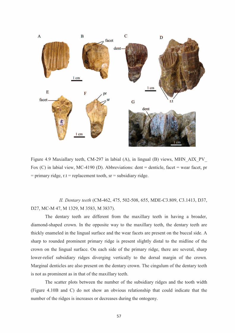
57
Figure 4.9 Maxiallary teeth, CM-297 in labial (A), in lingual (B) views, MHN_AIX_PV_
Fox (C) in labial view, MC-4190 (D). Abbreviations: dent = denticle, facet = wear facet, pr
= primary ridge, r.t = replacement tooth, sr = subsidiary ridge.
II. Dentary teeth (CM-462, 475, 502-508, 655, MDE-C3.809, C3.1413, D37,
D27, MC-M 47, M 1329, M 3583, M 3837).
The dentary teeth are different from the maxillary teeth in having a broader,
diamond-shaped crown. In the opposite way to the maxillary teeth, the dentary teeth are
thickly enameled in the lingual surface and the wear facets are present on the buccal side. A
sharp to rounded prominent primary ridge is present slightly distal to the midline of the
crown on the lingual surface. On each side of the primary ridge, there are several, sharp
lower-relief subsidiary ridges diverging vertically to the dorsal margin of the crown.
Marginal denticles are also present on the dentary crown. The cingulum of the dentary teeth
is not as prominent as in that of the maxillary teeth.
The scatter plots between the number of the subsidiary ridges and the tooth width
(Figure 4.10B and C) do not show an obvious relationship that could indicate that the
number of the ridges is increases or decreases during the ontogeny.

58
The root of the dentary teeth is labially curved, tapering and slightly narrower than
the crown width and it is longer than the crown. The mesial and distal sides of the root are
grooved, indicating a closely packed dentition. As in the case of the maxillary teeth, the
replacement sequence of dentary teeth cannot be determined.
Medially, the wear facet is shallowly concave, inclined buccally. The major facet is
formed on the distal portion of the occlusal surface and obliquely inclined toward the mesial
wear facet. The occlusal surface bears a single facet in the heavily worn teeth. The occlusal
facet bears microstriae oriented more or less perpendicularly to the transverse axis of the
tooth, as seen in dentary tooth MDE-D 37.

59
Figure 4.10 Scatter plots, (A) between the number of the ridges on the buccal surface of the
maxillary crown and the tooth width (Mx Mes-Dist L), (B) between the number of
secondary ridges mesial to the primary ridge on the lingual surface of the dentary crown and
the mesial width (Dent Mes W), (C) between the number of secondary ridges distal to the
primary ridge on the lingual surface of the dentary crown and the distal width (Dent Dist
W). (see Table of measurement in Appendix II)

60
4.2.3 Postcranial Skeleton
I. Axial skeleton
A complete articulated axial skeleton of Rhabdodon has never been found,
nevertheless partially articulated vertebrae have been reported from some localities,
especially from the late Campanian of the Vitrolles-Couperigne localities (Pincemaille-
Quillévéré, 2002; Chanthasit et al., in review). The vertebral column has been consequently
described on the basis of various isolated specimens from different localities. The total
number elements in the vertebral column cannot be determined.
I.1Cervical vertebrae and ribs (CM-86, 405, 441, 679; MC-M 30, M-4058)
Cervical vertebrae are rarely found. The total number of the cervical series cannot
be estimated. Some cervical remains have been recovered and are described here.
Only a single isolated atlas (CM – 86) (Figure 4.11) from Brégoux (Fox-Amphoux)
is known. The atlas consists of the intercentrum and two neural arches. The odontoid is
presumably unified as a part of the axis. In the atlas CM-86, the neural arch is fused to the
intercentrum as in the atlas of Ouranosaurus nigeriensis. The intercentrum is narrow and
subcrescentic. The anterior surface bears a large shallow depression for the occipital condyle
articulation. This depression is inclined obliquely to the long axis of the skull and expanded
dorsomedially toward the neural arches. The ventral edge of the intercemtrum is sharp while
the dorsal surface is rounded and strongly curved, resulting in a very narrow neural canal
compared with other ornithopods. Posteriorly, the intercentrum is convex because of a ridge
in the middle but concave dorsomedially for receiving the odontoid process (Figure 4.11C).
The neural arch is attached on the lateral side of the intercentrum. The neural arch continues
the depression formed by the intercentrum. Dorsally, the two neural arches are not fused to
each other even though they are also expanded medially (Figure 4.11B). An irregular
tapering horn-like process is developed dorsoposteriorly on the neural arch and curved
medially. The postzygapophysis is not well defined. The lateral and ventral surfaces are
rounded and show no evidence of diapophysis. This suggests that the cervical ribs are not
present yet on the atlas as in I. bernissartensis (Dollo, 1883) and Ouranosaurus (Taquet,
1976) whereas in I. atherfieldensis, Hypsilophodon, and maybe Tenontosaurus, the first pair
of cervical ribs is situated on the atlas (Hooley, 1925; Galton, 1974; Forster, 1990).

61
Figure 4.11. Atlas CM – 86 in anterior (A), dorsal (B) and posterior (C) views. Abbreviations: ic = intercentrum, na = neural arch, oc.c. = surface for occipital condyle, od.p = surface for odontoid process.
The axis is also rarely found. A single axis MC-M4058 (Figure 4.12) from the
Massecaps locality is not well-preserved. The odontoid process of the atlas is sutured in the
middle of the anterior facet of the axis centrum. The odontoid process is wedge-shaped,
triangular in lateral view and flattened dorsally. The odontoid process is tapering and
horizontally projects forward.
As preserved, the axis centrum is longer than wide and high and constricted
laterodorsally. It is difficult to distinguish the intercentrum of the axis from the centrum. The
neural spine arises between the prezygapophyses. The dorsal end of the neural spine is
broken, however it extends notably anteroposteriorly over the neural arch. The neural canal
is high and wide.
The prezygapophyses project anteriorly and face ventrolaterally as in Iguanodon
bernissartensis. The postzygapophyses are not preserved but seem to be situated lower than
the prezygapophyses. Although the lateral and posterior parts are damaged, on the left lateral
wall, a diapophysis is present on the lateral surface of the neural arch. Generally, the axis of
Rhabdodon is similar to that of Zalmoxes (Weishampel et al. 2003).

62
Figure 4.12 Axis MC-M4058 in left lateral (A) and anterior (B) views. Abbreviations: d = diapophysis, na = neural arch, nc = neural canal, od = odontoid process, prz = prezygopophysis.
Concerning the subsequent cervical vertebrae, four articulated anterior cervical
vertebrae have been discovered as part of the partial articulated skeleton of Rhabdodon
priscus from Vitrolles-Couperigne found in 1995. Pincemaille-Quillévéré (2002) described
the diagnostic features of these vertebrae which belong to the anterior part of the cervical
series. The centra are opisthocoelous, bearing well-developed neural spines with a cranial
orientation and the transverse processes extend anteroposteriorly. However, the
interpretation of the cervical vertebrae from Vitrolles is still uncertain because the
specimens are deformed.
Some other anterior cervical vertebrae are known from Massecaps (MC-M 30)
(Figure 4.13 A, B, C) and Bastide Neuve (CM-679) (Figure 4.13 D, E). Despite the fact that
MC- M 30 lacks the ventral part of the centrum, the neural arch and the zygapophyses are
quite well-preserved. It is the reverse in specimen CM-679, in which the centrum is
complete but the neural arch is incomplete.
The centrum of the anterior cervical vertebrae is as long as high and it is slightly
opisthocoelous to platycoelous. The anterior facet is broad, wider than high. The centrum is
transversely compressed, with concave lateral walls and a rounded ventral keel. The
parapophysis is situated just behind the lateral margin of the anterior articular facet, below
the neurocentral suture. This angular parapophysis is continued by a horizontal ridge at the
middle of the centrum. The neural arch is wide and robust, enclosing the sub-circular neural
canal. Anteriorly, the transverse processes project laterodorsally from the laterodorsal

63
surface of the neural arch. The transverse process supports the prezygapophysis dorsally as
well as the diapophysis laterally. The large curved postzygapophyses arise just after and
above the transverse processes. The postzygapophyses are widely divergent and prolonged
backward. They bear the large oval zygapophyseal facets which face ventrolaterally. The
neural spine is incompletely preserved, however it shows an oval, laterally compressed
neural spine base arising from the middle of the anterodorsal rim of the neural arch.
Although the size of the neural spine cannot be exactly estimated, it tends to be more
prominent than those of Zalmoxes which apparently are only a small conical spine.
Cervical CM-441 (Figure 4.13 F, G, H) is from the middle part of the cervical series.
The centrum is as long as wide and higher proportionally compared with the anterior
cervical vertebrae. The centrum is also compressed laterally and keeled ventrally. Compared
to the anterior cervicals, the neural arch of CM-441 is taller and supports more robust
transverse processes which are inclined more dorsally. The transverse processes still bear
both the prezygapophyses and the diapophyses. The articular facet of the prezygapophyses is
larger, erected upward and faces dorsomedially. The diapophysis is larger and developed
more posteroventrally on the transverse process. The parapophysis is situated more dorsally,
closer to the neurocentral suture. The postzygapophyses are less opened but project more
upward. The articular surface is also prominent and faces ventrolaterally as in the anterior
cervicals. The neural spine is displaced more posteriorly. As the main part of the spine is
broken, the orientation of the neural spine cannot be confirmed. However, I think the neural
spine of Rhabdodon seems not to be strongly oriented cranially, probably it is curved
initially anteriorly and subsequently turns backward. The cranial orientation of the spines of
the cervical vertebrae of Rhabdodon priscus from the Vitrolles-Couperigne locality is
presumably a result of deformation. Nevertheless, the neural spines of the cervical vertebrae
of Rhabdodon from Vitrolles-Couperigne are remarkable for their size which distinguishes it
from other ornithopod dinosaurs.
The cervical ribs are hardly known. The first pair of cervical ribs must have started at
the axis as mentioned above. According to the size and the position of the parapophysis and
the diapophysis, it is suggested that the cervical ribs of the anterior to mid cervical vertebrae
had well-separated double heads and that the capitulum was slightly larger than the
tuberculum.

64
Figure 4.13 Anterior cervical vertebrae, MC-M30 in anterior (A), left lateral (B) and dorsal (C) views, CM-679 in anterior (D) and left lateral (E) views. Mid-cervical vertebra CM- 441 in anterior (F), left lateral (G) and posterior (H) views. Abbreviations: d = diapophysis, na = neural arch, nc = neural canal, ns = neural spine, p = parapophysis, prz = prezygapophysis, poz = postzygapophysis. I.2 Dorsal vertebrae and ribs (CM-3, 142, 379, 380, 400, 416, 445, 446, 496,
498, 499, 564, 581, 527, 647, 710; MC- M 38, M 220, M 261, M 537, M 626, M 948, M
1080, M 1116, M 1131, M 1149, M 1699, M 2333, M 2828, M2705, M 3179, M 3395, M
3896, M 4012, M 4395, M 4532, MOB 56; MDE-C3 413, C3 973, C3 1400, C3 1556, C3
02.102, C3 02.137, C3 03.50; MNHN 8)
The most complete dorsal series was described by Pincemaille-Quillévéré (2002).
Sixteen dorsal vertebrae were found from a partial articulated skeleton of Rhabdodon

65
priscus from Vitrolles-Couperigne in 1995. This possibly indicates that Rhabdodon had at
least 16 dorsal vertebrae as in Iguanodon bernissartensis (Norman, 1980) and Ouranosaurus
nigeriensis (Taquet, 1976). The sixteen dorsal vertebrae described in that previous work are
not well preserved. Most of them were found as isolated centra or neural arches so that some
characters of dorsal vertebrae remain uncertain.
Additional isolated dorsal vertebrae described herein are more complete and well-
preserved though they are from different localities.
Anterior dorsal vertebrae (Figure 4.14) have a very short, higher than wide centrum.
The centrum of anterior dorsals is gently opisthocoeleous. The lateral wall is axially concave
and the ventral surface forms a thin keel in the middle. The centra from the middle dorsal
series (Figure 4.15) become longer and as wide as high while they are shortened and higher
than wide in posterior dorsal vertebrae (Figure 4.16). The articular facet is more circular
with a rounded edge through the anterior to middle series and the centrum becomes
platycoelous and amphicoelous in the posterior dorsal vertebrae. Laterally, the dorsal and
ventral margins of the centrum are parallel. The anterior and posterior articular facets of the
posterior dorsals are slightly convergent ventrally.
The neural arch of the anterior dorsal vertebrae is tall and wide then it becomes
shorter and narrower in more posterior elements. The neural arch extends from the anterior
surface to the posterior one along the centrum length in the anterior dorsals, then it is
slightly shortened through the subsequent dorsal vertebrae. The neural canal which is large
in the anterior dorsal vertebrae is reduced and rounder in cross-section in posterior
vertebrae.
The transverse processes are directed dorsolaterally (about 45 degree from the
vertical), curved and pointed slightly caudally in anterior dorsals. These processes become
shorter and reoriented more horizontally through the series. The parapophysis is positioned
on the lateral surface of the neural arch at the base of the transverse process in anterior
dorsals. Through the dorsal series, the parapophysis forms a vertically elongated concavity
and it gradually moves dorsally along the transverse process, closer to the diapophysis. The
parapophysis and the diapophysis are eventually merged into a large tubercle at the distal
end of the transverse process of the posterior dorsals (Figure 4.16). The prezygapophyses are
developed cranially on the proximal part of the transverse process of the anterior dorsal
vertebrae. They migrate medially closer to each other than on the cervical vertebrae and are
separately erected from the transverse process on the dorsal margin of the neural canal in

66
posterior dorsal vertebrae. The articular surfaces are slightly concave and they face
dorsomedially. The prezygapophyses are more prominent in more posterior dorsals.
The postzygapophyses of the anterior dorsal vertebrae are well separated and face
ventrolaterally but they do not extend as strongly backward as in the cervical vertebrae. The
postgygapophyses extend farther beyond the centrum in more posterior dorsals and the oval
articular surface becomes gradually larger in more posterior dorsals.
The neural spines in the proximal dorsal series are tall, forming more than a half of
the total height of the vertebra (See also fig.4 Chanthasit et al., in review). They are
compressed laterally, narrow, tapered upward and slightly curved posteriorly. The neural
spines are inclined more posteriorly in the mid-posterior series. The neural spines are
expanded anteroposteriorly as a large, long rectangular blade and gradually become longer,
being more than four times taller than the centrum in the most complete specimens (CM-
142, CM-44, CM-581 and MC-MN 32). The neural spines are axially longitudinal oval in
cross-section with sharp anterior and posterior ridges in the middle along the proximal
portion of the spines. The neural spine ends with a thick rugose dorsal tip. The neural spine
of some specimens (CM-142 and MC-MN 32) shows a slightly sigmoidal curvature in
anterior view (Figure 4.15 D, E). This may be a taphonomic effect.
Figure 4.14 Anterior dorsal vertebra CM-379 in anterior (A) and left lateral (B) views. Abbreviations: d = diapophysis, ns = neural spine, p = parapophysis, prz = prezygapophysis, poz = postzygapophysis.

67
Figure 4.15 Mid dorsal vertebrae, CM-581 in anterior (A), right lateral (B), posterior (C) views, CM-142 in anterior (D) and posterior (E) views and MC-MN32 in posterior-lateral view. Abbreviations: d = diapophysis, p = parapophysis, prz = prezygapophysis, poz = postzygapophysis, tp = transverse process.

68
Figure 4.16 Posterior dorsal vertebrae, CM-496 in anterior (A), right lateral (B) and posterior (C) views, MDE-C3.03.50 in right lateral (D) and posterior (E) views and MDE-C3.1400 in right lateral view. Abbreviations: pd = paradiapophysis, prz = prezygapophysis, poz = postzygapophysis, tp = transverse process.
Dorsal ribs are usually found fragmentary. They are of various sizes and shapes,
corresponding to their position along the dorsal series (Figure 4.17). Most of the dorsal ribs
are doubled-headed except for those articulating with the posterior dorsal vertebrae which

69
are single-headed. In anterior thoracic ribs, the capitulum and the tuberculum are widely
separated. The capitulum is supported by a long, slender capitular process with a slightly
expanded round articular surface. The tuberculum is at first sub-equal in size to the
capitulum then in more posterior ribs, the tuberculum is shortened, dorsoventrally expanded
and becomes more prominent than the capitulum. The articular surface of the tuberculum is
concave and oval in shape. Subsequently, the capitulum and the tuberculum move closer to
each other and the tubercular process disappears. The dorsal ribs are curved around the
thorax. On the proximal portion, the costal surface is anterodorsally convex and forms a
ridge continually from the tuberculum whereas it is concave posteriorly with a shallow
groove. The dorsal margin is proximally sharp while the ventral one is rounded along the
costal length. The dorsal ribs are anteroposteriorly compressed in the proximal part of the
shaft. The shaft is tapered at the distal tip in the anterior series but it gradually becomes
laterally compressed in its distal part in the more posterior ribs. In the posterior series, the
capitulum and the tuberculum are merged as a single articulation with the paradiapophysis
and costal shafts are short and tapered distally.
I.3 Sacrum (CM-621; MC-M 1233, M 1739, M 1882, PSP 1; MDE-C3 134, C3 135,
C3.04.72)
On the basis of the most complete sacral series of Rhabdodon from Vitrolles-
Couperigne (Pincemaille-Quillévéré, 2002; Chanthasit et al. (in review), the sacrum of
Rhabdodon is composed of six true co-ossified sacrals, which are possibly fused with the
sacrodorsal and the last dorsal vertebrae as in Iguanodon, Hypsilophodon and Zalmoxes
(Norman, 1980, 1986; Galton 1974; Weishampel et al. 2003; Godefroit et al. 2009). The
sacrum of Tenontosaurus consists of five fused true sacrals and a sacrodorsal. Besides the
sacrum from Vitrolles, the sacrum MC-PSP 1 (Figure 4.18) from Plo-Saint-Pons seems to be
the best preserved with six co-ossified vertebrae. The sacrum is arched anteroposteriorly.
The first vertebra of MC-PSP 1 is interpreted as a sacrodorsal. The centrum has a rounded
anterior articular surface and it is laterally compressed with a rounded keel on the ventral
surface as in the posterior dorsal vertebra. The transverse process is partly broken, situated
on the dorsal part of the neural arch. The centrum is posteriorly expanded and bears laterally
the first pair of sacral ribs. The first sacral ribs are developed at the joint between the
sacrodorsal and sacral one. The sacral ribs of sacral two to sacral five are also located at the
joint with the preceding sacral. The first sacral ribs are oriented caudally while the others are
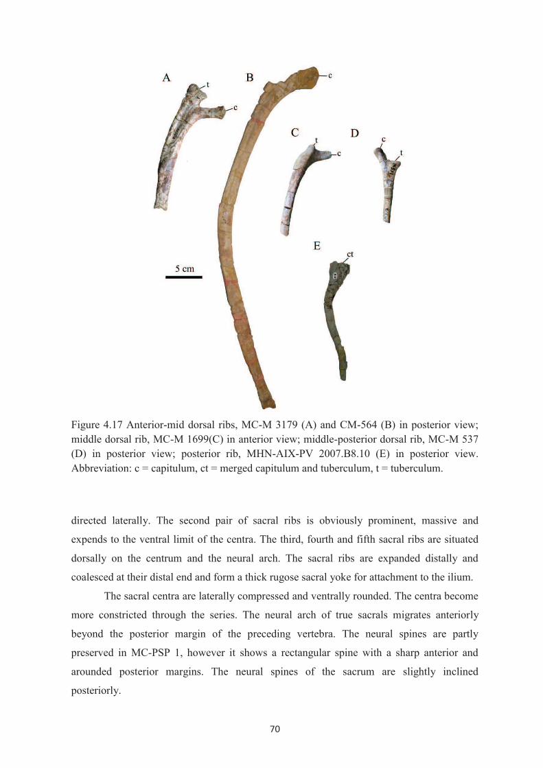
70
Figure 4.17 Anterior-mid dorsal ribs, MC-M 3179 (A) and CM-564 (B) in posterior view; middle dorsal rib, MC-M 1699(C) in anterior view; middle-posterior dorsal rib, MC-M 537 (D) in posterior view; posterior rib, MHN-AIX-PV 2007.B8.10 (E) in posterior view. Abbreviation: c = capitulum, ct = merged capitulum and tuberculum, t = tuberculum.
directed laterally. The second pair of sacral ribs is obviously prominent, massive and
expends to the ventral limit of the centra. The third, fourth and fifth sacral ribs are situated
dorsally on the centrum and the neural arch. The sacral ribs are expanded distally and
coalesced at their distal end and form a thick rugose sacral yoke for attachment to the ilium.
The sacral centra are laterally compressed and ventrally rounded. The centra become
more constricted through the series. The neural arch of true sacrals migrates anteriorly
beyond the posterior margin of the preceding vertebra. The neural spines are partly
preserved in MC-PSP 1, however it shows a rectangular spine with a sharp anterior and
arounded posterior margins. The neural spines of the sacrum are slightly inclined
posteriorly.

71
Figure 4.18 Sacrum MC-PSP 1 in right lateral (A, B) and ventral (C, D) views. Abbreviations: na = neural arch, ns = neural spine, s = sacral, sd = sacrodorsal, sy = sacral yoke, tp = transverse process.

72
I.4 Caudal vertebrae (CM- 2, 4, 8, 176, 378, 386- 389, 401- 404, 420- 422,
439, 447, 448, 454, 469, 491, 518, 525, 569, 575, 583, 585, 596, 601, 613, 614, 627, 646,
653, 688, A1, A3; MC-M 950, M 1403, M 1414, M 1800, M 2483, M 3589, M 4028, M
4119, MN 30, MN 36, PSP 5; MDE-C2.82.8, C3 28, C3 35, C3 36, C3 106-C3 110, C3 249,
C3 257, C3 337, C3 343, C3 344, C3 416, C3 504, C3 507, C3 611, C3 655, C3 775, C3
823, C3 881, C3 967, C3 980, C3 990, C3 995, C3 996, C3 1128 , C3 1136, C3 1145, C3
1152, C3 1152, C3 1156, C3 1176, C3 1177, C3 1183, C3 1191, C3 1197, C3 1282, C3
1300, C3 1414, C3 1421, C3 1551, C3 02.102, C3 02.137, C3 05.58, C3 08.55, C3 08.59,
RC 10-9, Vil 39; FSL 530515; MHN.AIX.PV.1999.76, 77, MHN_AIX_PV.2000.7,
MHN.AIX.PV.2001.12, 13, 16, 20, 112)
In the first caudal vertebrae, the centra are platycoelous, short and as wide as high,
with heart-shaped articular surfaces and a rounded ventral keel (Figure 4.19). The centra
retain the same shape but they are amphicoelous, increased in length and become gradually
higher than wide through the proximal to middle caudal series. Subsequently, the centra
become smaller in diameter but still proportionally lengthen along the tail. The lateral wall
of the caudals is compressed, concave anteroposteriorly, and then becomes flatter in
succeeding caudal vertebrae. Ventrally, the ventral keel is expanded anteriorly and
posteriorly and inclined upward to form the haemapophyseal facets for the chevrons. The
anterior haemapophyseal facet for the haemal arch is less prominent than the posterior one.
The haemapophyseal facets become insignificant in posterior caudal series. A ventral
groove is present in the middle caudal series and becomes less prominent in distal caudals.
The anterior caudal vertebrae bear transverse processes on the lateral wall of the
centrum below the neurocentral suture. The transverse processes are laterally projected
more or less horizontally and slightly curved cranially. The transverse processes decrease
gradually in size down the series and eventually disappear in more posterior caudals where
they become only an axial ridge on the dorsal half of the centra.
The neural arch of caudal vertebrae is low and narrow. The neural canal is small,
rounded in cross-section. In proximal caudals, the prezygapophyses are prominent and
extended anterodorsally and the articular facets are steeply inclined and face medially. The
articular facets become vertical, narrower and elongate cranially down the caudal series.
The postzygapophyses of the caudal vertebrae are less prominent than those of the
preceding vertebral series. Through the caudal series, the articular facets become smaller,
narrowly spaced and eventually merged as a single protuberance on the posterior margin of

73
the neural spine. The neural spine is still large, thick, straight and extended dorsally in
anterior to middle caudal vertebrae. The neural spine becomes shorter and narrower and
inclined caudally in successive caudals and the spine tends to disappear in most distal ones.
Figure 4.19 Anterior caudal vertebrae, MC-MN36 in anterior (A), left lateral (B), posterior (C), ventral (D) views and CM-469 (E) in right lateral view. Middle caudal vertebra MDE-C3 108 (F) in left lateral view. Mid-posterior caudal vertebrae CM-402 (G) and CM-646 (H) in left lateral view. Posterior vertebra CM-601 (I) in right lateral view Abbreviations: hf = haemapophyseal facet, prz = prezygapophysis, poz = postzygapophysis, tp = transverse process.

74
The chevrons of Rhabdodon (CM 355, 408, 409, 487, 555; FSL 300.005; MC-M 2,
3,543, 845, 2378) (Figure 4.20) were mostly found isolated from different localities. Thus,
the total number of chevrons in a series of caudal vertebrae cannot be determined. The first
chevron probably begins between the first and the second caudal vertebrae but its size
should be insignificant because of the diminished haemapophyseal facets on the ventral
surface of the most anterior caudal vertebrae. The chevrons are of various lengths. The
chevron length presumably increased from anterior to mid-caudal vertebrae then decreased
and the chevrons finally disappeared down the tail. However the transition in size along the
series cannot be exactly defined and hypotheses can be made on the basis of shape and the
haemapophyseal facets of the caudal vertebrae. In spite of the size, there are only few
differences between the chevrons along the caudal series.
The proximal end of the chevrons is expanded transversely. The articular facet is
unequally bisected, the anterior surface for the preceding caudal centrum being smaller than
the posterior surface for the following one. A shallow axial groove separates two lateral
bosses on the dorsal surface (Figure 4.20 B), these bosses go beyond the lateral walls of the
chevrons in some specimens (MC-M2378, CM-409, CM-487). The haemal canal is oval and
not very high compared with the chevron length. The haemal canal is completely enclosed
by the laterally compressed haemal arches which are reunited to form the chevron spine
below the canal. Posteriorly, below the haemal canal the chevron is concave and the haemal
arches continue as two ridges to the middle of the chevron spine. The haemal spine is
compressed transversely. The distal end is flat, slightly expanded and curved caudally. In
some specimens (CM-487 and FSL 300.005) the chevrons are compressed axially, straight
and the lateral surfaces are rounded. This variation of the chevrons could be related to the
different positions in the caudal series.

75
Figure 4.20 Chevrons, MC-M 543 in anterior (A), dorsal (B), posterior (C) views, CM-409
in right lateral view and CM-487 in posterior (E) and right lateral (F) views. Abbreviations:
ha = haemal arch, hc = haemal canal.
I.5 Ossified tendons
The ossified tendons are usually found fragmentary and it can hardly be recognized
whether they belong to Rhabdodon. The best occurrence of ossified tendons in Rhabdodon
has been described by Chanthasit et al. (in review) on the basis of the ossified tendons found
associated with the new partial skeleton of Rhabdodon priscus from the Vitrolles-
Couperigne locality. The ossified tendons are angular to rounded rods with striations along
the length. They are distributed along the vertebral column, particularly concentrated on the
posterior dorsal vertebrae and sacrum. Both epaxial and hypaxial ossified tendons are
present in the caudal series where we can see them on neural spines (Figure 4.21) and
chevrons (See fig.8G Chanthasit et al., in review) respectively. Moreover, as in figure 4.21,
the presence of the ossified tendons on the mid-posterior caudal vertebrae indicates that the
ossified tendons of Rhabdodon probably were present up to the posterior part of the
vertebral column. The arrangements of ossified tendons are difficule to discern. They seem
to be divided in two groups of tendons. The first group is represented by longitudinal

76
tendons running down the vertebrae with a small degree of inclination and the second group
represented by relatively short tendons crossing obliquely as present on the neural spine.
Figure 4.21 The ossified tendon is oriented obliquely on the neural spine of the mid-
posterior caudal vertebra. Abbreviation: os.t = ossified tendon.
II. Appendicular skeleton
II.1 Pectoral Girdle
II.1.1 Scapula (CM-611, MC-M 3036, MDE- C3 105, C3 116, C3
321, C3 412, C3 700, C3 713, C3 1013, C3 1295, C3 1559, C4 115, D 200)
The scapula of Rhabdodon is characterized by its remarkable posterodorsal
expansion (Figure 4.22), however it is less marked in a juvenile individual (MC-3036). The
posterodorsal border is arched. At the constricted neck, above the ventral articulation, the
anterior and posterior rims remain more or less parallel up to the mid-length of the scapula
before the posterior border gradually curves downward and diverges from the anterior one.
The anterior rim is nearly straight along all its length but it also slightly curves upward at
the distal end forming a rounded angle with the dorsal border.
The scapula is slightly bowed anteroposteriorly as it follows the outer contour of the
rib cage. The anterior and posterior edges are thin and rounded while the dorsal edge is
slightly thickened and rugose. From the neck, the scapula is thickened and flared to form the
articulation with the coracoid. The lateral surface of the scapular blade is continually convex
up to the neck then it is concave as a diagonal depression between the glenoid region and

77
the acromial process to form the deltoid fossa. The glenoid forms an acute angle with a
sharp caudally curved posteroventral margin from the neck and it is ventrally thickened and
concave to form a part of the glenoid cavity. The coracoid articular surface is convex
anteroposteriorly and particularly extended anteriorly where the acromial process is formed.
The coracoid articular surface covers more than 70% of the ventral border of the scapula.
Figure 4.22 Left scapulae, CM-611 (A) lateral view and MDE-C3 1013 in medial (B) and
lateral (C) views. Abbreviations: acr = acromial process, cor.a = coracoids articulation, del.f
= deltoid fossa, gl = glenoid.
II.1.2 Coracoid (CM-534, 612; MC-2567, MDE-C3 394, C3 457, C3
735, C3 994, C3 1549)
The coracoids of Rhabdodon are mostly not well preserved except for a nearly
complete left coracoid CM 612 (Figure 4.23). The coracoid forms a thin body with a
transversely thickened rugose scapular articulation and a well developed sternal process.
The swollen and rugose articular surface for the scapula is expanded on the
dorsomedial side of the coracoid. Next to the scapular articulation, the thickened glenoid
region is tilted laterally to continue the arc of the glenoid of the scapula. Just below the
glenoid, the smooth posterior rim of the coracoid is abruptly curved and joins the ventral
edge to form a hook-like sternal process. The ventral and anterior rims are rugose, slightly
thickened and give a rounded outline to the coracoid. The medial surface is concave. The

78
lateral surface shows distinctively a thick rugose muscle scar on the anterior border. The
round coracoid foramen is located beneath the articular surface for thje scapula and
approximately centered between the anterior and posterior borders. Medially, the coracoid
foramen runs posterodorsally, passes through the coracoid and appears to be more open in
lateral view.
Figure 4.23 Left coracoid CM-612 in medial (A) and lateral (B) views. Abbreviations: cf =
coracoids foramen, gl = glenoid, sc.a = scapula articulation, sp = sternal process.
II.2 Forelimb
II.2.1 Humerus (CM-451, 472, 597; MC-M 29, M 3925, M 4765;
MDE-C3.993, C4.9; MNHN-34, MNHN-035)
The humerus of Rhabdodon is robust with a well-developed deltopectoral crest. The
deltopectoral crest runs along the proximal half on the lateral rim of the humerus. The
deltopectoral crest is expanded and more rugose distally while it decreases in width and
becomes smoother toward the head. The deltopectoral crest turns anteromedially, forming
the lateral limit of an anterior concave surface (bicipital sulcus) on the proximal part of the
humerus. Immediately below this crest, the humeral shaft is constricted, narrower than the
proximal part. However, in the left humerus CM-597 (Figure 4.24 F), the humeral shaft is
not very slender proportionally compared to the width of the proximal and distal parts as in
a humerus of Rhabdodon from Vitrolles-Couperigne (Chanthasit et al., in review).

79
The humerus is curved medially and the proximal end of the humerus is transversely
expanded. The proximal end is also anteroposteriorly thickened particularly at the mid-
width to form a prominent boss of the humeral condyle on the posterior surface. The
humeral condyle is rounded and protrudes posteriorly.
The distal part of the humeral shaft is extended mediolaterally, slightly bowed and
twisted anteriorly relative to the upper part. The rounded, unequal articular condyles are
developed on the distal articular surface. The ulnar and radial condyles are separated
anteriorly by a narrow moderately developed coronoid fossa (flexor groove) and posteriorly
by a broad shallow olecranon fossa (extensor groove). The ulnar condyle is slightly larger
and extends below the radial condyle.
II.2.2 Ulna (CM-481, MDE-C3 1171, C3 1196, RC 4.01)
The ulna of Rhabdodon is proximally dominated by a triangular extended head with
a prominent olecranon process (Figure 4.25). The blunt, rugose olecranon process projects
dorsally above the ulna and the dorsal surface articulates with the humerus. Cranially to the
olecranon process, the rugose surface is extended, flattened and inclined forward to form the
narrow cranial coronoid process. This cranial process continues as a round, narrow,
dorsoventrally curved ridge down to the ulnar shaft. The lateral coronoid process projects
laterally to the olecranon process. The lateral coronoid process is larger than the cranial
coronoid process and forms a round ridge which is more posteriorly curved distally.
Laterocranially, between the cranial and lateral coronoid process, the proximal shaft form a
concave surface to receive the proximal head of the radius. This proximal concave surface
of the radii of both Rhabdodon and Zalmoxes is shallower and less prominent than that of
Tenontosaurus as the cranial coronoid process of the latter is prolonged more cranially from
the olecranon process.
The ulnar shaft is narrow and transversely compressed. Distally, the ulnar shaft is
slightly expanded and mildly bent mediocaudally. At the distal end, a shallow depression is
present craniomedially for the distal end of the radius.

80
Figure 4.24 Left humerus MC-4765 in anterior (A), lateral (B), dorsal (C), posterior (D)
views. Left humerus CM-451(E) and CM-597 (F) in anterior view. Abbreviations: dpc =
deltopectoral crest, hc = humeral condyle, ra.c = radial condyle, ul.c = ulnar condyle.

81
Figure 4.25 Right ulna MDE-RC 4.01 in posterolateral (A) anterior (B) and medial (C)
views. Abbreviations: ccp = cranial coronoid process, lateral coronoid process, ole =
olecranon process.
II.2.3 Radius (CM-610, 629; MDE-C3.118, C3.1003)
The radius is a straight, long bone with expanded extremities (Figure 4.26). The
shape of the head of the radius varies from round to oval as in Tenontosaurus (Foster,
1990). The radial head is rounded in MDE-C3.1003 while in CM-610, 629 and MDE-
C3.118, the head is laterally compressed with the tapered end pointing cranially. The
proximal articular surface is slightly concave and rugose. Only a short distance below the
compressed proximal articulation, the radial shaft bulges medially, becomes round and
forms a sub-triangular cross section. On the other hand, the radial shaft of MDE-C3.1003 is
rounded and slender almost along the whole radius length.
A shallow ridge is present on the medial surface and ends posteriorly as a small
rugose boss at the distal end. The distal part of the shaft is flattened and largely flared so
that it is broader than the proximal head in lateral view as shown in CM-629 and C3-118,
whereas this distal expansion is less marked in CM-610 and MDE-C3 1003. The distal end

82
is slightly twisted laterally relative to the proximal end. The distal articular surface is gently
convex. The anterior border of the distal articulation is narrower and extended more distally
than the posterior one.
Figure 4.26 Right radius CM-C3.1003 in lateral (A), anterior (B) and medial(C) views. Right radius CM-629 in anterior (D), dorsal (E) medial (F) and ventral (G) views.
II.2.4 Metacarpals and digits (CM 399, 663, 702; MC-M 4232,
MDE-C3 597)
The manus of Rhabdodon is hardly known. A phalangeal formula cannot be
provided because only a few hand elements are found (Figure 4.27).
Metacarpals MDE-C3.597 and CM-702 (Figure 4.27 A, B) are elongate and
dorsoventrally flattened. The proximal end is expanded more significantly than the distal
one. The shaft is contracted with curved lateral margins. The metacarpal CM-702 is more or
less symmetrical in shape. The distal end has a symmetrical articular surface which extends
dorsoventrally. The proximal end is not strongly curved as in MDE-C3.597. It could be
interpreted that the metacarpals CM-702 and MDE-C3.597 belong to digits III and II
respectively.

83
Metacarpal CM-399 (Figure 4.27 D) is short, robust and dorsoventrally compressed.
The proximal and distal ends are slightly expanded. The proximal articular facet is sub-
circular in outline. The surface is slightly concave and inclined laterally. The distal end has
a rounded articular surface which extends dorsoventrally. The ventral surface of the shaft is
flat. From comparison with the metacarpals of other ornithopods, this specimen is
metacarpal V.
Metacarpal CM- 663 is proportionally longer than CM-399. The proximal and distal
ends are expanded while the shaft is very constricted in the middle. The proximal articular
surface is large, rounded dorsoventrally and inclined inward. The shaft is dorsoventrally
compressed. The distal end is incomplete. Metacarpal CM-663 probably belongs to digit IV.
The morphological features of the metacarpals suggest that the manus of Rhabdodon
did not form a compact unit or was packed together only with a small contact area on the
proximal end.
An ungual phalanx MC-M 4232 is short, broad and dorsally convex. The proximal
surface is concave. The proximal articulation is extended distally for a short distance, then
the medial and lateral edges are expanded as a ledge on each side to support the nail groove.
The nail groove is not well defined in this specimen. The lateral and medial ledges are
shortly convergent at the distal tip. The ventral surface is concave and asymmetrically
divided by a prominent ridge which lies obliquely to the long axis of the ungual.
Figure 4.27 Metacarpals, MDE-C3.597 (A), CM-702 (B), CM-663(C), and CM-399 (D) in
dorsal view. Ungual phalanx MC-M 4232 (E) in dorsal view.

84
II.3 Pelvic Girdle
II.3.1 Ilium (CM-377, 397, 531, 567, 635, 686; MC-MOB 3, MOB
59, PSP 3; MDE-C3.1050, D157.28)
The ilium has a long ventrally curved anterior process (maximum 38 % of the total
length of the ilium). The anterior process is strongly arched in CM-531, CM-635, MC-MOB
59 while those of CM-377, 397, 567, 686 and MDE-D157.28 are less arched (Figure 4.28),
however the tip of the anterior process does reach the same level as the pubic peduncle in
lateral view as it does in Tenontosaurus (Forster, 1990). This anterior process is slightly
twisted laterally and the anterior end is expanded ventrally in some specimens (CM-397,
MDE-D157.28). Medially, the main blade of the ilium is slightly concave. This concave
surface is narrowed anteriorly and eventually becomes a ridge with a sub-triangular cross
section on the anterior process.
The striated, raised dorsal margin of the ilium is narrowed on the anterior process
but it is thickened toward the iliac blade. The dorsal edge is sub-equally thick and it is
neither flared nor significantly everted as in the ilium of Zalmoxes. The dorsoposterior
margin of the ilium is laterally thickened and rugose. The posterior margin curves
downward to join the ventral border where the brevis shelf is very poorly developed. In
lateral view, the dorsal border of the ilium is approximately straight above the acetabulum
and it is slightly convex on the postacetabular blade. In dorsal view, the ilium is slightly
sinuous.
The pubic peduncle extends anteroventrally, forming the anterior rim of the
acetabulum, and the pubic peduncle is triangular in cross section. The acetabulum forms a
round, quite deep embayment without a supraacetabular rim. The acetabulum is limited
posteriorly be a well developed ischial peduncle. The ischial peduncle is laterally expanded
as a thick, rounded boss with a rugose ventral surface to attach with the ischium. The ischial
peduncle is ventrally rounded and curved upward to meet the ventral margin of the
postacetabular blade.
Medially, the ilium bears a rugose depression covering all the ventral portion of the
iliac blade which indicates the attachment scars of the sacral ribs. This rough depression
extends initially from near the anterior tip of the pubic peduncle to the posterior rim of the
postacetabular blade. The attachment scar is limited by an irregular curved thin ridge
approximately at the mid height of the iliac blade.

85
Figure 4.28 Left ilium CM-635 in lateral (A), dorsal (B) and medial (C) views. Right ilium MDE-D157.28 (D) in lateral view. Abbreviations: acet = acetabulum, a.p = anterior process, is.p = ischial peduncle, p.p = pubic peduncle.

86
II.3.2 Pubis (CM-630; MDE-C3.02.104, C3.04.95)
The pubis of Rhabdodon consists of a pre-pubic process with a large pubic foramen
and a post-pubic rod although the latter is broken and incompletely preserved in all
specimens.
The pre-pubis (Figure 4.29) forms the anteroventral rim of the acetabulum. The
proximal articulation of the pubis is transversely expanded particularly at the rugose surface
for the acetabular margin. Dorsocranially to the acetabular margin, there is a rugose,
rounded puboiliac articular surface to which the pubic peduncle of the ilium is attached.
Ventral to the acetabular margin, a small rounded area for the ischium is present above the
proximal post-pubic rod. The post pubic rod is broken, only the proximal part is preserved
in CM-630. This rod seems to be thin and tapered, however its length could not be
estimated.
From the articular area, the pre-pubic process extends cranially and is slightly bent
medially at the distal end. The pre-pubic process of CM-630 is laterally compressed and
becomes thinner distally, while in MDE-C3.02.104 and MDE-C3.04.95, the pre-pubic
process is less compressed, narrower and oval in cross-section at the distal end (Figure 4.29
C, D). The pre-pubic process is dorsally curved. Although the pre-pubic process of the
Rhabdodon skeleton from Vitrolles (Chanthasit et al., in review) shows a ventrally
expanded border at the distal end, in lateral view the dorsal and ventral borders are more or
less parallel all along the pubic length in three pubes described here.
A deep oval pubic foramen is situated below the articular surface for the ischium.
The pubic foramen is completely enclosed and canted lateroventrally.

87
Figure 4.29 Right pubis CM-630 in dorsal (A) and lateral (B) views. Left pubis MDE-
C3.04.95 in medial (C) and ventral (D) views. Abbreviations: p.f = pubic foramen, pil =
puboiliac articular surface, pis = puboischial articular surface, p.pu = post-pubic rod, pre.p =
pre-pubic process.
II.3.3 Ischium (CM-398, 433, 565, 566; MC- M 24, M 1252, M
1960, M 2111, M 3180, M 4821, MOB-sn, PSP 2; MDE- C3.115, C3.167, C3.439, C3.722,
C3.991, C3.02.152, C3.08.63)
The ischium is a long shaft of bone with Y-like separated proximal articular ends for
the ilium and the pubis (Figure 4.30). Two articular peduncles are separated by a thin-edged,
embayment with a concave lateral surface which forms the posteroventral rim of the
acetabulum. The iliac peduncle is distinctly larger than the pubic peduncle. The iliac
peduncle of the ischium is laterally compressed, directed dorsally and ends with a slightly
concave oval articular surface. The pubic peduncle is more slender, elongate, directed
ventroanteriorly and terminated by a sub-circular articular facet.
Distally to these two peduncles, the ischial shaft is gradually tapered for a distance
and then the ventral margin forms a flared tab-like obturator process. The obturator process
projects ventromedially and is situated in the proximal half of the ischium. The ischial shaft

88
is laterally compressed and thickened posteriorly to the obturator process and then flattened
at the distal shaft. It is broad in its proximal part and approximately parallel-sided in the
middle then the ventral margin gradually expands up to the thickened rugose distal end. The
dorsal margin of the ischial shaft is rounded but it is sharpened at its distal end, conversely
to the ventral margin which is narrow, sharp on the proximal half and thickened on the distal
portion. The lateral surface of the shaft is convex while the medial surface is slightly
concave. Some ischia (CM-398, 433, 566) are bowed anteroposteriorly in the distal half and
some (CM-565, MC-1960, MC-MOB-sn, MDE-C3.02.152 (Figure 4.30D) have an
approximately straight ischial shaft. Both straight and bowed shafts are possibly found in
the same individual in some cases, for example the ischia from the Rhabdodon priscus
skeleton of Vitrolles-Couperigne (Pincemaille-Quillévéré, 2002) and those of
Tenontosaurus (Forster, 1990).
Figure 4.30 Right ischium CM-398 in lateral (A), ventral (B) and medial (C) views. Left ischium MDE-C3.02.152 (D) in medial view. Abbreviations: il.p = iliac peduncle, ob.p = obturator process, p.p = pubic peduncle.

89
II.4 Hindlimb
II.4.1 Femur (CM-287, 519, 543, 608; MC-M 1955, M 3088, MN41,
VC8, VC9; MDE-C3.114, C3.146, C3.267, C3.338, C3.349, C3.514, C3.546, C3.739,
C3.972, C3.974, C3.978, C3.992, C3.1118, C3.1168, C3.1270, C4.1, D204;
MHN.AIX.PV.2008.1.11)
The femur of Rhabdodon is robust and bowed anteroposteriorly (Figure 4.31). The
medial margin of the femur is concave whereas the lateral one is almost straight. The
femoral shaft is anteroposteriorly compressed, however, it is more rounded and curved
dorsoventrally in juvenile specimens (CM-287, MC-M 1955, MC-M 3088, MDE-C3.546,
MHN.AIX.PV.2008.1.11). The lateral and medial margins of the shaft are rounded. The
proximal part of the femur is concave on the anterior surface. Its mid shaft is convex
anteriorly, making it subtriangular in cross section.
The hemispheric femoral head is rugose and directed anteromedially. The head is
connected to the anteroposteriorly compressed proximal end which expands laterally into
large trochanters. The lesser trochanter is oriented anterolaterally and separated from the
greater trochanter by a shallow depression on the posterolateral side. The greater trochanter
is dominant as a large boss protruding posteriorly. The greater trochanter occupies
approximate 70% of the trochanteric area. Laterally the trochanters form a flat to slightly
concave surface and are narrowed distally to the femoral shaft.
The pendant fourth trochanter is positioned medioposteriorly on the posterior surface
and distally extended slightly beyond the mid length of the femur. The fourth trochanter
ends with a rounded hook pointing posteroventrally. A rugose, large, oval depression
extends dorsoventrally anterior to the fourth trochanter and this depression is limited by a
sharp border on the medial margin of the femur (Figure 4.31C).
The femur is largely expanded distally forming two well-developed distal condyles.
These two distal condyles extend posteriorly, showing a boot-like shape in medial view
(Figure 4.31C) and they are separated from each other anteriorly by a broad shallow
intercondylar or extensor groove and posteriorly by a deeper intercondylar (flexor) groove.
The medial condyle is transversely larger but the lateral condyle extends more distally. The
lateral condyle includes a condylid which is a long narrow posterior projection and situated
medially to the groove for m. iliofibularis.

90
Figure 4.31 Right femur MC-MN 41 in anterior (A), posterior (B), medial (C) and lateral
(D) views. Abbreviations: 4th – fourth trochanter, c = condylid, d = depression, eg =
extensor groove, fg = flexor groove, g.il = groove for m. iliofibularis, gt = greater
trochanter, h = femoral head, lc = lateral condyle, lt = lesser trachanter, mc = medial
condyle.
II.4.2 Tibia (CM-139, 364, 500, 631; MC-M3115; MDE-C3.112,
C3.195, C3.300, C3.305, C3.310, C3.975, C3.983, C3.1129, C3.1189, C3.1194, C3.08.49,
C4.06)
The tibia of Rhabdodon is a moderately robust long bone with an anteroposteriorly
expanded proximal end and transversely expanded distal end (Figure 4.32). It is relatively
straight, more slender and shorter than the femur.
The proximal end of the tibia is narrowed anteriorly to form a round-edged cnemial
crest which continues distally over only one fifth of the tibial length. Posteriorly to the
cnemial crest, the proximal end is widened and forms a lateral condyle which projects
laterally and is separated from the posterior end of the proximal articulation by a deep
narrow groove. This lateral condyle fits the caudal proximal articulation of the fibula.

91
Distally from the proximal end, the tibia is tapered and sub-rounded in cross section.
The rounded cnemial crest is transformed into a small sharp ridge and extends distally to
become the lateral edge of the distal end. The tibial shaft is twisted and lateromedially re-
expanded into a broad malleolus and the articulation with the tarsus. The distal end is
somewhat separated anteriorly by an open shallow flexor groove. The lateral malleolus of
the distal end extends more distally than the medial one and bears a flattened anterior
surface for articulation with the distal end of the fibula.
Figure 4.32 Right tibia MDE-C3.1189 in lateral (A), anterior (B) and posterior (C) views.
Abbreviations: ast.f = astragalar facet, cn.c = cnemial crest, lc = lateral condyle.
II.4.3 Fibula (CM-667, 677, MC-M 30MOB 62, MOB 85)
The fibula (Figure 4.33) is a long, thin bone with an expanded and laterally
compressed proximal end. The proximal end extends anteroposteriorly and the anterior end
is thickened. The proximal head is concave medially and convex laterally and shows
densely rugose and striated marks. Distal to the proximal end, the fibula tapers to a narrow
shaft. The proximal two thirds of two the shaft are convex laterally and slightly concave
medially with thin rounded anterior and posterior borders. Distally, the fibula is twisted,

92
forming a sharp ridge which gradually turns posteriorly from the medial surface. The
posteromedial surface is flattened and anterolaterally it is rough and slightly convex where it
contacts the tibia. The distal end is slightly expanded transversely, extended posteriorly and
slightly thickened to attach to the calcaneum.
Figure 4.33 Left fibula MC-MOB 85 in lateral (A), medial (B), anterior (C) and posterior
(D) views. Abbreviation: tib.f = tibial facet.
The tarsus of Rhabdodon is poorly known, probably because these small bones are
difficult to identify in the field so that it is possible that the tarsus is missed during the
excavations. Nevertheless, a calcaneum was found fused to a fibula of Rhabdodon
(MHN_AIX_PV.2008.1.18) discovered at the Jas-Neuf Sud site. The calcaneum is a small
nearly square bone with a posterior concave surface and it is thinner at the proximal
articulation with the fibula than at the distal end.

93
II.4.4 Metatarsals and digits (CM-116, 449, 590, 600, 632, 634; MC-
M 36, M 37, M 221, M 541, M 570, M 814, M 874, M 1222, M 2812, M 4232, M 4729,
MOB 9, MOB sn; MDE-C3.374, C3.510)
The pes of Rhabdodon consists of a compact unit of tightly fitting metatarsals and
the digits terminate with large hoof-like, pointed unguals.
Four different forms of metatarsals can be distinguished among all specimens. This
indicates that the pes of Rhabdodon consists of at least four packed metatarsals or four
digits. In CM-632 and MDE-C3.510, the metatarsal is short and compressed laterally and
expanded dorsoventrally at both proximal and distal ends (Figure 4.34 A). The shaft is
strongly constricted. The distal end is rounded. The lateral surface is flat. The medial
surface has a muscle scar on the proximal part of the shaft to attach to the adjacent
metatarsal. Both these specimens are interpreted as metatarsal I.
Metatarsals CM-116, CM-590, CM-634 are considered as metatarsals II. The
proximal end of metatarsal II is laterally compressed and markedly expanded dorsoventrally
beyond its shaft (Figure 4.34C). The proximal end is thickened in the dorsal part and thin in
the ventral part. The proximal surface is convex dorsoventrally. The shaft is strongly curved
dorsally and contracted from the proximal end then it is re-expanded up to the distal
articular condyle. The shaft is slightly divergent from metatarsal III in its distal portion
(Figure 4.34D).
MDE-C3.374 and MC-M 874 can presumably be identified as metatarsals III.
Metatarsal III is elongate, straight and proportionally the longest metatarsal. The proximal
end is dorsally expanded, more so on the lateral border than on the medial one, and the
lateral surface is proximally concave to fit metatarsal II. Ligament scars are markedly
present on both sides of the shaft. The distal articulation is rounded in lateral view and the
two distal condyles are more or less symmetrically divided and sub-equal in size.
Metatarsal CM-449 has an irregularly twisted shaft. It is scarred and concave on the
medial surface while it is rather smooth and rounded on the lateral side. The shaft is slightly
bowed laterally away from the neighboring metatarsal at the distal end. Metatarsal CM-449
ends with an oblique, rounded distal articular condyle. This specimen could be interpreted
as metatarsal IV.

94
Figure 4.34 Right metatarsal I MDE-C3.510 (A) in medial view. Right metatarsal II CM-
634 in medial (B) and dorsal (C) views. Right metatarsal III MC-M 874 in dorsal (D) and
lateral (E) views. Left metatarsal IV CM-449 in lateral (F), dorsal (G) and medial (H) views.
A phalangeal formula for the pes of Rhabdodon cannot be suggested since all
phalanges were found isolated. The phalangeal count of Tenontosaurus is 2-3-4-5-0 as in
Hypsilophodon, while the pes of Iguanodon and Ouranosaurus has a 0-3-4-5-0 phalangeal
formula. The morphological features of the digits as well as the number of the metatarsals of
Rhabdodon are comparable to those of Tenontosaurus. It could be suggested that
Rhabdodon has the same phalangeal count as Tenontosaurus, which is 2-3-4-5-0.
The proximal ends of all phalanges are concave and divided by a median ridge into
two depressions. The phalanges are slightly compressed dorsoventrally and end with an
expanded saddle-shaped distal surface. The ventral surface is slightly arched and wider than
the dorsal one. The proximal part of the ventral surface carries a scarred area for the
insertion of the flexor tendon. The first phalanx is massive and does not bear a dorsal
process on the proximal end as in other phalanges (Figure 4.35 A, B). The following

95
phalanges are shorter and smaller distally (Figure 4.35 D-F). The proximal articulation is
well-developed and deeply concave ascompared with the first phalanx. The proximal and
distal articular surfaces are limited, with everted margins. The depression on the distal
articulation is continued dorsally to receive the dorsal process of the next phalange
The ungual phalanges of Rhabdodon are arched proximodistally, broad, dorsally
convex and distally pointed (Figure 4.35 G-I). The proximal surface is concave and widened
ventrally. The proximal articulation is slightly contracted and extends distally for a short
distance, then the nail grooves are developed along the ungual sides. The nail grooves are
supported by the medial and lateral ledges which converge at the distal tip. The medial and
lateral edges are also prolonged and pointed proximally so the unguals are arrow-like in
outline. The ventral surface is concave and densely scarred on the proximal half.
Figure 4.35 Proximal phalanx MC-MOB-sn (A-C), distal phalanx MC-1222 (D-F) and
ungual phalanx MC-4729 (G-I) in dorsal (top), lateral (middle) and ventral (bottom) views.

96
Most of the postcranial material referred to Rhabdodon described above was found
isolated and originated from various localities. This poses a problem for the reconstruction
of this animal. Fortunately, among the recently discovered vertebrate remains at the
Vitrolles-Couperigne locality, there is a new partial skeleton of Rhabdodon priscus, which
is the second individual of Rhabdodon from this site. The description of this new skeleton
and the faunal diversity at the Vitrolles-Couperigne locality are described in the following
paper.
4.3 New partial articulated skeleton of Rhabdodon priscus (Dinosauria:
Ornithopoda) from the Late Cretaceous of Vitrolles-Couperigne, Bouches-du-Rhône,
southern France.
(in review, Bulletin de la Société géologique de France)

97
New partial articulated skeleton of Rhabdodon priscus (Dinosauria: Ornithopoda)
from the Late Cretaceous of Vitrolles-Couperigne, Bouches-du-Rhône, southern
France.
Phornphen CHANTHASIT 1, Thierry TORTOSA2,3, Yves DUTOUR3, Eric BUFFETAUT4,
Monique FEIST 5, Jean-François BABINOT 6, Jean-Claude RAGE7, Eric TURINI 3, Séverine
BERTON 3, Gilles CHEYLAN 3
Keywords – Dinosauria, Ornithopoda, Rhabdodontidae, Late Cretaceous, southern France
Abstract. - A new partial articulated skeleton of an ornithopod dinosaur, Rhabdodon priscus has
been discovered at the Vitrolles-Couperigne locality (Bouches-du-Rhône, France), not far from the
first partial articulated skeleton of R. priscus found in 1995. The new remains include only elements
of the postcranial skeleton. Many vertebrae and ribs are preserved. The posterior dorsal vertebrae
were found articulated and placed next to the fused sacrum. The pelvic girdle was partially
articulated. Numerous ossified tendons are well preserved. This new material is comparable in size
to the first one and there are many skeletal elements in common. This new find provides some
additional data on Rhabdodon which help to distinguish from its sister-taxon, Zalmoxes. New studies
on the stratigraphy and floral and faunal assemblages of the site indicate that this locality is late
Campanian in age.
Un nouveau squelette partiel de Rhabdodon priscus (Dinosauria : Ornithopoda) du
Crétacé supérieur de Vitrolles-Couperigne, Bouches-du-Rhône, Sud de la France.
Mots-clés. – Dinosauria, Ornithopoda, Rhabdodontidae, Crétacé supérieur, Sud de la France
Résumé. – Un nouveau squelette partiellement articulé du dinosaure ornithopode Rhabdodon
priscus, a été découvert dans le gisement de Vitrolles-Couperignes (Bouches-du-Rhône, France),
non loin du premier squelette partiellement articulé de R. priscus mis au jour en 1995. Les nouveaux
restes ne contiennent que du matériel post-crânien, avec de nombreuses vertèbres et côtes en bon état
de conservation. Les vertèbres dorsales postérieures ont été découvertes articulées et proches des

98
vertèbres sacrées fusionnées entre elles. La ceinture pelvienne est également partiellement articulée.
De nombreux tendons ossifiés sont bien préservés. Ce nouveau matériel est comparable en taille au
premier spécimen mis au jour et les deux squelettes partagent les mêmes caractéristiques
ostéologiques. Cette découverte fournit de nouvelles données plus précises sur Rhabdodon, qui
aident à le distinguer de son taxon-frère, Zalmoxes. De nouvelles études sur la stratigraphie et les
assemblages floristiques et fauniques du site indiquent un âge Campanien supérieur.
1 UMR 5125 - Paléoenvironnement & Paléobiosphère, Université Claude Bernard Lyon1, 2 rue Raphaël Dubois 69622 CEDEX Villeurbanne, France. E-mail : [email protected] 2 UMR 8538, Laboratoire de Géologie de l'Ecole Normale Supérieure, 24 rue Lhomond, 75031 Paris Cedex 05, France. E-mail: [email protected]
3 Muséum d’Histoire Naturelle, 6 rue Espariat 13100 Aix-en-Provence, France. E-mail : [email protected] 4 CNRS (UMR 8538), Laboratoire de Géologie de l'Ecole Normale Supérieure, 24 rue Lhomond, 75031 Paris Cedex 05, France. E-mail: [email protected] 5 Institut des Sciences de l’Evolution, Université Montpellier II, Place E. Bataillon, 34095 Montpellier, France
6 Centre de sédimentologie et paléontologie, Université de Provence, Centre Saint-Charles et Upresa 6019 du CNRS : « Dynamique des récifs et plates-formes carbonatées », 3 Place Victor-Hugo, 13331 Marseille CEDEX 03, France 7 CNRS (UMR 7207), Département « Histoire de la Terre », Muséum national d’histoire naturelle, CP 38, 57 rue Cuvier, 75231 Paris CEDEX 05, France. E-mail: [email protected]
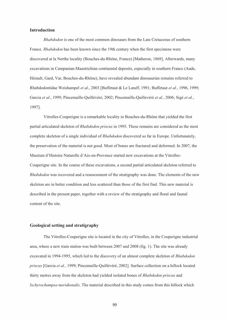
99
Introduction
Rhabdodon is one of the most common dinosaurs from the Late Cretaceous of southern
France. Rhabdodon has been known since the 19th century when the first specimens were
discovered at la Nerthe locality (Bouches-du-Rhône, France) [Matheron, 1869]. Afterwards, many
excavations in Campanian-Maastrichian continental deposits, especially in southern France (Aude,
Hérault, Gard, Var, Bouches-du-Rhône), have revealed abundant dinosaurian remains referred to
Rhabdodontidae Weishampel et al., 2003 [Buffetaut & Le Lœuff, 1991; Buffetaut et al., 1996, 1999;
Garcia et al., 1999; Pincemaille-Quillévéré, 2002; Pincemaille-Quillevéré et al., 2006; Sigé et al.,
1997].
Vitrolles-Couperigne is a remarkable locality in Bouches-du-Rhône that yielded the first
partial articulated skeleton of Rhabdodon priscus in 1995. These remains are considered as the most
complete skeleton of a single individual of Rhabdodon discovered so far in Europe. Unfortunately,
the preservation of the material is not good. Most of bones are fractured and deformed. In 2007, the
Muséum d’Histoire Naturelle d’Aix-en-Provence started new excavations at the Vitrolles-
Couperigne site. In the course of these excavations, a second partial articulated skeleton referred to
Rhabdodon was recovered and a reassessment of the stratigraphy was done. The elements of the new
skeleton are in better condition and less scattered than those of the first find. This new material is
described in the present paper, together with a review of the stratigraphy and floral and faunal
content of the site.
Geological setting and stratigraphy
The Vitrolles-Couperigne site is located in the city of Vitrolles, in the Couperigne industrial
area, where a new train station was built between 2007 and 2008 (fig. 1). The site was already
excavated in 1994-1995, which led to the discovery of an almost complete skeleton of Rhabdodon
priscus [Garcia et al., 1999; Pincemaille-Quillévéré, 2002]. Surface collection on a hillock located
thirty metres away from the skeleton had yielded isolated bones of Rhabdodon priscus and
Ischyrochampsa meridionalis. The material described in this study comes from this hillock which

100
was completely excavated layer by layer before the beginning of the construction of the station. This
hillock consisted of a succession of more or less calcareous marls and limestone layers. The
observed stratigraphic sequence belongs to the lower part of the Rognacian (or Lower Argiles
Rutilante Formation, Cojan and Moreau, 2006), a local stage corresponding to the top of the non-
marine Late Cretaceous, thirty metres below the Rognac Limestone (or Calcaire de Rognac
Formation, Cojan and Moreau, 2006) (fig. 2). It rests on an important red and yellow clay layer, also
known as “argiles bariolées” (variegated clays, B1). Above this level, the first four marly limestone
layers, passing from yellow-grey (B2) to grey colour (B3, B4) and the marly limestones (B5, B6) are
fossiliferous. The first compact limestone layer (B7), one metre thick, has yielded no macrofossils
and no organic microremains were found using chemical operations. It is followed by a last
fossiliferous grey marly level (B8). The studied sequence ends with another similar compact
limestone (B9), three metres thick, which was removed before the beginning of the fieldwork. The
two Rhabdodon sites at Vitrolles-Couperigne do not show the same stratigraphic sequences.
However, the top of the red and yellow clay level (B1) and the first limestone layer, under the
Rhabdodon skeleton (B7), can be used for a precise correlation. Thus, the first Rhabdodon
discovered in 1994 should be of the same age as the new material from level B8 described in this
study.
A considerable diversity of plant and animal fossils was recovered in the course of the
excavations (Appendix I). Macrofossils were discovered at three levels (B2, B4 and B8) and show
the presence of Rhabdodon priscus, dromaeosaurids, ankylosaurs, turtles and crocodiles. In addition,
large scale screen-washing operations have revealed remains of lissamphibians, squamates, fishes?,
various types of isolated eggshell fragments, invertebrates (gastropods, ostracodes) and green algae
(charophytes, dasycladales?), particularly in levels B4 and B6. The study of all these fossils (macro
and microremains) is still in progress. The fossil assemblages from the 1994-1995 excavations and
these presented here show a close resemblance at high taxonomic levels, with charophytes,
gastropods, turtles, dromaeosaurids and crocodiles. But precise comparisons are difficult because the
previous discoveries have not been described in detail. In addition, Garcia et al. [1999] did not
specify the exact origin of the Rhabdodon and Ischyrochampsa remains discovered from the hillock.

101
The preliminary list presented here (Appendix I) complements the general biotic assemblage from
Vitrolles-Couperigne with the addition of new taxa, especially among charophytes, (Microchara
sp.), gastropods, ostracodes, lissamphibians and turtles (Solemys aff. vermiculata).
The associated fossils provide precise stratigraphic indications, particularly on the basis of
charophytes and invertebrates. The best stratigraphic biomarkers are charophytes. Among them,
Peckichara cancellata Grambast indicates the Lower Rognacian [Feist and Freytet, 1983; Riveline
et al., 1996; Nieves Lopez-Martinez et al., 2001] whereas other taxa, some ostracodes and
gastropods, span the whole Rognacian [Babinot et al., 1983]. A recent magnetostratigraphic study of
the Aix Bassin places the Rognac Limestone at the level of the Campanian-Maastrichtian boundary
[Cojan and Moreau, 2006] and, consequently, the Vitrolles-Couperigne stratigraphic sequence in the
Upper Campanian (Chron 33N) and not in the Lower Maastrichtian [contra Garcia et al., 1999;
Pincemaille-Quillévéré, 2002].
Description of the Rhabdodon material
The material described in this work includes only postcranial elements, some of which
found in articulation and some isolated (fig. 3). This new partial articulated skeleton has several
elements in common with the partial skeleton referred to Rhabdodon priscus [Pincemaille-
Quillévéré, 2002], previously discovered from a nearby site. Moreover, these elements shared by the
two skeletons provide comparable diagnostic features. It thus appears that the Vitrolles-Couperigne
locality has yielded remains of two distinct partially articulated Rhabdodon skeletons. All specimens
are presently housed in the Muséum d’Histoire Naturelle d’Aix-en-Provence (Appendix II). The
measurements of complete and nearly complete elements are shown in Table 1.
Institutional Abbreviations
MHN-AIX-PV, Muséum d’Histoire Naturelle d’Aix-en-Provence

102
Systematics
Order ORNITHISCHIA Seeley, 1887
Suborder ORNITHOPODA Marsh, 1881
EUORNITHOPODA (sensu Weishampel 1990)
Infraorder IGUANODONTIA Sereno, 1986
Family RHABDODONTIDAE
Weishampel, Jianu, Cziki & Norman, 2003
Genus Rhabdodon Matheron 1869
Species Rhabdodon priscus, Matheron 1869
Material – a partial skeleton MHN-AIX-PV 2007.B8. 1-5, 7-51, 53-54, 56-67, 69,
72-73, 75-81, 83-87 and 89-99.
Description
Axial skeleton
No cervical vertebra has been preserved and some vertebrae (MHN-AIX-PV 2007. B8.11,
65, 77) could not be identified precisely. The anterior dorsal vertebra (MHN-AIX-PV 2007.B8.34)
(fig. 4) is short and the centrum is approximately as wide as high. The anterior articular facet is
concave whereas the posterior facet is slightly convex. The lateral walls are concave and the centrum
is keeled ventrally forming a heart-like shape in anterior view. The transverse processes are elongate
and oriented dorsolaterally. The parapophysis and diapophysis are separated, on the proximal and
distal part of the transverse process respectively. The prezygapophyses face dorsomedially. The
postzygapophyses are well separated and face ventrolaterally. The neural spine is tall, forming more
than half the total height of the vertebra. It is narrow, tapered upward and curved posteriorly at the
extremity.
The dorsal vertebra MHN-AIX-PV 2007.B8.1 which probably belongs to the middle to
posterior dorsal series is incomplete and deformed. Its centrum is more rounded, as wide as high and
it is amphicoelous. The posterior dorsal vertebrae (MHN-AIX-PV 2007.B8. 47, 56, 64, 66, 67) were

103
found partially articulated (fig. 5A, B). These vertebrae are amphicoelous. The transverse processes
become shorter and oriented more horizontally in the posterior dorsal series. The parapophysis shifts
progressively upward and finally merges with the diapophysis on the transverse process of the last
posterior dorsal vertebra (fig. 5E; pd). The neural spine of the posterior dorsal vertebrae is large,
rectangular in shape. It is at least two times higher than the centrum height. The posterior margin is
slightly curved and the upper part of the spine is extended anteroposteriorly. The prezygapophyses
are inclined dorsomedially. The postzygapophyses are less opened and their prominent oval facets
face ventrolaterally. The postzygapophyses are prolonged more posteriorly behind the centrum and
they are also situated more dorsally in more posterior vertebrae.
Dorsal ribs are preserved near the vertebrae. Their different forms and sizes indicate
different types of articulation with dorsal vertebrae. In anterior thoracic ribs, the capitulum and the
tuberculum are widely separated into two branches (fig. 6). The articular surfaces of anterior ribs are
sub-equal in size and subcircular. The shafts of these ribs are proximally concave in posterior view
and become rounded in the middle and mediolaterally compressed at the distal end. More posteriorly
the tuberculum is shortened but its articular surface is oval and more prominent than the capitulum.
The costal shaft is curved and more compressed anteroposteriorly and it is narrower distally. In the
most posterior ribs, the capitulum and the tuberculum are merged as a single articulation with the
para-diapophysis. These ribs are arched and tapered distally.
The sacrum of this Rhabdodon skeleton was found in two pieces separated by a minor fault
(fig. 7). They are deformed and compressed laterally. This fused sacrum comprises the last dorsal
vertebra, the sacrodorsal vertebra and sacrals one to five. Sacral six is not completely preserved. The
last dorsal vertebra which fused to the sacrum shows the same characters as the previous dorsal
vertebra (MHN-AIX-PV 2007.B8.47). The right lateral side of the centra of the sacrodorsal and the
first true sacral are covered by the ossified tendons but not the left lateral side. This suggests that the
ossified tendons should not be present on the centrum and must have been displaced from elsewhere
during fossilisation. Sacral two was cut by the fault; its central part is MHN-AIX-PV 2007.B8.14
and its neural spine MHN-AIX-PV 2007.B8.15. The sacral ribs of sacral two to four project
laterally while the ribs of sacral five are oriented more anteriorly. These sacral ribs arise at the co-

104
ossified junction between the centra and are linked to each other by a massive lateral bar which
contacts the medial surface of the ilium. The neural spines of the sacrum look like those of posterior
dorsal vertebrae, however they are higher and expanded dorsally and become very close to the
adjacent spines, and were possibly fused together.
Caudal vertebrae were found near the pelvic girdle, some of them are articulated. The caudal
vertebrae are not well preserved and deformed (fig. 8). The centrum is wide but short in the anterior
members of the caudal series whereas it is longer than wide and compressed laterally in the posterior
ones. The lateral wall of the caudals is concave anteroposteriorly, becoming flatter in succeeding
caudals. Ventrally, the centra bear the haemaporphyseal facets on the ventral margin of two articular
facets which the posterior haemaporphyseal facet is more prominent than the anterior one. The
transverse process is situated on the lateral wall of the centrum (fig. 6 A, B) and projects more or
less horizontally. The transverse process diminishes gradually and disappears in the posterior
caudals. The prezygapophyses of the anterior caudals cannot be observed, however, in a mid-
posterior caudal (MHN-AIX-PV 2007.B8. 19) the prezygapophysis is extended anterodorsally. It is
narrow and faces medially and tapers in the posterior series. The postzygapophyses are much less
prominent than those of the dorsal vertebrae. They are short and eventually merged as a single bulge
on the posterior margin of the neural spine. The neural spine is still large in anterior to middle caudal
vertebrae and then it becomes shorter and narrower in more posterior ones.
Some chevrons were recovered around the caudal vertebrae. Their proximal end is
expanded. Dorsally, the articular facet is unequally bisected by a transverse ridge into a larger
anterior and a smaller posterior facet for articulation with the haemapophyseal facets of the centrum.
Beneath this articular head, there is an oval haemal canal which is completely enclosed. The haemal
arches continue as two ridges on the posterior surface down to the middle of the chevron spine. The
distal margin is curved backward and flat at the distal end.
Ossified tendons are quite well preserved, some of them having been found in situ with the
vertebrae. The ossified tendons are distributed all along the dorsal, sacral and caudal vertebrae,
however they were found more densely packed over the posterior dorsal vertebrae and the sacrum.
Moreover, the tendons could be also arranged on the chevrons as shown in figure 8G. These ossified

105
rods tend to have mainly two shapes (angular and rounded) and to be oriented in two directions
(longitudinal and oblique). The longitudinal tendons lie side by side whereas the oblique ones lie
apart from each other (fig. 9). Long striations are present on the tendon surface. The displacement of
tendons, for example on the sacral centra (fig. 7), and scattered well-preserved tendons suggest that
ossified tendons were not fused to bones.
Forelimb
The right humerus (MHN-AIX-PV 2007.B8. 2) is incomplete, lacking the proximal
articulation, and it is compressed anteroposteriorly. The humeral condyle is not preserved. The
deltopectoral crest (fig. 10) is limited to the proximal half of the humerus. The crest is expanded and
more rugose distally while it decreases in width and becomes smoother toward the head. The
deltopectoral crest is bent anteromedially, forming a concave surface (bicipital sulcus) on the
proximal part of the humerus. At mid-length, the shaft is narrowed; however, it is not very slender
relative to the width of the proximal and distal parts. Distally, the humerus is also expanded to form
the distal articulation. Although the ulnar and radial condyles are incomplete, the coronoid and
olecranon fossae are visible between the distal condyles as broad triangular grooves in anterior and
posterior views respectively.
The right radius (MHN-AIX-PV 2007.B8.54) (fig. 10C) is not well preserved. It is
compressed lateromedially and bowed dorsoventrally. It is slender at mid-shaft and expanded at each
end. Because of poor preservation, the proximal and distal ends cannot be observed.
Pelvic girdle
The right ilium ((MHN-AIX-PV 2007.B8. 93) and ischium (MHN-AIX-PV 2007.B8. 17)
were found articulated (fig. 11). The ilium is not complete and compressed lateromedially. Only the
iliac blade posterior to the acetabulum is preserved. The dorsal margin is posteriorly convex. The
ischial peduncle of the ilium meets the iliac peduncle of the ischium by its ventral edge, forming the
posterior rim of the acetabulum. Above this process, a rugose depression is present on the medial
face and limited by an irregular curved thin ridge. This depression for the attachment of a sacral rib

106
extends to the posterior margin of the ilium. The posterior ventral margin of the ilium is slightly
inclined upward.
Both ischia were recovered but the left one (MHN -AIX-PV 2007.B8. 9) (fig. 12A) is
incomplete, lacking the proximal articulations. The iliac peduncle of the ischium is larger than the
pubic peduncle. These two peduncles are separated by an embayment which forms the
posteroventral rim of the acetabulum. The obturator process is situated on the proximal half of the
ischium and is projected medially. The ischial shaft is laterally flattened in cross-section and it is
broad in its proximal part and approximately parallel-sided in its middle part. The distal half of the
ischium is bowed and its distal end is slightly expanded ventrally.
Both the left and right pubes were recovered but only the pre-pubes are preserved (fig. 12).
The pre-pubis forms the anteroventral rim of the acetabulum. It is laterally compressed. The pre-
pubic process is extended forward and its distal end is expanded dorsoventrally and curves
downward. The proximal articulations are divided into the facet for the ilium projecting dorsally and
the facet for the ischium projecting ventrally. The former facet is larger than the latter one. A deep
oval pubic foramen with an anterior shallow depression is present just below the articulation for the
ischium.
Hindlimb
Few hindlimb bones have been found. There are only a fragment of femur and an ungual
phalanx which were recovered from the same bed as the other Rhabdodon remains. Nevertheless, a
left femur (MHN-AIX-PV 2008.B2.11) was found in a lower bed from which some other reptile
remains were recovered. The left femur is incomplete, lacking the proximal and distal ends. The
fourth trochanter is long (13 cm) and has a shallow depression anteriorly. The femoral shaft is large,
straight and transversely expanded at the proximal and distal ends.
The ungual phalanx (MHN-AIX-PV 2007.B8. 81) (fig. 13) is broad. It is sheared and
compressed dorsoventrally. The ungual is flattened and slightly arched ventrally. Its proximal end is
concave dorsoventrally and divided by a vertical ridge into sub-equal adjacent depressions. The
dorsal margin of this articular facet is prolonged more proximally than the ventral one. From the

107
articular margin, both sides of the ungual are slightly tapering in the proximal part where the
ligament scar is clearly visible in dorsal view. Then, the lateral and medial sides are expanded as a
long round ridge supporting well developed claw grooves extending to the distal tip of the ungual.
Discussion and conclusion
This new partial articulated skeleton of Rhabdodon priscus is comparable in size to the first
one. The new material described above provides information about some osteological features which
are missing in the first found skeleton, as listed below:
1) The neural spines of the dorsal vertebrae and the sacrum are two to three times higher
than the centra. They are still prominent in anterior caudal vertebrae, but less prominent in the more
posterior ones. Ouranosaurus and Iguanodon possess more developed neural spines whereas the
neural spines of Zalmoxes are short, as high as the centra of the dorsal vertebrae.
2) The sacrum consists of eight fused vertebrae; the last dorsal, the sacrodorsal and six true
sacral vertebrae. This character is shared with Iguanodon and Zalmoxes.
3) A great number of ossified tendons over the sacrum and the neighboring area around the
posterior dorsal and anterior caudal vertebrae suggest that the vertebral column was probably held
more or less horizontally. The ossified tendons were probably arranged in a double layer as in
Iguanodon. Both epaxial and hypaxial ossified tendons are present in the caudal skeleton, as in
Tenontosaurus. These characters are unknown in Zalmoxes.
4) The humerus is not very constricted at mid-shaft while it is narrowed in Zalmoxes. The
deltopectoral crest of the humerus is prominent and forms an angle with the humeral shaft. This
character is shown more distinctively in Zalmoxes shqiperorum.
5) An obturator process is present in the proximal half of the ischium. This process cannot
be observed on the right ischium of the first skeleton found in 1995, but it is present on the left one.
This may be a taphonomic effect. On the other hand this character is absent in Zalmoxes.
6) The ischial shaft is compressed laterally and slightly curved anteroposteriorly whereas
that of Zalmoxes is ovoid in cross-section and distinctively curved downward and forms a boot-like
distal end in Zalmoxes shqiperorum.

108
7) The iliac blade is deep and straight and it is sub-equally thick along the dorsal margin. In
contrast to Zalmoxes, the iliac blade faces ventrolaterally and the posterior dorsal margin is
expanded and everted.
The recent systematic analyses of Rhabdodon principally based on the first partial
articulated skeleton of Rhabdodon priscus [Pincemaille-Quillévéré, 1997, 2002] suggest that
Rhabdodon priscus has the closest relationship with Zalmoxes robustus and Z. shqiperorum from the
Late Maastrichtian of Romania [Weishampel et al., 2003]. All these taxa are placed in the same
family Rhabdodontidae of Iguanodontia, by Weishampel et al., [2003]. The anatomical information
provided by the new skeleton from Vitrolles-Couperigne discussed above confirms that Rhabdodon
priscus differs notably from Zalmoxes in having very tall neural spines on the vertebrae, possessing
an obturator process on the ischium, a flat ischiac shaft and a non-everted dorsal margin of the ilium.
These diagnostic postcranial characters were already included in the recent systematic analysis of
Weishampel et al. [2003]. No new cranial material is known from Vitrolles-Couperigne. The
identical fluted teeth and probably other cranial characters mentioned by Godefroit et al. [2009]
represent the best synapomorphies for Rhabdodontidae so far. While Zalmoxes and Rhabdodon are
closer to Tenontosaurus, from the Aptian of the United States, than to other iguanodontians,
Rhabdodon priscus from Vitrolles-Couperigne is closer to Tenontosaurus than Zalmoxes on the
basis of postcranial characters. This could suggest that the common ancestor of the rhabdodontid
dinosaurs evolved in North America, and then dispersed to Europe and eventually evolved in
geographical isolation on the various European landmasses during the Late Cretaceous.
Although the identification of floral and faunal elements is still in progress, the complete
exploitation of the Vitrolles-Couperigne site (layer by layer with screen-washing operations)
suggests that the local biodiversity was richer than previously described [Garcia et al., 1999],
especially at the level of the micro-flora and fauna. Nevertheless, the preliminary picture of the
biotic composition depicts a precise life environment. The alternating thick compact limestone
layers and marls are very rich in charoptytes typical of fresh water. The most represented gastropods
(Cyclophorus, Melanania, Physa, Pyrugulifera and Viviparus) are also characteristic of this
environment often charged in mud such as lakes, ponds and swamps in a warm and wet climatic

109
context [Fabre-Taxy, 1951, 1959]. This observation is in agreement with the presence of
lissamphibians which are essentially bound to this type of aquatic environment. The other present
vertebrates such as squamates, turtles (Solemys) and dinosaurs such as Rhabdodon and ankylosaurs,
which were not confined to the lakeside, may have come to the peri-lacustrine zone to find food or
nesting sites. From this zone, small bones and eggshell fragments were carried by streams and
accumulated in the lake or in the pond. The occurrence of small theropods such as dromaeosaurs was
probably linked to the presence of easily accessible animal carcasses in shallow water, a rather
opportunistic behaviour. The limited dislocation of the skeletons of Rhabdodon and the lack of some
anatomical parts (head, members and tail) could be explained by a combination of a slow current and
scavenger activity, the predators taking the extremities of the skeleton.
The stratigraphic distribution of the flora and fauna presented in this study can yet suffer
from a bias linked to the quantity of studied sediments. New studies will enable to confirm or enrich
the taxonomic lists for each layer and then to compare this biodiversity with those known from other
sites such as Champs-Garimond [Sigé et al., 1997], Cruzy [Buffetaut et al., 1999], La Neuve [Garcia
et al., 2000], La Rouquette [Massieux et al., 1987], Laño [Astibia et al., 1999] and Villeveyrac
[Buffetaut et al., 1996]. These comparisons are necessary in order to understand the
palaeobiogeographical relationships of some taxa (lissamphibians, squamates, turtles) in southern
France during the Late Cretaceous (Campanian and Maastrichtian).
Acknowledgements
We would like to express our gratitude to the excavation team of the Muséum d’Histoire
Naturelle d’Aix-en-Provence for their hard work both in fieldwork and in the laboratory. We are
deeply indebted to SNCF for their permission to access the area and to the city of Vitrolles and the
Region for their support. We also wish to acknowledge the help of Mohamed Berrabah and Frédéric
Perret during the fieldwork. We thank Loïc Villier and Marc Floquet (Université de Provence,
France) for their help in finding information for the determination of gastropods.

110
References
ASTIBIA H., CORRAL J.C., MURELAGA X., ORUE-ETXEBARRIA X., PEREDA-SUBERBIOLA X., coords
(1999). - Geology and palaeontology of the Late Cretaceous dinosaur-bearing beds of the
Laño quarry (Basque-Cantabrian Region, Iberian Peninsula). Est. .Mus. Cien. Nat. de Alava,
14 (núm. espec. 1), 1-380.
BABINOT J.F., FREYTET P., AMIOT M., BILOTTE M., DE BROIN F., COLOMBO F., DURAND J.P.,
FEIST M., FLOQUET M., GAYET M., LANGE-BADRE B., MASRIERA M., MEDUS J.,
TAMBAREAU Y., ULLASTRE J. &VILLATTE J. (1983). - Le Sénonien supérieur continental
de la France méridionale et de l’Espagne septentrionale : état des connaissances
biostratigraphiques. - Géol. Médit., X (3-4), 245-268.
BUFFETAUT E. & LE LŒUFF J. (1991). - Une nouvelle espèce de Rhabdodon (Dinosauria,
Ornitischia) du Crétacé Supérieur de l’Hérault (Sud de la France). - C. R. Acad. Sci., Paris,
Sér. II, 312, 943-948.
BUFFETAUT E., COSTA G., LE LŒUFF J., MARTIN M., RAGE J.-C., VALENTIN X. & TONG H.
(1996). - An Early Campanian vertebrate fauna from the Villeveyrac Basin (Hérault,
Southern France). - N. Jb. Geol. Paläont. Mh., 1, 1-16.
BUFFETAUT E., LE LŒUFF J., TONG H., DUFFAUD S., CAVIN L., GARCIA G., WARD D. &
L’ASSOCIATION CULTURELLE, ARCHEOLOGIQUE ET PALEONTOLOGIQUE DE CRUZY. (1999).
- Un nouveau gisement de vertébrés du Crétacé supérieur à Cruzy (Hérault, Sud de la
France). - C. R. Acad. Sci., Paris, 328, 203-208.
COJAN I. & MOREAU M.-G. (2006). - Correlation of terrestrial climatic fluctuations with global
signals during the Upper Cretaceous–Danian in a compressive setting (Provence, France).
J. Sediment. Res., 76, 589–604.
FABRE-TAXY S. (1951) Faunes lagunaires et continentales du Crétacé supérieur de Provence. II. –
Le Campanien fluvio-lacustre. – Ann, de Paléontol., XXXVII, 3-42.
111
FABRE-TAXY S. (1959) Faunes lagunaires et continentales du Crétacé supérieur de Provence. III –
Le Maestrichtien et le Danien. – Ann, de Paléontol., XLV, 55-124 .
FEIST M. & FREYTET P. (1983). - Conséquences stratigraphiques de la répartition des charophytes
dans le Campanien et le Maastrichtien du Languedoc. - Géol. Médit., X (3-4), 291-301.
GARCIA G., PINCEMAILLE M., VIANEY-LIAUD M., MARANDAT B., LORENZ E., CHEYLAN G.,
CAPETTA H., MICHAUX J. & SUDRE J. (1999). - Découverte du premier squelette presque
complet de Rhabdodon priscus (Dinosauria, Ornithopoda) du Maastrichtien inférieur de
Provence. - C. R. Acad. Sci., Paris, 328, 415-421.
GARCIA G., DUFFAUD S., FEIST M., MARANDAT B., TAMBAREAU Y., VILLATTE J. & SIGÉ B.
(2000). - La Neuve, gisement à plantes, invertébrés et vertébrés du Bégudien (Sénonien
supérieur continental) du bassin d'Aix-en-Provence. Geodiversitas, 22 (3), 325-348.
GODEFROIT P., CODREA V. & WEISHAMPEL D.B. (2009). - Osteology of Zalmoxes shqiperorum
(Dinosauria, Ornithopoda), based on new specimens from the Upper Cretaceous of N<laY-
Vad (Romania). – Geodiversitas, 31(3), 525-553.
LÓPEZ-MARTÍNEZ N., CANUDO J.I., ARDÈVOL L., PEREDA-SUBERBIOLA X., XABIERORUE-
ETXEBARRIA X., CUENCA-BESCÓS G., RUIZ-OMEÑACA J.I., MUREGAGA X. & FEIST M.
2001. - New dinosaur sites correlated with Upper Maastrichtian pelagic deposits in the
Spanish Pyrenees: implications for the dinosaur extinction pattern in Europe. - Cretaceous
Res., 22, 41-61.
MASSIEUX M., REY J. & VILLATTE J. (1987). – Sur l’âge maastrichtien de l’affleurement de la
Rouquette (Commune de Paraza, Minervois). Bull. Soc. Hist. Nat., Toulouse, 123, 151-157.
MATHERON P. (1869). - Notice sur les Reptiles fossiles des dépôts fluvio-lacustres crétacés du
bassin à lignite de Fuveau. - Mém. Acad. Sci., Bel. Lettr. Arts Marseille, 345-379.

112
PINCEMAILLE- QUILLÉVÉRÉ M. (2002). - Description d’un squelette partiel de Rhabdodon priscus
(Euornithopoda) du Crétacé supérieur de Vitrolles (Bouches du Rhône, France). Oryctos, 4,
39-70.
PINCEMAILLE- QUILLÉVÉRÉ M., BUFFETAUT E. & QUILLÉVÉRÉ F. (2006). - Description
ostéologique de l’arrière-crâne de Rhabdodon (Dinosauria,Euornithopoda) et implications
phylogénétiques. - Bull. Soc. géol. Fr., 177, 97-104.
RIVELINE J., BERGER J.-P., FEIST M., MARTIN-CLOSAS C., SCHUDACK M. & SOULIÉ-MARSCHE I.
(1996). European Mesozoic-Cenozoic charophyte biozonation. - Bull. Soc. géol. Fr., 167(3),
453-468.
SIGÉ B., BUSCALIONI A.D., DUFFAUD S., GAYET M., ORTH B., RAGE J.-C. & SANZ J.L. (1997). -
Etat des données sur le gisement crétacé supérieur continental de Champ-Garimond
(Gard, Sud de la France). Münchner Geowiss. Abh., A, 34, 111-130.
WEISHAMPEL D.B., JAINU C-M, CSIKI Z. & NORMAN D.B. (2003). - Osteology and
Phylogeny of Zalmoxes (N.G.), an unusual Euornithopod Dinosaur from the latest
Cretaceous of Romania. - Journal of the Systematic Paleontology, 1(2), 65-123.

113
FIG. 1 - Location map of Vitrolles-Couperigne localiy. 1: Vitrolles Limestone, 2 : Argiles Upper
Argiles Rutilantes, 3 : Calcaire de Rognac, 4 : Lower Argiles Rutilantes.
FIG. 1 – Carte localisant le gisement de Vitrolles-Couperigne. 1:Calcaire de Vitrolles, 2 : Argiles
Rutilantes supérieures, 3 : Calcaire de Rognac, 4 : Argiles Rutilantes inférieures
FIG. 2 - Log of Vitrolle-Couperigne site and positioning in chronotratigraphic and general
lithostratigraphic context (*not at scale). 1 : red and variegated (B1) clays, 2 : sandstone, 3 : clays
with gypsum, 4 : limestone (B7, B9), 5 : Alternation of marls and limestones, 6 : grey and yellow
marls (B2), or grey marls (B4, B8) or grey marls with black joint (B3), 7 : grey marl with limestone
nodules (B5), 8 : thin marly limestone lens (B6), 9 : Layers yielding Rhabdodon remains (B2, B8).
FIG. 2 - Log du site de Vitrolles-Couperigne et repositionnement dans les contextes
chronostratigraphiques et lithostratigraphiques généraux (*coupe non à l’échelle). 1 : argiles rouges
et bariolées (B1), 2 : grès, 3 : argiles à gypse, 4 : calcaire (B7, B9), 5 : alternance de marnes et
calcaires, 6 : marnes jaunes et grises (B2), ou seulement grises (B4, B8) ou grises avec jointures
noires (B3), 7 : marne grise avec nodules calcaires (B5), 8 : lentilles de calcaires fins et marneux
(B6), 9 : couches contenant les restes de Rhabdodon (B2, B8).
FIG. 3 - Map of the bones as they were found in the course of the excavations. The numbers are
those used in the present description (see Appendix: List of skeletal elements). Reconstruction of
partial skeleton showing positions of the bones discovered at the site in grey.
FIG. 3 – Plan des os, tels qu’ ils ont été trouvés au cours des fouilles. Les chiffres sont ceux utilisés
dans la description (voir Annexe: Liste des éléments du squelette). Reconstruction du squelette
partiel, montrant les positions des os découverts sur le site en gris.
FIG. 4 - Anterior dorsal vertebra (MHN-AIX-PV 2007.B8.34) in anterior (A), left lateral (B) and
posterior(C) views. Abbreviations: d = diapophysis, ns = neural spine, p = parapophysis, poz =
postzygapophysis, prz = prezygapophysis.

114
FIG. 4 - Vertèbre dorsale antérieure (MHN-AIX-PV 2007.B8.34) en vues antérieure (A), latérale
gauche (B) et postérieure(C). Abréviations: d = diapophyse, ns = épine neurale, p = parapophyse,
poz = postzygapophyse, prz = prézygapophyse.
FIG. 5 - Associated posterior dorsal vertebrae (MHN-AIX-PV 2007.B8.47, 67, 56) in right lateral
view (A). Dorsal vertebra MHN-AIX-PV 2007.B8. 66 found next to MHN-AIX-PV 2007.B8.56 in
posterolateral view. The second dorsal vertebra from the last (MHN-AIX-PV 2007.B8. 47) in
anterior (C), posterior (D) and left lateral (E) views. Abbreviations: d = diapophysis, ns = neural
spine, p = parapophysis, pd = paradiapophysis, poz = postzygapophysis, prz = prezygapophysis, tp =
transverse process.
FIG. 5 - Vertèbres dorsales postérieures associées (MHN-AIX-PV 2007.B8.47, 67, 56) vue latérale
droite (A). Vertèbre dorsale MHN-AIX-PV 2007.B8. 66 trouvé à côté de MHN-AIX-PV 2007.B8.56
en vue postérolatérale. La deuxième vertèbre dorsale à partir de l’arrière (MHN-AIX-PV 2007.B8.
47) en vues antérieure (C), postérieure (D) et latérale gauche (E). Abréviations: d = diapophyse, ns =
épine neurale, p = parapophyse, pd = paradiapophyse, poz = postzygapophyse, prz =
prézygapophyse, tp = processus transverse.
FIG. 6 - Anterior dorsal rib (MHN-AIX-PV 2007.B8. 3) in posterior view (A), mid-posterior dorsal
rib (MHN-AIX-PV 2007.B8. 41) in anterior view (B), posterior dorsal rib (MHN-AIX-PV 2007.B8.
10) in lateral view. Abbreviations: c = capitulum, ct = merged capitulum and tuberculum, t =
tuberculum.
FIG. 6 - Côte dorsale antérieure (MHN-AIX-PV 2007.B8. 3) en vue postérieure (A), côte dorsale
mi-postérieure (MHN-AIX-PV 2007.B8. 41) en vue antérieure (B), côte dorsale postérieure (MHN-
AIX-PV 2007.B8. 10) en vue laterale. Abréviations : c = capitulum, ct = capitulum et tuberculum,
fusionné, t = tuberculum.

115
FIG. 7- Sacrum MHN-AIX-PV 2007.B8. 14 (on the right) and 15(on the left) in right lateral view.
Abbreviations: ld = last dorsal vertebra, os.t = ossified tendons, s = sacral, sd = sacrodorsal vertebra,
sr = sacral rib.
FIG. 7- Sacrum MHN-AIX-PV 2007.B8. 14 (à droite) et 15(à gauche) en vue latérale droite.
Abréviations: ld = dernière vertèbre dorsale, os.t = tendons ossifiés, s = vertèbre sacrée, sd =
vertèbre sacrodorsale, sr = côte sacrée.
FIG. 8 - Anterior caudal vertebrae, MHN-AIX-PV 2007.B8.43 in anterior view (A), MHN-AIX-PV
2007.B8. 21 in left anterolateral view (B), MHN-AIX-PV 2007.B8. 4 in right anterolateral view (C)
and mid to posterior caudal vertebra MHN-AIX-PV 2007.B8. 19 in right lateral view (D). Chevrons,
MHN-AIX-PV 2007.B8.73 in anterolateral (E) and posterolateral (F) views, MHN-AIX-PV
2007.B8.90 in right lateral view (G). Abbreviation: hc = haemal canal, os.t = ossified tendon, poz =
postzygapophysis, prz = prezygapophysis, tp = transverse process. 5 cm and 3 cm scale bars are for
caudal vertebrae and chevrons respectively.
FIG. 8 - vertèbre caudale antérieure MHN-AIX-PV 2007.B8.43 en vue antérieure (A), MHN-AIX-
PV 2007.B8. 21 en vue antérolatérale gauche (B), MHN-AIX-PV 2007.B8. 4 en vue antérolatérale
droite (C) et vertèbre caudale moyenne à postérieure MHN-AIX-PV 2007.B8. 19 en vue latérale
droite (D). Chevrons, MHN-AIX-PV 2007.B8.73 en vues antérolaterale (E) et postérolatérale (F),
MHN-AIX-PV 2007.B8.90 en vue latérale droite (G). Abréviation: hc = canal hémal, os.t = tendon
ossifié, poz = postzygapophyse, prz = prézygapophyse, tp = processus transverse. 5 cm et 3 cm sont
des barres d’échelle pour les vertèbres caudales et les chevrons, respectivement.
FIG. 9 - Ossified tendons. Abbreviations: ns = neural spine, os.t = ossified tendons.
FIG. 9 - Tendons ossifiés. Abréviations: ns = neural spine, os.t = ossified tendons.
os.t = tendons ossifiés

116
FIG. 10 - Right humerus MHN-AIX-PV 2007.B8. 2 in anterior (A) and posterior (B) views. Right
radius MHN-AIX-PV 2007.B8. 54 in medial view(C). Abbreviations: dpc=deltopectoral crest,
rac=radial condyle, ulc=ulnar condyle.
FIG. 10 - Humérus droit MHN-AIX-PV 2007.B8. 2 en vues antérieure (A) et postérieure (B). Radius
droit MHN-AIX-PV 2007.B8. 54 en vue mediale (C). Abréviations: dpc= crête deltopectorale, rac=
condyle radiale, ulc= condyle ulnaire.
FIG. 11 - Articulated right ilium MHN-AIX-PV 2007.B8. 93 and ischium MHN-AIX-PV 2007.B8.
17 in medial view. Abbreviations: acet = acetabulum, il = ilium, il.p = iliac peduncle, is = ischium,
is.p = ischial peduncle, ob.p = obturator process, p.p = pubic peduncle.
FIG. 11 - Ilion droit MHN-AIX-PV 2007.B8. 93 et ischion droit MHN-AIX-PV 2007.B8. 17 en
connexion en vue médiale. Abréviations: acet = acétabulum, il = ilion, il.p = pédoncule iliaque, is =
ischion , is.p = pédoncule ischiatique, ob.p = processus obturé, p.p = pédoncule pubien.
FIG. 12 - Left ischium MHN-AIX-PV 2007.B8. 9 in medial view (A). Pre-pubis, MHN-AIX-PV
2007.B8.7 (B) and MHN-AIX-PV 2007.B8. 12 (B) in lateral view. Abbreviations: ob.p = obturator
process, p.f = pubic foramen, pre.p = pre-pubic process.
FIG. 12 – Ischion gauche MHN-AIX-PV 2007.B8. 9 en vue médiale (A). Pré-pubis, MHN-AIX-PV
2007.B8.7 (B) et MHN-AIX-PV 2007.B8. 12 (B) en vue laterale. Abréviations: ob.p = processus
obturé, p.f = foramen pubien, pre.p = processus pré-pubien.
FIG. 13 - An ungual phalanx ((MHN-AIX-PV 2007.B8. 81) in dorsal (A) and lateral (B) views.
FIG. 13 - Une phalange unguéale ((MHN-AIX-PV 2007.B8. 81) en vues dorsale (A) et laterale (B).

117
Appendix I: Floral and faunal assemblage found in the different layers of Vitrolles-Couperigne
stratigraphic sequence.
Taxa Layer
B1 B2 B3 B4 B5 B6 B7 B8 B9
Charophyta Heptorella brachyceras + Microchara cristata + Microchara sp. + Peckichara cancellata + + +
Peckichara sertulata + + + +
Charophyta? Dasycladaceae indet.? + +
Gastropoda Bauxia disjuncta + Clausilia patula + + Cyclophorus luneli + Cyclophorus solarium + Melania gourreti + Palaeostoa cureti + Palaeostoa meridionalis + Physa pygmæa + Pyrgulifera armata + + Viviparus beaumonti + taxa indet. + + + + + + +
Ostracoda aff. Cypridopsis sp. + Frambocythere tumiensis
ferreri + + Paracandona occitanica + + taxa indet. +
Pisces? taxa indet. + +
Lissamphibia Anura indet. + Palaeobatrachidae indet.? + Scapherpetontidae indet.? + Sirenidae or Proteidae indet.? +
Chelonia Solemys cf. vermiculata +
taxa indet. +
Squamata Lacertilia indet. + taxa indet. +
Crocodylia taxa indet. + + + + Dinosauria Ankylosauria indet. +
Dromaeosauridae indet. + + Rhabdodon priscus + +
Isolated Eggshells types indet. + + + + + + +

118
Appendix II: List of skeletal elements.
Number Nature Number Nature
MHN-AIX-PV 2007.B8.1 dorsal vertebra MHN-AIX-PV 2007.B8.34 dorsal vertebra
MHN-AIX-PV 2007.B8.2 right humerus MHN-AIX-PV 2007.B8.35 rib
MHN-AIX-PV 2007.B8.3 rib MHN-AIX-PV 2007.B8.36 rib
MHN-AIX-PV 2007.B8.4 caudal vertebra MHN-AIX-PV 2007.B8.37 rib
MHN-AIX-PV 2007.B8.5 tendon MHN-AIX-PV 2007.B8.38 rib
MHN-AIX-PV 2007.B8.7 left pre-pubis MHN-AIX-PV 2007.B8.39 rib
MHN-AIX-PV 2007.B8.8 rib MHN-AIX-PV 2007.B8.40 rib
MHN-AIX-PV 2007.B8.9 left ischium MHN-AIX-PV 2007.B8.41 rib
MHN-AIX-PV 2007.B8.10 rib MHN-AIX-PV 2007.B8.42 neural spine
MHN-AIX-PV 2007.B8.11 vertebra MHN-AIX-PV 2007.B8.43 caudal vertebra
MHN-AIX-PV 2007.B8.12 right pre-pubis MHN-AIX-PV 2007.B8.44 tendon
MHN-AIX-PV 2007.B8.13 tendon MHN-AIX-PV 2007.B8.45 tendon
MHN-AIX-PV 2007.B8.14 sacrum(1) MHN-AIX-PV 2007.B8.46 tendon
MHN-AIX-PV 2007.B8.15 sacrum(2) MHN-AIX-PV 2007.B8.47 dorsal vertebra
MHN-AIX-PV 2007.B8.16 tendon MHN-AIX-PV 2007.B8.48 rib
MHN-AIX-PV 2007.B8.17 right ischium MHN-AIX-PV 2007.B8.49 tendon
MHN-AIX-PV 2007.B8.18 caudal vertebra MHN-AIX-PV 2007.B8.50 rib
MHN-AIX-PV 2007.B8.19 caudal vertebra MHN-AIX-PV 2007.B8.51 tendon
MHN-AIX-PV 2007.B8.20 chevron MHN-AIX-PV 2007.B8.53 rib
MHN-AIX-PV 2007.B8.21 caudal vertebra MHN-AIX-PV 2007.B8.54 right radius
MHN-AIX-PV 2007.B8.22 caudal vertebra MHN-AIX-PV 2007.B8.56 dorsal vertebra
MHN-AIX-PV 2007.B8.23 caudal vertebra MHN-AIX-PV 2007.B8.57 caudal vertebra
MHN-AIX-PV 2007.B8.24 caudal vertebra MHN-AIX-PV 2007.B8.58 rib
MHN-AIX-PV 2007.B8.25 tendon MHN-AIX-PV 2007.B8.59 rib
MHN-AIX-PV 2007.B8.26 tendon MHN-AIX-PV 2007.B8.60 rib
MHN-AIX-PV 2007.B8.27 tendon MHN-AIX-PV 2007.B8.61 rib
MHN-AIX-PV 2007.B8.28 undetermined bone MHN-AIX-PV 2007.B8.62 rib
MHN-AIX-PV 2007.B8.29 vertebra MHN-AIX-PV 2007.B8.63 rib
MHN-AIX-PV 2007.B8.30 tendons MHN-AIX-PV 2007.B8.64 dorsal vertebra
MHN-AIX-PV 2007.B8.31 rib MHN-AIX-PV 2007.B8.65 vertebra
MHN-AIX-PV 2007.B8.32 rib MHN-AIX-PV 2007.B8.66 dorsal vertebra
MHN-AIX-PV 2007.B8.33 rib MHN-AIX-PV 2007.B8.67 dorsal vertebra
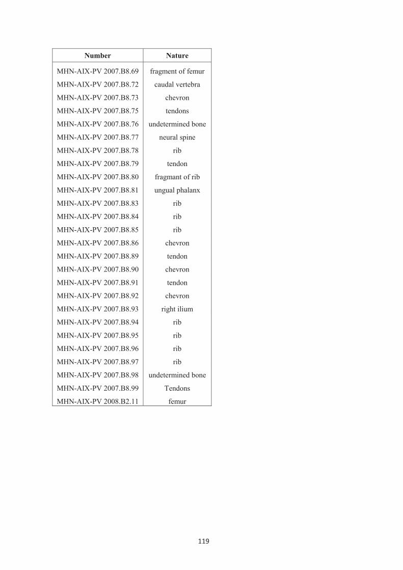
119
Number Nature
MHN-AIX-PV 2007.B8.69 fragment of femur
MHN-AIX-PV 2007.B8.72 caudal vertebra
MHN-AIX-PV 2007.B8.73 chevron
MHN-AIX-PV 2007.B8.75 tendons
MHN-AIX-PV 2007.B8.76 undetermined bone
MHN-AIX-PV 2007.B8.77 neural spine
MHN-AIX-PV 2007.B8.78 rib
MHN-AIX-PV 2007.B8.79 tendon
MHN-AIX-PV 2007.B8.80 fragmant of rib
MHN-AIX-PV 2007.B8.81 ungual phalanx
MHN-AIX-PV 2007.B8.83 rib
MHN-AIX-PV 2007.B8.84 rib
MHN-AIX-PV 2007.B8.85 rib
MHN-AIX-PV 2007.B8.86 chevron
MHN-AIX-PV 2007.B8.89 tendon
MHN-AIX-PV 2007.B8.90 chevron
MHN-AIX-PV 2007.B8.91 tendon
MHN-AIX-PV 2007.B8.92 chevron
MHN-AIX-PV 2007.B8.93 right ilium
MHN-AIX-PV 2007.B8.94 rib
MHN-AIX-PV 2007.B8.95 rib
MHN-AIX-PV 2007.B8.96 rib
MHN-AIX-PV 2007.B8.97 rib
MHN-AIX-PV 2007.B8.98 undetermined bone
MHN-AIX-PV 2007.B8.99 Tendons
MHN-AIX-PV 2008.B2.11 femur

120
Figure 1
Figure 2

121
Figure 3

122
Figure 4

123
Figure 5

124
Figure 6

125
Figure 7
Figure 8

126
Figure 9
Figure 10

127
Figure 11
Figure 12

128
Figure 13

129
References
Buffetaut, E., Le Lœuff, J. 1991. Une nouvelle espèce de Rhabdodon (Dinosauria,
Ornithischia) du Crétacé supérieur de l’Hérault (Sud de la France). Compte rendu de
l’Académie des Sciences de Paris, Série II 312, 943-948.
Chanthasit, P., Tortosa, T., Dutour, Y., Buffetaut, E., Feist, M., Babinot, J.F., Rage, J.C.,
Turini, E., Berton, S., Cheylan, G. (2010). New partial articulated skeleton of
Rhabdodon priscus (Dinosauria: Ornithopoda) from the Late Cretaceous of
Vitrolles-Couperigne, Bouches-du-Rhône, southern France. Bulletin de la Société
Géologique de France (in review).
Dollo, L. 1883. Note sur les restes de dinosauriens recontrés dans le Crétacé Supérieur de la
Belgique. Bulletin du Musée Royal d'Histoire Naturelle de Belgique 2, 205-221.
Forster, A.C. 1990. The postcranial skeleton of the ornithopod dinosaur Tenontosaurus
tilletti. Journal of Vertebrate Paleontology 10(3), 273-294.
Galton, P.M. 1974. The ornithischian dinosaur Hypsilophodon from the wealden of the Isle
of Wight. Bulletin of the British Museum (Natural History), Geology 25(1), 1-152.
Godefroit, P., Codrea, V., Weishampel, D.B. 2009. Osteology of Zalmoxes shqiperorum
(Dinosauria, Ornithopod), based on new specimens from the Upper Cretaceous of
N<laY-Vad (Romania). Geodiversitas 31(3), 525-553.
Lapparent, A. F. de. 1947. Les dinosauriens du Crétacé supérieur du Midi de la France.
Mémoires de la Société géologique de France 56, 1-54.
Hooley, R.W. 1925. On the skeleton of Iguanodon atherfieldensis from the Wealden. Q.J.
Geological Society of London 81(1), 1-61f.
Matheron, P. 1869. Notice sur les reptiles fossils des dépôts fluvio-lacustres crétacé du basin
à lignite de Fuveau. Bulletin de la Société Géologique de France 26(2), 781–795.
Norman, D.B. 1980. On the ornithischian dinosaur Iguanodon bernissartensis from
Belgium. Mémoires Institut Royal des Sciences Naturelles de Belgique 178, 1-103.

130
Norman, D.B. 1986. On the anatomy of Iguanodon atherfieldensis (Ornithischia :
Ornithopoda). Bulletin de l'Institut Royal des Sciences Naturalles de Belgique,
Sciences de la Terre 56, 281-372.
Norman, D.B. 1998. On Asian ornithopods (Dinosauria, Ornithischia). 3. A new species of
iguanodontid dinosaur. Zoological Journal of the Linnean Society. 122, 291-348.
Pincemaille-Quillévéré, M. 2002. Description d’un squelette partiel de Rhabdodon priscus
(Euornithopoda) du Crétacé supérieur de Vitrolles (Bouches du Rhône, France).
ORYTOS 4, 39-70.
Pincemaille-Quillévéré, M., Buffetaut, E., Quillévéré, F. 2006. Osteological description of
the braincase of Rhabdodon (Dinosauria, Euornithopoda) and phylogenetic
implications. Bulletin de la Société Géologique de France 177, 97-104.
Taquet, P. 1976. Géologie et Paléontologie du Gisement de Gadoufaoua (Aptien du Niger)
[Geology and Paleontology of the Gadoufaoua Locality (Aptian of Niger)]. Cahiers
de Paléontologie, Centre National de la Recherche Scientifique, Paris 1-191.
Weishampel, D.B., Jainu, C-M, Csiki Z., Norman, D.B. 2003. Osteology and Phylogeny of
Zalmoxes (N.G.), an unusual Euornithopod Dinosaur from the latest Cretaceous of
Romania. Journal of the Systematic Paleontology 1(2), 65-123.

131
Chapter 5:
Systematics
5.1 Discussion of the morphological variation in Rhabdodon and in
Rhabdodontidae.
Rhabdodon remains from different localities in France described in the previous part
reveal some morphological differences. The comparison between these new French
specimens and with the taxa in Rhabdodontidae including Rhabdodon priscus, Rhabdodon
septimanicus, Zalmoxes robustus, Zalmoxes shqiperorum (Matheron, 1869; Lapparent,
1947; Buffetaut and Le Lœuff, 1991; Buffetaut et al. 1996; Pereda-Suberbiola and Sanz,
1999; Pincemaille-Quillévéré, 2002; Pincemaille-Quillévéré, 2006; Weishampel et al.,
2003; Godefroit et al., 2009) suggest the following aspects of the morphological variability.
5.1.1 Cranial aspects
I. Premaxilla
- The maxillary (posterolateral) process of the premaxilla is relatively short
in Zalmoxes robustus while in the maxilla from Quarante, this process is prolonged with a
more prominent recess on the medial surface to receive the premaxillary process of the
maxilla.
- The external naris is more widely open in the specimen from Quarante.
These could suggest that at least two forms of snout are present among the
rhabdodontids. The skull of Rhabdodon from Quarante has a distinctive long and widely
open external naris with an arched nasal region whereas that of Zalmoxes has a short and
relatively less prominent nasal region. However, both premaxillae do not show a flared beak
as in hadrosaurine ornithopods.

132
II. Maxilla
- The premaxillary process of the maxilla is variable in Rhabdodon from
different localities in France. The maxilla from Quarante has a longer premaxillary process
than those of Rhabdodon from the Bellevue site and Highway A8 (Aix-en-Provence) and
those of Zalmoxes.
The lengthened premaxillary process of the maxilla from Quarante
corresponds to a posteriorly prolonged maxillary (posterolateral) process of the premaxilla
from the same locality. The specimens from the Bellevue site and the Highway A8 (Aix-en-
Provence) also suggest a short anterior region of the skull as in Zalmoxes.
III. Quadrate
- The asymmetrically developed articular condyles for the mandible of the
quadrate are present in French specimens, as in the quadrates of Zalmoxes. Weishampel et
al. (2003) mentioned that the asymmetry of the distal condyles associated with the
orientation of the squamosal cotylus implies an oblique orientation of the quadrate relative
to the sagittal plane of the skull. Consequently, this character is diagnostic not only for
Zalmoxes, as reported by Godefroit et al. (2009), but for Rhabdodontidae as a whole.
However, Godefroit et al. (2009) mentioned that the asymmetry of the distal condyles also
characterizes the more derived hadrosaurid clade (Weishampel et al. 1993; Godefroit et al.
1998)
- The robustness of the quadrate shows some variability in French specimens
although it is present in both Romanian species. The quadrate from Quarante is significantly
more robust, especially for the mandibular articular condyles, as compared with the
quadrates from Fox-Amphoux. This may reflect a difference in the mandibular joint and in
the resistance against the masticatory force.
-The caudal protuberance beneath the quadrate head which is bounded by a
postquadratic process of the squamosal is present as a narrow vertical buttress in French
quadrates. This buttress is slightly shorter and rounded in Zalmoxes robustus while it is
absent in Zalmoxes shqiperorum. The development of the caudal buttress appears to be also
variable in Tenontosaurus tilletti and Camptosaurus dispar (Weishampel, 1984).

133
IV. Braincase
Two juvenile braincases referred to Rhabdodon from Cruzy and Fox-
Amphoux do not show any significant differences between them but they can be
distinguished from the braincase of Zalmoxes.
- The lateral walls of the foramen magnum of Rhabdodon are shorter than
those of Zalmoxes.
- The paroccipital processes of Rhabdodon are directed more or less
horizontally in occipital view while those of Zalmoxes are more curved and oriented more
dorsally.
- The basipterygoid processes of the braincase of Rhabdodon are relatively
longer and directed more laterally than those of Zalmoxes.
V. Dentary
The morphological features of the dentary are clearly variable among the
rhabdodontid dinosaurs. Buffetaut and Le Lœuff (1991) erected Rhabdodon septimanicus
and distinguished this species from R. priscus on the basis of the diagnostic characters of the
dentary from Montouliers. The new dentaries described in this thesis show a variability
similar to that present in R. septimanicus in the following characters.
- The strong curvature of the alveolar row is regarded as one of diagnostic
features of Rhabdodon septimanicus while most dentaries of R. priscus and Zalmoxes are
relatively straight or slightly concave. However, a dentary discovered in the Late
Campanian La Boucharde locality (Allain and Suberbiola, 2003) and the newly found
dentaries from Quarante have a tooth row that is remarkably curved outward, as in R.
septimanicus.
- The shelf lateral to the tooth row is usually present in Rhabdodon and
Zalmoxes dentaries. This well-marked angled shelf is not obvious in a dentary from
Bellevue and it is absent in the dentaries from Quarante and La Boucharde, as in R.
septimanicus. However, the dentary of R. septimanicus shows a more smoothly rounded
lateral surface.
- The coronoid process of the R. septimanicus dentary is in prolongation of
the alveolar row. This character is also present in the dentaries from Quarante and La
Boucharde while in other dentaries the distal part of the dentition is laterally shrouded by
the anterior region of the coronoid process.

134
On the basis of the dentary characters, the dentary from La Boucharde is
possibly referred to Rhabdodon septimanicus. Regarding the dentary from Quarante, besides
the shared characters with R. septimanicus, it has a distinctively anteriorly displaced dentary
symphysis and the anterior end is obviously curved downward. Moreover, its size is
significantly larger than that of other dentaries. The dentary from Quarante may belong to
an adult R. septimanicus in which the symphyseal region is well-developed and the general
size is large, or it may belong to another species of the genus Rhabdodon.
Besides the variability of the dentary mentioned above, the coronoid process
has two different forms, robust and gracile, in both R. priscus and R. septimanicus.
Moreover, the inclination of the coronoid process is also shown in various degrees ranging
from nearly 90° to 125°. These features may reflect sexual dimorphism and/or individual
variation.
VI. Dentition
The most obvious type of variation in the teeth in Rhabdodontidae is the
density of ridges on the tooth surface.
- The number of ridges on the buccal surface of maxillary teeth and the
number of secondary ridges on the labial surface of dentary teeth of Rhabdodontidae can be
divided generally into two groups. As shown in the scatter plot below (Figure 5.1), the
number of ridges on the buccal surface of maxillary teeth of Rhabdodon is generally lower
than that of Zalmoxes, however some maxillary teeth of Rhabdodon have a greater width.
This happens to be the same in dentary teeth. The wider Rhabdodon dentary teeth do not
bear more secondary ridges on the labial surface. When they are of the same size, teeth of
Zalmoxes bear a greater number of ridges than those of Rhabdodon.

135
Figure 5.1 Scatter plots modified from Weishampel et al. (2003) and including the plots in
figure 8; A) between the number of the ridges on the buccal surface of the maxillary crown
and the tooth width (Mx Mes-Dist L), B) between the number of secondary ridges mesial to
the primary ridge on the lingual surface of the dentary crown and the mesial width (Dent
Mes W), C) between the number of secondary ridges distal to the primary ridge on the
lingual surface of the dentary crown and the distal width (Dent Dist W).

136
5.1.2 Postcranial aspects
I. Vertebrae
- The neural spine of the dorsal vertebrae of Rhabdodon from southern
France is much more prominent than in Zalmoxes (at least twice and a half higher than the
height of the centrum). Among French specimens, the neural spine of adult individuals
shows some differences. The dorsal vertebrae of the partial articulated skeletons of
Rhabdodon from Vitrolles-Couperigne bear comparatively broader, but shorter neural
spines (two-three times the height of the centrum) while in the dorsal vertebrae from Saint-
Chinian, Fox-Amphoux and Villeveyrac (Buffetaut et al. 1996) the neural spines are four
times taller than the centrum. This variation in French specimens may suggest sexual
dimorphism.
II. Scapula
- Two distinct forms, robust and slender, are present in both Zalmoxes and
Rhabdodon. The scapular neck of the robust scapula is not very constricted compared with
the proximal articulation with the coracoid.
- The anterior rim of the scapula of Zalmoxes is nearly straight throughout its
entire length while the anterior rim of the Rhabdodon scapula is slightly curved upward and
diverges from the main shaft.
- The strong expansion of the posterodorsal border of the scapula is common
in Rhabdodon as the anterior and posterior rims are widely divergent at the distal end. The
dorsal expansion varies in Zalmoxes robustus (less expanded in the slender form and more
so in the robust form)
Zalmoxes shqiperorum seems to have a distinctive scapula compared with
other rhabdodontids. According to Weishampel et al. (2003) and Godefroit et al. (2009), the
scapula of Zalmoxes shqiperorum possesses some autapomorphic characters in having a
narrow, strap-like scapular blade with a sharply expanded posterodistal end and a prominent
flared acromial process.
III. Humerus
- The robustness of the humeral shaft varies from robust to slender. Pereda-
Suberbiola and Sanz (1999) remarked that the ratio of the humeral length to the minimal
transversal diameter of the shaft ranges from 6 to7 in a large humerus from Provence to 9-

137
10 in a small Transylvanian specimen. The robustness ratio of the humeri described in this
work ranges widely from 5 to 9. It appears that the humeri of Rhabdodontidae are greatly
variable in size, however this is probably related to the ontogenic changes as mentioned in
Pereda-Suberbiola and Sanz’s work.
- The deltopectosal crest is more distinctively projected anterolaterally in the
the humeri of Zalmoxes than in those of Rhabdodon. Moreover, the deltopectoral crest of
the humerus of Zalmoxes shqiperorum is particularly prominent, extending beyond the
proximal half of the humerus while in other rhabdodontids this crest is limited to the
proximal half of the humerus.
IV. Ilium
- The acetabulum of the ilium of Zalmoxes robustus is of limited extent,
consisting of a narrow, shallow breach between the rugose pubic peduncle and the
enormous, lenticular ischial peduncle while the acetabulum of Rhabdodon and Z.
shqiperorum is well-developed and forms a concave rounded embayment on the ventral part
of the ilium. This narrow acetabulum is an autapomorphic character for Zalmoxes robustus
- A supra-acetabular shelf is present on the dorsal margin of the ilium of
Zalmoxes but it is absent in Rhabdodon.
- The preacetabular process of the ilium of Zalmoxes is remarkably twisted
and faces ventrolaterally while that of Rhabdodon is more or less in the same plane as the
iliac blade. The preacetabular process of ilia of Rhabdodon is strongly arched in some
specimens and less so in others. The latter character may indicate individual variation/sexual
dimorphism. This variation is also reported in Hypsilophodon (Galton, 1974 and 2009).
V. Ischium
- The obturator process is located on the proximal half of the ischial shaft in
Rhabdodon while this process is absent in the ischium of Zalmoxes.
- The ischial shaft is notably curved ventrally in the ischium of Zalmoxes.
This character was regarded as synapomorphic for Zalmoxes. According to new material of
Rhabdodon from Vitrolles-Couperigne, the ventrally curved ischial shaft is present in
Rhabdodon as well. This variability is also present in Tenontosaurus tilletti (Forster 1990).
Consequently, this character is excluded from the diagnosis of the genus Zalmoxes.
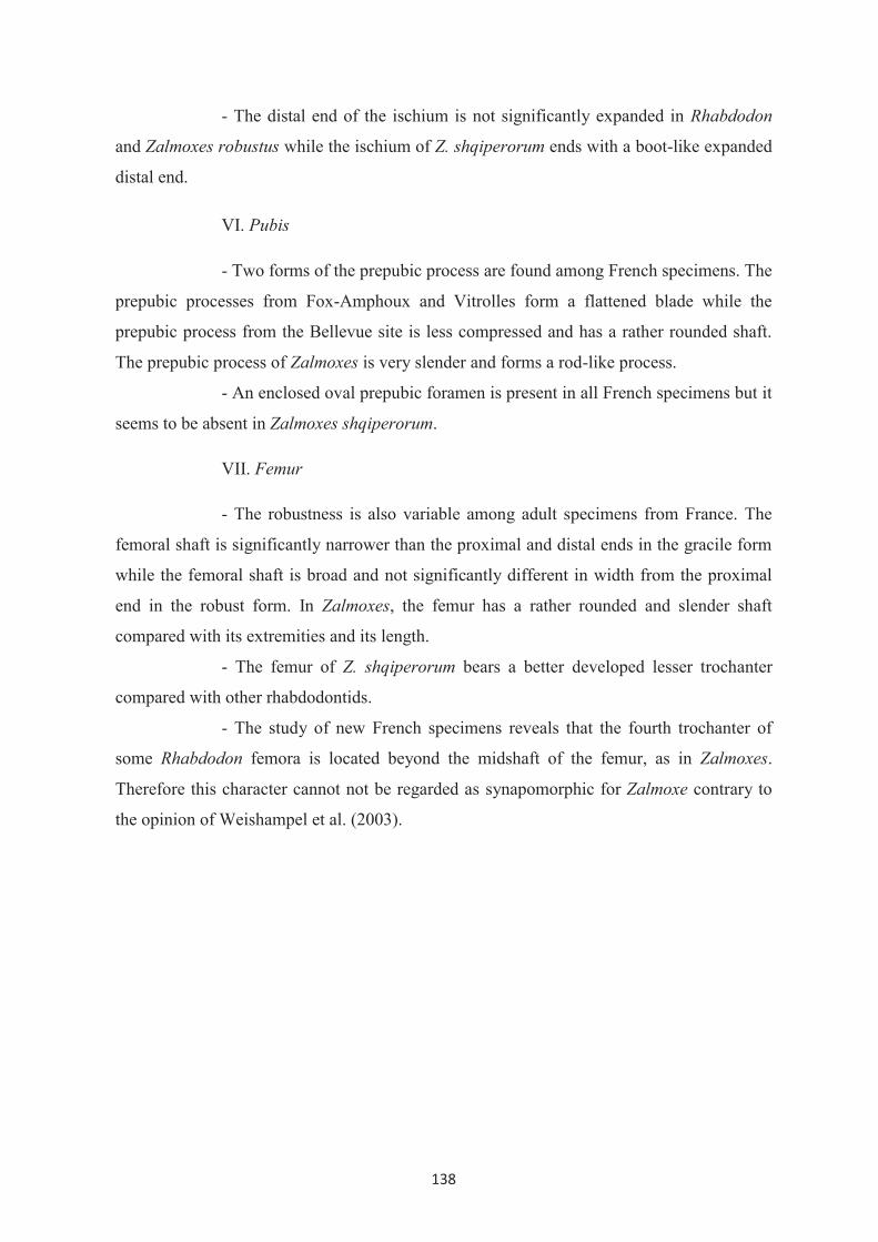
138
- The distal end of the ischium is not significantly expanded in Rhabdodon
and Zalmoxes robustus while the ischium of Z. shqiperorum ends with a boot-like expanded
distal end.
VI. Pubis
- Two forms of the prepubic process are found among French specimens. The
prepubic processes from Fox-Amphoux and Vitrolles form a flattened blade while the
prepubic process from the Bellevue site is less compressed and has a rather rounded shaft.
The prepubic process of Zalmoxes is very slender and forms a rod-like process.
- An enclosed oval prepubic foramen is present in all French specimens but it
seems to be absent in Zalmoxes shqiperorum.
VII. Femur
- The robustness is also variable among adult specimens from France. The
femoral shaft is significantly narrower than the proximal and distal ends in the gracile form
while the femoral shaft is broad and not significantly different in width from the proximal
end in the robust form. In Zalmoxes, the femur has a rather rounded and slender shaft
compared with its extremities and its length.
- The femur of Z. shqiperorum bears a better developed lesser trochanter
compared with other rhabdodontids.
- The study of new French specimens reveals that the fourth trochanter of
some Rhabdodon femora is located beyond the midshaft of the femur, as in Zalmoxes.
Therefore this character cannot not be regarded as synapomorphic for Zalmoxe contrary to
the opinion of Weishampel et al. (2003).

139
5.1.3 Conclusion
1. According to morphological observations on new Rhabdodon remains
from southern France, at least two species were represented, Rhabdodon priscus and
Rhabdodon septimanicus. Rhabdodon priscus includes all specimens except the type
specimen of R. septimanicus, a dentary from La Boucharde and the cranial material from
Quarante. The dentary from the Late Campanian of La Boucharde is referred to Rhabdodon
septimanicus. The cranial elements from Quarante provide sufficiently diagnostic characters
to suggest another variety of Rhabdodon in France, however the dentary from Quarante
shares three diagnostic characters with R. spetimanicus. I therefore consider that the
Quarante material can be referred to as cf. Rhabdodon septimanicus.
2. Besides the dentary that distinguishes R. septimanicus from R. priscus,
there are no other sufficiently distinctive postcranial characters to describe French
specimens as another different taxon. The variation within Rhabdodon priscus could be
interpreted as the intraspecific variability. The differences in robustness as well as the
differences in the neural spine probably suggest sexual dimorphism. However the extent of
sexual dimorphism in the postcranial skeleton is poorly known. The different morphological
features are possibly due to the individual or ontogenic variability as reported in other
groups of Ornithopoda (Forster, 1990; Galton, 2009).
3. New data from French material confirm some diagnostic characters of
Rhabdodon and suggest the exclusion of some synapomorphic characters of
Rhabdodontidae and of Zalmoxes defined by Weishampel et al. (2003). The systematic and
phylogenetic analysis will be reevaluated and discussed in the following part.

140
5.2 Phylogenetic relationships of Rhabdodon
As briefly mentioned in Chapter 2, since the first description of Rhabdodon this
European taxon was diversely allied to hypsilophodontids, camptosaurids, iguanodontids
and to the recently erected group of rhabdodontids. The study of Weishampel et al. (2003)
apparently represents the most recent phylogenetic analysis which includes Rhabdodon in
the cladogram. However in their work a great number of data about Rhabdodon priscus are
missing. This study will follow mainly the cladistic analysis of non-hadrosaurid
Euornithopoda of Weishampel et al. (2003) and also includes new data about Rhabdodon
priscus in the dataset to reevaluate the relationships of Rhabdodontidae and the
phylogenetic position of Rhabdodon.
The analyses were based on 15 terminal species following Weishampel et al. (2003).
75 characters (23 cranial, 12 dental and 40 postcranial) from Weishampel et al. (2003)
matrix were polarized by successive out-group comparisons with Heterodontosaurus tucki
and Marginocephalia. The description of new remains referred to Rhabdodon priscus from
southern France provides some additional characters missing in previous works. Although
the validity of Rhabdodon septimanicus is accepted in this work and new material referred
to cf. Rhabdodon septimanicus is reported, most of its skeleton is still poorly known. I
therefore do not include this species in this analysis.
Rhabdodon priscus has been recoded in the matrix of Weishampel et al. (2003) (See
the matrix in Appendix III.2). Some differences in coding are noted in Appendix III.1. As
coded in the dataset, Rhabdodon priscus is 60% complete instead of 40% in previous works.
The analysis was run under PAUP* (version 4.0b10 for 32-bit Microsoft Window,
Swofford 2001) under a branch-and-bound search with both accelerated transformation
(ACCTRAN) and delayed transformation (DELTRAN) options. All characters were run
unordered without weighting.
5.2.1 Results
The analyses result in three most parsimonious trees of 151 steps with a consistency
index (CI) of 0.530, a retention index (RI) of 0.770 and rescaled consistency index (RCI) of
0.408. The strict consensus tree which is given by this analysis is identical to that of
Weishampel et al. 2003) in Figure X.

141
Figure 5.2 Consensus tree of the three most parsimonious trees from this study.
This study focused on the systematic revision of Rhabdodon priscus, together with
Zalmoxes and Rhabdodontidae. Only the characters attributed to Rhabdodon priscus and
Zalmoxes will be discussed in detail and associated with the morphological variation
mentioned above to suggest the amended diagnoses of these taxa.
From the present cladistic analysis, Rhabdodon is distinguished from its sister group
by the following characters:
1. Ossified epaxial tendons arranged in double-layered lattice. (character 42,
identified with both ACCTRAN and DELTRAN; convergent in Ankylopollexia)
2. A wide scapular neck (character 43, identified with both ACCTRAN and
DELTRAN; convergent in Iguanodon, Hypsilophodon, and the clade consisting of the
common ancestor of Parksosaurus and Thescelosaurus)
3. Robust radius (minimum radial width is equal or more than 10% of radial length)
(character 47, identified with DELTRAN, with ACCTRAN; it is a synapomorphy for
Rhabdodontidae; convergent in Ankylopollexia and it is identified as an apomorphy of
Thescelosaurus.

142
4. Hoof-shaped ungual of manual digits II-IV (character 54, identified with
DELTRAN; with ACCTRAN; it is a synapomorphy for Rhabdodontidae; convergent in
Iguanodon).
5. An elongate flattened prepubic process (character 63, with both ACCTRAN and
DELTRAN; convergent in Iguanodontia). However, this feature shows a variability in the
prepubis from Bellevue in which the rounded prepubic process is rather rounded than
laterally flattened.
6. Hoof-shaped ungual of pedal digit II-IV (character 75, identified with DELTRAN;
with ACCTRAN; it is a synapomorphy for Rhabdodontidae; convergent in Iguanodon)
Zalmoxes robustus and Zalmoxes shqiperorum are united as sister-species on the
basis of the following characters:
1. Long, narrow sternal process of the coracoid (character 44, identified with
ACCTRAN; with DELTRAN), this character is autapomorphic for Z. shqiperorum.
2. Deltopectoral crest emerges much more abruptly from the humeral shaft.
(character 46, identified with both ACCTRAN and DELTRAN; this character is convergent
in Tenontosaurus and Hypsilophodon)
3. Strongly twisted preacetabular process of the ilium (character 56, identified in
both ACCTRAN and DELTRAN).
4. Absence of the obturator process (character 58 identified in both ACCTRAN and
DELTRAN; reversal of the condition at Euornithopoda)
5. Distinctly downwardly curved ischial shaft (character 60, identified in both
ACCTRAN and DELTRAN; convergent in Dryomorpha)
6. Ovoid to subcylindrical cross-section of the ischial shaft (character 61, identified
in both ACCTRAN and DELTRAN; convergent in Dryomorpha)
7. Distinctively bowed femur in anterior view (character 65, identified in both
ACCTRAN and DELTRAN; convergent in Dryosaurus and Parksosaurus)
In the present study, Rhabdodon and Zalmoxes form a node-based clade of
Rhabdodontidae as in the analysis of Weishampel et al. (2003). According to this study,
Rhabdodontidae is diagnosed by the following 3 features:
1. Expansion of the posterior process of the jugal to form the ventral margin of the
infratemporal fenestra (character 11, identified with ACCTRAN; with DELTRAN it is

143
apomorphic for Z. robustus, this feature is also apomorphic for Agilisaurus and
convergently apomorphic for Dryomorpha)
2. More than 10 ridges on the lingual side of the dentary tooth crowns. (character 32,
identified with both ACCTRAN and DELTRAN)
3. A straight to slightly convex dorsal margin of the ilium in lateral view (reversal of
character 55, identified with both ACCTRAN and DELTRAN; this feature, a sinuous dorsal
iliac margin, is apomorphic at the level of the clade consisting of the common ancestor of
Gasparinisaura and Iguanodontia, it is also reversed in Camptosaurus.
3. Absence of metatarsal V (character 74, identified with ACCTRAN; with
DELTRAN it is restricted to be apomorphic to Z. robustus; it is also convergently
apomorphic for Ankylopollexia).
5.3 Conclusion
1. Rhabdodon differs from Zalmoxes in having horizontally directed paroccipital
processes, long and laterally directed basipterygoid processes, a lower ridge density on the
maxillary and dentary tooth crowns, prominently high neural spines on dorsal vertebrae (at
least twice and a half higher than the height of the centrum), ossified epaxial tendons
arranged in double-layered lattice, a wide scapular neck, an elongated flattened prepubic
process, a straight to slightly bowed femur in anterior view, hoof-shaped unguals of manual
and pedal digits II-IV.
2. Rhabdodontidae is considered as a sister group of Iguanodontia. It is diagnosed by
the following features: more than 10 sharp ridges on the lingual side of the crowns of the
dentary teeth, a straight to slightly convex dorsal margin of the ilium. Potential apomorphies
of Rhabdodontidae include an expansion of the posterior process of the jugal to form the
ventral margin of the infratemporal fenestra, a robust radius, hoof-shaped unguals of manual
and pedal digits II-IV, the absence of metatarsal V.
3. An amended diagnosis of Zalmoxes may include the following features:
deltopectoral crest emerging much more abruptly from the humeral shaft, strongly twisted
preacetabular process of the ilium, absence of the obturator process, distinctly downwardly
curved ischial shaft, ovoid to subcylindrical cross-section of the ischial shaft, distinctively
bowed femur in anterior view.

144
References
Allain, R., Pereda-Suberbiola, X. 2003. Dinosaurs of France. Comptes Rendus Palevol 2,
27–44.
Pereda-Suberbiola, X., Sanz, J.L. (1999) - The Ornithopod dinosaurs Rhabdodon from the
Upper Cretaceous of Laõo (Iberian Peninsula). Est. Mus. Cienc. Nat. de Àlava, 14
(N° Espec. 1), 257-272.
Buffetaut, E., Le Lœuff, J. 1991. Une nouvelle espèce de Rhabdodon (Dinosauria
Ornithischia) du Crétacé de l’Hérault (Sud de la France). C.R. Acad. Sci. Paris. Sér.2
312, 943-948.
Buffetaut, E., Costa, G., Le Lœuff, J., Martin, M., Rage, J.C, Valentin, X., Tong, H. 1996
An Early Campanian vertebrate fauna from the Villeveyrac Basin (Hérault, Southern
France). Neues Jahrbuch für Geologie und Paläontologie Monatshefte 1, 1-16.
Forster, A.C. 1990. The postcranial skeleton of the ornithopod dinosaur Tenontosaurus
Tilletti. Journal of Vertebrate Paleontology 10(3), 273-294.
Galton, P.M. 1974. The ornithischian dinosaur Hypsilophodon from the wealden of the Isle
of Wight. Bulletin of the British Museum (Natural History), Geology 25(1), 1-152.
Galton, P.M. 2009. Notes on Neocomian (Late Cretaceous) ornithopod dinosaurs from
England – Hyosilophodon, Valdosaurus, “Camptosaurus”, “Iguanodon”- and
referred specimens from Romania and elsewhere. Revue de Paléobiologie, Genève
28(1), 211-273.
Godefroit, P., Dong, Z.M., Bultynck, P., Li, H., Feng, L. 1998. New Bactrosaurus
(Dinosauria: Hadrosauroidea) material from Iren Dabasu (Inner Mongolia, P. R.
China). Bulletin de l'Institut Royal des Sciences Naturelles de Belgique, Sciences de
la Terre 68(supplement), 3-70.
Godefroit, P., Codrea, V., Weishampel, D.B. 2009. Osteology of Zalmoxes shqiperorum
(Dinosauria, Ornithopod), based on new specimens from the Upper Cretaceous of
N<laY-Vad (Romania). Geodiversitas 31(3), 525-553.

145
Lapparent, A. F. de. 1947. Les dinosauriens du Crétacé supérieur du Midi de la France.
Mémoires de la Société géologique de France 56, 1-54.
Matheron, P. 1869a. Notice sur les reptiles fossils des dépôts fluvio-lacustres crétacé du
basin à lignite de Fuveau. Mémoires de l’Académie impériale des Sciences, Belles-
Lettres et Arts de Marseille, 1-39.
Pereda-Suberbiola, X., Sanz, J.L. 1999. The ornithopod dinosaur Rhabdodon from the
Upper Cretaceous of Laño (Iberian Peninsula). Estudios del Museo de Ciencias
Naturales de Alva 14 (Número especial 1), 257–272.
Pincemaille-Quillévéré, M. 2002. Description d’un squelette partiel de Rhabdodon priscus
(Euornithopoda) du Crétacé supérieur de Vitrolles (Bouches du Rhône, France).
ORYTOS 4, 39-70.
Pincemaille-Quillévéré, M., Buffetaut, E., Quillévéré, F. 2006. Osteological description of
the braincase of Rhabdodon (Dinosauria, Euornithopoda) and phylogenetic
implications. Bulletin de la Société Géologique de France 177, 97-104.
Swofford, D. L. 2001. PAUP*: Phylogenetic Analysis Using Parsimony (* and Other
Methods), version 4.0b10. Sinauer Associates, Inc., Sunderland, Massachusetts.
Weishampel, D.B. 1984. Evolution of Jaw Mechanisms in Ornithopod Dinosaurs. Advances
in Anatomy Embryology and Cell Biology, 87, 1-109.
Weishampel, D. B., Norman, D. B., Grigorescu, D. 1993. Telmatosaurus transsylvanicus
from the Late Cretaceous of Romania: the most basal hadrosaurid. Palaeontology 36,
361-385.
Weishampel, D.B., Jainu, C-M, Csiki Z., Norman, D.B. 2003. Osteology and Phylogeny of
Zalmoxes (N.G.), an unusual Euornithopod Dinosaur from the latest Cretaceous of
Romania. Journal of the Systematic Paleontology 1(2), 65-123.

146

147
Chapter 6:
Discussion
6.1 Geographical and temporal distribution of Rhabdodon
Rhabdodon was initially erected on the basis of a dentary with its characteristic
fluted teeth from La Nerthe (Bouches-du-Rhône, France) (Matheron, 1869). Medium-sized
ornithopods, distinguished from other groups by this distinctive type of dentition have been
recovered from several Late Cretaceous terrestrial localities and appear to be endemic to
Europe (France, Spain, Romania, Austria and Hungary).
According to the previous works and this present study, a certain amount of diversity
within this dinosaur group has been suggested. Rhabdodon and its sister taxon, Zalmoxes
form a recently defined family, Rhabdodontidae Weishampel, Jianu, Cziki and Norman,
2003. At the moment, the non-hadrosaurid ornithopod remains discovered in France and
Spain are referred to genus Rhabdodon and the remains from the Early Campanian of
Austria and the Maastrichtian of Romania are referred to Zalmoxes (Weishampel et al.,
2003; Sachs and Hornung, 2006, Godefroit et al., 2009). The material from Hungary does
not provide sufficiently diagnostic features to define it at the generic level, it is consequently
considered as Rhabdodontidae indet. on the basis of the teeth (Osi, 2004). The remains from
the Santonian Iharkut locality of Hungary seem to be the oldest among the rhabdodontid
dinosaurs.
Rhabdodon is widely distributed in Campanian-Maastrichtian continental localities
of southern France as mentioned in Chapter 3. The earliest appareance of Rhabdodon in
France is represented by Rhabdodon priscus remains from the Early Campanian of
Villeveyrac basin (Buffetaut et al., 1996). The age of Villeveyrac basin has been confirmed
recently by the magnetostratigraphic study of Benammi et al. (2006). The most recent
remains of Rhabdodon seem to be from the Vitrolles-La-Plaine locality which is dated as
Maastrichtian (Valentin et al. 2009).
Both Rhabdodon priscus and R. septimanicus (including cf. R. septimanicus
described in this study) from the Saint-Chinian area were discovered in the Grès à Reptiles

148
Formation (Late Campanian - Early Maastrichtian). The type of Rhabdodon septimanicus
was found at a locality near Montouliers while a productive site, Massecaps, yielding
Rhabdodon priscus is located at the neighbouring village, Cruzy. Despite the fact that the
precise dating of most of the French localities is usually difficult and is still uncertain, it
cannot be excluded that these two species coexisted during the same time interval as in the
case of Zalmoxes robustus and Z. shqiperorum which are regarded as sympatric (Godefroit
et al., 2009).
The Late Campanian to Maastrichtian dinosaur fauna of southern France is
dominated by Rhabdodon and titanosaurs. This faunal assemblage seems to be replaced
during the late Maastrichtian by a different assemblage, from which Rhabdodon is absent
and in which hadrosaurs play an important part (Le Lœuff et al., 1994). The co-occurrence
of Rhabdodon and a basal hadrosaurid in southern France is reported from a Maastrichtian
locality in Provence (Valentin et al., 2009). Rhabdodon and the hadrosaurids apparently
occupied a similar ecological niche in the Maastrichtian. They most probably competed for
food resources. With the evolutionary success of their jaw mechanism and dentition, the
hadrosaurs may have started to occupy that niche in southern France prior to the Late
Maastrichtian and finally replaced Rhabdodon in the Late Maastrichtian.
6.2 Paleobiogeographical implications
The fossil record shows that Rhabdodon is distributed in southern France over a long
period ranging from Campanian to Maastrichtian. As revealed in the phylogenetic study, the
European family, Rhabdodontidae, is a close relative to Iguanodontia. As described in
Chapter 4 and according to previous studies (Pincemaille-Quillévéré, 2002; Chanthasit et al.
in review), Rhabdodon shares several osteological characters in particular the postcranial
elements with Tenontosaurus tilletti. This could suggest that the common ancestor of the
rhabdodontid dinosaurs evolved in North America, and then dispersed to Europe. For much
of the Jurassic and Cretaceous, Europe was composed of several major islands and some of
which had occasional contact with North America or Asia. The split-up of the Euroamerican
palaeogeographical province during the Aptian caused the European taxa to evolve
separately from North American taxa (Upchurch et al., 2002).
As discussed in the preceding chapter, Zalmoxes can be clearly distinguished from
Rhabdodon, it indicates that these two rhabdodontids evolved separatedly in geographical

149
isolation. The geographical and temporal distribution of Rhabdodon and Zalmoxes suggest
that these two taxa diverged as early as the Early Campanian.
In the Late Cretaceous, Europe was indeed a vast archipelago; the southern, western,
central and eastern domains of Europe consisted of a string of medium to small-sized
islands separated from each other by shallow epicontinental seas (Pereda-Suberbiola, 2009)
(Figure 6.1). The taxonomic variability among Rhabdodontidae would be the result of
geographical, ecological (island) and temporal (localities with various stratigraphic
positions) variability.
Figure 6.1 Simplified palaeogeographical map of Europe and Peri-Tethyan area between
latitutes 10° and 50° N for the Late Cretaceous (Maastrichtian), the black squares indicate
the distribution of Rhabdodontidae. Key: A, Apennine platform; Al, Alboran islands; Ap,
Apulian platform; AA, Austroalpine island; AD, Adriatic-Dinaric island; AS, Anglo-
Scottish island; C, Crimean island; FS, Fennoscandian shield; I, Irish island; IA, Ibero-
Armorican island; M, Moesian platform; RB, Rhenish-Bohemian island; Rh, Rhodope; T,
Transylvanian basin; US, Ukrainian shield. Colour marks: light, exposed land; light grey,
epicontinental seas; dark grey, oceanic basins; white, unknown; black, volcanics. (Modified
from Pereda-Suberbiola, 2009)

150
However, the palaeobiogeographical affinities of the Late Cretaceous continental
fauna of Europe are complex. The faunas are commonly considered to show a mixed pattern
resulting from the addition of "Asiamerican" and Gondwanan forms to European taxa. An
example from southern France is provided by the dromaeosaurid, Variraptor mechinorum
which shows the closest affinities with the Asian taxa (Chanthasit and Buffetaut, 2009)
(APPENDIX IV) while in this study Rhabdodon is considered as relatively close to North
American taxa.
6.3 Feeding
The information concerning the jaw function is quite poor as the cranial elements
related to the jaw mechanism are rarely found. A pleurokinetic jaw mechanism could be
suggested for Rhabdodon. The posterior broadening of the maxillary process of the
premaxilla provides a loosened maxilla-premaxilla joint and allows the lateral motion of the
maxilla. The dentition of Rhabdodon shows an elaborate pattern. The maxillary and dentary
teeth are covered with a thicker layer of enamel on one side than on the other. The thickly
enameled surface is more durable to wear so that a sharp edge is formed while the thinly
enameled surface is less resistant and forms an obliquely inclined occlusal surface. The
sharp edge would provide the cutting action. The wear of the longitudinal ridges on the
thickly enameled surface provides the serrations which enhance the sharpness of the cutting
edge. The surangular bears a well developed lateral lip shrouding the lateroventral portion
of the quadrate. This structure indicates a strong attachment for the ligaments and thus
stability at the quadrate-mandible joint. The robust dentary and a well-developed coronoid
process indicate a powerful jaw closure. This could be related to a resistant type of food.
The narrow premaxillae influence the shape of the beak that is also narrow. As suggested by
Weishampel et al. (2003), Zalmoxes had the potential to be a selective feeder by using its
narrow beak to select preferred food items. Rhabdodon could also have same feeding habits.

151
6.4 Posture and locomotion
A great number of ossified tendons over the sacrum and the neighbouring area
around the posterior dorsal and anterior caudal vertebrae suggest that the vertebral column
was probably held more or less in horizontal position. Rhabdodon may have been primarily
quadrupedal. It has a robust forelimb. The humerus possesses a well-developed
deltopectoral crest. The ulna bears a prominent olecranon. The manus is short and robust. It
is likely that the forelimb supported the body weight and was adapted for standing or
walking as a quadruped. However, the metacarpals were not firmly packed together and
with a short manus and forearm, Rhabdodon was probably incapable of rapid quadrupedal
locomotion but probably stood bipedally for rapid locomotion. The tibia is shorter than the
femur and the position of the fourth trochanter is relatively low compared with the condition
in a cursorial animal (Gregory, 1912). These characters limited the capacity of Rhabdodon
for fast running. On the basis of the osteological description, a reconstruction of Rhabdodon
can be proposed as shown on the following figure.
Figure 6.2 A skeletal reconstruction of Rhabdodon based on composite elements from
southern France.

152
References
Benammi, M., Urrutia-Fucugauchi, J., Vianey-Liaud, M. 2006. Preliminary
magnetostratigraphic study of the Late Cretaceous dinosaur site from Villeveyrac-
Mèze Basin, Southern France. International Geology Review, 48(1), 89 – 96.
Buffetaut, E., Le Lœuff, J. 1996. Late Cretaceous dinosaur faunas of Europe: some
correlation problems. Cretaceous Research, 12, 159-176.
Chanthasit, P., Buffetaut, E. 2009. New data on the Dromaeosauridae (Dinosauria:
Theropoda) from the Late Cretaceous of southern France. Bulletin de la Société
Géologique de France 180(2), 145-154.
Godefroit, P., Codrea, V., Weishampel, D.B. 2009. Osteology of Zalmoxes shqiperorum
(Dinosauria, Ornithopod), based on new specimens from the Upper Cretaceous of
Nãlat-Vad (Romania). Geodiversitas 31(3), 525-553.
Gregory, W.K. 1912. Notes on the principles of quadrupedal locomotion and on the
mechanism of the limb in hoofed animals. Annals of the New York Academy of
Sciences 22, 267-294.
Le Lœuff J., Buffetaut E., Martin M. 1994. The last stages of dinosaur faunal history in
Europe: a succession of Maastrichtian dinosaur assemblages from the Corbières
(southern France). Geological Magazine, Cambridge 131, 625-630.
Pereda-Suberbiola, X. 2009. Biogeographical affinities of Late Cretaceous continental
tetrapods of Europe: a review. Bulletin de la Société Géologique de France 180(1),
57-71.
Matheron, P. 1869. Notice sur les reptiles fossils des dépôts fluvio-lacustres crétacé du basin
à lignite de Fuveau. Mémoires de l’Académie impériale des Sciences, Belles-Lettres
et Arts de Marseille, 1-39.
Sachs, S., Hornung, J. J. 2006. Juvenile ornithopod (Dinosauria: Rhabdodontidae) remains
from the Upper Cretaceous (lower Campanian, Gosau Group) of Muthmannsdorf
(Lower Austria). Géobios 39, 415-425.

153
Ösi, A. 2004. The first dinosaur remains from the Upper Creataceous of Hungary
(Csehbànxa Formation, Bakony Mts). Geobios 37, 749–753.
Upchurch, P., Hunn, C. A., and Norman, D. B. 2002. An analysis of dinosaurian
biogeography: evidence for the existence of vicariance and dispersal patterns caused
by geological events. Proceedings of the Royal Society of London 269, 613-621.
Valentin, X., Godefroit, P., Tabuce, R., Vianey-Liaud, M., Garcia, G., Wenhao, W.U., 2009.
First maastrichtian vetebrate assemblage from Provence (Vitrolles-la-plaine, France).
In Godefroit, P. Lambert, O. (Eds.). Tribute to Charles Darwin and Bernissart
Iguanodons: New perspectives on vertebrate evolution and Early Cretaceous
ecosystems, Brussels 2009. Programme, Abstracts and Field Trips Guidebook, p.94.
Weishampel, D.B., Jainu, C-M, Csiki Z., Norman, D.B. 2003. Osteology and Phylogeny of
Zalmoxes (N.G.), an unusual Euornithopod Dinosaur from the latest Cretaceous of
Romania. Journal of the Systematic Paleontology 1(2), 65-123.

154
155
Chapter 7:
Conclusions
The study of the ornithopod dinosaur Rhabdodon from the Late Cretaceous of France can be concluded as follows:
7.1 The validity of Rhabdodon septimanicus
The study of undescribed material of Rhabdodon having been discovered in
various localities in southern France provides more information on the osteology of
Rhabdodon. New cranial elements have been described. Most of them have been discovered
at a locality near the village of Quarante (Hérault) and it can be assumed that these elements
belong to a single individual. The material from Quarante, particularly the premaxilla,
maxilla and dentary, shows several distinctive characters and can be distinguished from the
material from other localities. The premaxilla bears an elongated posterolateral (maxillary)
process and a large opening of the external naris. The premaxillary process of the maxilla
from Quarante is relatively long compared with those of other localities. Nevertheless, the
dentary from Quarante shares diagnostic characters with that of Rhabdodon septimanicus
Buffetaut & Le Lœuff, 1991. Consequently, on the basis of the dentary the Quarante
material can be referred to as cf. Rhabdodon septimanicus. Furthermore, the Rhabdodon
remains from Quarante provide additional elements and diagnostic characters for R.
septimanicus. The morphological diagnosis of R. septimanicus including the Quarante
material supports the validity of this species. However, besides the skull, no postcranial
elements referred to R. septimanicus can be distinguished from other taxa.
7.2 The variation of the genus Rhabdodon
There is some morphological variation in the cranial and postcranial skeleton
between Rhabdodon specimens from different localities and between specimens coming
from the same locality. Concerning the cranial skeleton, excluding the diagnosis of the
dentary of R. septimanicus and of the maxilla from Quarante mentioned above, the dentaries
of Rhabdodon show robust and gracile coronoid processes and various degrees of coronoid

156
process inclination. In the postcranial skeleton, there are some differences in the
development of neural spines, in the robustness of the pectoral girdle and limb bones and in
various features of the pelvic bones. These morphological differences are not distinct and
are insufficient to define French specimens as another separate taxon. Consequently, the
variation within Rhabdodon priscus could be interpreted as an intraspecific variability
which is possibly due to individual or ontogenic variability or sexual dimorphism. However
the associated cranial and postcranial material of R. septimanicus is still unknown, it cannot
be excluded that some postcranial bones which show the morphological differences could
belong to R. septimanicus. In the present state of our knowledge, it can be concluded that
two species, Rhabdodon priscus and Rhabdodon septimanicus, were represented in the Late
Cretaceous of France. R. septimanicus can be distinguished from R. priscus by the dentary
diagnosis defined by Buffetaut and Le Lœuff (1991) and the diagnostic characters of
referred material from Quarante mentioned above.
7.3 Anatomical comparison and systematics
Rhabdodon is placed in Rhabdodontidae with its sister taxon, Zalmoxes. The new
specimens described in this work suggest several distinctive morphological features
between Rhabdodon and Zalmoxes especially the postcranial elements. The osteological
description of new specimens from southern France allows the reassessment of the
phylogenetic relationships of Rhabdodon among Rhabdodontidae and with other
Euornithopoda. This study follows the most recent cladistic analysis by Weishampel et al.
(2003). New data from French material of Rhabdodon priscus have been recoded in the
matrix of Weishampel et al. 2003 with 18 additional characters which were missing in
previous work (APPENDIX III).
In spite of the fact that the phylogenetic position of Rhabdodon given by the
present study is in agreement with previous work, French material suggests the exclusion of
some synapomorphic characters of Rhabdodontidae and of Zalmoxes defined by
Weishampel et al. (2003). The anatomical comparison and the phylogenetic analysis suggest
an amended diagnosis of Rhabdodon, of Zalmoxes and of Rhabdodontidae.

157
7.4 Paleobiology
Rhabdodon was most likely a facultatively bipedal ornithopod with at least
occasional quadrupedal locomotion. It is characterized primarily by its elaborate dentition
and probably had a sophisticated mastication. Rhabdodon appears to be exclusively
distributed in Europe ranging from the Early Campanian to the Early Maastrichtian. It is
probably represented only in France and Spain as the remains from Romania and Austria are
referred to its sister taxon Zalmoxes (Pereda-Suberbiola and Sanz, 1999; Weishampel et al.,
2003; Sachs and Hornung, 2006, Godefroit et al., 2009) and the Hungarian material has not
yet been verified at the generic level (Ôsi, 2004) nevertheless the teeth are clearly different
from those of Rhabdodon.( Buffetaut, pers.comm. 2010)
The phylogenetic study suggests that the European family, Rhabdodontidae, is a
close relative to Iguanodontia. Among the iguanodontids, Rhabdodon is particularly close to
Tenontosaurus tilletti as they share several postcranial features. This could suggest that the
common ancestor of the rhabdodontid dinosaurs evolved in North America, and then
dispersed to Europe. Rhabdodon and Zalmoxes eventually evolved separately on the
European archipelago. The geographical and temporal distribution of Rhabdodon and
Zalmoxes suggest that these two taxa diverged as early as the Early Campanian.

158
References
Buffetaut, E., Le Lœuff, J. 1991. Une nouvelle espèce de Rhabdodon (Dinosauria,
Ornithischia) du Crétacé supérieur de l’Hérault (Sud de la France). Compte rendu de
l’Académie des Sciences de Paris, Série II 312, 943-948.
Godefroit, P., Codrea, V., Weishampel, D.B. 2009. Osteology of Zalmoxes shqiperorum
(Dinosauria, Ornithopod), based on new specimens from the Upper Cretaceous of
Nãlat-Vad (Romania). Geodiversitas 31(3), 525-553.
Ösi, A. 2004. The first dinosaur remains from the Upper Creataceous of Hungary
(Csehbànxa Formation, Bakony Mts). Geobios 37, 749–753.
Pereda-Suberbiola, X., Sanz, J.L. 1999. The ornithopod dinosaur Rhabdodon from the
Upper Cretaceous of Laño (Iberian Peninsula). Estudios del Museo de Ciencias
Naturales de Alva 14 (Número especial 1), 257–272.
Sachs, S., Hornung. J. J. 2006. Juvenile ornithopod (Dinosauria: Rhabdodontidae) remains
from the Upper Cretaceous (lower Campanian, Gosau Group) of Muthmannsdorf
(Lower Austria). Géobios 39, 415-425.
Weishampel, D.B., Jainu, C-M, Csiki Z., Norman, D.B. 2003. Osteology and Phylogeny of
Zalmoxes (N.G.), an unusual Euornithopod Dinosaur from the latest Cretaceous of
Romania. Journal of the Systematic Paleontology 1(2), 65-123.

159
APPENDIX I
Rhabdodontid Locality List
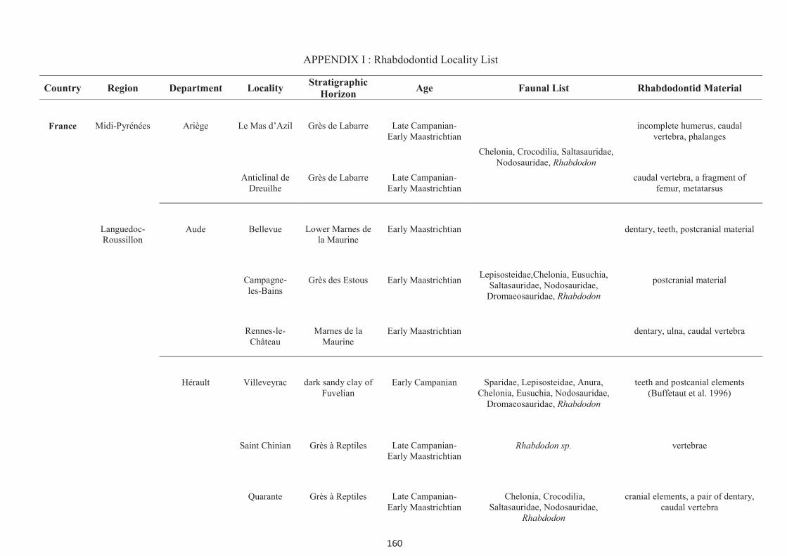
160
APPENDIX I : Rhabdodontid Locality List
Country Region Department Locality Stratigraphic
Horizon Age Faunal List Rhabdodontid Material
France
Midi-Pyrénées
Ariège
Le Mas d’Azil
Grès de Labarre
Late Campanian-Early Maastrichtian
Chelonia, Crocodilia, Saltasauridae, Nodosauridae, Rhabdodon
incomplete humerus, caudal vertebra, phalanges
Anticlinal de Dreuilhe
Grès de Labarre Late Campanian-Early Maastrichtian
caudal vertebra, a fragment of femur, metatarsus
Languedoc-Roussillon
Aude Bellevue Lower Marnes de la Maurine
Early Maastrichtian
Lepisosteidae,Chelonia, Eusuchia, Saltasauridae, Nodosauridae,
Dromaeosauridae, Rhabdodon
dentary, teeth, postcranial material
Campagne-les-Bains
Grès des Estous Early Maastrichtian postcranial material
Rennes-le-Château
Marnes de la Maurine
Early Maastrichtian dentary, ulna, caudal vertebra
Hérault Villeveyrac dark sandy clay of Fuvelian
Early Campanian Sparidae, Lepisosteidae, Anura, Chelonia, Eusuchia, Nodosauridae,
Dromaeosauridae, Rhabdodon
teeth and postcanial elements (Buffetaut et al. 1996)
Saint Chinian Grès à Reptiles Late Campanian-Early Maastrichtian
Rhabdodon sp. vertebrae
Quarante Grès à Reptiles Late Campanian-Early Maastrichtian
Chelonia, Crocodilia, Saltasauridae, Nodosauridae,
Rhabdodon
cranial elements, a pair of dentary, caudal vertebra

161
Country Region Department Locality Stratigraphic
Horizon Age Faunal List Rhabdodontid Material
France
Languedoc-Roussillon
Hérault
Montouliers
Grès à Reptiles
Late Campanian-Early Maastrichtian
Chelonia, Theropoda, Maniraptora, Saltasauridae, Nodosauridae, cf
Rhabdodon, Rhabdodon
septimanicus
dentary (type) (Buffetaut and Le Lœuff, 1991), braincase
(Pincemaille-Quillévéré et al. 2006) , maxillary fragment, long bone fragment, phalanges, unguals
Massecaps Grès à Reptiles Late Campanian-Early Maastrichtian
Lepisosteidae, coelacanth, Chelonia, Amphibia, Squamata,
Crocodilia, Pterosauria, Dromaeosauridae, Enantiornithes,
Saltasauria, Nodosauridae, Rhabdodon, Mammalia
some cranial elements and postcranial material
Combebelle Grès à Reptiles Late Campanian-Early Maastrichtian
2 femora
Sainte-Foy Grès à Reptiles Late Campanian-Early Maastrichtian
femoral fragment
Plo Saint Pons
Grès à Reptiles Late Campanian-Early Maastrichtian
vertebra, sacrum, ilia
Mont Plo Nord
Grès à Reptiles Late Campanian-Early Maastrichtian
vertebrae, femur,
Quarante Grès à Reptiles Late Campanian-Early Maastrichtian
cranial elements, vertebra, fibula
fragment
Gard Champ-
Garimond
red clays of Begudo-
Rognacien Maastrichtian
Lepisosteidae, coelacanth, Chelonia, Amphibia, Squamata,
Crocodilia, Pterosauria, Dromaeosauridae, Saltasauria,
Nodosauridae, Rhabdodon
teeth, vertebra, fragment of humerus

162
Country Region Department Locality Stratigraphic
Horizon Age Faunal List Rhabdodontid Material
France
Provence-Alpes-Côte
d'Azur
Bouches-du-Rhône
Vitrolles-Couperigne
sandy marl of Lower Rognacian
Early Maastrichtian
Crocodilia, another reptile, Rhabdodon priscus
cranial elements, 2 partial articulated skeletons (Pincemaille-
Quillévéré, 2002)
La Boucharde Fluvio-lacustrine sandstone of
Begudian
Late Campanian-Early Maastrichtian
Chelonia, Crocodilia,Dromaeosauridae,
Saltasauria, Nodosauridae, Ceratosauria, Rhabdodon
cranial and post cranial material
La Nerthe lacustrine marl of upper Rognacian
Maastrichtian Chelonia, Crocodilia,Saltasauria, Rhabdodon priscus
dentary (Type), teeth, postcranial material ( Matheron, 1869)
Roques-Hautes
Red marl with lenticular
sandstone of Lower Rognacian
Early Maastrichtian Crocodilia, Dromaeosauridae, Rhabdodon
femur
Rousset Clay and sandstone of
Lower Rognacian
Early Maastrichtian Rhabdodon femur
Var Métisson Grès à Reptiles Late Campanian-Early Maastrichtian
Chelonia, Crocodilia, Abelisauridae, Dromaeosauridae,
Saltasauria, Nodosauridae, Rhabdodon
teeth, vertebrae, femora, tibiae, metatarsi, phalange

163
Country Region Department Locality Stratigraphic
Horizon Age Faunal List Rhabdodontid Material
France
Provence-Alpes-Côte
d'Azur
Var
La Bastide Neuve
Grès à Reptiles
Late Campanian-Early Maastrichtian
Chelonia, Crocodilia, Pterosauria, Abelisauridae, Dromaeosauridae,
Enantiornithes, Gargantuavis, Saltasauria, Nodosauridae,
Rhabdodon
cranial and postcranial material
Basségat Grès à Reptiles
Late Campanian-Early Maastrichtian
vertebrae, fragment of femur, tibia, metatarsus, phalanges
Mourfrey champ
Grès à Reptiles Late Campanian-Early Maastrichtian
vertebrae, femur, tibia, metatarsus
Spain Castilla y León Burgos Laño L1A, L1B, L2 of Laño quarry
Late Campanian Chondrichthyes, Osteichthyes, Chelonia, Amphibia, Squamata,
Crocodilia, Pterosauria, Dromaeosauridae, Saltasauria,
Nodosauridae, Rhabdodon, mammalia
teeth, vertebrae, humerus, femora (Pereda-Suberbiola and Sanz, 1999)
Austria Niederösterreich Wiener Neustadt
Muthmannsdorf
Grünbach Formation, Gosau
Group
Early Campanian Chelonia, Crocodilia, Pterosauria, Nodosauridae, Zalmoxes
Dentary, teeth, fragment of parietal, fragment of scapula, radius?, femur,
tibia, manual ungual ( Sachs and Hornung, 2006)
Hungary Central Transdanubia
Veszprèm Iharkút Csehbánya Formation
Santonian fish, frog, turtle, crocodile, Theropoda, Nodosauridae,
Rhabdodontidae
Teeth, dentaries (Osi, 2004)

164
Country Region Department Locality Stratigraphic
Horizon Age Faunal List Rhabdodontid Material
Romania
Transylvania
Hunedoara
Sânpetru
Sânpetru Formation
Maastrichtian
Chelonia, Amphibia, Squamata, Crocodilia, mammalia, Theropoda, Sauropoda, Ankylosauria, Zalmoxes
robustus and Z. shqiperorum
cranial and postcranial material (Weishampel et al., 2003)
N<laY-Vad Sânpetru Formation
Maastrichtian birds, mammalia, Theropoda, Sauropoda, Zalmoxes shqiperorum
cranial and postcranial material (Godefroit et al. 2009)
S<laj Someş Odorhei
Jibou Formation Maastrichtian Chelonia, Crocodilia, Zalmoxes vertebrae, scapula (Codrea and Godefroit, 2008)

165
APPENDIX II
The anatomical measurements of the Rhabdodon
elements described in this work

166
Appendix II.1: Diagram showing the basis of the measurements of the specimens.
The measurements of other long bones use the same basis as for the femur
L = Total length, Wp = Width of the proximal end and Wd = Width of the distal end

167
Appendix II.2 Origin of the specimens:
CM = Fox-Amphoux localities
MC-CY.QR/MC-QR = Quarante locality
MC-M = Massecaps locality, Cruzy
MC-MN = Mont-plo locality
MC-MOB = Montouliers locality
MC-PSP = Plo-Saint-Pons locality
MC-VC = Combebelle locality
MDE-C2 = Le Pylône locality, Campagne-sur-Aude
MDE-C3 = Bellevue locality, Campagne-sur-Aude
MDE-C4 = Campagne-les-Bains locality, Campagne-sur-Aude
MDE-D = Montouliers locality
MDE-RC = Rennes-le-Château locality
MDE-Vil = Villeveyrac locality
Appendix II.3 Table of measurements of dentaries.
No. L/R L H ♂ No. of alvioli
CM-587 L >186 54 - 9
CM-443 R 236 52 125 9
CM-709 L 152 38 115 9
MC-CY.QR 1 R 330 6.3 104 11
MC-CY.QR 2 L 368 6.3 110 11
MC-M23 L 177 3.7 114 10
MDE-C3 1280 L 126 30.0 91 9
MDE-C3 1407 L >180 53 120 10

168
Appendix II.4 Measurements of maxillary teeth.
No. Mesial-distal length (Tooth Width)(mm)
Number of ridges
CM-297 24 12
CM-326 25 12
CM-554 22 12
MC-M 4190 17 8
MDE-C3 892 21 12
MDE-C3 1412 22 7
MNH_AIX_PV_Fox 21 9
Appendix II.5 Measurements of dentary teeth.
No. Mesial width
(mm) No.of mesial
ridges Distal width
(mm) No.of distal
ridges
CM-462 9.5 3 8.5 6
CM-475 9.7 4 7.97 2
CM-502 12.3 4 11.7 5
CM-503 17 4 9.8 3
CM-504 14.5 4 9.5 6
CM-505 11 6 8 7
CM-506 7.5 5 4.5 4
CM-507 11 8 10 6
CM-508 12.5 4 9 4
CM-655 10.5 3 6 2
MC-M 3583 13.5 5 10.5 3
MDE-C3 809 13 6 10 5
MDE-D27 10 4 8 4
MDE-D37 8.6 5 7.4 5

169
Appendix II.6 Measurements of vertebrae (mm).
Nature No. L W H
Atlas CM-86 21 38 33
Axis MC-4058 58 29 32
Anterior cervical CM-679 39 47 38
MC-M 30 28 29 >12
MDE-C3.1417 35 27 32
Posterior cervical CM-441 49 47 60
Anterior dorsal CM-379 28 38 48
Middle dorsal CM-142 43 38 39
CM-445 63 58 59
CM-446 37 47 55
CM-581 75 48 60
CM-527 40 47 50
CM-710 60 57 63
MC-MN 32 77 59 53
MDE-C3 1556 72 55 56
Posterior dorsal CM-3 59 56 81
CM-416 57 82 77
CM-496 39 45 43
MC-M 948 58 54 55
MC-M 1149 53 57 46
MC-M 2705 37 34 39
MC-M 3395 50 47 55
MC-M 3896 50 46 46
MC-M 4012 52 60 46
MDE-C3 413 50 63 56
MDE-C3 973 65 57 60
MDE-C3 1400 65 56 65
MDE-C3 03.50 65 64 51
MNHN 8 64 43 62

170
Nature No. L W H
Dorsal? MC-M 626 34 38 38
MC-M 1116 34 35 31
MC-M 1080 36 41 36
MC-M 220 55 72 59
MC-M 261 58 49 54
MC-M 38 56 68 57
MDE-C3 02.102 68 79 85
MDE-C3 02.137 62 64 61
Anterior caudal CM-403 59 55 85
CM-404 42 69 74
CM-569 49 61 64
CM-585 43 50 50
CM-688 79 57.5 98
MC-M 0950 55 60 49
MC-M 2483 59 72 76
MC-M 4028 35 36 39
MC-MN 30 62 53 51
MC-MN 36 42 63 68
MDE-C3 35 65 60 70
MDE-C3 110 62 65 72
MDE-C3 257 61 65 61
MDE-C3 337 67 86 91
MDE-C3 343 31 40 43
MDE-C3 344 68 85 88
MDE-C3 507 70 67 56
MDE-C3 611 72 70 73
MDE-C3 655 64 85 62
MDE-C3 775 64 60 70
MDE-C3 881 50 60 65
MDE-C3 1136 68 60 60
MDE-C3 1145 58 90 80
MDE-C3 1177 30 14 20
MDE-C3 1183 79 38 47
MDE-C3 1197 75 59 70
MDE-C3 1421 62 52 65

171
Nature No. L W H
Proximal-Mid caudal CM-2 75 60 60
CM-389 50 55 70
CM-469 42 34 59
CM-525 43 59 73
CM-653 43 38 48
MC-M 3589 36 38 41
MC-PSP 5 46 51 48
MDE-C3 109 67 62 76
MDE-C3 416 74 80 83
MDE-C3 980 68 48 68
MDE-C3 1191 47 35 44
MDE-C3 1414 65 54 68
MDE-Vil 39 48 23 42
MHN.AIX.PV.2001.12 61 67 71
MHN.AIX.PV.2001.16 74 69 66
MHN.AIX.PV.2001.20 69 63 63
MHN.AIX.PV.2001.112 73 47 77
Mid-caudal CM-4 76 49 55
CM-378 59 33 50
CM-386 67 47 70
CM-387 70 40 60
CM-388 57 40 75
CM-401 71 34 57
CM-402 71 42 64
CM-447 86 63 64
CM-448 83 33 61.5
CM-575 80 45 68
CM-583 77 43 88
CM-596 81 40 59
CM-613 56 39 48
CM-646 80 44 58
CM-A1 81 36 62
CM-A3 61 46 60
MC-M 4119 54 45 43
MDE-C3 28 81 50 65

172
Nature No. L W H
Mid-caudal MDE-C3 106 82 63 78
MDE-C3 107 92 49 63
MDE-C3 249 72 53 56
MDE-C3 823 77 55 53
MDE-C3 990 75 40 50
MDE-C3 995 63 50 55
MDE-C3 996 65 43 55
MDE-C3 1152 82 42 45
MDE-C3 1152 70 58 58
MDE-C3 1156 78 45 57
MDE-C3 1176 66 42 67
MDE-RC 10-9 85 50 58
MDE-C2.82.8 78 38 45
Posterior caudal CM-8 72 37 49
CM-176 63 31 38
CM-420 56 17 30
CM-421 51 23 28
CM-422 42 21 25
CM-439 79 37 54
CM-454 52 28 39
CM-491 56 23 29
CM-518 53.5 25 36
CM-601 55.5 22 28
CM-614 56 27.5 37
CM-627 71 30 37
MC-M 1414 69 50 47
MDE-C3 36 82 46 68
MDE-C3 108 73 47 73
MDE-C3 504 86 40 53
MDE-C3 967 49 25 29
MDE-C3 1128 - 35 35
MDE-C3 1282 77 38 53
MDE-C3 1300 74 38 38
MDE-C3 03.45 53 36 47
MDE-C3 08.55 79 37 47

173
Nature No. L W H
Posterior caudal MDE-C3 08.59 76 38 52
MHN.AIX.PV.1999.76 65 54 41
MHN.AIX.PV.1999.77 85 50 -
MHN.AIX.PV.2000.7 54 35 29.3
MHN.AIX.PV.2001.13 61 25 46
Sacrum MC-PSP 1 sc s1 s1 s3 s4 s5
L 40 45 42 36 38 56
W 36 35 34 24 21 20

174
Appendix II.7 Measurements of appendicular skeleton (mm).
Nature No. L/R L Wp Wd Wm Wsc
Scapula CM-611 R 400 185 275 98 130
MDE-C3.321 L 423 - - 85 133
MDE-C3.1013 L 470 200 - 95 150
MDE-C3.1559 R > 220 63 - - -
H W Wsc
Coracoid CM-534 L 145 177 100
CM-612 L 171 225 120
MC-M 2567 R - 167 93
MDE-C3.994 L 92 82 62
MDE-C3.735 R 115 120 80
MDE-C3.457 R 145 153 83
L Wp Wd Ldel Wm
Humerus CM-451 L 359 122 98 120 51
CM-472 L 310 107 87.7 102 54
CM-597 L >285 102 80 110 60
MC-M 0029 L 325 - - 185 37
MC-M 3925 L 260 73 69 124 44
MC-M 4765 L 280 96 79 154 50
MDE-C3.993 R 233 61 52 120 51
MDE-C4.9 L 260 68 62 130 -
MNHN-034 L 380 107 90 190 60
MNHN-035 R >330 112 107 160 57
Ulna CM-481 L >245 - 63
MDE-C3.1171 R 276 86 46
MDE-C3.1196 R 163 48 28
MDE-RC.4.01 R 322 105 56
Radius CM-610 R 225 46.5 55
CM-629 R 273 ±54 83
MDE-C3.118 R 350 - 115
MDE-C3.1003 R 250 50 56

175
L Wp Wd
Metarcarpal CM-399 37 25 20
CM-663 60 31 22
CM-702 69 31 22
MDE-C3.597 78 33 -
L Lap H Ilium CM-377 R 345 92 92
CM-397 L 345 97 112
CM-531 R 323 122 85
CM-567 R 425 147 110
CM-635 L 285 98 67
CM-686 L 119 - 44
MC-PSP3 L >222 - 81
MDE-C3.1050 R - 162 -
MDE-D 157.28 R 535 228 106
L Wp Wd Pre-pubis CM-630 R 317 150 47
MDE-C3.04.95 L 309 131 37
L Wp Hob Wip Wpp Ischium CM-398 L 490 160 111 55 29
CM-433 R 490 150 112 63 43
CM-565 L 540 ±170 140 59 40
CM-566 L 367 122 113 45 32
MC-M 1252 L - 70 87 - -
MC-M 1960 L ±227 75 60 - 22
MC-M 2111 L - >65 75 27 -
MC-M 3180 R - - 95 31 -
MC-M 4821 R - 105 104 41 29
MC-MOB sn R 420 190 113 75 40
MDE-C3.115 R - 163 111 60 43
MDE-C3.439 L - 128 119 - -
MDE-C3.722 L - 150 120 66 45
MDE-C3.991 L - 146 110 ±47 31
MDE-C3.02.152 R 320 94 97 35 18

176
L Wp Wd L4th 4th
Femur CM-287 R ±375 90 110 188 80 CM-519* L >350 90 90 - 58
CM-543 L 530 150 162 300 82
CM-608 L 535 160 >130 310 85
MC-M1955 R >260 75 70 130 60
MC-M3088 R 340 105 120 180 80
MC-MN41 R 500 168 200 245 85
MC-VC8 L ±570 ±152 ±190 ±280 100
MC-VC9* R >580 - ±180 ±290 96
MDE-C3.146* L - 170 - 280 100
MDE-C3.114 L 405 110 120 190 70
MDE-C3.267 R 530 170 190 295 9.5
MDE-C3.338 R 540 170 180 280 95
MDE-C3.349 L ±385 97 107 190 75
MDE-C3.546 R 199 34 50 - -
MDE-C3.739* L ±360 80 110 180 56
MDE-C3.972* L ±410 95 120 190 67
MDE-C3.974 L 610 170 175 290 90
MDE-C3.978 L 540 165 >160 310 110
MDE-C3.992 R >305 - - 152 60
MDE-C3.1118 L 570 150 190 280 100
MDE-C3.1168 L 560 ±140 170 ±270 90
MDE-C3.1270 L 360 90 ±90 - -
MHN_AIX_PV_ 2008.1.11 R 258 64 72 134 54
L Wp Wd
Tibia CM-139 R 420 105 145 CM-364 R 380 100 150
CM-631 R 455 91 145
MC-M3115 R >163 - 48
MDE-C3.112 L 275 84 93
MDE-C3.195 L - 92 -
MDE-C3.300 ? >470 235 -
MDE-C3.310 L 485 144 150
MDE-C3.975 R 485 105 151

177
L Wp Wd
Tibia MDE-C3.1129 R 435 125 145
MDE-C3.1189 R 335 90 102
MDE-C3.1194 R 490 62 114
MDE-C4.06 L >275 80 ±70
Fibula CM-677 L >290 75 27
CM-667 L 390 68.5 31 MC-MOB 62 L ±410 72 32
MC-MOB 85 L ±400 89 36
MC-3072 R 284 45 23
I-IV L Wp Wd
Metatarsus CM-116 II 144 84 46 CM-449 IV 121 44 34
CM-590 II 130 61 38
CM-632 I 117 71.5 55.3
CM-634 II 132 80 38
MC-874 III 115 45 30
MDE-C3.374 III 127 47 28
MDE-C3.510 I 68 47 31
L
Phalange MC-M 36 19
MC-M 37 21
MC-M 1222 22
MC-MOB 9 50
MC-MOBsn 32
Ungual CM-600 65
MC-M 221 37
MC-M 541 40
MC-M 570 53
MC-M 814 40
MC-M 2812 38
MC-M 4232 30
MC-M 4729 48
MC-MOBsn1 22
MC-MOBsn2 70

178

179
APPENDIX III

180
APPENDIX III.1
Based on the new French remains described in this thesis, Rhabdodon priscus
has been recoded in the matrix of Weishampel et al. (2003). Some additional charaters
of Z. shqiperorum from Godefroit et al. (2009) missing in previous analysis have been
also coded. Some differences in coding are following:
13. Paroccipital process, horizontal and slightly widened distally (0), distinctly pendant
distal end (1). The paroccipital processes of the braincase from France laterally direct
more or less horizontally and its distal ends are not distinctively expanded. It is therefore
coded as (0).
14. Supraoccipital. Supraoccipital forms the dorsal margin of the foramen magnum (0),
supraoccipital is excluded from participating in the dorsal margin of the foramen
magnum by the paired exoccipitals (1). The supraoccipital forms partly the dorsal
margin of the foramen magnum in R. priscus; it is coded as (0)
22. Coronoid process shape, oblique and short (0), distinctly elevated (1). The coronoid
process of Rhabdodon dentary is elongated dorsally; it is coded as (1).
23. Shrouding of the distal dentition by the coronoid process, the dentary dentition ends
immediately prior to the elevetion of the coronoid process (0), the distal extent of the
dentary dentition is laterally shrouded by the anterior region of the coronoid process
(1). The distal dentition is shrouded by the anterior region of the coronoid process of the
dentary in R. priscus. It is coded as (1)
25. Number of maxillary teeth 12 or fewer tooth positions (0), between 13 and 15 (1),
more than 15 positions (2). All maxillae of R. priscus bear less than 12 tooth position.
This character is coded as (0)
30. Number of dentary tooth positions. 13 or fewer tooth positions (0), between 14 and
17 positions (1), 18 or more positions (2). Most of Rhabdodon dentary has less than 13
tooth positions, it is coded as (0)

181
37. Number of dorsal vertebrae, 15 or fewer (0), 16 or more dorsals (1). At least 16
dorsal vertebrae were found with the partial articulated skeleton of R. pricus
(Pincemaille-Quillévéré, 2002). It is consequently coded as (1)
41. Hypaxial tendons within the tail. absent (0), present (1). Ossified tendons are found
also on the tendon of R.priscus. It is coded as (1)
42. Ossified epaxial tendons longitudinally arranged along the epaxial region of the
dorsal, sacral and caudal vertebrae (0), epaxial tendons arranged in a double-layered
lattice (1). The ossified tendons associated with the partial articulated skeleton of R.
priscus from Vitrolles were found in two forms on the dorsal and sacrul vertebrae. The
first form is long and somehow longitudinal arranged and the second one is relatively
short and obliquely oriented. It could be thereofore coded as (1).
46. Deltopectoral crest of the humerus rounded in lateral view (0), emerges much more
abruptly from the humeral shaft and angular profile to the crest. The humerus of
R.priscus bear a prominent deltopectoral crest but this crest do not emerge much more
abruptly from the humeral shaft. This character is coded as (0)
47. Robust radius. gracile (minimal radial width is lesser than 10%) (0), robust
(minimal radial width is equal to or greater than 10% radial length) (1). The radii of
R.priscus has minimal width equal or greater than 10%. It is therefore coded as (1)
54. Hoof-shaped ungula of manual digits II and III. claw-like (0), hoof-shaped (1).
The ungual of pedal digits in R. priscus is hoof-shaped. It is coded as (0).
56. Precetabular process of the ilium same plane as the iliac body (0), distinctly twisted
(1). The precetabular process of R. priscus is in the same plane as the iliac body, coded
as (0).
57. Acetabulum and supra-acetabular rim. Relatively large and concave with a dorsal
supra-cetabular rim (0), narrow and reduced no supra-acetabular rim (1). Relatively
large and no supra-acetabulur rim (2). In this study, another condition is added. The

182
ilium of R. priscus and Z.shqiperorum has a large and concave but the supra-acatabulum
rim is absent.
61. Cross-section of the ischial shaft laterally flattened in cross-section (0), ovoid to
subcylindrical (1). The ischial shaft of Rhabdodon is transversely compressed thus it is
coded as (0).
69. Position of the fourth trochanter entirely on the proximal half of the femur (0), at
midshaft or on the distal half of the femur (1). The fourth trochanter of several femora
from southern France is located at the midshaft of the femur, this character is therefore
coded as (1) (contra Weishampel, 2003)
74. Metatarsal V. present (0), absent (1). This character was coded as (1). It is coded as
unknown (?) in this work. Although, the metatarsal has never been found, it is still
uncertain whether the Metatarsal V is present in R. priscus.
75. Hoof-shaped unguals in pedal digits II-IV. claw-shaped (0), hoof-shaped (1). The
ungual in pedal digits of R. priscus is in hoof-shaped however it is pointed at the distal
end.(1)

183
APPENDIX III.2 Taxon-Matrix (modified from Weishampel et al. 2003)
10 20 30 40 50 60 70
Ancestor 0000 0 00000 0 0 00000000 0000000000 000 0 000000 0000000000 0000000000 0000000000 000 0 0
Marginocephalia 0000 0 00000 0 0 00000000 0000000000 000 0 000000 0000000000 0000100000 0000000000 000 0 0
Heterodontosauridae 0000 0 00100 0 0 10000000 0010000010 000 0 000000 0000010000 0000000000 0000000001 000 0 0
A.multidens 0?00 1 0?000 1 1 00000??? 010?201002 001 0 000000 ???0000000 ?000000100 0020000000 000 0 0
Th.neglectus 1??0 ? ????? ? 1 11?????? 010??01002 001 0 101011 1010001000 0000100100 0021010010 ?00 0 0
P.warreni 1?10 1 ??0?? ? 1 ????0??? 0???2110?2 001 ? ??1011 1010000??? ????10?100 0021101000 ?00 0 0
H.foxii 0000 0 00000 0 1 00001000 0000001011 101 1 100001 1010100000 0000000100 0020000000 001 0 0
G.cincosaltensis 0?10 1 ????0 0 ? 0001???? 01??001110 00? ? 1??1?? 100001???? ????1001?? ??20001000 0?? ? 0
T.tilletti 1111 1 10011 0 0 10011110 1112010010 100 1 111110 1000110000 0110100100 0011010011 100 0 0
T.dossi 1111 ? 10011 0 0 10?1??10 1?11010010 100 1 1?0110 10000?0?0? 01?0100100 0011010011 100 0 0
D.altus 0110 0 10110 1 0 100????1 0112211111 100 1 1?0110 000?010000 ?1?0100111 1110110001 111 0 0
D.lettowvorbeckii 0110 0 10110 1 0 100??011 0112111110 100 1 1?0110 0000010??? ?1?0100111 1110110001 111 0 0
C.dispar 1110 0 11110 1 0 10110111 1112210111 100 0 101110 0100011111 1110000111 1111010011 101 1 0
I.atherfieldensis 1110 1 11110 1 0 11110111 1112210112 100 0 111110 0110011111 1111100111 1111011111 111 1 1
I.bernissartensis 1110 1 11110 1 0 11110111 1112210112 100 0 111110 0110011111 1111100111 1111011111 111 1 1
R.priscus ???? ? ????? ? ? 00?????? 111?011010 111 1 1?111? 1110011??? ???1002100 0011010011 1?? ? 1
Z.robustus 1011 ? 00001 1 0 10010101 1112022010 111 1 1??11? ??0?1????? ????011001 10?1110011 1?? 1 ?
Z.shqiperorum ???? ? ????? ? 1 ???????? 111??????0 111 1 1??1?? 1001?????? ????012001 1121110?11 1?? ? ?
A, “Agilisaurus”; Th, Thescelosaurus; P, Parksosaurus; H, Hypsilophodon; G, Gasparinisaura; T, Tenontosaurus; D, Dryosaurus; C,Camptosaurus; I, Iguanodon; R,
Rhabdodon; Z, Zalmoxes

184

185
APPENDIX IV
186

187

188

189

190

191

192
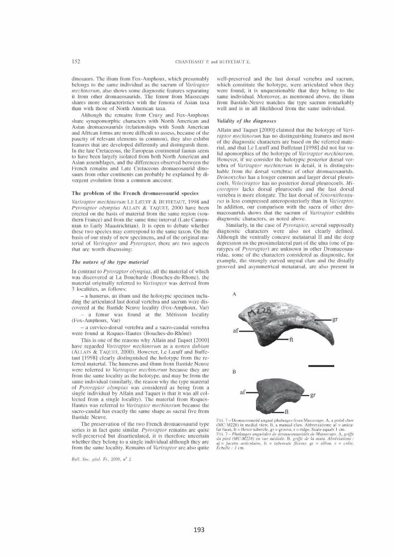
193

194
195

Title: The ornithopod dinosaur Rhabdodon from the Late Cretaceous of southern France:
anatomy, systematics and paleobiology.
Abstract: The ornithopod dinosaur Rhabdodon was named by Matheron in 1869 and a first species,
Rhabdodon priscus, was proposed on the basis of a dentary fragment with its diagnostic teeth from the Late
Cretaceous of Bouches-du-Rhône in southern France. A new species, R. septimanicus, was subsequently
erected by Buffetaut and Le Loeuff (1991) based on a characteristic dentary from a Late Cretaceous locality at
Montouliers (Hérault). The validity of the latter species was in debate, the question being how many species of
Rhabdodon existed in Southern France. Abundant unpublished material (both cranial and postcranial) of
Rhabdodon discovered in recent excavations at various localities in southern France is described, as a result of
which the anatomy of this animal is now better known. A reconstruction of Rhabdodon is proposed. R.
septimanicus is considered as valid by including new cranial elements from Quarante (Hérault). Two species of
Rhabdodon undoubtedly coexisted in the same time interval (Late Campanian – Early Maastrichtian) in
southern France. The phylogenetic analysis confirms that Rhabdodon can be placed in a distinct family,
Rhabdodontidae, which also includes its sister taxon, Zalmoxes, from which it is distinguished by numerous
postcranial characters. Rhabdodontidae has a close relation with Iguanodontia. It is suggested that the common
ancestor of the rhabdodontid dinosaurs evolved in North America, and then dispersed to Europe and eventually
evolved in geographical isolation on the various European landmasses during the Late Cretaceous.
Key-words: Ornithopoda, Rhabdodontidae, Rhabdodon, anatomy, Late Cretaceous, Southern France
Titre : Le dinosaure ornithopode Rhabdodon dans le Crétacé supérieur de sud de la France:
anatomie, systématique et paléobiologie.
Resumé : Le dinosaure ornithopode Rhabdodon a été nommé par Matheron en 1869 et la première espèce,
Rhabdodon priscus, a été proposée sur la base d'un fragment dentaire avec ses dents très caractéristiques
découvert dans le Crétacé supérieur des Bouches-du-Rhône dans le Sud de la France. Une nouvelle espèce, R.
septimanicus., a ensuite été proposée par Buffetaut et Le Loeuff en 1991 sur la base d’un dentaire trouvé dans
une localité du Crétacé supérieur à Montouliers (Hérault). La validité de cette nouvelle espèce était en débat, la
question étant de savoir combien d'espèces de Rhabdodon existaient dans le Sud de la France. Un abondant
matériel inédit (crânien et postcrânien) de Rhabdodon, découvert récemment au cours de fouilles sur divers
sites du Sud de la France, est décrit, de sorte que l’'anatomie de cet animal est désormais mieux connue. Une
reconstitution de Rhabdodon est proposée. R. septimanicus est considéré comme valide à la suite de l'inclusion
de nouveaux éléments crâniens de Quarante (Hérault). Deux espèces de Rhabdodon ont certainement coexisté
durant le même période (Campanien supérieur – Maastrichtien inférieur) dans le Sud de la France. L'analyse
phylogénétique confirme que Rhabdodon peut être placé dans une famille des Rhabdodontidae avec son taxon-
frère, Zalmoxes, dont il se distingue par de nombreux caractères postcrâniens. Les Rhabdodontidae sont
proches des Iguanodontia. Il est suggéré que l'ancêtre commun des rhabdodontidés a évolué en Amérique du
Nord, puis s’est dispersé en Europe et a finalement évolué dans l'isolement géographique de l’archipel
européen au cours de du Crétacé supérieur.
Mots-clés : Ornithopoda, Rhabdodontidae, Rhabdodon, anatomie, Crétacé supérieur, Sud de la France