The Open Ophthalmology Journal · OCT examination, using Cirrus HD OCT (Zeiss Cirrus TM HD-OCT...
12
Send Orders for Reprints to [email protected] 334 The Open Ophthalmology Journal, 2017, 11, 334-345 1874-3641/17 2017 Bentham Open The Open Ophthalmology Journal Content list available at: www.benthamopen.com/TOOPHTJ/ DOI: 10.2174/1874364101711010334 RESEARCH ARTICLE Assessment of Posterior Segment Using Spectral Domain OCT in Highly Myopic Eyes Heba Radi AttaAllah * , Ismail Ahmed Nagib Omar and Ahmed Shawkat Abdelhalim Ophthalmology Department, Lecturer of Ophthalmology, Minia University Hospital, Faculty of Medicine, Minia University, Al-Minya, Egypt Received: July 28, 2017 Revised: October 19, 2017 Accepted: October 30, 2017 Abstract: Purpose: Spectral Domain Optical Coherence Tomography (SD-OCT) was used to evaluate retinal and vitreo-retinal changes that occur in highly myopic patients. Methods: This prospective study included 472 eyes of 472 patients suffering from high myopia (> -6.00 D), between May 2012 and December 2015. All patients were examined, using Cirrus HD OCT (Zeiss Cirrus TM HD-OCT model 4000), to detect any retinal or vitreo- retinal interface abnormalities. All obtained data was analyzed using Statistical Package for the Social Sciences software version 17 (SPSS Inc, Chicago, IL, USA) and the paired two-sided t-test. Bivariate correlations were performed between different parameters using the Spearman correlation coefficient (r). Results: Mean spherical equivalent (MSE) was -13.11± 4.35D. Mean axial length (AL) was 28.5±1.62 mm. Posterior vitreous detachment (PVD) was the most frequent OCT finding; representing 33.4% of the cases, 13.7% of them were associated with macular traction. A statistically significant positive correlation was found between AL and MTM, full thickness macular hole, PVD with traction, and dome shaped macula (r = 0.49 and P = 0.001, r = 0.422 and P = 0.0001, r = 0.25 and P = 0.03, r=0.475, P=0.001 respectively) Conclusion: OCT is a valuable tool in detecting retinal and vitreo-retinal interface abnormalities in highly myopic eyes, and it can be used for follow up of those patients to avoid advanced retinal damage. Keywords: Dome shaped macula, Foveoschisis, High myopia, Myopic traction maculopathy, Myopic macular hole, Myopic CNV. 1. INTRODUCTION High or pathologic myopia is defined as a refractive error of ≥ -6.00 diopters (D) and an axial length >26 mm [1]. In pathologic myopia, fundus changes are numerous and related to the degree of myopia, axial length of the globe, presence of a posterior staphyloma and age [2]. * Address correspondence to this author at the Ophthalmology Department, Lecturer of Ophthalmology, Minia University Hospital, Faculty of Medicine, Minia University, Al-Minia, Postal code: 61111, Egypt; Tel: +20862342505; E-mail: [email protected]
-
Upload
duonghuong -
Category
Documents
-
view
214 -
download
0
Transcript of The Open Ophthalmology Journal · OCT examination, using Cirrus HD OCT (Zeiss Cirrus TM HD-OCT...
Send Orders for Reprints to [email protected]
334 The Open Ophthalmology Journal, 2017, 11, 334-345
1874-3641/17 2017 Bentham Open
The Open Ophthalmology Journal
Content list available at: www.benthamopen.com/TOOPHTJ/
DOI: 10.2174/1874364101711010334
RESEARCH ARTICLE
Assessment of Posterior Segment Using Spectral Domain OCT inHighly Myopic Eyes
Heba Radi AttaAllah*, Ismail Ahmed Nagib Omar and Ahmed Shawkat Abdelhalim
Ophthalmology Department, Lecturer of Ophthalmology, Minia University Hospital, Faculty of Medicine, MiniaUniversity, Al-Minya, Egypt
Received: July 28, 2017 Revised: October 19, 2017 Accepted: October 30, 2017
Abstract:
Purpose:
Spectral Domain Optical Coherence Tomography (SD-OCT) was used to evaluate retinal and vitreo-retinal changes that occur inhighly myopic patients.
Methods:
This prospective study included 472 eyes of 472 patients suffering from high myopia (> -6.00 D), between May 2012 and December2015. All patients were examined, using Cirrus HD OCT (Zeiss Cirrus TM HD-OCT model 4000), to detect any retinal or vitreo-retinal interface abnormalities.
All obtained data was analyzed using Statistical Package for the Social Sciences software version 17 (SPSS Inc, Chicago, IL, USA)and the paired two-sided t-test. Bivariate correlations were performed between different parameters using the Spearman correlationcoefficient (r).
Results:
Mean spherical equivalent (MSE) was -13.11± 4.35D. Mean axial length (AL) was 28.5±1.62 mm. Posterior vitreous detachment(PVD) was the most frequent OCT finding; representing 33.4% of the cases, 13.7% of them were associated with macular traction. Astatistically significant positive correlation was found between AL and MTM, full thickness macular hole, PVD with traction, anddome shaped macula (r = 0.49 and P = 0.001, r = 0.422 and P = 0.0001, r = 0.25 and P = 0.03, r=0.475, P=0.001 respectively)
Conclusion:
OCT is a valuable tool in detecting retinal and vitreo-retinal interface abnormalities in highly myopic eyes, and it can be used forfollow up of those patients to avoid advanced retinal damage.
Keywords: Dome shaped macula, Foveoschisis, High myopia, Myopic traction maculopathy, Myopic macular hole, Myopic CNV.
1. INTRODUCTION
High or pathologic myopia is defined as a refractive error of ≥ -6.00 diopters (D) and an axial length >26 mm [1]. Inpathologic myopia, fundus changes are numerous and related to the degree of myopia, axial length of the globe,presence of a posterior staphyloma and age [2].
* Address correspondence to this author at the Ophthalmology Department, Lecturer of Ophthalmology, Minia University Hospital, Faculty ofMedicine, Minia University, Al-Minia, Postal code: 61111, Egypt; Tel: +20862342505; E-mail: [email protected]
OCT Evaluation in High Myopia The Open Ophthalmology Journal, 2017, Volume 11 335
The unique combination of retinal traction generated by the epiretinal membrane (ERM) and/or residual focalvitreomacular traction (VMT), together with the complex and distinctive anatomy of degenerative myopia, leads to thefrequent presence of macular damage in these eyes, such as retinoschisis, lamellar holes, macular hole formation andposterior retinal detachment. Such findings may be difficult to diagnose on routine fundus examination or byfluorescein angiography [3]. However, the recent increased use of optical coherence tomography (OCT) has revealedthe presence of posterior pole related conditions that are difficult to visualize with other imaging techniques.[2]
While working in Upper Egypt, we have observed an increase in the number of highly myopic patients; aconsiderable number of those patients prefer to have their refractive errors corrected by refractive surgeries, rather thanwearing eye glasses, in pre-operative evaluation of those patients we noted significant visual impairment, in somepatients, not explained by routine fundus examination. In this study, we aimed to evaluate retinal and vitreo-retinalchanges using spectral domain (SD) OCT in such highly myopic eyes with recent visual complaints, either referred forrefractive surgery preparation or attending our outpatient clinic. Furthermore, we aimed to study any abnormalities thatmight be missed on routine examination and that could lead to significant retinal damage, thus keeping such patients onregular follow-up visits, with the aim of decreasing the number of visually disabled, highly myopic patients.
2. SUBJECTS AND METHODS
This prospective study was done between May 2012 and December 2015. It included 472 eyes of 472 patients whoattended the ophthalmic outpatient clinic in Minia University hospital, and Minia investigation eye center; 40.4% (191)were male and 59.5% (281) were female. Age ranged between 22 and 65 years, with a mean and standard deviation of50.38 ± 11.4.
We included all symptomatic patients with high myopia (> -6.00 D), who had recent metamorphopsia orunexplained visual symptoms on routine examination, only one eye for each patient was selected for analysis (the eyewith more findings, or with recent complaint). Patients with media opacities that may have interfered with scan signalstrength, and with a previous history of intraocular surgeries, or with other ocular or systemic diseases that could affectthe macular area (dystrophy, age related macular degeneration, diabetic retinopathy, etc.) were excluded from thisstudy.
Patients' assessment included: Best corrected visual acuity, slit lamp examination for anterior segment evaluation,fundus examination using +78 D lens, a cycloplejc refraction using the Nidek autoref/keratometer (LS 900, Haag-StreitDiagnostics, Switzerland), and fluorescein angiography using IMAGE net 2000™; fundus camera (TOPCONCorporation, Japan). Axial length measurement was made using al Quantel medical Aviso (A&B scan OphthalmicEchography, 2008, France).
OCT examination, using Cirrus HD OCT (Zeiss Cirrus TM HD-OCT model 4000), was performed after appropriatepupillary dilation using cyclopentolate 1% eye drops; high definition HD 5line Raster scan was selected and used atfour different orientations: oblique (45°and 135°), vertical scans (90°) and horizontal scans (180°), to scan the posteriorpole thoroughly (at the macular area, and nasally). The scan length was 9 mm to cover a larger examination area (exceptfor the vertical scan, where we used a 6 mm scan). All OCT examinations were performed by one observer (H.R.). OCTimaging was optimized by maintaining a good tear film, by applying artificial tears.
This study gained approval from the Local Research Ethics Committee and it was in accordance with the HelsinkiDeclaration. All patients gave their written consent.
3. STATISTICAL ANALYSIS
All obtained data was analyzed using Statistical Package for the Social Sciences software version 17 (SPSS Inc,Chicago, IL, USA) and the paired two-sided t-test. A P-value < 0.05 was considered to be statistically significant.Bivariate correlations were performed between different parameters using the Spearman correlation coefficient (r).
4. RESULTS
This study included 472 eyes of 472 patients. Patients’ age, spherical equivalent (SE), and axial length (AL) areshown in Table 1.
336 The Open Ophthalmology Journal, 2017, Volume 11 AttaAllah et al.
Table 1. Patients' age, SE (spherical equivalent), and AL (axial length).
Range Mean ±SDAge 22 to 65 years 50.38±11.4 years
Spherical equivalent -6.50 to -23D -13.11± 4.35DAxial length 26 to 33.85 mm 28.5±1.62 mm
Abnormal macular findings were observed by two observers, recorded and analyzed.
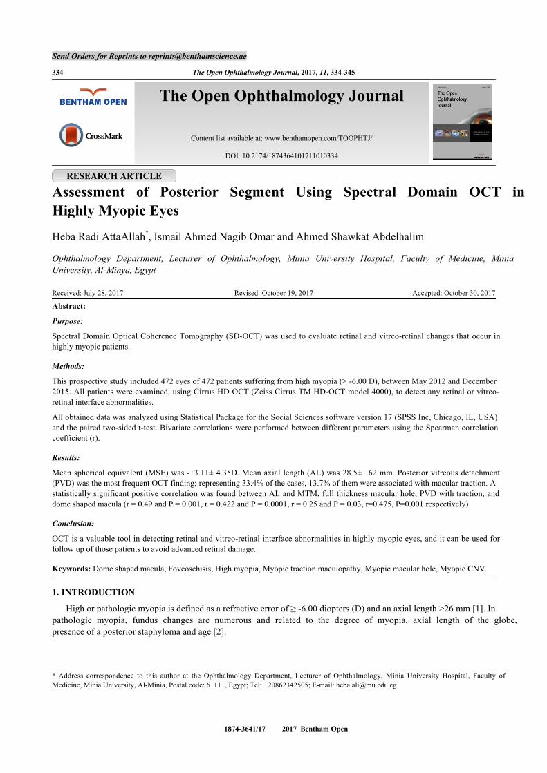
Myopic foveoschisis (MF) was defined as a separation of intraretinal layers, predominantly outer layers, into athinner outer layer and a thicker inner layer, with hyper-reflective bridging columns, and subsequent retinal destruction[4] (Fig. 1).
Fig. (1). HD OCT scan showing myopic foveoschisis, (white arrows).
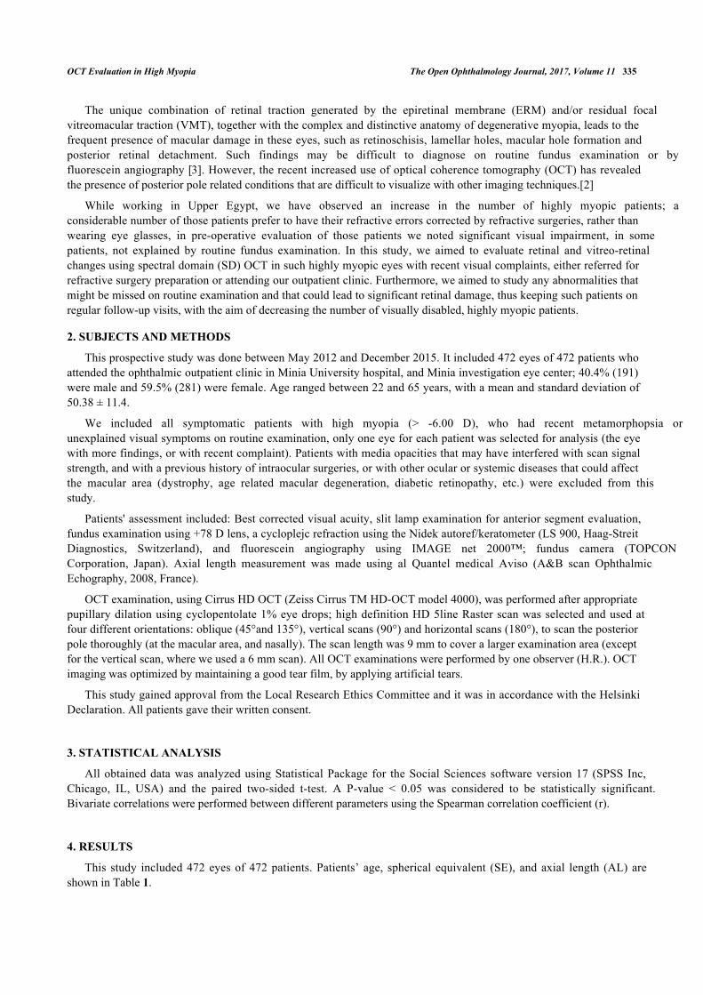
Myopic traction maculopathy (MTM; Fig. 2), was diagnosed as follows: the presence of an epiretinal membrane,vitreomacular traction due to incomplete vitreomacular separation, retinal thickening with or without cystoid edema,separation of the neurosensory retina into two or more layers, retinal detachment, lamellar or full-thickness macularholes [2, 5].
Fig. (2). HD OCT scan showing myopic traction maculopathy (MTM).
OCT Evaluation in High Myopia The Open Ophthalmology Journal, 2017, Volume 11 337
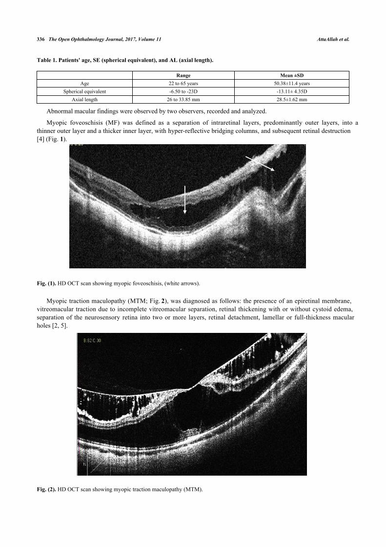
A paravascular retinal cyst appears as a small hollow space mainly around large retinal vessels [4], as seen inFig. (3).
Fig. (3). HD OCT scan showing paravascular retinal cyst (white arrow).
Retinal traction was subdivided into: an epiretinal membrane with multifocal attachments causing tangentialtraction, and incomplete vitreomacular separation (partial posterior vitreous detachment) causing anteroposteriorvitreomacular traction [4].
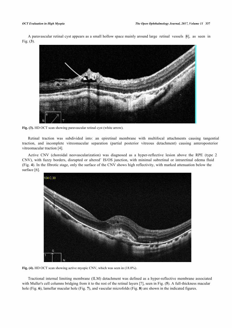
Active CNV (choroidal neovascularization) was diagnosed as a hyper-reflective lesion above the RPE (type 2CNV), with fuzzy borders, disrupted or altered’ IS/OS junction, with minimal subretinal or intraretinal edema fluid(Fig. 4). In the fibrotic stage, only the surface of the CNV shows high reflectivity, with marked attenuation below thesurface [6].
Fig. (4). HD OCT scan showing active myopic CNV, which was seen in (18.0%).
Tractional internal limiting membrane (ILM) detachment was defined as a hyper-reflective membrane associatedwith Muller's cell columns bridging from it to the rest of the retinal layers [7], seen in Fig. (5). A full-thickness macularhole (Fig. 6), lamellar macular hole (Fig. 7), and vascular microfolds (Fig. 8) are shown in the indicated figures.

338 The Open Ophthalmology Journal, 2017, Volume 11 AttaAllah et al.
Fig. (5). HD OCT scan showing tractional internal limiting membrane (ILM) detachment (white arrow).
Fig. (6). HD OCT scan showing full thickness macular hole (FTMH), which was seen in (8.2%), in this case there is an evidence ofantero-posterior vitreo-retinal traction at the inferior retinal edge (white arrow).
Fig. (7). HD OCT scan showing lamellar macular hole that was seen in (4.6%).

OCT Evaluation in High Myopia The Open Ophthalmology Journal, 2017, Volume 11 339
Fig. (8). HD OCT scan showing 2 vascular microfolds (white arrows).
Dome shaped macula was defined on vertical OCT scans, as inward bulge of RPE of more than 50 µm, above apresumed tangential line joining the outer border of RPE, at the bottom of the posterior staphyloma [8], as seen in Fig.(9).
Fig. (9). HD OCT scan showing a dome shaped macula, which is defined as inward bulge of RPE of more than 50 µm (red arrow),above a presumed tangential line (yellow line) joining the outer border of RPE, at the bottom of the posterior staphyloma.
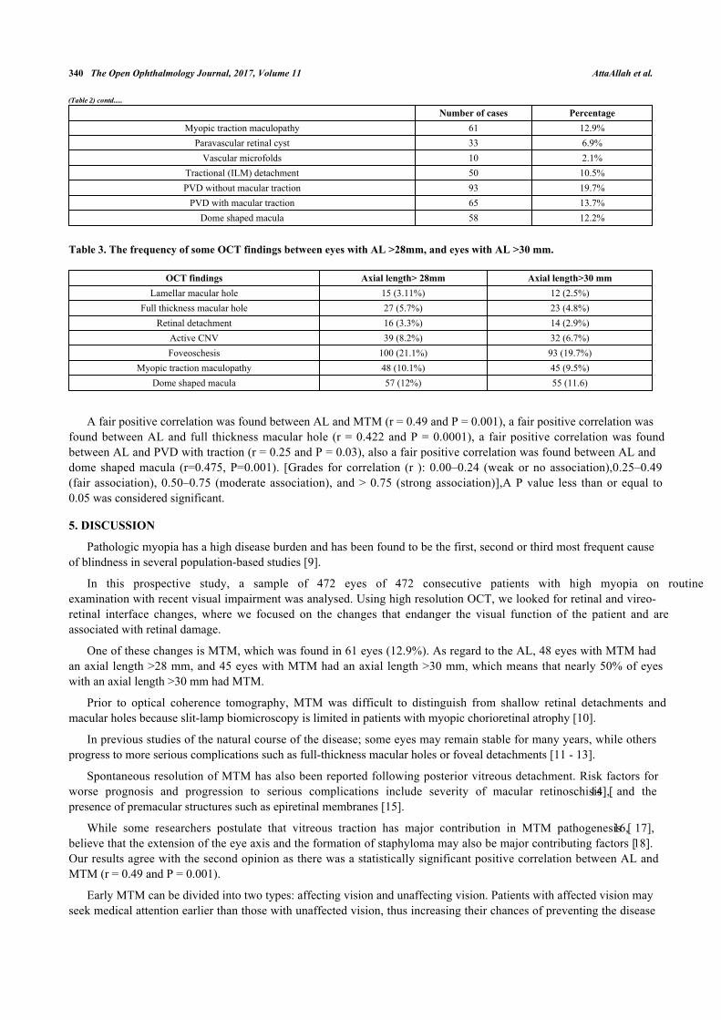
The frequency of OCT findings is shown in Table 2. On further analysis, it was found that 287 eyes (60.8%) had anaxial length >28 mm, while 90 eyes (19%) had an axial length >30 mm, the frequency of some OCT findings as regardto the AL is shown in Table 3.
Table 2. The frequency of OCT findings.
Number of cases PercentageLamellar macular hole 22 4.6%
Full thickness macular hole 39 8.2%Retinal detachment 18 3.8%
Inactive CNV 34 7.2%Active CNV 85 18.0%
Myopic Foveoschisis 104 22%Epiretinal membrane 74 15.6%
340 The Open Ophthalmology Journal, 2017, Volume 11 AttaAllah et al.
Number of cases PercentageMyopic traction maculopathy 61 12.9%
Paravascular retinal cyst 33 6.9%Vascular microfolds 10 2.1%
Tractional (ILM) detachment 50 10.5%PVD without macular traction 93 19.7%
PVD with macular traction 65 13.7%Dome shaped macula 58 12.2%
Table 3. The frequency of some OCT findings between eyes with AL >28mm, and eyes with AL >30 mm.
OCT findings Axial length> 28mm Axial length>30 mmLamellar macular hole 15 (3.11%) 12 (2.5%)
Full thickness macular hole 27 (5.7%) 23 (4.8%)Retinal detachment 16 (3.3%) 14 (2.9%)
Active CNV 39 (8.2%) 32 (6.7%)Foveoschesis 100 (21.1%) 93 (19.7%)
Myopic traction maculopathy 48 (10.1%) 45 (9.5%)Dome shaped macula 57 (12%) 55 (11.6)
A fair positive correlation was found between AL and MTM (r = 0.49 and P = 0.001), a fair positive correlation wasfound between AL and full thickness macular hole (r = 0.422 and P = 0.0001), a fair positive correlation was foundbetween AL and PVD with traction (r = 0.25 and P = 0.03), also a fair positive correlation was found between AL anddome shaped macula (r=0.475, P=0.001). [Grades for correlation (r ): 0.00–0.24 (weak or no association),0.25–0.49(fair association), 0.50–0.75 (moderate association), and > 0.75 (strong association)],A P value less than or equal to0.05 was considered significant.
5. DISCUSSION
Pathologic myopia has a high disease burden and has been found to be the first, second or third most frequent causeof blindness in several population-based studies [9].
In this prospective study, a sample of 472 eyes of 472 consecutive patients with high myopia on routineexamination with recent visual impairment was analysed. Using high resolution OCT, we looked for retinal and vireo-retinal interface changes, where we focused on the changes that endanger the visual function of the patient and areassociated with retinal damage.
One of these changes is MTM, which was found in 61 eyes (12.9%). As regard to the AL, 48 eyes with MTM hadan axial length >28 mm, and 45 eyes with MTM had an axial length >30 mm, which means that nearly 50% of eyeswith an axial length >30 mm had MTM.
Prior to optical coherence tomography, MTM was difficult to distinguish from shallow retinal detachments andmacular holes because slit-lamp biomicroscopy is limited in patients with myopic chorioretinal atrophy [10].
In previous studies of the natural course of the disease; some eyes may remain stable for many years, while othersprogress to more serious complications such as full-thickness macular holes or foveal detachments [11 - 13].
Spontaneous resolution of MTM has also been reported following posterior vitreous detachment. Risk factors forworse prognosis and progression to serious complications include severity of macular retinoschisis [14], and thepresence of premacular structures such as epiretinal membranes [15].
While some researchers postulate that vitreous traction has major contribution in MTM pathogenesis [16, 17],believe that the extension of the eye axis and the formation of staphyloma may also be major contributing factors [18].Our results agree with the second opinion as there was a statistically significant positive correlation between AL andMTM (r = 0.49 and P = 0.001).
Early MTM can be divided into two types: affecting vision and unaffecting vision. Patients with affected vision mayseek medical attention earlier than those with unaffected vision, thus increasing their chances of preventing the disease
(Table 2) contd.....

OCT Evaluation in High Myopia The Open Ophthalmology Journal, 2017, Volume 11 341
from reaching a more advanced stage [19, 20].
Some authors advocate that surgical treatment is indicated in MTM when VA is impaired or the patient complainsof visual disturbances [15]. Another study shows persistent photoreceptor defects and irregular choroidal detachmentsin MTM patients with poor post-operative visual outcomes, and recommends the timing of surgery for when there isthreatened disruption of the outer retina on OCT [21].
The effect of epiretinal traction on highly myopic eyes is more serious than on non-myopic eyes, because itproduces more pronounced retinal damage. Therefore, it should be taken as a major risk factor for retinal damage inthose patients [22]. In this study, 139 eyes (29.4%) had retinal traction, ERM with tangential traction was found in 74eyes (15.6%), and partial posterior vitreous detachment with antero-posterior traction was found in 65 eyes (13.7%). Astatistically significant positive correlation was found between AL and PVD with traction (r = 0.25 and P = 0.03).
Gomaa and Abo Hussein, who conducted their study on 100 highly myopic Egyptian patients, report that epiretinalmembranes were present in 65 eyes (they did not mention whether all epiretinal membranes were associated withtangential traction or not), while vitreomacular traction (anterior-posterior traction) was detected in ten eyes [23].
The importance of OCT was to detect subtle degrees of traction that could not be noticed on routine biomicroscopicexamination.
As demonstrated by Gallemore et al., retinal damage is defined by the presence of retinoschisis, lamellar or full-thickness macular hole and shallow retinal detachment [24]. Accordingly, in the current study, we found retinal damagein 34.9% of cases (165 eyes; lamellar macular holes in 22 eyes, full-thickness macular holes in 39 eyes and foveoschisisin 104 eyes). Such a high frequency of retinal damage in a myopic population corresponds to the 34% reported byTakano and Kishi in a smaller sample (32 eyes) [25].
In this study, myopic foveoschisis was found in 104 eyes, representing 22% of the cases, 100 eyes had an axiallength >28 mm, alone or associated with other lesions. It is characterized by an intraretinal cleavage in a myopicposterior staphyloma, and it can only be detected by OCT.
Smiddy suggested that myopic maculoschisis, foveal schisis and vitreoschisis in high myopia should all fall underthe family of MTM [26].
Gaucher et al. report that the risk of worsening vision seems to increase when MF is associated with premacularstructures such as epiretinal membranes or a partially detached vitreous cortex [15].
Shimada et al. found that half of patients with MF have been reported to develop retinal detachment or macularholes within two or more years of follow-up, and recommend that serial OCT examinations should be performed inthese patients [11]. Unfortunately, in our study we couldn’t obtain such data, as we were concerned with the frequencyof findings, more than follow up of these findings.
Shimada et al. suggest in another longitudinal study of five eyes with retinoschisis that progressed to a foveal retinaldetachment using time-domain OCT that inward traction was transmitted to the outer retina through the fovealcolumnar structures in the retinoschisis layer [27].
Tractional ILM detachment was reported in 50 eyes (10.5%) in the current study. Sayanagi et al [28], report that thisphenomenon may be an important contributor to the separation of the inner layers of the neural retina resulting inmacular retinoschisis. It also seems that traction myopic maculopathy, as described by Panozzo and Mercanti in 2004, isa variation of longstanding tractional ILM detachment, with loss of column bridges due to extreme traction and retinalatrophy [2].
The cause of the ILM detachment is still uncertain. In Goma and Abo Hussein’s study, they record ILMdetachments in seven out of 100 eyes; they were always in the peripheral macula (extra foveal), where there wereretinal vessels including arterioles [23], but in this series some cases (13 eyes) had ILM detachments reaching juxta andparafoveal locations starting from peripheral locations.
Axial length elongation and/or formation of posterior staphyloma in highly myopic eyes may generate the inwardtraction force exerted by these factors. This is strongly supported by the remission of the retinoschisis after vitrectomycombined with ILM peeling, which theoretically releases the traction forces exerted by the posterior vitreous cortex andthe ILM and partly by the retinal vessels in the area from which the ILM was peeled [29].
Macular holes were found in 61 eyes (12.8%); 39 eyes had full-thickness macular holes, 27 eyes had an axial length

342 The Open Ophthalmology Journal, 2017, Volume 11 AttaAllah et al.
>28 mm, while 23 eyes had an axial length >30 mm. There was a statistically significant positive correlation betweenAL and full thickness macular holes (r = 0.422 and P = 0.0001).
Prognosis is generally better in cases involving only high-myopia macular holes without foveoschisis than in caseswith both [30].
Retinal detachment (shallow) was found in 3.8% (18 eyes) of the cases. Almost all of them (16 eyes) had an axiallength >28 mm. In 2004, Panozzo and Mercanti reported 1.6% of shallow RD in a sample of 125 myopic eyes [2], butin the Baba et al. study they found 9% of 134 eyes [31].
In our study, 119 eyes (25.2%) had choroidal nonvascular membranes, 34 eyes (7.2%) were in an inactive form andthe remaining were active. Only 39 eyes of those with active CNV had an axial length >28 mm, and 32 eyes had anaxial length of >30 mm.
Ikuno et al [32] made a study of 23 consecutive patients with bilaterally high myopia (axial length ≥26.5 mm, orrefractive error ≤8 D) and they hypothesized that axial length elongation alone did not cause myopic CNV, and thatthere may be other latent factors. They confirmed that the axial length and refractive error did not differ between theaffected eyes and fellow eyes, indicating that these factors were not major risk factors of myopic CNV. They also foundthat in eyes with myopic CNV the sub foveal and inferior choroid were significantly thinner than in fellow eyes;furthermore, they found that the posterior staphyloma was steeper in eyes with myopic CNV, explaining the greaterchoroidal thinning caused by more mechanical stress related to stretching at the macula [32].
Dome shaped macula, first described by Gaucher et al. as inward bulge of the macula within the concavity of aposterior staphyloma in highly myopic eyes based on OCT observations [33]. In the current study, it was found in 58eyes (12.2%). Its prevalence is estimated to be 10.7% by an European study and 9.3% by a Japanese study [4].Development of dome shaped macula can be explained by different mechanisms including: ocular hypotony, localchoroidal and scleral thickening, tangential vitreoretinal traction, inward folding of the sclera through collapsedposterior portion of the eye wall, asymmetry in staphyloma progression, or resistance to deformation of scleralstaphyloma [34]. Visually threatening macular complications have been suggested to be more frequent in eyes withdome-shaped macula than eyes with low or no myopia, which include myopic CNV, macular holes (MHs), serous RD,foveal and extrafoveal schisis [35]. In the current study the prevalence of such complications in dome shaped macula,was as the following: 10 eyes with myopic CNV, 20 eyes with full thickness macular hole, foveoschisis in 26 eyes, and2 eyes with shallow serous retinal detachment.
There was a statistically significant positive correlation between AL and dome shaped macula (r=0.475 andP=0.001).
6. LIMITATIONS
To this study includes: the study was concerned with the frequency of OCT findings in highly myopic eyes, withoutstudying the course of some important changes (MTM, foveoschisis, traction), so further longitudinal studies arerequired to establish the findings that are definitely associated with progression. Visually threatening macularcomplications occurring in eyes with dome-shaped macula, should be studied in correlation with the height of domeshaped macula.
CONCLUSION
In this study, we demonstrated that retinal abnormalities in the macular area can be a frequent finding in eyes withdegenerative myopia. Epiretinal traction and CNV play a major role in the prognosis of these cases.
Optical coherence tomography can explain patient's visual complaints in highly myopic eyes and may even showabnormalities in asymptomatic cases. It can also detect some undetectable abnormalities on routine ophthalmoscopeexaminations in order to avoid advanced retinal damage.
ETHICS APPROVAL AND CONSENT TO PARTICIPATE:
The study has gained approval from the Local Research Ethics Committee, Faculty of Medicine, Minia UniversityHospitals.
Informed consent was obtained from all patients for being included in this study.

OCT Evaluation in High Myopia The Open Ophthalmology Journal, 2017, Volume 11 343
HUMAN AND ANIMAL RIGHTS
This research is in accordance with the Helsinki Declaration of 1964, as revised in 2013.
CONSENT FOR PUBLICATION
Not applicable.
CONFLICT OF INTEREST
The authors declare no conflict of interest, financial or otherwise.
ACKNOWLEDGEMENTS
Funding:
No funding or sponsorship was received for this study or publication of this article
Disclosures:
The authors declare that there is no conflict of interest regarding the publication of this paper.
Authorship:
All named authors meet the International Committee of Medical Journal Editors (ICMJE) criteria for authorship forthis manuscript, take responsibility for the integrity of the work as a whole, and have given final approval to the versionto be published.
REFERENCES
[1] Balacco-Gabrieli C. Aetiopathogenesis of degenerative myopia. A hypothesis. Ophthalmologica 1982; 185(4): 199-204.[http://dx.doi.org/10.1159/000309243] [PMID: 6216444]
[2] Panozzo G, Mercanti A. Optical coherence tomography findings in myopic traction maculopathy. Arch Ophthalmol 2004; 122(10): 1455-60.[http://dx.doi.org/10.1001/archopht.122.10.1455] [PMID: 15477456]
[3] Sanghvi C, Jalil A, Chaudhry N L, et al. Update on OCT findings in high myopia. Retina Today 2007; 4(1): 110-21.
[4] Cicinelli MV, Pierro L, Gagliardi M, Bandello F. Optical coherence tomography and pathological myopia: An update of the literature. IntOphthalmol 2015; 35(6): 897-902.[http://dx.doi.org/10.1007/s10792-015-0118-y] [PMID: 26265324]
[5] Champion M. Myopic Traction Maculopathy 2013. Available at: http://eyewiki.aao.org/Myopic_Traction_Maculopathy
[6] Introini U, Casalino G, Querques G, Gimeno AT, Scotti F, Bandello F. Spectral-domain OCT in anti-VEGF treatment of myopic choroidalneovascularization. Eye (Lond) 2012; 26(7): 976-82.[http://dx.doi.org/10.1038/eye.2012.75] [PMID: 22538218]
[7] Fujimoto M, Hangai M, Suda K, Yoshimura N. Features associated with foveal retinal detachment in myopic macular retinoschisis. Am JOphthalmol 2010; 150(6): 863-70.[http://dx.doi.org/10.1016/j.ajo.2010.06.023] [PMID: 20951972]
[8] Caillaux V, Gaucher D, Gualino V, Massin P, Tadayoni R, Gaudric A. Morphologic characterization of dome-shaped macula in myopic eyeswith serous macular detachment. Am J Ophthalmol 2013; 156(5): 958-967.e1.[http://dx.doi.org/10.1016/j.ajo.2013.06.032] [PMID: 23972305]
[9] Wong TY, Ferreira A, Hughes R, Carter G, Mitchell P. Epidemiology and disease burden of pathologic myopia and myopic choroidalneovascularization: An evidence-based systematic review. Am J Ophthalmol 2014; 157(1): 9-25.e12.[http://dx.doi.org/10.1016/j.ajo.2013.08.010] [PMID: 24099276]
[10] Sayanagi K, Morimoto Y, Ikuno Y, Tano Y. Spectral-domain optical coherence tomographic findings in myopic foveoschisis. Retina 2010;30(4): 623-8.[http://dx.doi.org/10.1097/IAE.0b013e3181ca4e7c] [PMID: 20394112]
[11] Shimada N, Ohno-Matsui K, Baba T, Futagami S, Tokoro T, Mochizuki M. Natural course of macular retinoschisis in highly myopic eyeswithout macular hole or retinal detachment. Am J Ophthalmol 2006; 142(3): 497-500.[http://dx.doi.org/10.1016/j.ajo.2006.03.048] [PMID: 16935601]
[12] Sun CB, Liu Z, Xue AQ, Yao K. Natural evolution from macular retinoschisis to full-thickness macular hole in highly myopic eyes. Eye(Lond) 2010; 24(12): 1787-91.[http://dx.doi.org/10.1038/eye.2010.123] [PMID: 20829889]

344 The Open Ophthalmology Journal, 2017, Volume 11 AttaAllah et al.
[13] Ripandelli G, Rossi T, Scarinci F, Scassa C, Parisi V, Stirpe M. Macular vitreoretinal interface abnormalities in highly myopic eyes withposterior staphyloma: 5-year follow-up. Retina 2012; 32(8): 1531-8.[http://dx.doi.org/10.1097/IAE.0b013e318255062c] [PMID: 22614742]
[14] Shimada N, Tanaka Y, Tokoro T, Ohno-Matsui K. Natural course of myopic traction maculopathy and factors associated with progression orresolution. Am J Ophthalmol 2013; 156(5): 948-57.[http://dx.doi.org/10.1016/j.ajo.2013.06.031]
[15] Gaucher D, Haouchine B, Tadayoni R, et al. Long-term follow-up of high myopic foveoschisis: Natural course and surgical outcome. Am JOphthalmol 2007; 143(3): 455-62.[http://dx.doi.org/10.1016/j.ajo.2006.10.053] [PMID: 17222382]
[16] Ishida S, Yamazaki K, Shinoda K, Kawashima S, Oguchi Y. Macular hole retinal detachment in highly myopic eyes: Ultrastructure ofsurgically removed epiretinal membrane and clinicopathologic correlation. Retina 2000; 20(2): 176-83.[http://dx.doi.org/10.1097/00006982-200002000-00011] [PMID: 10783951]
[17] Kobayashi H, Kishi S. Vitreous surgery for highly myopic eyes with foveal detachment and retinoschisis. Ophthalmology 2003; 110(9):1702-7.[http://dx.doi.org/10.1016/S0161-6420(03)00714-0] [PMID: 13129865]
[18] Robichaud JL, Besada E, Basler L, Frauens BJ. Spectral domain optical coherence tomography of myopic traction maculopathy. OphthalmicSurg Lasers Imaging 2011; 82(10): 607-13.[http://dx.doi.org/10.1016/j.optm.2011.03.013] [PMID: 21840263]
[19] Silva R. Myopic maculopathy: A review. Ophthalmologica 2012; 228(4): 197-213.[http://dx.doi.org/10.1159/000339893] [PMID: 22846778]
[20] Ouyang PB, Duan XC, Zhu XH. Diagnosis and treatment of myopic traction maculopathy. Int J Ophthalmol 2012; 5(6): 754-8.[PMID: 23275913]
[21] Shin JY, Yu HG. Visual prognosis and spectral-domain optical coherence tomography findings of myopic foveoschisis surgery using 25-gauge transconjunctival sutureless vitrectomy. Retina Mar 2012; 32(3): 486-92.[http://dx.doi.org/10.1097/IAE.0b013e31822058d1] [PMID: 21955988]
[22] Panozzo G, Mercanti A. Vitrectomy for myopic traction maculopathy. Arch Ophthalmol 2007; 125(6): 767-72.[http://dx.doi.org/10.1001/archopht.125.6.767] [PMID: 17562987]
[23] Gomaa AR, Abouhussein MA. Optical coherence tomography findings in high myopia. Egypt Retina J 2013; 1: 13-7.[http://dx.doi.org/10.4103/2347-5617.135241]
[24] Gallemore RP, Jumper JM, McCuen BW II, Jaffe GJ, Postel EA, Toth CA. Diagnosis of vitreoretinal adhesions in macular disease withoptical coherence tomography. Retina 2000; 20(2): 115-20.[http://dx.doi.org/10.1097/00006982-200002000-00002] [PMID: 10783942]
[25] Takano M, Kishi S. Foveal retinoschisis and retinal detachment in severely myopic eyes with posterior staphyloma. Am J Ophthalmol 1999;128(4): 472-6.[http://dx.doi.org/10.1016/S0002-9394(99)00186-5] [PMID: 10577588]
[26] Smiddy WE, Kim SS, Lujan BJ, Gregori G. Myopic traction maculopathy: Spectral domain optical coherence tomographic imaging and ahypothesized mechanism. Ophthalmic Surg Lasers Imaging 2009; 40(2): 169-73.[http://dx.doi.org/10.3928/15428877-20090301-21] [PMID: 19320306]
[27] Shimada N, Ohno-Matsui K, Yoshida T, Futagami S, Tokoro T, Mochizuki M. Development of macular hole and macular retinoschisis ineyes with myopic choroidal neovascularization. Am J Ophthalmol 2008; 145(1): 155-61.[http://dx.doi.org/10.1016/j.ajo.2007.08.029] [PMID: 17988641]
[28] Sayanagi K, Ikuno Y, Tano Y. Tractional internal limiting membrane detachment in highly myopic eyes. Am J Ophthalmol 2006; 142(5):850-2.[http://dx.doi.org/10.1016/j.ajo.2006.05.031] [PMID: 17056366]
[29] Ikuno Y, Sayanagi K, Ohji M, et al. Vitrectomy and internal limiting membrane peeling for myopic foveoschisis. Am J Ophthalmol 2004;137(4): 719-24.[PMID: 15059711]
[30] Alkabes M, Pichi F, Nucci P, et al. Anatomical and visual outcomes in high myopic macular hole (HM-MH) without retinal detachment: Areview. Graefes Arch Clin Exp Ophthalmol 2014; 252(2): 191-9.[http://dx.doi.org/10.1007/s00417-013-2555-5] [PMID: 24384802]
[31] Baba T, Ohno-Matsui K, Yoshida T, et al. Optical coherence tomography of choroidal neovascularization in high myopia. Acta OphthalmolScand 2002; 80(1): 82-7.[http://dx.doi.org/10.1034/j.1600-0420.2002.800116.x] [PMID: 11906310]
[32] Ikuno Y, Jo Y, Hamasaki T, Tano Y. Ocular risk factors for choroidal neovascularization in pathologic myopia. Invest Ophthalmol Vis Sci2010; 51(7): 3721-5.[http://dx.doi.org/10.1167/iovs.09-3493] [PMID: 20207975]

OCT Evaluation in High Myopia The Open Ophthalmology Journal, 2017, Volume 11 345
[33] Gaucher D, Erginay A, Lecleire-Collet A, et al. Dome-shaped macula in eyes with myopic posterior staphyloma. Am J Ophthalmol 2008;145(5): 909-14.[http://dx.doi.org/10.1016/j.ajo.2008.01.012] [PMID: 18342827]
[34] Ng DS, Cheung CY, Luk FO, et al. Advances of optical coherence tomography in myopia and pathologic myopia. Eye (Lond) 2016; 30(7):901-16.[http://dx.doi.org/10.1038/eye.2016.47] [PMID: 27055674]
[35] Coco RM, Sanabria MR, Alegría J. Pathology associated with optical coherence tomography macular bending due to either dome-shapedmacula or inferior staphyloma in myopic patients. Ophthalmologica 2012; 228(1): 7-12.[http://dx.doi.org/10.1159/000336910] [PMID: 22488163]
© 2017 AttaAllah et al.
This is an open access article distributed under the terms of the Creative Commons Attribution 4.0 International Public License (CC-BY 4.0), acopy of which is available at: https://creativecommons.org/licenses/by/4.0/legalcode. This license permits unrestricted use, distribution, andreproduction in any medium, provided the original author and source are credited.