
Afr. J. Food Agric. Nutr. Dev. 2020; 20(3):15936-15953 DOI ...
THE JOURNAL 0 1989 by The American Society for Biochemistry and Molecular Biology, Inc.
OF BIOLOGICAL CHEMISTRY Vol. 264, No. 27, Issue of September 25, pp. 15936-15942,1989 Printed in U. S. A.
An Alternate Promoter in the Glucokinase Gene Is Active in the Pancreatic ,8 Cell*
(Received for publication, March 16, 1989)
Mark A. Magnuson@ and Kathy D. Shelton From the Departments of Molecular Phvsiolom and Biowhysics and $Medicine, Vanderbilt University Medical School, Nashville, Tennessee 37232
1 “
An alternate promoter in the glucokinase gene is active in the cell and produces a glucokinase mRNA which is longer and that has a different leader sequence and translation start site than the hepatic glucokinase mRNA. The glucokinase B cell promoter is located at least 12 kilobases upstream from the glucokinase he- patic promoter. Transcription from the glucokinase B cell promoter initiates over a region of 62 bases. The absence of a TATA box homology in the proximal promoter region may account for the diffuse transcrip- tional initiation. Translation of the B cell glucokinase mRNA predicts a glucokinase isozyme that is different from the hepatic isozyme by 15 amino acids at the N terminus. The use of alternative promoters apparently enables the glucokinase gene to be regulated by insulin in the liver and by glucose in the B cell, thus possibly constituting an important feedback control loop for maintaining glucose homeostasis.
Alternate RNA splicing of the B cell glucokinase mRNA predicts at least two /3 cell glucokinase isoforms. An alternate splice acceptor site in the 4th exon of the glucokinase gene was identified in two glucokinase cDNAs from rat insulinoma tissue. Use of the alternate splice acceptor site results in a 51-nucleotide in frame deletion in the @ cell glucokinase mRNA and removal of 17 amino acids from a region of the protein situated between the putative glucose and ATP binding do- mains. Analysis of the pattern of RNA splicing in tis- sues containing B cells indicates that the splice acceptor site utilized in producing hepatic glucokinase mRNA is also utilized in the /3 cell.
Glucose is the major physiological stimulus for the secretion of insulin by pancreatic p cells. The electrical, ionic, and secretory responses of p cells to glucose are mediated by the metabolism of this sugar (1-3). Glucokinase (ATP:D-hexose 6-phosphotransferase, EC 2.7.1.1) catalyzes the initial step in the utilization of glucose by the cell and liver a t physiological glucose concentration (4-16 mM) and appears to play a crucial role in regulating insulin secretion by the p cell (for general
* This work was supported by grants from the American Diabetes Association and the Juvenile Diabetes Foundation and by Vanderbilt Diabetes Research and Training Center Grant GK 07061. The costs of publication of this article were defrayed in part by the payment of page charges. This article must therefore be hereby marked “aduer- tisement” in accordance with 18 U.S.C. Section 1734 solely to indicate this fact.
The nucleotide sequence(s) reported in this paper has been submitted to the GenBankTM/EMBL Data Bank with accession nurnbeds) M25806andM25807.
$ To whom correspondence should be addressed 708 Light Hall, Vanderbilt University Medical School, Nashville, TN 37232. Tel.: 615-322-7006.
reviews see Refs. 4 and 5). The high K , of glucokinase for glucose, coupled with the lack of end product inhibition com- pared to other hexokinases, enables glucokinase in the p cell to act as a pacesetter of glycolysis and, hence, of insulin secretion. Inhibition of glucokinase activity by mannoheptu- lose, alloxan, or other inhibitors prevents glucose-stimulated insulin release by p cells (5), observations which are consistent with the view that the enzyme plays an important role in modulating insulin secretion.
The function of glucokinase in the liver is different from that in the /3 cell. Hepatic glucokinase helps to facilitate the uptake and conversion of glucose by acting as an insulin- sensitive determinant of hepatic glucose usage. An increase in glucose phosphorylation, mediated by glucokinase, helps to maintain a gradient for glucose transport into the liver. The different functions of glucokinase in cells and the liver impose different requirements for the regulation of the en- zyme in these two tissues. Indeed, while insulin is the primary stimulant of hepatic glucokinase gene expression, islet gluco- kinase activity appears dependent upon glucose (6). Bedoya et al. (6) reported glucokinase activity decreased 70% in insulinoma-bearing, hypoglycemic animals. Glucokinase ac- tivity recovered to control levels within 24 h of removing the insulinoma from these animals, and a glucose infusion also resulted in a partial restoration of glucokinase activity in animals with an insulinoma. An understanding of how the glucokinase gene is expressed and regulated in the pancreatic @ cell is pertinent to both the physiology of glucose homeosta- sis and p cell-specific gene expression.
The availability of cDNAs encoding hepatic glucokinase (7) has enabled studies on the expression and regulation of the glucokinase gene in the pancreatic (3 cell. We have character- ized the transcription unit of insulinoma glucokinase mRNA and have compared it to the hepatic glucokinase mRNA transcription unit. Interestingly, two different promoters are contained in the glucokinase gene, one being active in p cells and the other in the liver. This finding offers an explanation for the differential regulation of glucokinase in liver and p cells. The promoter/regulatory sequences utilized in the liver apparently enable regulation by insulin while the promoter/ regulatory sequences utilized in the p cell may enable regula- tion by glucose. Translation of glucokinase is initiated within tissue-specific first exon sequences thus leading to 15 different amino acids at the N terminus of glucokinase in the liver and the p cell. Furthermore, utilization of an alternate splice acceptor site in the 4th exon of the gene predicts additional heterogeneity in the glucokinase isozymes present in the p cell.
EXPERIMENTAL PROCEDURES
General Techniques-Standard procedures were used for screening phage libraries, DNA labeling, restriction enzyme mapping, subclon-
15936
An Alternate Promoter for /3 Cell Glucokinase 15937
ing, and Southern blot transfers (8) unless otherwise stated. Oligo- nucleotides were synthesized on an Applied Biosystems 380A DNA synthesizer utilizing phosphotriester chemistry. Complementary DNA probes used for library screening and blot transfer hybridization experiments were labeled by random oligonucletide priming (9).
Isolation of Poly(A)+ RNAs-Total cellular RNA was isolated by homogenization in guanidinium isothiocyanate and centrifugation through a 6 M CsCl cushi,on. Poly(A)+ RNA was separated from total RNA by chromatography on oligo(dT)-cellulose (Type 111, Collabo-
vided by Dr. William Chick (Dept. of Biochemistry, University of rative Research). Frozen rat insulinoma tissue (10) was kindly pro-
Massachusetts) and used as a source for insulinoma poly(A)+ RNA. Hepatic poly(A)+ RNA was prepared from streptozotocin diabetic rats which were treated with insulin for either 4 or 8 h (7).
Northern Blot Ana1ysi.-Poly(A)+ RNAs were size-fractionated in a 1.4% agarose gel containing 6.3% formaldehyde, 1 mM EDTA, and 20 mM MOPS' at pH 7.4. The RNA was blot-transferred to a nitrocellulose filter and prehybridized for 4 h in a buffer containing 50% formamide, 5 X S8PE (1 X SSPE is 0.18 M NaCl, 10 mM NaH2P04, pH 7.4, 1 mM EDTA), 5 X Denhardt's (0.1% Ficoll, 0.1% polyvinylpyrrolidone, 0.1% albumin), 0.1% sodium dodecyl sulfate, and 100 pg/ml sheared, denatured salmon testes DNA. 1 X lo6 cpm/ ml of a denatured cDNA probe was added and allowed to hybridize overnight. The filter was then washed three times for 20 min each at 65 "C with 0.2 X SSC (where 1 X SSC is 0.15 M sodium chloride and 0.015 M sodium citrate) Bollowed by autoradiography for 2 days.
Isolation of Glucokinase cDNA and Genomic DNA-Rat insulinoma poly(A)+ RNA was used -to construct a cDNA library in XZap (Stra- tagene). Double-stranded cDNA was synthesized (11) and inserted into the EcoRI site of XZAP DNA using EcoRI linkers. The ligated DNAs were packaged yielding a library of approximately 1.5 X lo6 recombinant bacteriophage. The library was plated on a lawn of Escherichia coli BB4 for screening. After purification of individual plaques, the bacteriophage clones were converted to plasmid clones in Bluescript M13+ by :wperinfection with R407 helper phage ac- cording to the suppliers protocol (Stratagene). The rat genomic DNA library in EMBL 3 from which XGK7 was isolated was obtained from G. Scherer (Institute for Human Genetics, Freiberg, West Germany) (12). The restriction map of XGK7 was determined using both mul- tiple restriction enzyme digests and the indirect end-labeling method of Rackwitz et al. (13).
DNA Sequence Analysis-Fragments of the GK.Z9 cDNA and XGK7 genomic DNA were subcloned into pEMBL 18 and pEMBL 19 (16) using cohesive end ligations. Single-stranded DNA was pro- duced by superinfection with M13K07 helper phage. DNA sequence analysis of the single-stranded DNA templates was accomplished using a modified T7 polymerase (Sequenase 11, United States Bio- chemical Corp.) and dideoxy chain termination reactions (15). The reaction products were separated on 6% acrylamide, 7 M urea sequenc- ing gels. Sequencing reactions were primed using either the universal M13 forward primer or custom 18-mer oligonucleotides.
RNase Protection Analysis-The plasmid pGK7.HX was con- structed by ligating a HindIII-XbaI fragment (482 base pairs (bp)) from XGK7 into Bluescript M13+ (Stratagene). An antisense RNA probe, uniformly labeled with 32P, was generated from HindIII-line- arized pGK7.HX using T7 RNA polymerase (Bethesda Research Laboratories) and [CP~'P~CTP (800 Ci/mmol, Amersham Corp.) un- der standard conditions l:16). This RNA probe was used to map the transcription initiation sites of the glucokinase p cell promoter. 10- pg samples of either tRNA or poly(A)+ RNA were lyophilized and dissolved in hybridization buffer containing 5 X lo5 cpm of the RNA probe. The RNAs were heated at 85 "C for 10 min, allowed to hybridize at 45 "C overnight, and then digested with RNase A (50 pg/ ml, Sigma) and RNase TI. (1400 units/ml, BRL) at 37 "C for 1 h. The RNA fragments were precipitated with ethanol, size-fractionated on a 6% acrylamide-urea sequencing gel, and visualized by autoradiog- raphy.
Primer Extension Analysis-A 30-mer oligonucleotide (5"GTA- ATGAGGCAACAGGTA.ACTCTGTCCCTC-3') complementary to sequences near the 5' end of the pancreatic glucokinase isozyme was synthesized and end-labeled using [Y-~'P]ATP (>5000 Ci/mmol) and T4 polynucleotide kinase (New England Biolabs). The primer (1 X lo6 cpm) was annealed tc, 10 pg of either tRNA or poly(A)+ RNA in a total volume of 20 p1 for 1 h at 65 "C; the annealing buffer contained
The abbreviations used are: MOPS, 4-morpholinepropanesulfonic acid; kb, kilobase pairs; bp, base pairs; PCR, polymerase chain reac- tion.
20 mM Tris at pH 7.5, 250 mM NaCI, and 1 mM EDTA. Following hybridization, the mixture was diluted with 130 p1 of a solution containing 50 mM Tris at pH 7.5, 40 mM KCl, 10 mM dithiothreitol, 3 mM MgC12, 75 pg/ml actinomycin D, 0.5 mM of each deoxyribonu- cleotide, and 2 units of avian myeloblastosis virus reverse transcrip- tase (Promega) and incubated at 37 "C for 1 h. The products were precipitated with ethanol, size-fractionated on a 5% acrylamide, 7 M urea gel, and visualized by autoradiography using an intensifying screen.
Polymerase Chain Reaction Amplification of cDNA-First strand cDNA synthesis for polymerase chain reaction (PCR) amplification was accomplished as described above. After quantitating cDNA syn- thesis by the incorporation of 32P-labeled dATP, the RNA template was removed by alkaline hydrolysis, neutralization, and ethanol pre- cipitation. Amplification of the cDNA was performed according to the method of Saiki et al. (17) using Thermus aquaticus DNA polym- erase (Perkin-Elmer Cetus). 100-pl reactions were run under condi- tions previously described (17) except that the DNA polymerase was added after heating the reaction mixture to 94 "C for 2 min and cooling to 72 "C. Between 50 and 200 ng of first strand cDNA was used in each PCR amplification reaction. Forty amplification cycles were performed using a DNA thermal cycler (Perkin-Elmus Cetus). Each cycle consisted of a step program (94 "C, 1 min; 42 "C, 1 min; 72 "C, 2 min). Products of the PCR amplification were visualized on a 1.2% agarose gel containing ethidium bromide by fluorescence with
primers (5' to 3') used are GK768 CTCTTTGACTACATCTCT; UV illumination. The sequences of the 18-mer oligonucleotide
TACAAG; GK1276: TTTCCTTCTTGGTGGCCT and GK1351: GACTGTGACTGAGCTCGG.
GK357: GGGTTCGCTGAGCTTTCA; GK806: GGCTATGGATAC-
RESULTS
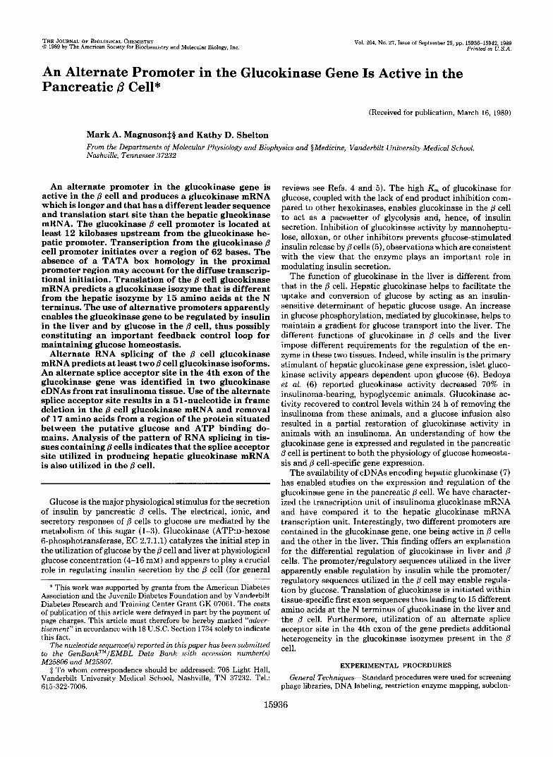
Analysis and Cloning of the Glucokinase mRNA in Insuli- noma Tissue-The size of the glucokinase mRNA in rat insulinoma tissue was estimated by Northern blot analysis. A single glucokinase mRNA species was detected in both rat insulinoma and liver poly(A)+ RNA using the hepatic gluco- kinase GK.Z2 cDNA as a probe. The size of the glucokinase mRNA in insulinoma was, however, larger than that from liver (Fig. 1, compare lanes 1 and 2). The hepatic glucokinase mRNA is 2346 nucleotides long, not including the poly(A) tail (18). The glucokinase mRNA in insulinoma tissue appeared to be approximately 2600 nucleotides in length.
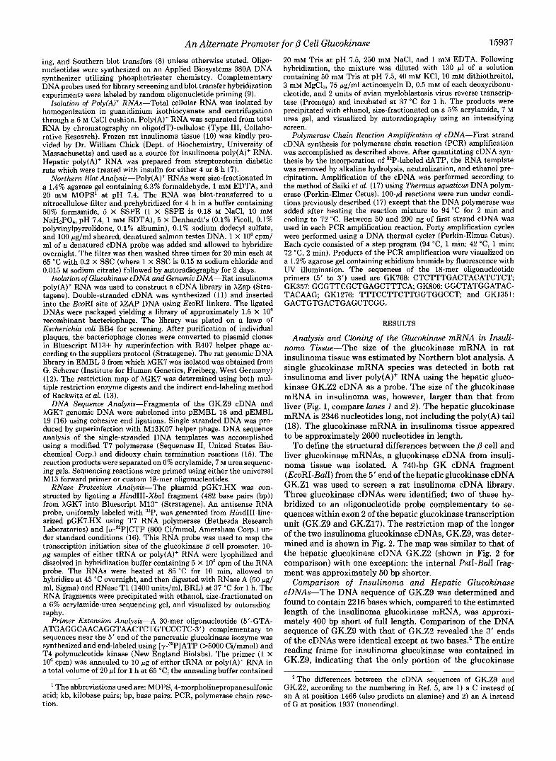
To define the structural differences between the @ cell and liver glucokinase mRNAs, a glucokinase cDNA from insuii- noma tissue was isolated. A 740-bp GK cDNA fragment (EcoRI-BalI) from the 5' end of the hepatic glucokinase cDNA GK.Z1 was used to screen a rat insulinoma cDNA library. Three glucokinase cDNAs were identified; two of these hy- bridized to an oligonucleotide probe complementary to se- quences within exon 2 of the hepatic glucokinase transcription unit (GK.Z9 and GK.Zl7). The restriction map of the longer of the two insulinoma glucokinase cDNAs, GK.Z9, was deter- mined and is shown in Fig. 2. The map was similar to that of the hepatic glucokinase cDNA GK.Z2 (shown in Fig. 2 for comparison) with one exception: the internal PstI-BalI frag- ment was approximately 50 bp shorter.
Comparison of Insulinoma and Hepatic Glucokinase cDNAs-The DNA sequence of GK.Z9 was determined and found to contain 2216 bases which, compared to the estimated length of the insulinoma glucokinase mRNA, was approxi- mately 400 bp short of full length. Comparison of the DNA sequence of GK.Z9 with that of GK.Z2 revealed the 3' ends of the cDNAs were identical except a t two bases.' The entire reading frame for insulinoma glucokinase was contained in GK.Z9, indicating that the only portion of the glucokinase
' The differences between the cDNA sequences of GK.Z9 and GK.Z2, according to the numbering in Ref. 5, are 1) a C instead of an A at position 1466 (also predicts an alanine) and 2) an A instead of G at position 1937 (noncoding).
15938 An Alternate Promoter for p Cell Glucokinase
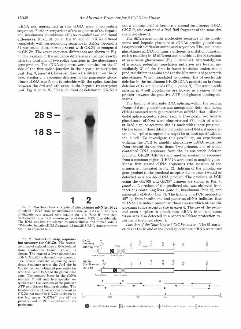
mRNA not represented in this cDNA were 5'-noncoding sequences. Further comparison of the sequences of the hepatic and insulinoma glucokinase cDNAs revealed two additional differences. First, 81 bp at the 5' end of GK.Z9 differed completely with corresponding sequence in GK.Z2. Second, a 51 nucleotide deletion was present with GK.Z9 as compared to GK.Z2. The exact sequence differences are shown in Fig. 3. The location of the sequence differences coincided exactly with the locations of two splice junctions in the glucokinase gene product. The cDNA sequences were identical on the 3' side of the first splice junction in the hepatic transcription unit (Fig. 3, panel A ) ; however, they were different on the 5' side. Similarly, a sequence deletion in the pancreatic gluco- kinase cDNA was found to coincide with the splice junction between the 3rd and 4th exon in the hepatic transcription unit (Fig. 3, panel I?). The 51-nucleotide deletion in GK.Z9 is
28S-
18S-
1 2 FIG. 1. Northern blot analysis of glucokinase mRNAs. 10 pg
of poly(A)+ RNA from rat insulinoma tissue (lune A ) and the livers of diabetic rats treated with insulin for 4 h (lune B ) was size- fractionated in a 1.4% agarose gel containing 6.3% formaldehyde. The RNA was blot transferred to nitrocellulose and probed with a "P-labeled hepatic cDNA fragment. 18 and 28 S RNA standards were run in an adjacent lane.
not a cloning artifact because a second insulinoma cDNA, GK.Zl7, also contained a PstI-BalI fragment of the same size (data not shown).
The differences in the nucleotide sequence of the insuli- noma and hepatic glucokinase cDNAs predict glucokinase isozymes with different amino acid sequences. The insulinoma glucokinase mRNA contains a different translation initiation codon resulting in 15 different amino acids at the N terminus of pancreatic glucokinase (Fig. 3, panel A ) . Alternately, use of a second potential translation initiation site located im- mediately 3' of the first in-frame initiation codon would predict 8 different amino acids at the N terminus of pancreatic glucokinase. When translated to protein, the 51-nucleotide deletion in the insulinoma GK.Z9 cDNA predicts an in frame deletion of 17 amino acids (Fig. 3, panel B ) . The amino acids missing in /3 cell glucokinase are located in a region of the protein between the putative ATP and glucose binding do- mains.
The finding of alternate RNA splicing within the reading frame of /3 cell glucokinase was unexpected. Both insulinoma cDNAs isolated were generated from mRNAs that utilized a distal splice acceptor site in exon 4. Previously, two hepatic glucokinase cDNAs were characterized (7), both of which utilized a splice acceptor site 51 nucleotides more proximal. On the basis of these different glucokinase cDNAs, it appeared the distal splice acceptor site might be utilized specifically in the /3 cell. To investigate this possibility, an experiment utilizing the PCR to amplify glucokinase cDNA sequences from several tissues was done. Two primers, one of which contained DNA sequence from the 51-nucleotide deletion found in GK.Z9 (GK768) and another containing sequence from a common region (GK357), were used to amplify gluco- kinase first strand cDNA sequences (the location of the primers is illustrated in Fig. 2). Splicing of the glucokinase gene product to the proximal acceptor site in exon 4 would be detected as a 487-bp cDNA product. The products of PCR using the GK768 and GK357 primers are shown in Fig. 4, panel A. A product of the predicted size was observed from reactions containing liver (lane I) , insulinoma (lane 2), and pancreatic cDNAs (lane 3 ) . The finding of a PCR product of 487 bp from insulinoma and pancreas cDNA indicates that mRNAs are indeed present in these tissues which utilize the proximal splice acceptor site in exon 4. The use of the proxi- mal exon 4 splice in glucokinase mRNA from insulinoma tissue was also detected in a separate RNase protection ex- periment (data not shown).
Location of the Glucokinase /3 Cell Promoter-The 81 nucle- otides at the 5' end of the /3 cell glucokinase mRNA were used
FIG. 2. Restriction map sequenc- ing strategy for GK.Z9. The restric- tion map of a glucokinase cDNA isolated from insulinoma tissue (GK.ZS) is shown. The map of a liver glucokinase cDNA (GK.Z2) is shown for comparison. The arrows indicate sequencing reac- tions. Sequence across the PstI site in GK.ZS has been obtained previously for both the liver cDNA and the glucokinase gene. The hatched boxes in the cDNA indicate p cell and liver-specific se- quences and the locations of the putative ATP and glucose binding domains. The location of the 51 nucleotides present in GK.Z2, not found in GK.ZS, is shown by the box under "GK768," one of the primers used in PCR amplification ex- periments.
GK.22 - -
I I I 1 I I 2500 2000 1500 1000 500 o b p
An Alternate Promoter for /3 Cell Glucokinase 15939
B l
U.29 ( I ~ u l i n o u )
A W C ____ ~- (51 bp) mr~1ac1~m--"---- (17 a.a.) ------------ae~Lyrnir~yr
T h . ~ ~ = c ~ v l l e l l r u P h e h r p h r I l v ~ ~ l ~ ~ r ~ " ~ ~ ~ p ~ ~ ~ ~ L y ~ n i ~ ~ ~ = ~ L y s n i r L y s I I I I I I I I I I I I I I I I I I I I I I I I
A C ~ A ~ ~ ~ ~ ~ C ~ ~ ~ T ~ ~ ~ ~ ~ C
won 3 I IDLO" 4 I exon 4 ( a l l e m l e ]
u.w (8ep.Ilc)
FIG. 3. Nucleotide and predicted amino acid sequence dif- ferences between the B cell and hepatic glucokinase cDNAs and isozymes. Panel A, the nucleotide sequence of the 5' end of GK.Z9 and the translated open reading frame from the first in-frame ATG are shown. The nucleotide and amino acid sequences are com- pared to those from the hepatic glucokinase cDNA GK.Z2. The location of the splice junctions is indicated below the sequences. Panel B, the nucleotide and translated amino acid sequences flanking the internal deletion of the insulinoma glucokinase cDNA GK.Z9 is shown. The splice junction separating sequences derived from the 3rd and 4th exons is indicated below the sequences. a.a., amino acids.
A B C
1 2 3 1 2 1 2 3 FIG. 4. Amplification of glucokinase cDNA fragments using
the polymerase chain reaction. First strand cDNA was prepared from poly(A)' RNAs isolated from rat insulinoma, rat pancreas, and insulin-treated diabetic rat liver. PCR amplifications were performed as described under "Experimental Procedures." The products were separated in a 1.2% agarose gel containing ethidium bromide and visualized by fluorescence under UV illumination. The products hy- bridized to glucokinase DNA probes in a separate Southern blot hybridization experiment (not shown). Panel A, PCR amplification products generated using GK768 as the 5' primer and GK357 as the 3' primer are shown: lane I, liver cDNA; lane 2, rat insulinoma cDNA; and lane 3, pancreas cDNA. Panel B, PCR amplification products generated using GK1351 as the 5' primer and GK1276 as the 3' primer are shown: lane I, pGK7.B2 DNA; and lane 2, insulinoma cDNA. Panel C, PCR amplification products generated using GK806 as the 5' primer and GK357 as the 3' primer are shown: lane I, liver cDNA; lane 2, insulinoma cDNA; and lane 3, pancreas cDNA.
to identify the promoter and first exon sequences in the glucokinase gene which are expressed in the @ cell. These sequences were not detected in XGK5 (18), the genomic DNA clone containing the hepatic glucokinase promoter (data not shown). The glucokinase @ cell promoter and first exon se- quences were, therefore, located further upstream in the gene. To identify a genomic DNA fragment containing the @ cell- specific glucokinase sequences, a rat genomic DNA library was screened using the EcoRI-PstI fragment from GK.Z9 as a probe. From approximately 1 X IO6 plaques, a single clone, XGK7, was isolated which hybridized with the pancreas-spe- cific portion of the cDNA probe. Analysis of the genomic DNA contained in this recombinant bacteriophage indicated the sequences hybridizing with the GK.Z9 fragment were located near the 3' end of the DNA fragment (Fig. 5). Unfor- tunately, the XGK7 DNA insert did not overlap the XGK5 DNA insert, thus the distance between the hepatic and @ cell glucokinase promoters or the size of the first intron in the glucokinase @ cell transcription unit were not determined.
/ \
w M W
FIG. 5. Restriction map, subclones, and sequencing strategy of the B cell glucokinase promoter. Shown from top to bottom are 1) the restriction map of XGK7.2) the restriction map of pGK7.B2, and 3) the strategy and subclones used to sequence the DNA. The location of the first exon used in the j3 cell glucokinase isozyme is indicated as the hatched box below pGK7.B2. The adjoining arrow indicates the approximate location of the transcription initiation sites. The location of two primers used in PCR reactions is also indicated.
However, based on the amount of DNA downstream of the j3 cell glucokinase promoter and upstream of the hepatic glu- cokinase promoter, this distance is greater than 12 kb. A 4- kb BamHI fragment which hybridized with GK.Z9 was sub- cloned and analyzed further (see pGK7.B2 in Fig. 5).
Identification of Transcription Initiation Sites in the @ Cell- To determine whether the @ cell glucokinase mRNA leader sequences were contained in a single exon, another PCR experiment was performed. A 3' primer (GK1276), comple- mentary to pancreas-specific sequences found in GK.Z9, and a 5' primer (GK1351), complementary to genomic DNA se- quences located further upstream in the gene, were used. The location of the sequences complimentary to the primers is indicated in Fig. 5. A 472-bp DNA fragment was amplified from both pGK7.B2 (Fig. 4, panel B, lane 1 ) and insulinoma cDNA (Fig. 4, panel B, lane 2). This result indicates that sequences from the glucokinase gene and cDNA are co-linear between the locations of the two oligonucleotide primers and that the 5"noncoding sequence of the glucokinase mRNA, not fully represented in GK.Z9, is present as a single exon. Thus, sequences from the 5' end of the glucokinase @ cell mRNA can accurately be deduced from the genomic DNA sequences between the primers GK1276 and GK1351.
An RNase protection experiment was done to determine whether DNA sequences in XGK7 upstream of the GK.Z9 homology contained the glucokinase @ cell promoter. An an- tisense RNA probe was generated from DNA sequences be- tween the HindIII-XbaI sites of pGK7.B2 (see Fig. 5). Poly(A)+ RNAs from rat insulinoma, insulin-treated diabetic rat liver, and rat brain were tested. Only the insulinoma poly(A)+ RNA protected the antisense RNA probe from diges- tion (Fig. 6, panel A, lane 2) indicating sequences correspond- ing to the 5' end of the glucokinase mRNA in the @ cell were present in this genomic DNA fragment. Interestingly, many different sized fragments were protected from RNase diges- tion by the insulinoma poly(A)+ RNA, indicating that tran- scription initiation from the glucokinase @ cell promoter spans a 62-base region.
A primer extension assay was used to confirm the multiple transcription initiation sites from the glucokinase @ cell pro- moter. Fig. 6, panel B, shows the products synthesized from a 30-mer oligonucleotide primer complementary to sequences near the 5' end of the pancreatic glucokinase mRNA. Poly(A)+ RNAs from rat insulinoma, insulin-treated diabetic rat liver, and rat brain were tested again. Results similar to

An Alternate Promoter for f i Cell Glucokinase 15940
A
- 201 - 190 - 180
- 160
- 147
- 123
- 110
1 2 3 4
C
RNase Protection:
B -110
1 2 3 4
-90
- 76
-67
. . . . . . . 00. CAGGAGCACAGAGGCCCTGACGGGAGGCATCTAC~CCACACCTGGTTAG~CAG~C~TCGAC?G ..................
Primer Extension:
FIG. 6. Identification of @ cell glucokinase transcription ini- tiation sites. Panel A, RNase protection analysis. An RNase protec- tion assay was performed as described under "Experimental Proce- dures." 10 pg of RNA was used in each assay: lane I, tRNA; lane 2, rat insulinoma poly(A)+ RNA; lane 3, insulin-treated diabetic rat liver poly(A)+ RNA; and lane 4, rat brain poly(A)+ RNA. Size markers were run in an adjacent lane. Panel B, primer extension analysis. A primer extension assay was performed as described under "Experi- mental Procedures." 10 pg of RNA was used in each assay: lane I, tRNA; lane 2, rat insulinoma poly(A)+ RNA; lane 3, insulin-treated diabetic rat liver poly(A)+ RNA; and lane 4, rat brain poly(A)+ RNA. Size markers were run in an adjacent lane. Panel C, location of the multiple transcription initiation sites. The nucleotides corresponding to the bands observed in both the RNase protection assay (top) and primer extension assay (bottom) are shown. The large and small dots correspond to strong and weak bands observed from each assay, respectively.
those for the RNase protection assay were obtained. Primer extension products were detected only from insulinoma poly(A)+ RNA ( l a n e 2) and not from the other RNAs tested (lanes 1, 3, and 4 ) . In addition, the transcription initiation sites indicated by the primer extension experiment mapped to the same region on the glucokinase gene as those deter- mined by the RNase protection assay. The location of the transcription initiation sites identified by the two experiments corresponded well (Fig. 6, panel C ) , although the RNase protection assay, which used a probe of higher specific activ- ity, detected two sites further upstream. The wide region of transcription initiation makes the numbering of upstream DNA bases rather arbitrary. However, for this purpose we designated an adenine, which lies between the most proximal initiation sites detected in the RNase protection and primer extension assays, as +1.
DNA Sequence of the Glucokinase /3 Cell First Exon and Promoter-The sequence of the DNA flanking the transcrip- tion initiation sites was determined. The limit for sequencing in the 5' direction was a BamHI site (-2299). A portion of that sequence is shown in Fig. 7 and contains 1000 bp of DNA upstream of the designated transcription initiation site, the 473-bp first exon in the j3 cell glucokinase transcription unit, and 27 bp from the first intron of the glucokinase gene.
Expression of the Hepatic Glucokinase Promoter-The downstream glucokinase promoter has been shown to be active in the liver (18). To determine whether it might also be active in the j3 cell, a third PCR experiment was performed. Primers were chosen such that the 3' primer (GK357) would anneal to sequences common to both liver and /3 cell gluco-
-loo0
-900
-800
-700
"x)
-ma
- 4 0 0
-100
- 2 0 0
-lW
1
.lo1
.201
.lo1
1101
FIG. 7. Genomic DNA sequences flanking the /3 cell gluco- kinase promoter. The sequence of DNA flanking the transcription initiation sites of the glucokinase B cell promoter is shown. Notewor- thy features are underlined and numbered I, the predicted translation initiation codon; 2, the region of transcription initiation; 3, eight nucleotides similar to an enhancer in the insulin gene which is discussed in the text; and 4, a repetitive purine/pyrimidine tract with potential to form Z-DNA. In addition, two short reading frames are indicated which lie upstream of the reading frame for glucokinase.
kinase cDNA sequences, but the 5' primer (GK806) would anneal only to hepatic glucokinase sequences. Synthesis of a 847-bp product would, therefore, be specific for expression of the downstream glucokinase promoter. A product of this size was observed only when cDNA from insulin-treated rat liver was amplified (Fig. 4, panel C, lane 1 ). Products were not seen from amplification reactions using insulinoma ( l a n e 2) or pancreas cDNAs ( l u n e 3) indicating the downstream gluco- kinase promoter is active in the liver but not in the /3 cell.
DISCUSSION
Alternate Promoters in the Glucokinase Gene-The exist- ence of two different promoters in a single glucokinase gene leads to the production of tissue-specific glucokinase mRNAs as illustrated in Fig. 8. The glucokinase promoter active in /3 cells is located at least 12 kb upstream of the promoter active in the liver resulting in a transcription unit of greater than 27.5 kb compared to the hepatic glucokinase transcription unit of 15.5 kb. The use of alternative promoters is a means of regulating the tissue- and developmental state-specific expression of proteins (19). Several genes have been described that use multiple promoters active in different tissues or at different developmental stages (19). For instance, the a-am- ylase gene is expressed in both the liver and the salivary gland and contains two promoters, one of which is active only in the salivary gland while the other is active in both the liver and salivary gland (20). The al-antitrypsin gene, however, utilizes two promoters which are specifically expressed in single tissues: one promoter is active in the liver, the other in the macrophage (21). The glucokinase gene is similar to the al-antitrypsin gene because the two promoters it contains each appear to be active in a specific tissue.
Glucokinase Isozymes in the j3 Cell-Hepatic and islet glu- cokinase have been shown to be indistinguishable by any chromatographic, enzymatic, or immunologic criteria (22,23). The cloning of an insulinoma glucokinase cDNA, and com- parison of its sequence to a hepatic glucokinase cDNA, indi- cates that the enzymes in these tissues are indeed structurally

An Alternate Promoter for 0 Cell Glucokinase
p Cell Glucokinase mRNA
t
15941
H 18
FIG. 8. Alternate promoters in the glucokinase gene. The use of alternate promoters in the rat glucokinase gene is diagramatically illustrated. 1P 1H 2 3 4/4a 5 6 7 8 9 1 0
Hepatic Glucokinase mRNA m quite similar. An important difference, however, is the 15 amino acids at N-terminal ends of the proteins. Initiation of translation from different codons in the glucokinase mRNAs in the liver and p cell, due to the use of tissue-specific first exons, gives rise to the different terminal amino acids. The tissue-specific N-terminal amino acids have little effect on either the mass or ch,arge of the predicted glucokinase iso- zymes. Furthermore, the identical enzymatic properties of glucokinase from the liver and p cell suggests altering these N-terminal amino acidk has little functional impact.
Alternative RNA splicing at the splice acceptor site in the 4th exon appears to generate additional diversity in glucoki- nase isozymes in the p cell. The masses of the p cell glucoki- nase isozymes are predlicted to differ by less than 4%: 52,087 uersus 50,066 Da, respectively. The isoelectric points of the isozymes differ by a similar amount: the larger has a calculated PI of 5.08 whereas the smaller has a PI of 5.18. The portion of the protein affected lies between the putative ATP and glucose binding domains. Whether deletion of 17 amino acids in this region has an effect on enzymatic behavior is not known and can be determined only by functional expression of the different cDNAs.
The Glucokinase ,f3 Cell Promoter-The glucokinase /3 cell promoter initiates tramcription from multiple transcription initiation sites. Interestingly, examination of the proximal promoter sequence did not reveal either a TATA or CAAT homology, features usually found in the proximal promoter regions of tissue-specific promoters. The absence of the TATA homology may explain the wide range of transcription initia- tion from this promoter since this element is thought to be important for the precise positioning of transcription initia- tion sites (24).
An important consequence of these studies is the finding of a promoter whose activity appears to be restricted to p cells. The only other promoters described whose activity is limited primarily to /3 cells arc! those for insulin. Genes which are expressed specifically in a given tissue might use the same transcription factors as a basis for coordinated, tissue-specific control of gene transcription. For example, the same liver- specific factors interact with the promoters of the fibrinogen and al-antitrypsin genes (25), two genes that have promoters active only in the liver. I t is possible, therefore, that ,8 cell- specific nuclear factors recognize elements in genes expressed specifically in the p cell. The insulin promoter has been studied extensively in order to locate /3 cell-specific enhancer elements, and at least one sequence appears to be important in this regard (26-28). A,s a first step in locating p cell-specific enhancer elements in the glucokinase p cell promoter, we compared the 5”flanking DNA sequences of the glucokinase p cell and rat insulin I genes for conserved DNA sequences
that might suggest such conserved functional elements. Al- though such a simple analysis does not establish whether conserved sequences are functionally important, it might sug- gest a starting point for functional studies. The sequence GCCATCAG, located a t positions -126 to -133 upstream of the designated initiation site in the glucokinase gene, is a 7 out of 8 bp match for the sequence GCCATCTG, an element crucial for the expression of the rat insulin I promoter in insulinoma cells (26-28). The homology within this region actually extends further 5‘ and, in total, includes a 12 out of 14 bp match with rat insulin I sequences. I t is possible this sequence is important for expression of glucokinase in the p cell.
Tissue Expression of the Dual Glucokinase Promoters- Based on existing reports and our data, it appears that the upstream glucokinase promoter is expressed primarily or solely in p cells while the downstream promoter is expressed primarily or solely in the liver. Glucokinase immunoreactivity has been detected in cytoplasmic protein from liver and islets but not from intestinal mucosa, exocrine pancreas, epididymal adipose, kidney, brain, or spleen (29). Our studies indicate the upstream glucokinase promoter is active in insulinoma tissue and not in the liver or brain, and the downstream glucokinase promoter is active in the liver and not in tissues containing p cells. Expression of the hepatic glucokinase promoter was detected only in the liver and not in insulinoma or pancreatic tissues. Furthermore, identification of sequences in the he- patic glucokinase promoter which are similar to elements known to be important for liver-specific gene expression (18) lends further support to the notion that the downstream glucokinase promoter is expressed specifically in liver. Similar arguments also apply to the upstream glucokinase promoter which appears to be expressed specifically in p cells.
Functional Implications of Multiple Promoters in the Glu- cokinase Gene-Our finding of alternate promoters in the glucokinase gene is not entirely surprising. Liver and pan- creatic islets are distantly related during development, and expression of genes in these different tissues probably requires different transcription factors. More importantly, glucokinase is regulated differently in the liver and p cell, and the proposed functional roles for the enzyme are different in the two tissues. The finding of alternate promoters in the glucokinase gene offers a likely mechanism for the differential regulation of hepatic and /3 cell glucokinase. Regulation of hepatic gluco- kinase gene transcription by insulin (18, 30) suggests that factors, probably modulated by changes in plasma insulin concentration, act to affect the rate of RNA transcription from the hepatic glucokinase promoter. However, for gluco- kinase to function as a moderator of p cell glycolysis and insulin secretion, the enzyme cannot, a priori, be regulated by

15942 An Alternate Promoter for p Cell Glucokinase
insulin. Therefore, it is reasonable to think there are factors in p cells that regulate activity of the p cell glucokinase promoter in response to plasma glucose. Although it needs to be established whether this is indeed the case, characteriza- tion of the glucokinase transcription unit and identification of an alternate promoter which is active in /? cells now enables such experiments to be undertaken.
A Possible Feedback Control Loop-Dual promoters in the glucokinase gene may constitute an important feedback con- trol loop. Hepatic glucokinase is regulated by insulin which, by affecting glucose usage, alters the concentration of blood glucose. /3 cell glucokinase, on the other hand, may be regu- lated by glucose. If so, then alterations in glucokinase activity would affect the rate of glycolysis and, hence, insulin secretion by the /3 cell. The regulation of glucokinase by insulin in the liver and by glucose in the ,8 cell may constitute an important feedback loop for maintaining glucose homeostasis as sug- gested previously (6). Alternate promoters in the glucokinase gene provide a physical explanation by which differential regulation could occur, thus, providing a basis for feedback regulation to occur within a single glucokinase gene expressed in the liver and pancreatic p cells.
Concluding Remarks-Characterization of the glucokinase gene p cell transcription unit is an important step toward understanding the expression of glucokinase in the /? cell. Identification of /3 cell-specific glucokinase isozymes and an alternate glucokinase promoter active in p cells provides the necessary framework and reagents for the study of the expres- sion and regulation of glucokinase in normal and diabetic animals. Studies to characterize both the factors regulating expression of the glucokinase /3 cell promoter and the function of the enzyme in the p cell are now possible.
Acknowledgments-We thank Dr. W. Chick for providing rat in- sulinoma tissue, Drs. M. Tamkun and P. A. Weil for critical reading of the manuscript, and V. Kim for her energetic involvement in early stages of this work.
REFERENCES
1. Ashcroft, S. J. H. (1980) Diabetologica 18,5-15 2. Taljedal, 1.-B. (1981) Diabetologica 21, 1-17 3. Ashcroft, F. M., Harrison, D. E., and Ashcroft, S. J. H. (1984)
4. Weinhouse, S. (1976) Curr. Top. Cell. Regul. 11, 1-50 5. Meglasson, M. D., and Matschinsky, F. M. (1986) Diabetes/
6. Bedoya, F. J., Matschinsky, F. M., Shimizu, T., O'Neil, J. J., and
Nature 312,446-448
Metabolism Rev. 2 , 163-214
Appel, M.C. (1986) J. Biol. Chem. 261 , 10760-10764 7. Andreone, T. L., Printz, R. L., Pilkis, S. J., Magnuson, M. A.,
and Granner, D. K. (1989) J. Biol. Chem. 264,363-369 8. Maniatis, T., Fritsch, E. F., and Sambrook, J. (1982) Molecular
Cloning: A Laboratory Manual, Cold Spring Harbor Laboratory, Cold Spring Harbor, NY
9. Feinberg, A. P., and Vogelstein, B. (1983) Anal. Biochem. 132 , 6-13
10. Chick, W. L., Warren, S., Chute, R. N., Like, A. A., Lauris, V., and Kitchen, K. C. (1977) Proc. Natl. Acad. Sci. U. S. A. 74,
11. Gubler, U., and Hoffman, B. J. (1983) Gene (Amst.) 2 5 , 263-269 12. Shinomiya, T., Scherer, G., Schmid, W., Zentgraf, H., and Schutz,
G. (1984) Proc. Natl. Acad. Sci. U. S. A. 81, 1346-1350 13. Rachwitz, H.-R., Zehetner, G., Frischauf, A.-M., and Lehrach, H.
(1984) Gene (Amst.) 3 0 , 195-200 14. Dente, L., Cesareni, G., and Cortese, R. (1983) Nucleic Acids Res.
11,1645-1655 15. Sanger, R., Nicklen, S., and Coulson, A. R. (1977) Proc. Natl.
Acad. Sci. U. S. A. 7 4 , 5463-5467 16. Melton, D. A., Kreig, P. A., Rebagliati, M. R., Maniatis, T., Zinn,
K., and Green, M. R. (1984) Nucleic Acids Res. 12 , 7035-7056 17. Saiki, R. K., Gelfand, D. H., Stoffel, S., Scharf, S. J., Higuchi,
R., Horn, G. T., Mullis, K. B., and Erlich, H. A. (1988) Science
18. Magnuson, M. A., Andreone, T. L., Printz, R. L., Kock, S., Granner, D. K. (1989) Proc. Natl. Acad. Sci. U. S. A, , 86,4838- 4842
19. Breitbart, R. E., Andreadis, A., and Nadal-Ginard, B. (1987) Annu. Rev. Biochem. 56,467-95
20. Young, R. A., Hagenbuchle, O., and Schibler, U. (1981) Cell 2 3 ,
21. Perlino, E., Cortese, R., and Ciliberto, G. (1987) EMBO J. 6 ,
22. Meglasson, M. D., Burch, P. T., Berner, D. K., Najafi, H., Vogin, A. P., and Matschinsky, F. M. (1983) Proc. Natl. Acad. Sci. U.
23. Vischer, U., Blondel, B., Wollheim, C. B., Hoppner, W., Seitz, H. J., and Iynedjian, P. B. (1987) Biochem. J. 241,249-255
24. Breathnach, R., and Chambon, P. (1981) Annu. Rev. Biochem.
25. Courtois, G., Morgan, J. G., Campbell, L. A., Fourel, G., and
26. Ohlsson, H., Karlsson, O., and Edlund, T. (1988) Proc. Natl.
27. Karlsson, O., Edlund, T., Moss, J. B., Rutter, W. J., and Walker,
28. Moss, L. G., Moss, J. B., and Rutter, W. J. (1988) Mol. Cell. Biol.
29. Iynedjian, P., Mobius, G., Seitz, H. J., Wollheim, C. B., and
2001 Renold, A. E. (1986) Proc. Natl. Acad. Sci. U. S. A. 8 3 , 1998-
30. Iynedjian, P. B., Gjinovci, A., and Renold, A. E. (1988) J. Biol. Chem. 263,740-744
628-632
239,487-491
451-458
2767-2771
S. A. 80,85-89
50,349-383
Crabtree, G. R. (1987) Science 238,688-692
Acad. Sci. U. S. A. 85,4228-4231
M. D. (1987) Proc. Natl. Acad. Sci. U. S. A. 84,8819-8823
8,2620-2627


![Ascheanalytik 190116 kurz.pptx [Schreibgeschützt] · 2019. 1. 25. · Kohlenstoffbestimmung • MB VDLUFA Bd. II.1 10.2 bzw. DIN EN 15936 (Boden, Abfall,..) • Prinzip: Bestimmung](https://static.fdocuments.net/doc/165x107/60f94e1f8e59913cac00ec28/ascheanalytik-190116-kurzpptx-schreibgeschtzt-2019-1-25-kohlenstoffbestimmung.jpg)
