The Neuronal Calcium Sensor Protein Acrocalcin: A … · broader evolutionary history of...
8
The Neuronal Calcium Sensor Protein Acrocalcin: A Potential Target of Calmodulin Regulation during Development in the Coral Acropora millepora Alejandro Reyes-Bermudez 1,2 , David J. Miller 1 , Susanne Sprungala 1 * 1 ARC Centre of Excellence for Coral Reef Studies and School of Pharmacy and Molecular Sciences, James Cook University, Townsville, Queensland, Australia, 2 Okinawa Institute of Science and Technology, Okinawa, Japan Abstract To understand the calcium-mediated signalling pathways underlying settlement and metamorphosis in the Scleractinian coral Acropora millepora, a predicted protein set derived from larval cDNAs was scanned for the presence of EF-hand domains (Pfam Id: PF00036). This approach led to the identification of a canonical calmodulin (AmCaM) protein and an uncharacterised member of the Neuronal Calcium Sensor (NCS) family of proteins known here as Acrocalcin (AmAC). While AmCaM transcripts were present throughout development, AmAC transcripts were not detected prior to gastrulation, after which relatively constant mRNA levels were detected until metamorphosis and settlement. The AmAC protein contains an internal CaM-binding site and was shown to interact in vitro with AmCaM. These results are consistent with the idea that AmAC is a target of AmCaM in vivo, suggesting that this interaction may regulate calcium-dependent processes during the development of Acropora millepora. Citation: Reyes-Bermudez A, Miller DJ, Sprungala S (2012) The Neuronal Calcium Sensor Protein Acrocalcin: A Potential Target of Calmodulin Regulation during Development in the Coral Acropora millepora. PLoS ONE 7(12): e51689. doi:10.1371/journal.pone.0051689 Editor: Eugene A. Permyakov, Russian Academy of Sciences, Institute for Biological Instrumentation, Russian Federation Received July 30, 2012; Accepted November 5, 2012; Published December 17, 2012 Copyright: ß 2012 Reyes-Bermudez et al. This is an open-access article distributed under the terms of the Creative Commons Attribution License, which permits unrestricted use, distribution, and reproduction in any medium, provided the original author and source are credited. Funding: The work was supported by the School of Pharmacy and Molecular Sciences, James Cook University and the ARC Centre of Excellence for Coral Reef Studies. The funders had no role in study design, data collection and analysis, decision to publish, or preparation of the manuscript. Competing Interests: The authors have declared that no competing interests exist. * E-mail: [email protected] Introduction Scleractinian corals play important ecological roles, as they are responsible for the underlying framework of coral reefs, one of the most productive ecosystems on earth [1,2]. However, the molecular mechanisms underlying many aspects of their biology, including symbiosis, calcification and regeneration, are still poorly understood. Calcium metabolism and homeostasis are of partic- ular interest in corals in the context of calcification. Recent microarray studies [3,4,5,6] suggest that calcium-dependent signalling pathways may regulate metamorphosis, symbiosis and skeleton deposition in scleractinian corals. Consistent with this, clear counterparts of many of the molecules known to play key roles in calcium signalling and homeostasis in vertebrates are present in Acropora [7,8]. However, surprisingly little is known about either calcium metabolism or calcium-dependent signalling pathways in corals. Eukaryotes use changes in intracellular calcium concentration to regulate a diverse variety of cellular signalling pathways [9,10]. Calcium signalling is regulated by calcium itself via calcium- modulating proteins, which are involved in all aspects of cell function [11]. The EF-hand family is the most studied group of intracellular calcium-binding proteins able to implement the calcium signal or to buffer its cytosolic concentration [9,12]. Despite sequence and structural similarity, the responses of these ‘‘calcium sensors’’ to binding of calcium are diverse [13]. Upon calcium binding, this group of molecules typically undergoes topological changes within the EF-hand domain, a helix-loop-helix motif [14], enabling interaction with specific target proteins initiating a signalling cascade that will lead to specific cellular responses [13]. Calmodulin (CaM) is considered the most versatile ‘‘calcium sensor’’ due to its role regulating essential cellular processes such as cell cycle and calcium homeostasis across eukaryotes [15,16]. CaM sequences are known for several cnidarians [17,18,19,20] in- cluding Hydra magnipapillata, Nematostella vectensis and Acropora species [18] and CaM expression is up regulated during metamorphosis in the coral Montastraea faveolata [5]. Although CaM has been extensively investigated in the context of regulation of many calcium dependent processes, little is known about its interactions in early diverging metazoans and, as a key regulator of calcium- dependent processes, the identification of CaM targets may shed some light on the control of calcium carbonate deposition in corals as well as other processes such as metamorphosis and symbiont interactions. Furthermore, because Scleractinia represent an early diverging animal phylum, unravelling the roles of calcium- dependent process in corals may contribute to understanding the broader evolutionary history of calcium-dependent cellular path- ways. A number of transcripts encoding putative calcium sensor proteins were identified in the transcriptome of Acropora millepora [21], amongst which an uncharacterized NCS protein known here as Acrocalcin (AmAC) emerged as a putative AmCaM target as it contains a predicted CaM-binding site. In this study, we characterized AmCaM and AmAC expression profiles as well as the ability of the AmCaM and AmAC proteins to interact in vitro. PLOS ONE | www.plosone.org 1 December 2012 | Volume 7 | Issue 12 | e51689
Transcript of The Neuronal Calcium Sensor Protein Acrocalcin: A … · broader evolutionary history of...

The Neuronal Calcium Sensor Protein Acrocalcin: APotential Target of Calmodulin Regulation duringDevelopment in the Coral Acropora milleporaAlejandro Reyes-Bermudez1,2, David J. Miller1, Susanne Sprungala1*
1ARC Centre of Excellence for Coral Reef Studies and School of Pharmacy and Molecular Sciences, James Cook University, Townsville, Queensland, Australia, 2Okinawa
Institute of Science and Technology, Okinawa, Japan
Abstract
To understand the calcium-mediated signalling pathways underlying settlement and metamorphosis in the Scleractiniancoral Acropora millepora, a predicted protein set derived from larval cDNAs was scanned for the presence of EF-handdomains (Pfam Id: PF00036). This approach led to the identification of a canonical calmodulin (AmCaM) protein and anuncharacterised member of the Neuronal Calcium Sensor (NCS) family of proteins known here as Acrocalcin (AmAC). WhileAmCaM transcripts were present throughout development, AmAC transcripts were not detected prior to gastrulation, afterwhich relatively constant mRNA levels were detected until metamorphosis and settlement. The AmAC protein contains aninternal CaM-binding site and was shown to interact in vitro with AmCaM. These results are consistent with the idea thatAmAC is a target of AmCaM in vivo, suggesting that this interaction may regulate calcium-dependent processes during thedevelopment of Acropora millepora.
Citation: Reyes-Bermudez A, Miller DJ, Sprungala S (2012) The Neuronal Calcium Sensor Protein Acrocalcin: A Potential Target of Calmodulin Regulation duringDevelopment in the Coral Acropora millepora. PLoS ONE 7(12): e51689. doi:10.1371/journal.pone.0051689
Editor: Eugene A. Permyakov, Russian Academy of Sciences, Institute for Biological Instrumentation, Russian Federation
Received July 30, 2012; Accepted November 5, 2012; Published December 17, 2012
Copyright: � 2012 Reyes-Bermudez et al. This is an open-access article distributed under the terms of the Creative Commons Attribution License, which permitsunrestricted use, distribution, and reproduction in any medium, provided the original author and source are credited.
Funding: The work was supported by the School of Pharmacy and Molecular Sciences, James Cook University and the ARC Centre of Excellence for Coral ReefStudies. The funders had no role in study design, data collection and analysis, decision to publish, or preparation of the manuscript.
Competing Interests: The authors have declared that no competing interests exist.
* E-mail: [email protected]
Introduction
Scleractinian corals play important ecological roles, as they are
responsible for the underlying framework of coral reefs, one of the
most productive ecosystems on earth [1,2]. However, the
molecular mechanisms underlying many aspects of their biology,
including symbiosis, calcification and regeneration, are still poorly
understood. Calcium metabolism and homeostasis are of partic-
ular interest in corals in the context of calcification. Recent
microarray studies [3,4,5,6] suggest that calcium-dependent
signalling pathways may regulate metamorphosis, symbiosis and
skeleton deposition in scleractinian corals. Consistent with this,
clear counterparts of many of the molecules known to play key
roles in calcium signalling and homeostasis in vertebrates are
present in Acropora [7,8]. However, surprisingly little is known
about either calcium metabolism or calcium-dependent signalling
pathways in corals.
Eukaryotes use changes in intracellular calcium concentration
to regulate a diverse variety of cellular signalling pathways [9,10].
Calcium signalling is regulated by calcium itself via calcium-
modulating proteins, which are involved in all aspects of cell
function [11]. The EF-hand family is the most studied group of
intracellular calcium-binding proteins able to implement the
calcium signal or to buffer its cytosolic concentration [9,12].
Despite sequence and structural similarity, the responses of these
‘‘calcium sensors’’ to binding of calcium are diverse [13]. Upon
calcium binding, this group of molecules typically undergoes
topological changes within the EF-hand domain, a helix-loop-helix
motif [14], enabling interaction with specific target proteins
initiating a signalling cascade that will lead to specific cellular
responses [13].
Calmodulin (CaM) is considered the most versatile ‘‘calcium
sensor’’ due to its role regulating essential cellular processes such as
cell cycle and calcium homeostasis across eukaryotes [15,16]. CaM
sequences are known for several cnidarians [17,18,19,20] in-
cluding Hydra magnipapillata, Nematostella vectensis and Acropora species
[18] and CaM expression is up regulated during metamorphosis in
the coral Montastraea faveolata [5]. Although CaM has been
extensively investigated in the context of regulation of many
calcium dependent processes, little is known about its interactions
in early diverging metazoans and, as a key regulator of calcium-
dependent processes, the identification of CaM targets may shed
some light on the control of calcium carbonate deposition in corals
as well as other processes such as metamorphosis and symbiont
interactions. Furthermore, because Scleractinia represent an early
diverging animal phylum, unravelling the roles of calcium-
dependent process in corals may contribute to understanding the
broader evolutionary history of calcium-dependent cellular path-
ways.
A number of transcripts encoding putative calcium sensor
proteins were identified in the transcriptome of Acropora millepora
[21], amongst which an uncharacterized NCS protein known here
as Acrocalcin (AmAC) emerged as a putative AmCaM target as it
contains a predicted CaM-binding site. In this study, we
characterized AmCaM and AmAC expression profiles as well as
the ability of the AmCaM and AmAC proteins to interact in vitro.
PLOS ONE | www.plosone.org 1 December 2012 | Volume 7 | Issue 12 | e51689

This interaction may be significant during settlement and meta-
morphosis.
Materials and Methods
Collection of Coral Life History StageEarly coral life history stages were collected on Magnetic Island
(Queensland, Australia, GBRMPA Marine Park Permit G10/
33232.1) and maintained in fresh filtered seawater (1 mm) prior to
either snap freezing in liquid nitrogen (for RNA extraction) or
fixation in 4% formaldehyde (for in-situ hybridisation; [22]).
RNA Extraction and Virtual Northern BlottingTotal RNA was extracted from the following key stages of
Acropora millepora development: 1) the pre-gastrulation
‘‘prawnchip’’ (PC) stage, 2) late gastrula ‘‘sphere’’ (S), 3) early
planula ‘‘pear’’ (Pea), 4) planula larva (Pla) and 5) settled
juvenile polyps (Post). RNA extractions were performed using
Ambion RNAwizTM RNA isolation Briefly,1 mg samples of total
RNA were used from each sample to synthesise first strand
cDNA, 300 ng aliquots of which were subjected to limited PCR
amplification using the SMART PCR cDNA synthesis kit as
described previously [23]. To generate virtual northern blots,
2 ml aliquots of the resulting double-stranded (stage specific)
cDNA samples were loaded into independent wells on agarose
gels and transferred to nylon membranes (Hybond-N+, Amer-
sham) by capillary blot transfer and hybridised as described
[24]. Because the amount of starting material (RNA) is uniform
and subject to PCR within the range where amplification is
directly proportional to the number of cycles performed,
hybridisation signals detected after virtual northern analysis
are directly comparable and reflect levels of transcript present at
each developmental stage.
Generation of Radioactive ProbesRadioactive probes were prepared by random oligonucleotide-
primed synthesis (oligolabelling) using a-32P dATP (Geneworks).
Linear DNA (25 ng) was radioactively labelled using the
Megaprime oligolabelling kit (Amersham Biosciences). Mem-
branes were exposed to Phosphorimager screens (Molecular
Dynamics) for 5 h.
Expression of Recombinant ProteinsComplete coding sequences of AmCaM and AmAC were
cloned into pGEX-6P (GE-Healthcare) or,pProEX HTb (Invitro-
gen) respectively, allowing expression in E. coli BL21 of fusion
proteins carrying either GST- (pGEX-6P) or 66His (pProEX) tags
at their N-termini. To induce expression of the fusion proteins,
IPTG was added to cultures of optical density 0.5–0.8 at 600 nm
to a final concentration of 1 mM. Three hours after IPTG
treatment, cells were harvested by centrifugation at 4000 rpm for
15 min at 4uC. Pellets were suspended in 10 ml aliquots of ice cold
PBS and lysed by sonication. Cell debris was pelleted by
centrifugation at 10,000 rpm for 5 min and supernatants sub-
jected to affinity chromatography on the appropriate ligand. Using
the manufacturers’ recommended protocols.
Affinity Purification of Recombinant ProteinsTo 0.25 ml of either equilibrated 50% Glutathione Sepharose
4B suspension beads (Pharmacia Biotech) or Ni-NTA resin
(QIAGEN) recovered supernatants were added and purifications
were carried out according to the manufacturers’ protocols. Eluted
samples were subjected to standard protein electrophoresis on
a 10% acrylamide gel according to [24]. Gels were stained with
Coomassie brilliant blue and the sizes of recombinant proteins
estimated by comparison with standard commercial protein
standards.
Protein Interaction Experiments Using AffinityChromatographyAliquots (0.5 ml) of sonicated soluble fractions of GST-AmCaM
and His-AmAC preparations were incubated with 0.25 ml of Ni-
NTA resin for one hour at 4uC with shaking in the presence of
either 1 mM CaCl2 or 5 mM EGTA. Mixtures were then loaded
onto a protein purification column and treated with wash and
elution buffers containing either 1 mM CaCl2 or 5 mM EGTA.
Eluted and flow through samples were subjected to standard
protein electrophoresis to test for co-localization of GST-AmCaM
and His-AmAC proteins within the same fraction.
Protein Interaction Experiments UsingImmunoprecipitationAliquots (0.5 ml) of sonicated soluble fractions of GST-AmCaM
and His-AmAC preparations were mixed and incubated at 4uC for
1 h with shaking in the presence of either 1 mM CaCl2 or 5 mM
EGTA. After this time, an aliquot (50 ml) of agarose-conjugatedmouse antibody to human calmodulin, raised against amino acids
1–149 of the full length human Calmodulin I (CaM-I; Santa Cruz
sc-5537 AC) in a 500 mg/ml stock solution, was added and
incubation continued for another 1 h at 4uC with shaking, prior to
collection of immunoprecipitates by centrifugation and washing of
the pellets (X3) with 0.5 ml aliquots of PBS containing either
1 mM CaCl2 or 5 mM EGTA. The resulting immunoprecipitates
and supernatants were analysed by protein electrophoresis
followed by western blotting [24].
In situ HybridizationThe template for riboprobe production was generated from
mixed stage cDNA by PCR using the following primer pair:
forward (59-GCACGAGTGGCACTGTACG) and reverse (59-
TGAAATTCTAGCTCACGGAAAA) and the product cloned
into pGEM-T (Promega). Antisense and control sense strand
RNA-probes were generated, and in situ hybridization performed
as previously described [22], with the exception that clearing and
photography were carried out as described by [25].
Results
Identification and Characterization of EF-hand ProteinsA predicted protein set derived from early stage A. millepora
cDNA libraries was scanned for the presence of EF-hand domains
(Pfam Id: PF00036). Several cDNAs encoding putative calcium
sensor proteins were identified; two of these clearly corresponded
to widely distributed proteins - a canonical calmodulin (CaM)
protein, designated here as AmCaM and an uncharacterised
member of the Neuronal Calcium Sensor (NCS) family of proteins
known here as Acrocalcin (AmAC).
AmCaM encodes an acidic protein of 149 AA with predicted
molecular weight (MW) of ,17 kDa and isoelectric point (pI) of
4.15. The AmCaM protein (corresponding to Cluster 043479;
Fig. 1A) has a high level of similarity with canonical CaM
molecules from other species, e.g. 100% identity to sequences
from Acropora muricata and A. digitifera (ACA51013.1, and
aug_v2a.01102.t1 respectively), approximately 99% identity with
Nematostella XP_001638581.1 and 97% identity to the human
CaM (NP_001734.1). Interestingly, we were unable to identify
a canonical calmodulin in the genome of the sponge
NCS-Protein - Calmodulin Interaction in Coral
PLOS ONE | www.plosone.org 2 December 2012 | Volume 7 | Issue 12 | e51689

Amphimedon, although clear orthologs were present in two other
sponges, Suberites domuncula and Halicondria okadi, the former of
which is included in the alignment shown as Fig. 1A. The
canonical CaM protein is remarkably similar across the
eukaryotes, the yeast (Schizosaccharomyces) sequence having 73%
identity with the coral sequence. AmCaM is typical in terms of
structure; the protein contains four EF-hand motifs (each of
,13 AA), each of which fulfils the criteria for Ca2+-binding
activity.
The coral NCS protein AmAC (corresponding to Cluster
013002) (Fig. 1B) encodes a protein of 190AA with a predicted
MW of ,22 kDa and predicted pI of 5.03. BlastP significance
and identity values against the canonical NCS-B proteins
Neurocalcin from Drosophila (NP_788543.1, E= 3e2116 and
85% identity) and Hippocalcin from the mosquito Aedes aegypti
(XP_001648788.1, E= 6e2116 and 84% identity) identify the
AmAC protein as an uncharacterised NCS-B class member
[26]. Like some other NCS-B proteins, AmAC contains three
EF-hand motifs and an N-terminal myristoylation site (MGK).
This modification allows the association of NCS proteins with
targets that are usually membrane associated or directs them to
specific subcellular compartments in a calcium dependent
manner [10,26].
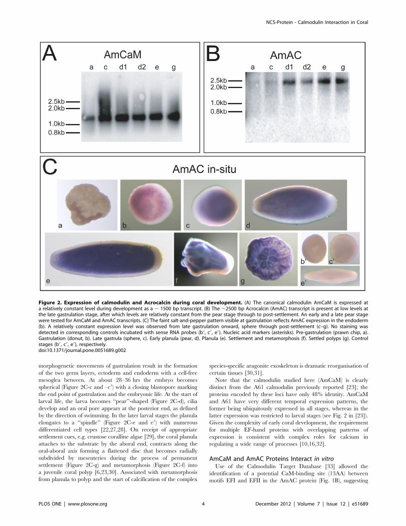
After Gastrulation AmCaM and AmAC Genes are Co-expressed during Coral DevelopmentNorthern blotting revealed that the developmental profiles of
the transcripts encoding the two Acropora EF-hand proteins
differed. Whilst AmCaM transcripts (,1500 bp) were present at
relatively uniform levels across each of the developmental stages
examined (prawn chip to post settlement; Fig. 2A), the (,2500 bp)
AmAC transcript was first detected at late gastrulation and was
present higher levels from the pear stage through to post
settlement (Fig. 2B). In situ hybridization with AmAC probes
revealed a faint, salt-and-pepper like pattern of expression in the
gastrula (donut) (Fig. 2C: b), expression becoming uniform but
restricted to the endoderm from the early planula (sphere) stage
through to post-settlement (Fig. 2C, c–g). No staining was detected
prior to gastrulation (Fig. 2C, a) or when using the sense probe
(Fig. 2C: b9, c9 and e9). In situ hybridization with various probes
implied that AmCaM was expressed ubiquitously throughout
development (data not shown).
The major developmental stages of embryonic development
until early settlement of Acropora together with an approximate
timeline are summarized in [27]. The embryo at ‘‘prawn chip’’
stage (Figure 2C-a) consists of an irregularly shaped cellular
bilayer. During the next stage ‘‘donut’’ (Figure 2C-b and –b9), the
Figure 1. Primary structure of the coral EF-hand proteins. (A) As in the canonical calmodulins of a wide range of other eukaryotes, the AmCaMprotein contains four predicted EF-hand motifs, each of which fulfils the criteria for activity. Genbank identifiers for the sequences: Acropora Cluster043479; Nematostella XP_00163858.1; Homo NP_001734.1; Drosophila NP_523710.1; Aedes XP_001662431.1; Suberites O97341; Trichoplax EDV29861.1;Monosiga XP_001749021.1; Schizosaccharomyces XP_002175972. (B) The coral Acrocalcin (AmAC) protein is a typical member of the NCS-B class,possessing an N-terminal myristoylation site (MGK, orange box), three EF-hand motifs (indicated by red boxes) and a predicted CaM-binding site (bluebox). Genbank identifiers for sequences: Acropora Cluster 013002; Nematostella1 XP_001639634.1; Nematostella2 XP_001639635.1; HS (Homo sapiens)hippocalcin NP_002140.2; HS (Homo sapiens) neurocalcin NP_114430; Drosophila NP_788543.1; Aedes XP_001648788.1; AmphimedonXP_003386697.1; Trichoplax EDV23214.1; Monosiga EDQ90181.1.doi:10.1371/journal.pone.0051689.g001
NCS-Protein - Calmodulin Interaction in Coral
PLOS ONE | www.plosone.org 3 December 2012 | Volume 7 | Issue 12 | e51689
morphogenetic movements of gastrulation result in the formation
of the two germ layers, ectoderm and endoderm with a cell-free
mesoglea between. At about 28–36 hrs the embryo becomes
spherical (Figure 2C-c and –c9) with a closing blastopore marking
the end point of gastrulation and the embryonic life. At the start of
larval life, the larva becomes ‘‘pear’’-shaped (Figure 2C-d), cilia
develop and an oral pore appears at the posterior end, as defined
by the direction of swimming. In the later larval stages the planula
elongates to a ‘‘spindle’’ (Figure 2C-e and e9) with numerous
differentiated cell types [22,27,28]. On receipt of appropriate
settlement cues, e.g. crustose coralline algae [29], the coral planula
attaches to the substrate by the aboral end, contracts along the
oral-aboral axis forming a flattened disc that becomes radially
subdivided by mesenteries during the process of permanent
settlement (Figure 2C-g) and metamorphosis (Figure 2C-f) into
a juvenile coral polyp [6,23,30]. Associated with metamorphosis
from planula to polyp and the start of calcification of the complex
species-specific aragonite exoskeleton is dramatic reorganisation of
certain tissues [30,31].
Note that the calmodulin studied here (AmCaM) is clearly
distinct from the A61 calmodulin previously reported [23]; the
proteins encoded by these loci have only 48% identity. AmCaM
and A61 have very different temporal expression patterns, the
former being ubiquitously expressed in all stages, whereas in the
latter expression was restricted to larval stages (see Fig. 2 in [23]).
Given the complexity of early coral development, the requirement
for multiple EF-hand proteins with overlapping patterns of
expression is consistent with complex roles for calcium in
regulating a wide range of processes [10,16,32].
AmCaM and AmAC Proteins Interact in vitroUse of the Calmodulin Target Database [33] allowed the
identification of a potential CaM-binding site (13AA) between
motifs EFI and EFII in the AmAC protein (Fig. 1B), suggesting
Figure 2. Expression of calmodulin and Acrocalcin during coral development. (A) The canonical calmodulin AmCaM is expressed ata relatively constant level during development as a , 1500 bp transcript. (B) The ,2500 bp Acrocalcin (AmAC) transcript is present at low levels atthe late gastrulation stage, after which levels are relatively constant from the pear stage through to post-settlement. An early and a late pear stagewere tested for AmCaM and AmAC transcripts. (C) The faint salt-and-pepper pattern visible at gastrulation reflects AmAC expression in the endoderm(b). A relatively constant expression level was observed from late gastrulation onward, sphere through post-settlement (c–g). No staining wasdetected in corresponding controls incubated with sense RNA probes (b9, c9, e9). Nucleic acid markers (asterisks). Pre-gastrulation (prawn chip, a).Gastrulation (donut, b). Late gastrula (sphere, c). Early planula (pear, d). Planula (e). Settlement and metamorphosis (f). Settled polyps (g). Controlstages (b9, c9, e9), respectively.doi:10.1371/journal.pone.0051689.g002
NCS-Protein - Calmodulin Interaction in Coral
PLOS ONE | www.plosone.org 4 December 2012 | Volume 7 | Issue 12 | e51689

that the AmAC and AmCaM proteins might interact in vivo. The
AmAC CaM binding motif (LSVTSRGSLEKKL) seems to be
a novel structure combining characteristics of canonical IQ and 1-
8-14 motifs [34]. Although this site lacks some canonical IQ
residues, the terminal residues (KKL) are characteristic of IQ
motifs from neurite growth related proteins and the internal RG
sequence is commonly associated with the IQ motifs of myosin
molecules [34]. Furthermore, the presence of flanking and internal
bulky hydrophobic residues such as leucines (L) resembles the
structure of 1-8-14 motifs [33] with the difference that the
anchoring residues of the calmodulin binding site are in position 1-
9-13.
Virtual northern blotting experiments (Fig. 2A, B) showed that
AmCaM and AmAC are both expressed during late larval
development, when metamorphosis and settlement occur and
calcification is initiated. As CaM transcript levels are relatively
constant through development, the interaction may be regulated
by the availability of AmAC [12,35]. To test the ability of AmCaM
to interact with AmAC, affinity chromatography and immuno-
precipitation experiments were conducted in the presence of
available calcium (1 mM CaCl2) or in its absence (5 mM EGTA).
For this purpose, recombinant GST-AmCaM (,42 KDa) and
His-AmAC (,24 KDa) proteins were produced and purified
(Fig. 3A).
Affinity chromatography confirmed the interaction of the
AmCaM and AmAC proteins predicted on the basis of sequence
analysis. After incubating the fusion-proteins together in the
presence or absence of calcium, the resulting protein complexes
were subjected to chromatography using the His-tag affinity ligand
Ni-NTA (Fig. 3C). As can be seen in the right panel of Fig. 3C, in
the presence of Ca2+, GST-AmCaM was retained on the Ni-NTA
affinity column via its interaction with AmAC (which carries the
poly(His) affinity tag), as the two proteins are eluted together in E1
and E2. In the absence of Ca2+ (left panel of Fig. 3C), neither
protein was retained by the column, because the interaction of the
affinity (poly-His) tag with the column is prevented in the presence
of EGTA.
The observed discrepancies in size of the fusion-proteins (GST-
AmCaM and His-AmAC) between the 5 mM EGTA and the
1 mM CaCl2 treatments (Fig. 3C), is explained by topological
changes experienced in Ca2+-binding molecules under denaturing
gel electrophoresis. In the presence of calcium, CaM molecules are
known to migrate faster on the SDS gels, thus having smaller
apparent molecular weights than in the absence of calcium [36,37]
(Fig. 3C, black arrow heads). The same phenomenon was
observed during denaturing gel electrophoresis of the calcium-
binding protein AmAC (Fig. 3C, red arrow heads).
To test whether the AmAC/AmCaM interaction occurs in the
absence of Ca2+, the recombinant proteins were incubated
together in medium containing 1 mM CaCl2 or 5 mM EGTA
and then subjected to immunoprecipitation using anti-human
calmodulin. The anti-human CaM-I antibody detected specifically
the Acropora CaM (Fig. 3B). As can be seen in Fig. 3D, AmAC was
co-precipitated with AmCaM in both the presence and absence of
Ca2+, implying that the AmAC/AmCaM interaction occurs in
a calcium-independent manner in vitro.
Discussion
In eukaryotes, regulatory EF-hand proteins are the primary
mediators of calcium signalling pathways [38], and amongst these,
the Neuronal Calcium Sensor (NCS) protein family appear to have
a wide variety of roles [39,40]. Five classes (A-E) of NCS protein
are recognized based on sequence similarity [10,39].
While members of the NCS-A class, including NCS-1 and
frequenin [41,42], are widely distributed across eukaryotes, the
coral NCS protein reported here clearly belongs to the NCS-B
class, members of which were previously known only from
bilaterians [38,43]. Searching the database revealed the presence
of two uncharacterised genes encoding predicted NCS-B proteins
(Nv1: XP_001639634 and Nv2: XP_001639635) in the genome of
the sea anemone Nematostella vectensis genome [44]. Clearly related
NCS-B sequences are also present in the sponge Amphimedon
(XP_003386697.1) and the placozoan Trichoplax (EDV23214.1).
NCS proteins are also present in the choanoflagellate Monosiga, but
at this time it is unclear to which class the closest Monosiga match
(EDQ90181.1) belongs. Thus the NCS-B protein class clearly pre-
dates the Bilateria, but whether it predates the Metazoa is unclear.
Despite canonical calmodulins being highly conserved, they
participate in a diverse range of biological processes due to the
ability to interact with an enormous range of target proteins, many
of which are taxonomically restricted [12,35]. For example, in
another coral, Stylophora pistillata, CaM is thought to interact with
a calcium-ATPase, and this interaction may be relevant to the
regulation of skeleton deposition [45]. The presence of clear
counterparts of most of the key molecules involved in vertebrate
calcium signalling as well as a number of coral-specific calcium
sensors (in preparation) suggests that both conserved and coral-
specific calcium dependent signalling pathways function during
settlement and metamorphosis in A. millepora. Given the wide
phylogenetic distribution and sequence conservation displayed by
both calmodulin and NCS-B proteins, it is surprising that the
interaction identified here has not been previously reported,
however, we were unable to find precedents in the literature or the
String interaction databases [string-db.org] or IntAct [ebi.ac.uk/
intact].
The affinity chromatography and immunoprecipitation experi-
ments presented here demonstrate that AmCaM interacts with
AmAC in vitro in the presence of calcium (Figs. 3C and D)., and
immunoprecipitation experiments indicate that this interaction is
likely to also occur in the absence of calcium (Figure 3D, left
panel).
The temporal expression data (Fig. 2) are consistent with this
interaction occurring during late larval development in Acropora,
although the in situ data indicate that this is more likely to occur in
the endoderm than in the aboral ectoderm that is the site of
calcification initiation. The fact that the putative CaM binding site
is located between two EF-hand domains in the AmAC protein
suggests that cooperative coordination may occur between the
calcium binding motifs of the two proteins. For some target
proteins, CaM coordination is necessary to enable calcium binding
or to induce exposure of hydrophobic residues [10,46,47,48].
Most NCS family members are thought to be multifunctional
regulators of neuronal cellular processes [39,49], only NCS-A
types being known to function in both neuronal and non-neuronal
cell types [39,50,51]. Whereas NCS-B proteins have neuronal
functions in vertebrates, the broad endodermal expression pattern
observed for AmAC (Fig. 3) suggests a non-neuronal role during
coral development. Analogy with the vertebrate NCS proteins
neurocalcin and hippocalcin suggests possible roles in vesicle or
mRNA transport, regulation of cGMP intracellular levels and/or
apoptosis [38,52,53] in the larval endoderm.
Coral larvae accumulate calcium in endodermal lipid contain-
ing vesicles prior to skeleton deposition [54]. During the de-
velopment of Acropora larvae, endodermal tissue functions primar-
ily in the mobilisation of stored lipids for energy metabolism and
buoyancy control [31,55,56], presumably also releasing calcium
from endodermal lipid stores. AmAC might therefore have
NCS-Protein - Calmodulin Interaction in Coral
PLOS ONE | www.plosone.org 5 December 2012 | Volume 7 | Issue 12 | e51689

NCS-Protein - Calmodulin Interaction in Coral
PLOS ONE | www.plosone.org 6 December 2012 | Volume 7 | Issue 12 | e51689

a regulatory role during the calcium release process that
accompanies metamorphosis and skeleton deposition. Clarifying
the role of AmAC and the biological relevance of the AmAC/
AmCaM interaction will require the development of tools to allow
functional analysis of coral genes.
Acknowledgments
The authors thank Eldon Ball and David Hayward for all manner of advice
on protocols and techniques.
Author Contributions
Conceived and designed the experiments: DJM SS. Performed the
experiments: ARB SS. Analyzed the data: ARB DJM SS. Contributed
reagents/materials/analysis tools: ARB DJM SS. Wrote the paper: ARB
DJM SS.
References
1. Gunderson L (2007) Ecology: a different route to recovery for coral reefs. Curr
Biol 17: R27–28.
2. Veron J (2000) Corals of the world. Townsville: Australian Institute of Marine
Sciences.
3. DeSalvo MK, Voolstra CR, Sunagawa S, Schwarz JA, Stillman JH, et al. (2008)
Differential gene expression during thermal stress and bleaching in the
Caribbean coral Montastraea faveolata. Mol Ecol 17: 3952–3971.
4. Grasso LC, Maindonald J, Rudd S, Hayward DC, Saint R, et al. (2008)
Microarray analysis identifies candidate genes for key roles in coral de-
velopment. BMC Genomics 9: 540.
5. Reyes-Bermudez A, Desalvo MK, Voolstra CR, Sunagawa S, Szmant AM, et al.
(2009) Gene expression microarray analysis encompassing metamorphosis and
the onset of calcification in the scleractinian coral Montastraea faveolata. Marine
Genomics 2: 149–159.
6. Grasso LC, Negri AP, Foret S, Saint R, Hayward DC, et al. (2011) The biology
of coral metamorphosis: molecular responses of larvae to inducers of settlement
and metamorphosis. Dev Biol 353: 411–419.
7. Miller DJ (2011) Coralbase: Acropora millepora genome assembly pre-release www.
coralbase.org.
8. Shinzato C, Shoguchi E, Kawashima T, Hamada M, Hisata K, et al. (2011)
Using the Acropora digitifera genome to understand coral responses to
environmental change. Nature 476: 320–323.
9. Carafoli E (2002) Calcium signaling: a tale for all seasons. Proc Natl Acad
Sci U S A 99: 1115–1122.
10. Haeseleer F, Imanishi Y, Sokal I, Filipek S, Palczewski K (2002) Calcium-
binding proteins: intracellular sensors from the calmodulin superfamily. Biochem
Biophys Res Comm 290: 615–623.
11. Carafoli E, Genazzani A, Guerini D (1999) Calcium controls the transcription of
its own transporters and channels in developing neurons. Biochem Biophys Res
Comm 266: 624–632.
12. Bahler M, Rhoads A (2002) Calmodulin signalling via the IQ motif. FEBS
Letters 513: 107–113.
13. Bhattacharya S, Bunick CG, Chazin WJ (2004) Target selectivity in EF-hand
calcium binding proteins. Biochim Biophys Acta 1742: 69–79.
14. Gifford JL, Walsh MP, Vogel HJ (2007) Structures and metal-ion-binding
properties of the Ca2+-binding helix-loop-helix EF-hand motifs. Biochem J 405:
199–221.
15. Cyert MS (2001) Genetic analysis of calmodulin and its targets in Saccharo-
myces cerevisiae. Ann Rev Genet 35: 647–672.
16. Carafoli E (2005) Calcium–a universal carrier of biological signals. Delivered on
3 July 2003 at the Special FEBS Meeting in Brussels. FEBS J 272: 1073–1089.
17. Jamieson GA, Bronson DD, Schachat FH, Vanaman TC (1980) Structure and
function relationships among Calmodulins and tropinin C-like proteins from
divergent eukaryotic organisms. Annals NY Acad Sci 356: 1–13.
18. Morita M, Iguchi A, Takemura A (2009) Roles of calmodulin and calcium/
calmodulin-dependent protein kinase in flagellar motility regulation in the coral
Acropora digitifera. Mar Biotech 11: 118–123.
19. Yuasa HJ, Suzuki T, Yazawa M (2001) Structural organization of lower marine
nonvertebrate calmodulin genes. Gene 279: 205–212.
20. Chen IP, Tang CY, Chiou CY, Hsu JH, Wei NV, et al. (2009) Comparative
analyses of coding and noncoding DNA regions indicate that Acropora (Anthozoa:
Scleractina) possesses a similar evolutionary tempo of nuclear vs. mitochondrial
genomes as in plants. Mar Biotech 11: 141–152.
21. Moya A, Huisman L, Ball EE, Hayward DC, Grasso LC, et al. (2012) Whole
transcriptome analysis of the coral Acropora millepora reveals complex responses to
CO2-driven acidification during the initiation of calcification. Mol Ecol 21:
2440–2454.
22. Hayward DC, Catmull J, Reece-Hoyes JS, Berghammer H, Dodd H, et al.
(2001) Gene structure and larval expression of cnox-2Am from the coral Acropora
millepora. Dev Genes Evol 211: 10–19.
23. Hayward DC, Hetherington S, Behm CA, Grasso LC, Foret S, et al. (2011)
Differential gene expression at coral settlement and metamorphosis - a sub-
tractive hybridization study. PloS One 6: e26411.
24. Sambrook JF, Fritsch EF, Maniatis T (1989) Molecular Cloning: A laboratory
manual. Cold Spring Harbour, New York: Cold Spring Harbour Press.
25. de Jong DM, Hislop NR, Hayward DC, Reece-Hoyes JS, Pontynen PC, et al.
(2006) Components of both major axial patterning systems of the Bilateria are
differentially expressed along the primary axis of a ‘radiate’ animal, the
anthozoan cnidarian Acropora millepora. Dev Biol 298: 632–643.
26. Burgoyne RD, O’Callaghan DW, Hasdemir B, Haynes LP, Tepikin AV (2004)
Neuronal Ca2+-sensor proteins: multitalented regulators of neuronal function.
Trends Neurosci 27: 203–209.
27. Ball EE, Hayward DC, Reece-Hoyes JS, Hislop NR, Samuel G, et al. (2002)
Coral development: from classical embryology to molecular control. Int J Dev
Biol 46: 671–678.
28. Ball EE, Hayward DC, Catmull J, Reece-Hoyes JS, Hislop NR, et al. (2000)
Molecular control of development in the reef coral, Acropora millepora. Proc 9th Int
Coral Reefs Symp: 395–401.
29. Morse ANC, Iwao K, Baba M, Shimoike K, Hayashibara T, et al. (1996) An
ancient chemosensory mechanism brings new life to coral reefs. Biol Bull 191:
149–154.
30. Harrison PL, Wallace CC (1990) Reproduction, dispersal and recruitment of
Scleractinian corals. In: Dubinsky Z, editor. Coral Reefs. Amsterdam: Elsevier
Science Publisher BV 133–207.
31. Vandermeulen JH (1975) Studies on Coral Reefs.III. Fine Structural Changes of
Calicoblast Cells in Pocillopora damicornis during Settling and Calcification. Mar
Biol 31: 69–77.
32. Vetter SW, Leclerc E (2003) Novel aspects of calmodulin target recognition and
activation. Eur J Biochem 270: 404–414.
33. Lab I (2002) Calmodulin Target Database http://calcium.uhnres.utoronto.ca/
ctdb/.
34. Rhoads AR, Friedberg F (1997) Sequence motifs for calmodulin recognition.
FASEB J 11: 331–340.
35. Benaim G, Villalobo A (2002) Phosphorylation of calmodulin. Functional
implications. Eur J Biochem 269: 3619–3631.
36. Wallace RW, Tallant EA, Dockter ME, Cheung WY (1982) Calcium binding
domains of calmodulin. Sequence of fill as determined with terbium
luminescence. J Biol Chem 257: 1845–1854.
37. Burgess WH, Jemiolo DK, Kretsinger RH (1980) Interaction of calcium and
calmodulin in the presence of sodium dodecyl sulfate. Biochim Biophys Acta
623: 257–270.
38. Burgoyne RD, Weiss JL (2001) The neuronal calcium sensor family of Ca2+-
binding proteins. Biochem J 353: 1–12.
39. Burgoyne RD (2007) Neuronal calcium sensor proteins: generating diversity in
neuronal Ca2+ signalling. Nat Rev Neurosci 8: 182–193.
40. Ames JB, Lim S (2012) Molecular structure and target recognition of neuronal
calcium sensor proteins. Biochim Biophys Acta 1820: 1205–1213.
41. Pongs O, Lindemeier J, Zhu XR, Theil T, Engelkamp D, et al. (1993)
Frequenin-a novel calcium-binding protein that modulates synaptic efficacy in
the Drosophila nervous system. Neuron 11: 15–28.
Figure 3. The Acrocalcin and calmodulin proteins interact in vitro. (A) Purity of preparations of the recombinant proteins used in the in vitrointeraction experiments: Acropora calmodulin carrying an N-terminal GST-tag (GST-AmCaM; ,42 KDa) and Acrocalcin bearing an N-terminal poly-Histag (His-AmAC; ,24 KDa). (B) Polyclonal antibody against human canonical calmodulin specifically recognises recombinant AmCaM (black arrow),whereas recombinant AmAC (red arrow) is not recognised. (C) In the presence of Ca2+ (right panel), AmCaM is retained on a Ni-NTA affinity columnvia its interaction with the recombinant AmAC that attaches to the matrix via the poly(His) tag that it contains, whereas in the absence of Ca2+ (leftpanel) neither protein is retained by the column. (D) Recombinant AmAC is co-precipitated with AmCaM after incubation with antibody againsthuman calmodulin (agarose-conjugated human CaM-I antibody; Santa Cruz) both in the presence (right panel) and absence (left panel) of Ca2+. GST-AmCaM (black arrows). His-AmAC (red arrows). Flow through (F), Elution 1 (E1), Elution 2 (E2). Protein markers (asterisks).doi:10.1371/journal.pone.0051689.g003
NCS-Protein - Calmodulin Interaction in Coral
PLOS ONE | www.plosone.org 7 December 2012 | Volume 7 | Issue 12 | e51689

42. McCue HV, Haynes LP, Burgoyne RD (2010) The diversity of calcium sensor
proteins in the regulation of neuronal function. Cold Spring Harbor Perspectivesin Biology 2: a004085.
43. De Castro E, Nef S, Fiumelli H, Lenz SE, Kawamura S, et al. (1995) Regulation
of rhodopsin phosphorylation by a family of neuronal calcium sensors. BiochemBiophys Res Comm 216: 133–140.
44. Putnam NH, Srivastava M, Hellsten U, Dirks B, Chapman J, et al. (2007) Seaanemone genome reveals ancestral eumetazoan gene repertoire and genomic
organization. Science 317: 86–94.
45. Zoccola D, Tambutte E, Kulhanek E, Puverel S, Scimeca JC, et al. (2004)Molecular cloning and localization of a PMCA P-type calcium ATPase from the
coral Stylophora pistillata. Biochim Biophys Acta 1663: 117–126.46. Ishida H, Nakashima K, Kumaki Y, Nakata M, Hikichi K, et al. (2002) The
solution structure of apocalmodulin from Saccharomyces cerevisiae impliesa mechanism for its unique Ca2+ binding property. Biochem 41: 15536–15542.
47. O’Donnell SE, Newman RA, Witt TJ, Hultman R, Froehlig JR, et al. (2009)
Thermodynamics and conformational change governing domain-domaininteractions of calmodulin. Methods Enzymol 466: 503–526.
48. O’Donnell SE, Yu L, Fowler CA, Shea MA (2011) Recognition of beta-calcineurin by the domains of calmodulin: thermodynamic and structural
evidence for distinct roles. Proteins 79: 765–786.
49. Amici M, Doherty A, Jo J, Jane D, Cho K, et al. (2009) Neuronal calciumsensors and synaptic plasticity. Biochem Soc Trans 37: 1359–1363.
50. Hilfiker S (2003) Neuronal calcium sensor-1: a multifunctional regulator of
secretion. Biochem Soc Trans 31: 828–832.
51. Weiss JL, Hui H, Burgoyne RD (2010) Neuronal calcium sensor-1 regulation of
calcium channels, secretion, and neuronal outgrowth. Cell Mol Neurobiol 30:
1283–1292.
52. Haynes LP, Fitzgerald DJ, Wareing B, O’Callaghan DW, Morgan A, et al.
(2006) Analysis of the interacting partners of the neuronal calcium-binding
proteins L-CaBP1, hippocalcin, NCS-1 and neurocalcin delta. Proteomics 6:
1822–1832.
53. Ivings L, Pennington SR, Jenkins R, Weiss JL, Burgoyne RD (2002)
Identification of Ca2+-dependent binding partners for the neuronal calcium
sensor protein neuroncalcin: interaction with actin, clathrin and tubulin.
Biochem J 363: 599–608.
54. Clode PL, Marshall AT (2004) Calcium localisation by X-ray microanalysis and
fluorescence microscopy in larvae of zooxanthellate and azooxanthellate corals.
Tissue Cell 36: 379–390.
55. Vandermeulen JH, Watabe N (1973) Studies on Reef Corals. I. Skeleton
formation by newly settled planula larva of Pocillopora damicornis. Mar Biol 23: 47–
57.
56. Vandermeulen JH (1974) Studies on Reefs Corals II. Fine structure of planktonic
planula larva of Pocillopora damicornis with emphasis on the aboral epidermis. Mar
Biol 27: 239–249.
NCS-Protein - Calmodulin Interaction in Coral
PLOS ONE | www.plosone.org 8 December 2012 | Volume 7 | Issue 12 | e51689