The Nervous System - ksumsc.com. Foundation Block/Male...Objectives At the end of the lecture, the...
23
The Nervous System Prof. Musaad Alfayez
Transcript of The Nervous System - ksumsc.com. Foundation Block/Male...Objectives At the end of the lecture, the...

The Nervous System
Prof. Musaad Alfayez
Objectives
At the end of the lecture, the students should be able
to:
List the subdivisions of the nervous system
Define the terms: grey matter, white matter, nucleus,
ganglion, tract and nerve.
List the parts of the brain.
Identify the external and internal features of spinal cord.
Enumerate the cranial nerves
Describe the parts and distribution of the spinal nerve.
Define the term ‘dermatome’
List the structures protecting the central nervous
system
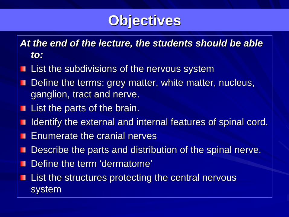
The nervous system has
three functions:
Collection of sensory
input : Identifies changes
occurring inside and
outside the body by using
sensory receptors. These
changes are called stimuli
Integration: Processes,
analyses and interprets
these changes and makes
decisions
Effects a response by
activating muscles or
glands (effectors) via
motor output
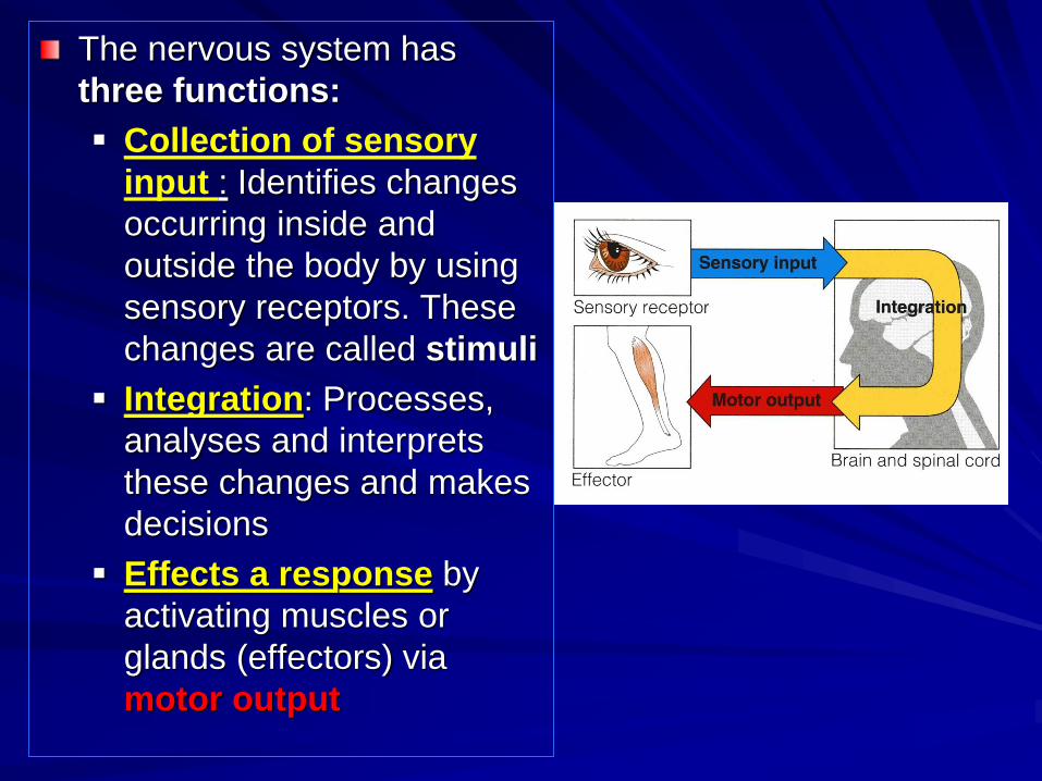
Structural Organization
Central Nervous
System (CNS):
Brain & Spinal
cord
Peripheral
Nervous System
(PNS): Nerves
(cranial, spinal) &
ganglia

Sensory/Motor division
Functional Organization
Somatic/Autonomic (Visceral) division
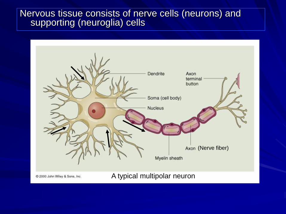
Nervous tissue consists of nerve cells (neurons) and supporting (neuroglia) cells
(Nerve fiber)
A typical multipolar neuron

Nervous tissue is organized as:
Grey matter: which contains the cell bodies & the short processes of the neurons, the neuroglia and the blood vessels.
White matter: which contains the long processes of the neurons (no cell bodies), the neuroglia and the blood vessels

A group of neurons within the
CNS is called a nucleui
A group of neurons outside
the CNS is called a ganglia
Remember…
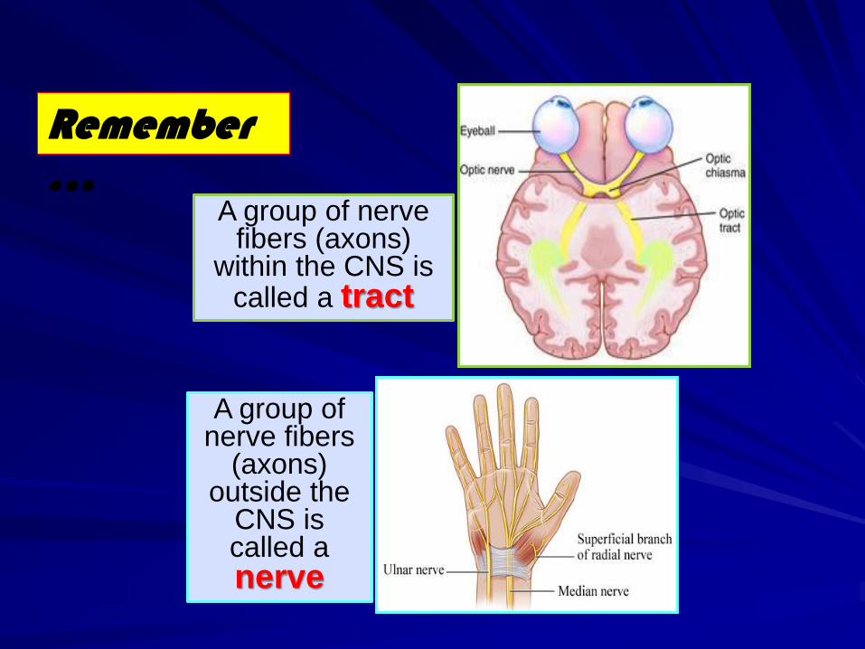
A group of nerve fibers (axons)
within the CNS is called a tract
A group of nerve fibers
(axons) outside the
CNS is called a nerve
Remember…

The Brain
Cerebrum (2
Cerebral
hemispheres)
Cerebellum
Brainstem:
Midbrain
Pons
Medulla oblongata
Diencephalon:
Thalamus,
Hypothalamus,
Subthalamus &
Epithalamus
The brain is a large mass of nervous tissue located in
the cranial cavity. It has four major regions
p
mo
mb

Corpus callosum
Right hemisphere
Left hemisphere
The largest part of the brain, has two
hemispheres
The cerebral hemispheres are
connected by a thick bundle of nerve
fibers called corpus callosum
The surface shows ridges of tissue,
called gyri, separated by grooves
called sulci
Sulcus
Divided by deeper
sulci, into 4 lobes:
Frontal
Parietal
Temporal
Occipital
CEREBRUM
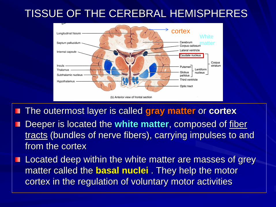
TISSUE OF THE CEREBRAL HEMISPHERES
The outermost layer is called gray matter or cortex
Deeper is located the white matter, composed of fiber
tracts (bundles of nerve fibers), carrying impulses to and
from the cortex
Located deep within the white matter are masses of grey
matter called the basal nuclei . They help the motor
cortex in the regulation of voluntary motor activities
cortexWhite
matter
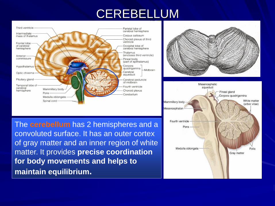
CEREBELLUM
The cerebellum has 2 hemispheres and a
convoluted surface. It has an outer cortex
of gray matter and an inner region of white
matter. It provides precise coordination
for body movements and helps to
maintain equilibrium.

Spinal CordIt is a two-way conduction pathway to the brain & a major reflex center
42-45 cm long, cylindrical in shape, lies within the vertebral canal.
Extends from foramen magnum to L2 vertebra
Continuous above with medulla oblongata
Caudal tapering end is called conusmedullaris
Has 2 enlargements: cervical and lumbosacral
Gives rise to 31 pairs of spinal nerves
Group of spinal nerves at the end of the spinal cord is called caudaequina
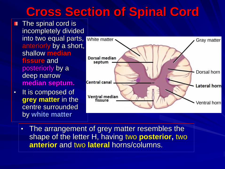
Cross Section of Spinal CordThe spinal cord is incompletely divided into two equal parts, anteriorly by a short, shallow median fissure and posteriorly by a deep narrow median septum.
• It is composed of grey matter in the centre surrounded by white matter
• The arrangement of grey matter resembles the shape of the letter H, having two posterior, two anterior and two lateral horns/columns.
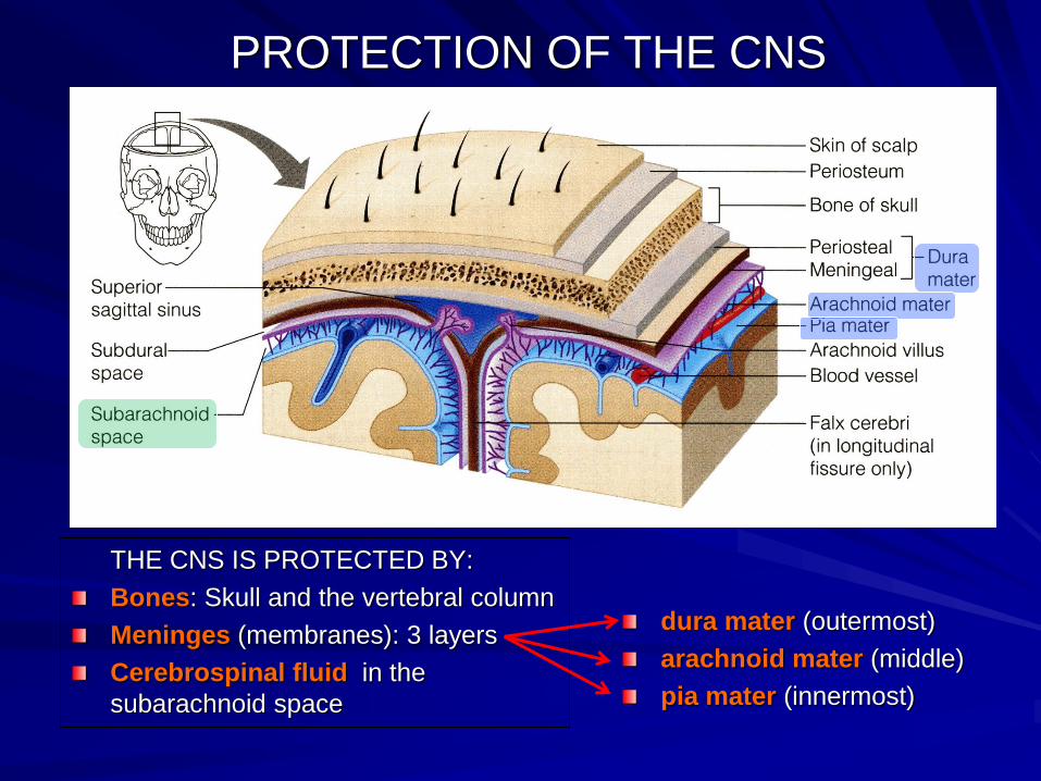
PROTECTION OF THE CNS
THE CNS IS PROTECTED BY:
Bones: Skull and the vertebral column
Meninges (membranes): 3 layers
Cerebrospinal fluid in the
subarachnoid space
dura mater (outermost)
arachnoid mater (middle)
pia mater (innermost)
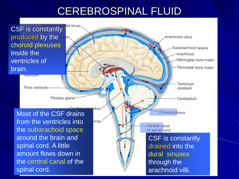
CEREBROSPINAL FLUID
CSF is constantly
produced by the
choroid plexuses
inside the
ventricles of
brain.
Most of the CSF drains
from the ventricles into
the subarachoid space
around the brain and
spinal cord. A little
amount flows down in
the central canal of the
spinal cord.
CSF is constantly
drained into the
dural sinuses
through the
arachnoid villi.

Peripheral Nerves
Cranial:
• 12 pairs,
• attached to brain
• named & numbered
from 1-12
May be sensory, motor or mixed
Two types:
Spinal:
31 pairs
attached to spinal cord
named and numbered
according to the region of
the spinal cord
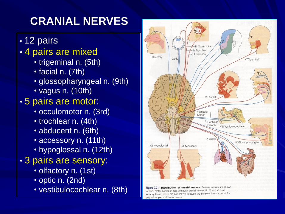
CRANIAL NERVES
• 12 pairs
• 4 pairs are mixed• trigeminal n. (5th)
• facial n. (7th)
• glossopharyngeal n. (9th)
• vagus n. (10th)
• 5 pairs are motor:• occulomotor n. (3rd)
• trochlear n. (4th)
• abducent n. (6th)
• accessory n. (11th)
• hypoglossal n. (12th)
• 3 pairs are sensory:• olfactory n. (1st)
• optic n. (2nd)
• vestibulocochlear n. (8th)
Spinal Nerves and Nerve Plexuses
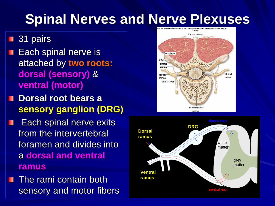
31 pairs
Each spinal nerve is
attached by two roots:
dorsal (sensory) &
ventral (motor)
Dorsal root bears a
sensory ganglion (DRG)
Each spinal nerve exits
from the intervertebral
foramen and divides into
a dorsal and ventral
ramus
The rami contain both
sensory and motor fibers
DRG
Ventral
ramus
Dorsal
ramus

The dorsal rami are
distributed individually,
supply the skin and
muscles of the back
the ventral rami form
plexuses (except in
thoracic region where
they form the intercostal
nerves), and supply the
anterior part of the body
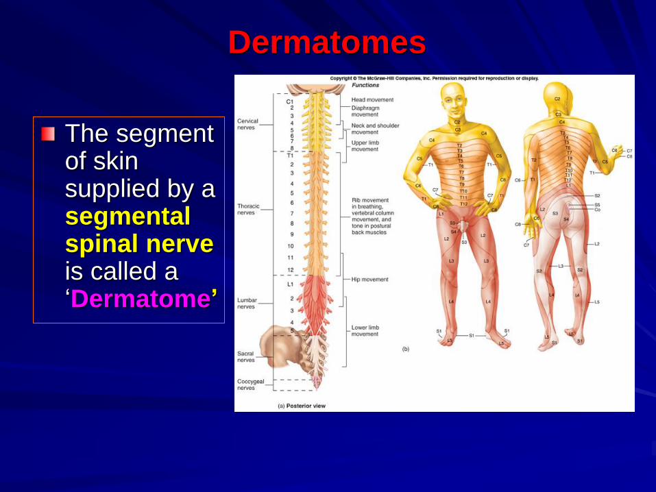
Dermatomes
The segment of skin supplied by a segmental spinal nerve is called a ‘Dermatome’