Nyiragongo DRC Predicting the next big eruption. Nyiragongo DRC.

The N-DRC forms a conserved biochemicalcomplex that maintains outer doublet alignmentand limits microtubule sliding in motile axonemesRaqual Bower, University of MinnesotaDouglas Tritschler, University of MinnesotaKristyn VanderWaal, University of MinnesotaCatherine A. Perrone, University of MinnesotaJoshua Mueller, University of MinnesotaLaura Fox, Emory UniversityWinfield S Sale, Emory UniversityM. E. Porter, University of Minnesota
Journal Title: Molecular Biology of the CellVolume: Volume 24, Number 8Publisher: American Society for Cell Biology | 2013-04-15, Pages 1134-1152Type of Work: Article | Final Publisher PDFPublisher DOI: 10.1091/mbc.E12-11-0801Permanent URL: https://pid.emory.edu/ark:/25593/s7bct
Final published version: http://dx.doi.org/10.1091/mbc.E12-11-0801
Copyright information:© 2013 Bower et al.This is an Open Access work distributed under the terms of the CreativeCommons Attribution-NonCommercial-ShareAlike 3.0 Unported License(http://creativecommons.org/licenses/by-nc-sa/3.0/).
Accessed December 12, 2021 8:05 AM EST

1134 | R. Bower et al. Molecular Biology of the Cell
MBoC | ARTICLE
The N-DRC forms a conserved biochemical complex that maintains outer doublet alignment and limits microtubule sliding in motile axonemesRaqual Bowera, Douglas Tritschlera, Kristyn VanderWaala, Catherine A. Perronea, Joshua Muellera, Laura Foxb, Winfield S. Saleb, and M. E. Portera
aDepartment of Genetics, Cell Biology, and Development, University of Minnesota, Minneapolis, MN 55455; bDepartment of Cell Biology, Emory University School of Medicine, Atlanta, GA 30322
ABSTRACT The nexin–dynein regulatory complex (N-DRC) is proposed to coordinate dynein arm activity and interconnect doublet microtubules. Here we identify a conserved region in DRC4 critical for assembly of the N-DRC into the axoneme. At least 10 subunits associate with DRC4 to form a discrete complex distinct from other axonemal substructures. Transfor-mation of drc4 mutants with epitope-tagged DRC4 rescues the motility defects and restores assembly of missing DRC subunits and associated inner-arm dyneins. Four new DRC subunits contain calcium-signaling motifs and/or AAA domains and are nearly ubiquitous in species with motile cilia. However, drc mutants are motile and maintain the 9 + 2 organization of the axoneme. To evaluate the function of the N-DRC, we analyzed ATP-induced reactivation of isolated axonemes. Rather than the reactivated bending observed with wild-type axonemes, ATP addition to drc-mutant axonemes resulted in splaying of doublets in the distal region, followed by oscillatory bending between pairs of doublets. Thus the N-DRC provides some but not all of the resistance to microtubule sliding and helps to maintain optimal alignment of doublets for productive flagellar motility. These findings provide new insights into the mechanisms that regulate motility and further highlight the importance of the proximal re-gion of the axoneme in generating flagellar bending.
INTRODUCTIONCilia and flagella are highly conserved, microtubule-based organ-elles that extend from the surfaces of eukaryotic cells and play critical roles in cell signaling and motility. Defects in components involved in ciliary assembly, signaling, or motility have profound consequences for an organism, and in humans they can lead to a wide variety of
diseases collectively known as ciliopathies (Hildebrandt et al., 2011; Drummond, 2012). Defects in motility are often associated with chronic respiratory disease (primary ciliary dyskinesia [PCD]), male sterility, hydrocephalus, and defects in the development of the left–right body axis (Fliegauf et al., 2007; Lee, 2011; Zariwala et al., 2011). Given that >400 proteins have been identified by both genomic and proteomic strategies as components of the ciliary and flagellar pro-teome but less than half have been clearly correlated with a specific structure or function (Li et al., 2004; Pazour et al., 2005; Ishikawa et al., 2012; Mizuno et al., 2012), there is considerable interest in a more comprehensive definition of axoneme substructures, including conserved protein complexes required for regulation of motility.
Most motile cilia and flagella contain a structure known as the 9 + 2 axoneme, which consists of nine outer doublet microtubules arranged in a ring surrounding a central pair (CP) of two singlet mi-crotubules. The outer and inner dynein arms (ODA and IDA, respec-tively) are the motor enzymes that drive ciliary motility, and they are arranged in two distinct rows along the length of the A-tubule of each outer doublet. The motor domains of the dyneins walk along
Monitoring EditorErika Holzbaur University of Pennsylvania
Received: Nov 13, 2012Revised: Feb 1, 2013Accepted: Feb 8, 2013
This article was published online ahead of print in MBoC in Press (http://www .molbiolcell.org/cgi/doi/10.1091/mbc.E12-11-0801) on February 20, 2013.Address correspondence to: Mary E. Porter ([email protected]).
© 2013 Bower et al. This article is distributed by The American Society for Cell Biology under license from the author(s). Two months after publication it is avail-able to the public under an Attribution–Noncommercial–Share Alike 3.0 Unported Creative Commons License (http://creativecommons.org/licenses/by-nc-sa/3.0).“ASCB®,” “The American Society for Cell Biology®,” and “Molecular Biology of the Cell®” are registered trademarks of The American Society of Cell Biology.
Abbreviations used: CP, central pair; CSC, calmodulin spoke complex; DHC, dynein heavy chain; DRC, dynein regulatory complex; FAP, flagellar associated polypeptide; GFP, green fluorescent protein; IDA, inner dynein arm; iTRAQ, isobaric tag for relative and absolute quantitation; MS, mass spectrometry; N-DRC, nexin–dynein regulatory complex; ODA, outer dynein arm; PCD, primary ciliary dyskinesia; PF, paralyzed flagella; RS, radial spoke; WT, wild type.

Volume 24 April 15, 2013 Role of the N-DRC in flagellar motility | 1135
Reexamination of the drc mutants by cryo–electron tomography (cryo-ET) demonstrated that the DRC is an integral part of the nexin link, now referred to as the nexin–dynein regulatory complex (N-DRC; Heuser et al., 2009). Both improvements in resolution and the ability to view the N-DRC in three dimensions have further re-vealed numerous connections between the N-DRC and other structures, such as the ODAs, IDAs, RS2, and CSC (Heuser et al., 2009, 2012a,b; Pigino et al., 2011, 2012; Barber et al., 2012; Bui et al., 2012). As a result, there is considerable interest in defining the different subunits of the DRC and their interactions with other axonemal complexes (Lin et al., 2011; Heuser et al., 2012a).
In this study, we used DRC4 and associated mutations as a starting point to reanalyze the biochemical composition of the N-DRC, identify subcomplexes with the N-DRC, and further test the role of the N-DRC in regulating microtubule sliding. We previ-ously showed that the PF2 gene product, DRC4, is the Chlamydomonas orthologue of a highly conserved coiled-coil protein known as Gas11 in humans, Gas8 in other vertebrates, and trypanin in trypanosomes (Rupp and Porter, 2003; Ralston and Hill, 2006; Colantonio et al., 2009). Here we identify a con-served DRC4 region missing in sup-pf3 that is critical for the as-sembly of the N-DRC linker. We also used coimmunoprecipita-tion, iTRAQ labeling, and quantitative mass spectrometry to identify additional N-DRC subunits beyond those previously de-scribed by two-dimensional (2D) gel-based methods (Piperno et al., 1992, 1994; Lin et al., 2011). Spectral counting was used to more rigorously define the IDA isoforms most closely associated with the N-DRC. In addition, we generated and characterized sev-eral new antibodies and epitope-tagged strains for the analysis of N-DRC in vitro and in vivo. This work shows that the N-DRC be-haves as a discrete biochemical complex, distinct from other sub-structures within the axoneme such as the RS, CSC, and protofila-ment ribbon. Finally, we used ATP-induced reactivation and microtubule-sliding assays to demonstrate that the N-DRC pro-vides some but not all of the resistance to microtubule sliding and also serves to maintain alignment between outer doublet microtu-bules during flagellar bending.
RESULTSDistribution of DRC4 in drc mutantsWe previously identified the PF2 gene product as a highly con-served orthologue of the vertebrate protein GAS8/GAS11 that cor-responds to a spot on 2D gels known as DRC4 (Rupp and Porter, 2003). To further characterize the properties and distribution of DRC4, we generated polyclonal antibodies against a conserved peptide sequence, EERHQVEIKVYKQKKVKHLLYEH, and a GAS8 fu-sion protein and tested the affinity-purified antibodies on Western blots of axonemes. Both antibodies recognized an ∼50-kDa poly-peptide that is present in wild type (WT), missing in pf2, and shifted ∼5 kDa in the PF2-HA rescued sample (Supplemental Figure S1A), thereby demonstrating their specificity for DRC4. The affinity-puri-fied antibodies were then used to probe blots of axonemes isolated from a series of motility mutants (Figure 1A and Supplemental Table S1). DRC4 is present at WT levels in mutants affecting the assembly of the ODAs (pf28, sup-pf2) and IDAs (pf9, ida4) but is altered to various degrees in drc mutants. DRC4 is present in pf3 and sup-pf4 but migrates as two smaller bands in sup-pf3 (Figure 1A). These re-sults are consistent with previous studies using 2D gels: the loss of the WT DRC4 spot in sup-pf3 correlated with the appearance of two smaller spots (Huang et al., 1982; Lin et al., 2011). Collectively these observations suggested that sup-pf3 might be an unusual PF2 mu-tation that results in modification of DRC4.
the surface of the B-tubule of the neighboring doublet in an ATP-sensitive manner and thereby generate sliding forces between the doublet microtubules. Because the outer doublets are attached to basal bodies at their base and cross-linked by nexin linkages every 96 nm along their length, microtubule sliding between the doublets is constrained and converted into axonemal bending. In addition, both the ODAs and IDAs are minus end–directed motors that push adjacent doublets toward the tip of the axoneme during bending (Sale and Satir, 1977; Fox and Sale, 1987). Dynein cross-bridge ac-tivity must therefore be switched on and off on opposite sides of the axoneme for effective bending to occur (Wais-Steider and Satir, 1979; Satir and Matsuoka, 1989; Brokaw, 2009a).
Three distinct but overlapping models have been proposed for how dynein activity might be regulated during the ciliary beat cycle (Brokaw, 2009a; Lindemann and Lesich, 2010; Lindemann, 2011; Porter, 2012). One model (the sliding control model) suggests that the basic mechanics of the dynein cross-bridge cycle coupled with the linear arrangement of dynein motors along the length of the outer doublets results in the sequential activation and termination of dynein activity during bending (Riedel-Kruse et al., 2007; Brokaw, 2009a; Lindemann and Lesich, 2010; Mitchison and Mitchison, 2010). Another view is that ciliary bending alters the activity of dy-nein cross-bridges by changes in doublet curvature or doublet spac-ing (curvature control or geometric clutch models; Morita and Shingyoji, 2004; Hayashi and Shingyoji, 2008; Lindemann and Mitchell, 2007; Lindemann and Lesich, 2010; Brokaw, 2009a). These changes increase or decrease the probability that a dynein arm can form a cross-bridge with a neighboring doublet. Both of these mod-els are influenced by factors that affect the affinity of the dynein cross-bridges and/or the behavior of the nexin linkages.
A third view is that dynein activity is modulated by a series of mechanical and enzymatic interactions that involve the CP microtu-bules and their associated projections, the radial spokes (RSs), the calmodulin spoke complex (CSC), and the dynein regulatory com-plex (DRC). This model (the distributor model) is based on the need for additional mechanisms to modify dynein activity in response to intracellular and extracellular signals (e.g., calcium) and also on a large body of work with Chlamydomonas flagellar mutants (Porter and Sale, 2000; Porter, 2012; Smith and Yang, 2004). The model proposes that the CP projections make contacts with the radial spokes in an asymmetric manner during axonemal bending. Differ-ences in strain or tension are transduced through the RS to the CSC, the DRC, and/or other, unidentified components to inhibit or acti-vate different subsets of inner dynein arms. The detailed mechanism is not clear, but pharmacological studies indicate that several tightly bound axonemal kinases and phosphatases may also be involved in regulating the velocity of dynein-driven sliding (Porter and Sale, 2000; Wirschell et al., 2011).
Although these three models reflect different perspectives on the various mechanisms that regulate dynein activity, all three models require some component within the axoneme that limits dynein activity and the extent of microtubule sliding (Brokaw, 2009a; Lindemann and Lesich, 2010; Woolley, 2010; Porter, 2012). In the distributor model, this component is the DRC, because mu-tations in the DRC can override the paralysis observed with CP and RS mutations and restore some form of flagellar motility. Mutations in the DRC are also associated with defects in the assembly of a subset of tightly bound axonemal polypeptides (Huang et al., 1982; Piperno et al., 1992, 1994) that have been correlated with defects in the assembly of a large structure at the distal end of the 96-nm axoneme repeat, between RS2 and the dynein arms (Sup-plemental Table S1; Mastronarde et al., 1992; Gardner et al., 1994).

1136 | R. Bower et al. Molecular Biology of the Cell
a deletion of 96 nucleotides in the larger product (sup-pf3 splice form B). All of exon 4 was missing in splice form A, whereas splice form B used a cryptic splice site downstream of the TOC1 insertion site, at nucleotides 3384–3391 of the original exon 4 (Supple-mental Figure S2D). The DRC4 subunits encoded by the two novel transcripts contain in-frame deletions of 56 and 32 amino acids, respectively (Figure 1B). The sup-pf3 splice A product deletes amino acids 96–151 and has a predicted molecular weight of 48.4 kDa and pI of 6.02. The splice B product deletes amino acids 96–127 and has a predicted molecular weight of 51.3 kDa and a pI of 6.18. These changes are consistent with the shifts seen on Western blots (Figure 1A) and 2D gels (Huang et al., 1982; Lin et al., 2011).
The sup-pf3 mutation deletes a conserved region of the DRC4 subunitDRC4 is a highly conserved, coiled-coil pro-tein with significant degree of sequence and structural similarity across diverse organisms that assemble motile cilia (Figure 1B; Rupp and Porter, 2003). Exon 4 encodes amino acid residues 96–151, which overlaps with the end of the first predicted coiled-coil do-main, CC1 (residues 25–104) and the begin-ning of the second coiled-coil domain, CC2 (residues 132–239; Figure 1C). CC1 and CC2 have been described as a region medi-
ating an interaction between the mouse GAS8 subunit and Rab3B in vitro (Nishimura et al., 2008), whereas the region containing CC2 has been proposed as a potential microtubule-binding site (GMAD, residues 118–260) in human GAS11 (Hill et al., 2000; Bekker et al., 2007). Deletion of exon 4 might be expected to have a significant impact on DRC4 assembly and function. Analysis of the modified polypeptide sequences predicts that the truncated region could form a contiguous coiled coil (CC1-CC2; Figure 1C).
Rescue of pf2 and sup-pf3 by transformation with PF2–green fluorescent proteinDikaryon rescue experiments previously showed that motility de-fects in pf2 could be rescued by mating to WT cells, but rescue of sup-pf3 required deflagellation and reassembly of new flagella (Huang et al., 1982). These observations suggested that the sup-pf3 subunits do not readily exchange with their WT counterparts in in-tact flagella but that sup-pf3 is recessive to WT after turnover of flagellar proteins after regeneration. To test this hypothesis directly, we transformed pf2 and sup-pf3 cells with a green fluorescent pro-tein (GFP)–tagged version of the PF2 gene, screened GFP-positive cells for rescue of motility, and then analyzed isolated axonemes on Western blots (Figure 2).
Live-cell imaging or immunofluorescence with a GFP antibody revealed DRC4-GFP signal along the length of both flagella and at two spots at the anterior end of the cell, near the basal body re-gion, in rescued strains but not in mutant cells (Figure 2, A and B). As measured by phase contrast microscopy, the forward swimming velocities of the GFP-positive strains were significantly increased relative to either sup-pf3 or pf2, demonstrating that the PF2-GFP
The sup-pf3 mutation is caused by insertion of a transposable element in the PF2 geneTo assess how the sup-pf3 mutation might affect the PF2 gene, we amplified 500– to 1000–base pair fragments by PCR from WT and sup-pf3 genomic DNA and sequenced directly (Supplemental Table S2). No sequence differences were observed, with the excep-tion that we were unable to amplify one small region of the PF2 gene in sup-pf3. We therefore compared SacI digests of WT and sup-pf3 genomic DNA on Southern blots probed with PF2 sub-clones, including an ∼1.2-kb NheI restriction fragment correspond-ing to nucleotides 2146–3362 (Supplemental Figure S2A). This probe hybridized to 701– and 755–base pair SacI fragments in WT DNA, but the 701–base pair band was shifted to ∼7 kb in sup-pf3 genomic DNA (Supplemental Figure S2B). The shift of the 701–base pair fragment indicated that a major rearrangement had occurred in the region between nucleotides 2744 and 3445.
To characterize the mutation, we constructed a size-fractionated minilibrary from sup-pf3 genomic DNA and isolated an ∼7-kb SacI subclone containing the modified region of PF2 gene. Sequence analysis revealed the insertion of a large (>5.6 kb) TOC1 transpos-able element (Day and Rochaix, 1991) just downstream of the splice acceptor site for exon 4 (Supplemental Figure S2C). To assess how TOC1 might affect splicing of the PF2 transcript, we used primers from exons 1 and 5 to amplify reverse transcription PCR (RT-PCR) products from WT and sup-pf3 cDNA (Supplemental Figure S2D). A 495–base pair RT-PCR product was recovered from control cDNA, but only two smaller products were recovered from sup-pf3 cDNA (Supplemental Figure S2E). Direct sequencing indicated a deletion of 168 nucleotides in the smaller product (sup-pf3 splice form A) and
FIGURE 1: DRC4 is truncated by alternative splicing in sup-pf3. (A) Western blot of WT and mutant axonemes probed with antibodies to DRC4 reveals the presence of truncated DRC4 subunits in sup-pf3. Anti-Rib43 serves as a loading control. The outer-arm subunit IC2 is reduced in the outer-arm mutants pf28 and sup-pf2. DRC2 and tektin are reduced only in pf3. (B) Clustal W alignment of the human GAS11 and Chlamydomonas DRC4 polypeptide sequences. The deleted amino acids (outlined in the black box) correspond to a region that is highly conserved between DRC4 orthologues. (C) Predicted structural domains in WT and sup-pf3 DRC4 subunits. The deleted regions are predicted to alter the arrangement of coiled-coil domains 1 and 2 in DRC4.

Volume 24 April 15, 2013 Role of the N-DRC in flagellar motility | 1137
(Rupp and Porter, 2003; Heuser et al., 2009). However, the complex-ity of dynein heavy chain (DHC) composition in Chlamydomonas axonemes (Porter et al., 1996; Yagi et al., 2009; Bui et al., 2012) has made a clear description of the specific DHC defects difficult. In addition, some DHCs are more resistant to high salt extraction than others (Pazour et al., 2005; Yagi et al., 2009), and so analysis of dy-nein extracts can be potentially misleading.
As an alternative strategy for the identification of dynein defects in the drc mutants, we used spectral counting (Zhu et al., 2010) to quantify inner-arm DHCs in isolated axonemes. Axonemes from WT, pf2, and pf2::PF2-HA rescued strains were separated by SDS–PAGE, and the DHC region was excised from the gels. Each sample was extracted and trypsin digested, and then three to five replicates per sample were analyzed by tandem mass spectrometry (MS/MS). Both the total number of unique peptides and the total number of as-signed spectra per DHC subspecies were analyzed. Because assem-bly of the I1 dynein is largely unaffected in pf2 mutants (Mastronarde et al., 1992; Gardner et al., 1994), the total number of assigned spectra for each DHC was normalized to the total number of as-signed spectra for the 1-α and 1-β DHCs of the I1 dynein. As shown in Figure 3A, assembly of DHC8 was significantly reduced (p < 0.005) in pf2 relative to both the WT and the pf2::PF2-HA strain. We also detected a slight but statistically significant decrease (p < 0.005) in DHC2 in pf2 relative to both WT and pf2::PF2-HA. The observed changes in the relative abundance of other inner-arm DHCs in pf2, including DHC5, were not statistically significant when compared with both WT and pf2::PF2-HA rescued strains. Western blots of WT and pf2 axonemes probed with antibodies against several dynein subunits confirmed that most of the other dynein subspecies were not significantly reduced in pf2 (Figure 3B). We conclude that dynein e (DHC8) is the major inner-arm subspecies whose assembly is dis-rupted in pf2, consistent with our previous analysis of pf2 and sup-pf3 dynein extracts by fast-performance liquid chromatography (FPLC; Gardner et al., 1994), and that the recovery of DHC8 corre-lates with the reassembly of IA4 in the rescued strains (Rupp and Porter, 2003; Heuser et al., 2009).
Interactions between DRC4, the RS, the CSC, and the protofilament ribbonTo identify other polypeptides closely associated with DRC4, we ex-tracted isolated flagella with different buffers to define conditions that release DRC4 from the axoneme. Flagellar components in-volved in intraflagellar transport (IFT) are typically extracted by treat-ment with nonionic detergents and/or high concentrations of MgATP (Cole et al., 1998), whereas most of the inner and outer dy-nein arms are released by treatment with 0.6 M NaCl (Piperno and Luck, 1979). In contrast, extraction of the central pair (CP) and RS complexes usually requires treatment with 0.2–0.6 M NaI (Mitchell and Sale, 1999; Yang et al., 2001). Very little DRC4 was released af-ter extraction with nonionic detergents, 10 mM ATP, or 0.5 M NaCl, but significant quantities of DRC4 were released by extraction of axonemes with 0.5 M NaI (Supplemental Figure S3A). Because both structural and biochemical studies have shown that the N-DRC is closely associated with radial spoke 2 (RS2) and components of a calmodulin spoke–associated complex (CSC; Mastronarde et al., 1992; Gardner et al., 1994; Piperno, 1995; Rupp and Porter, 2003; Heuser et al., 2009, 2012a; Dymek et al., 2011), we also analyzed extraction of the N-DRC in the RS mutant pf14 and extraction of the RS and CSC in the pf2 mutant. High-salt treatment of pf14 axonemes released the CSC subunit CaM-IP3 (flagellar associated polypeptide 61 [FAP61]) at relatively low ionic strength, but extraction of DRC4 was unaffected by the absence of the RS (Supplemental Figure S3B).
transgene is functional with respect to flagellar motility (Figure 2C). However, the swimming velocities of the GFP rescued strains were slightly slower than those in WT cells. These results could indicate that expression from the PF2-GFP transgene is reduced relative to that from the endogenous PF2 gene.
To assess the extent of DRC4-GFP assembly, we analyzed ax-onemes from WT, mutant, and rescued cells on Western blots. Both the affinity-purified DRC4 antibody and the GFP antibody recog-nized an ∼75-kDa DRC4-GFP band in the pf2::PF2-GFP rescued strain (Figure 2D). However, the sup-pf3::PF2-GFP strains contained three bands recognized by the DRC4 antibody: a major band cor-responding to DRC4-GFP and minor bands corresponding to the modified DRC4 subunits in sup-pf3 (Figure 2D). Thus DRC4-GFP assembles into the axoneme more efficiently than the truncated sup-pf3 DRC4 subunits, but it does not completely displace them. The phenotype of truncated sup-pf3 polypeptides appears to be codominant in the PF2-GFP rescued strains. Backcrossing pf2::PF2-GFP rescued cells to WT cells confirmed that DRC4-GFP subunits assemble into axonemes at approximately the same stoichiometry as the endogenous WT DRC4 subunits (Bower and Porter, unpub-lished results). Collectively, the results demonstrate that the PF2-GFP transgene can rescue motility in both pf2 and sup-pf3 and that DRC4-GFP assembles into flagella at near-WT levels.
Turnover of DRC4 subunits in dikaryon flagellaFlagellar assembly and maintenance require the continuous delivery of precursors to the tips of the flagella by intraflagellar transport before assembly into the axoneme (Qin et al., 2004; Hou et al., 2007; Ahmed et al., 2008). However, earlier efforts to rescue sup-pf3 mu-tants by mating to WT cells failed unless the flagella were amputated and allowed to regenerate (Huang et al., 1982). To better under-stand the process of DRC assembly, we mated pf2::PF2-GFP strains to both pf2 and WT cells and analyzed quadriflagellate dikaryons for the appearance of DRC4-GFP in the unlabeled flagella. Cells of op-posite mating type were mixed together, fixed at specific time points, and then processed for immunofluorescence. When pf2::PF2-GFP cells were mated to a WT strain, DRC4-GFP signal was clearly visible in the two flagella derived from the pf2::PF2-GFP parent (Figure 2E). However, no DRC4-GFP signal was detected in flagella derived from the WT parent, even if the dikaryons were fixed at 10, 30, or 60 min after mixing. These results indicated that there was little exchange between the donor DRC4-GFP subunits and the en-dogenous, unlabeled DRC4 subunits in WT flagella during the first hour. In contrast, if pf2::PF2-GFP cells were mated to pf2 cells, DRC4-GFP signal could be detected at the tips of the two mutant flagella in <10 min (Figure 2F). DRC4-GFP staining was more evident at 30 min, and by 60 min, DRC4-GFP labeling of the pf2 flagella had clearly extended proximally, toward the cell body (Figure 2F). This pattern of initial staining at the flagellar tip followed by the spread of the signal proximally toward the flagellar base is similar to the pat-tern described previously for the assembly of radial spokes in WT × pf14 dikaryons (Johnson and Rosenbaum, 1992).
Reassembly of inner-arm dyneins in PF2-rescued strainsSeveral studies identified deficiencies in assembly of single-headed, IDA isoforms in pf2 and sup-pf3 axonemes, especially dynein e (Piperno et al., 1992, 1994; Gardner et al., 1994; Supplemental Table S1). These deficiencies have been correlated with defects in inner arm structures closely associated with the N-DRC (Gardner et al., 1994; Rupp and Porter, 2003; Bui et al., 2009; Heuser et al., 2009). Moreover, rescue of pf2 with either the PF2-HA or PF2-GFP transgene resulted in the reassembly of the missing IDA structures

1138 | R. Bower et al. Molecular Biology of the Cell
FIGURE 2: PF2-GFP rescues the mutant phenotypes associated with pf2 and sup-pf3. (A) Live imaging of an immobilized PF2-GFP rescued cell by DIC and fluorescence microscopy reveals that DRC4-GFP is located near the basal body region and along the length of the flagella. (B) DIC and fluorescence imaging of fixed PF2-GFP rescued cells (left)

Volume 24 April 15, 2013 Role of the N-DRC in flagellar motility | 1139
and mutant cells (right, pf2-4 and sup-pf3) shows that the GFP signal is observed exclusively in the rescued strains. (C) The forward swimming velocities of WT, mutant, and rescued strains were measured by phase contrast microscopy. The total number of cells measured for each strain (n) is indicated. Both mutants were significantly slower than WT (p < 0.005). Rescued strains were faster than the mutant strains but not completely WT (p < 0.005). The sup-pf3::PF2-GFP rescued strains were also slightly but significantly slower than pf2::PF2-GFP (p < 0.05). (D) A Western blot of isolated axonemes was probed with antibodies against the DRC4 fusion protein antibody (top) and the RSP16 subunit (bottom). Truncated DRC4 subunits are found in sup-pf3 and two rescued strains (left), whereas a larger, more abundant band at ∼75 kDa is observed in the PF2-GFP rescued strains. (E) DIC and fluorescence images of dikaryons fixed at 10 and 30 min after mixing WT and pf2-4::PF2-GFP cells and stained with an antibody against GFP. No significant accumulation of the GFP signal was observed in the WT flagella. (F) Images of dikaryons fixed at 10 and 60 min after mixing pf2-1 and pf2-4::PF2-4-GFP cells and stained with the GFP antibody. Accumulation of GFP signal was initially observed at the flagellar tips and gradually spread to the proximal region. Scale bars, 5 μm.
or Rib72 (Wirschell et al., 2013; Bower and Porter, unpublished re-sults) in any of the drc mutants. To test whether DRC4 might remain bound to the ribbon during sarkosyl extraction, we analyzed WT axonemes and ribbon fractions on Western blots. Rib43 and Rib72 were quantitatively retained in the ribbon fraction after sarkosyl ex-traction, as previously described (Norrander et al., 2000; Ikeda et al., 2003), but DRC4 was not (Supplemental Figure S3D). Thus DRC4 is not a major component of the protofilament ribbon, and any poten-tial interaction between the ribbon and DRC4 is probably indirect.
Identification of DRC4-associated polypeptides by coimmunoprecipitation and iTRAQ analysisTo identify the polypeptides most closely associated with DRC4/PF2, we performed a series of coimmunoprecipitation experiments using extracts prepared from WT, mutant, and rescued axonemes. To define optimal conditions for immunoprecipitation, we incubated dialyzed extracts with CaM-IP2, GFP, and DRC4 antibodies and ana-lyzed the resulting immunoprecipitates on Western blots probed with antibodies to CaM-IP3 and DRC4. As expected, the CaM-IP2 antibody immunoprecipitated CaM-IP3 but not DRC4, the DRC4 antibody immunoprecipitated DRC4 but not CaM-IP3, and the GFP antibody immunoprecipitated DRC4-GFP but not WT DRC4 (Sup-plemental Figure S3E). However, the WT DRC4 subunit (∼55 kDa) migrated very close to the tubulin bands in the extract and the im-munoglobulin bands in the immunoprecipitates, making it difficult to resolve DRC4 as a separate band. We therefore performed most immunoprecipitations using PF2-HA or PF2-GFP extracts, where DRC4 migrated at ∼60 and ∼75 kDa, respectively. Silver-stained gels revealed a large number of bands present in immunoprecipitates prepared from PF2-HA extracts relative to immunoprecipitates from control extracts (Figure 4A). Bands from both the HA-positive and control lanes were excised from the gel, extracted, trypsin digested, and analyzed by MS/MS. During the course of this study, we also identified FAP155 as the likely gene product of the SUP-PF4 locus, based on the deletion of a large genomic region containing the FAP155 gene (Lin et al., 2011). We therefore transformed sup-pf4 with a HA-tagged FAP155 gene and performed additional immuno-precipitation experiments using WT, sup-pf4, and SUP-PF4-HA ex-tracts (Supplemental Figure S4). Those polypeptides identified by minimum of three unique peptides in multiple experiments are listed in Table 1, and the individual peptides are shown in Supple-mental Figure S5. Four polypeptides correspond to missing proteins identified by 2D gel-based methods (Lin et al., 2011), but three polypeptides are novel: FAP122, FAP84, and FAP82 (Figure 4B). Western blots further demonstrated that the DRC1 subunit specifically coimmunoprecipitated with both DRC4-HA and FAP155-HA (Supplemental Figures 3E and 4D).
To confirm that the polypeptides identified by coimmunoprecipi-tation are bona fide subunits of the N-DRC, we analyzed WT and mutant axonemes by iTRAQ labeling and MS/MS. iTRAQ labeling
In contrast, both RSP16 and CaM-IP3 required extraction with 0.4–0.6 M NaI in the pf2 mutant (Supplemental Figure S3C). The extrac-tion profiles are consistent with the close physical interaction be-tween RS2 and the CSC (Dymek and Smith, 2007; Dymek et al., 2011; Heuser et al., 2012a) but also suggest that DRC4 is part of a distinct complex.
The extraction of DRC4 by 0.4–0.6 M NaI implies that the N-DRC is tightly bound to the surface of the microtubule but not an integral component of the underlying outer doublet. This prediction is con-sistent with observation that pf2 axonemes lacked the linker region of the N-DRC but the outer doublet microtubules appeared unal-tered when viewed in cross section or by cryo-ET (Mastronarde et al., 1992; Gardner et al., 1994; Rupp and Porter, 2003; Heuser et al., 2009). However, this region has also been proposed as bind-ing site of several proteins (Rib72, PACRG) associated with the protofilament ribbon that might contribute to the periodic spacing and assembly of the N-DRC (Ikeda et al., 2003, 2007). In addition, the surface of the outer doublet microtubules was altered in the drc mutant pf3 (Heuser et al., 2009). To determine whether there might be any interaction between DRC4 and subunits of the protofilament ribbon, we probed Western blots of drc mutant axonemes with an-tibodies against ribbon subunits. However, we observed no evi-dence for a significant change in the levels of either Rib43 (Figure 1A)
FIGURE 3: Inner-arm dynein subspecies in pf2. (A) Isolated axonemes from WT, pf2-4, and pf2-4::PF2-HA were separated on a 5% SDS–PAGE gel, the DHC regions were excised, and three to five replicates were analyzed by MS/MS and spectral counting. The total number of spectra for each DHC subspecies was expressed as a percentage of the total number of spectra for the two DHCs of the I1 dynein. DHC8 was substantially reduced in pf2, and DHC2 was slightly reduced (p <0.005). (B) Western blot of WT and pf2 axonemes probed with antibodies against various dynein subunits. No obvious defects were observed with antibodies against the outer-arm (OA) subunit IC69 or the inner-arm subunits shown here. No antibodies were available for DHC2 or DHC8.

1140 | R. Bower et al. Molecular Biology of the Cell
results, we raised antibodies against several candidates and used them to probe West-ern blots of WT, pf2, and PF2-HA axonemes. Antibodies against FAP50, FAP82, FAP134, FAP155, and FAP200 showed that each pro-tein was present in WT, missing or reduced in pf2, and restored to WT levels in PF2-HA axonemes (Supplemental Figure S1B). Antibodies against other axonemal proteins, such the CaM-IP3 subunit of the CSC (FAP61), the IC140 subunit of the I1 dynein, and the RSP16 subunit of the RS, showed that the levels of these proteins were largely unchanged in pf2 axonemes, consistent with the iTRAQ data. FAP250 was unchanged in pf2 but reduced in pf3 (Figure 1A), consis-tent with the hypothesis that FAP250 corre-sponds to DRC2 (Kabututu et al., 2010; Lin et al., 2011; Tritschler, Bower, and Porter, un-published results).
Because sup-pf3 and sup-pf4 lack only a subset of the DRC subunits missing in pf2, we compared duplicate samples of WT, pf2, sup-pf3, and sup-pf4 using iTRAQ labeling and an eight-plex analysis (Table 2). In this experiment, only 188 polypeptides were detected at a 5% false discovery rate, and the total number of peptides detected per protein was lower. The predicted protein ra-tios are likely less accurate (Mahoney et al., 2011), but the general trends remained the same. Those proteins identified as reduced in pf2 in the four-plex experiment were also reduced in the eight-plex experiment. The same proteins were reduced to a lesser extent in sup-pf3, and only two proteins (FAP169 and FAP155) were reduced in sup-pf4. Western blot analysis of WT, pf2, sup-pf3, and sup-pf4 axonemes confirmed these observations (Figure 4C). The two proteins reduced in sup-pf4 (FAP155 and FAP169) and two other proteins reduced in pf2 and sup-pf3 (FAP134 and FAP50) were recently identified by proteomic analysis of spots on 1D and 2D gels and designated as DRC5, DRC6, DRC3, and DRC7, respec-tively (Lin et al., 2011). However, FAP200, FAP122, FAP84, and FAP82 are candidates
for additional DRC subunits, now designated as DRC8–DRC11 (see Table 2, Figure 4B, and Supplemental Figure S5). Furthermore, these proteins contain domains (EF hand, IQ domains) implicated in calcium signaling, and DRC11 also contains an AAA domain typi-cally associated with nucleotide hydrolysis (Figure 4B).
Coextraction and cosedimentation of DRC subunitsBecause drc mutations disrupt the assembly of several distinct ax-onemal structures (Gardner et al., 1994; Heuser et al., 2009), it is critical to determine whether the polypeptides associated with DRC4 are bona fide subunits of the same protein complex or com-ponents of peripheral structures. We therefore analyzed high-salt extracts by sucrose density gradient centrifugation and Western blots. As mentioned earlier, DRC4 was extracted from WT axonemes
allows one to compare complex protein samples such as flagella and identify a small subset of polypeptides that are quantitatively differ-ent between samples (Portman et al., 2009). Duplicate samples of WT and pf2 axonemes were labeled with four different iTRAQ tags, digested with trypsin, fractionated by ion exchange and capillary LC, spotted on a matrix-assisted laser desorption/ionization (MALDI) tar-get, and analyzed by mass spectrometry. A total of 252 polypep-tides were identified with high confidence at a false discovery rate of 5%, and for most proteins, no significant differences were observed between WT and pf2 samples. However, a small number of proteins were reproducibly reduced in pf2; these include all of the polypep-tides identified by coimmunoprecipitation except FAP250 and DRC1 (Table 2). Other axonemal polypeptides, such as subunits of the RS, CSC, and protofilament ribbon, were unchanged. To verify these
FIGURE 4: Identification of polypeptides that interact with DRC4. (A) Silver-stained gel of immunoprecipitates obtained with an HA antibody and extracts from pf2 and PF2-HA axonemes. Although several polypeptides were nonspecifically bound to HA beads in both samples, several unique bands were observed in the PF2-HA sample. See Table 1 for polypeptides identified by MS/MS. (B) Diagrams showing predicted polypeptide domains in the candidate DRC subunits identified by coimmunoprecipitation and/or iTRAQ analyses. (C) Western blot of WT, pf2, sup-pf3, and sup-pf4 axonemes probed with antibodies against candidate DRC subunits and other axonemal polypeptides. Several subunits identified as missing in pf2 were also missing or reduced in sup-pf3. Only FAP155/DRC5 was missing in sup-pf4. Antibodies against subunits of the I1 dynein (IC140), CSC (FAP61/CaM-IP3), RS (RSP16), and FAP59/CCDC39 serve as loading controls. (D) Western blots of isolated axonemes (WA) and extracts obtained by sequential treatment with 0.6 M NaCl (HS), 0.2, 0.4, and 0.6 M NaI, and the final extracted outer doublets (OD). DRC subunits were coextracted from WT and PF2-HA with 0.4–0.6 M NaI, but some subunits were more readily extracted from sup-pf3 at lower ionic strengths.

Volume 24 April 15, 2013 Role of the N-DRC in flagellar motility | 1141
that lack the RS, even though the CSC was shifted to the upper por-tion of the gradient (Supplemental Figure S3G). These observations demonstrate that DRC4 is part of a discrete complex distinct from both the RS and the CSC. Sedimentation of the RS and CSC was unchanged in pf2 extracts (Supplemental Figure S3H), consistent with this hypothesis. We therefore analyzed extracts from WT, mu-tant, and rescued strains to assess which DRC subunits might be most closely associated with DRC4.
Because loss of one DRC subunit might affect extraction of other DRC components, we extracted axonemes sequentially with
in ∼0.5 M NaI, along with subunits of the RS and CSC (Supplemental Figure S3A). Reprobing the blots with antibodies against several N-DRC candidates confirmed that these proteins were also preferen-tially extracted in 0.5 M NaI (Supplemental Figure S3A). To deter-mine whether the N-DRC could be physically separated from the RS and CSC, we dialyzed the high-salt extracts against low salt and fractionated by sucrose density gradient centrifugation. DRC4 sedi-mented at >19 S in WT extracts, in a region that overlaps with sub-units of the RS and CSC complexes (Supplemental Figure S3F). However, sedimentation of DRC4 was unchanged in pf14 extracts
Complex name Protein
Four-plex peptides WT/WT pf2/WT
Eight-plex peptides WT/WT pf2/WT sup-pf3/WT sup-pf4/WT
DRC1 14(17) 0.88 0.84 8 (10) 0.67 1.37 1.04 1.40
DRC2 FAP250 6(9) 0.96 0.92 5 (10) 1.12 1.00 0.99 1.03
DRC3 FAP134 4(5) 0.96 0.07 2 (2) 1.05 0.55 0.72 1.08
DRC4 PF2 15(16) 1.11 0.07 4 (4) 1.00 0.36 0.62 1.00
DRC5 FAP155 1(2) 0.89 0.11 1 (1) 0.86 0.40 0.47 0.37
DRC6 FAP169 4(4) 1.02 0.11 1 (1) 1.09 0.24 0.35 0.20
DRC7 FAP50 10(12) 0.94 0.06 4 (4) 1.00 0.61 0.68 1.01
DRC8 FAP200 2(5) 1.07 0.12 1 (1) 1.02 0.44 0.30 0.95
DRC9 FAP122 3(3) 1.02 0.06 3 (2) 1.08 0.50 0.66 1.18
DRC10 FAP84 2(2) 1.05 0.34 ND
DRC11 FAP82 2(2) 0.91 0.43 1 (1) 0.90 0.50 0.41 1.28
Ribbon Rib43a 19(25) 0.99 0.98 12 (16) 1.12 0.76 0.82 1.23
Ribbon Rib72 53(62) 0.97 1.08 28 (38) 1.37 1.58 1.41 1.50
CCDC39a FAP59 17(19) 0.98 1.01 5 (11) 1.07 0.98 0.98 0.99
CaM-IP3 FAP61 7(9) 0.94 0.89 2 (3) 1.08 0.97 0.90 0.93
CaM-IP4 FAP251 6(6) 0.96 0.98 4 (6) 0.97 1.05 1.05 1.04
Radial spoke
RSP16 20(30) 1.03 1.19 14 (12) 0.98 0.96 0.94 0.97
MBO MBO2 5(6) 0.91 0.92 1(2) 1.01 0.90 0.99 0.96
The relative amount of each protein in each sample was compared with that present in the WT sample to obtain a protein ratio. The WT/WT ratios indicate the variability in labeling and protein loading between replicates of the same sample (typically <10%). For each protein, duplicate mutant/WT ratios were averaged. Proteins whose mutant/WT ratios were <75% in both experiments are indicated in bold, with the exception of FAP84, which was only detected in the four-plex experiment. The total number of unique peptides detected at the 95% confidence interval is shown first, and the number of peptides used for quantification is indicated in parentheses. aCCDC39 is the name of the human orthologue of FAP59.
TABLE 2: Polypeptide ratios in WT and drc-mutant axonemes determined by iTRAQ analysis.
Protein Molecular weight (kDa) DRC4-HA (peptides, spectra) DRC5-HA (peptides, spectra)
DRC1 79 WB WB
FAP250 (DRC2) 65 7 (21) 6 (6)
FAP134 (DRC3) 60 3 (3) 4 (4)
PF2 (DRC4) 55 4 (6) 9 (10)
FAP155 (DRC5) 43 WB 3 (6)
FAP50 (DRC7) 177 21 (64) 15 (20)
FAP122 (DRC9) 46 6 (11) 8 (14)
FAP84 (DRC10) 41 7 (20) 7 (10)
FAP82 (DRC11) 95 WB 5 (6)
Both the total number of unique peptides and the total number of assigned spectra are shown. WB indicates a protein that was not identified in the initial mass spectroscopic analysis but was subsequently detected in the immunoprecipitates by Western blots (Supplemental Figures S3 and S4).
TABLE 1: Polypeptides identified by coimmunoprecipitation with DRC4 and DRC5.

1142 | R. Bower et al. Molecular Biology of the Cell
the defects in DRC4 weaken its interactions with other DRC sub-units, including DRC2.
To further explore the relationship between the different DRC subunits, we subjected extracts from several strains to sucrose density gradient centrifugation and analyzed the resulting fractions on Western blots. DRC subunits present in extracts prepared from WT and HA rescued strains were observed to cosediment near the bottom (>19 S) of 5–20% sucrose density gradients, although a sig-nificant portion of DRC7 was also observed near the top of the gra-dient at ∼8 S (Figure 5 and Supplemental Figure S3F). In contrast, several other proteins sediment near the top of the gradients, in-cluding Rib43, Rib72, and tektin (Supplemental Figure S3F). FAP59 is the Chlamydomonas orthologue of the CCDC39 protein in hu-mans, which was recently proposed as a potential DRC subunit (Merveille et al., 2011). However, FAP59 did not appear to be very tightly associated with the DRC in Chlamydomonas, as it also sedi-mented near the top of the sucrose gradients (Figure 5 and Supple-mental Figure S3F). Thus only a subset of the proposed DRC sub-units cosediment as a discrete complex on gradients of WT and rescued extracts.
The sup-pf4 mutant is the least defective drc mutant identified to date (Huang et al., 1982). Its motility phenotype is difficult to
different salt solutions to identify optimal conditions for release of the DRC. For instance, sup-pf3 axonemes lack a subset of DRC polypeptides (Table 2 and Figure 4C), and the sup-pf3 mutation deletes a conserved region of DRC4 (Figure 1B) that overlaps with a predicted microtubule-binding site (Bekker et al., 2007). The modified DRC4 subunits might therefore be more readily ex-tracted from sup-pf3 axonemes in buffers of lower ionic strength. However, the DRC4 subunits in sup-pf3 were not released until treatment with 0.4 M NaI (Figure 4D), which suggests that the in-teraction of DRC4 with the outer doublet was not significantly al-tered in sup-pf3. In contrast, some of the other DRC subunits were more easily extracted from sup-pf3 axonemes than from WT or PF2-HA axonemes (Figure 4D). This susceptibility to salt extrac-tion was most obvious with DRC2. A significant proportion of DRC2 was released at each step in the 0.6 M NaCl, 0.2 M NaI, and 0.4 M NaI extracts, and very little protein remained in the 0.6 M NaI extract or final pellet of outer doublets. In contrast, DRC2 was not released from WT or PF2-HA axonemes until treatment with 0.4–0.6 M NaI, and even then, a significant fraction remained bound to the final pellet of outer doublets. Similar effects were observed with other DRC subunits (Figure 4D). These results sug-gest that even though DRC2 is present at WT levels in sup-pf3,
FIGURE 5: Sucrose density gradient centrifugation identifies subcomplexes within the DRC. Western blots of fractions obtained by analyzing DRC-containing extracts on 5–20% sucrose density gradients. The bottoms (20% sucrose) of the gradients are on the left. (A) PF2-HA, all DRC subunits cosediment near the bottom of the gradient; (B) sup-pf4, DRC subunits dissociate into multiple peaks; (C) sup-pf3, DRC subunits form three distinct subcomplexes; (D) SUP-PF4-HA, most DRC subunits reassociate to form a large complex that sediments near the bottom of the gradient.

Volume 24 April 15, 2013 Role of the N-DRC in flagellar motility | 1143
axonemes (Figure 6A), as previously de-scribed (Okagaki and Kamiya, 1986; Smith and Sale, 1992). In contrast, when full-length axonemes from drc mutants were incubated in the presence of 0.1 mM ATP, they were observed to fray into individual doublets at their distal ends but remained intact at their proximal ends (Figure 6). Of interest, pairs or small subsets of doublets often displayed cycles of repetitive microtubule sliding and bending, similar to that observed previ-ously with protease-treated WT axonemes (Aoyama and Kamiya, 2005). However, the splayed drc-mutant axonemes rarely exhib-ited complete sliding disintegration in the absence of protease (Figure 6). Addition of both ATP and protease to drc-mutant ax-onemes induced splaying, followed by mi-crotubule sliding and disintegration. Iso-lated axonemes from the rescued strains remained intact and did not splay in the presence of ATP alone (Figure 6B), demon-strating that the structural connections be-
tween the outer doublets were restored in the rescued strains. In addition, axonemes from the rescued strains displayed linear mi-crotubule sliding disintegration when treated with both ATP and protease, similar to WT axonemes (Figure 6A).
Thus, as predicted, addition of ATP alone to drc-mutant ax-onemes resulted in the active disruption of the axoneme by splaying of doublet microtubules. However, the experiments also revealed that the outer doublets are held together at the proximal end of the axoneme. These results suggest that additional structures, presum-ably sensitive to added proteases, must be present in the proximal region of the axoneme and that these proximal structures help to maintain the 9 + 2 arrangement of the axoneme in the absence of N-DRC connections (see Discussion).
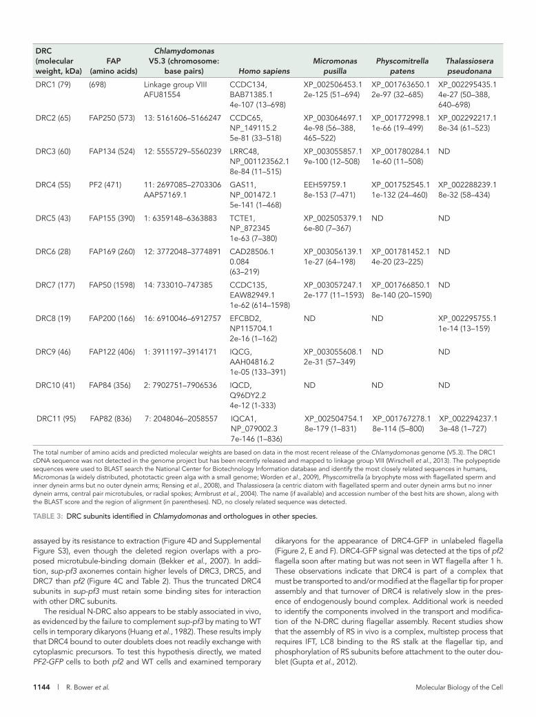
DISCUSSIONDRC4 plays a central role in the assembly of the N-DRC linkerDRC4 is one of the most highly conserved subunits of the N-DRC, and closely related sequences have been identified in nearly all spe-cies with motile axonemes (Table 3; Rupp and Porter, 2003; Ralston and Hill, 2006; Merchant et al., 2007; Colantonio et al., 2009; Hodges et al., 2011). Characterization of sup-pf3 as a transposon-induced, in-frame deletion in the PF2 gene (Figure 1 and Supple-mental Figure S2) further demonstrates that DRC4 plays a central role in the assembly and function of the N-DRC linker. The identifica-tion of sup-pf3 as a PF2 allele was unexpected, given that sup-pf3 was previously mapped to linkage group VI (Huang et al., 1982), but rescue by transformation with PF2 (Figure 2) clearly shows that sup-pf3 and pf2 are two mutations in the same gene on linkage group IX. These observations are also consistent with the similarities in their biochemical and structural phenotypes (Piperno et al., 1992, 1994; Gardner et al., 1994; Heuser et al., 2009). The presence of a large TOC1 transposon within the PF2 gene might have been ex-pected to completely block its function, but alternative splicing to remove TOC1 has been observed in other genes (Rushforth and Anderson, 1996). Moreover, the modified DRC4 subunits in sup-pf3 clearly retain some function. Although the amount of DRC4 is reduced (Figures 1A, 2D, and 4C and Table 2), binding of DRC4 to the outer doublet does not appear to be significantly altered, as
distinguish from WT (Brokaw and Luck, 1985; Gardner et al., 1994), and only a small portion of the linker domain is missing when ana-lyzed by cryo-ET (Heuser et al., 2009). Only two polypeptides (DRC5 and DRC6) are missing in sup-pf4 axonemes (Table 2; Huang et al., 1982; Piperno et al., 1992, 1994; Lin et al., 2011). However, analysis of sup-pf4 extracts suggests that loss of DRC5 and DRC6 destabilizes other subunits of the N-DRC. For example, DRC7 was more readily solubilized than DRC2 and DRC4 in sup-pf4 extracts (Supplemental Figure S4C). Second, DRC4 and DRC7 dissociated into multiple peaks on sup-pf4 gradients, but DRC1, DRC2, and DRC11 still cosed-imented near the bottom (Figure 5B). However, after rescue of sup-pf4 with the FAP155-HA construct, all of the DRC subunits reassoci-ated and cosedimented on the SUP-PF4-HA gradient (Figure 5D).
The dissociation of the N-DRC was even more pronounced in sup-pf3 gradients (Figure 5C). Truncated DRC4 subunits were al-ways observed near the top of the gradient, peaking in fraction 16. DRC7 sedimented slightly further down, peaking in fraction 17, and DRC1 and DRC2 cosedimented near the middle of the gradient as a third peak in fractions 11–13. The dissociation of the N-DRC into at least three distinct subcomplexes in sup-pf3 is consistent with its greater susceptibility to salt extraction described earlier (Figure 4D) and further highlights the central role of DRC4 in the assembly of the N-DRC linker.
Evidence for the role of the N-DRC subunits in maintaining outer doublet alignment and limiting microtubule slidingBased on diverse data, the nexin link of the N-DRC is postulated to play a role in the alignment of adjacent doublet microtubules and the maintenance of the integrity of the axoneme (Gibbons, 1963, 1965; Stephens, 1970; Stephens et al., 1989; Summers and Gibbons, 1971, 1973; Linck, 1973; Witman et al., 1978; Heuser et al., 2009; Porter, 2012). To address this hypothesis, we isolated full-length ax-onemes from WT, drc mutants, and rescued strains and tested their ability to undergo either ATP-induced reactivation or micro tubule-sliding disintegration (Figure 6). When full-length, WT axonemes were incubated with 0.1 mM ATP in the absence of protease, they often exhibited cycles of reactivated beating, but the axonemes remained intact. Once protease was added to WT axonemes, 0.1 mM ATP induced linear microtubule-sliding dis integration of the
FIGURE 6: The N-DRC maintains alignment between outer doublet microtubules in the presence of ATP. (A) Responses of full-length WT or mutant axonemes upon addition of 0.1 mM ATP or 0.1 mM ATP and protease. (B) Quantification of the numbers of axonemes that remained intact, splayed at their distal ends, or underwent sliding disintegration in the presence of 0.1 mM ATP alone. The majority of WT axonemes remained intact (and occasionally were observed to reactivate flagellar beating), whereas axonemes from drc-mutant strains splayed at their distal ends. Axonemes from rescued strains remained intact. Scale bar is 4 μm.
1144 | R. Bower et al. Molecular Biology of the Cell
dikaryons for the appearance of DRC4-GFP in unlabeled flagella (Figure 2, E and F). DRC4-GFP signal was detected at the tips of pf2 flagella soon after mating but was not seen in WT flagella after 1 h. These observations indicate that DRC4 is part of a complex that must be transported to and/or modified at the flagellar tip for proper assembly and that turnover of DRC4 is relatively slow in the pres-ence of endogenously bound complex. Additional work is needed to identify the components involved in the transport and modifica-tion of the N-DRC during flagellar assembly. Recent studies show that the assembly of RS in vivo is a complex, multistep process that requires IFT, LC8 binding to the RS stalk at the flagellar tip, and phosphorylation of RS subunits before attachment to the outer dou-blet (Gupta et al., 2012).
assayed by its resistance to extraction (Figure 4D and Supplemental Figure S3), even though the deleted region overlaps with a pro-posed microtubule-binding domain (Bekker et al., 2007). In addi-tion, sup-pf3 axonemes contain higher levels of DRC3, DRC5, and DRC7 than pf2 (Figure 4C and Table 2). Thus the truncated DRC4 subunits in sup-pf3 must retain some binding sites for interaction with other DRC subunits.
The residual N-DRC also appears to be stably associated in vivo, as evidenced by the failure to complement sup-pf3 by mating to WT cells in temporary dikaryons (Huang et al., 1982). These results imply that DRC4 bound to outer doublets does not readily exchange with cytoplasmic precursors. To test this hypothesis directly, we mated PF2-GFP cells to both pf2 and WT cells and examined temporary
DRC (molecular weight, kDa)
FAP (amino acids)
Chlamydomonas V5.3 (chromosome:
base pairs) Homo sapiensMicromonas
pusillaPhyscomitrella
patensThalassiosera pseudonana
DRC1 (79) (698) Linkage group VIII AFU81554
CCDC134, BAB71385.1 4e-107 (13–698)
XP_002506453.1 2e-125 (51–694)
XP_001763650.1 2e-97 (32–685)
XP_002295435.1 4e-27 (50–388, 640–698)
DRC2 (65) FAP250 (573) 13: 5161606–5166247 CCDC65, NP_149115.2 5e-81 (33–518)
XP_003064697.1 4e-98 (56–388, 465–522)
XP_001772998.1 1e-66 (19–499)
XP_002292217.1 8e-34 (61–523)
DRC3 (60) FAP134 (524) 12: 5555729–5560239 LRRC48, NP_001123562.1 8e-84 (11–515)
XP_003055857.1 9e-100 (12–508)
XP_001780284.1 1e-60 (11–508)
ND
DRC4 (55) PF2 (471) 11: 2697085–2703306 AAP57169.1
GAS11, NP_001472.1 5e-141 (1–468)
EEH59759.1 8e-153 (7–471)
XP_001752545.1 1e-132 (24–460)
XP_002288239.1 8e-32 (58–434)
DRC5 (43) FAP155 (390) 1: 6359148–6363883 TCTE1, NP_872345 1e-63 (7–380)
XP_002505379.1 6e-80 (7–367)
ND ND
DRC6 (28) FAP169 (260) 12: 3772048–3774891 CAD28506.1 0.084 (63–219)
XP_003056139.1 1e-27 (64–198)
XP_001781452.1 4e-20 (23–225)
ND
DRC7 (177) FAP50 (1598) 14: 733010–747385 CCDC135, EAW82949.1 1e-62 (614–1598)
XP_003057247.1 2e-177 (11–1593)
XP_001766850.1 8e-140 (20–1590)
ND
DRC8 (19) FAP200 (166) 16: 6910046–6912757 EFCBD2, NP115704.1 2e-16 (1–162)
ND ND XP_002295755.1 1e-14 (13–159)
DRC9 (46) FAP122 (406) 1: 3911197–3914171 IQCG, AAH04816.2 1e-05 (133–391)
XP_003055608.1 2e-31 (57–349)
ND ND
DRC10 (41) FAP84 (356) 2: 7902751–7906536 IQCD, Q96DY2.2 4e-12 (1-333)
ND ND ND
DRC11 (95) FAP82 (836) 7: 2048046–2058557 IQCA1, NP_079002.3 7e-146 (1–836)
XP_002504754.1 8e-179 (1–831)
XP_001767278.1 8e-114 (5–800)
XP_002294237.1 3e-48 (1–727)
The total number of amino acids and predicted molecular weights are based on data in the most recent release of the Chlamydomonas genome (V5.3). The DRC1 cDNA sequence was not detected in the genome project but has been recently released and mapped to linkage group VIII (Wirschell et al., 2013). The polypeptide sequences were used to BLAST search the National Center for Biotechnology Information database and identify the most closely related sequences in humans, Micromonas (a widely distributed, phototactic green alga with a small genome; Worden et al., 2009), Physcomitrella (a bryophyte moss with flagellated sperm and inner dynein arms but no outer dynein arms; Rensing et al., 2008), and Thalassiosera (a centric diatom with flagellated sperm and outer dynein arms but no inner dynein arms, central pair microtubules, or radial spokes; Armbrust et al., 2004). The name (if available) and accession number of the best hits are shown, along with the BLAST score and the region of alignment (in parentheses). ND, no closely related sequence was detected.
TABLE 3: DRC subunits identified in Chlamydomonas and orthologues in other species.

Volume 24 April 15, 2013 Role of the N-DRC in flagellar motility | 1145
et al., 1994; Wirschell et al., 2013). Therefore we have strong evi-dence for at least 11 DRC subunits.
At least six DRC subunits cosediment with DRC4 as part of a large complex in extracts from WT and rescued strains, but the N-DRC dissociates into smaller subcomplexes in drc-mutant extracts (Figure 5 and Supplemental Figure S3). In particular, the loss of DRC5 and DRC6 destabilizes the interaction between DRC7 and DRC4 in sup-pf4 extracts (Figure 5B and Supplemental Figure S4C), and the truncation of DRC4 in sup-pf3 leads to the loss of DRC11 and the formation of at least three distinct subcomplexes (Figure 5C). Antibodies to other DRC subunits are needed to further charac-terize the individual subcomplexes.
Significant defects in the assembly of ICL1, IC140, or FAP61 were not observed in pf2, sup-pf3, or sup-pf4 axonemes by either iTRAQ or Western blot analyses (Figure 4). These results differ from previ-ous observations based on 2D gels (Lin et al., 2011). The reasons for this discrepancy are unknown but may reflect the difficulties associ-ated with the resolution of higher–molecular weight polypeptides on 2D gels. We previously found evidence for changes in the phos-phorylation status of FAP119, FAP206, FAP230, and FAP252 in sev-eral n-drc mutants (Lin et al., 2011). Of interest, FAP206 was recently recovered by coimmunoprecipitation with RSP3 (Gupta et al., 2012), and FAP252 was previously associated with Rib72 based on blot overlay (Ikeda et al., 2007). These polypeptides may therefore be associated with structures that are located in close proximity to the N-DRC, but whether they represent bona fide N-DRC subunits is unresolved. Our current understanding of N-DRC subunit composi-tion is summarized in Supplemental Table S4.
Characteristics and distribution of DRC subunitsMost DRC subunits are structural proteins that are widely conserved in species that assemble motile axonemes (Table 3; Merchant et al., 2007; Hodges et al., 2011; Lin et al., 2011). Eight subunits contain coiled-coil domains and/or leucine-rich repeats likely to be impor-tant for subunit interactions and complex assembly (Figure 3B). However, four new DRC candidates (DRC8–DRC11) contain EF hand or IQ domains involved in binding calcium- or calmodulin-related proteins. Whether any of these subunits also interacts with RS2 and/or the CSC remains to be determined. DRC11 also contains an AAA domain typically associated with nucleotide binding and hydrolysis (Snider and Houry, 2008). Whether the IQ domain or the AAA do-main of DRC11 contributes to the proposed conformational changes of the nexin link (Bozkurt and Woolley, 1993; Minoura et al., 1999; Lindemann et al., 2005; Lindemann, 2011) is unknown but is an in-triguing possibility. Furthermore, the absence of DRC11 in both pf2 and sup-pf3 suggests that it may correspond to one of the two un-identified densities in the linker region of the N-DRC that makes contact with the neighboring B-tubule (Heuser et al., 2009).
The identification of additional mutant alleles will be an impor-tant next step toward understanding the contribution of each sub-unit in assembly and/or motility. For instance, the characterization of the pf3 mutation in DRC1 and another mutation in the human orthologue CCDC135 demonstrates that this DRC subunit plays a conserved role in assembly of the N-DRC and ciliary motility (Wirschell et al., 2013). A FAP134 (DRC3) mutant has been identi-fied (Lechtreck et al., 2009a), but little is known about its motility phenotype or effects on the assembly of other DRC subunits. DRC3 has been associated with DRC4 based on its presence in sup-pf3 and absence in pf2 (Huang et al., 1982; Piperno et al., 1992, 1994; Lin et al., 2011). DRC3 might be one of the 65- to 85-kDa polypep-tides that interact directly with DRC4 using chemical cross-linking (Rupp and Porter, 2003).
The N-DRC stabilizes the attachment of dynein e (DHC8) to the axonemeSeveral studies proposed that the N-DRC functions as an adaptor for the binding of a subset of inner-arm dyneins to the outer doublets (Piperno et al., 1992, 1994; Gardner et al., 1994). However, the com-plexity of DHC composition and the heterogeneity in DHC distribu-tion both around and along the length of the axoneme have compli-cated efforts to identify the specific dyneins affected (Bui et al., 2009; Yagi et al., 2009). Quantification of inner-arm DHCs by spec-tral counting demonstrated that DHC8 is significantly reduced in pf2 and restored to WT levels in PF2-HA axonemes (Figure 3), consis-tent with previous speculation that the IA4 structure corresponds to dynein e (Gardner et al., 1994; Bui et al., 2009; Heuser et al., 2009). No significant changes were observed in DHC5 (dynein b), unlike our previous study based on the FPLC analysis of dynein extracts (Gardner et al., 1994), but a slight decrease was detected with DHC2 (dynein d). Additional work is needed to identify the other DHCs most closely associated with the DRC. Recent studies suggest that both DHC2 (dynein d) and DHC7 (dynein g) may be located in this region (Bui et al., 2012) and also confirm that DHC8 is reduced in pf2 (Kubo et al., 2012). The inner-arm DHC defects detected by spectral counting in pf2 are also consistent with the recent charac-terization of DHC defects in pf3 (Wirschell et al., 2013) and the ear-lier description of these strains as inner-arm motility mutants (Brokaw and Kamiya, 1987).
The N-DRC is a discrete, tightly bound axonemal complexAlthough the N-DRC is believed to mediate signals between the radial spokes and dynein arms (Piperno et al., 1992, 1994; Gardner et al., 1994; Heuser et al., 2009), the nature of the biochemical inter-actions between these different structures is poorly understood. We therefore generated epitope-tagged DRC constructs and several an-tibodies against DRC subunits as tools for the analysis of the N-DRC in vivo and in vitro (Supplemental Tables S1 and S4 and Supplemen-tal Figure S1). Biochemical fractionation revealed that DRC4 is highly enriched in isolated flagella and axonemes relative to the cell body or flagellar membrane fraction (Figure 2 and Supplemental Figure S3A). Moreover, DRC4 is uniformly distributed along the length of the axoneme when viewed by fluorescence microscopy (Figure 2), consistent with previous work (Rupp and Porter, 2003). DRC4 and other DRC subunits are most efficiently extracted with 0.4–0.6 M NaI, along with subunits of the RS and CSC (Figure 4 and Supple-mental Figure S3A). However, DRC subunits can be separated from subunits of the RS, CSC, or protofilament ribbon by differential ex-traction and/or by sucrose density gradient centrifugation of WT or mutant extracts (Figure 4 and Supplemental Figure S3), consistent with the hypothesis that the N-DRC is a discrete complex.
Coimmunoprecipitation and iTRAQ analysis have identified 10 polypeptides closely associated with DRC4 (Tables 1 and 2), includ-ing four subunits not previously identified by 2D gel-based ap-proaches (Lin et al., 2011). To verify these observations, we gener-ated antibodies to six of the DRC candidates (Supplemental Figure S1). All except FAP250 are missing or reduced in pf2 (Figure 4). However, FAP250 is missing or reduced in pf3 (Figure 1). The pf3 mutant is associated with defects in assembly of the DRC1 and DRC2 subunits (Huang et al., 1982; Piperno et al., 1992, 1994; Lin et al., 2011) believed to be major components of the N-DRC base plate (Heuser et al., 2009). We propose that FAP250 corresponds to DRC2 based on the similarities in their predicted molecular weights (65 and 73 kDa. respectively). In addition, the gene encoding FAP250 maps to chromosome 13, whereas the gene encoding DRC1 has been linked to pf3 mutation on chromosome 8 (Piperno

1146 | R. Bower et al. Molecular Biology of the Cell
mary cilia (Ishikawa et al., 2012). The latter is consistent with the observations that the organization of the outer doublets is often more variable in primary cilia than in motile cilia (Gluenz et al., 2010). Taken together, these observations suggest the primary function of the N-DRC is to maintain outer doublet alignment in motile ax-onemes and limit microtubule sliding during flagellar bending. To further test this idea, we examined ATP-induced reactivation and dynein-driven microtubule sliding in drc-mutant axonemes.
The N-DRC maintains the alignment and integrity of the distal axonemeThe discovery that the DRC is a component of the nexin link that interconnects outer doublet microtubules (Heuser et al., 2009) was somewhat unexpected, given that two of the drc mutants display near-wild-type motility (Brokaw et al., 1982; Brokaw and Luck, 1985). In addition, no significant defects in axoneme organization had been observed, with the caveat that the samples had been pre-pared in the absence of ATP (Gardner et al., 1994; Heuser et al., 2009). Based on the hypothesis that the N-DRC contains at least some of the protease-sensitive components that normally maintain the integrity of the isolated axoneme (Summers and Gibbons, 1971, 1973), we predicted that addition of ATP to axonemes with N-DRC deficiencies would result in dynein-driven microtubule sliding and axoneme disintegration rather than ATP-induced reactivation of ax-onemal bending. However, in full-length, unsonicated axonemes, the disruption occurred by the splaying of individual outer doublet microtubules in the medial and distal regions of the axoneme. In contrast, the proximal ∼2 μm of the drc-mutant axonemes remained intact (Figure 6).
These results are consistent with a model in which the N-DRC plays a role in maintaining the alignment of outer doublets in the medial/distal region of the axoneme during motility. In the absence of N-DRC connections, the integrity of the isolated axoneme and alignment of outer doublets are apparently maintained by the dy-nein arms: addition of ATP to the drc mutant axonemes leads to dynein arm detachment and results in splaying of the outer doublet microtubules. In addition, we frequently observed repetitive bend-ing, sliding, and reannealing between pairs or small groups of outer doublet microtubules in the splayed axonemes from the drc mu-tants, similar to that seen in frayed doublets from WT axonemes (Aoyama and Kamiya, 2005). Thus, with the assembly of a stable axonemal base, the dynein motors and outer doublet microtubules are sufficient for producing simple, repetitive sliding-bending (Aoyama and Kamiya, 2005; Brokaw, 2009b), and, based on this study, repetitive motion between the outer doublet microtubules does not require the presence of the N-DRC.
Although simple, repetitive sliding-bending does not require the N-DRC, the connections provided by the N-DRC do appear to facili-tate resistance to sliding along the length of the axoneme. This per-spective is consistent with a distributor model in which the radial spokes (and central pair projections) activate subsets of dynein arms to exert force against the N-DRC linkages (Porter and Sale, 2000; Porter, 2012; Smith and Yang, 2004). In the absence of signals from the CP/RS complex, dynein activity is reduced, leading to decreased microtubule sliding and flagellar paralysis. Mutations in the N-DRC that weaken the connections between the outer doublet microtu-bules allow the dynein arms to generate microtubule sliding, even in the absence of signals from the CP/RS complex, and some form of motility is restored (Huang et al., 1982). Thus the wild-type N-DRC functions to limit dynein arm activity indirectly along the length of the axoneme. Given that N-DRC subunits appear to be ubiquitous in motile axonemes (Table 3), the putative role of
A mutation has been identified in the Drosophila orthologue of DRC7 known as CG34110/CCDC135 (Yang et al., 2011). The lost boys (lobo) mutation was recovered by screening for defects in the movement of Drosophila sperm in the female reproductive tract, a calcium-regulated process that requires both waveform conversion and regulation of wave propagation. The lobo phenotype is similar to that of a Pkd2 mutant that disrupts a polycystin-related calcium channel in the flagellar membrane, and so the authors proposed that CG34110 functions downstream of Pkd2 in a common signaling pathway (Yang et al., 2011). Because no calcium-binding domain has been identified in CG34110, some other DRC subunits with cal-cium- or calmodulin-binding domains may also be involved in this pathway. Phylogenetic comparisons of ciliary proteins have recently identified a conserved transglutaminase-like (TGL) peptidase do-main in CCDC135/CG34110/FAP50 that is predicted to bind tightly to glutamylated proteins (Zhang and Aravind, 2012). If the TGL do-main is functional in DRC7, it might facilitate interactions with glu-tamylated tubulin residues on the B-tubule. Of interest, a recent study of a glutamylation-deficient strain (tpg1) provided evidence for an interaction between glutamylated tubulin and dynein e and the DRC during microtubule sliding (Kubo et al., 2010, 2012).
The total mass of DRC subunits identified thus far is ∼750 kDa, about half the size of the N-DRC estimated by cryo-ET (Heuser et al., 2009). Nothing is known about the absolute stoichiometry of the subunits, but it is possible to use the flagellar proteome (Pazour et al., 2005) to estimate the relative stoichiometries of the DRC subunits compared with other flagellar proteins, as done previously for subunits of the IFT and BBS complexes (Lechtreck et al., 2009b). These estimates assume that the total number of peptides detected per polypeptide is roughly proportional to the size of the protein and its relative abundance in the flagel-lum. Several peptides were detected for each DRC subunit, rang-ing from 1.7 to 3.3 peptides per 10 kDa, with an average of ∼2.5 peptides per 10 kDa. These ratios are similar to those ob-tained for subunits of the CSC (1.1–2.1 peptides/10 kDa) and FAP59 (2.5 peptides/10 kDa) but approximately one-half of those observed for RSP3 (4.8 peptides per 10 kDa). Current models of the RS vary considerably in their estimates of RSP subunit stoi-chiometries (Yang and Smith, 2009; Pigino et al., 2011), but the only direct evidence indicates that RSP3 is present as a dimer (Wirschell et al., 2008). In addition, the DRC and CSC are associ-ated with only one spoke (RS2) per 96-nm repeat (Gardner et al., 1994; Heuser et al., 2009, 2012a). Based on these estimates, each N-DRC structure could contain two copies of each subunit, which would total ∼1500 kDa, close to the size of the N-DRC es-timated by cryo-ET (Heuser et al., 2009). Further work is needed to purify the N-DRC and determine the stoichiometry of each subunit directly.
Orthologues of the DRC subunits are widely distributed in or-ganisms with motile axonemes (Merchant et al., 2007; Hodges et al., 2011), including species such as Physcomitrella that lack outer dy-nein arms but retain radial spokes and inner dynein arms (Rensing et al., 2008; Table 3). Of interest, however, at least four of the DRC subunits (DRC1, DRC2, DRC4, and DRC11) are also retained in spe-cies such as Thalassiosira that only assemble outer dynein arms but lack the CP/RS complex and inner dynein arms (Armbrust et al., 2004). These comparisons suggest that the N-DRC plays a funda-mental role in regulating motility that is even more basic than the higher-order control provided by the CP/RS complex. Indeed others have noted that both microtubule spacing and interdoublet link-ages are highly conserved in motile 9 + 0 cilia (Lindemann, 2011). In addition, DRC subunits have not been detected in nonmotile pri-

Volume 24 April 15, 2013 Role of the N-DRC in flagellar motility | 1147
of the proximal region of the axoneme is required to identify these components and characterize their role in the organization of the axoneme and the regulation of ciliary and flagellar motility.
MATERIALS AND METHODSCulture conditions, genetic analyses, and strain constructionThe strains used in this study are listed in Supplemental Table S1. Cells were maintained on Tris-acetate phosphate medium but resus-pended overnight in liquid minimal medium or 10 mM 4-(2-hydroxyethyl)-1-piperazineethanesulfonic acid (HEPES), pH 7.6, to facilitate flagellar assembly and mating.
A GFP-tagged version of PF2 was constructed by insertion of a codon-modified GFP into a 6.9-kb XmaI subclone (p9b11-X1.2) containing the complete WT PF2 gene (Rupp and Porter, 2003). A BstEII site was eliminated by site-directed mutagenesis of a CTG codon to a CTC codon in the pHisCrGFP plasmid (Fuhrmann et al., 1999). The GFP tag was amplified using primers with BstEII restric-tion sites, ligated into pGEM-T Easy (Promega, Madison, WI), and then digested and religated into a unique BstEII site in the PF2 plas-mid (Supplemental Table S2). Sequence analysis of the PF2-GFP subclone confirmed that the GFP tag was inserted into the 12th exon of the PF2 gene in the proper orientation and reading frame. The GFP sequence is followed by 52 nucleotides (14 codons) en-coding the last 13 amino acids of the PF2 polypeptide.
PF2-GFP rescued strains were generated by transforming pf2-4 or sup-pf3 with the PF2-GFP plasmid and the selectable marker pSI103 containing the aphVIII gene (Sizova et al., 2001) and plating cells on Tris-acetate-phosphate medium containing 10 μg/ml paro-momycin. GFP-positive cells were identified by fluorescence micros-copy, and motility phenotypes were assessed by phase contrast microscopy. The sup-pf4 strain was rescued by transformation with an ∼7.4-kb BamH1/XhoI subclone containing an HA-tagged FAP155 gene derived from BAC 1i23. The stop codon in FAP155 was mu-tated to an FseI site, and the triple-HA tag was amplified using prim-ers with FseI sites (Supplemental Table S2).
Southern blots, PCR and sequence analysis, and library constructionIsolation of genomic DNA or total RNA, restriction enzyme digests, PCRs, and Southern blots were performed as previously described (Perrone et al., 2000, 2003; Rupp et al., 2001; Rupp and Porter, 2003). PCR primers were designed using the published sequence for the PF2 gene (accession numbers AY309087 and AY3090088). DNA sequencing was performed by the Biomedical Genomics Center (St. Paul, MN) and analyzed using the Sequencher and MacVector software available through the Minnesota Supercom-puting Institute (Minneapolis, MN). The predicted amino acid se-quence (accession number AAP57169 or Q7XJ96) differs from the sequence reported by the Chlamydomonas genome project (XP_001697295) in the region corresponding to amino acid resi-dues 152–197. Our predicted amino acid sequence was based on the direct sequencing of RT-PCR products (Rupp and Porter, 2003), whereas the genome project used gene-modeling programs to in-fer exon splice sites (Merchant et al., 2007). Alignment of the pre-dicted amino sequence with the most recent assembly of Chlamy-domonas expressed sequence tags for PF2 confirms our previously published sequence.
For identification of the site of the mutation in sup-pf3, genomic DNA was digested with SacI, and restriction fragments ranging in size from 6 to 8 kb were used to create a mini–genomic library as de-scribed previously (Perrone et al., 2000; Rupp and Porter, 2003). Colonies containing the modified ∼7-kb SacI fragment in sup-pf3
the N-DRC in providing resistance to sliding should also be consid-ered in the other models for regulating dynein cross-bridge activity and axonemal bending (see Introduction).
One unresolved question is how axonemes maintain their 9 + 2 organization in drc-mutant cells. We propose that that intact flagella of live cells contain other structures that contribute to axoneme in-tegrity. These may include the capping structures at the flagellar tips that insert into the A-tubules of the outer doublets, connections between the outer doublets and the flagellar membrane, and/or other axonemal structures. Mutations have been identified in other organisms that lead to disorganization of the outer doublets (Dawe et al., 2005; Baron et al., 2007; Merveille et al., 2011), but the pre-cise location of the gene products is unresolved. The further charac-terization of the large collection of flagellar motility mutants avail-able in Chlamydomonas may soon reveal the identity and location of the other components that contribute to axoneme integrity and coordinated motility.
The proximal axoneme is a relatively stable structure that provides resistance and contributes to motilityIn most cilia and eukaryotic flagella, bends are initiated at the proxi-mal end and then propagated distally. With notable exceptions (Holwill and McGregor, 1976; Woolley et al., 2006), bends are not propagated in the reverse direction. In addition, the ability to initi-ate bending appears to be localized to a small region at the proxi-mal axoneme (Goldstein, 1969), and ATP-induced reactivated movement of isolated axonemes also results in initiation and propa-gation of bends from the base (Gibbons and Gibbons, 1972; Shingyoji et al., 1977). Furthermore, both experimental and theo-retical evidence indicates the mechanical properties of the base of the axoneme are important for oscillatory bending (Goldstein, 1969; Fujimura and Okuno, 2006; Riedel-Kruse et al., 2007; Mitchison and Mitchison, 2010; Woolley, 2010).
Our study of ATP-treated axonemes from drc mutants also re-veals that the proximal axoneme is a relatively stable structure. We propose that the stability of the proximal axoneme prevents signifi-cant dislocation of adjacent doublet microtubules in the drc mu-tants. As a consequence, ATP addition results in splaying and rean-nealing of outer doublets rather than the linear sliding that leads to axoneme disintegration in protease-treated axonemes. We also conclude that the stability of the proximal axoneme is not based on known components of the N-DRC. Although the drc mutants ex-amined in this study differ in the extent of their polypeptide defi-ciencies (Supplemental Table S4), we have no evidence to suggest that any of the subunits identified thus far vary along the length of the axoneme (Figure 2 and Supplemental Figure S4). However, we previously noted that the structural defects in drc mutants are more pronounced in medial/distal region of the axoneme than in the proximal region (Gardner et al., 1994). The proximal region must therefore contain additional unique structures that link the outer doublets and maintain the integrity of the axoneme. However, with limited exceptions, we know little about the specialized structures (Witman et al., 1978; Hoops and Witman, 1983; Bui et al., 2012) and proteins (Mahjoub et al., 2004; Iomini et al., 2006; Yagi et al., 2009) assembled at the base of the axoneme. Candidate compo-nents include minor inner-arm dyneins localized to the proximal axoneme (Yagi et al., 2009; Bui et al., 2012) and other interdoublet linkers of unknown composition (Witman et al., 1978; Bui et al., 2012; Lin et al., 2012). We predict that these minor dyneins and/or uncharacterized interdoublet linkers in the proximal axoneme play an essential role in the initiation of axonemal bending and in pro-viding additional resistance to dynein-driven sliding. Further study

1148 | R. Bower et al. Molecular Biology of the Cell
were identified by hybridization with a 1.2-kb NheI fragment corre-sponding to nucleotides 2146–3362 of the PF2 gene. Sequence analysis of the subclone revealed the presence of a TOC1 transposon in the middle of the PF2 transcription unit (Supplemental Figure S2). The identification of a large genomic deletion in sup-pf4 that includes the FAP155 gene and two neighboring genes was previously de-scribed (Lin et al., 2011).
Antibody production and characterizationA peptide corresponding to amino acid residues 87–108 (EERHQVEIKVYKQKKVKHLLYEH) of the Chlamydomonas PF2 se-quence was synthesized by Research Genetics (Huntsville, AL), coupled to keyhole limpet hemocyanin, and used to immunize two rabbits. Polyclonal serum from both rabbits was pooled and puri-fied over a peptide affinity column. A second antibody was raised against a fusion protein corresponding to amino acid residues 1–430 of the mouse GAS8 sequence (GenBank Accession number AA499529). A mouse cDNA clone (937316; Stratagene, Santa Clara, CA) was digested with NcoI and BamHI and ligated into pET30a (Novagen, Gibbstown, NJ). The subclone was digested with BamHI and HindIII, and a small synthetic BamHI/HindIII linker containing two stop codons was ligated in-frame with the GAS8 sequence. Expression of the histidine (His)-tagged GAS8 fusion protein was induced overnight at 20°C, and soluble protein was purified on a Ni2+ column (Novagen), dialyzed into phosphate-buff-ered saline, and used to immunize three rabbits (Covance, Denver, PA). A second fusion protein was prepared by transferring the GAS8 sequence into pGEX-2T (Amersham) and purification of a glutathi-one S-transferase (GST)–GAS8 fusion protein on a glutathione–Sep-harose column. The polyclonal antibody was blot affinity purified against either the His-tagged or the GST-tagged GAS8 fusion pro-tein as described by Tang (1993). Alternatively, the GST-tagged GAS8 fusion protein was cross-linked to glutathione–Sepharose beads and used for column affinity purification of the antibody as described by the manufacturer. The affinity-purified antibodies were tested against blots of WT and pf2-4 axonemes at a 1:1000 dilution. Some batches of affinity-purified antibodies demonstrated additional cross-reactivity against an ∼100-kDa band in both WT and pf2-4 axonemes. To reduce this background, the affinity-puri-fied antibody was absorbed against methanol-extracted pf2-4 cells or blots of pf2-4 axonemes as previously described (Perrone et al., 2003).
Peptides and/or fusion proteins from DRC4-associated polypep-tides identified by coimmunoprecipitation and/or iTRAQ analysis were used to generate polyclonal antibodies in rabbits at Spring Valley Laboratories (Woodbine, MD) or Covance. The following pep-tides were synthesized and coupled to KLH via a C-terminal cysteine: FAP134 (AEINKGQYNGYFTQVRC), FAP50 (RRSTRKSKRGGDSKRLC), FAP169 (LPDANTWPTWGALRERC), FAP155 (ADEDYWRRRSQARW-KNC), FAP200 (CASLFRDDYHTLLRAFRC), and FAP82 (EKRNPDT-GEYPDFPDPC and GKPGTAGSKPGTADKKC). Peptides were cou-pled to SulfoLink (Pierce Chemical, Rockford, IL) for affinity purification of the resulting antisera. Fusion proteins corresponding to amino acid residues 853–1258 of FAP50, 311–500 of FAP82, and 1–573 of FAP250 were also generated using His, GST, and MBP tags. Antisera against FAP50 were generated with gel-purified 6xHis-FAP50 fusion protein and affinity purified against the FAP50-MBP fusion protein bound to an NHS agarose (ThermoFisher Scientific, Rockford, IL) col-umn. Antisera against FAP250 were generated with gel-purified 6xHis-FAP250 fusion protein and blot affinity purified against the FAP250-GST protein. Antisera against FAP82 were generated with the FAP82-GST fusion protein purified from inclusion bodies.
Isolation of flagella and fractionation of the dynein regulatory complexFlagella were isolated by pH shock or dibucaine treatment and demembranated using 0.1–0.5% IGEPAL CA-630 (Sigma Aldrich, St. Louis, MO) as previously described (Witman, 1986). Axonemes were resuspended in HMEEN (10 mM HEPES, pH 7.4, 5 mM MgSO4, 1 mM ethylene glycol tetraacetic acid, 0.1 mM EDTA, 30 mM NaCl) plus 1 mM dithiothreitol (DTT) and 0.1 μg/ml protease inhibitors (leupeptin, aprotinin, pepstatin) and sequentially extracted with HMEEN containing 0.6 M NaCl, 0.2 M NaI, 0.4 M NaI, or 0.6 M NaI to identify optimal conditions for extraction of DRC4 in WT and dif-ferent drc mutants. Extracts were dialyzed against HMEEN, clarified by centrifugation, and then fractionated on 5–20% sucrose density gradients. Sarkosyl-extracted protofilament ribbons were prepared as described in Norrander et al. (2000).
SDS–PAGE and Western blot analysesSamples were analyzed on 5–15% polyacrylamide, 0–2.4 M glycerol gradient gels. Protein was transferred to Immobilon P (Millipore, Billerica, MA), and total protein on the blot was visualized with the reversible Blot FastStain (Chemicon, Temecula, CA) to ensure equal loading. Antibodies were diluted in Tropix I-Block (Applied Biosystems, Bedford, MA) at the concentrations indicated in Sup-plemental Table S3. Immunoreactive bands were detected with alkaline phosphatase–conjugated secondary antibodies and the Tropix chemiluminescent detection system.
Immunoprecipitation and mass spectrophotometry of DRC4-associated proteinsImmunoprecipitations were performed as described by Wargo et al. (2005) and Dymek and Smith (2007) with the following modifica-tions. Isolated axonemes from WT, mutant, and rescued strains were extracted with 0.4-0.6 M NaI in HMEEN for 30 min on ice. Extracts were dialyzed against HMEEN or Na-low buffer (10 mM HEPES, pH 7.4, 5 mM MgSO4, 1 mM DTT, 0.5 mM EDTA, and 30 mM NaCl) for a minimum of 2 h to overnight and clarified by centrifugation. From 40 to 200 μl of clarified extract (containing 40–300 μg of pro-tein) were incubated for 3–24 h with Tris-buffered saline (TBS), 0.1% Tween, protein A or protein G beads, and 20–40 μg of one of the following antibodies: rabbit anti–CaM-IP2, rabbit anti-GAS8, Covance mouse anti-HA (clone 16B120), Roche rat anti-HA (clone 3F10; Roche, Indianapolis, IN), or rabbit anti-GFP (A11122; Invitro-gen, Carlsbad, CA) for several hours. The beads were collected by centrifugation and washed four or five times with TBS/0.1% Tween and twice with TBS. Beads were boiled in sample buffer, and sam-ples were loaded on 5–15% polyacrylamide gels for Western blot-ting and silver staining. Gels were stained with PROTSIL2-1KT (Sigma-Aldrich, St. Louis, MO) or SilverQuest (LC6070; Invitrogen).
Coomassie blue– or silver-stained bands from both experimental and control lanes were excised from the gel, destained, and sub-jected to cysteine reduction, alkylation and tryptic digestion on the Genomic Solution ProPrep robot (Ann Arbor, MI) in the Center for Mass Spectrometry and Proteomics at the University of Minnesota (St. Paul, MN) and processed as described in Andersen et al. (2010) with the following modifications. Tryptic peptides were extracted, dried down, and rehydrated in water/acetonitrile/formic acid (95:5:0.1). MS was performed on a linear ion trap (LTQ; Thermo Elec-tron, San Jose, CA) using nanoscale microcapillary reversed-phase liquid chromatography electrospray ionization MS/MS. Peptide mix-tures were autosampled on a Paradigm MS4 system (Michrom Biore-sources, Auburn, CA). Samples were desalted and concentrated on a Paradigm Platinum Peptide Nanotrap (Michrom Bioresources)

Volume 24 April 15, 2013 Role of the N-DRC in flagellar motility | 1149
precolumn (0.15 × 50 mm, 400-μl volume) and subsequently to a fused silica microcapillary column (75-μm internal diameter) packed in-house using a high-pressure helium bomb to a length of 13 cm with Magic C18AQ reversed-phase material on a flow splitter (Michrom Bioresources) at a flow rate of 250 nl/min. The samples were subjected to a 60-min (10–40% acetonitrile) gradient and di-rectly eluted into the microcapillary column set to 2.0 kV. The LTQ was operated in the positive-ion mode using data-dependent acqui-sition methods initiated by a survey MS scan, which was followed by MS/MS (collision energy of 29%) on the four most abundant ions detected in the survey scan. The m/z values selected in the survey scan for MS/MS were dynamically excluded from further data-de-pendent MS/MS for a time of 30 s. The signal intensity threshold for an ion to be selected for MS/MS was set to a lower limit of 1000.
Sequence analysis and criteria for protein identificationMass spectra were extracted and analyzed using BioWorks SeQuest Analysis Software package, SeQuest version SRF v. 27, revision 13 (ThermoFisher Scientific, San Jose, CA) against Chlamydomonas sequences at National Center for Biotechnology Information or the Chlamydomonas genome database (version 3.0; http://genome.jgi -psf.org/Chlre/Chlre3.home.html) for protein identification. The ex-tracted data were also subjected to X! Tandem version 2007.01.01.1 searches within the Scaffold program (v_3_00_03; Proteome Soft-ware, Portland, OR). The data were searched with a fragment-ion mass tolerance of 1.00 Da and a parent-ion tolerance of 1.00 Da. Iodoacetamide derivative of cysteine was specified as a fixed modi-fication and oxidation of methionine as a variable modification.
The Scaffold program was also used to validate MS/MS-based peptide and protein identifications. Peptide IDs were accepted if they could be established at >95.0% probability as specified by the Peptide Prophet algorithm (Keller et al., 2002). Protein IDs were ac-cepted if they could be established at >99.9% probability by the Protein Prophet algorithm (Nesvizhskii et al., 2003) and contained at least three identified peptides. Proteins that contained similar pep-tides and could not be differentiated based on MS/MS analysis alone were grouped to satisfy the principles of parsimony. Any poly-peptides detected in both experimental and control samples (e.g., tubulin) were considered contamination.
Preparation of samples for iTRAQ analysisAxoneme samples were resuspended in 10 mM HEPES, pH 7.4, for estimation of the protein concentration and to remove salt, DTT, and protease inhibitors. After centrifugation at 16,000 rpm, the pel-lets were resuspended in 0.5 M triethylammonium bicarbonate, pH 8.5, at a concentration of 0.6–5.0 mg/ml. Proteins were dena-tured and reduced; cysteine residues were blocked with methyl-methane thiosulfonate, and then samples were digested overnight at 37°C with trypsin. Aliquots of each sample (50 μg for the four-plex and 25 μg for the eight-plex) were reacted in duplicate with four-plex (114–117) or eight-plex (113–119, 121) iTRAQ labeling reagents (AB SCIEX, Foster City, CA). The labeled samples were combined, dried under vacuum, and purified with an MCX cartridge (Waters Corporation, Milford, MA) to remove excess trypsin, unreacted iTRAQ reagents, and buffer.
The labeled peptide mixture was first fractionated by strong-cation-exchange chromatography into 14 fractions, and then each fraction was subjected to capillary-LC with Magic C18AQ columns (Michrom Bioresources). The LC elutes were spotted onto MALDI targets in a 1232-spot format. MS data on all LC spots were ac-quired on 4800 MALDI-TOF/TOF analyzer (AB/MDS) with a 200-Hz repetition rate Nd:YAG laser. Full-scan MS spectra were acquired
from m/z 800 to 4000. Data-dependent tandem MS settings in-cluded acquisition of the top 30 most intense ion signals per spot. Additional details were as previously reported (Akkina et al., 2009).
Protein Pilot software version 3 (AB SCIEX) was used for data pro-cessing (Shilov et al., 2007). Searches were performed against the Chlamydomonas version 3 database (translated from genomic se-quences; http://genome.jgi-psf.org/Chlre3/Chlre3.home.html) plus common lab contaminant proteins (www.thegpm.org/crap/index .html). The quality of the protein identification was further assessed by the false-discovery-rate analysis (Tang et al., 2008), and a 5% local false-discovery-rate threshold was set as the cut-off value for proteins subjected to quantification. Only those proteins identified with >95% confidence and whose WT/WT ratio varied <20% were considered further. A total of 252 proteins were detected in the four-plex experiment, and 188 proteins were detected in the eight-plex experiment.
The predicted amino acid sequences of proteins identified in the V3 database were compared with revised sequences in later re-leases of the Chlamydomonas genome project (V4, http://genome .jgi-psf.org/Chlre4/Chlre4.home.html; and V5, www.phytozome .net/search.php?show=text&org=Org_Creinhardtii_v5.3). Any am-biguous sequences were verified directly by RT-PCR of WT cDNA (Supplemental Table S2). The location of predicted protein domains was determined using the SMART program (available at http://smart.embl-heidelberg.de/).
Phase contrast and fluorescence microscopySwimming phenotypes were assessed by phase contrast micros-copy using a 40× objective and 1.6× eyepiece (Rupp and Porter, 2003). The distribution of DRC4-GFP in live cells was analyzed using either a Zeiss Axioskop (Carl Zeiss, Jena, Germany) as described later or a Nikon Eclipse E800 photomicroscope (Nikon, Melville, NY) using a 100×/1.3 numerical aperture (NA) Plan Apo objective, a Pho-tometrics Cascade 512B camera (Photometric, Tucson, AZ), and the ImagePro Plus software package available at the University Imaging Center (St. Paul, MN). Live cells were immobilized with 1% low–melting point agarose and/or allowed to adhere to polyetha-nolamine-treated coverslips before imaging. Image stacks were col-lected using z-steps of 0.339 μm. For the image shown in Figure 2A, sections 11–21 were deconvolved using ImagePro Plus 6.2 Sharp Stack and then used to make a maximum-intensity projection.
Because the DRC4-GFP signal bleaches quickly, most cells were fixed using ice-cold methanol (Sanders and Salisbury, 1995) and stained for immunofluorescence using a mouse monoclonal anti-body to GFP (Molecular Probes) or a rat monoclonal antibody to HA (clone 3F10; Roche) and Alexa Fluor–488 conjugated secondary an-tibodies (Molecular Probes) as described by Mueller et al. (2005). Slides were sealed using either SlowFade or Prolong Gold mounting medium (Invitrogen Life Technologies, Grand Island, NY). Images were collected on a Zeiss Axioscop equipped with differential inter-ference contrast (DIC) and wide-field fluorescence optics using a 100×/1.3NA Plan Neofluar objective, a CoolSNAP-ES monochrome charge-coupled device camera (Photometrics), and the MetaMorph Software package, version 7.1.7.0 (Molecular Devices, Downington, PA). Selected images were cropped, rotated, and labeled in Photo-shop (Adobe, San Jose, CA).
Analysis of microtubule sliding and splaying in isolated axonemesMicrotubule sliding and splaying was measured as previously de-scribed (Okagaki and Kamiya, 1986; Habermacher and Sale, 1996) with the following modifications. Briefly, isolated flagella were

1150 | R. Bower et al. Molecular Biology of the Cell
resuspended in buffer without protease inhibitors and demembran-ated with buffer containing 0.5% Nonidet P-40 in 10 mM HEPES, pH 7.4, 5 mM MgSO4, 1 mM DTT, 0.5 mM EDTA, 1% polyethylene gly-col (20,000 molecular weight), and 25 mM potassium acetate. The unsonicated, full-length axonemes were added to a perfusion cham-ber, constructed using double-stick tape as spacers between the coverslip and slide, and permitted to adhere to the glass surfaces, and microtubule sliding/splaying was initiated by the addition of buf-fer containing 0.1 mM ATP or 0.1 mM ATP and subtilisin A type VIII protease at 5 μg/ml (Sigma-Aldrich). Assays of microtubule sliding disintegration often use short axoneme fragments obtained by soni-cation (e.g., Kubo et al., 2012), but we found that it was essential to use unsonicated, full-length axonemes and lower concentrations of ATP to detect the splaying phenomenon. Images were recorded us-ing a Zeiss Axiovert 35 microscope equipped with dark-field optics, a Zeiss 40× Plan-Apo lens, and a silicon intensified camera (VE-1000; Dage-MTI, Michigan City, IN). The video images were converted to a digital format using Labview 7.1 software (National Instruments, Aus-tin, TX), and sliding or splaying was determined manually by count-ing multiple fields of axonemes at ∼15 s after motility was initiated. All axonemes observed within a field were scored as intact, splayed (held together at the proximal end), or disrupted by linear sliding. At least 70 axonemes were counted per treatment in each experiment.
ACKNOWLEDGMENTSWe thank Lorraine Anderson, LeeAnn Higgins, Todd Markowski, and Bruce Witthun of the Center for Mass Spectrometry and Com-putational Proteomics at the University of Minnesota for assistance with iTRAQ labeling, mass spectrometry, and spectral counting. The Center is supported by multiple grants, including National Science Foundation Major Research Instrumentation Grants 9871237 and NSF-DBI-0215759 as described at www.cbs.umn.edu/msp/about. We also acknowledge the University of Minnesota Supercomputing Institute for software support and data storage and Mark Sanders and the University Imaging Center for assistance with live-cell imag-ing. We also thank Matt Laudon and the Chlamydomonas Genetics Center (University of Minnesota) for strains. Richard Linck (University of Minnesota), Mark Sanders (University of Minnesota), Ritsu Kamiya (University of Tokyo, Tokyo, Japan), Toshiki Yagi (Kyoto University, Kyoto, Japan), Pinfen Yang (Marquette University, Milwaukee, WI), Elizabeth Smith (Dartmouth College, Hanover, NH), and Gianni Piperno (Mount Sinai School of Medicine, New York, NY) generously supplied antibodies as listed in Supplemental Table S3. Preliminary reports of this work were presented at American Society for Cell Biology meetings in 2006 and 2010. This work was supported by National Institutes of Health grants to M.E.P. (GM-055667) and W.S.S. (GM-051173). K.V.M. was supported in part by an American Heart Association Pre-Doctoral Fellowship and a University of Minnesota Graduate School Fellowship, and M.E.P. was recipient of University of Minnesota Graduate School Grant 20828.
REFERENCESAhmed NT, Gao C, Lucker BF, Cole DG, Mitchell DR (2008). ODA16 aids
axonemal outer row dynein assembly through an interaction with the intraflagellar transport machinery. J Cell Biol 183, 313–322.
Akkina SK, Zhang Y, Nelsestuen GL, Oetting WS, Ibrahlm HN (2009). Temporal stability of the urinary proteome after kidney transplant: more sensitive than protein composition? J Proteome Res 8, 94–103.
Andersen JD, Boylan KL, Xue FS, Anderson LB, Witthuhn BA, Markowski TW, Higgins L, Skubitz AP (2010). Identification of candidate biomarkers in ovarian cancer serum by depletion of highly abundant proteins and differential in-gel electrophoresis. Electrophoresis 31, 599–610.
Aoyama S, Kamiya R (2005). Cyclical interactions between two outer dou-blet microtubules in split flagellar axonemes. Biophys J 89, 3261–3268.
Armbrust EV et al. (2004). The genome of the diatom Thalassiosira pseudo-nana: ecology, evolution, and metabolism. Science 306, 79–86.
Barber CF, Heuser T, Carbajal-Gonzalez BI, Botchkarev VV Jr, Nicastro D (2012). Three-dimensional structure of the radial spokes reveals het-erogeneity and interactions with dyneins in Chlamydomonas flagella. Mol Biol Cell 23, 111–120.
Baron DM, Ralston KS, Kabututu ZP, Hill KL (2007). Functional genomics in Trypanosoma brucei identifies evolutionarily conserved components of motile flagella. J Cell Sci 120, 478–491.
Bekker JM, Colantonio JR, Stephens AD, Clarke WT, King SJ, Hill KL, Crosbie RH (2007). Direct interaction of Gas11 with microtubules: implications for the dynein regulatory complex. Cell Motil Cytoskeleton 64, 461–473.
Bozkurt HH, Woolley DM (1993). Morphology of nexin links in relation to interdoublet sliding in the sperm flagellum. Cell Motil Cytoskeleton 24, 109–118.
Brokaw CJ (2009a). Thinking about flagellar oscillation. Cell Motil Cytoskeleton 66, 425–436.
Brokaw CJ (2009b). Simulation of cyclic dynein-driven sliding, splitting, and reassociation in an outer doublet pair. Biophys J 97, 2939–2947.
Brokaw CJ, Kamiya R (1987). Bending patterns of Chlamydomonas flagella: IV. Mutants with defects in inner and outer dynein arms indicate differ-ences in dynein arm function. Cell Motil Cytoskeleton 8, 68–75.
Brokaw CJ, Luck DJ (1985). Bending patterns of Chlamydomonas flagella: III. A radial spoke head deficient mutant and a central pair deficient mutant. Cell Motil 5, 195–208.
Brokaw CJ, Luck DJ, Huang B (1982). Analysis of the movement of Chlamydomonas flagella: the function of the radial-spoke system is revealed by comparison of wild-type and mutant flagella. J Cell Biol 92, 722–732.
Bui KH, Sakakibara H, Movassagh T, Oiwa K, Ishikawa T (2009). Asymmetry of inner dynein arms and inter-doublet links in Chlamydomonas flagella. J Cell Biol 186, 437–446.
Bui KH, Yagi T, Yamamoto R, Kamiya R, Ishikawa T (2012). Polarity and asymmetry in the arrangement of dynein and related structures in the Chlamydomonas axoneme. J Cell Biol 198, 913–925.
Colantonio JR, Vermot J, Wu D, Langenbacher AD, Fraser S, Chen JN, Hill KL (2009). The dynein regulatory complex is required for ciliary motility and otolith biogenesis in the inner ear. Nature 457, 205–209.
Cole DG, Diener DR, Himelblau AL, Beech PL, Fuster JC, Rosenbaum JL (1998). Chlamydomonas kinesin-II-dependent intraflagellar transport (IFT): IFT particles contain proteins required for ciliary assembly in Caenorhabditis elegans sensory neurons. J Cell Biol 141, 993–1008.
Dawe HR, Farr H, Portman N, Shaw MK, Gull K (2005). The Parkin co-reg-ulated gene product, PACRG, is an evolutionarily conserved axonemal protein that functions in outer-doublet microtubule morphogenesis. J Cell Sci 118, 5421–5430.
Day A, Rochaix JD (1991). A transposon with an unusual LTR arrangement from Chlamydomonas reinhardtii contains an internal tandem array of 76 bp repeats. Nucleic Acids Res 19, 1259–1266.
Drummond IA (2012). Cilia functions in development. Curr Opin Cell Biol 24, 24–30.
Dymek EE, Heuser T, Nicastro D, Smith EF (2011). The CSC is required for complete radial spoke assembly and wild-type ciliary motility. Mol Biol Cell 22, 2520–2531.
Dymek EE, Smith EF (2007). A conserved CaM- and radial spoke associated complex mediates regulation of flagellar dynein activity. J Cell Biol 179, 515–526.
Fliegauf M, Benzing T, Omran H (2007). When cilia go bad: cilia defects and ciliopathies. Nat Rev Mol Cell Biol 8, 880–893.
Fox LA, Sale WS (1987). Direction of force generated by the inner row of dynein arms on flagellar microtubules. J Cell Biol 105, 1781–1787.
Fuhrmann M, Oertel W, Hegemann P (1999). A synthetic gene cod-ing for the green fluorescent protein (GFP) is a versatile reporter in Chlamydomonas reinhardtii. Plant J 19, 353–361.
Fujimura M, Okuno M (2006). Requirement of the fixed end for spontane-ous beating in flagella. J Exp Biol 209, 1336–1343.
Gardner LC, O’Toole E, Perrone CA, Giddings T, Porter ME (1994). Components of a “dynein regulatory complex” are located at the junc-tion between the radial spokes and the dynein arms in Chlamydomonas flagella. J Cell Biol 127, 1311–1325.
Gibbons BH, Gibbons IR (1972). Flagellar movement and ATPase activity in sea urchin sperm extracted with Triton X-100. J Cell Biol 54, 75–97.

Volume 24 April 15, 2013 Role of the N-DRC in flagellar motility | 1151
in Chlamydomonas reinhardtii identifies a novel protein of intraflagellar transport complex B. Cell Motil Cytoskeleton 66, 469–482.
Lechtreck KF, Johnson EC, Sakai T, Cochran D, Ballif BA, Rush J, Pazour GJ, Ikebe M, Witman GB (2009b). The Chlamydomonas reinhardtii BBSome is an IFT cargo required for export of specific signaling proteins from flagella. J Cell Biol 187, 1117–1132.
Lee L (2011). Mechanisms of mammalian ciliary motility: Insights from pri-mary ciliary dyskinesia genetics. Gene 473, 57–66.
Li JB et al. (2004). Comparative genomics identifies a flagellar and basal body proteome that includes the BBS5 human disease gene. Cell 117, 541–552.
Lin J, Heuser T, Song K, Fu X, Nicastro D (2012). One of the nine doublet microtubules of eukaryotic flagella exhibits unique and partially con-served structures. PLoS One 7, e46494.
Lin J, Tritschler D, Song K, Barber CF, Cobb JS, Porter ME, Nicastro D (2011). Building blocks of the nexin-dynein regulatory complex in Chla-mydomonas flagella. J Biol Chem 286, 29175–29191.
Linck RW (1973). Chemical and structural differences between cilia and flagella from the lamellibranch mollusc, Aequipecten irradians. J Cell Sci 12, 951–981.
Lindemann CB (2011). Experimental evidence for the geometric clutch hypothesis. Curr Top Dev Biol 95, 1–31.
Lindemann CB, Lesich KA (2010). Flagellar and ciliary beating: the proven and the possible. J Cell Sci 123, 519–528.
Lindemann CB, Macauley LJ, Lesich KA (2005). The counterbend phenom-enon in dynein-disabled rat sperm flagella and what it reveals about the interdoublet elasticity. Biophys J 89, 1165–1174.
Lindemann CB, Mitchell DR (2007). Evidence for axonemal distortion dur-ing the flagellar beat of Chlamydomonas. Cell Motil Cytoskeleton 64, 580–589.
Mahjoub MR, Qasim Rasi M, Quarmby LM (2004). A NIMA-related kinase, Fa2p, localizes to a novel site in the proximal cilia of Chlamydomonas and mouse kidney cells. Mol Biol Cell 15, 5172–5186.
Mahoney DW, Therneau TM, Heppelmann CJ, Higgins L, Benson LM, Zenka RM, Jagtap P, Nelsestuen GL, Bergen HR, Oberg AL (2011). Relative quantification: characterization of bias, variability and fold changes in mass spectrometry data from iTRAQ-labeled peptides. J Proteome Res 10, 4325–4333.
Mastronarde DN, O’Toole ET, McDonald KL, McIntosh JR, Porter ME (1992). Arrangement of inner dynein arms in wild-type and mutant flagella of Chlamydomonas. J Cell Biol 118, 1145–1162.
Merchant SS et al. (2007). The Chlamydomonas genome reveals the evolu-tion of key animal and plant functions. Science 318, 245–250.
Merveille AC et al. (2011). CCDC39 is required for assembly of inner dynein arms and the dynein regulatory complex and for normal ciliary motility in humans and dogs. Nat Genet 43, 72–78.
Mueller J, Perrone CA, Bower R, Cole DG, Porter ME (2005). The FLA3 KAP subunit is required for localization of kinesin-2 to the site of flagellar assembly and processive anterograde intraflagellar transport. Mol Biol Cell 16, 1341–1354.
Minoura I, Yagi T, Kamiya R (1999). Direct measurement of inter-doublet elasticity in flagellar axonemes. Cell Struct Funct 24, 27–33.
Mitchell DR, Sale WS (1999). Characterization of a Chlamydomonas inser-tional mutant that disrupts flagellar central pair microtubule-associated structures. J Cell Biol 144, 293–304.
Mitchison TJ, Mitchison HM (2010). Cell biology: how cilia beat. Nature 463, 308–309.
Mizuno N, Taschner M, Engel BD, Lorentzen E (2012). Structural studies of ciliary components. J Mol Biol 422, 163–180.
Morita Y, Shingyoji C (2004). Effects of imposed bending on microtubule sliding in sperm flagella. Curr Biol 14, 2113–2118.
Nesvizhskii AI, Keller A, Kolker E, Aebersold R (2003). A statistical model for identifying proteins by tandem mass spectrometry. Anal Chem 75, 4646–4658.
Nishimura N, Araki K, Shinahara W, Nakano Y, Nishimura K, Higashio H, Sasaki T (2008). Interaction of Rab3B with microtubule-binding protein Gas8 in NIH 3T3 cells. Arch Biochem Biophys 474, 136–142.
Norrander JM, deCathelineau AM, Brown JA, Porter ME, Linck RW (2000). The Rib43a protein is associated with forming the specialized protofila-ment ribbons of flagellar microtubules in Chlamydomonas. Mol Biol Cell 11, 201–215.
Okagaki T, Kamiya R (1986). Microtubule sliding in mutant Chlamydomonas devoid of outer dynein arms. J Cell Biol 103, 1895–1902.
Pazour GJ, Agrin N, Leszyk J, Witman GB (2005). Proteomic analysis of a eukaryotic cilium. J Cell Biol 170, 103–113.
Gibbons IR (1963). Studies on the protein components of cilia from Tetrahymena pyriformis. Proc Natl Acad Sci USA 50, 1002–1010.
Gibbons IR (1965). Chemical dissection of cilia. Arch Biol (Liege) 76, 317–352.Gluenz E, Hoog JL, Smith AE, Dawe HR, Shaw MK, Gull K (2010). Beyond
9+0: noncanonical axoneme structures characterize sensory cilia from protists to humans. FASEB J 24, 3117–3121.
Goldstein SF (1969). Irradiation of sperm tails by laser microbeam. J Exp Biol 51, 431–441.
Gupta A, Diener DR, Sivadas P, Rosenbaum JL, Yang P (2012). The versatile molecular complex component LC8 promotes several distinct steps of flagellar assembly. J Cell Biol 198, 115–126.
Habermacher G, Sale WS (1996). Regulation of flagellar dynein by an axonemal type-1 phosphatase in Chlamydomonas. J Cell Sci 109 Pt 7, 1899–1907.
Hayashi S, Shingyoji C (2008). Mechanism of flagellar oscillation-bending-induced switching of dynein activity in elastase-treated axonemes of sea urchin sperm. J Cell Sci 121, 2833–2843.
Heuser T, Barber CF, Lin J, Krell J, Rebesco M, Porter ME, Nicastro D (2012b). Cryoelectron tomography reveals doublet-specific structures and unique interactions in the I1 dynein. Proc Natl Acad Sci USA 109, E2067–E2076.
Heuser T, Dymek EE, Lin J, Smith EF, Nicastro D (2012a). The CSC connects three major axonemal complexes involved in dynein regulation. Mol Biol Cell 23, 3143–3155.
Heuser T, Raytchev M, Krell J, Porter ME, Nicastro D (2009). The dynein regulatory complex is the nexin link and a major regulatory node in cilia and flagella. J Cell Biol 187, 921–933.
Hildebrandt F, Benzing T, Katsanis N (2011). Ciliopathies. N Engl J Med 364, 1533–1543.
Hill KL, Hutchings NR, Grandgenett PM, Donelson JE (2000). T lymphocyte-triggering factor of African trypanosomes is associated with the flagellar fraction of the cytoskeleton and represents a new family of proteins that are present in several divergent eukaryotes. J Biol Chem 275, 39369–39378.
Hodges ME, Wickstead B, Gull K, Langdale JA (2011). Conservation of cili-ary proteins in plants with no cilia. BMC Plant Biol 11, 185.
Holwill MEJ, McGregor JL (1976). Effects of calcium on flagellar movement in the trypanosome Crithidia oncopelti. J Exp Biol 65, 222–242.
Hoops HJ, Witman GB (1983). Outer doublet heterogeneity reveals struc-tural polarity related to beat direction in Chlamydomonas flagella. J Cell Biol 97, 902–908.
Hou Y, Qin H, Follit JA, Pazour GJ, Rosenbaum JL, Witman GB (2007). Func-tional analysis of an individual IFT protein: IFT46 is required for transport of outer dynein arms into flagella. J Cell Biol 176, 653–665.
Huang B, Ramanis Z, Luck DJ (1982). Suppressor mutations in Chlamy-domonas reveal a regulatory mechanism for flagellar function. Cell 28, 115–124.
Ikeda K, Brown JA, Yagi T, Norrander JM, Hirono M, Eccleston E, Kamiya R, Linck RW (2003). Rib72, a conserved protein associated with the rib-bon compartment of flagellar A-microtubules and potentially involved in the linkage between outer doublet microtubules. J Biol Chem 278, 7725–7734.
Ikeda K, Ikeda T, Morikawa K, Kamiya R (2007). Axonemal localization of Chlamydomonas PACRG, a homologue of the human Parkin-coregu-lated gene product. Cell Motil Cytoskeleton 64, 814–821.
Iomini C, Li L, Mo W, Dutcher SK, Piperno G (2006). Two flagellar genes, AGG2 and AGG3, mediate orientation to light in Chlamydomonas. Curr Biol 16, 1147–1153.
Ishikawa H, Thompson J, Yates JR 3rd, Marshall WF (2012). Proteomic analy-sis of mammalian primary cilia. Curr Biol 22, 414–419.
Johnson KA, Rosenbaum JL (1992). Polarity of flagellar assembly in Chlamy-domonas. J Cell Biol 119, 1605–1611.
Kabututu ZP, Thayer M, Melehani JH, Hill KL (2010). CMF70 is a subunit of the dynein regulatory complex. J Cell Sci 123, 3587–3595.
Keller A, Nesvizhskii AI, Kolker E, Aebersold R (2002). Empirical statistical model to estimate the accuracy of peptide identifications made by MS/MS and database search. Anal Chem 74, 5383–5392.
Kubo T, Yagi T, Kamiya R (2012). Tubulin polyglutamylation regulates flagel-lar motility by controlling a specific inner-arm dynein that interacts with the dynein regulatory complex. Cytoskeleton (Hoboken) 69, 1059–1068.
Kubo T, Yanagisawa HA, Yagi T, Hirono M, Kamiya R (2010). Tubulin poly-glutamylation regulates axonemal motility by modulating activities of inner-arm dyneins. Curr Biol 20, 441–445.
Lechtreck KF, Johnson EC, Sakai T, Cochran D, Ballif BA, Rush J, Pazour G, Ikebe M, Witman GB (2009a). HA-tagging of putative flagellar proteins

1152 | R. Bower et al. Molecular Biology of the Cell
Perrone CA, Myster SH, Bower R, O’Toole ET, Porter ME (2000). Insights into the structural organization of the I1 inner arm dynein from a domain analysis of the 1beta dynein heavy chain. Mol Biol Cell 11, 2297–2313.
Perrone CA, Tritschler D, Taulman P, Bower R, Yoder BK, Porter ME (2003). A novel dynein light intermediate chain colocalizes with the retrograde motor for intraflagellar transport at sites of axoneme as-sembly in Chlamydomonas and mammalian cells. Mol Biol Cell 14, 2041–2056.
Pigino G, Bui KH, Maheshwari A, Lupetti P, Diener D, Ishikawa T (2011). Cryoelectron tomography of radial spokes in cilia and flagella. J Cell Biol 195, 673–687.
Pigino G, Maheshwari A, Bui KH, Shingyoji C, Kamimura S, Ishikawa T (2012). Comparative structural analysis of eukaryotic flagella and cilia from Chlamydomonas, Tetrahymena, and sea urchins. J Struct Biol 178, 199–206.
Piperno G (1995). Regulation of dynein activity within Chlamydomonas flagella. Cell Motil Cytoskeleton 32, 103–105.
Piperno G, Luck DJL (1979). Axonemal adenosine triphosphatases from flagella of Chlamydomonas reinhardtii. Purification of two dyneins. J Biol Chem 254, 3084–3090.
Piperno G, Mead K, LeDizet M, Moscatelli A (1994). Mutations in the “dy-nein regulatory complex” alter the ATP-insensitive binding sites for inner arm dyneins in Chlamydomonas axonemes. J Cell Biol 125, 1109–1117.
Piperno G, Mead K, Shestak W (1992). The inner dynein arms I2 interact with a “dynein regulatory complex” in Chlamydomonas flagella. J Cell Biol 118, 1455–1463.
Porter ME (2012). King SM (2012). Flagellar motility and the dynein regula-tory complex. In: Dyneins: Structure, Biology, and Disease, Waltham, MA: Elsevier, 337–365.
Porter ME, Knott JA, Myster SH, Farlow SJ (1996). The dynein gene family in Chlamydomonas reinhardtii. Genetics 144, 569–585.
Porter ME, Sale WS (2000). The 9 + 2 axoneme anchors multiple inner arm dyneins and a network of kinases and phosphatases that control motility. J Cell Biol 151, F37–F42.
Portman N, Lacomble S, Thomas B, McKean PG, Gull K (2009). Combin-ing RNA interference mutants and comparative proteomics to identify protein components and dependences in a eukaryotic flagellum. J Biol Chem 284, 5610–5619.
Qin H, Diener DR, Geimer S, Cole DG, Rosenbaum JL (2004). Intraflagellar transport (IFT) cargo: IFT transports flagellar precursors to the tip and turnover products to the cell body. J Cell Biol 164, 255–266.
Ralston KS, Hill KL (2006). Trypanin, a component of the flagellar dynein regulatory complex, is essential in bloodstream form African trypano-somes. PLoS pathogens 2, e101.
Rensing SA et al. (2008). The Physcomitrella genome reveals evolutionary insights into the conquest of land by plants. Science 319, 64–69.
Riedel-Kruse IH, Hilfinger A, Howard J, Julicher F (2007). How molecular motors shape the flagellar beat. HFSP J 1, 192–208.
Rupp G, O’Toole E, Porter ME (2001). The Chlamydomonas PF6 locus encodes a large alanine/proline-rich polypeptide that is required for assembly of a central pair projection and regulates flagellar motility. Mol Biol Cell 12, 739–751.
Rupp G, Porter ME (2003). A subunit of the dynein regulatory complex in Chlamydomonas is a homologue of a growth arrest-specific gene prod-uct. J Cell Biol 162, 47–57.
Rushforth AM, Anderson P (1996). Splicing removes the Caenorhabditis elegans transposon Tc1 from most mutant pre-mRNAs. Mol Cell Biol 16, 422–429.
Sale WS, Satir P (1977). Direction of active sliding of microtubules in Tet-rahymena cilia. Proc Natl Acad Sci USA 74, 2045–2049.
Sanders MA, Salisbury JL (1995). Immunofluorescence microscopy of cilia and flagella. Methods Cell Biol 47, 163–169.
Satir P, Matsuoka T (1989). Splitting the ciliary axoneme: implications for a “switch-point” model of dynein arm activity in ciliary motion. Cell Motil Cytoskeleton 14, 345–358.
Shilov IV, Seymour SL, Patel AA, Loboda A, Tang WH, Keating SP, Hunter CL, Nuwaysir LM, Schaeffer DA (2007). The paragon algorithm, a next generation search engine that uses sequence temperature values and feature probabilities to identify peptides from tandem mass spectra. Mol Cell Proteomics 6, 1638–1655.
Shingyoji C, Murakami A, Takahashi K (1977). Local reactivation of Triton-ex-tracted flagella by iontophoretic application of ATP. Nature 265, 269–270.
Sizova I, Fuhrmann M, Hegemann P (2001). A Streptomyces rimosus aphVIII gene coding for a new type phosphotransferase provides stable antibi-otic resistance to Chlamydomonas reinhardtii. Gene 277, 221–229.
Smith EF, Sale WS (1992). Regulation of dynein-driven microtubule sliding by the radial spokes in flagella. Science 257, 1557–1559.
Smith EF, Yang P (2004). The radial spokes and central apparatus: mechano-chemical transducers that regulate flagellar motility. Cell Motil Cytoskel-eton 57, 8–17.
Snider J, Houry WA (2008). AAA+ proteins: diversity in function, similarity in structure. Biochem Soc Trans 36, 72–77.
Stephens RE (1970). Thermal fractionation of outer fiber doublet microtu-bules into A- and B-subfiber components. A- and B-tubulin. J Mol Biol 47, 353–363.
Stephens RE, Oleszko-Szuts S, Linck RW (1989). Retention of ciliary ninefold structure after removal of microtubules. J Cell Sci 92 Pt 3, 391–402.
Summers KE, Gibbons IR (1971). Adenosine triphosphate-induced sliding of tubules in trypsin-treated flagella of sea-urchin sperm. Proc Natl Acad Sci USA 68, 3092–3096.
Summers KE, Gibbons IR (1973). Effects of trypsin digestion on flagellar structures and their relationship to motility. J Cell Biol 58, 618–629.
Tang WH, Shilov IV, Seymour SL (2008). Nonlinear fitting method for determining local false discovery rates from decoy database searches. J Proteome Res 7, 3661–3667.
Tang WJ (1993). Blot-affinity purification of antibodies. Methods Cell Biol 37, 95–104.
Wais-Steider J, Satir P (1979). Effect of vanadate on gill cilia: switching mechanism in ciliary beat. J Supramol Struct 11, 339–347.
Wargo MJ, Dymek EE, Smith EF (2005). Calmodulin and PF6 are compo-nents of a complex that localizes to the C1 microtubule of the flagellar central apparatus. J Cell Sci 118, 4655–4665.
Wirschell M et al. (2013). The nexin-dynein regulatory complex subunit DRC1 is essential for motile cilia function in algae and humans. Nat Genet 45, 262–268.
Wirschell M, Yamamoto R, Alford L, Gokhale A, Gaillard A, Sale WS (2011). Regulation of ciliary motility: conserved protein kinases and phosphatases are targeted and anchored in the ciliary axoneme. Arch Biochem Biophys 510, 93–100.
Wirschell M, Zhao F, Yang C, Yang P, Diener D, Gaillard A, Rosenbaum JL, Sale WS (2008). Building a radial spoke: flagellar radial spoke protein 3 (RSP3) is a dimer. Cell Motil Cytoskeleton 65, 238–248.
Witman GB (1986). Isolation of Chlamydomonas flagella and flagellar axonemes. Methods Enzymol 134, 280–290.
Witman GB, Plummer J, Sander G (1978). Chlamydomonas flagellar mu-tants lacking radial spokes and central tubules. Structure, composition, and function of specific axonemal components. J Cell Biol 76, 729–747.
Woolley D, Gadelha C, Gull K (2006). Evidence for a sliding-resistance at the tip of the trypanosome flagellum. Cell Motil Cytoskeleton 63, 741–746.
Woolley DM (2010). Flagellar oscillation: a commentary on proposed mechanisms. Biol Rev Camb Philos Soc 85, 453–470.
Worden AZ et al. (2009). Green evolution and dynamic adaptations re-vealed by genomes of the marine picoeukaryotes Micromonas. Science 324, 268–272.
Yagi T, Uematsu K, Liu Z, Kamiya R (2009). Identification of dyneins that localize exclusively to the proximal portion of Chlamydomonas flagella. J Cell Sci 122, 1306–1314.
Yang P, Diener DR, Rosenbaum JL, Sale WS (2001). Localization of calmodu-lin and dynein light chain LC8 in flagellar radial spokes. J Cell Biol 153, 1315–1326.
Yang P, Smith EF (2009). The flagellar radial spokes. In: The Chlamydomonas Sourcebook, ed. GB Witman, San Diego, CA: Elsevier, 3, 209–234.
Yang Y et al. (2011). Regulation of flagellar motility by the conserved flagel-lar protein CG34110/Ccdc135/FAP50. Mol Biol Cell 22, 976–987.
Zariwala MA, Omran H, Ferkol TW (2011). The emerging genetics of pri-mary ciliary dyskinesia. Proc Am Thorac Soc 8, 430–433.
Zhang D, Aravind L (2012). Novel transglutaminase-like peptidase and C2 domains elucidate the structure, biogenesis and evolution of the ciliary compartment. Cell Cycle 11, 3861–3875.
Zhu W, Smith JW, Huang CM (2010). Mass spectrometry-based label-free quantitative proteomics. J Biomed Biotechnol 2010, 840518.