The Mouse Cytosine-5 RNA Methyltransferase NSun2 Is a Component of the Chromatoid Body
10
The Mouse Cytosine-5 RNA Methyltransferase NSun2 Is a Component of the Chromatoid Body and Required for Testis Differentiation Shobbir Hussain, a Francesca Tuorto, b Suraj Menon, c Sandra Blanco, a Claire Cox, a Joana V. Flores, a Stephen Watt, c Nobuaki R. Kudo, d Frank Lyko, b Michaela Frye a Wellcome Trust-Medical Research Council Stem Cell Institute, University of Cambridge, Cambridge, United Kingdom a ; Division of Epigenetics, German Cancer Research Center, Heidelberg, Germany b ; CR-UK Cambridge Research Institute, Li Ka Shing Centre, Cambridge, United Kingdom c ; IRDB, Department of Surgery and Cancer, Imperial College London, London, United Kingdom d Posttranscriptional regulatory mechanisms are crucial for protein synthesis during spermatogenesis and are often organized by the chromatoid body. Here, we identify the RNA methyltransferase NSun2 as a novel component of the chromatoid body and, further, show that NSun2 is essential for germ cell differentiation in the mouse testis. In NSun2-depleted testes, genes encoding Ddx4, Miwi, and Tudor domain-containing (Tdr) proteins are repressed, indicating that RNA-processing and posttranscrip- tional pathways are impaired. Loss of NSun2 specifically blocked meiotic progression of germ cells into the pachytene stage, as spermatogonial and Sertoli cells were unaffected in knockout mice. We observed the same phenotype when we simultaneously deleted NSun2 and Dnmt2, the only other cytosine-5 RNA methyltransferase characterized to date, indicating that Dnmt2 was not functionally redundant with NSun2 in spermatogonial stem cells or Sertoli cells. Specific NSun2- and Dnmt2-methylated tRNAs decreased in abundance when both methyltransferases were deleted, suggesting that RNA methylation pathways play an essential role in male germ cell differentiation. D uring spermatogenesis, diploid spermatogonia differentiate into meiotic spermatocytes, which transform into haploid spermatids and then mature into sperm (see Fig. S1 in the supple- mental material). All premeiotic cells are called spermatogonia; they include regenerating stem cells, as well as those that have taken the path to terminal differentiation. In the mouse testis, spermatogonia are attached to the basal lamina close to the tubu- lar wall. Spermatogonia undergo several rounds of mitotic divi- sions before they enter two meiotic divisions as primary sper- matocytes, which leads to the production of haploid round spermatids (1). Round spermatids undergo morphological changes and transform into elongated spermatids and finally into spermatozoa. Germ cell differentiation is supported by Sertoli cells, which mature until the second postnatal week, when they lose their proliferative capacity and acquire functions to support spermatogenesis (2). NSun2 is a posttranscriptional regulator, methylating cyto- sine-5 in tRNA and possibly other RNA species, and its deletion in mice causes male infertility (3, 4). Especially during mam- malian spermatogenesis, transcriptional and posttranscrip- tional mechanisms need to be tightly controlled, because syn- thesis and translation of mRNAs are temporally uncoupled twice during the differentiation program (see Fig. S1 in the supplemental material) (5, 6). Transcription is first stalled dur- ing the first meiotic prophase to allow DNA repair after homol- ogous recombination at the leptotene and zygotene stages (7). A wave of intense transcription during the pachytene stage then allows the spermatocytes to sustain two consecutive rounds of cell division without a real interphase (8–10). Transcription then continues until the transition from round to elongated spermatids, when a second phase of transcriptional silencing occurs (10, 11). Thus, ongoing protein translation in elongated spermatids at the final steps of spermiogenesis entirely depends on the stability of mRNAs (12, 13). To compensate for the lack of de novo RNA synthesis, tran- scriptionally competent pachytene spermatocytes and round spermatids store mRNA in ribonuclear particles that protect the mRNA until translation is required (8, 10, 14). Accordingly, a large number of RNA-binding proteins are highly or uniquely expressed in germ cells, and many knockout mouse models for genes encoding RNA-binding proteins are infertile (15, 16). Many of these RNA-binding proteins localize to the chromatoid body, an RNA-processing organelle in the cytoplasm close to the peri- nuclear region (17). The chromatoid body appears for the first time in the cyto- plasm of meiotic pachytene spermatocytes as an electron-dense fibrous-granular structure in the interstices of mitochondrial clusters (see Fig. S1 in the supplemental material) (18). The chro- matoid body contains proteins involved in RNA processing, such as RNA helicases, decapping enzymes, and Argonaute proteins, as well as mRNAs and microRNAs (19). The proper formation and function of chromatoid bodies is essential for posttranscriptional regulation of gene expression in haploid germ cells (17). Here, we identify the RNA methyltransferase NSun2 as a novel component of the chromatoid body in male haploid germ cells. Loss of NSun2 leads to reduced expression of genes involved in RNA processing, and we find an essential role for NSun2 in the progression of the first prophase of male meiosis. Received 13 November 2012 Returned for modification 26 December 2012 Accepted 30 January 2013 Published ahead of print 11 February 2013 Address correspondence to Michaela Frye, [email protected]. Supplemental material for this article may be found at http://dx.doi.org/10.1128 /MCB.01523-12. Copyright © 2013, American Society for Microbiology. All Rights Reserved. doi:10.1128/MCB.01523-12 The authors have paid a fee to allow immediate free access to this article. April 2013 Volume 33 Number 8 Molecular and Cellular Biology p. 1561–1570 mcb.asm.org 1561 Downloaded from https://journals.asm.org/journal/mcb on 08 February 2022 by 178.205.143.3.
Transcript of The Mouse Cytosine-5 RNA Methyltransferase NSun2 Is a Component of the Chromatoid Body

The Mouse Cytosine-5 RNA Methyltransferase NSun2 Is a Componentof the Chromatoid Body and Required for Testis Differentiation
Shobbir Hussain,a Francesca Tuorto,b Suraj Menon,c Sandra Blanco,a Claire Cox,a Joana V. Flores,a Stephen Watt,c Nobuaki R. Kudo,d
Frank Lyko,b Michaela Fryea
Wellcome Trust-Medical Research Council Stem Cell Institute, University of Cambridge, Cambridge, United Kingdoma; Division of Epigenetics, German Cancer ResearchCenter, Heidelberg, Germanyb; CR-UK Cambridge Research Institute, Li Ka Shing Centre, Cambridge, United Kingdomc; IRDB, Department of Surgery and Cancer, ImperialCollege London, London, United Kingdomd
Posttranscriptional regulatory mechanisms are crucial for protein synthesis during spermatogenesis and are often organized bythe chromatoid body. Here, we identify the RNA methyltransferase NSun2 as a novel component of the chromatoid body and,further, show that NSun2 is essential for germ cell differentiation in the mouse testis. In NSun2-depleted testes, genes encodingDdx4, Miwi, and Tudor domain-containing (Tdr) proteins are repressed, indicating that RNA-processing and posttranscrip-tional pathways are impaired. Loss of NSun2 specifically blocked meiotic progression of germ cells into the pachytene stage, asspermatogonial and Sertoli cells were unaffected in knockout mice. We observed the same phenotype when we simultaneouslydeleted NSun2 and Dnmt2, the only other cytosine-5 RNA methyltransferase characterized to date, indicating that Dnmt2 wasnot functionally redundant with NSun2 in spermatogonial stem cells or Sertoli cells. Specific NSun2- and Dnmt2-methylatedtRNAs decreased in abundance when both methyltransferases were deleted, suggesting that RNA methylation pathways play anessential role in male germ cell differentiation.
During spermatogenesis, diploid spermatogonia differentiateinto meiotic spermatocytes, which transform into haploid
spermatids and then mature into sperm (see Fig. S1 in the supple-mental material). All premeiotic cells are called spermatogonia;they include regenerating stem cells, as well as those that havetaken the path to terminal differentiation. In the mouse testis,spermatogonia are attached to the basal lamina close to the tubu-lar wall. Spermatogonia undergo several rounds of mitotic divi-sions before they enter two meiotic divisions as primary sper-matocytes, which leads to the production of haploid roundspermatids (1). Round spermatids undergo morphologicalchanges and transform into elongated spermatids and finally intospermatozoa. Germ cell differentiation is supported by Sertolicells, which mature until the second postnatal week, when theylose their proliferative capacity and acquire functions to supportspermatogenesis (2).
NSun2 is a posttranscriptional regulator, methylating cyto-sine-5 in tRNA and possibly other RNA species, and its deletionin mice causes male infertility (3, 4). Especially during mam-malian spermatogenesis, transcriptional and posttranscrip-tional mechanisms need to be tightly controlled, because syn-thesis and translation of mRNAs are temporally uncoupledtwice during the differentiation program (see Fig. S1 in thesupplemental material) (5, 6). Transcription is first stalled dur-ing the first meiotic prophase to allow DNA repair after homol-ogous recombination at the leptotene and zygotene stages (7).A wave of intense transcription during the pachytene stage thenallows the spermatocytes to sustain two consecutive rounds ofcell division without a real interphase (8–10). Transcriptionthen continues until the transition from round to elongatedspermatids, when a second phase of transcriptional silencingoccurs (10, 11). Thus, ongoing protein translation in elongatedspermatids at the final steps of spermiogenesis entirely dependson the stability of mRNAs (12, 13).
To compensate for the lack of de novo RNA synthesis, tran-
scriptionally competent pachytene spermatocytes and roundspermatids store mRNA in ribonuclear particles that protect themRNA until translation is required (8, 10, 14). Accordingly, alarge number of RNA-binding proteins are highly or uniquelyexpressed in germ cells, and many knockout mouse models forgenes encoding RNA-binding proteins are infertile (15, 16). Manyof these RNA-binding proteins localize to the chromatoid body,an RNA-processing organelle in the cytoplasm close to the peri-nuclear region (17).
The chromatoid body appears for the first time in the cyto-plasm of meiotic pachytene spermatocytes as an electron-densefibrous-granular structure in the interstices of mitochondrialclusters (see Fig. S1 in the supplemental material) (18). The chro-matoid body contains proteins involved in RNA processing, suchas RNA helicases, decapping enzymes, and Argonaute proteins, aswell as mRNAs and microRNAs (19). The proper formation andfunction of chromatoid bodies is essential for posttranscriptionalregulation of gene expression in haploid germ cells (17).
Here, we identify the RNA methyltransferase NSun2 as a novelcomponent of the chromatoid body in male haploid germ cells.Loss of NSun2 leads to reduced expression of genes involved inRNA processing, and we find an essential role for NSun2 in theprogression of the first prophase of male meiosis.
Received 13 November 2012 Returned for modification 26 December 2012Accepted 30 January 2013
Published ahead of print 11 February 2013
Address correspondence to Michaela Frye, [email protected].
Supplemental material for this article may be found at http://dx.doi.org/10.1128/MCB.01523-12.
Copyright © 2013, American Society for Microbiology. All Rights Reserved.
doi:10.1128/MCB.01523-12
The authors have paid a fee to allow immediate free access to this article.
April 2013 Volume 33 Number 8 Molecular and Cellular Biology p. 1561–1570 mcb.asm.org 1561
Dow
nloa
ded
from
http
s://j
ourn
als.
asm
.org
/jour
nal/m
cb o
n 08
Feb
ruar
y 20
22 b
y 17
8.20
5.14
3.3.

MATERIALS AND METHODSEthics statement. All mouse husbandry and experiments were carried outaccording to the guidelines of the local ethics committee under the termsof a United Kingdom Home Office license.
Mice. Two lines of NSun2�/� mice were generated using the embry-onic stem (ES) cell line D014D11 (German Gene Trap Consortium) andthe mouse line Nsun2tm1a(EUCOMM)Wtsi (The Wellcome Trust Sanger In-stitute). Generation and genotyping were performed as described previ-ously (3). NSun2�/� mice were crossed with Dnmt2�/� mice(Dnmt2tm1Bes/J) to generate NSun2-Dnmt2 double-knockout (DKO)mice (20, 21).
Chromosome spreads. Meiotic chromosome spreads were performedas described previously (22). Testes were dissected from mice and placedin phosphate-buffered saline (PBS). The tunica albuginea and extracellu-lar material were removed from the seminiferous tubules, which werewashed further in PBS. The tubules were then placed in hypotonic extrac-tion buffer, which consisted of 30 mM Tris, 50 mM sucrose, 17 mMtrisodium citrate dihydrate, 5 mM EDTA, 0.5 mM dithiothreitol (DTT),and 0.5 mM phenylmethylsulfonyl fluoride (PMSF), pH 8.2, for 1 h. Thetubules were then removed from the extraction buffer and placed on aglass slide, on which they were resuspended in 40 �l of 100 mM sucrose,pH 8.2. The tubules were torn using forceps, and the tubule remnantswere removed. The remaining suspension was then placed onto one endof a glass slide that had previously been dipped in 1% paraformaldehyde(PFA), pH 9.2, containing 0.15% Triton X-100, and the suspension wasspread over the surface of the slide by tilting the slide. The chromosomeswere dried for 4 h in a closed box with high humidity before proceeding toimmunofluorescence staining.
Immunofluorescence and antibodies. Freshly dissected testes wereplaced in 4% PFA for 16 to 20 h and then transferred to 70% ethanol. Thetestes were then embedded in paraffin, and sections were made on glassslides. The testis sections were then rehydrated in xylene and an ethanolseries before antigen retrieval was performed by boiling the slides for 20min in 10 mM tribasic sodium citrate, pH 6. The slides were washed inPBS and then immersed in 0.5% Triton X-100 for 10 min before beingplaced in blocking buffer (5% fetal calf serum [FCS] in PBS) for 1 h.Antibody stainings were performed in blocking buffer before washing andmounting of slides in Vectashield. For NSun2 staining, a purified rabbitpolyclonal antibody (NSun2 antibody Meth2) (1:500) or 20854-1-AP (1:500) (NSun2p; Proteintech) were used. Mouse monoclonal antibodyNPM1 (1:500) was from Sigma and sp56 (1:200) from Sourde BioscienceUK Ltd. Rabbit polyclonal Mili and Miwi antibodies (1:500) were fromCell Signaling Technologies; goat polyclonal Ddx4 (1:100), Ddx25 (1:100), Maelstrom (1:100), and Gata4 (1:100) and rabbit polyclonal Dmrt1(1:100) were from Santa Cruz. Rabbit polyclonal Sycp3 (1:500) and Ki67(1:100) were from Abcam, and mouse monoclonal gH2AX antibody (1:200) was from Millipore.
Coimmunoprecipitation and protein expression. Seminiferous tu-bules were lysed in lysis buffer (1% NP-40, 150 mM NaCl, 20 mM Tris-HCl, pH 8, 1 mM DTT, and protease inhibitor cocktail) for 1 h on ice. Thelysates were cleared by centrifugation at 13,000 rpm, the protein concen-tration was normalized, and the lysates were diluted 1:10 in immunopre-cipitation (IP) buffer (150 mM NaCl, 20 mM Tris-HCl, pH 8, 1 mM DTT,10% glycerol, and protease inhibitor cocktail) with protein A dynabeads(Invitrogen) and either NSun2p (Proteintech) antibody or rabbit preim-mune serum. The IP mixtures were incubated for 16 h at 4°C with gentlemixing. After five 10-ml washes with IP buffer, the beads were resus-pended in SDS protein sample buffer, and samples were electrophoresedon a 10% SDS-polyacrylamide gel. The gels were blotted onto nitrocellu-lose membranes, which were incubated in TBST blocking solution (Tris-buffered saline, pH 8.8, with 5% skim milk powder). The blots were in-cubated with primary antibodies (Ddx4 or Maelstrom; Santa Cruz) inblocking solution, followed by incubation with the anti-goat horseradishperoxidase (HRP)-conjugated secondary antibody. The chemilumines-cent signal was detected using an enhanced-chemiluminescence (ECL) kit
(GE Healthcare) according to the manufacturer’s instructions. Proteinexpression of NSun2 (Covalab; 1:1,000) and Dnmt2 (Santa Cruz; 1:500)was analyzed by standard Western blotting using 80 �g of total testisprotein extract.
RNA isolation and quantitative reverse transcription-PCR (qRT-PCR). Expression of Dnmt2 and NSun2 RNA was measured as describedpreviously (21). RNA was isolated from testes using TRIzol (Life Technol-ogies), and cDNA synthesis was performed on 1 �g of RNA using theSuperscript III Reverse Transcriptase kit from Invitrogen with randomhexamers. Miwi, Tnp2, and Prm1 TaqMan probes were from AppliedBiosystems, and quantitative PCR (qPCR) was performed according tothe manufacturer’s instructions. qPCR for the retrotransposons Line1 5=untranslated region (UTR), Line1 open reading frame 2 (ORF2), intracis-ternal A particle (IAP) 3= long terminal repeat (LTR), and IAP GAG wasperformed using the SYBR green method, as described previously (23).The primers used were as follows: Line15=UTR-F, GGC GAA AGG CAAACG TAA GA; Line15=UTR-R, GGA GTG CTG CGT TCT GAT GA;Line1ORF2-F, GGA GGG ACA TTT CAT TCT CAT CA; Line1ORF2-R,GCT GCT CTT GTA TTT GGA GCA TAG A; IAP3=LTR-F, GCA CATGCG CAG ATT ATT TGT T; IAP3=LTR-R, CCA CAT TCG CCG TTACAA GAT; IAPGAG-F, AAC CAA TGC TAA TTT CAC CTT GGT; IAP-GAG-R, GCC AAT CAG CAG GCG TTA GT. Northern blot analyses fortRNAs were performed as described previously (21).
Gene expression arrays and analyses. Total RNA (250 ng) was con-verted to cRNA target using the Illumina TotalPrep-96 kit (Ambion;4397949). Total RNA was reverse transcribed and converted to double-stranded cDNA using a T7 promoter-oligo(dT) primer and purified withmagnetic oligo(dT) beads. This formed the template for an in vitro tran-scription (IVT) reaction that included a biotinylated nucleotide-ribonu-cleotide mixture for both cRNA amplification and biotin labeling. Afterpurification, quality control, and quantity normalization, the cRNAs ofsix samples per genotype and condition were hybridized to arrays. Hy-bridization, washing, staining, and scanning were performed according tostandard Illlumina protocols (Illumina WGGX DirectHyb Assay Guide11286331 RevA). Microarray hybridization, washing, and scanning wereperformed at the Genomics Core Facility of Cancer Research UnitedKingdom (CR-UK) (Cambridge Research Institute [CRI], Cambridge,United Kingdom) (24).
Gene expression analysis was carried out on MouseWG-6 v2.0 Expres-sion BeadChip (Illumina) arrays. All data analyses were carried out in Rusing Bioconductor packages (25). Raw intensity data from the arrayscanner were processed using the BASH (26) and HULK algorithms asimplemented in the beadarray package (27). Log2 transformation andquantile normalization of the data were performed across all samplegroups. Differential expression analysis was carried out using the limmapackage (28). Differentially expressed genes were selected using a P valuecutoff of �0.01 after application of false discovery rate (FDR) correctionfor multiple testing applied globally to correct for multiple contrasts.
Gene ontology categorizations were performed using DAVID Bioin-formatics Resources 6.7 (http://david.abcc.ncifcrf.gov/).
Microarray data accession number. The data discussed in this publi-cation have been deposited in NCBI’s Gene Expression Omnibus (GEO)(29) and are accessible through GEO series accession number GSE39480(http://www.ncbi.nlm.nih.gov/geo/query/acc.cgi?acc�GSE39480).
RESULTSNSun2 is essential for normal male fertility. Despite repeatedmatings between the ages of 6 and 21 weeks, male mice with a ho-mozygous deletion of the NSun2 gene in two independent knockoutlines [Nsun2Gt(D014D11)Wrst and Nsun2tm1a(EUCOMM)Wtsi] failed toproduce pregnant females (3). To confirm male infertility in the ab-sence of NSun2 (NSun2�/�), we isolated the testes from both knock-out lines and found a marked decrease in size compared to those fromwild-type (wt) littermates (Fig. 1A and B; see Fig. S2A in the supple-mental material). To explore the biological functions of NSun2 dur-
Hussain et al.
1562 mcb.asm.org Molecular and Cellular Biology
Dow
nloa
ded
from
http
s://j
ourn
als.
asm
.org
/jour
nal/m
cb o
n 08
Feb
ruar
y 20
22 b
y 17
8.20
5.14
3.3.

ing spermatogenesis, we first analyzed the consequences of its dele-tion for testis morphology in Nsun2tm1a(EUCOMM)Wtsi males at 12weeks of age (Fig. 1C and D). Lack of NSun2 caused a loss of elon-gated spermatids in NSun2�/� males, but not spermatogonia or pri-mary spermatocytes (Fig. 1C, arrows, and D). The same morpholog-ical defects were observed in Nsun2Gt(D014D11)Wrst mice (see Fig. S2Band C in the supplemental material). Loss of spermatids was con-firmed by RNA expression analyses for two markers of round sper-matids, transition protein 2 (Tnp2) and Prm1 (30, 31). Both markerswere more than 10-fold repressed when NSun2 was deleted (Fig. 1E).Thus, testes of NSun2�/� mice contained spermatocytes but lackedspermatids, indicating that NSun2 is required for successful meiosisduring spermatogenesis.
To determine the precise developmental stage at which themorphological changes became apparent, we analyzed histologi-cal sections from wild-type and NSun2�/� testes at postnatal days
6 (P6), 12, 15, and 26 (Fig. 1F to I). We observed morphologicaldifferences between NSun2�/� and wild-type testes at P26 only bythe lack of round spermatids in NSun2�/� testes (Fig. 1I). Wetherefore focused our further studies on adult mice 3 months ofage, if not otherwise indicated.
To identify the defective meiotic stage in the absence of NSun2,we immunolabeled surface-spread testicular cells for Sycp3, amarker of the lateral component of the synaptonemal complex,and �H2AX, which marks double-strand breaks and the sex body(Fig. 2A to E). The dynamic localization of both markers duringmeiosis has been well described (32). The vast majority of germcells in NSun2�/� testes failed to progress beyond early spermato-cyte differentiation and arrested at the leptotene and zygotenestages (Fig. 2A, B, and F). We observed a 6-fold reduction of cellsat the pachytene stage in the absence of NSun2 (Fig. 2C, D, and F).The vast reduction of pachytene and lack of diplotene spermato-
FIG 1 Testis size is reduced and sperm differentiation is blocked in NSun2�/�
mice [Nsun2tm1a(EUCOMM)Wtsi]. (A) A testis from an NSun2�/� mouse (�/�)is smaller than one from a wild-type littermate (wt). (B) Quantification of datafrom panel A. Error bars, standard deviations (SD). (C and D) Hematoxylinand eosin staining of testis sections from wild-type and NSun2�/� mice. Thearrows indicate the absence of elongated sperm in the NSun2�/� testis. ps,primary spermatocytes; rs, round spermatids; es, elongated spermatids. (E)qPCR confirming repression of NSun2, Tnp2, and Prm1 RNAs in theNSun2�/� testis. (F to I) Hematoxylin and eosin staining of sections from wtand NSun2�/� testes at the indicated time points.
FIG 2 Germ cells arrest at the leptotene and zygotene stages of prophase I at 6weeks of age. (A to E) Surface-spread germ cells isolated from wild-type (A, C,and E) and NSun2�/� (B and D) testes labeled for Sycp3 (green) and �H2X(red). DAPI (4=,6-diamidino-2-phenylindole) (blue) served as a counterstain.(F) Quantification of germ cells at the indicated stages of early prophase. Theerror bars indicate SD.
NSun2 Is a Regulator of Male Germ Cell Differentiation
April 2013 Volume 33 Number 8 mcb.asm.org 1563
Dow
nloa
ded
from
http
s://j
ourn
als.
asm
.org
/jour
nal/m
cb o
n 08
Feb
ruar
y 20
22 b
y 17
8.20
5.14
3.3.
cytes indicate that spermatogenesis is aborted during the pachytenestage, which is also confirmed by the presence of spermatocytes witha normal sex body (Fig. 2D; see Fig. S3A and B in the supplementalmaterial). NSun2�/� testes lacked diplotene germ cells (Fig. 2E andF). The increase in germ cells at leptotene and zygotene stages furtherindicated that spermatocyte differentiation is blocked at the entry tothe pachytene stage in NSun2�/� testes (Fig. 2F), an effect that was
not due to increased apoptosis (see Fig. S3C and D in the supplemen-tal material). We further confirmed the lack of pachytene cells as earlyas P15 (see Fig. S4 in the supplemental material). Although wild-typetestis was morphologically indistinguishable from knockout testis atP15 (Fig. 1H), in rare cases we did observe the appearance ofpachytene spermatocytes in wild-type, but not in knockout tubules(see Fig. S4A to C in the supplemental material).
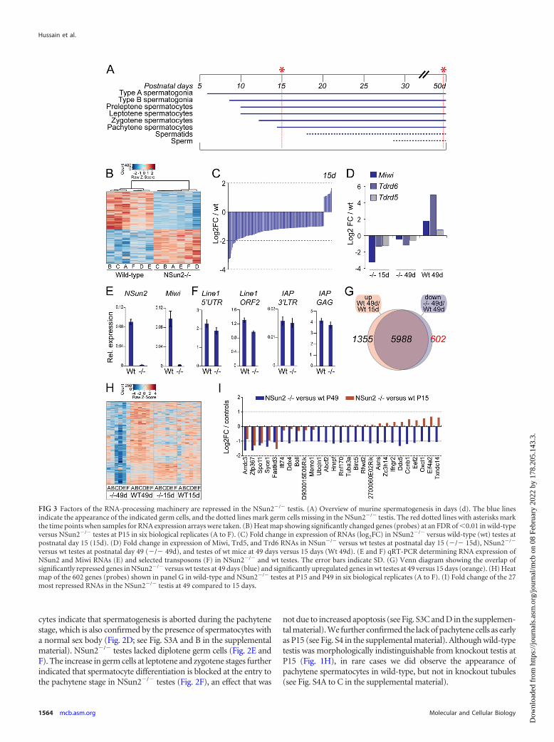
FIG 3 Factors of the RNA-processing machinery are repressed in the NSun2�/� testis. (A) Overview of murine spermatogenesis in days (d). The blue linesindicate the appearance of the indicated germ cells, and the dotted lines mark germ cells missing in the NSun2�/� testis. The red dotted lines with asterisks markthe time points when samples for RNA expression arrays were taken. (B) Heat map showing significantly changed genes (probes) at an FDR of �0.01 in wild-typeversus NSun2�/� testes at P15 in six biological replicates (A to F). (C) Fold change in expression of RNAs (log2FC) in NSun2�/� versus wild-type (wt) testes atpostnatal day 15 (15d). (D) Fold change in expression of Miwi, Trd5, and Trd6 RNAs in NSun�/� versus wt testes at postnatal day 15 (�/� 15d), NSun2�/�
versus wt testes at postnatal day 49 (�/� 49d), and testes of wt mice at 49 days versus 15 days (Wt 49d). (E and F) qRT-PCR determining RNA expression ofNSun2 and Miwi RNAs (E) and selected transposons (F) in NSun2�/� and wt testes. The error bars indicate SD. (G) Venn diagram showing the overlap ofsignificantly repressed genes in NSun2�/� versus wt testes at 49 days (blue) and significantly upregulated genes in wt testes at 49 versus 15 days (orange). (H) Heatmap of the 602 genes (probes) shown in panel G in wild-type and NSun2�/� testes at P15 and P49 in six biological replicates (A to F). (I) Fold change of the 27most repressed RNAs in the NSun2�/� testis at 49 compared to 15 days.
Hussain et al.
1564 mcb.asm.org Molecular and Cellular Biology
Dow
nloa
ded
from
http
s://j
ourn
als.
asm
.org
/jour
nal/m
cb o
n 08
Feb
ruar
y 20
22 b
y 17
8.20
5.14
3.3.

Since transcription is stalled before but highly increased duringthe pachytene stage (see Fig. S1 in the supplemental material), weasked whether lack of the posttranscriptional modification of cy-tosine-5 methylation in RNAs might hinder transcriptional pro-cesses at the pachytene stage.
Transcriptional and posttranscriptional pathways are re-pressed in the absence of NSun2. Murine spermatogenesis initi-ates a few days after birth and takes approximately 35 days(Fig. 3A). To determine gene transcription levels in testes thatlacked NSun2, we performed gene expression profiling in wild-type and NSun2�/� mice at postnatal days 15 and 49. Postnatalday 15 was chosen because pachytene is initiated at this age, and
we did not observe any morphological differences in wild-typeand knockout mouse models. At postnatal day 49, spermatogen-esis is completed, and NSun2�/� mice lack spermatids and sperm(Fig. 3A, dotted lines).
Despite the lack of morphological differences at P15, RNA mi-croarray analyses distinguished gene expression profiles accordingto genotype (see Fig. S5 in the supplemental material). A total of3,155 genes (probes) were found to be differentially expressed inwild-type versus NSun2�/� testes (FDR � 0.01) (Fig. 3B; see Ta-ble S1 in the supplemental material). To analyze whether inhibi-tion of transcriptional processes might hinder entry into thepachytene stage, we determined the gene ontology categories ofthe 1,347 repressed genes in NSun2�/� testes (Table 1). Genesencoding proteins involved in RNA processing, such as nucleotideand RNA-binding proteins, as well as ATP-dependent helicases,were significantly underrepresented in NSun2�/� testes at P15(Table 1; see Table S2 in the supplemental material).
To identify potential candidate genes causing the block in germ
TABLE 1 Gene ontology categorization of significantly (FDR � 0.01)underrepresented genes (probes) in NSun2�/� testes at P15 and P49
Term Count % P value
Underrepresented in NSun2�/� at 15 days(1,186 probes)
DAVID biological processes(GOTERM_BP_FAT)
Spermatogenesis 46 3.88 5.50E�12Cell cycle 78 6.57 1.44E�11Multicellular-organism reproduction 54 4.55 9.83E�09Protein localization 79 6.66 9.21E�08RNA processing 53 4.47 2.26E�07DNA metabolic process 51 4.30 4.10E�07
DAVID molecular processes(GOTERM_MF_FAT)
Nucleotide binding 214 18.03 1.00E�19Helicase activity 28 2.36 1.13E�09ATPase activity 43 3.62 2.19E�09RNA binding 74 6.23 9.35E�09ATP-dependent helicase activity 19 1.60 6.71E�07Serine/threonine kinase activity 46 3.88 1.12E�05tRNA binding 7 0.59 5.14E�04
KEGG database pathwaysAminoacyl-tRNA biosynthesis 8 0.67 0.002Cell cycle 13 1.10 0.007RNA degradation 8 0.67 0.012Ubiquitin-mediated proteolysis 12 1.01 0.026Nucleotide excision repair 6 0.51 0.032Spliceosome 11 0.93 0.034
Underrepresented in NSun2�/� at 49 days(602 probes)
DAVID biological processes(GOTERM_BP_FAT)
Protein transport 36 7.83 1.78E�05Chromosome organization 25 5.43 8.68E�05Translation 20 4.35 4.53E�04Cell cycle 30 6.52 7.60E�04
DAVID molecular processes(GOTERM_MF_FAT)
Nucleotide binding 92 20 1.80E�08RNA binding 34 7.39 6.25E�05GTP binding 20 4.35 8.30E�04Translation initiation factor activity 9 1.96 0.002ATP binding 50 10.87 0.006Nucleoside binding 52 11.30 0.011
KEGG database pathwaysBasal transcription factors 6 1.30 5.90E�04Pathways in cancer 12 2.61 0.081
FIG 4 Localization of NSun2 in adult testis. (A and B) Immunofluorescencelabeling of NSun2 (NSun2p; green) in wild-type (wt) (A) and NSun2�/�
(�/�) (B) testes. (C and D) Higher magnification of wt (C) and NSun2�/�
(D) testes showing that NSun2 protein is lost in round spermatids. (E and F)Colocalization of NSun2 (green) and Ddx25 (red) in wt (E) and NSun2�/� (F)testes. Nuclei were counterstained in blue (DAPI) (A to F). The arrows (A, C,and E) point to NSun2 staining in round spermatids.
NSun2 Is a Regulator of Male Germ Cell Differentiation
April 2013 Volume 33 Number 8 mcb.asm.org 1565
Dow
nloa
ded
from
http
s://j
ourn
als.
asm
.org
/jour
nal/m
cb o
n 08
Feb
ruar
y 20
22 b
y 17
8.20
5.14
3.3.

cell development, we asked which genes showed the highestchanges in expression in NSun2�/� testes. We found a total of 118genes more than 2-fold downregulated and very few (33) genes2-fold or more upregulated in NSun2�/� versus wild-type testis(Fig. 3C; see Table S3 in the supplemental material). One of themost repressed genes in NSun2�/� testes was Miwi (Fig. 3D andE). Miwi is specifically expressed in spermatocytes and sperma-tids, and lack of Miwi causes spermatogenic arrest at the roundspermatid stage (34). Interestingly, Miwi localizes to chromatoidbodies, which function in RNA storage and processing, and wefound two more chromatoid body components (Tdrd5 andTdrd6) downregulated in NSun2�/� testes (Fig. 3D). Miwi di-rectly interacts with Tdrd6, and also in Tdrd6�/� mice, the devel-opment from round to elongated spermatids is abrogated (35). Inwild-type mice, expression of Miwi, Tdrd5, and Tdrd6 were up-regulated during spermatogenesis (Fig. 3D, Wt 49d). Miwi hasbeen shown to regulate retrotransposon silencing in mouse testes(36). However, we did not observe increased expression of trans-posons, such as Line1 and IAP (Fig. 3F). Thus, male infertility inNSun2�/� mice is not due to deleterious transposon activation(36–39).
RNA microarray analyses of testes from adult mice (P49) re-vealed that 6,590 genes (probes) were significantly (FDR � 0.01)downregulated in NSun2�/� testes (see Table S4 in the supple-mental material). To exclude genes that were repressed because ofthe specific loss of spermatids and sperm in NSun2�/� testes
(Fig. 3A), we compared this gene list with transcripts that aresignificantly upregulated during normal spermatogenesis by com-paring the gene expression profiles of wild-type mice at P49 versusP15 (see Table S4 in the supplemental material). Only repressionof 602 genes (probes) in NSun2�/� testes at P49 was due to lack ofNSUN2 rather than loss of spermatids and sperm (Fig. 3G and H;see Table S5 in the supplemental material). Gene ontology cate-gorization using the 602 probes confirmed that genes encodingproteins involved in transcription and RNA processing were againoverrepresented (Table 1). When we plotted the top 27 genesshowing the highest fold change in expression in NSun2�/� testesat P49 but less than 1.5-fold change in expression in wild-typecontrols (P49 versus P15), we found three ATP-dependent RNAhelicases (Ddx4, Ddx5, and Eif4a2) within this group (Fig. 3I).
Together, the RNA expression assays demonstrated that maleinfertility of NSun2�/� mice may be caused by misregulation ofthe transcriptional process during the pachytene stage, as thegenes showing the highest fold change in repression were involvedin RNA processing.
Proteins of the RNA-processing machinery are reduced inNSun2�/� testes. We next asked whether NSun2 might be part ofthe RNA-processing machinery in testes and determined the lo-calization of the NSun2 protein during spermatogenesis (Fig. 4).The NSun2 protein was present in round spermatids localized tocytoplasmic granules close to the nuclei (Fig. 4A and C, arrows).The specificity of the NSun2 staining was confirmed using two
FIG 5 NSun2 localizes to the chromatoid body. (A and B) NSun2 (green) colocalizes with Ddx4 (red) in round spermatids using two different antibodies forNSun2 (Meth2) (A) and 20854-AP (NSun2p) (B). (C) Coimmunoprecipitation of NSun2 with Ddx4 (top) and Maelstrom (bottom) in wild-type and NSun2�/�
testes. Preimmune serum served as the negative control. (D and E) NSun2 (green) does not colocalize with Ddx4 (red) at the intermitochondrial cement inspermatocytes (D) or sp56 (red) at the acrosomal matrix in spermatids (E). (i to iv) Higher magnifications of the respective boxed areas in panels A, B, D, and E).(A, B, D, and E) Nuclei were counterstained in blue (DAPI).
Hussain et al.
1566 mcb.asm.org Molecular and Cellular Biology
Dow
nloa
ded
from
http
s://j
ourn
als.
asm
.org
/jour
nal/m
cb o
n 08
Feb
ruar
y 20
22 b
y 17
8.20
5.14
3.3.

different antibodies (NSun2p and NSun2) (see Materials andMethods) (Fig. 4A to D). Although NSun2 was also expressed innucleoli of Sertoli cells, neither the formation of the nucleoli northe number of Sertoli cells was affected when NSun2 was deleted(see Fig. S6A to C in the supplemental material). Both signals inthe nucleoli of Sertoli cells and in round spermatids were lost inNSun2�/� testes (Fig. 4B and D; see Fig. S6B in the supplementalmaterial).
We next asked whether the NSun2-positive granules werechromatoid bodies. We detected colocalization of NSun2 withDdx25 and Ddx4 only in wild-type testes (Fig. 4E and F, arrows,and 5A and B; see Fig. S7 in the supplemental material). Ddx4 andDdx25 are ATP-dependent RNA helicases that localize to thechromatoid body and are required for germ cell development (40–42). We further confirmed localization of NSun2 to chromatoidbodies using two different antibodies (NSun2p and NSun2) (seeMaterials and Methods) (Fig. 4E and F and 5A and B; see Fig. S7 inthe supplemental material) and by coimmunoprecipitation withDdx4 and Maelstrom (Fig. 5C) (33). The small amount of NSun2protein in the coimmunoprecipitations with Ddx4 and Mael-strom might be due to the fact that NSun2 colocalized with bothmarkers only in round spermatids, but not the intermitochondrialcement of spermatocytes, where Nsun2 was found in nucleolarstructures (Fig. 5D; see Fig. S8A in the supplemental material).NSun2 showed no overlap with the acrosomal protein sp56 inspermatids (Fig. 5E) or Ddx4 in prospermatogonia at embryonicday 16.5 (E16.5) (see Fig. S8B in the supplemental material).
Cytosine-5 tRNA methyltransferases are dispensable for thespermatogonium and early spermatocytes. Methylation of tRNAat cytosine-5 is catalyzed by Nsun2 and Dnmt2 (20, 43, 44). tRNAsare uniquely methylated by NSun2 and Dnmt2, since tRNAs iso-lated from testes lacking both enzymes are not methylated (21).Therefore, we considered that the unaffected development ofspermatogonia and early spermatocytes up to pachytene stage inNsun2�/� testes might be due to complementation of NSun2 de-ficiency by Dnmt2. Dnmt2�/� mice are viable and fertile and donot exhibit any gross phenotype (20).
To confirm that both RNA methyltransferases were coex-pressed in the same cell types, we measured RNA and proteinlevels of NSun2 and Dnmt2 during germ cell differentiation (Fig.6A and B). Until P15, NSun2 RNA was weakly expressed, but itwas 6-fold upregulated from P20, which coincides with the ap-pearance of spermatids (Fig. 3A and 6A, left). We observed a sim-ilar expression pattern for Dnmt2 RNA, although Dnmt2 in-creased slightly earlier during germ cell development (P15)(Fig. 6A, right). We observed upregulation of the NSun2 andDnmt2 proteins with similar kinetics (Fig. 6B).
The low abundances of both proteins from P1 to P15 indicatedthat they might not be functionally active in early stages of germcell differentiation. Although recent studies suggest that cyto-sine-5 methylation also occurs in mRNA (4), we were unable todetect any significant overlap between putative methylatedmRNAs and differentially abundant mRNAs in NSun2�/� testes(data not shown). The confirmed target substrates of NSun2 andDnmt2 are tRNAs, and loss of cytosine-5 methylation in tRNAsdecreases their overall stability (21). In line with these data, wefind that the abundances of the NSun2 and Dnmt2 target substratetRNAs AspGTC, GlyGCC, and LeuCAA decreased after deletion ofNsun2 (NSun2�/�) or NSun2 and Dnmt2 (DKO) both at P15 andin adult testes, whereas the negative-control tRNA IleTAT re-
mained unchanged (Fig. 6C). Thus, although only weakly ex-pressed at P15, deletion of NSun2 and Dnmt2 already decreasedtRNA stability early in germ cell differentiation, even before thepachytene stage.
Comparable to NSun2 protein localization in wild-type testes,we found Mili to be absent in the spermatogonium, but then bothlocalized to the cytoplasm of primary spermatocytes of double-knockout testes (Fig. 4A and 7A, wild type) (45). Whereas local-ization of Mili in Dnmt2�/� testes was comparable to that in thewild type (Fig. 7A, Dnmt2�/�), the number of Mili-positive cellswas dramatically reduced in the absence of NSun2 (Fig. 7A,NSun2�/�). Similarly, Miwi, which normally localizes to sper-matocytes and the chromatoid bodies of spermatids, was reducedonly upon deletion of NSun2 (Fig. 7B, NSun2�/�). We observedthe same reduction in Mili- and Miwi-positive cells in the secondindependent NSun2 knockout line (Nsun2Gt(D014D11)Wrst) (seeFig. S9 in the supplemental material). Double deletion of NSun2and Dnmt2 (DKO) resulted in a comparable loss of Mili- andMiwi-positive germ cells in NSun2�/� testes, whereas Dnmt2�/�
testes showed normal levels of both proteins (Fig. 7A and B).
FIG 6 Deletion of NSun2 and Dnmt2 reduced tRNA stability in testes at P15and 3 months of age. (A) RNA expression levels of NSun2 (left) and Dnmt2(right) at the indicated postnatal days. The error bars indicate SD. (B) Proteinexpression levels of NSun2 (top) and Dnmt2 (middle) at the indicated post-natal days (P) in wild-type and NSun2-Dnmt2 double knockouts (DKO).GAPDH served as a loading control (bottom). (C) Northern blot analyses ofNSun2- and Dnmt2-methylated tRNAs at P15 and 3 months of age. 5S RNAserved as a loading control.
NSun2 Is a Regulator of Male Germ Cell Differentiation
April 2013 Volume 33 Number 8 mcb.asm.org 1567
Dow
nloa
ded
from
http
s://j
ourn
als.
asm
.org
/jour
nal/m
cb o
n 08
Feb
ruar
y 20
22 b
y 17
8.20
5.14
3.3.

To confirm that spermatogonia were not affected by deletionof NSun2 and Dnmt2, we stained testis sections for Ki67, a markerfor dividing cells. The number of Ki67-positive spermatogonialcells remained unchanged in NSun2�/� testes (Fig. 7C). Althoughwe observed a slight reduction in proliferating Ki67-positive cellsin DKO testes (Fig. 7C; see Fig. S10A in the supplemental mate-rial), the number and distribution of Sertoli cells labeled by twodifferent antibodies to Gata4 and spermatogonial stem cellsmarked by Dmrt1 were unchanged in the absence of both NSun2and Dnmt2 proteins (Fig. 7D; see Fig. S10B in the supplementalmaterial) (46). Gata4 labeled a dot-like structure in wild-typeround spermatids, which were not representative of chromatoidbodies and are likely to be nonspecific (see Fig. S10C in the sup-plemental material).
In conclusion, our data suggested that in mouse testes, post-transcriptional cytosine-5 modifications are dispensable in sper-matogonial stem cells and early spermatocytes but are specificallyrequired for the meiotic progression from the leptotene/zygotenestages to pachytene.
DISCUSSION
Our finding that the RNA methyltransferase NSun2 is required forthe progression of the first prophase of male meiosis highlights theessential roles of posttranscriptional mechanisms during sper-matogenesis. Spermatids and sperm cannot be formed in the ab-sence of NSun2. Although the lack of NSun2 and Dnmt2 can cause
differentiation defects in somatic tissues, such as brain and skin,the complete lack of a specific differentiated lineage seems to beunique to the male testis (3, 21, 43).
In somatic cells, the NSun2 protein localizes to the nucleoli ininterphase of the cell cycle (47–49). The nucleolus is a distinctnuclear domain in which RNA processing and maturation, as wellas tRNA methylation, take place (50). Also in the testis, we findNSun2-positive nucleolar structures in Sertoli cells and primaryspermatocytes at interphase of the cell cycle (51). The deletion ofNSun2 in Sertoli cells may contribute to the impaired spermato-genesis in NSun2 knockout mice, because the correct proliferationand maturation of Sertoli cells are essential for germ cells to prog-ress through differentiation and meiosis (2). However, two obser-vations argue against a contribution by impaired Sertoli cells tothe observed NSun2�/� phenotype. First, the number and loca-tion of Sertoli cells were unaffected by deletion of NSun2. Second,in the absence of NSun2, Sertoli cells still showed the characteristicnucleolar tripartite structure typical of mature, adult mouse Ser-toli cells, indicating that the maturation of Sertoli cells was unaf-fected (52).
During cell divisions at the beginning of both mitosis and mei-osis, nucleolar structures disassemble, but the components arestored at various cellular locations throughout the cell cycle (48,51, 53, 54). Nucleolar reassembly starts at telophase in somaticcells and depends on the activation of RNA-processing complexes(55). During spermatogenesis, the nucleolus becomes fragmented
FIG 7 Cytosine-5 methylation is dispensable in spermatogonial and Sertoli cells. (A to D) Immunofluorescence labeling of Mili (green) (A), Miwi (green) (B),Ki67 (green) (C), and Gata4 (sc-1237) (red) and Dmrt1 (green) to mark Sertoli and spermatogonial stem cells, respectively (D), in wild-type testis or upondeletion of Dnmt2 (Dnmt2�/�), NSun2 (NSun2�/�), or both Dnmt2 and NSun2 (DKO). DAPI served as a nuclear counterstain.
Hussain et al.
1568 mcb.asm.org Molecular and Cellular Biology
Dow
nloa
ded
from
http
s://j
ourn
als.
asm
.org
/jour
nal/m
cb o
n 08
Feb
ruar
y 20
22 b
y 17
8.20
5.14
3.3.

at the zygotene-pachytene stage and migrates to the cytoplasm,where it forms a structure that has been suggested to be the originof the chromatoid body (51, 56, 57).
Lack of NSun2 causes a block of progression of the first pro-phase of male meiosis at the zygotene-pachytene stage before thechromatoid bodies first appear in the cytoplasm at the latepachytene stage (18), indicating that NSun2’s functions are essen-tial for meiotic prophase progression before the chromatoid bodyis formed. Since NSun2�/� testis lacks germ cells containing achromatoid body, it is difficult to determine whether NSun2 isalso required for chromatoid body assembly, or even RNA pro-cessing at the chromatoid body. However, RNA expression pro-filing revealed that genes encoding proteins involved in transcrip-tional and posttranscriptional processes are already reduced atP15 before any chromatoid body is formed, indicating that maleinfertility in NSun2�/� mice is not simply due to the lack of chro-matoid bodies. However, we did find downregulation of a numberof mRNAs encoding proteins linked to functions of the chroma-toid body. Dissecting whether deletion of NSun2 is directly linkedto decay of those specific mRNAs or indirectly prevents the for-mation of functional chromatoid bodies is hampered by the factthat these processes are intertwined (35).
NSun2 generally localizes to cellular RNA-processing centers,and while NSun2 is the first RNA methyltransferase identified as acomponent of the chromatoid body, Drosophila NSun2 is also acomponent of ribonuclear particles involved in RNA silencingand germ cell development (58). How NSun2 mechanisticallyblocks the progression of the first prophase of male meiosis beforethe pachytene stage remains to be determined but may, at least inpart, be dependent on its tRNA methyltransferase activity.
NSun2 is the orthologue of Saccharomyces cerevisiae Trm4tRNA methylase (47, 59, 60). NSun2 catalyzes the formation ofcytosine-5 methylation (m5C) in several tRNAs in vivo in tissues,including skin, liver, and testis (3, 21). Functionally, the m5C post-transcriptional modification influences translation rates, as well ascorrect RNA folding and stability (21, 61–64). In the absence ofNSun2, tRNAs lack specific m5C modifications, which can causereduced protein translation rates (3, 21). Thus, the methylation oftRNAs by NSun2 at ribonuclear particles might allow translationof stalled mRNAs. In this scenario, global transcription should beunaffected, and reduced expression of specific mRNAs might re-flect a delay in germ cell development before the first meioticprophase is blocked.
ACKNOWLEDGMENTS
We are most grateful to everybody who provided us with reagents. Inparticular, we thank Shinseog Kim, Noora Kotaja, and Duncan Odom foradvice and helpful comments on the manuscript and Tanja Musch fortechnical assistance. We also thank the CRI Genomics and BioinformaticsCore Facilities. We gratefully acknowledge the support of the CambridgeStem Cell Initiative and Stephen Evans-Freke.
This work was funded by the Deutsche Forschungsgemeinschaft(FOR1082), CR-UK, and the Medical Research Council. F.T. is supportedby the Institute of Genetics and Biophysics A. Buzzati-Traverso, CNR,Italy.
REFERENCES1. de Rooij DG. 2001. Proliferation and differentiation of spermatogonial
stem cells. Reproduction 121:347–354.2. Sharpe RM, McKinnell C, Kivlin C, Fisher JS. 2003. Proliferation and
functional maturation of Sertoli cells, and their relevance to disorders oftestis function in adulthood. Reproduction 125:769 –784.
3. Blanco S, Kurowski A, Nichols J, Watt FM, Benitah SA, Frye M.2011. The RNA-methyltransferase Misu (NSun2) poises epidermalstem cells to differentiate. PLoS Genet. 7:e1002403. doi:10.1371/journal.pgen.1002403.
4. Squires JE, Patel HR, Nousch M, Sibbritt T, Humphreys DT, Parker BJ,Suter CM, Preiss T. 2012. Widespread occurrence of 5-methylcytosine inhuman coding and non-coding RNA. Nucleic Acids Res. 40:5023–5033.
5. Braun RE. 1998. Post-transcriptional control of gene expression duringspermatogenesis. Semin. Cell Dev. Biol. 9:483– 489.
6. Paronetto MP, Messina V, Barchi M, Geremia R, Richard S, Sette C.2011. Sam68 marks the transcriptionally active stages of spermatogenesisand modulates alternative splicing in male germ cells. Nucleic Acids Res.39:4961– 4974.
7. Turner JM, Mahadevaiah SK, Fernandez-Capetillo O, Nussenzweig A,Xu X, Deng CX, Burgoyne PS. 2005. Silencing of unsynapsed meioticchromosomes in the mouse. Nat. Genet. 37:41– 47.
8. Geremia R, Boitani C, Conti M, Monesi V. 1977. RNA synthesis inspermatocytes and spermatids and preservation of meiotic RNA duringspermiogenesis in the mouse. Cell Differ. 5:343–355.
9. Monesi V, Geremia R, D’Agostino A, Boitani C. 1978. Biochemistry ofmale germ cell differentiation in mammals: RNA synthesis in meiotic andpostmeiotic cells. Curr. Top. Dev. Biol. 12:11–36.
10. Paronetto MP, Sette C. 2010. Role of RNA-binding proteins in mamma-lian spermatogenesis. Int. J. Androl. 33:2–12.
11. Monesi V. 1964. Ribonucleic acid synthesis during mitosis and meiosis inthe mouse testis. J. Cell Biol. 22:521–532.
12. Yang J, Medvedev S, Reddi PP, Schultz RM, Hecht NB. 2005. TheDNA/RNA-binding protein MSY2 marks specific transcripts for cytoplas-mic storage in mouse male germ cells. Proc. Natl. Acad. Sci. U. S. A.102:1513–1518.
13. Zhong J, Peters AH, Lee K, Braun RE. 1999. A double-stranded RNA-binding protein required for activation of repressed messages in mamma-lian germ cells. Nat. Genet. 22:171–174.
14. Kleene KC. 2001. A possible meiotic function of the peculiar patterns ofgene expression in mammalian spermatogenic cells. Mech. Dev. 106:3–23.
15. Venables JP, Cooke HJ. 2000. Lessons from knockout and transgenicmice for infertility in men. J. Endocrinol. Invest. 23:584 –591.
16. Venables JP, Eperon I. 1999. The roles of RNA-binding proteins inspermatogenesis and male infertility. Curr. Opin. Genet. Dev. 9:346 –354.
17. Kotaja N, Lin H, Parvinen M, Sassone-Corsi P. 2006. Interplay ofPIWI/Argonaute protein MIWI and kinesin KIF17b in chromatoid bodiesof male germ cells. J. Cell Sci. 119:2819 –2825.
18. Fawcett DW, Eddy EM, Phillips DM. 1970. Observations on the finestructure and relationships of the chromatoid body in mammalian sper-matogenesis. Biol. Reprod. 2:129 –153.
19. Kotaja N, Sassone-Corsi P. 2007. The chromatoid body: a germ-cell-specific RNA-processing centre. Nat. Rev. Mol. Cell Biol. 8:85–90.
20. Goll MG, Kirpekar F, Maggert KA, Yoder JA, Hsieh CL, Zhang X, GolicKG, Jacobsen SE, Bestor TH. 2006. Methylation of tRNAAsp by the DNAmethyltransferase homolog Dnmt2. Science 311:395–398.
21. Tuorto F, Liebers R, Musch T, Schaefer M, Hofmann S, Kellner S, FryeM, Helm M, Stoecklin G, Lyko F. 2012. RNA cytosine methylation byDnmt2 and NSun2 promotes tRNA stability and protein synthesis. Nat.Struct. Mol. Biol. 19:900 –905.
22. Peters AH, Plug AW, van Vugt MJ, de Boer P. 1997. A drying-downtechnique for the spreading of mammalian meiocytes from the male andfemale germline. Chromosome Res. 5:66 – 68.
23. Carmell MA, Girard A, van de Kant HJ, Bourc’his D, Bestor TH, deRooij DG, Hannon GJ. 2007. MIWI2 is essential for spermatogenesis andrepression of transposons in the mouse male germline. DevelopmentalCell 12:503–514.
24. Nascimento EM, Cox CL, MacArthur S, Hussain S, Trotter M, BlancoS, Suraj M, Nichols J, Kubler B, Benitah SA, Hendrich B, Odom DT,Frye M. 2011. The opposing transcriptional functions of Sin3a and c-Mycare required to maintain tissue homeostasis. Nat. Cell Biol. 13:1395–1405.
25. Gentleman RC, Carey VJ, Bates DM, Bolstad B, Dettling M, Dudoit S,Ellis B, Gautier L, Ge Y, Gentry J, Hornik K, Hothorn T, Huber W,Iacus S, Irizarry R, Leisch F, Li C, Maechler M, Rossini AJ, Sawitzki G,Smith C, Smyth G, Tierney L, Yang JY, Zhang J. 2004. Bioconductor:open software development for computational biology and bioinformat-ics. Genome Biol. 5:R80. doi:10.1186/gb-2004-5-10-r80.
26. Cairns JM, Dunning MJ, Ritchie ME, Russell R, Lynch AG. 2008. BASH:
NSun2 Is a Regulator of Male Germ Cell Differentiation
April 2013 Volume 33 Number 8 mcb.asm.org 1569
Dow
nloa
ded
from
http
s://j
ourn
als.
asm
.org
/jour
nal/m
cb o
n 08
Feb
ruar
y 20
22 b
y 17
8.20
5.14
3.3.
a tool for managing BeadArray spatial artefacts. Bioinformatics 24:2921–2922.
27. Dunning MJ, Smith ML, Ritchie ME, Tavare S. 2007. Beadarray: Rclasses and methods for Illumina bead-based data. Bioinformatics 23:2183–2184.
28. Smyth G. 2005. Limma: linear models for microarray data, p 397– 420. InGentleman VCR, Dudoit S, Huber W, Irizarry R (ed), Bioinformatics andcomputational biology solutions using R and Bioconductor. Springer,New York, NY.
29. Edgar R, Domrachev M, Lash AE. 2002. Gene Expression Omnibus:NCBI gene expression and hybridization array data repository. NucleicAcids Res. 30:207–210.
30. Marret C, Avallet O, Perrard-Sapori MH, Durand P. 1998. Localizationand quantitative expression of mRNAs encoding the testis-specific histoneTH2B, the phosphoprotein p19, the transition proteins 1 and 2 duringpubertal development and throughout the spermatogenic cycle of the rat.Mol. Reprod. Dev. 51:22–35.
31. Wykes SM, Nelson JE, Visscher DW, Djakiew D, Krawetz SA. 1995.Coordinate expression of the PRM1, PRM2, and TNP2 multigene locus inhuman testis. DNA Cell Biol. 14:155–161.
32. Turner JM, Burgoyne PS, Singh PB. 2001. M31 and macroH2A1.2colocalise at the pseudoautosomal region during mouse meiosis. J. CellSci. 114:3367–3375.
33. Costa Y, Speed RM, Gautier P, Semple CA, Maratou K, Turner JM,Cooke HJ. 2006. Mouse MAELSTROM: the link between meiotic silenc-ing of unsynapsed chromatin and microRNA pathway? Hum. Mol. Genet.15:2324 –2334.
34. Deng W, Lin H. 2002. Miwi, a murine homolog of piwi, encodes acytoplasmic protein essential for spermatogenesis. Developmental Cell2:819 – 830.
35. Vasileva A, Tiedau D, Firooznia A, Muller-Reichert T, Jessberger R.2009. Tdrd6 is required for spermiogenesis, chromatoid body architec-ture, and regulation of miRNA expression. Curr. Biol. 19:630 – 639.
36. Reuter M, Berninger P, Chuma S, Shah H, Hosokawa M, Funaya C,Antony C, Sachidanandam R, Pillai RS. 2011. Miwi catalysis is requiredfor piRNA amplification-independent LINE1 transposon silencing. Na-ture 480:264 –267.
37. Aravin AA, Sachidanandam R, Girard A, Fejes-Toth K, Hannon GJ.2007. Developmentally regulated piRNA clusters implicate MILI in trans-poson control. Science 316:744 –747.
38. De Fazio S, Bartonicek N, Di Giacomo M, Abreu-Goodger C, Sankar A,Funaya C, Antony C, Moreira PN, Enright AJ, O’Carroll D. 2011. Theendonuclease activity of Mili fuels piRNA amplification that silencesLINE1 elements. Nature 480:259 –263.
39. Kuramochi-Miyagawa S, Watanabe T, Gotoh K, Totoki Y, Toyoda A,Ikawa M, Asada N, Kojima K, Yamaguchi Y, Ijiri TW, Hata K, Li E,Matsuda Y, Kimura T, Okabe M, Sakaki Y, Sasaki H, Nakano T. 2008.DNA methylation of retrotransposon genes is regulated by Piwi familymembers MILI and MIWI2 in murine fetal testes. Genes Dev. 22:908 –917.
40. Kotaja N, Bhattacharyya SN, Jaskiewicz L, Kimmins S, Parvinen M,Filipowicz W, Sassone-Corsi P. 2006. The chromatoid body of male germcells: similarity with processing bodies and presence of Dicer and mi-croRNA pathway components. Proc. Natl. Acad. Sci. U. S. A. 103:2647–2652.
41. Tanaka SS, Toyooka Y, Akasu R, Katoh-Fukui Y, Nakahara Y, Suzuki R,Yokoyama M, Noce T. 2000. The mouse homolog of Drosophila Vasa isrequired for the development of male germ cells. Genes Dev. 14:841– 853.
42. Tsai-Morris CH, Sheng Y, Lee E, Lei KJ, Dufau ML. 2004. Gonadotro-pin-regulated testicular RNA helicase (GRTH/Ddx25) is essential for sper-matid development and completion of spermatogenesis. Proc. Natl. Acad.Sci. U. S. A. 101:6373– 6378.
43. Rai K, Chidester S, Zavala CV, Manos EJ, James SR, Karpf AR, JonesDA, Cairns BR. 2007. Dnmt2 functions in the cytoplasm to promote liver,brain, and retina development in zebrafish. Genes Dev. 21:261–266.
44. Schaefer M, Pollex T, Hanna K, Tuorto F, Meusburger M, Helm M,
Lyko F. 2010. RNA methylation by Dnmt2 protects transfer RNAs againststress-induced cleavage. Genes Dev. 24:1590 –1595.
45. Kuramochi-Miyagawa S, Kimura T, Ijiri TW, Isobe T, Asada N, FujitaY, Ikawa M, Iwai N, Okabe M, Deng W, Lin H, Matsuda Y, Nakano T.2004. Mili, a mammalian member of piwi family gene, is essential forspermatogenesis. Development 131:839 – 849.
46. Matson CK, Murphy MW, Griswold MD, Yoshida S, Bardwell VJ,Zarkower D. 2010. The mammalian doublesex homolog DMRT1 is atranscriptional gatekeeper that controls the mitosis versus meiosis deci-sion in male germ cells. Developmental Cell 19:612– 624.
47. Frye M, Watt FM. 2006. The RNA methyltransferase Misu (NSun2)mediates Myc-induced proliferation and is upregulated in tumors. Curr.Biol. 16:971–981.
48. Hussain S, Benavente SB, Nascimento E, Dragoni I, Kurowski A,Gillich A, Humphreys P, Frye M. 2009. The nucleolar RNA methyl-transferase Misu (NSun2) is required for mitotic spindle stability. J.Cell Biol. 186:27– 40.
49. Khan MA, Rafiq MA, Noor A, Hussain S, Flores JV, Rupp V, VincentAK, Malli R, Ali G, Khan FS, Ishak GE, Doherty D, Weksberg R, AyubM, Windpassinger C, Ibrahim S, Frye M, Ansar M, Vincent JB. 2012.Mutation in NSUN2, which encodes an RNA methyltransferase, causesautosomal-recessive intellectual disability. Am. J. Hum. Genet. 90:856 –863.
50. Colonna A, Kerr SJ. 1980. The nucleus as the site of tRNA methylation. J.Cell. Physiol. 103:29 –33.
51. Paniagua R, Nistal M, Amat P, Rodriguez MC. 1986. Ultrastructuralobservations on nucleoli and related structures during human spermato-genesis. Anat Embryol. 174:301–306.
52. Myers M, Ebling FJ, Nwagwu M, Boulton R, Wadhwa K, Stewart J, KerrJB. 2005. Atypical development of Sertoli cells and impairment of sper-matogenesis in the hypogonadal (hpg) mouse. J. Anat. 207:797– 811.
53. Hernandez-Verdun D. 2011. Assembly and disassembly of the nucleolusduring the cell cycle. Nucleus 2:189 –194.
54. Takeuchi IK, Takeuchi YK. 1990. Ethanol-phosphotungstic acid andbismuth staining of spermatid nucleoli in mouse spermiogenesis. J. Struct.Biol. 103:104 –112.
55. Hernandez-Verdun D, Roussel P, Gebrane-Younes J. 2002. Emergingconcepts of nucleolar assembly. J. Cell Sci. 115:2265–2270.
56. Andonov MD, Chaldakov GN. 1989. Morphological evidence for cal-cium storage in the chromatoid body of rat spermatids. Experientia 45:377–378.
57. Comings DE, Okada TA. 1972. The chromatoid body in mouse sper-matogenesis: evidence that it may be formed by the extrusion of nucleolarcomponents. J. Ultrastruct. Res. 39:15–23.
58. Gerbasi VR, Preall JB, Golden DE, Powell DW, Cummins TD,Sontheimer EJ. 2011. Blanks, a nuclear siRNA/dsRNA-binding complexcomponent, is required for Drosophila spermiogenesis. Proc. Natl. Acad.Sci. U. S. A. 108:3204 –3209.
59. Brzezicha B, Schmidt M, Makalowska I, Jarmolowski A, Pienkowska J,Szweykowska-Kulinska Z. 2006. Identification of human tRNA:m5Cmethyltransferase catalysing intron-dependent m5C formation in the firstposition of the anticodon of the pre-tRNA Leu (CAA). Nucleic Acids Res.34:6034 – 6043.
60. Motorin Y, Grosjean H. 1999. Multisite-specific tRNA:m5C-methyltransferase (Trm4) in yeast Saccharomyces cerevisiae: identifica-tion of the gene and substrate specificity of the enzyme. RNA 5:1105–1118.
61. Agris PF. 2004. Decoding the genome: a modified view. Nucleic Acids Res.32:223–238.
62. Grosjean B, Ha R. 2005. Modification and editing of RNA. Springer,Berlin, Germany.
63. Motorin Y, Helm M. 2010. tRNA stabilization by modified nucleotides.Biochemistry 49:4934 – 4944.
64. Motorin Y, Lyko F, Helm M. 2010. 5-Methylcytosine in RNA: detection,enzymatic formation and biological functions. Nucleic Acids Res. 38:1415–1430.
Hussain et al.
1570 mcb.asm.org Molecular and Cellular Biology
Dow
nloa
ded
from
http
s://j
ourn
als.
asm
.org
/jour
nal/m
cb o
n 08
Feb
ruar
y 20
22 b
y 17
8.20
5.14
3.3.