The mid‑sternal length, a practical anatomical landmark for...
4
Journal of Research in Medical Sciences | May 2013 | 383 The mid‑sternal length, a practical anatomical landmark for optimal positioning of long‑term central venous catheters Fereshte Salimi, Mohammad Reza Imani, Navab Ghasemi 1 , Amir Keshavarzian 2 , Amir Hosein Davarpanah Jazi 3 Departments of Vascular Surgery, 1 Cardiology, 2 Specialist Recourses, 3 Medical Education Research Center, Isfahan University of Medical Sciences, Isfahan, Iran Background: Long‑term tunneled catheters are used for the hemodialysis or chemotherapy in many patients. Proper placement of the catheter tip could reduce early and late catheter related complications. Aim of the present study was to evaluate a new formula for proper placement of tunneled hemodialysis or infusion port device by using an external anatomic landmark. Materials and Methods: A total of 64 adult patients undergoing elective placement of tunneled Central Venous Catheter (CVC) requiring hemodialysis or chemotherapy were enrolled in this prospective study during 2011‑2012 in the university hospital. e catheter length to be inserted in the right internal jugular vein (IJV) was calculated by adding two measurements (the shortest straight length between the insertion point of the needle and the suprasternal notch plus and half of sternal length). e catheter position was considered correct if the tip was positioned in the right atrium (RA) or Superior vena cava (SVC)‑RA junction. Results: e patients were 55.28 ± 19.85 years of age, weighed 5.78 ± 16.62 kg and were 166.07 ± 10.27 cm tall. Catheters were inserted successfully in 88% of patients (n = 56). Catheter tip positions in the failures were SVC (n = 5), tricuspid valve (n = 2), and right ventricle (n = 1) in our patients. Conclusion: Long‑term hemodialysis or port CVC could easily insert in the right IJV by using half of the sternal length as an external land marks among adult patients. Key words: External landmark, hemodialysis, port device Address for correspondence: Dr. M. R. Imani, Department of Vascular Surgery, Al Zahra Hospital, Soffeh St. Isfahan, Iran. E‑mail: [email protected] Received: 27‑08‑2012; Revised: 13‑01‑2013; Accepted: 11‑03‑2013 radiographic landmarks for correct positioning have been proposed. [9] Carina, as a reliable radiographic marker, for the safe placement of hemodialysis catheter was suggested by some authors. [3,9] Using this marker for placement of CVC required pre‑procedural Chest X‑ray (CXR) and additional radiation and costs. Aim of the present study was to evaluate a new formula for proper placement of tunneled hemodialysis or port device by using an external anatomic landmark. Although, chest radiography has been used to determine proper position of the catheter tip routinely; however, the SVC‑RA junction and the RA is not easily identifiable by simple observation of the chest roentgenogram. Therefore in order to precise determination of the catheter tip position we used transthoracic echocardiography to outline exact localization of CVC. Aim of the present study was to evaluate a new formula for proper placement of tunneled hemodialysis or port device by using external anatomic landmarks. MATERIALS AND METHODS This study was performed over a 20 weeks period during 2011‑2012 in Al Zahra hospital, Isfahan, Iran, with approval from the Ethics Commiee of Isfahan INTRODUCTION Generally internal jugular veins (IJV) are preferred the preferred site for inserting tunneled central venous catheter (CVC). To minimize the risk of early and late catheter related complications the goal is to place the tip of catheter at the junction of the superior vena cava (SVC) and the right atrium (RA) or within atrium. [1] Several methods such as landmarks, [2,3] simple formulas, [4,5] and advanced techniques such as intracardiac electrocardiography, live fluoroscopy, [6,7] and trans‑esophageal echocardiography (TEE) [8] are suggested for placement of CVC to an adequate depth in adults. Sophisticated technologies such as intracardiac Electrocardiogram (ECG) and TEE are not often available in an operating room and considered time consuming and expensive methods for exact positioning of tunneled CVCs. Fluoroscopy is preferred by some surgeons and interventionists, but this method puts patients in a small amount of X‑ray exposure. In addition, fluoroscopy may not available in many operating rooms. However, surface anatomic landmarks are easily identifiable, practical in all patients, and rapidly available for patients candidate for long term dialysis venous access regardless of the gender, age, and operator’s experience. Various ORIGINAL ARTICLE
Transcript of The mid‑sternal length, a practical anatomical landmark for...
Journal of Research in Medical Sciences | May 2013 |383
The mid‑sternal length, a practical anatomical landmark for optimal positioning of long‑term central venous catheters
Fereshte Salimi, Mohammad Reza Imani, Navab Ghasemi1, Amir Keshavarzian2, Amir Hosein Davarpanah Jazi3
Departments of Vascular Surgery, 1Cardiology, 2Specialist Recourses, 3Medical Education Research Center, Isfahan University of Medical Sciences, Isfahan, Iran
Background: Long‑term tunneled catheters are used for the hemodialysis or chemotherapy in many patients. Proper placement of the catheter tip could reduce early and late catheter related complications. Aim of the present study was to evaluate a new formula for proper placement of tunneled hemodialysis or infusion port device by using an external anatomic landmark. Materials and Methods: A total of 64 adult patients undergoing elective placement of tunneled Central Venous Catheter (CVC) requiring hemodialysis or chemotherapy were enrolled in this prospective study during 2011‑2012 in the university hospital. The catheter length to be inserted in the right internal jugular vein (IJV) was calculated by adding two measurements (the shortest straight length between the insertion point of the needle and the suprasternal notch plus and half of sternal length). The catheter position was considered correct if the tip was positioned in the right atrium (RA) or Superior vena cava (SVC)‑RA junction. Results: The patients were 55.28 ± 19.85 years of age, weighed 5.78 ± 16.62 kg and were 166.07 ± 10.27 cm tall. Catheters were inserted successfully in 88% of patients (n = 56). Catheter tip positions in the failures were SVC (n = 5), tricuspid valve (n = 2), and right ventricle (n = 1) in our patients. Conclusion: Long‑term hemodialysis or port CVC could easily insert in the right IJV by using half of the sternal length as an external land marks among adult patients.
Key words: External landmark, hemodialysis, port device
Address for correspondence: Dr. M. R. Imani, Department of Vascular Surgery, Al Zahra Hospital, Soffeh St. Isfahan, Iran. E‑mail: [email protected]: 27‑08‑2012; Revised: 13‑01‑2013; Accepted: 11‑03‑2013
radiographic landmarks for correct positioning have been proposed.[9] Carina, as a reliable radiographic marker, for the safe placement of hemodialysis catheter was suggested by some authors.[3,9] Using this marker for placement of CVC required pre‑procedural Chest X‑ray (CXR) and additional radiation and costs. Aim of the present study was to evaluate a new formula for proper placement of tunneled hemodialysis or port device by using an external anatomic landmark.
Although, chest radiography has been used to determine proper position of the catheter tip routinely; however, the SVC‑RA junction and the RA is not easily identifiable by simple observation of the chest roentgenogram. Therefore in order to precise determination of the catheter tip position we used transthoracic echocardiography to outline exact localization of CVC. Aim of the present study was to evaluate a new formula for proper placement of tunneled hemodialysis or port device by using external anatomic landmarks.
MATERIALS AND METHODS
This study was performed over a 20 weeks period during 2011‑2012 in Al Zahra hospital, Isfahan, Iran, with approval from the Ethics Committee of Isfahan
INTRODUCTION
Generally internal jugular veins (IJV) are preferred the preferred site for inserting tunneled central venous catheter (CVC). To minimize the risk of early and late catheter related complications the goal is to place the tip of catheter at the junction of the superior vena cava (SVC) and the right atrium (RA) or within atrium.[1] Several methods such as landmarks,[2,3] simple formulas,[4,5] and advanced techniques such as intracardiac electrocardiography, live fluoroscopy,[6,7] and trans‑esophageal echocardiography (TEE)[8] are suggested for placement of CVC to an adequate depth in adults. Sophisticated technologies such as intracardiac Electrocardiogram (ECG) and TEE are not often available in an operating room and considered time consuming and expensive methods for exact positioning of tunneled CVCs. Fluoroscopy is preferred by some surgeons and interventionists, but this method puts patients in a small amount of X‑ray exposure. In addition, fluoroscopy may not available in many operating rooms. However, surface anatomic landmarks are easily identifiable, practical in all patients, and rapidly available for patients candidate for long term dialysis venous access regardless of the gender, age, and operator’s experience. Various
Or
igin
al a
rt
icl
e
Salimi, et al.: A new method for placement of long‑term central venous catheter
Journal of Research in Medical Sciences| May 2013 | 384
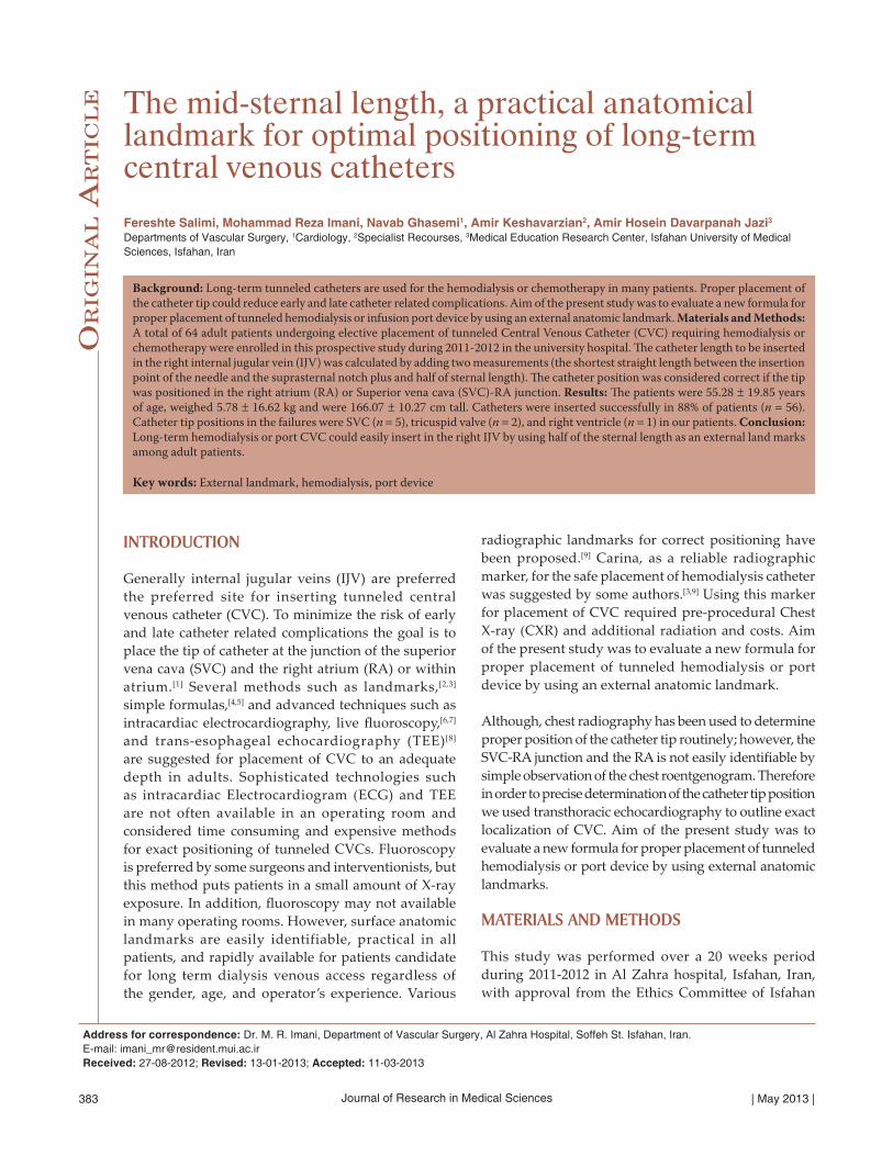
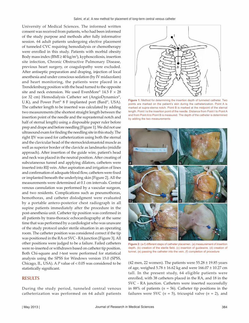
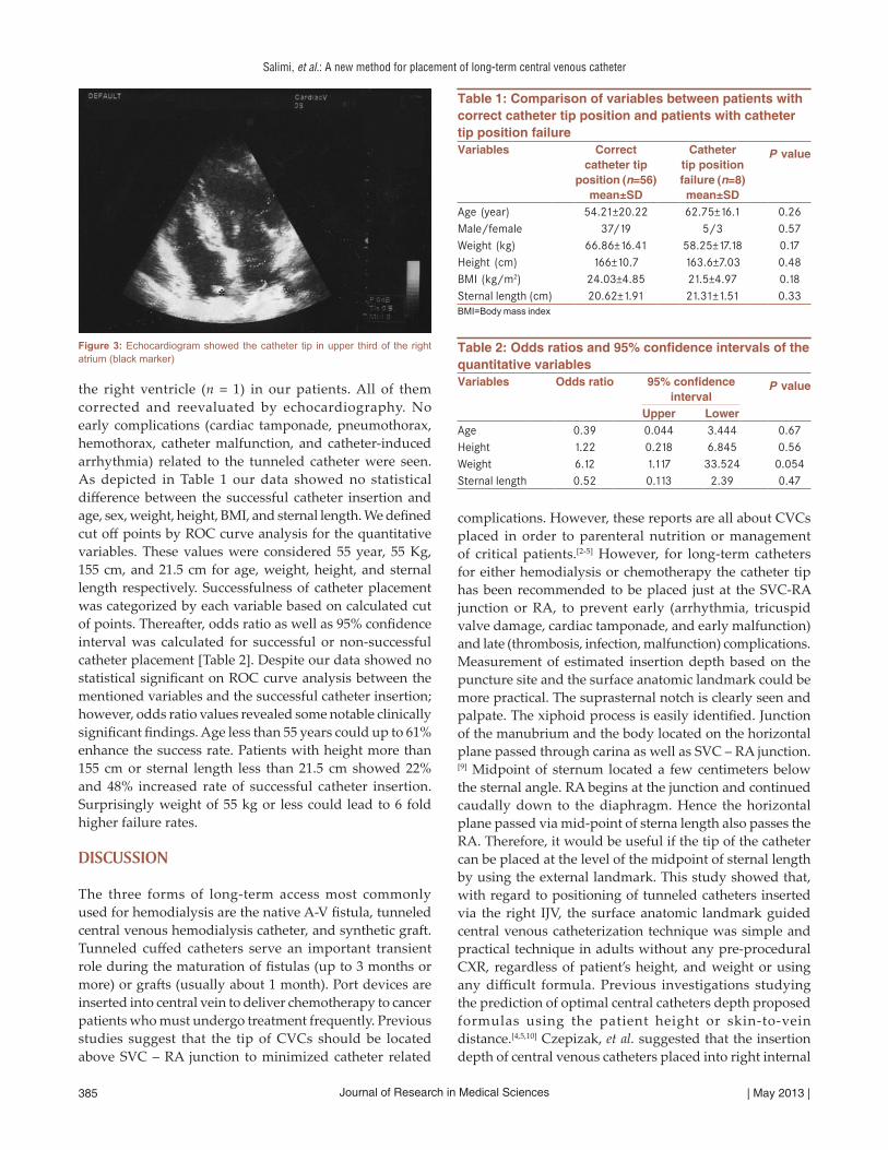
University of Medical Sciences. The informed written consent was received from patients, who had been informed of the study purpose and methods after fully informative session. 64 adult patients undergoing elective placement of tunneled CVC requiring hemodialysis or chemotherapy were enrolled in this study. Patients with morbid obesity Body mass index (BMI ≥ 40 kg/m2), kyphoscoliosis, insertion site infection, Chronic Obstructive Pulmonary Disease, previous heart surgery, or coagulopathy were excluded. After antiseptic preparation and draping, injection of local anesthesia and under conscious sedation (by IV midazolam) and heart monitoring, the patients were placed in a Trendelenburg position with the head turned to the opposite site and neck extension. We used EvenMore® 14.5 F × 28 (or 32 cm) Hemodialysis Catheter set (AngioDynamics®, U.K), and Power Port® 8 F implanted port (Bard®, USA). The catheter length to be inserted was calculated by adding two measurements (the shortest straight length between the insertion point of the needle and the suprasternal notch and half of sternal length) using a disposable paper ruler before prep and drape and before needling [Figure 1]. We did not use ultrasound exam for finding the needling site in this study. The right IJV was used for catheterization using both the sternal and the clavicular head of the sternocleidomastoid muscle as well as superior border of the clavicle as landmarks (middle approach). After insertion of the guide wire, patient’s head and neck was placed in the neutral position. After creating of subcutaneous tunnel and applying dilators, catheters were inserted into RIJ vein. After aspiration and irrigation of lines and confirmation of adequate blood flow, catheters were fixed or implanted beneath the underlying skin [Figure 2]. All the measurements were determined at 0.1 cm intervals. Central venous cannulation was performed by a vascular surgeon, and two residents. Complications such as pneumothorax, hemothorax, and catheter dislodgment were evaluated by a portable antero‑posterior chest radiograph in all supine patients immediately after the procedure in the post‑anesthesia unit. Catheter tip position was confirmed in all patients by trans‑thoracic echocardiography at the same time that was performed by a cardiologist who was unaware of the study protocol under sterile situation in an operating room. The catheter position was considered correct if the tip was positioned in the RA or SVC ‑ RA junction [Figure 3]. All other positions were judged to be a failure. Failed catheters were re‑inserted or withdrawn based on catheter tip position. Both Chi‑square and t‑test were performed for statistical analysis using the SPSS for Windows version 15.0 (SPSS, Chicago, IL, USA). A P value of < 0.05 was considered to be statistically significant.
RESULTS
During the study period, tunneled central venous catheterization was performed on 64 adult patients
(42 men, 22 women). The patients were 55.28 ± 19.85 years of age, weighed 5.78 ± 16.62 kg and were 166.07 ± 10.27 cm tall. In the present study, 64 eligible patients were enrolled, with 38 catheters placed in the RA, and 18 in the SVC ‑ RA junction. Catheters were inserted successfully in 88% of patients (n = 56). Catheter tip positions in the failures were SVC (n = 5), tricuspid valve (n = 2), and
Figure 1: Method for determining the insertion depth of tunneled catheter. Two points are marked on the patient’s skin during the catheterization. Point A is marked at supra‑sterna notch. Point B is marked at the midpoint of the sternal length. Point I is the insertion point of the needle. Distance from Point I to Point A and from Point A to Point B is measured. The depth of the catheter is determined by adding the two measurements
Figure 2: (a‑f) different steps of catheter placemen. (a) measurement of insertion depth, (b) creation of the sterile field, (c) insertion of guidewire, (d) creation of tunnel, (e) passing the catheter into the vein, (f) completion of procedure
dc
b
f
a
e
Salimi, et al.: A new method for placement of long‑term central venous catheter
Journal of Research in Medical Sciences | May 2013 |385
the right ventricle (n = 1) in our patients. All of them corrected and reevaluated by echocardiography. No early complications (cardiac tamponade, pneumothorax, hemothorax, catheter malfunction, and catheter‑induced arrhythmia) related to the tunneled catheter were seen. As depicted in Table 1 our data showed no statistical difference between the successful catheter insertion and age, sex, weight, height, BMI, and sternal length. We defined cut off points by ROC curve analysis for the quantitative variables. These values were considered 55 year, 55 Kg, 155 cm, and 21.5 cm for age, weight, height, and sternal length respectively. Successfulness of catheter placement was categorized by each variable based on calculated cut of points. Thereafter, odds ratio as well as 95% confidence interval was calculated for successful or non‑successful catheter placement [Table 2]. Despite our data showed no statistical significant on ROC curve analysis between the mentioned variables and the successful catheter insertion; however, odds ratio values revealed some notable clinically significant findings. Age less than 55 years could up to 61% enhance the success rate. Patients with height more than 155 cm or sternal length less than 21.5 cm showed 22% and 48% increased rate of successful catheter insertion. Surprisingly weight of 55 kg or less could lead to 6 fold higher failure rates.
DISCUSSION
The three forms of long‑term access most commonly used for hemodialysis are the native A‑V fistula, tunneled central venous hemodialysis catheter, and synthetic graft. Tunneled cuffed catheters serve an important transient role during the maturation of fistulas (up to 3 months or more) or grafts (usually about 1 month). Port devices are inserted into central vein to deliver chemotherapy to cancer patients who must undergo treatment frequently. Previous studies suggest that the tip of CVCs should be located above SVC – RA junction to minimized catheter related
complications. However, these reports are all about CVCs placed in order to parenteral nutrition or management of critical patients.[2‑5] However, for long‑term catheters for either hemodialysis or chemotherapy the catheter tip has been recommended to be placed just at the SVC‑RA junction or RA, to prevent early (arrhythmia, tricuspid valve damage, cardiac tamponade, and early malfunction) and late (thrombosis, infection, malfunction) complications. Measurement of estimated insertion depth based on the puncture site and the surface anatomic landmark could be more practical. The suprasternal notch is clearly seen and palpate. The xiphoid process is easily identified. Junction of the manubrium and the body located on the horizontal plane passed through carina as well as SVC – RA junction.[9] Midpoint of sternum located a few centimeters below the sternal angle. RA begins at the junction and continued caudally down to the diaphragm. Hence the horizontal plane passed via mid‑point of sterna length also passes the RA. Therefore, it would be useful if the tip of the catheter can be placed at the level of the midpoint of sternal length by using the external landmark. This study showed that, with regard to positioning of tunneled catheters inserted via the right IJV, the surface anatomic landmark guided central venous catheterization technique was simple and practical technique in adults without any pre‑procedural CXR, regardless of patient’s height, and weight or using any difficult formula. Previous investigations studying the prediction of optimal central catheters depth proposed formulas using the patient height or skin‑to‑vein distance.[4,5,10] Czepizak, et al. suggested that the insertion depth of central venous catheters placed into right internal
Table 1: Comparison of variables between patients with correct catheter tip position and patients with catheter tip position failureVariables Correct
catheter tip position (n=56)
mean±SD
Catheter tip position failure (n=8) mean±SD
P value
Age (year) 54.21±20.22 62.75±16.1 0.26Male/female 37/19 5/3 0.57Weight (kg) 66.86±16.41 58.25±17.18 0.17Height (cm) 166±10.7 163.6±7.03 0.48BMI (kg/m2) 24.03±4.85 21.5±4.97 0.18Sternal length (cm) 20.62±1.91 21.31±1.51 0.33BMI=Body mass index
Table 2: Odds ratios and 95% confidence intervals of the quantitative variablesVariables Odds ratio 95% confidence
intervalP value
Upper LowerAge 0.39 0.044 3.444 0.67Height 1.22 0.218 6.845 0.56Weight 6.12 1.117 33.524 0.054Sternal length 0.52 0.113 2.39 0.47
Figure 3: Echocardiogram showed the catheter tip in upper third of the right atrium (black marker)
Salimi, et al.: A new method for placement of long‑term central venous catheter
Journal of Research in Medical Sciences| May 2013 | 386
jugular vein could be calculated by dividing the patient’s height into 10. By this formula 90% of catheters placed properly in desired position.[4] Hence, this method needs pre‑procedural height measurement that is limited among unconscious or disabled patients due to inability to the exact height measurement in erect position. Several studies regarding the radiographic landmarks in adults relevant to catheter positioning have been carried out.[3,11,12] Lee et al. indicated pre‑measured two radiographic landmarks, the carina, and the edge of the right transverse process of the first thoracic spine, on posteroanterior CXR to determine the insertion length of catheter in adults for proper placement of CVCs. However, this needs more radiation exposure before the operation and also additional costs.
Three previous studies using only external landmarks to decide the central catheter depth are available to the best of our knowledge two in children and other among adults. Right third intercostal space,[13] sternal head of the right clavicle and the line connecting both nipples[14] were used in two studies among children while in other study thyroid notch, and manubrium‑costal joint was used for proper positioning of central catheters.[15] However, it seems mentioned external land marks, third intercostal space, head of the right clavicle, nipples, (which are mobile among adults), thyroid notch, and manubrium‑costal joint are not easy to find by palpation. Despite other investigations verification of land marks used in the present study is easier than the previous markers. The main advantage of the present study is independency to use complex formulae or paraclinic methods before procedure. External land marks used in the present study regardless of age, gender, weight (except morbid obese patients) and height is palpable in patient’s bedside. Despite this is the first time that half of sternal length is used for estimation of insertion depth and no cadaver study revealed this issue. However, we used this new formula for the first time without previous documents because all patients underwent post‑procedural echocardiography for identification of possible catheter malposition and further needed correction.
CONCLUSION
In conclusion, long‑term hemodialysis or port central venous catheter could easily insert in the right IJV by using half of sternal length as an external land marks, half of distance between the suprasternal notch and the xiphoid process, among adult patients in centers, which
new technologies like intracardiac ECG or live fluoroscopy are not exist.
REFERENCES
1. Ash SR. Advances in tunneled central venous catheters for dialysis: Design and performance. Semin Dial 2008;21:504‑15.
2. Stonelake PA, Bodenham AR. The carina as a radiological landmark for central venous catheter tip position. Br J Anaesth 2006;96:335‑40.
3. Albrecht K, Nave H, Breitmeier D, Panning B, Tröger HD. Applied anatomy of the superior vena cava‑the carina as a landmark to guide central venous catheter placement. Br J Anaesth 2004;92:75‑7.
4. Czepizak CA, O’Callaghan JM, Venus B. Evaluation of formulas for optimal positioning of central venous catheters. Chest 1995;107:1662‑4.
5. Peres PW. Positioning central venous catheters: A prospective survey. Anaesth Intensive Care 1990;18:536‑9.
6. Wilson RG, Gaer JA. Right atrial electrocardiography in placement of central venous catheters. Lancet 1988;1:462‑3.
7. Jeon Y, Ryu HG, Yoon SZ, Kim JH, Bahk JH. Transesophageal echocardiographic evaluation of ECG‑guided central venous catheter placement. Can J Anaesth 2006;53:978‑83.
8. Andropoulos DB, Stayer SA, Bent ST, Campos CJ, Bezold LI, Alvarez M, et al. A controlled study of transesophageal echocardiography to guide central venous catheter placement in congenital heart surgery patients. Anesth Analg 1999;89:65‑70.
9. Schuster M, Nave H, Piepenbrock S, Pabst R, Panning B. The carina as a landmark in central venous catheter placement. Br J Anaesth 2000;85:192‑4.
10. Chen PT, Ting CK, Wang YC, Cheng HW, Chan KH, Chang WK. Practical preprocedure measurement to estimate the required insertion depth and select the optimal size of tunneled dialysis catheter in uremic patients. Semin Dial 2010;23:431‑9.
11. Caruso LJ, Gravenstein N, Layon AJ, Peters K, Gabrielli A. A better landmark for positioning a central venous catheter. J Clin Monit Comput 2002;17:331‑4.
12. Lee JB, Lee YM. Pre‑measured length using landmarks on posteroanterior chest radiographs for placement of the tip of a central venous catheter in the superior vena cava. J Int Med Res 2010;38:134‑41.
13. Kim KO, Jo JO, Kim HS, Kim CS. Positioning internal jugular venous catheters using the right third intercostal space in children. Acta Anaesthesiol Scand 2003;47:1284‑6.
14. Na HS, Kim JT, Kim HS, Bahk JH, Kim CS, Kim SD. Practical anatomic landmarks for determining the insertion depth of central venous catheter in paediatric patients. Br J Anaesth 2009;102:820‑3.
15. Ezri T, Weisenberg M, Sessler DI, Berkenstadt H, Elias S, Szmuk P, et al. Correct depth of insertion of right internal jugular central venous catheters based on external landmarks: Avoiding the right atrium. J Cardiothorac Vasc Anesth 2007;21:497‑501.
How to cite this article: Salimi F, Imani MR, Ghasemi N, Keshavarzian A, Jazi AHD. The mid‑sternal length, a practical anatomical landmark for optimal positioning of long‑term central venous catheters. J Res Med Sci 2013;18:383‑6.
Source of Support: Nil, Conflict of Interest: None declared.