The Mediator Complex Subunit PFT1 Is a Key Regulator of Jasmonate-Dependent Defense in
16
The Mediator Complex Subunit PFT1 Is a Key Regulator of Jasmonate-Dependent Defense in Arabidopsis C W Brendan N. Kidd, a,b Cameron I. Edgar, a,b Krish K. Kumar, a,1 Elizabeth A. Aitken, b Peer M. Schenk, b John M. Manners, a and Kemal Kazan a,2 a Commonwealth Scientific and Industrial Research Organization Plant Industry, Queensland Bioscience Precinct, St. Lucia, Queensland, 4067, Australia b School of Biological Sciences, University of Queensland, St. Lucia, Queensland, 4072, Australia Jasmonate signaling plays an important role in both plant defense and development. Here, we have identified a subunit of the Mediator complex as a regulator of the jasmonate signaling pathway in Arabidopsis thaliana. The Mediator complex is a conserved multiprotein complex that acts as a universal adaptor between transcription factors and the RNA polymerase II transcriptional machinery. We report that the PHYTOCHROME AND FLOWERING TIME1 (PFT1) gene, which encodes the MEDIATOR25 subunit of Mediator, is required for jasmonate-dependent defense gene expression and resistance to leaf- infecting necrotrophic fungal pathogens. Conversely, PFT1 appears to confer susceptibility to Fusarium oxysporum, a root- infecting hemibiotrophic fungal pathogen known to hijack jasmonate responses for disease development. Consistent with this, jasmonate gene expression was suppressed in the pft1 mutant during infection with F. oxysporum. In addition, a wheat (Triticum aestivum) homolog of PFT1 complemented the defense and the developmental phenotypes of the pft1 mutant, suggesting that the jasmonate signaling functions of PFT1 may be conserved in higher plants. Overall, our results identify an important control point in the regulation of the jasmonate signaling pathway within the transcriptional machinery. INTRODUCTION In response to an attempted infection, plants activate an inbuilt system of defense, which results in the production of a variety of pathogenesis-related (PR) proteins and secondary metabolites, as well as low molecular weight defense-related hormones, such as salicylates (SAs) and jasmonates (JAs). These defense-related hormones act both locally and systemically to orchestrate the plant’s defense signaling network through the activation of transcription factors. Thus, transcriptional regulation of defense gene expression plays a major role in determining whether a plant is more or less resistant to pathogen attack (McGrath et al., 2005). Recently, a new component of the plant’s transcriptional machinery has been identified with the purification of the Medi- ator complex in Arabidopsis thaliana (Ba ¨ ckstro ¨ m et al., 2007). The Mediator complex is a large multiprotein complex that is conserved in all eukaryotes, from yeast to humans, whose characterization had until recently remained elusive in plants. The function of the Mediator complex is to act as a bridge between the RNA polymerase II complex and the myriad of transcription factors present within the cell (Kim et al., 1994; Koleske and Young, 1994). By binding to distal activators/re- pressors as well as general transcription factors at the promoter site, Mediator fine-tunes diverse regulatory inputs and presents a balanced output to the RNA polymerase II complex to initiate transcription (Malik and Roeder, 2005). Mediator subunits are organized into three core modules, termed the head, middle, and tail, as well as an additional detachable kinase module. The tail module of Mediator is thought to interact primarily with DNA-bound transcription fac- tors, while the head and middle modules bind to the C-terminal domain of RNA polymerase II (reviewed in Malik and Roeder, 2005). Depending on the organism, the Mediator complex con- tains ;20 to 30 subunits. For example, the Mediator from the yeast Saccharomyces cerevisiae is composed of 25 subunits, of which 22 subunits are at least partially conserved among eukary- otes (Boube et al., 2002; Bourbon et al., 2004). Ba ¨ ckstro ¨ m et al. (2007) identified 21 conserved and six putative plant-specific Mediator subunits in Arabidopsis. It is expected that individual Mediator subunits recognize and respond to a subset of the ;1500 transcription factors present in the Arabidopsis genome. Therefore, determining which transcription factors each Media- tor subunit recognizes would provide a step forward in the understanding of how the Mediator complex manages to inte- grate the complex transcriptional information in plants. Prior to its identification as a Mediator subunit, MEDIATOR25 (MED25) was first described as a positive regulator of shade avoidance in Arabidopsis and was termed PHYTOCHROME AND FLOWERING TIME1 (PFT1) (Cerda ´ n and Chory, 2003). Cerda ´ n and Chory (2003) hypothesized that PFT1 might act 1 Current address: Department of Plant Molecular Biology and Biotech- nology, Centre for Plant Molecular Biology, Tamil Nadu Agricultural University, Coimbatore, 641 003, Tamil Nadu, India. 2 Address correspondence to [email protected]. The author responsible for distribution of materials integral to the findings presented in this article in accordance with the policy described in the Instructions for Authors (www.plantcell.org) is: Kemal Kazan ([email protected]). C Some figures in this article are displayed in color online but in black and white in the print edition. W Online version contains Web-only data. www.plantcell.org/cgi/doi/10.1105/tpc.109.066910 The Plant Cell, Vol. 21: 2237–2252, August 2009, www.plantcell.org ã 2009 American Society of Plant Biologists Downloaded from https://academic.oup.com/plcell/article/21/8/2237/6095505 by guest on 21 December 2021
Transcript of The Mediator Complex Subunit PFT1 Is a Key Regulator of Jasmonate-Dependent Defense in
The Mediator Complex Subunit PFT1 Is a Key Regulator ofJasmonate-Dependent Defense in Arabidopsis C W
Brendan N. Kidd,a,b Cameron I. Edgar,a,b Krish K. Kumar,a,1 Elizabeth A. Aitken,b Peer M. Schenk,b
John M. Manners,a and Kemal Kazana,2
a Commonwealth Scientific and Industrial Research Organization Plant Industry, Queensland Bioscience Precinct, St. Lucia,
Queensland, 4067, Australiab School of Biological Sciences, University of Queensland, St. Lucia, Queensland, 4072, Australia
Jasmonate signaling plays an important role in both plant defense and development. Here, we have identified a subunit of
the Mediator complex as a regulator of the jasmonate signaling pathway in Arabidopsis thaliana. The Mediator complex is a
conserved multiprotein complex that acts as a universal adaptor between transcription factors and the RNA polymerase II
transcriptional machinery. We report that the PHYTOCHROME AND FLOWERING TIME1 (PFT1) gene, which encodes the
MEDIATOR25 subunit of Mediator, is required for jasmonate-dependent defense gene expression and resistance to leaf-
infecting necrotrophic fungal pathogens. Conversely, PFT1 appears to confer susceptibility to Fusarium oxysporum, a root-
infecting hemibiotrophic fungal pathogen known to hijack jasmonate responses for disease development. Consistent with
this, jasmonate gene expression was suppressed in the pft1 mutant during infection with F. oxysporum. In addition, a wheat
(Triticum aestivum) homolog of PFT1 complemented the defense and the developmental phenotypes of the pft1 mutant,
suggesting that the jasmonate signaling functions of PFT1 may be conserved in higher plants. Overall, our results identify an
important control point in the regulation of the jasmonate signaling pathway within the transcriptional machinery.
INTRODUCTION
In response to an attempted infection, plants activate an inbuilt
system of defense, which results in the production of a variety of
pathogenesis-related (PR) proteins and secondary metabolites,
as well as lowmolecular weight defense-related hormones, such
as salicylates (SAs) and jasmonates (JAs). These defense-related
hormones act both locally and systemically to orchestrate the
plant’s defense signaling network through the activation of
transcription factors. Thus, transcriptional regulation of defense
gene expression plays a major role in determining whether a
plant is more or less resistant to pathogen attack (McGrath et al.,
2005).
Recently, a new component of the plant’s transcriptional
machinery has been identified with the purification of the Medi-
ator complex in Arabidopsis thaliana (Backstrom et al., 2007).
The Mediator complex is a large multiprotein complex that is
conserved in all eukaryotes, from yeast to humans, whose
characterization had until recently remained elusive in plants.
The function of the Mediator complex is to act as a bridge
between the RNA polymerase II complex and the myriad of
transcription factors present within the cell (Kim et al., 1994;
Koleske and Young, 1994). By binding to distal activators/re-
pressors as well as general transcription factors at the promoter
site,Mediator fine-tunes diverse regulatory inputs and presents a
balanced output to the RNA polymerase II complex to initiate
transcription (Malik and Roeder, 2005).
Mediator subunits are organized into three core modules,
termed the head, middle, and tail, as well as an additional
detachable kinase module. The tail module of Mediator is
thought to interact primarily with DNA-bound transcription fac-
tors, while the head and middle modules bind to the C-terminal
domain of RNA polymerase II (reviewed in Malik and Roeder,
2005). Depending on the organism, the Mediator complex con-
tains ;20 to 30 subunits. For example, the Mediator from the
yeast Saccharomyces cerevisiae is composed of 25 subunits, of
which 22 subunits are at least partially conserved among eukary-
otes (Boube et al., 2002; Bourbon et al., 2004). Backstrom et al.
(2007) identified 21 conserved and six putative plant-specific
Mediator subunits in Arabidopsis. It is expected that individual
Mediator subunits recognize and respond to a subset of the
;1500 transcription factors present in the Arabidopsis genome.
Therefore, determining which transcription factors each Media-
tor subunit recognizes would provide a step forward in the
understanding of how the Mediator complex manages to inte-
grate the complex transcriptional information in plants.
Prior to its identification as a Mediator subunit, MEDIATOR25
(MED25) was first described as a positive regulator of shade
avoidance in Arabidopsis and was termed PHYTOCHROME
AND FLOWERING TIME1 (PFT1) (Cerdan and Chory, 2003).
Cerdan and Chory (2003) hypothesized that PFT1 might act
1Current address: Department of Plant Molecular Biology and Biotech-nology, Centre for Plant Molecular Biology, Tamil Nadu AgriculturalUniversity, Coimbatore, 641 003, Tamil Nadu, India.2 Address correspondence to [email protected] author responsible for distribution of materials integral to thefindings presented in this article in accordance with the policy describedin the Instructions for Authors (www.plantcell.org) is: Kemal Kazan([email protected]).CSome figures in this article are displayed in color online but in blackand white in the print edition.WOnline version contains Web-only data.www.plantcell.org/cgi/doi/10.1105/tpc.109.066910
The Plant Cell, Vol. 21: 2237–2252, August 2009, www.plantcell.org ã 2009 American Society of Plant Biologists
Dow
nloaded from https://academ
ic.oup.com/plcell/article/21/8/2237/6095505 by guest on 21 D
ecember 2021
downstream of phytochrome B to promote flowering in response
to shade. However, Wollenberg et al. (2008) have since found
that the pft1 mutant did not show an altered flowering response
when grown in simulated shade (far-red light–enriched long-day
[LD] conditions). As a result,PFT1 is now considered to be a gene
that negatively regulates the phytochrome signaling pathway as
opposed to being a component of the phytochrome signaling
pathway itself.
Here, we report that PFT1 also acts as a positive regulator of
JA signaling that regulates plant defense responses during
fungal pathogen infection. In a genome-wide analysis of Arabi-
dopsis transcription factor gene expression, we previously
reported that PFT1 expression was reduced in response to
methyl jasmonate (MeJA) and during the defense response to an
incompatible isolate of the necrotrophic fungal pathogen Alter-
naria brassicicola (McGrath et al., 2005). In this report, we show
that PFT1 is required for uncompromised expression of JA-
dependent defense genes and resistance to the leaf-infecting
necrotrophic pathogens A. brassicicola and Botrytis cinerea. We
have also found that PFT1 is necessary for susceptibility to the
root-infecting hemibiotrophic fungal pathogen Fusarium oxy-
sporum, which requires intact JA signaling in the host to promote
disease symptoms (Thatcher et al., 2009). In addition, through
the analysis of several Mediator subunit mutants, we have
identified an additional Mediator subunit, MED8, which is also
a regulator of both flowering time and disease resistance. These
results provide new insights into the regulatory role of plant
Mediator subunits in determining fungal disease outcomes as
well as the initiation of flowering.
RESULTS
PFT1 Is Required for Basal Resistance to Necrotrophic
Fungal Pathogens
Previous research investigating the responses of theArabidopsis
transcription factor transcriptome during plant defense had
demonstrated that PFT1 transcript levels were reduced in
Arabidopsis leaves either challenged with A. brassicicola or
treated with MeJA (McGrath et al., 2005; see Supplemental
Table 1 online). To analyze potential roles of PFT1 in plant
defense, we isolated two homozygous lines harboring indepen-
dent T-DNA insertions in the PFT1 gene (Alonso et al., 2003).
These lines were designated as pft1-2 and pft1-3 in sequence
after the pft1-1 mutant previously characterized by Cerdan and
Chory (2003). Like the pft1-1mutant, the pft1-2mutant contains
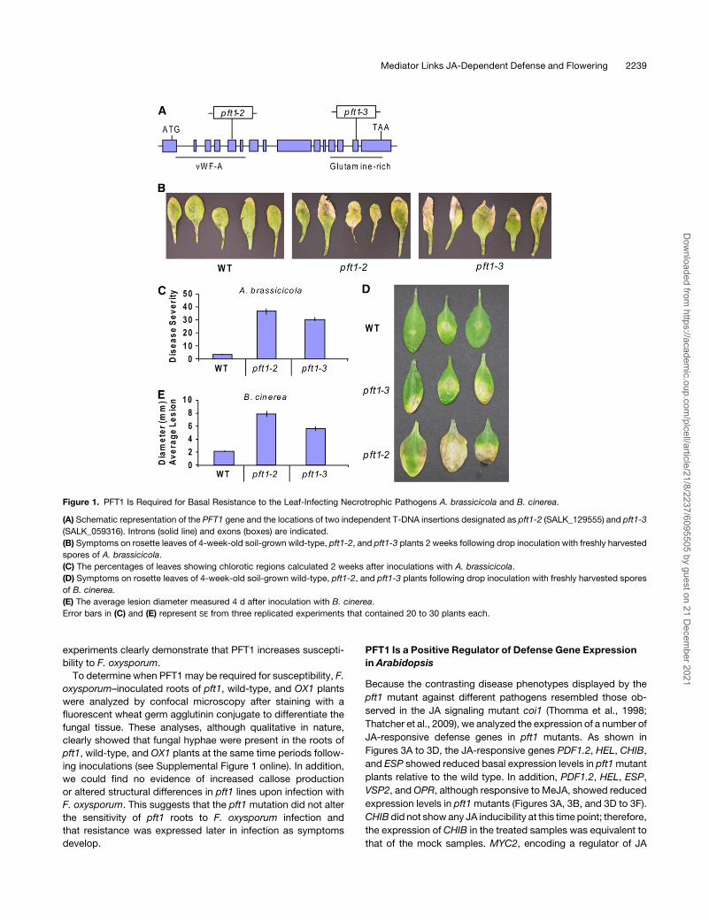
a T-DNA insertion located in the 5th exon of the PFT1 gene. This
insertion lies within a genomic region that encodes a von
Willebrand Factor A domain (vWF-A) located at the N terminus
of the PFT1 protein (Figure 1A). The vWF-A domain is a widely
distributed protein–protein interaction domain (Whittaker and
Hynes, 2002) and has been shown to be critical for the binding of
MED25 to theMediator complex in human cell lines (Mittler et al.,
2003). The T-DNA in the pft1-3 mutant is inserted into the 14th
exon of PFT1 (Figure 1A) and disrupts the Gln-rich region
predicted to function as a putative transcriptional activation
domain (Cerdan and Chory, 2003).
To determine whether PFT1 is required during plant defense,
pft1-2, pft1-3, andwild-type plants were inoculated with the leaf-
infecting necrotrophic fungal pathogens A. brassicicola and B.
cinerea. The A. brassicicola isolate used here is incompatible on
wild-type (Columbia-0 [Col-0]) Arabidopsis (Schenk et al., 2000,
2003) but has been shown to be capable of causing lesions on
Arabidopsis mutants with attenuated plant defenses (Trusov
et al., 2006). As shown in Figure 1B, inoculations with A.
brassicicola resulted in the development of distinct chlorotic
regions restricted to older rosette leaves of the pft1 mutant. In
these experiments, 35 and 30% of the inoculated pft1-2 and
pft1-3 leaves, respectively, showed chlorosis (Figure 1C). Fur-
thermore, the pathogen was often able to sporulate on these
chlorotic lesions. By contrast, only 3% of the inoculated leaves
from wild-type plants had similar chlorotic regions (Figure 1C).
Similarly, B. cinerea, although poorly compatible on wild-type
plants, produced larger chlorotic regions on the pft1 mutant
leaves than those on wild-type leaves (Figures 1D and 1E). Thus,
these pathogen inoculation experiments suggest that PFT1 is an
important component of basal resistance to these necrotrophic
pathogens in Arabidopsis.
PFT1 Is a F. oxysporum Susceptibility Gene
To determine whether PFT1 is also required for resistance to a
pathogen with a different lifestyle, we conducted inoculation
experiments with the root-infecting hemibiotrophic fungal path-
ogen F. oxysporum, which causes vascular wilt disease in a wide
range of plants, including Arabidopsis (Diener and Ausubel,
2005; Edgar et al., 2006; Berrocal-Lobo and Molina, 2008;
Michielse and Rep, 2009). The number of leaves with chlorosis/
necrosis and the percentage of plant death after inoculations
were substantially lower (P < 0.01) in the pft1mutants than those
in the wild-type plants (Figures 2A to 2C). The increased resis-
tance to F. oxysporum and increased susceptibility against B.
cinerea andA. brassicicola inpft1mutantswas reminiscent of the
reaction to these pathogens in the coi1 mutant, which has
impaired JA signaling (Thatcher et al., 2009).
To confirm that the increased F. oxysporum resistance was
due to a loss-of-function mutation in the PFT1 gene, the Fusar-
ium resistance phenotypes of the pft1-2 mutant, a 35S-PFT1
overexpressing line (OX1) as well as the pft1mutant transformed
with a genomic copy of PFT1 (G1), were analyzed in replicated
inoculation experiments. The PFT1-overexpressing line and the
PFT1 genomic complement, designated as OX1 and G1, re-
spectively, have been previously characterized in relation to the
function of PFT1 in phytochrome signaling (Cerdan and Chory,
2003). Consistent with the previous inoculation experiments, the
pft1-2 plants were mostly free of visible symptoms, while typical
F. oxysporum disease symptoms (e.g., vein chlorosis) appeared
on a high proportion of the wild-type,OX1, andG1 plants (Figure
2D). The OX1 plants were the most affected, with ;90% of the
OX1 leaves showing visible symptoms, while the G1 plants, as
expected, showed symptoms comparable to those on wild-type
plants (Figures 2D and 2E). Finally, becauseG1was generated in
the pft1-1 background (Cerdan and Chory, 2003), we tested the
response of the pft1-1 mutant to F. oxysporum. As expected,
pft1-1 was also resistant to F. oxysporum. Together, these
2238 The Plant Cell
Dow
nloaded from https://academ
ic.oup.com/plcell/article/21/8/2237/6095505 by guest on 21 D
ecember 2021
experiments clearly demonstrate that PFT1 increases suscepti-
bility to F. oxysporum.
To determine when PFT1 may be required for susceptibility, F.
oxysporum–inoculated roots of pft1, wild-type, and OX1 plants
were analyzed by confocal microscopy after staining with a
fluorescent wheat germ agglutinin conjugate to differentiate the
fungal tissue. These analyses, although qualitative in nature,
clearly showed that fungal hyphae were present in the roots of
pft1, wild-type, and OX1 plants at the same time periods follow-
ing inoculations (see Supplemental Figure 1 online). In addition,
we could find no evidence of increased callose production
or altered structural differences in pft1 lines upon infection with
F. oxysporum. This suggests that the pft1 mutation did not alter
the sensitivity of pft1 roots to F. oxysporum infection and
that resistance was expressed later in infection as symptoms
develop.
PFT1 Is a Positive Regulator of Defense Gene Expression
in Arabidopsis
Because the contrasting disease phenotypes displayed by the
pft1 mutant against different pathogens resembled those ob-
served in the JA signaling mutant coi1 (Thomma et al., 1998;
Thatcher et al., 2009), we analyzed the expression of a number of
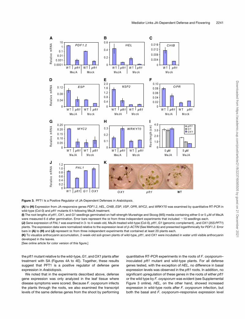
JA-responsive defense genes in pft1 mutants. As shown in
Figures 3A to 3D, the JA-responsive genes PDF1.2, HEL, CHIB,
and ESP showed reduced basal expression levels in pft1mutant
plants relative to the wild type. In addition, PDF1.2, HEL, ESP,
VSP2, andOPR, although responsive to MeJA, showed reduced
expression levels in pft1mutants (Figures 3A, 3B, and 3D to 3F).
CHIB did not showany JA inducibility at this time point; therefore,
the expression of CHIB in the treated samples was equivalent to
that of the mock samples. MYC2, encoding a regulator of JA
Figure 1. PFT1 Is Required for Basal Resistance to the Leaf-Infecting Necrotrophic Pathogens A. brassicicola and B. cinerea.
(A) Schematic representation of the PFT1 gene and the locations of two independent T-DNA insertions designated as pft1-2 (SALK_129555) and pft1-3
(SALK_059316). Introns (solid line) and exons (boxes) are indicated.
(B) Symptoms on rosette leaves of 4-week-old soil-grown wild-type, pft1-2, and pft1-3 plants 2 weeks following drop inoculation with freshly harvested
spores of A. brassicicola.
(C) The percentages of leaves showing chlorotic regions calculated 2 weeks after inoculations with A. brassicicola.
(D) Symptoms on rosette leaves of 4-week-old soil-grown wild-type, pft1-2, and pft1-3 plants following drop inoculation with freshly harvested spores
of B. cinerea.
(E) The average lesion diameter measured 4 d after inoculation with B. cinerea.
Error bars in (C) and (E) represent SE from three replicated experiments that contained 20 to 30 plants each.
Mediator Links JA-Dependent Defense and Flowering 2239
Dow
nloaded from https://academ
ic.oup.com/plcell/article/21/8/2237/6095505 by guest on 21 D
ecember 2021
signaling, was inducible by MeJA in the wild type but not
inducible in pft1 mutants (Figure 3G). By contrast, WRKY70,
encoding a negative regulator of JA signaling, showed higher
expression levels in pft1 mutants in response to MeJA (Figure
3H). In separate sets of experiments, we further examined the
expression of these defense genes in wild-type, G1, and OX1
plants and found increased transcript levels of JA-responsive
genes in OX1 plants (see Supplemental Figure 2 online). In
addition, pft1 roots were less sensitive to growth inhibition by
MeJA than G1 and OX1 roots (Figure 3I). Together, these data
suggest a positive regulatory role for PFT1 in JA signaling.
We also noted that MYC2 and WRKY70 (differentially regu-
lated in pft1mutants) encode a positive and a negative regulator
of the anthocyanin pathway, respectively (Li et al., 2006;
Dombrecht et al., 2007). In addition, the basal transcript levels
of PAL1, which encodes a major isoform of the enzyme Phe
ammonia lyase involved in phenylpropanoid biosynthesis, was
lower in the pft1 mutant than in wild-type, G1, and OX1 plants
(Figure 3J). Consistent with these expression data, the pft1
mutant showed a lack of anthocyanin production, whilewild-type
and OX1 plants produced strong anthocyanin accumulation
(Figure 3K).
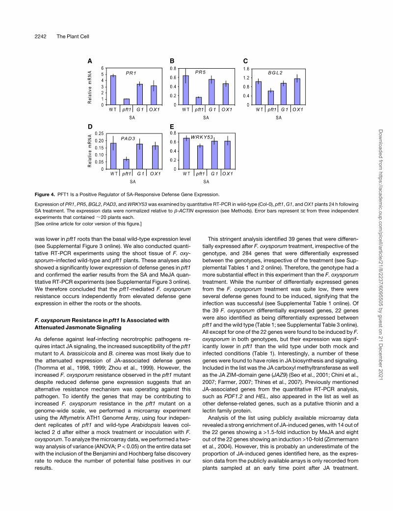
As the pft1 mutant showed attenuated JA defense gene
expression, we also investigated whether SA-responsive de-
fense genes showed any differential expression in the mutant.
Interestingly, the SA-responsive defense genes PR1, PR5, and
BGL2, the phytoalexin biosynthesis gene PAD3, and the tran-
scriptional activatorWRKY33 all showed attenuated induction in
Figure 2. PFT1 Is an F. oxysporum Susceptibility Gene.
(A) Typical disease symptoms and plant death observed on wild-type (Col-0) and pft1 mutants 2 weeks after inoculations of the roots of 3- to 4-week-
old plants.
(B) The number of leaves showing chlorosis and necrosis 10 d after inoculations and expressed as the percentage of total number of leaves.
(C) Percentages of plant death 2 weeks after the inoculation of roots with F. oxysporum.
(D) Typical disease phenotypes of wild-type (Col-0), pft1, OX1 (35S:PFT1), and G1 (the pft1mutant complemented with a genomic PFT1) plants at 12 d
after F. oxysporum inoculation.
(E) Average percentage of disease severity measured in the same lines shown in (D). Error bars represent SE from three replicated experiments that
contained 20 to 30 plants each.
2240 The Plant Cell
Dow
nloaded from https://academ
ic.oup.com/plcell/article/21/8/2237/6095505 by guest on 21 D
ecember 2021
the pft1mutant relative to thewild-type,G1, andOX1 plants after
treatment with SA (Figures 4A to 4E). Together, these results
suggest that PFT1 is a positive regulator of defense gene
expression in Arabidopsis.
We noted that in the experiments described above, defense
gene expression was only analyzed in the leaf tissue where
disease symptoms were scored. Because F. oxysporum infects
the plants through the roots, we also examined the transcript
levels of the same defense genes from the shoot by performing
quantitative RT-PCR experiments in the roots of F. oxysporum–
inoculated pft1 mutant and wild-type plants. For all defense
genes tested, with the exception of HEL, no difference in basal
expression levels was observed in the pft1 roots. In addition, no
significant upregulation of these genes in the roots of either pft1
or the wild type by F. oxysporum was evident (see Supplemental
Figure 3 online). HEL, on the other hand, showed increased
expression in wild-type roots after F. oxysporum infection, but
both the basal and F. oxysporum–responsive expression level
Figure 3. PFT1 Is a Positive Regulator of JA-Dependent Defenses in Arabidopsis.
(A) to (H) Expression from JA-responsive genes PDF1.2, HEL, CHIB, ESP, VSP, OPR, MYC2, and WRKY70 was examined by quantitative RT-PCR in
wild-type (Col-0) and pft1 mutants 6 h following MeJA treatment.
(I) The root lengths of pft1, OX1, and G1 seedlings germinated on half-strength Murashige and Skoog (MS) media containing either 0 or 5 mM of MeJA
were measured 6 d after germination. Error bars represent the SE from three independent experiments that included ;10 seedlings each.
(J) Gene expression of PAL1 was examined in 3- to 4-week-old, MeJA-treated wild-type (Col-0), pft1, G1 (genomic complement) , and OX1 (35S:PFT1)
plants. The expression data were normalized relative to the expression level of b-ACTIN (See Methods) and presented logarithmically for PDF1.2. Error
bars in (A) to (H) and (J) represent SE from three independent experiments that contained at least 20 plants each.
(K) To visualize anthocyanin accumulation, 2-week-old soil-grown plants of wild-type, pft1, and OX1 were incubated in water until visible anthocyanin
developed in the leaves.
[See online article for color version of this figure.]
Mediator Links JA-Dependent Defense and Flowering 2241
Dow
nloaded from https://academ
ic.oup.com/plcell/article/21/8/2237/6095505 by guest on 21 D
ecember 2021
was lower in pft1 roots than the basal wild-type expression level
(see Supplemental Figure 3 online). We also conducted quanti-
tative RT-PCR experiments using the shoot tissue of F. oxy-
sporum–infected wild-type and pft1 plants. These analyses also
showed a significantly lower expression of defense genes in pft1
and confirmed the earlier results from the SA and MeJA quan-
titative RT-PCR experiments (see Supplemental Figure 3 online).
We therefore concluded that the pft1-mediated F. oxysporum
resistance occurs independently from elevated defense gene
expression in either the roots or the shoots.
F. oxysporum Resistance in pft1 Is Associated with
Attenuated Jasmonate Signaling
As defense against leaf-infecting necrotrophic pathogens re-
quires intact JA signaling, the increased susceptibility of the pft1
mutant to A. brassicicola and B. cinerea was most likely due to
the attenuated expression of JA-associated defense genes
(Thomma et al., 1998, 1999; Zhou et al., 1999). However, the
increased F. oxysporum resistance observed in the pft1 mutant
despite reduced defense gene expression suggests that an
alternative resistance mechanism was operating against this
pathogen. To identify the genes that may be contributing to
increased F. oxysporum resistance in the pft1 mutant on a
genome-wide scale, we performed a microarray experiment
using the Affymetrix ATH1 Genome Array, using four indepen-
dent replicates of pft1 and wild-type Arabidopsis leaves col-
lected 2 d after either a mock treatment or inoculation with F.
oxysporum. To analyze themicroarray data, weperformed a two-
way analysis of variance (ANOVA; P < 0.05) on the entire data set
with the inclusion of the Benjamini and Hochberg false discovery
rate to reduce the number of potential false positives in our
results.
This stringent analysis identified 39 genes that were differen-
tially expressed after F. oxysporum treatment, irrespective of the
genotype, and 284 genes that were differentially expressed
between the genotypes, irrespective of the treatment (see Sup-
plemental Tables 1 and 2 online). Therefore, the genotype had a
more substantial effect in this experiment than the F. oxysporum
treatment. While the number of differentially expressed genes
from the F. oxysporum treatment was quite low, there were
several defense genes found to be induced, signifying that the
infection was successful (see Supplemental Table 1 online). Of
the 39 F. oxysporum differentially expressed genes, 22 genes
were also identified as being differentially expressed between
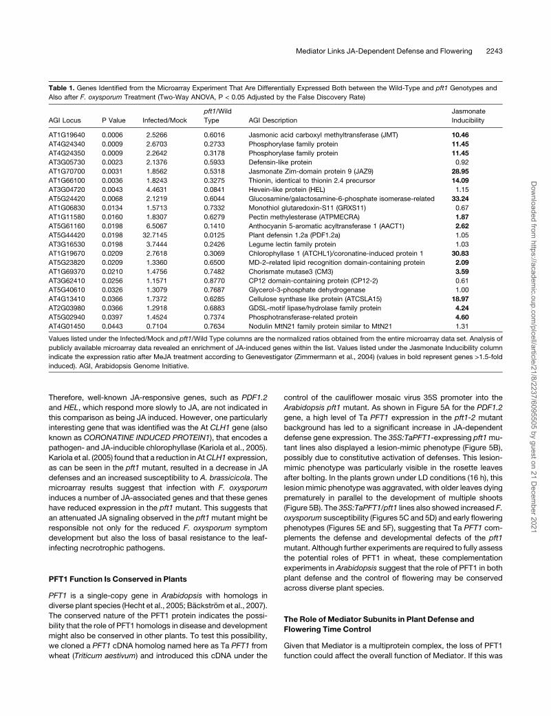
pft1 and the wild type (Table 1; see Supplemental Table 3 online).
All except for one of the 22 genes were found to be induced by F.
oxysporum in both genotypes, but their expression was signif-
icantly lower in pft1 than the wild type under both mock and
infected conditions (Table 1). Interestingly, a number of these
genes were found to have roles in JA biosynthesis and signaling.
Included in the list was the JA carboxyl methyltransferase as well
as the JA ZIM-domain gene (JAZ9) (Seo et al., 2001; Chini et al.,
2007; Farmer, 2007; Thines et al., 2007). Previously mentioned
JA-associated genes from the quantitative RT-PCR analysis,
such as PDF1.2 and HEL, also appeared in the list as well as
other defense-related genes, such as a putative thionin and a
lectin family protein.
Analysis of the list using publicly available microarray data
revealed a strong enrichment of JA-induced genes, with 14 out of
the 22 genes showing a >1.5-fold induction by MeJA and eight
out of the 22 genes showing an induction >10-fold (Zimmermann
et al., 2004). However, this is probably an underestimate of the
proportion of JA-induced genes identified here, as the expres-
sion data from the publicly available arrays is only recorded from
plants sampled at an early time point after JA treatment.
Figure 4. PFT1 Is a Positive Regulator of SA-Responsive Defense Gene Expression.
Expression of PR1, PR5, BGL2, PAD3, andWRKY53was examined by quantitative RT-PCR in wild-type (Col-0), pft1,G1, andOX1 plants 24 h following
SA treatment. The expression data were normalized relative to b-ACTIN expression (see Methods). Error bars represent SE from three independent
experiments that contained ;20 plants each.
[See online article for color version of this figure.]
2242 The Plant Cell
Dow
nloaded from https://academ
ic.oup.com/plcell/article/21/8/2237/6095505 by guest on 21 D
ecember 2021
Therefore, well-known JA-responsive genes, such as PDF1.2
and HEL, which respond more slowly to JA, are not indicated in
this comparison as being JA induced. However, one particularly
interesting gene that was identified was the At CLH1 gene (also
known as CORONATINE INDUCED PROTEIN1), that encodes a
pathogen- and JA-inducible chlorophyllase (Kariola et al., 2005).
Kariola et al. (2005) found that a reduction in AtCLH1 expression,
as can be seen in the pft1 mutant, resulted in a decrease in JA
defenses and an increased susceptibility to A. brassicicola. The
microarray results suggest that infection with F. oxysporum
induces a number of JA-associated genes and that these genes
have reduced expression in the pft1 mutant. This suggests that
an attenuated JA signaling observed in the pft1mutant might be
responsible not only for the reduced F. oxysporum symptom
development but also the loss of basal resistance to the leaf-
infecting necrotrophic pathogens.
PFT1 Function Is Conserved in Plants
PFT1 is a single-copy gene in Arabidopsis with homologs in
diverse plant species (Hecht et al., 2005; Backstrom et al., 2007).
The conserved nature of the PFT1 protein indicates the possi-
bility that the role of PFT1 homologs in disease and development
might also be conserved in other plants. To test this possibility,
we cloned a PFT1 cDNA homolog named here as Ta PFT1 from
wheat (Triticum aestivum) and introduced this cDNA under the
control of the cauliflower mosaic virus 35S promoter into the
Arabidopsis pft1 mutant. As shown in Figure 5A for the PDF1.2
gene, a high level of Ta PFT1 expression in the pft1-2 mutant
background has led to a significant increase in JA-dependent
defense gene expression. The 35S:TaPFT1-expressing pft1mu-
tant lines also displayed a lesion-mimic phenotype (Figure 5B),
possibly due to constitutive activation of defenses. This lesion-
mimic phenotype was particularly visible in the rosette leaves
after bolting. In the plants grown under LD conditions (16 h), this
lesion mimic phenotype was aggravated, with older leaves dying
prematurely in parallel to the development of multiple shoots
(Figure 5B). The 35S:TaPFT1/pft1 lines also showed increased F.
oxysporum susceptibility (Figures 5C and 5D) and early flowering
phenotypes (Figures 5E and 5F), suggesting that Ta PFT1 com-
plements the defense and developmental defects of the pft1
mutant. Although further experiments are required to fully assess
the potential roles of PFT1 in wheat, these complementation
experiments in Arabidopsis suggest that the role of PFT1 in both
plant defense and the control of flowering may be conserved
across diverse plant species.
The Role of Mediator Subunits in Plant Defense and
Flowering Time Control
Given that Mediator is a multiprotein complex, the loss of PFT1
function could affect the overall function of Mediator. If this was
Table 1. Genes Identified from the Microarray Experiment That Are Differentially Expressed Both between the Wild-Type and pft1 Genotypes and
Also after F. oxysporum Treatment (Two-Way ANOVA, P < 0.05 Adjusted by the False Discovery Rate)
AGI Locus P Value Infected/Mock
pft1/Wild
Type AGI Description
Jasmonate
Inducibility
AT1G19640 0.0006 2.5266 0.6016 Jasmonic acid carboxyl methyltransferase (JMT) 10.46
AT4G24340 0.0009 2.6703 0.2733 Phosphorylase family protein 11.45
AT4G24350 0.0009 2.2642 0.3178 Phosphorylase family protein 11.45
AT3G05730 0.0023 2.1376 0.5933 Defensin-like protein 0.92
AT1G70700 0.0031 1.8562 0.5318 Jasmonate Zim-domain protein 9 (JAZ9) 28.95
AT1G66100 0.0036 1.8243 0.3275 Thionin, identical to thionin 2.4 precursor 14.09
AT3G04720 0.0043 4.4631 0.0841 Hevein-like protein (HEL) 1.15
AT5G24420 0.0068 2.1219 0.6044 Glucosamine/galactosamine-6-phosphate isomerase-related 33.24
AT1G06830 0.0134 1.5713 0.7332 Monothiol glutaredoxin-S11 (GRXS11) 0.67
AT1G11580 0.0160 1.8307 0.6279 Pectin methylesterase (ATPMECRA) 1.87
AT5G61160 0.0198 6.5067 0.1410 Anthocyanin 5-aromatic acyltransferase 1 (AACT1) 2.62
AT5G44420 0.0198 32.7145 0.0125 Plant defensin 1.2a (PDF1.2a) 1.05
AT3G16530 0.0198 3.7444 0.2426 Legume lectin family protein 1.03
AT1G19670 0.0209 2.7618 0.3069 Chlorophyllase 1 (ATCHL1)/coronatine-induced protein 1 30.83
AT5G23820 0.0209 1.3360 0.6500 MD-2–related lipid recognition domain-containing protein 2.09
AT1G69370 0.0210 1.4756 0.7482 Chorismate mutase3 (CM3) 3.59
AT3G62410 0.0256 1.1571 0.8770 CP12 domain-containing protein (CP12-2) 0.61
AT5G40610 0.0326 1.3079 0.7687 Glycerol-3-phosphate dehydrogenase 1.00
AT4G13410 0.0366 1.7372 0.6285 Cellulose synthase like protein (ATCSLA15) 18.97
AT2G03980 0.0366 1.2918 0.6883 GDSL-motif lipase/hydrolase family protein 4.24
AT5G02940 0.0397 1.4524 0.7374 Phosphotransferase-related protein 4.60
AT4G01450 0.0443 0.7104 0.7634 Nodulin MtN21 family protein similar to MtN21 1.31
Values listed under the Infected/Mock and pft1/Wild Type columns are the normalized ratios obtained from the entire microarray data set. Analysis of
publicly available microarray data revealed an enrichment of JA-induced genes within the list. Values listed under the Jasmonate Inducibility column
indicate the expression ratio after MeJA treatment according to Genevestigator (Zimmermann et al., 2004) (values in bold represent genes >1.5-fold
induced). AGI, Arabidopsis Genome Initiative.
Mediator Links JA-Dependent Defense and Flowering 2243
Dow
nloaded from https://academ
ic.oup.com/plcell/article/21/8/2237/6095505 by guest on 21 D
ecember 2021

the case, then inactivation of other Mediator subunits could lead
to similar defense and developmental phenotypes observed in
the pft1 plants. To test this hypothesis, we isolated homozygous
T-DNA insertion lines for 11 individual Arabidopsis Medi-
ator subunit mutants publicly available in the SALK Arabidopsis
T-DNA insertion collection (see Methods). Of the Mediator mu-
tants tested, only an insertion in the MED8 subunit (Figure 6A;
see Supplemental Figure 4 online) produced an F. oxysporum
resistance phenotype that was comparable to that of the pft1
mutant in terms of delayed symptom development (Figures 6B
and 6C) and increased survival rates (see Supplemental Figure 5
online). In addition, themed8mutant had increased susceptibility
to A. brassicicola with 42% of the inoculated leaves showing
relatively large chlorotic lesions (Figure 6D) as compared with
21%ofwild-type leaves that showed some degree of chlorosis in
these experiments.
Figure 5. PFT1 Function Is Conserved in Plants.
(A) Transgenic expression of Ta PFT1, a PFT1 homolog from wheat, in the pft1 mutant background complements the compromised defense gene
expression.
(B) The 35S:TaPFT1/pft1 line shows spontaneous lesion development (arrow).
(C) and (D) Expression of Ta PFT1 complements the increased F. oxysporum resistance of the pft1 mutant.
(E) and (F) Expression of Ta PFT1 complements the delayed flowering phenotype of pft1. PDF1.2 transcript levels were examined by quantitative RT-
PCR, and the expression data were normalized relative to b-ACTIN expression and presented logarithmically.
Numbers on each bar in (A) represent fold difference in expression of PDF1.2 in untreated plants of pft1 and 35S:TaPFT1/pft1 relative to untreated wild-
type plants. Error bars represent SE from three independent replicates that contained ;20 plants each. F. oxysporum inoculation experiments were
conducted as described above, and symptom development was scored 10 d after inoculation.
2244 The Plant Cell
Dow
nloaded from https://academ
ic.oup.com/plcell/article/21/8/2237/6095505 by guest on 21 D
ecember 2021
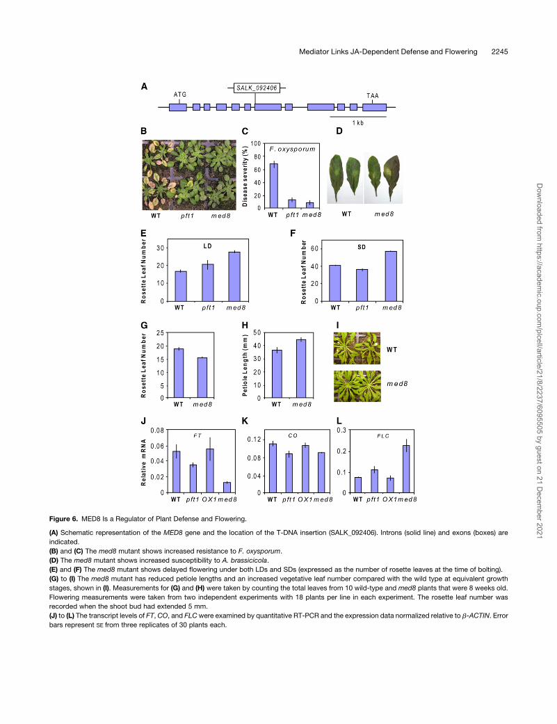
Figure 6. MED8 Is a Regulator of Plant Defense and Flowering.
(A) Schematic representation of the MED8 gene and the location of the T-DNA insertion (SALK_092406). Introns (solid line) and exons (boxes) are
indicated.
(B) and (C) The med8 mutant shows increased resistance to F. oxysporum.
(D) The med8 mutant shows increased susceptibility to A. brassicicola.
(E) and (F) The med8 mutant shows delayed flowering under both LDs and SDs (expressed as the number of rosette leaves at the time of bolting).
(G) to (I) The med8 mutant has reduced petiole lengths and an increased vegetative leaf number compared with the wild type at equivalent growth
stages, shown in (I). Measurements for (G) and (H) were taken by counting the total leaves from 10 wild-type and med8 plants that were 8 weeks old.
Flowering measurements were taken from two independent experiments with 18 plants per line in each experiment. The rosette leaf number was
recorded when the shoot bud had extended 5 mm.
(J) to (L) The transcript levels of FT, CO, and FLCwere examined by quantitative RT-PCR and the expression data normalized relative to b-ACTIN. Error
bars represent SE from three replicates of 30 plants each.
Mediator Links JA-Dependent Defense and Flowering 2245
Dow
nloaded from https://academ
ic.oup.com/plcell/article/21/8/2237/6095505 by guest on 21 D
ecember 2021
Interestingly, the med8 mutant also demonstrated an altered
flowering time with a strong delay in flowering under both short-
day (SD) and LD conditions (Figures 6E and 6F). The med8
mutant also possessed an increased number of leaves and
shorter petioles under vegetative conditions, giving it a distinc-
tive phenotype (Figures 6G to 6I). In addition to med8, we also
measured the flowering time of pft1 as a comparison. Our results
confirmed the results of Cerdan and Chory (2003), with a small
but statistically significant (P < 0.05) decrease and increase in
flowering time under SD and LD, respectively, when determined
by rosette leaf number (Figures 6E and 6F), and an increased
flowering time under both SD and LD when determined by the
number of days to bolting (see Supplemental Figure 6 online).
To explore the molecular mechanism behind the late flowering
phenotype ofmed8, we also quantified the expression of the key
flowering genes, FLOWERING LOCUS T (FT) and CONSTANS
(CO), which positively regulate flowering, and FLOWERING
LOCUS C (FLC), which negatively regulates flowering (reviewed
in Farrona et al., 2008) in LD-grown pft1 andmed8 plants (Figures
6J to 6L). Cerdan and Chory (2003) previously reported reduced
FT and CO expression in pft1, and our results confirmed this
finding. We also found reduced expression of the floral promot-
ing genes FT and CO in med8 compared with the wild type. In
addition, we found increased expression of the floral repressor
FLC in bothpft1 andmed8. Furthermore, the levels of FT and FLC
expression in pft1 and med8 correlated well with the severity of
the flowering delay seen in these two mutants, with the later
flowering med8 having a noticeably lower and higher level of FT
and FLC expression, respectively, than pft1 and the wild type
(Figures 6J and 6L). Together, these experiments suggest that
MED8, similar to PFT1, is a regulator of both plant defense and
flowering time.
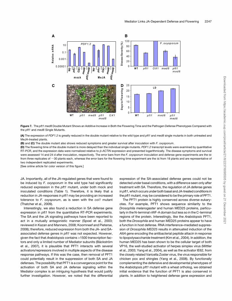
Genetic Evidence for the Independent and Additive
Functions of PFT1 and MED8
As the pft1 and med8 mutants appear to similarly affect both
flowering time and pathogen resistance, we sought to determine
whether these two mutations act independently to produce
similar phenotypes. To test this possibility genetically, we con-
structed a pft1 med8 double mutant and analyzed its defense
and flowering time phenotypes together with those of the pft1
and med8 single mutants. First, we looked at the expression of
PDF1.2 in the double and single mutants after treatment with
MeJA.We found that the expression ofPDF1.2was slightly lower
in both untreated and 6-h MeJA-treated med8 plants than in
similarly treatedwild-type plants (Figure 7A). However, in the pft1
med8 double mutant, PDF1.2 expression was greatly reduced,
with the expression level in the double mutant being;300- and
370-fold less in the MeJA-treated and untreated plants, respec-
tively, than similarly treated pft1 plants (Figure 7A). Inoculation of
the pft1 med8 double mutant with F. oxysporum also revealed a
relatively small but additive increase in resistance relative to the
individual single mutants (Figures 7B and 7C). At 24 d after
inoculation, the increased resistance of the pft1 med8 double
mutant was more evident, with a noticeable difference in the
survival rate and overall vigor of the double mutant compared
with the pft1 and med8 lines (Figure 7C).
In addition, we compared the flowering time of the double
mutant to the wild-type and the single pft1 and med8 mutants.
We found that the pft1med8 doublemutant had a similar number
of leaves at flowering under LD to the pft1 mutant (see Supple-
mental Figure 7 online). However, the double mutant flowered
later than pft1 and med8 when flowering time was measured by
days to flowering under LD, suggesting an additive effect on
flowering time (Figure 7D). The stronger effects of the pft1 med8
double mutant on flowering time and defense than either of the
single mutants suggest that the pft1 and med8 mutations might
be affecting these two phenotypes by independent and additive
mechanisms.
DISCUSSION
The Mediator complex was first purified from yeast in the early
1990s (Kim et al., 1994). Subsequent studies have discovered
that the Mediator complex is an essential part of the transcrip-
tional machinery in all eukaryotes (Bourbon, 2008). Surprisingly,
the existence of theMediator complex in plants has only recently
been shown in Arabidopsis (Backstrom et al., 2007). Using a
biochemical purification strategy, Backstrom et al. (2007) iden-
tified 21 conserved subunits and six plant-specific Mediator
subunits. Two of the Mediator subunits identified by Backstrom
et al. (2007), MED14 and MED25, were previously studied plant
proteins, SWP1 and PFT1, which are involved in regulating leaf
cell number in aerial organs and phytochrome signaling, respec-
tively (Autran et al., 2002; Cerdan and Chory, 2003). More
recently, another Mediator subunit, MED21, has been shown to
be required for resistance to necrotrophic pathogens in Arabi-
dopsis (Dhawan et al., 2009), although the mechanism of this
resistance is currently unknown.
In this article, we show that in addition to its known role in
phytochrome signaling, PFT1 is also an important component of
the plant’s basal defense and is required for uncompromised
expression of JA-dependent defenses as well as resistance to
necrotrophic fungal pathogens, such as A. brassicicola and B.
cinerea (Figures 1 to 4). Interestingly, PFT1 is also essential for
susceptibility to the root-infecting fungus F. oxysporum, which is
thought to use the host JA pathway to promote host senescence
and necrosis (Thatcher et al., 2009).
JA-dependent defense genes and the phytoalexin camalexin
have previously been shown to be active against necrotrophic
pathogens, such as A. brassicicola and B. cinerea (Thomma
et al., 1998, 1999; Zhou et al., 1999). Therefore, it is likely that the
attenuated expression of JA-responsive defense genes as well
as PAD3 is the cause of increased A. brassicicola and B. cinerea
susceptibility in pft1.
The mechanism(s) behind the increased F. oxysporum resis-
tance was initially less apparent. However, as recently reported,
the coi1 mutant with compromised JA signaling and JA-depen-
dent gene expression shows a remarkable increase in resistance
to this pathogen (Thatcher et al., 2009). Through stringent
analysis of the microarray data, we identified that a large pro-
portion of the genes that were differentially expressed after
infection by F. oxysporum treatment, aswell as between thewild-
type and pft1 genotypes, were genes that are also regulated by
2246 The Plant Cell
Dow
nloaded from https://academ
ic.oup.com/plcell/article/21/8/2237/6095505 by guest on 21 D
ecember 2021
JA. Importantly, all of the JA-regulated genes that were found to
be induced by F. oxysporum in the wild type had significantly
reduced expression in the pft1 mutant, under both mock and
inoculated conditions (Table 1). Therefore, it is likely that a
reduction in JA-responses in pft1may be providing an increased
tolerance to F. oxysporum, as is seen with the coi1 mutant
(Thatcher et al., 2009).
Interestingly, we also found a reduction in SA defense gene
expression in pft1 from the quantitative RT-PCR experiments.
The SA and the JA signaling pathways have been reported to
act in a mutually antagonistic manner (Spoel et al., 2003;
reviewed in Kazan andManners, 2008; Koornneef and Pieterse,
2008); therefore, reduced expression from both the JA- and SA-
associated defense genes in pft1 was not expected. However,
given the fact that Arabidopsis contains >1500 transcription fac-
tors and only a limited number of Mediator subunits (Backstrom
et al., 2007), it is plausible that PFT1 interacts with several
activators/repressors involved in multiple aspects of the disease
response pathways. If this was the case, then removal of PFT1
could potentially result in the suppression of both SA and JA
defenses. The possibility that PFT1 is a convergence point for the
activation of both SA and JA defense signaling within the
Mediator complex is an intriguing hypothesis that would justify
further investigation. However, we noted that the differential
expression of the SA-associated defense genes could not be
detected under basal conditions, with a difference seen only after
treatment with SA. Therefore, the regulation of JA defense genes
inpft1, whichoccurs under bothbasal andJA-treatedconditions in
the pft1mutant,may be considered to be the primary role of PFT1.
The PFT1 protein is highly conserved across diverse eukary-
otes. For example, PFT1 shows sequence similarity to the
Drosophila melanogaster and human MED25 proteins, particu-
larly in the N-terminal vWF-A domain but less so in the C-terminal
regions of the protein. Interestingly, like the Arabidopsis PFT1,
both the Drosophila and human MED25 proteins appear to have
a function in host defense. RNA interference–mediated suppres-
sion of Drosophila MED25 results in attenuated induction of the
AttA gene encoding the antibacterial peptide attacin in response
to lipopolysaccharide treatment (Kim et al., 2004). In addition, the
human MED25 has been shown to be the cellular target of both
VP16, the well-studied activator of herpes simplex virus (Mittler
et al., 2003; Yang et al., 2004), as well as the activator IE62, from
the closely related Varicella Zoster virus, the virus responsible for
chicken pox and shingles (Yang et al., 2008). By functionally
complementing the defensive and developmental phenotypes of
the Arabidopsis pft1 mutant with a wheat ortholog, we obtained
initial evidence that the function of PFT1 is also conserved in
plants. In addition to heightened defense gene expression and
Figure 7. The pft1 med8 Double Mutant Shows an Additive Increase in Both the Flowering Time and the Pathogen Defense Phenotypes Compared with
the pft1 and med8 Single Mutants.
(A) The expression of PDF1.2 is greatly reduced in the double mutant relative to the wild type and pft1 and med8 single mutants in both untreated and
MeJA-treated plants.
(B) and (C) The double mutant also shows reduced symptoms and greater survival after inoculation with F. oxysporum.
(D) The flowering time of the double mutant is more delayed than the individual single mutants. PDF1.2 transcript levels were examined by quantitative
RT-PCR, and the expression data were normalized relative to b-ACTIN expression and presented logarithmically. The disease symptoms and survival
were assessed 14 and 24 d after inoculation, respectively. The error bars from the F. oxysporum inoculation and defense gene experiments are the SE
from three replicates of;30 plants each, whereas the error bars for the flowering time experiment are the SE from 18 plants and are representative of
two independent replicated experiments.
[See online article for color version of this figure.]
Mediator Links JA-Dependent Defense and Flowering 2247
Dow
nloaded from https://academ
ic.oup.com/plcell/article/21/8/2237/6095505 by guest on 21 D
ecember 2021
accelerated flowering relative to the pft1 mutants, the 35S:
TaPFT1/pft1 plants displayed a spontaneous lesion phenotype.
Transgenic expression of the vWF-A domain of the BONZAI/
COPINE1 protein of Arabidopsis in transgenic tobacco (Nicoti-
ana tabacum) activates defense responses and also produces a
lesion mimic phenotype (Liu et al., 2005), suggesting that the
lesionmimic phenotypewe observed in TaPFT1-expressing pft1
mutant plants could be mediated by the vWF-A domain of Ta
PFT1.
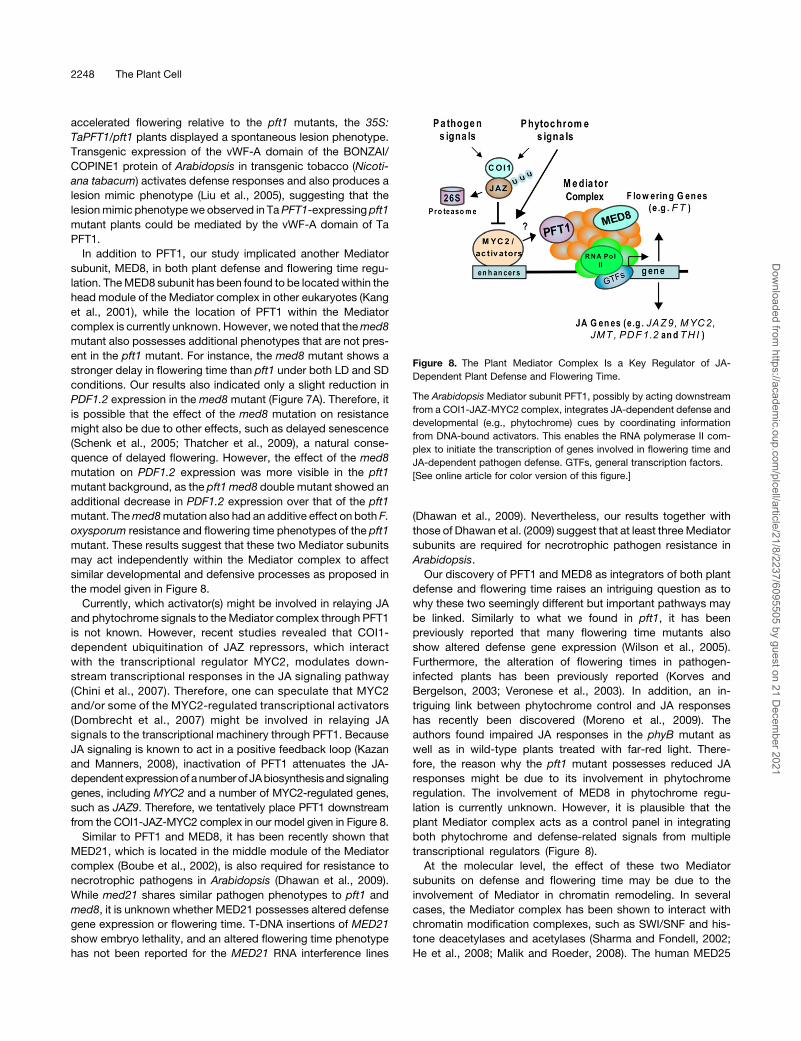
In addition to PFT1, our study implicated another Mediator
subunit, MED8, in both plant defense and flowering time regu-
lation. TheMED8 subunit has been found to be located within the
head module of the Mediator complex in other eukaryotes (Kang
et al., 2001), while the location of PFT1 within the Mediator
complex is currently unknown. However, we noted that themed8
mutant also possesses additional phenotypes that are not pres-
ent in the pft1 mutant. For instance, the med8 mutant shows a
stronger delay in flowering time than pft1 under both LD and SD
conditions. Our results also indicated only a slight reduction in
PDF1.2 expression in the med8 mutant (Figure 7A). Therefore, it
is possible that the effect of the med8 mutation on resistance
might also be due to other effects, such as delayed senescence
(Schenk et al., 2005; Thatcher et al., 2009), a natural conse-
quence of delayed flowering. However, the effect of the med8
mutation on PDF1.2 expression was more visible in the pft1
mutant background, as the pft1 med8 double mutant showed an
additional decrease in PDF1.2 expression over that of the pft1
mutant. Themed8mutation also had an additive effect on both F.
oxysporum resistance and flowering time phenotypes of the pft1
mutant. These results suggest that these two Mediator subunits
may act independently within the Mediator complex to affect
similar developmental and defensive processes as proposed in
the model given in Figure 8.
Currently, which activator(s) might be involved in relaying JA
and phytochrome signals to theMediator complex through PFT1
is not known. However, recent studies revealed that COI1-
dependent ubiquitination of JAZ repressors, which interact
with the transcriptional regulator MYC2, modulates down-
stream transcriptional responses in the JA signaling pathway
(Chini et al., 2007). Therefore, one can speculate that MYC2
and/or some of the MYC2-regulated transcriptional activators
(Dombrecht et al., 2007) might be involved in relaying JA
signals to the transcriptional machinery through PFT1. Because
JA signaling is known to act in a positive feedback loop (Kazan
and Manners, 2008), inactivation of PFT1 attenuates the JA-
dependentexpressionof anumberof JAbiosynthesisandsignaling
genes, including MYC2 and a number of MYC2-regulated genes,
such as JAZ9. Therefore, we tentatively place PFT1 downstream
from the COI1-JAZ-MYC2 complex in our model given in Figure 8.
Similar to PFT1 and MED8, it has been recently shown that
MED21, which is located in the middle module of the Mediator
complex (Boube et al., 2002), is also required for resistance to
necrotrophic pathogens in Arabidopsis (Dhawan et al., 2009).
While med21 shares similar pathogen phenotypes to pft1 and
med8, it is unknown whether MED21 possesses altered defense
gene expression or flowering time. T-DNA insertions of MED21
show embryo lethality, and an altered flowering time phenotype
has not been reported for the MED21 RNA interference lines
(Dhawan et al., 2009). Nevertheless, our results together with
those of Dhawan et al. (2009) suggest that at least threeMediator
subunits are required for necrotrophic pathogen resistance in
Arabidopsis.
Our discovery of PFT1 and MED8 as integrators of both plant
defense and flowering time raises an intriguing question as to
why these two seemingly different but important pathways may
be linked. Similarly to what we found in pft1, it has been
previously reported that many flowering time mutants also
show altered defense gene expression (Wilson et al., 2005).
Furthermore, the alteration of flowering times in pathogen-
infected plants has been previously reported (Korves and
Bergelson, 2003; Veronese et al., 2003). In addition, an in-
triguing link between phytochrome control and JA responses
has recently been discovered (Moreno et al., 2009). The
authors found impaired JA responses in the phyB mutant as
well as in wild-type plants treated with far-red light. There-
fore, the reason why the pft1 mutant possesses reduced JA
responses might be due to its involvement in phytochrome
regulation. The involvement of MED8 in phytochrome regu-
lation is currently unknown. However, it is plausible that the
plant Mediator complex acts as a control panel in integrating
both phytochrome and defense-related signals from multiple
transcriptional regulators (Figure 8).
At the molecular level, the effect of these two Mediator
subunits on defense and flowering time may be due to the
involvement of Mediator in chromatin remodeling. In several
cases, the Mediator complex has been shown to interact with
chromatin modification complexes, such as SWI/SNF and his-
tone deacetylases and acetylases (Sharma and Fondell, 2002;
He et al., 2008; Malik and Roeder, 2008). The human MED25
Figure 8. The Plant Mediator Complex Is a Key Regulator of JA-
Dependent Plant Defense and Flowering Time.
The Arabidopsis Mediator subunit PFT1, possibly by acting downstream
from a COI1-JAZ-MYC2 complex, integrates JA-dependent defense and
developmental (e.g., phytochrome) cues by coordinating information
from DNA-bound activators. This enables the RNA polymerase II com-
plex to initiate the transcription of genes involved in flowering time and
JA-dependent pathogen defense. GTFs, general transcription factors.
[See online article for color version of this figure.]
2248 The Plant Cell
Dow
nloaded from https://academ
ic.oup.com/plcell/article/21/8/2237/6095505 by guest on 21 D
ecember 2021
protein has been shown to interact with histone acetylases
(Black et al., 2006; Lee et al., 2007). Chromatin remodeling
complexes are known to be functionally conserved evolutionarily
(Farrona et al., 2008). Thus, if this function ofMED25 is conserved
between plants and animals, then one would expect that
Arabidopsis PFT1 would also play a role in chromatin modifica-
tion. SWP1/MED14 is known to interact with the transcriptional
corepressor LEUNIG, which interacts with the histone deacety-
lase HDA19 (Gonzalez et al., 2007). Interestingly, a number of
genes involved in chromatin modification and remodeling affect
both flowering and plant defense (Wagner and Meyerowitz,
2002; He and Amasino, 2005; Zhou et al., 2005; Walley et al.,
2008; March-Dıaz et al., 2008; Wu et al., 2008).
Similar to our results with the pft1 andmed8mutants, a recent
study found that the loss of activity in SPLAYED, one of the SWI/
SNF classes of chromatin remodeling ATPases in Arabidopsis,
leads to reduced JA-responsive defense gene expression and
increased susceptibility to B. cinerea (Walley et al., 2008). The
sydmutants also show defects in reproductive development and
flowering time (Wagner and Meyerowitz, 2002). Similarly, chro-
matin modification by the histone deacetylases HDA19 and
HDA6 is required for JA-responsive defense gene expression
and resistance to necrotrophic pathogens (Zhou et al., 2005; Wu
et al., 2008). Again, similarly to pft1 and med8, loss-of-function
mutations in the HDA6 gene delays flowering (Wu et al., 2008).
HDA6 is also known to interact with COI1 (Devoto et al., 2002),
further linking JA responses to chromatin modification.
Finally, theMediator subunit MED21, required for necrotrophic
pathogen resistance, has been shown to interact with HUB1, an
E3 ligase responsible for the ubiquination of H2B histones. The
hub1 mutant also shows alterations in flowering time (Dhawan
et al., 2009). Although further research is required to determine
whether perturbation of the Mediator complex in pft1 and med8
would compromise chromatin remodeling, overall, these studies
support the view that multiple components of the plant tran-
scriptional machinery are required for the regulation of both plant
development, such as flowering time and pathogen defense.
In conclusion, our results reported here link two plant Mediator
subunits as integrators of flowering time and JA-dependent
defense-related signals to the transcriptional machinery. Future
research should reveal new insights into the specific roles of the
remaining Mediator subunits and help to advance our under-
standing of the transcriptional regulation of gene expression in
plants.
METHODS
Plant Growth Conditions, Chemical Treatments, and
Pathogen Inoculations
Plant growth conditions and MeJA and SA treatments were described
previously (Schenk et al., 2000; Campbell et al., 2003; Anderson et al.,
2004). Briefly, plants were grown in a controlled environment room with a
temperature of 248C and a light intensity of 150 mmol·m22·s21. Photo-
synthetically active radiation was supplied by high pressure metal halide
lamps (Sylvania) and tungsten halogen lamps (Phillips). The red:far-red
ratio was;1.13, which is within 10% of the observed daylight. All plants
used were in the Arabidopsis thaliana Col-0 background. Mutant lines
used are listed in the Accession Numbers section at the end of Methods.
Homozygous plants of pft1, med8, and the other Mediator subunit
mutants were identified using the primer sequences given at http://
signal.salk.edu/tdnaprimers.html and used in the experiments described
here. The pft1 med8 double mutant was created by pollinating an
emasculated med8 floral bud with the pollen from the pft1.2 mutant. All
treatments were performed on soil-grown 4- to 5-week-old plants, unless
otherwise stated. The Fusarium oxysporum isolate used in this study was
strain Fo5176 obtained from Roger Shivas, Queensland Plant Pathology
Herbarium, Queensland Department of Primary Industries and Fisheries
(QDPIandF), Brisbane, Australia. Inoculations were performed as de-
scribed by Anderson et al. (2004) and Edgar et al. (2006). The Alternaria
brassicicola (UQ4273) infection assays were performed as described
previously (Campbell et al., 2003). Botrytis cinerea (BRIP25539) infection
assays were performed in a similar manner to the A. brassicicola assays by
harvesting spores from half-strength potato dextrose agar plates and
pipetting 5-mL droplets (13 106 spores/mL) onmatureArabidopsis leaves.
Quantitative RT-PCR Expression Analyses
Quantitative RT-PCR experiments were done as described previously
using the Applied Biosystems 7900HT Fast real-time PCR system in
conjunction with SYBR Green fluorescence to detect transcript levels
(McGrath et al., 2005). Briefly, for all data analysis, the PCR primer
efficiency (E-value) of each primer pair in each reaction was calculated
from theDRn values of each amplification plot using LinRegPCRsoftware
(Ramakers et al., 2003). Amplification plots were analyzed using a cycle
threshold value of 0.2 across all experiments. Absolute gene expres-
sion levels relative to the previously validated reference genes b-ACTIN2,
b-ACTIN7, and b-ACTIN8 were used for each cDNA sample using
the equation: relative abundance = (Egene^(–Ct gene))/(EACTIN^(–Ct
ACTIN)). The mean expression range of the reference gene was found to
be within 61 Ct across all experiments. Three biological replicates of
mock and treated samples were used, and the average ratio of these
values was used to determine the fold change in transcript level in
treatment samples compared with control. The sequences of the primer
pairs have been previously published (Anderson et al., 2004; McGrath
et al., 2005; Dombrecht et al., 2007).
Flowering Time Measurements
Flowering time measurements were recorded from at least 18 plants per
genotype that were grown in soil under either LD or SD conditions. Plants
grown in LD conditions had 16 h of light at 288Cand a night period of 218C.
The plants grown in SD conditions had 8 h of light at 248C and a night
period of 218C. Plants were grown in trays containing 30 (5 3 5 cm) cells
and were separated into individual cells once the first true leaves had
expanded. The rosette leaf number as well as days to flowering were
recorded when the shoot bud had extended 5 mm. For quantitative
RT-PCR analysis of flowering control genes, 30 plants of each genotype
were grown in LD conditions for 4 weeks before being harvested at the
end of the 16-h photoperiod. RNAwas extracted, converted to cDNA, and
used for quantitative RT-PCR analysis as described above.
Microarray Analysis
Wild-type and pft1 plants were grown and inoculated as described
above. Four independent biological replicates consisting of 20 plants
eachwere root dipped in either water or a F. oxysporum spore suspension
of 106 spores/mL in water and replanted in soil. The shoot material was
harvested 48 h later and total RNA extracted using the RNeasy plant mini
kit (Qiagen). The RNA was labeled, hybridized, washed, and scanned by
the Australian Genome Research Facility onto 16 ATH1 GeneChip arrays
and the resulting data analyzed using GenespringGX 7.3.1 (Agilent) as
previously described (Dombrecht et al., 2007). Briefly, the raw CEL files
Mediator Links JA-Dependent Defense and Flowering 2249
Dow
nloaded from https://academ
ic.oup.com/plcell/article/21/8/2237/6095505 by guest on 21 D
ecember 2021
were normalized using the RMA algorithm, and then the resulting ex-
pression values were normalized per chip to the median across all chips.
A two-way ANOVAwas used to investigate differentially expressed genes
between both the treatment and genotype. A P value cutoff of 0.05 aswell
as multiple testing correction using the Benjamini and Hochberg false
discovery rate was applied to the data, and the significant genes in both
treatment and genotype parameters were obtained. The microarray data
have been submitted to the National Center for Biotechnology Informa-
tion (NCBI) Gene ExpressionOmnibus (http://www.ncbi.nlm.nih.gov/geo)
under accession number GSE15236.
Root Growth Inhibition and Anthocyanin Assays
Surface-sterilized Arabidopsis seeds were plated on half-strength MS
medium (supplied with 5% sucrose and 0.7% Bacto Agar, pH 6.0)
supplemented with either 0.01% ethanol (mock treatment) or 5 mMMeJA
(Aldrich; solubilized in ethanol). Plates were incubated under continuous
light at 228C, and seedlings were monitored 6 d for root growth. For
anthocyanin assays, 2-week-old pft1, wild-type, and OX1 plants were
detached from their roots and incubated in 6-well microtitre plates (Iwaki)
containing distilled water for 3 weeks under SD conditions. Two inde-
pendent experiments were performed with each containing four plants
per genotype.
Generation of 35S:TaPFT1/pft1 Arabidopsis Lines
The PFT1 protein of Arabidopsis (NP_173925.3) was used to search the
homologous gene of wheat in The Institute of Genomic Research data-
base using tBLASTn program. The search resulted in identification of the
UniGene (Ta.39294) that is 61% identical to theArabidopsisPFT1 protein.
Ta.39294 is named here as Ta PFT1. To clone Ta PFT1, total RNA was
isolated from young seedling of the wheat (Triticum aestivum) variety
Kennedy using Promega SV total RNA isolation system. cDNA syn-
thesis was done using the cDNA synthesis kit Superscript III (Invitrogen).
The Ta PFT1 cDNA was amplified using the following primers:
59-CCCGGATCCCGGATTCGCGAGGGCGAG-39 and 59-CCCGGATC-
CACTCGCAATGCTCTGTAC-39. The amplification product was cloned
into the pBlunt vector (Invitrogen) and confirmed by sequencing. Ta PFT1
was released by digesting the plasmid with BamHI and cloned into the
BamH1-digested binary vector pPCV91 (Strizhov et al., 1996), which was
then mobilized into the Agrobacterium tumefaciens strain GV3101. The
pft1mutant plants were transformed using the floral dip method, and the
seeds collected from infiltrated plants were grown on half-strength MS
medium containing 15 mg/L hygromycin (Sigma-Aldrich) to select the
transformants. The presence of Ta PFT1 was confirmed by PCR. Homo-
zygous lines were used in gene expression and phenotypic analyses.
Accession Numbers
Sequence data from this article can be found in the Arabidopsis Genome
Initiative or GenBank/EMBL databases under the following accession
numbers: PFT1 (At1g25540, NP_173925.3), MED8 (At2g03070), PDF1.2
(At5g44420),CHIB (At3g12500), PR1 (At2g14610), PR5 (At1G75040), PAL1
(At2g37040),MYC2 (At1g32640), HEL (At3g04720), ESP (At1g54040),OPR
(At1g17990 and At1g18020), BGL2 (At3g57260), PAD3 (At3g26830),
WRKY70 (At3g56400), WRKY53 (At4g23810), iASK (At5g26751), FT
(At1G65480), CO (At5G15840), FLC (At5G10140), b-ACTIN2 (At3g18780),
b-ACTIN7 (At5g09810), b-ACTIN8 (At1g49240), and Ta PFT1 (Unigene
Ta.39294). The microarray data have been submitted to the NCBI Gene
Expression Omnibus (http://www.ncbi.nlm.nih.gov/geo) under the acces-
sion number GSE15236. The following mutant lines were used: pft1-2
(At1g25540, SALK_129555), pft1-3 (SALK_059316), med6 (At3g21350,
SALK_055723C), med8 (At2g03070, SALK_092406), med9 (At1g55080,
SALK_115775),med10a (At5g41910, SALK_115673),med22b (At1g07950,
SALK_001024C),med31 (At5g19910,SALK_051025),med19a (At5g12230,
SALK_020936), med32 (At1g11760, SALK_028490), med33a (At3g23590,
SALK_119561), med33b (At2g48110, SALK_015532), and med34
(At1g31360, CS87663).
Supplemental Data
The following materials are available in the online version of this article.
Supplemental Figure 1. Confocal Microscopy and Fungal Quantifi-
cation of F. oxysporum–Infected Wild-Type, pft1, and OX1 Lines.
Supplemental Figure 2. Quantitative RT-PCR Expression of JA-
Associated Genes in the Wild-Type, G1, and OX1 Lines.
Supplemental Figure 3. The Expression of Defense Genes after F.
oxysporum Infection in the Roots and Shoots.
Supplemental Figure 4. Disease Severity following the Inoculation of
11 Mediator Subunit Mutant Lines with F. oxysporum.
Supplemental Figure 5. The Survival of Wild-Type, pft1, OX1, med8,
and the pft1 med8 Double Mutant after Infection with F. oxysporum.
Supplemental Figure 6. The Flowering Phenotype of pft1 and med8.
Supplemental Figure 7. The Flowering Phenotype of the pft1 med8
Double Mutant.
Supplemental Table 1. The List of Genes Differentially Regulated by
the F. oxysporum Treatment from the Microarray.
Supplemental Table 2. The List of Genes Differentially Regulated by
the Genotype from the Microarray.
Supplemental Table 3. The List of Genes That Are Differentially
Regulated by Both the Genotype and the F. oxysporum Treatment.
ACKNOWLEDGMENTS
B.N.K. and C.I.E. were supported by postgraduate scholarships from
the Cooperative Research Centre for Tropical Plant Protection as well as
the Grains Research and Development Corporation. K.K.K. was the
recipient of a fellowship from the Department of Biotechnology of the
Indian Government. We thank the ABRC for the seeds of Arabidopsis
T-DNA insertion lines used in the study; Pablo Cerdan and Joan Chory
for kindly providing the pft1-1, OX1, and G1 seeds; Roger Shivas for
the F. oxysporum and B. cinerea isolates used in the study; Christina
Bakker and Carol Kistler for the isolation of homozygous lines of the
PFT1 T-DNA insertion lines; Christina Bakker for optimization of the B.
cinerea inoculation assay; Rosemary White for assistance in confocal
microscopy; Bruno Dombrecht for useful discussions at the early stages
of this work; and Louise Thatcher, Donald Gardiner, and Timothy
Fitzgerald for critical manuscript reading and useful discussions.
ReceivedMarch 16, 2009; revised June 26, 2009; accepted July 25, 2009;
published August 11, 2009.
REFERENCES
Alonso, J.M., et al. (2003). Genome-wide insertional mutagenesis of
Arabidopsis thaliana. Science 301: 653–657.
Anderson, J.P., Badruzsaufari, E., Schenk, P.M., Manners, J.M.,
Desmond, O.J., Ehlert, C., Maclean, D.J., Ebert, P.R., and Kazan,
K. (2004). Antagonistic interaction between abscisic acid and
jasmonate-ethylene signaling pathways modulates defense gene
2250 The Plant Cell
Dow
nloaded from https://academ
ic.oup.com/plcell/article/21/8/2237/6095505 by guest on 21 D
ecember 2021
expression and disease resistance in Arabidopsis. Plant Cell 16:
3460–3479.
Autran, D., Jonak, C., Belcram, K., Beemster, G.T., Kronenberger,
J., Grandjean, O., Inze, D., and Traas, J. (2002). Cell numbers and
leaf development in Arabidopsis: A functional analysis of the STRUW-
WELPETER gene. EMBO J. 21: 6036–6049.
Backstrom, S., Elfving, N., Nilsson, R., Wingsle, G., and Bjorklund,
S. (2007). Purification of a plant Mediator from Arabidopsis thaliana
identifies PFT1 as the Med25 subunit. Mol. Cell 26: 717–729.
Berrocal-Lobo, M., and Molina, A. (2008). Arabidopsis defense re-
sponse against Fusarium oxysporum. Trends Plant Sci. 13: 145–150.
Black, J.C., Choi, J.E., Lombardo, S.R., and Carey, M. (2006). A
Mechanism for coordinating chromatin modification and preinitiation
complex assembly. Mol. Cell 23: 809–818.
Boube, M., Joulia, L., Cribbs, D.L., and Bourbon, H.M. (2002).
Evidence for a Mediator of RNA polymerase II transcriptional regula-
tion conserved from yeast to man. Cell 110: 143–151.
Bourbon, H.M. (2008). Comparative genomics supports a deep evolu-
tionary origin for the large, four-module transcriptional Mediator
complex. Nucleic Acids Res. 36: 3993–4008.
Bourbon, H.M., et al. (2004). A unified nomenclature for protein
subunits of Mediator complexes linking transcriptional regulators to
RNA polymerase II. Mol. Cell 14: 553–557.
Campbell, E.J., Schenk, P.M., Kazan, K., Penninckx, I.A., Anderson,
J.P., Maclean, D.J., Cammue, B.P., Ebert, P.R., and Manners, J.M.
(2003). Pathogen-responsive expression of a putative ATP-binding
cassette transporter gene conferring resistance to the diterpenoid
sclareol is regulated by multiple defense signaling pathways in
Arabidopsis. Plant Physiol. 133: 1272–1284.
Cerdan, P.D., and Chory, J. (2003). Regulation of flowering time by light
quality. Nature 423: 881–885.
Chini, A., Fonseca, S., Fernandez, G., Adie, B., Chico, J.M., Lorenzo,
O., Garcıa-Casado, G., Lopez-Vidriero, I., Lozano, F.M., Ponce,
M.R., Micol, J.L., and Solano, R. (2007). The JAZ family of repressors
is the missing link in jasmonate signalling. Nature 448: 666–671.
Devoto, A., Nieto-Rostro, M., Xie, D., Ellis, C., Harmston, R., Patrick,
E., Davis, J., Sherratt, L., Coleman, M., and Turner, J.G. (2002).
COI1 links jasmonate signalling and fertility to the SCF ubiquitin-ligase
complex in Arabidopsis. Plant J. 32: 457–466.
Dhawan, R., Luo, H., Foerster, A.M., AbuQamar, S., Du, H.-N.,
Briggs, S.D., Scheid, O.M., and Mengiste, T. (2009). HISTONE
MONOUBIQUITINATION1 interacts with a subunit of the Mediator
complex and regulates defense against necrotrophic fungal patho-
gens in Arabidopsis. Plant Cell 21: 1000–1019.
Diener, A.C., and Ausubel, F.M. (2005). RESISTANCE TO FUSARIUM
OXYSPORUM 1, a dominant Arabidopsis disease-resistance gene, is
not race specific. Genetics 171: 305–321.
Dombrecht, B., Xue, G.P., Sprague, S.J., Kirkegaard, J.A., Ross,
J.J., Reid, J.B., Fitt, G.P., Sewelam, N., Schenk, P.M., Manners,
J.M., and Kazan, K. (2007). MYC2 differentially modulates diverse
jasmonate-dependent functions in Arabidopsis. Plant Cell 19: 2225–
2245.
Edgar, C.I., McGrath, K.C., Dombrecht, B., Manners, J.M., Schenk,
P.M., Maclean, D.J., and Kazan, K. (2006). Salicylic acid mediates
resistance to the vascular wilt pathogen Fusarium oxysporum in
the model host Arabidopsis thaliana. Australas. Plant Pathol. 35:
581–591.
Farmer, E.E. (2007). Jasmonate perception machines. Nature 448:
659–660.
Farrona, S., Coupland, G., and Turck, F. (2008). The impact of
chromatin regulation on the floral transition. Semin. Cell Dev. Biol.
19: 560–573.
Gonzalez, D., Bowen, A.J., Carroll, T.S., and Conlan, R.S. (2007). The
transcription corepressor LEUNIG interacts with the histone deacety-
lase HDA19 and Mediator components MED14 (SWP) and CDK8
(HEN3) to repress transcription. Mol. Cell. Biol. 15: 5306–5315.
He, Q., Battistella, L., and Morse, R.H. (2008). Mediator requirement
downstream of chromatin remodeling during transcriptional activation
of CHA1 in yeast. J. Biol. Chem. 283: 5276–5286.
He, Y., and Amasino, R.M. (2005). Role of chromatin modification in
flowering-time control. Trends Plant Sci. 10: 30–35.
Hecht, V., Foucher, F., Ferrandiz, C., Macknight, R., Navarro, C.,
Morin, J., Vardy, M.E., Ellis, N., Beltran, J.P., Rameau, C., and
Weller, J.L. (2005). Conservation of Arabidopsis flowering genes in
model legumes. Plant Physiol. 137: 1420–1434.
Kang, J.S., Kim, S.H., Hwang, M.S., Han, S.J., Lee, Y.C., and Kim,
Y.J. (2001). The structural and functional organization of the yeast
mediator complex. J. Biol. Chem. 276: 42003–42010.
Kariola, T., Brader, G., Li, J., and Palva, E.T. (2005). Chlorophyllase 1,
a damage control enzyme, affects the balance between defense
pathways in plants. Plant Cell 17: 282–294.
Kazan, K., and Manners, J.M. (2008). Jasmonate signaling: toward an
integrated view. Plant Physiol. 146: 1459–1468.
Kim, T.W., Kwon, Y.J., Kim, J.M., Song, Y.H., Kim, S.N., and Kim,
Y.J. (2004). MED16 and MED23 of Mediator are coactivators of
lipopolysaccharide- and heat-shock-induced transcriptional activa-
tors. Proc. Natl. Acad. Sci. USA 101: 12153–12158.
Kim, Y.J., Bjorklund, S., Li, Y., Sayre, M.H., and Kornberg, R.D.
(1994). A multiprotein Mediator of transcriptional activation and its
interaction with the C-terminal repeat domain of RNA polymerase II.
Cell 4: 599–608.
Koleske, A.J., and Young, R.A. (1994). An RNA polymerase II holoen-
zyme responsive to activators. Nature 368: 466–469.
Koornneef, A., and Pieterse, C.M. (2008). Cross talk in defense
signaling. Plant Physiol. 146: 839–844.
Korves, T.M., and Bergelson, J. (2003). A developmental response to
pathogen infection in Arabidopsis. Plant Physiol. 133: 339–347.
Lee, H.K., Park, U.H., Kim, E.J., and Um, S.J. (2007). MED25 is distinct
from TRAP220/MED1 in cooperating with CBP for retinoid receptor
activation. EMBO J. 26: 3545–3557.
Li, J., Brader, G., Kariola, T., and Palva, E.T. (2006). WRKY70
modulates the selection of signaling pathways in plant defense. Plant
J. 46: 477–491.
Liu, J., Jambunathan, N., and McNellis, T.W. (2005). Transgenic
expression of the von Willebrand A domain of the BONZAI 1/COPINE
1 protein triggers a lesion-mimic phenotype in Arabidopsis. Planta
221: 85–94.
Malik, S., and Roeder, R.G. (2005). Dynamic regulation of pol II
transcription by the mammalian Mediator complex. Trends Biochem.
Sci. 30: 256–263.
Malik, S., and Roeder, R.G. (2008). Epigenetics? Mediator does that
too! Mol. Cell 31: 305–306.
March-Dıaz, R., Garcıa-Domınguez, M., Lozano-Juste, J., Leon, J.,
Florencio, F.J., and Reyes, J.C. (2008). Histone H2A.Z and homo-
logues of components of the SWR1 complex are required to control
immunity in Arabidopsis. Plant J. 53: 475–487.
McGrath, K.C., Dombrecht, B., Manners, J.M., Schenk, P.M., Edgar,
C.I., Maclean, D.J., Scheible, W.R., Udvardi, M.K., and Kazan, K.
(2005). Repressor- and activator-type ethylene response factors
functioning in jasmonate signaling and disease resistance identified
via a genome-wide screen of Arabidopsis transcription factor gene
expression. Plant Physiol. 139: 949–959.
Michielse, C.B., and Rep, M. (2009). Pathogen profile update: Fusarium
oxysporum. Mol. Plant Pathol. 10: 311–324.
Mittler, G., Stuhler, T., Santolin, L., Uhlmann, T., Kremmer, E.,
Lottspeich, F., Berti, L., and Meisterernst, M. (2003). A novel
Mediator Links JA-Dependent Defense and Flowering 2251
Dow
nloaded from https://academ
ic.oup.com/plcell/article/21/8/2237/6095505 by guest on 21 D
ecember 2021
docking site on Mediator is critical for activation by VP16 in mam-
malian cells. EMBO J. 22: 6494–6504.
Moreno, J.E., Tao, Y., Chory, J., and Ballare, C.L. (2009). Ecological
modulation of plant defense via phytochrome control of jasmonate
sensitivity. Proc. Natl. Acad. Sci. USA 106: 4935–4940.
Ramakers, C., Ruijter, J.M., Deprez, R.H.L., and Moorman, A.F.M.
(2003). Assumption-free analysis of quantitative real-time polymerase
chain reaction (PCR) data. Neurosci. Lett. 339: 62–66.
Schenk, P.M., Kazan, K., Manners, J.M., Anderson, J.P., Simpson,
R.S., Wilson, I.W., Somerville, S.C., and Maclean, D.J. (2003).
Systemic gene expression in Arabidopsis during an incompatible
interaction with Alternaria brassicicola. Plant Physiol. 132: 999–1010.
Schenk, P.M., Kazan, K., Rusu, A.G., Manners, J.M., and Maclean,
D.J. (2005). The SEN1 gene of Arabidopsis is regulated by signals that
link plant defense responses and senescence. Plant Physiol. Bio-
chem. 43: 997–1005.
Schenk, P.M., Kazan, K., Wilson, I., Anderson, J.P., Richmond, T.,
Somerville, S.C., and Manners, J.M. (2000). Coordinated plant
defense responses in Arabidopsis revealed by microarray analysis.
Proc. Natl. Acad. Sci. USA 97: 11655–11660.
Seo, H.S., Song, J.T., Cheong, J.J., Lee, Y.H., Lee, Y.W., Hwang, I.,
Lee, J.S., and Choi, Y.D. (2001). Jasmonic acid carboxyl methyl-
transferase: A key enzyme for jasmonate-regulated plant responses.
Proc. Natl. Acad. Sci. USA 98: 4788–4793.
Sharma, D., and Fondell, J.D. (2002). Ordered recruitment of histone
acetyltransferases and the TRAP/Mediator complex to thyroid
hormone-responsive promoters in vivo. Proc. Natl. Acad. Sci.
USA 99: 7934–7939.
Spoel, S.H., et al. (2003). NPR1 modulates cross-talk between salicy-
late- and jasmonate-dependent defense pathways through a novel
function in the cytosol. Plant Cell 15: 760–770.
Strizhov, N., Keller, M., Mathur, J., Koncz-Kalman, Z., Bosch, D.,
Prudovsky, E., Schell, J., Sneh, B., Koncz, C., and Zilberstein, A.
(1996). A synthetic cryIC gene, encoding a Bacillus thuringiensis delta-
endotoxin, confers Spodoptera resistance in alfalfa and tobacco.
Proc. Natl. Acad. Sci. USA 93: 15012–15017.
Thatcher, L.F., Manners, J.M., and Kazan, K. (2009). Fusarium oxy-
sporum hijacks COI1-regulated jasmonate signaling to promote dis-
ease in Arabidopsis. Plant J. 58: 927–939.
Thines, B., Katsir, L., Melotto, M., Niu, Y., Mandaokar, A., Liu, G.H.,
Nomura, K., He, S.Y., Howe, G.A., and Browse, J. (2007). JAZ
repressor proteins are targets of the SCFCO11 complex during
jasmonate signalling. Nature 448: 661–665.
Thomma, B., Eggermont, K., Penninckx, I., Mauch-Mani, B.,
Vogelsang, R., Cammue, B.P.A., and Broekaert, W.F. (1998).
Separate jasmonate-dependent and salicylate-dependent defense
response pathways in Arabidopsis are essential for resistance to
distinct microbial pathogens. Proc. Natl. Acad. Sci. USA 95: 15107–
15111.
Thomma, B.P., Nelissen, I., Eggermont, K., and Broekaert, W.F.
(1999). Deficiency in phytoalexin production causes enhanced sus-
ceptibility of Arabidopsis thaliana to the fungus Alternaria brassicicola.
Plant J. 19: 163–171.
Trusov, Y., Rookes, J.E., Chakravorty, D., Armour, D., Schenk, P.M.,
and Botella, J.R. (2006). Heterotrimeric G proteins facilitate Arabi-
dopsis resistance to necrotrophic pathogens and are involved in
jasmonate signaling. Plant Physiol. 140: 210–220.
Veronese, P., Narasimhan, M.L., Stevenson, R.A., Zhu, J.K., Weller,
S.C., Subbarao, K.V., and Bressan, R.A. (2003). Identification of a
locus controlling Verticillium disease symptom response in Arabidop-
sis thaliana. Plant J. 35: 574–587.
Wagner, D., and Meyerowitz, E.M. (2002). SPLAYED, a novel SWI/SNF
ATPase homolog, controls reproductive development in Arabidopsis.
Curr. Biol. 12: 85–94.
Walley, J.W., Rowe, H.C., Xiao, Y., Chehab, E.W., Kliebenstein, D.J.,
Wagner, D., and Dehesh, K. (2008). The chromatin remodeler
SPLAYED regulates specific stress signaling pathways. PLoS Pathog.
4: e1000237.
Whittaker, C.A., and Hynes, R.O. (2002). Distribution and evolution of
von Willebrand/integrin A domains: Widely dispersed domains with
roles in cell adhesion and elsewhere. Mol. Biol. Cell 13: 3369–3387.
Wilson, I.W., Kennedy, G.C., Peacock, J.W., and Dennis, E.S. (2005).
Microarray analysis reveals vegetative molecular phenotypes of
Arabidopsis flowering-time mutants. Plant Cell Physiol. 46: 1190–
1201.
Wollenberg, A.C., Strasser, B., Cerdan, P.D., and Amasino, R.M.
(2008). Acceleration of flowering during shade-avoidance in Arabi-
dopsis thaliana alters the balance between FLOWERING LOCUS
C-mediated repression and photoperiodic induction of flowering.
Plant Physiol. 148: 1681–1694.
Wu, K., Zhang, L., Zhou, C., Yu, C.W., and Chaikam, V. (2008). HDA6
is required for jasmonate response, senescence and flowering in
Arabidopsis. J. Exp. Bot. 59: 225–234.
Yang, F., DeBeaumont, R., Zhou, S., and Naar, A.M. (2004). The
activator-recruited cofactor/Mediator coactivator subunit ARC92 is a
functionally important target of the VP16 transcriptional activator.
Proc. Natl. Acad. Sci. USA 101: 2339–2344.
Yang, M., Hay, J., and Ruyechan, W.T. (2008). The Varicella Zoster
Virus IE62 protein utilizes the human Mediator complex in promoter
activation. J. Virol. 82: 12154–12163.
Zhou, C., Zhang, L., Duan, J., Miki, B., and Wu, K. (2005). HISTONE
DEACETYLASE19 is involved in jasmonic acid and ethylene sig-
naling of pathogen response in Arabidopsis. Plant Cell 17: 1196–
1204.
Zhou, N., Tootle, T.L., and Glazebrook, J. (1999). Arabidopsis PAD3, a
gene required for camalexin biosynthesis, encodes a putative cyto-
chrome P450 monooxygenase. Plant Cell 11: 2419–2428.
Zimmermann, P., Hirsch-Hoffmann, M., Hennig, L., and Gruissem,
W. (2004). GENEVESTIGATOR. Arabidopsis microarray database and
analysis toolbox. Plant Physiol. 136: 2621–2632.
2252 The Plant Cell
Dow
nloaded from https://academ
ic.oup.com/plcell/article/21/8/2237/6095505 by guest on 21 D
ecember 2021