The Malaria Parasite's Chloroquine Resistance Transporter is a
12
The Malaria Parasite’s Chloroquine Resistance Transporter is a Member of the Drug/Metabolite Transporter Superfamily Rowena E. Martin and Kiaran Kirk School of Biochemistry and Molecular Biology, Faculty of Science, The Australian National University, Canberra, Australia The malaria parasite’s chloroquine resistance transporter (CRT) is an integral membrane protein localized to the parasite’s acidic digestive vacuole. The function of CRT is not known and the protein was originally described as a transporter simply because it possesses 10 transmembrane domains. In wild-type (chloroquine-sensitive) parasites, chloroquine accumulates to high concentrations within the digestive vacuole and it is through interactions in this compartment that it exerts its antimalarial effect. Mutations in CRT can cause a decreased intravacuolar concentration of chloroquine and thereby confer chloroquine resistance. However, the mechanism by which they do so is not understood. In this paper we present the results of a detailed bioinformatic analysis that reveals that CRT is a member of a previously undefined family of proteins, falling within the drug/metabolite transporter superfamily. Comparisons between CRT and other members of the superfamily provide insight into the possible role of the protein and into the significance of the mutations associated with the chloroquine resistance phenotype. The protein is predicted to function as a dimer and to be oriented with its termini in the parasite cytosol. The key chloroquine-resistance-conferring mutation (K76T) is localized in a region of the protein implicated in substrate selectivity. The mutation is predicted to alter the selectivity of the protein such that it is able to transport the cationic (protonated) form of chloroquine down its steep concentration gradient, out of the acidic vacuole, and therefore away from its site of action. Introduction The emergence and spread of malaria parasites that are resistant to the widely-used antimalarial drug chloro- quine (CQ) has been a disaster for world health. CQ is a weak base that accumulates in the parasite’s digestive vacuole, a lysosomal compartment in which haemoglobin, taken up from the host cell cytosol via an endocytotic feeding mechanism, is degraded to its component peptides and haem. Within the vacuole CQ interferes with the mechanism by which the potentially toxic haem monomers are converted to the inert crystalline substance haemozoin, causing monomeric haem to accumulate to levels that kill the parasite. Two proteins—Pgh1 (P-glycoprotein homolog 1; Reed et al. 2000) and CRT (CQ resistance transporter; Fidock et al. 2000)—have been implicated as playing a role in CQ resistance. Both are integral membrane proteins, localized to the parasite’s digestive vacuole membrane. In Plasmodium falciparum, the most virulent of the human malaria parasites, mutations in the CRT protein (PfCRT) confer CQ resistance on otherwise sensitive parasite strains (Sidhu, Verdier-Pinard, and Fidock 2002). CQ-resistant parasites have a markedly reduced concentration of CQ in their digestive vacuole (Fitch 1970; Saliba, Folb, and Smith 1998); however, neither the mechanism by which PfCRT influences the intravacuolar concentration of the drug nor the normal physiological role of this protein are understood. In their original description of the protein, Fidock et al. (2000) described PfCRT, together with orthologs from other Plasmodium species and a more distant homolog from the slime mould Dictyostelium discoideum, as being putative channels or transporters containing 10 transmembrane domains (TMDs). Very recently, two preliminary reports have assigned the PfCRT protein to the drug/metabolite transporter superfamily (Martin, Trueman, and Kirk 2003; Tran and Saier 2004). Here we present a detailed bio- informatic analysis of the protein and of the family and superfamily to which it belongs. Comparisons between PfCRT and members of the superfamily provide insight into the possible role of the protein and into the significance of the mutations associated with the CQ resistance phenotype. Materials and Methods Identifying Homologs of PfCRT The amino acid sequence of PfCRT was queried against the NCBI nonredundant protein database using a BlastP (Altschul et al. 1990) search in which the query sequence was not masked in areas of low compositional complexity. This option was chosen because the filter sometimes excludes from the analysis stretches of hydro- phobic residues that correspond to the membrane-spanning regions of the protein and which are therefore of biological interest. Proteins retrieved by this search but excluded from the subsequent phylogenetic study included duplicate sequences and truncated proteins. Several proteins dis- played areas of reasonable alignment with PfCRT (as appraised by eye), even though the Blast output suggested that the probability of homology was low (P . 10 24 ). A detailed bioinformatic analysis was performed on such proteins; for each, the number and position of putative TMDs was used to assess whether the regions of sequence similarity corresponded to regions of alignment between the predicted secondary structures of the retrieved protein and PfCRT. Proteins that satisfied this criteria were then queried against the NCBI nonredundant protein database (using BlastP) and the Entrez Conserved Domain Database (using Reverse Position-Specific Blast; Marchler-Bauer et al. 2002) to determine their relationships to other proteins. PfCRT was also queried against the Entrez Conserved Domain Database as well as the Conserved Domain Architecture Retrieval Tool database (Geer et al. 2002) and was the subject of a Position-Specific Iterated Blast Key words: malaria, Plasmodium falciparum, chloroquine, drug resistance, PfCRT, drug/metabolite transporter. E-mail: [email protected]. Mol. Biol. Evol. 21(10):1938–1949. 2004 doi:10.1093/molbev/msh205 Advance Access publication July 7, 2004 Molecular Biology and Evolution vol. 21 no. 10 Ó Society for Molecular Biology and Evolution 2004; all rights reserved. Downloaded from https://academic.oup.com/mbe/article/21/10/1938/1025185 by guest on 20 December 2021
Transcript of The Malaria Parasite's Chloroquine Resistance Transporter is a

The Malaria Parasite’s Chloroquine Resistance Transporter is aMember of the Drug/Metabolite Transporter Superfamily
Rowena E. Martin and Kiaran KirkSchool of Biochemistry and Molecular Biology, Faculty of Science, The Australian National University, Canberra, Australia
The malaria parasite’s chloroquine resistance transporter (CRT) is an integral membrane protein localized to theparasite’s acidic digestive vacuole. The function of CRT is not known and the protein was originally described asa transporter simply because it possesses 10 transmembrane domains. In wild-type (chloroquine-sensitive) parasites,chloroquine accumulates to high concentrations within the digestive vacuole and it is through interactions in thiscompartment that it exerts its antimalarial effect. Mutations in CRT can cause a decreased intravacuolar concentration ofchloroquine and thereby confer chloroquine resistance. However, the mechanism by which they do so is not understood.In this paper we present the results of a detailed bioinformatic analysis that reveals that CRT is a member of a previouslyundefined family of proteins, falling within the drug/metabolite transporter superfamily. Comparisons between CRT andother members of the superfamily provide insight into the possible role of the protein and into the significance of themutations associated with the chloroquine resistance phenotype. The protein is predicted to function as a dimer and to beoriented with its termini in the parasite cytosol. The key chloroquine-resistance-conferring mutation (K76T) is localizedin a region of the protein implicated in substrate selectivity. The mutation is predicted to alter the selectivity of the proteinsuch that it is able to transport the cationic (protonated) form of chloroquine down its steep concentration gradient, out ofthe acidic vacuole, and therefore away from its site of action.
Introduction
The emergence and spread of malaria parasites thatare resistant to the widely-used antimalarial drug chloro-quine (CQ) has been a disaster for world health. CQ isa weak base that accumulates in the parasite’s digestivevacuole, a lysosomal compartment in which haemoglobin,taken up from the host cell cytosol via an endocytoticfeeding mechanism, is degraded to its component peptidesand haem. Within the vacuole CQ interferes with themechanism by which the potentially toxic haem monomersare converted to the inert crystalline substance haemozoin,causing monomeric haem to accumulate to levels that killthe parasite.
Two proteins—Pgh1 (P-glycoprotein homolog 1;Reed et al. 2000) and CRT (CQ resistance transporter;Fidock et al. 2000)—have been implicated as playinga role in CQ resistance. Both are integral membraneproteins, localized to the parasite’s digestive vacuolemembrane. In Plasmodium falciparum, the most virulentof the human malaria parasites, mutations in the CRTprotein (PfCRT) confer CQ resistance on otherwisesensitive parasite strains (Sidhu, Verdier-Pinard, andFidock 2002). CQ-resistant parasites have a markedlyreduced concentration of CQ in their digestive vacuole(Fitch 1970; Saliba, Folb, and Smith 1998); however,neither the mechanism by which PfCRT influences theintravacuolar concentration of the drug nor the normalphysiological role of this protein are understood.
In their original description of the protein, Fidock et al.(2000) described PfCRT, together with orthologs from otherPlasmodium species and a more distant homolog from theslime mould Dictyostelium discoideum, as being putativechannels or transporters containing 10 transmembranedomains (TMDs). Very recently, two preliminary reports
have assigned the PfCRT protein to the drug/metabolitetransporter superfamily (Martin, Trueman, and Kirk 2003;Tran and Saier 2004). Here we present a detailed bio-informatic analysis of the protein and of the family andsuperfamily to which it belongs. Comparisons betweenPfCRT and members of the superfamily provide insight intothe possible role of the protein and into the significance ofthe mutations associated with the CQ resistance phenotype.
Materials and MethodsIdentifying Homologs of PfCRT
The amino acid sequence of PfCRT was queriedagainst the NCBI nonredundant protein database usinga BlastP (Altschul et al. 1990) search in which the querysequence was not masked in areas of low compositionalcomplexity. This option was chosen because the filtersometimes excludes from the analysis stretches of hydro-phobic residues that correspond to the membrane-spanningregions of the protein and which are therefore of biologicalinterest. Proteins retrieved by this search but excluded fromthe subsequent phylogenetic study included duplicatesequences and truncated proteins. Several proteins dis-played areas of reasonable alignment with PfCRT (asappraised by eye), even though the Blast output suggestedthat the probability of homology was low (P . 1024).A detailed bioinformatic analysis was performed on suchproteins; for each, the number and position of putativeTMDs was used to assess whether the regions of sequencesimilarity corresponded to regions of alignment betweenthe predicted secondary structures of the retrieved proteinand PfCRT. Proteins that satisfied this criteria were thenqueried against the NCBI nonredundant protein database(using BlastP) and the Entrez Conserved Domain Database(using Reverse Position-Specific Blast; Marchler-Bauer etal. 2002) to determine their relationships to other proteins.PfCRT was also queried against the Entrez ConservedDomain Database as well as the Conserved DomainArchitecture Retrieval Tool database (Geer et al. 2002)and was the subject of a Position-Specific Iterated Blast
Key words: malaria, Plasmodium falciparum, chloroquine, drugresistance, PfCRT, drug/metabolite transporter.
E-mail: [email protected].
Mol. Biol. Evol. 21(10):1938–1949. 2004doi:10.1093/molbev/msh205Advance Access publication July 7, 2004
Molecular Biology and Evolution vol. 21 no. 10 � Society for Molecular Biology and Evolution 2004; all rights reserved.
Dow
nloaded from https://academ
ic.oup.com/m
be/article/21/10/1938/1025185 by guest on 20 Decem
ber 2021

(Altschul et al. 1997) search of the NCBI nonredundantprotein database.
Alignment of Hydropathy Profiles
Hydropathy profile alignments were generated athttp://bioinformatics.weizmann.ac.il/hydroph/ using Kyte-Doolittle (x 2 1) values, a window size of 17, and analgorithm that introduces gaps into the alignment to findthe best match between two profiles. The final alignmentof the hydropathy plots was compiled and edited inAdobe� Photoshop� 6.0.1.
Secondary Structure Predictions
TMpred (www.ch.embnet.org/software/TMPRED_form.html) and TMMHM (www.cbs.dtu.dk/services/TMHMM-2.0/) were used to detect putative membrane-spanning domains in the sequences of the proteins ofinterest. Predictions of protein orientation were made onthe basis of the ‘positive inside’ rule (von Heijne 1986;van Klompenburg et al. 1997) as well as by TMMHM(which incorporates the positive inside rule in its pre-diction of membrane protein topology [Sonnhammer, vonHeijne, and Krogh 1998]). The predicted secondarystructure of loop 7 of the CRT family proteins wasobtained using the PredictProtein server (http://cubic.bioc.columbia.edu/predictprotein/).
Construction of Alignments
The ClustalW program (Thompson, Higgins, andGibson 1994) in MacVector� 7.1 was used to generateand edit alignments. Sequences for various DMT proteinshave been described (e.g., Jack, Yang, and Saier 2001),and these were used to retrieve many DMT members fromthe NCBI database. Proteins of different DMT familieshave diverged considerably at the amino acid level and thiscan make a one-step alignment method error-prone. Wetherefore first aligned proteins within a family and thenused the ClustalW profile-alignment tool to assemble thefamilies into one large alignment. This alignment corre-sponded very well with the alignment of the predictedTMDs in these proteins. The first half of the DMTsuperfamily alignment was aligned to the second using theClustalW profile alignment tool.
Phylogenetic Analyses
Regions of the alignment that could not be alignedunambiguously were excluded prior to analysis. Aphylogenetic tree was estimated using the Neighbor-Joining method (Saitou and Nei 1987) and uncorrected(‘‘p’’) amino acid distances in MacVector� 7.1. Ties in thetree were resolved randomly and a bootstrap analysis(Felsenstein 1985) was performed with 1,000 replicates.
Results and DiscussionPfCRT Belongs to a Family of Proteins
The proteins with the greatest similarity to PfCRT arehomologs from other Plasmodia species (P. vivax, P.knowlesi, P. yoelii yoelii, P. chabaudi, and P. berghei) andthese are retrieved by a BlastP search with P values in therange of 102162 to 102137. The P value is the probabilitythat the sequence similarity shared by the query proteinand the retrieved protein arose by chance. A P value,1024 is considered to indicate a significant sequencesimilarity between the two proteins, consistent with theirhaving a related biological function. Following thePlasmodia proteins, the next best hits against PfCRTinclude a protein from Cryptosporidium parvum (1 310225), the D. discoideum protein (4 3 10220), and sev-eral proteins from Arabidopsis thaliana (�2 3 1025). Aphylogenetic analysis performed on the sequence align-ment of the Plasmodia, C. parvum, D. discoideum, andA. thaliana proteins (fig. 1) provides good evidence insupport of the hypothesis that these proteins form a family.
The CRT Proteins Are Related to Known Transporters
The search for relatives of PfCRT in the NCBI databaseretrieved many proteins that had P values . 1024, indicat-ing low, if any, homology to PfCRT. Nevertheless, severalof these proteins showed a reasonable similarity in sequenceto PfCRT over specific regions of the alignment. All suchproteins are members of the same group of transportproteins, the drug/metabolite transporter (DMT) superfamily.When the D. discoideum, C. parvum, and A. thalianaproteins were queried against the NCBI database, they alsoretrieved many proven or putative transporters of the DMTsuperfamily. Three iterations of a Position-Specific IteratedBlast search of the NCBI database using PfCRT as the querysequence retrieved, with good significance, several charac-terized transporters of the DMT superfamily. Proteins of thesame family typically share distinct modules or domains thathave a common evolutionary origin and function. ThePfCRT protein was queried against the Entrez ConservedDomain Database using Reverse Position-Specific Blast andwas found to have weak, but significant, hits to DMTsuperfamily conserved domains. These observations areconsistent with the hypothesis that the proteins of the CRTfamily form part of, or are related to, the DMT superfamily.
Members of the CRT and DMT Families HaveSimilar Hydropathy Plots
A simple and commonly used predictor of thestructure of a membrane protein is the hydropathy plot,
FIG. 1.—Neighbour-Joining tree of CRT family based on un-corrected distances. Numbers indicate the percentage of 1,000 bootstrapreplicates which support the topology shown. The scale bar represents thenumber of substitutions per site for a unit branch length.
The Chloroquine Resistance Transporter 1939
Dow
nloaded from https://academ
ic.oup.com/m
be/article/21/10/1938/1025185 by guest on 20 Decem
ber 2021
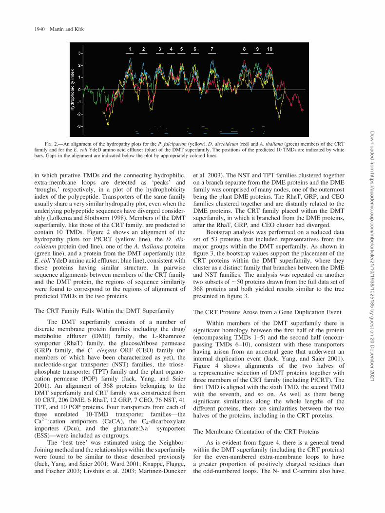
in which putative TMDs and the connecting hydrophilic,extra-membrane loops are detected as ‘peaks’ and‘troughs,’ respectively, in a plot of the hydrophobicityindex of the polypeptide. Transporters of the same familyusually share a very similar hydropathy plot, even when theunderlying polypeptide sequences have diverged consider-ably (Lolkema and Slotboom 1998). Members of the DMTsuperfamily, like those of the CRT family, are predicted tocontain 10 TMDs. Figure 2 shows an alignment of thehydropathy plots for PfCRT (yellow line), the D. dis-coideum protein (red line), one of the A. thaliana proteins(green line), and a protein from the DMT superfamily (theE. coli YdeD amino acid effluxer; blue line), consistent withthese proteins having similar structure. In pairwisesequence alignments between members of the CRT familyand the DMT protein, the regions of sequence similaritywere found to correspond to the regions of alignment ofpredicted TMDs in the two proteins.
The CRT Family Falls Within the DMT Superfamily
The DMT superfamily consists of a number ofdiscrete membrane protein families including the drug/metabolite effluxer (DME) family, the L-Rhamnosesymporter (RhaT) family, the glucose/ribose permease(GRP) family, the C. elegans ORF (CEO) family (nomembers of which have been characterized as yet), thenucleotide-sugar transporter (NST) families, the triose-phosphate transporter (TPT) family and the plant organo-cation permease (POP) family (Jack, Yang, and Saier2001). An alignment of 368 proteins belonging to theDMT superfamily and CRT family was constructed from10 CRT, 206 DME, 6 RhaT, 12 GRP, 7 CEO, 76 NST, 41TPT, and 10 POP proteins. Four transporters from each ofthree unrelated 10-TMD transporter families—theCa21:cation antiporters (CaCA), the C4-dicarboxylateimporters (Dcu), and the glutamate:Na1 symporters(ESS)—were included as outgroups.
The ‘best tree’ was estimated using the Neighbor-Joining method and the relationships within the superfamilywere found to be similar to those described previously(Jack, Yang, and Saier 2001; Ward 2001; Knappe, Flugge,and Fischer 2003; Livshits et al. 2003; Martinez-Duncker
et al. 2003). The NST and TPT families clustered togetheron a branch separate from the DME proteins and the DMEfamily was comprised of many nodes, one of the outermostbeing the plant DME proteins. The RhaT, GRP, and CEOfamilies clustered together and are distantly related to theDME proteins. The CRT family placed within the DMTsuperfamily, in which it branched from the DME proteins,after the RhaT, GRP, and CEO cluster had diverged.
Bootstrap analysis was performed on a reduced dataset of 53 proteins that included representatives from themajor groups within the DMT superfamily. As shown infigure 3, the bootstrap values support the placement of theCRT proteins within the DMT superfamily, where theycluster as a distinct family that branches between the DMEand NST families. The analysis was repeated on anothertwo subsets of ;50 proteins drawn from the full data set of368 proteins and both yielded results similar to the treepresented in figure 3.
The CRT Proteins Arose from a Gene Duplication Event
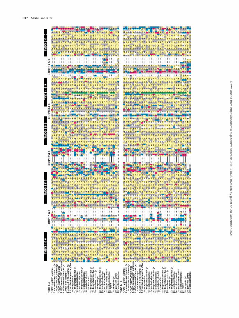
Within members of the DMT superfamily there issignificant homology between the first half of the protein(encompassing TMDs 1–5) and the second half (encom-passing TMDs 6–10), consistent with these transportershaving arisen from an ancestral gene that underwent aninternal duplication event (Jack, Yang, and Saier 2001).Figure 4 shows alignments of the two halves ofa representative selection of DMT proteins together withthree members of the CRT family (including PfCRT). Thefirst TMD is aligned with the sixth TMD, the second TMDwith the seventh, and so on. As well as there beingsignificant similarities along the whole lengths of thedifferent proteins, there are similarities between the twohalves of the proteins, including in the CRT proteins.
The Membrane Orientation of the CRT Proteins
As is evident from figure 4, there is a general trendwithin the DMT superfamily (including the CRT proteins)for the even-numbered extra-membrane loops to havea greater proportion of positively charged residues thanthe odd-numbered loops. The N- and C-termini also have
FIG. 2.—An alignment of the hydropathy plots for the P. falciparum (yellow), D. discoideum (red) and A. thaliana (green) members of the CRTfamily and for the E. coli YdeD amino acid effluxer (blue) of the DMT superfamily. The positions of the predicted 10 TMDs are indicated by whitebars. Gaps in the alignment are indicated below the plot by appropriately colored lines.
1940 Martin and Kirk
Dow
nloaded from https://academ
ic.oup.com/m
be/article/21/10/1938/1025185 by guest on 20 Decem
ber 2021

FIG. 4.—The alignment of the first half (encompassing TMDs 1–5) with the second half (encompassing TMDs 6–10) of a representative selectionof DMT proteins, including three CRT family members. Residues are shaded as follows: positively charged, blue; negatively charged, red; tryptophanand tyrosine, white; polar, proline, and glycine, grey; remaining nonpolar, yellow. The conserved proline in TMDs 4 and 9 is highlighted in green. Thevariant N- and C-termini have been omitted. In some proteins loop 2 and/or 7 has been truncated and this is indicated by a solid black line. ‘(p)’indicates that the assignment of function is only putative. The proteins are as follows: 1, M. musculus gi 2499227 (Eckhardt et al. 1996); 2,D. melanogster gi 21355345 (Martinez-Duncker et al. 2003); 3, K. lactis gi 6016590 (Abeijon, Robbins, and Hirschberg 1996); 4, C. elegans gi20140026 (Berninsone et al. 2001); 5, C. albicans gi 14971021 (Nishikawa et al. 2002); 6, H. sapiens gi 14009667 (Lubke et al. 2001; Luhn et al.2001); 7, Z. mays gi 1352200 (Fischer et al. 1994); 8, A. thaliana gi 15219121; 9, E. coli gi 26107188 (Livshits et al. 2003); 10, E. coli gi 25367911(Santiviago et al. 2002); 11, B. subtilis gi 1175623 (Jack, Yang, and Saier 2001); 12, P. agglomerans gi 4098977; 13, E. coli gi 26250715 (Jack, Yang,and Saier 2001); 14, F. nucleatum gi 19705391; 15, M. loti gi 13473318; 16, M. rubra gi 2062658 (Berg, Hilbi, and Dimroth 1997); 17, E. coli gi13361606 (Dassler et al. 2000); 18, T. erythraeum gi 23043464; 19, B. fungorum gi 22988226; 20, P. marinus gi 33862898; 21, T. fusca gi 23018393;22, B. subtilis gi 16081133 (Jack, Yang, and Saier 2001); 23, C. aurantiacus gi 22972802; 24, A. thaliana gi 25408555; 25, A. thaliana gi 18415262;26, A. thaliana gi 21536591; 27, P. falciparum gi 23612473 (Fidock et al. 2000); 28, D. discoideum gi 11139714; 29, S. xylosus gi 2226001 (Fiegleret al. 1999); 30, C. elegans gi 13384556.
!
FIG. 3.—Neighbour-Joining tree of the DMT superfamily based on uncorrected distances. The analysis included three members of the CRT familyand 38 DMT sequences. Four of the DMT proteins are characterized transporters of the nucleotide-sugar transporter (NST) family and 34 are known orputative members of the drug/metabolite efflux (DME) family. Included for comparison were four sequences from each of three unrelated 10 TMDtransporter families: the Ca21:cation antiporters (CaCA), the C4-dicarboxylate importers (Dcu), and the Glutamate:Na1 symporters (ESS). Numbersindicate the percentage of 1,000 bootstrap replicates which support the topology shown. The scale bar represents the number of substitutions per site fora unit branch length.
The Chloroquine Resistance Transporter 1941
Dow
nloaded from https://academ
ic.oup.com/m
be/article/21/10/1938/1025185 by guest on 20 Decem
ber 2021
1942 Martin and Kirk
Dow
nloaded from https://academ
ic.oup.com/m
be/article/21/10/1938/1025185 by guest on 20 Decem
ber 2021

a preponderance of positive charge. This makes it likely thatthe even-numbered loops, together with the N- and C-termini, are located at the cytoplasmic face of the membrane(predicted by the positive inside rule [von Heijne 1986; vanKlompenburg et al. 1997] and by TMHMM [Sonnhammer,von Heijne, and Krogh 1998]). Such an orientation (i.e.,termini in the cytosol) has been proven experimentally fortwo DMT proteins, the mouse CMP-sialic acid transporter(NST family; Eckhardt, Gotza, and Gerardy-Schahn 1999)and the PecM protein of Erwinia chrysanthemi (DMEfamily Rouanet and Nasser 2001).
For the RhaT, GRP, and CEO proteins it is the odd-numbered loops that have the higher proportion of positivecharge, suggestive of the opposite topology, and this hasbeen demonstrated experimentally for the S. typhimuriumRhaT protein (for which the termini were found to benoncytosolic [Tate and Henderson 1993]).
The Significance of the Extra-Membrane Loops
Having established that PfCRT is a member of theDMT superfamily it is possible to assign putative functionsto different regions of the PfCRT protein on the basisof previous studies of other members of the superfamily(fig. 4).
A striking feature of figure 4 is the conservation ofstructural elements throughout the superfamily, despite theconsiderable divergence in sequence, mode of transport,and substrate specificity. For instance, the length andcomposition of the loops are conserved between proteinsfrom different families. Furthermore, the loop regions inthe second half of the transporter bear significant similarityto the corresponding loops in the first half. Loops 3 and 8and 4 and 9, are particularly well conserved. Studies withproteins from both the NST and DME families haverevealed that the insertion of an epitope tag or reportermolecule in loops 3, 4, 8, and 9 can inactivate thetransporter and in some instances cause the protein to belocalized incorrectly within the cell and/or degraded (Tateand Henderson 1993; Eckhardt, Gotza, and Gerardy-Schahn 1999; Rouanet and Nasser 2001). By contrast, thesame sequences introduced into loops 1, 2, 5, and 6 haveno effect on transporter activity or localization.
Compared to the other loops, 2 and 7 show lesssequence conservation and show significant variation inlength between DMT proteins. In most DMT proteins loop
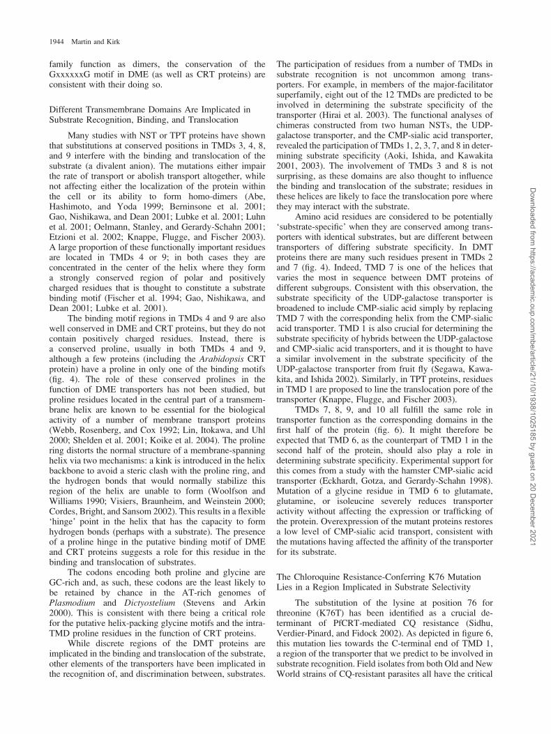
7 is a relatively long hydrophilic domain, changes in whichhave been shown to influence transporter activity. Theactivity of the mouse CMP-sialic acid transporter isreduced when an epitope is inserted into loop 7 (but notloop 2), and increasing the length of the tag causesa further reduction in transporter activity (Eckhardt, Gotza,and Gerardy-Schahn 1999). As illustrated in figure 5, loop 7is especially long and well conserved in composition inthe proteins of the CRT family, and the predicted structureof this region (given by the PredictProtein sever) wassimilar for each protein: a ‘compact globular domain’ thatis formed by a nine amino acid alpha helix followed by twoshort beta sheets (although in some proteins the first betasheet may instead be an alpha-helix).
The predicted orientation of PfCRT in the membrane(i.e., with the protein termini in the cytosol) places loop 7in the digestive vacuole, where it may have a role inmodulating the activity of the transporter.
The Presence of Helix Packing Motifs inTransmembrane Domains 5 and 10 Indicatesthat PfCRT May Function as a Dimer
While the structure of the DMT transporters has beenretained over time and evolution, the underlying aminoacid sequence has proven more plastic. Nevertheless, someregions of sequence have been strongly conserved. InTMDs 5 and 10 there are two conserved glycines that areseparated by six hydrophobic residues (fig. 4; Knappe,Flugge, and Fischer 2003). This motif (GxxxxxxG) isa common feature of membrane proteins, in which it isthought to facilitate the packing of membrane-spanninghelices, leading to the association of TMDs to formoligomers (Liu, Engelman, and Gerstein 2002). Othersmall residues (alanine, serine, and threonine) can replaceone of the glycines in a glycine-packing motif (Russ andEngelman 2000; Eilers et al. 2002) and such a substitutionhas occurred in a number of the DMT proteins, includingPfCRT (fig. 4). TMDs 5 and 10 are known to have a role inmediating the formation of homo-dimers by the NST andTPT transporters (Abe, Hashimoto, and Yoda 1999; Ishidaet al. 1999; Streatfield et al. 1999; Gao and Dean 2000)and nonconservative mutations within the putative packingmotifs in TMDs 5 and 10 abrogate transporter activity aswell as stability (Ishida et al. 1999; Streatfield et al. 1999).Although it is not known whether members of the DME
FIG. 5.—Alignment of CRT proteins over TMD 1 and loop 7. Residues are shaded as follows: positively charged, black; negatively charged, darkgrey; tryptophan and tyrosine, white; polar, proline and glycine, mid-grey; remaining nonpolar, light grey. The position of the K76T mutation in TMD1 is indicated (star) and CQ-resistant parasite strains (numbered 5 and 6) are highlighted.
The Chloroquine Resistance Transporter 1943
Dow
nloaded from https://academ
ic.oup.com/m
be/article/21/10/1938/1025185 by guest on 20 Decem
ber 2021
family function as dimers, the conservation of theGxxxxxxG motif in DME (as well as CRT proteins) areconsistent with their doing so.
Different Transmembrane Domains Are Implicated inSubstrate Recognition, Binding, and Translocation
Many studies with NST or TPT proteins have shownthat substitutions at conserved positions in TMDs 3, 4, 8,and 9 interfere with the binding and translocation of thesubstrate (a divalent anion). The mutations either impairthe rate of transport or abolish transport altogether, whilenot affecting either the localization of the protein withinthe cell or its ability to form homo-dimers (Abe,Hashimoto, and Yoda 1999; Berninsone et al. 2001;Gao, Nishikawa, and Dean 2001; Lubke et al. 2001; Luhnet al. 2001; Oelmann, Stanley, and Gerardy-Schahn 2001;Etzioni et al. 2002; Knappe, Flugge, and Fischer 2003).A large proportion of these functionally important residuesare located in TMDs 4 or 9; in both cases they areconcentrated in the center of the helix where they forma strongly conserved region of polar and positivelycharged residues that is thought to constitute a substratebinding motif (Fischer et al. 1994; Gao, Nishikawa, andDean 2001; Lubke et al. 2001).
The binding motif regions in TMDs 4 and 9 are alsowell conserved in DME and CRT proteins, but they do notcontain positively charged residues. Instead, there isa conserved proline, usually in both TMDs 4 and 9,although a few proteins (including the Arabidopsis CRTprotein) have a proline in only one of the binding motifs(fig. 4). The role of these conserved prolines in thefunction of DME transporters has not been studied, butproline residues located in the central part of a transmem-brane helix are known to be essential for the biologicalactivity of a number of membrane transport proteins(Webb, Rosenberg, and Cox 1992; Lin, Itokawa, and Uhl2000; Shelden et al. 2001; Koike et al. 2004). The prolinering distorts the normal structure of a membrane-spanninghelix via two mechanisms: a kink is introduced in the helixbackbone to avoid a steric clash with the proline ring, andthe hydrogen bonds that would normally stabilize thisregion of the helix are unable to form (Woolfson andWilliams 1990; Visiers, Braunheim, and Weinstein 2000;Cordes, Bright, and Sansom 2002). This results in a flexible‘hinge’ point in the helix that has the capacity to formhydrogen bonds (perhaps with a substrate). The presenceof a proline hinge in the putative binding motif of DMEand CRT proteins suggests a role for this residue in thebinding and translocation of substrates.
The codons encoding both proline and glycine areGC-rich and, as such, these codons are the least likely tobe retained by chance in the AT-rich genomes ofPlasmodium and Dictyostelium (Stevens and Arkin2000). This is consistent with there being a critical rolefor the putative helix-packing glycine motifs and the intra-TMD proline residues in the function of CRT proteins.
While discrete regions of the DMT proteins areimplicated in the binding and translocation of the substrate,other elements of the transporters have been implicated inthe recognition of, and discrimination between, substrates.
The participation of residues from a number of TMDs insubstrate recognition is not uncommon among trans-porters. For example, in members of the major-facilitatorsuperfamily, eight out of the 12 TMDs are predicted to beinvolved in determining the substrate specificity of thetransporter (Hirai et al. 2003). The functional analyses ofchimeras constructed from two human NSTs, the UDP-galactose transporter, and the CMP-sialic acid transporter,revealed the participation of TMDs 1, 2, 3, 7, and 8 in deter-mining substrate specificity (Aoki, Ishida, and Kawakita2001, 2003). The involvement of TMDs 3 and 8 is notsurprising, as these domains are also thought to influencethe binding and translocation of the substrate; residues inthese helices are likely to face the translocation pore wherethey may interact with the substrate.
Amino acid residues are considered to be potentially‘substrate-specific’ when they are conserved among trans-porters with identical substrates, but are different betweentransporters of differing substrate specificity. In DMTproteins there are many such residues present in TMDs 2and 7 (fig. 4). Indeed, TMD 7 is one of the helices thatvaries the most in sequence between DMT proteins ofdifferent subgroups. Consistent with this observation, thesubstrate specificity of the UDP-galactose transporter isbroadened to include CMP-sialic acid simply by replacingTMD 7 with the corresponding helix from the CMP-sialicacid transporter. TMD 1 is also crucial for determining thesubstrate specificity of hybrids between the UDP-galactoseand CMP-sialic acid transporters, and it is thought to havea similar involvement in the substrate specificity of theUDP-galactose transporter from fruit fly (Segawa, Kawa-kita, and Ishida 2002). Similarly, in TPT proteins, residuesin TMD 1 are proposed to line the translocation pore of thetransporter (Knappe, Flugge, and Fischer 2003).
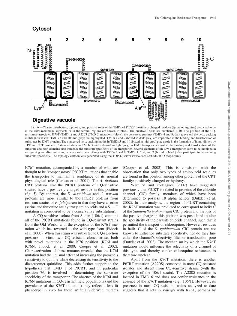
TMDs 7, 8, 9, and 10 all fulfill the same role intransporter function as the corresponding domains in thefirst half of the protein (fig. 6). It might therefore beexpected that TMD 6, as the counterpart of TMD 1 in thesecond half of the protein, should also play a role indetermining substrate specificity. Experimental support forthis comes from a study with the hamster CMP-sialic acidtransporter (Eckhardt, Gotza, and Gerardy-Schahn 1998).Mutation of a glycine residue in TMD 6 to glutamate,glutamine, or isoleucine severely reduces transporteractivity without affecting the expression or trafficking ofthe protein. Overexpression of the mutant proteins restoresa low level of CMP-sialic acid transport, consistent withthe mutations having affected the affinity of the transporterfor its substrate.
The Chloroquine Resistance-Conferring K76 MutationLies in a Region Implicated in Substrate Selectivity
The substitution of the lysine at position 76 forthreonine (K76T) has been identified as a crucial de-terminant of PfCRT-mediated CQ resistance (Sidhu,Verdier-Pinard, and Fidock 2002). As depicted in figure 6,this mutation lies towards the C-terminal end of TMD 1,a region of the transporter that we predict to be involved insubstrate recognition. Field isolates from both Old and NewWorld strains of CQ-resistant parasites all have the critical
1944 Martin and Kirk
Dow
nloaded from https://academ
ic.oup.com/m
be/article/21/10/1938/1025185 by guest on 20 Decem
ber 2021
K76T mutation, accompanied by a number of what arethought to be ‘compensatory’ PfCRT mutations that enablethe transporter to maintain a semblance of its normalphysiological role (Carlton et al. 2001). The A. thalianaCRT proteins, like the PfCRT proteins of CQ-sensitivestrains, have a positively charged residue in this position(fig. 5). By contrast, the D. discoideum and C. parvumproteins are more similar to the PfCRT proteins fromresistant strains of P. falciparum in that they have a serine(serine and threonine are hydroxy amino acids and a S ! Tmutation is considered to be a conservative substitution).
A CQ-sensitive isolate from Sudan (106/1) containsall of the PfCRT mutations found in CQ-resistant strainsfrom the Old World, with the exception of the K76T mu-tation which has reverted to the wild-type form (Fidocket al. 2000). When this strain was subjected to CQ-selectionpressure in vitro, two CQ-resistant clones arose, bothwith novel mutations in the K76 position (K76I andK76N; Fidock et al. 2000; Cooper et al. 2002).Characterization of these mutants revealed that the K76Imutation had the unusual effect of increasing the parasite’ssensitivity to quinine while decreasing its sensitivity to thediastereomer quinidine. This adds further support to thehypothesis that TMD 1 of PfCRT, and in particularposition 76, is involved in determining the substratespecificity of the transporter. The absence of the K76I andK76N mutations in CQ-resistant field populations (and theprevalence of the K76T mutation) may reflect a less fitphenotype in vivo for these artificially-derived mutants
(Cooper et al. 2002). This is consistent with theobservation that only two types of amino acid residuesare found in this position among other proteins of the CRTfamily: positively charged or hydroxy.
Warhurst and colleagues (2002) have suggestedpreviously that PfCRT is related to proteins of the chloridechannel (ClC) family, members of which have beendetermined to possess 18 alpha helices (Dutzler et al.2002). In their analysis, the region of PfCRT containingthe K76T mutation was predicted to correspond to helix Cof the Salmonella typhimurium ClC protein and the loss ofthe positive charge in this position was postulated to alterthe specificity of the parasite chloride channel, such that itpermitted the transport of chloroquine. However, residuesin helix C of the S. typhimurium ClC protein are notknown to influence substrate specificity, nor do they lineeither the channel’s selectivity filter or translocation pore(Dutzler et al. 2002). The mechanism by which the K76Tmutation would influence the selectivity of a channel ofthis type, and thereby confer chloroquine resistance, istherefore unclear.
Apart from the K76T mutation, there is anotherPfCRT mutation (A220S) conserved in most CQ-resistantisolates and absent from CQ-sensitive strains (with theexception of the 106/1 strain). The A220S mutation islocated in TMD 6 and does not confer resistance in theabsence of the K76T mutation (e.g., 106/1). However, itspresence in most CQ-resistant strains analyzed to datesuggests that it acts in synergy with K76T, perhaps by
FIG. 6.—Charge distribution, topology, and putative roles of the TMDs of PfCRT. Positively charged residues (lysine or arginine) predicted to liein the extra-membrane segments or in the termini regions are shown in black. The putative TMDs are numbered 1–10. The position of the CQ-resistance-associated K76T (TMD 1) and A220S (TMD 6) mutations (black), the conserved prolines (TMDs 4 and 9; dark grey) and the helix packingmotifs (GxxxxxxT; TMDs 5 and 10; mid-grey) are highlighted. TMDs 4 and 9 (boxed in dark grey) are implicated in the binding and translocation ofsubstrates by DMT proteins. The conserved helix packing motifs in TMDs 5 and 10 (boxed in mid-grey) play a role in the formation of homo-dimers byTPT and NST proteins. Certain residues in TMDs 3 and 8 (boxed in light grey) in DMT transporters assist in the binding and translocation of thesubstrate and both domains also influence the substrate specificity of the transporter. Several elements of the DMT transporter seem to be involved inrecognizing and discriminating between substrates. Along with TMDs 3 and 8, TMDs 1, 2, 6, and 7 (boxed in black) also participate in determiningsubstrate specificity. The topology cartoon was generated using the TOPO2 server (www.sacs.ucsf.edu/TOPO/topo.html).
The Chloroquine Resistance Transporter 1945
Dow
nloaded from https://academ
ic.oup.com/m
be/article/21/10/1938/1025185 by guest on 20 Decem
ber 2021

aiding the recognition of CQ as a substrate for PfCRT or asa compensatory mutation that stabilizes the interaction ofthe transporter with its physiological substrate(s). Thelocation of the A220 mutation in a PfCRT domainpredicted to participate in substrate recognition is consis-tent with both of these scenarios.
Substrates Effluxed by DME Transporters
The members of the DMT superfamily bearing theclosest similarity to the CRT proteins in the region of thesubstrate binding motif fall within the DME transportersubfamily. Substrates for DME transporters include aminoacids, weak bases, and organic cations. The YdeD proteinof E. coli exports cysteine metabolites (Dassler et al. 2000),whereas the E. coli YbiF protein exports a broad range ofamino acids including homoserine, threonine, lysine, andhistidine (Livshits et al. 2003). In other species of bacteria,DME proteins are implicated in the efflux of methylamine(MttP; Ferguson and Krzycki 1997), the di-cationicherbicide methyl viologen (YddG; Santiviago et al. 2002)and the pigment indigoidine, which is, like chloroquine,a weak base (PecM; Rouanet and Nasser 2001). The factthat DME transporters are known to transport both weakbases and divalent organic cations lends support to thehypothesis that the CQ-resistant form of PfCRT transportsthe chloroquine in the di-cationic form.
DME systems are postulated to be H1-coupled andthis has been confirmed experimentally for at least oneDME transporter (the E. coli YbiF protein) [Livshits et al.1993].
The Role of PfCRT in Chloroquine Resistance
Figure 7 shows a model for the mechanism ofPfCRT-mediated CQ-resistance, based on the insightsgained from the bioinformatic analysis presented here, andconsistent with previous reports of enhanced CQ effluxfrom CQ-resistant parasites (Krogstad et al. 1987;
Sanchez, Stein, and Lanzer 2003). The protein is shownas a dimer, functioning to export ‘metabolites’ from theparasite’s digestive vacuole. The facts that (1) a number ofrelated DME proteins transport amino acids, and (2) thatthe only known metabolite transport function of thedigestive vacuole is the efflux of peptides (Kolakovich etal. 1997) and perhaps amino acids, prompt the hypothesisthat PfCRT is an amino acid/peptide transporter (perhapsH1-coupled), but this remains to be tested.
The protein is predicted, on the basis of the ‘positive-inside’ rule as well as by TMHMM, to be oriented with theN- and C-termini in the parasite cytosol and the ‘compactglobular domain’ of loop 7 located at the vacuolar face ofthe membrane. In this orientation, the crucial CQR-conferring K76T mutation lies close to the surface of thevacuolar face of the membrane, within a region of theprotein postulated to be involved in substrate selectivity.The positive charge is predicted to play a key role inrepulsing the protonated (cationic) form of chloroquine(CQ21, the predominant species present within the acidicvacuole) and preventing it from interacting with thetransporter. The CQR-conferring mutation of Lys to Thr(or Ile or Asn) removes the positive charge, allowingCQ21 to interact with and be transported by the protein,down the steep outward CQ21 concentration gradient(again perhaps in symport with H1). On exiting thevacuole and entering the parasite cytosol (pH 7.3; Salibaand Kirk 1999), the CQ is deprotonated and diffuses out ofthe parasite as the neutral (membrane-permeant) species.The net result is a decrease in the overall concentration ofCQ at its site of action within the digestive vacuole, andhence a decreased CQ-sensitivity of the parasite.
Experimental testing of the key aspects of the modelpresented in figure 7 await the expression of PfCRT ina heterologous system in a form in which the transportproperties of the protein can be investigated directly. PfCRThas been successfully expressed in yeast (Zhang, Howard,and Roepe 2002); however, there has not, as yet, been anydirect demonstration of its transport function. Efforts are
FIG. 7.—Model for the mechanism of PfCRT-mediated resistance to CQ. The protein is shown as a dimer, functioning to export ‘metabolites’(perhaps amino acids or peptides, and perhaps in symport with H1) from the parasite’s digestive vacuole (DV). The ‘positive-inside’ rule, and a presumedinwardly-positive electrical potential across the DV membrane, predicts the N- and C-termini to be cytosolic and the ‘compact globular domain’ of loop 7to be located at the vacuolar face of the membrane. CQ is a diprotic weak base and therefore accumulates in the acidic DV in the protonated (positivelycharged, CQ21) form. In parasites expressing ‘wild type’ PfCRT, the positive charge of K76 prevents the interaction of CQ21 with the transporter. TheCQ resistance–conferring K to T mutation removes the positive charge and alters the substrate selectivity, allowing CQ21 to interact with the transporterand to be effluxed from the DV, perhaps in symport with H1. This results in a reduction of the concentration of CQ within the vacuole.
1946 Martin and Kirk
Dow
nloaded from https://academ
ic.oup.com/m
be/article/21/10/1938/1025185 by guest on 20 Decem
ber 2021
presently underway to express the protein in Xenopus laevisoocytes and to measure and compare the transport ofradiolabeled chloroquine via PfCRT with and without theK76T mutation. It is predicted that oocytes expressingPfCRT from CQ-resistant strains (i.e., having the K76Tmutation) in their plasma membrane will transport [3H]CQ,whereas those expressing wild-type PfCRT from CQ-sensitive strains (having K76) will not. The successfulexpression of PfCRT inXenopus oocytes will also allow: (1)a direct test of the hypothesis that PfCRT proteins from bothCQ-resistant and CQ-sensitive strains transport aminoacids/peptides; (2) screening of other classes of substratefor their ability to be transported via PfCRT; (3) thedetermination of whether PfCRT-mediated transport is H1
coupled; and (4) an investigation of whether, as has beenproposed (Warhurst 2003), the chloroquine resistancereversal agent verapamil interacts directly with PfCRT (inTMD 1) to inhibit the transport of CQ. Such experimentshave the potential to yield important insights into themolecular mechanism underlying chloroquine resistance.
Acknowledgments
The authors are grateful to Stephen Allen and JanMartin for technical assistance and to Rhys Hayward,Kevin Saliba, and John Trueman for helpful discussions.This work was supported by the Australian NationalHealth and Medical Research Council (grant ID179804).
Literature Cited
Abe, M., H. Hashimoto, and K. Yoda. 1999. Molecularcharacterization of Vig4/Vrg4 GDP-mannose transporter ofthe yeast Saccharomyces cerevisiae. FEBS Lett. 458:309–312.
Abeijon, C., P. W. Robbins, and C. B. Hirschberg. 1996.Molecular cloning of the Golgi apparatus uridine diphosphate-N-acetylglucosamine transporter from Kluyveromyces lactis.Proc. Natl. Acad. Sci. USA 93:5963–5968.
Altschul, S. F., W. Gish, W. Miller, E. W. Myers, and D. J.Lipman. 1990. Basic local alignment search tool. J. Mol. Biol.215:403–410.
Altschul, S. F., T. L. Madden, A. A. Schaffer, J. Zhang, Z.Zhang, W. Miller, and D. J. Lipman. 1997. Gapped Blast andPSI-Blast: a new generation of protein database searchprograms. Nucleic Acids Res. 25:3389–3402.
Aoki, K., N. Ishida, and M. Kawakita. 2001. Substraterecognition by UDP-galactose and CMP-sialic acid trans-porters. Different sets of transmembrane helices are utilizedfor the specific recognition of UDP-galactose and CMP-sialicacid. J. Biol. Chem. 276:21555–21561.
———. 2003. Substrate recognition by nucleotide sugar trans-porters: further characterization of substrate recognitionregions by analyses of UDP-galactose/CMP-sialic acid trans-porter chimeras and biochemical analysis of the substratespecificity of parental and chimeric transporters. J. Biol.Chem. 278:22887–22893.
Berg, M., H. Hilbi, and P. Dimroth. 1997. Sequence of a genecluster from Malonomonas rubra encoding components of themalonate decarboxylase Na1 pump and evidence for theirfunction. Eur. J. Biochem. 245:103–115.
Berninsone, P., H. Y. Hwang, I. Zemtseva, H. R. Horvitz, and C.B. Hirschberg. 2001. SQV-7, a protein involved inCaenorhabditis elegans epithelial invagination and earlyembryogenesis, transports UDP-glucuronic acid, UDP-N-
acetylgalactosamine, and UDP-galactose. Proc. Natl. Acad.Sci. USA 98:3738–3743.
Carlton, J. M., D. A. Fidock, A. Djimde, C. V. Plowe, and T. E.Wellems. 2001. Conservation of a novel vacuolar transporterin Plasmodium species and its central role in chloroquineresistance of P. falciparum. Curr. Opin. Microbiol. 4:415–420.
Cooper, R. A., M. T. Ferdig, X. Z. Su, L. M. Ursos, J. Mu, T.Nomura, H. Fujioka, D. A. Fidock, P. D. Roepe, and T. E.Wellems. 2002. Alternative mutations at position 76 of thevacuolar transmembrane protein PfCRT are associated withchloroquine resistance and unique stereospecific quinine andquinidine responses in Plasmodium falciparum. Mol. Phar-macol. 61:35–42.
Cordes, F. S., J. N. Bright, and M. S. Sansom. 2002. Proline-induced distortions of transmembrane helices. J. Mol. Biol.323:951–960.
Dassler, T., T. Maier, C. Winterhalter, and A. Bock. 2000.Identification of a major facilitator protein from Escherichiacoli involved in efflux of metabolites of the cysteine pathway.Mol. Microbiol. 36:1101–1112.
Dutzler, R., E. B. Campbell, M. Cadene, B. T. Chait, and R.MacKinnon. 2002. X-ray structure of a ClC chloride channelat 3.0 A reveals the molecular basis of anion selectivity.Nature 415:287–294.
Eckhardt, M., B. Gotza, and R. Gerardy-Schahn. 1998. Mutantsof the CMP-sialic acid transporter causing the Lec2phenotype. J. Biol. Chem. 273:20189–20195.
———. 1999. Membrane topology of the mammalian CMP-sialic acid transporter. J. Biol. Chem. 274:8779–8787.
Eckhardt, M., M. Muhlenhoff, A. Bethe, and R. Gerardy-Schahn.1996. Expression cloning of the Golgi CMP-sialic acidtransporter. Proc. Natl. Acad. Sci. USA 93:7572–7576.
Eilers, M., A. B. Patel, W. Liu, and S. O. Smith. 2002. Com-parison of helix interactions in membrane and soluble alpha-bundle proteins. Biophys. J. 82:2720–2736.
Etzioni, A., L. Sturla, A. Antonellis, E. D. Green, R. Gershoni-Baruch, P. M. Berninsone, C. B. Hirschberg, and M. Tonetti.2002. Leukocyte adhesion deficiency (LAD) type II/carbohy-drate deficient glycoprotein (CDG) IIc founder effect andgenotype/phenotype correlation. Am. J. Med. Genet. 110:131–135.
Felsenstein, J. 1985. Confidence limits on phylogenies: an app-roach using the bootstrap. Evolution 39:783–791.
Ferguson, D. J. Jr., and J. A. Krzycki. 1997. Reconstitution oftrimethylamine-dependent coenzyme M methylation with thetrimethylamine corrinoid protein and the isozymes of methyl-transferase II from Methanosarcina barkeri. J. Bacteriol.179:846–852.
Fidock, D. A., T. Nomura, A. K. Talley et al. (14 co-authors).2000. Mutations in the P. falciparum digestive vacuoletransmembrane protein PfCRT and evidence for their role inchloroquine resistance. Mol. Cell 6:861–871.
Fiegler, H., J. Bassias, I. Jankovic, and R. Bruckner. 1999.Identification of a gene in Staphylococcus xylosus encod-ing a novel glucose uptake protein. J. Bacteriol. 181:4929–4936.
Fischer, K., B. Arbinger, B. Kammerer, C. Busch, S. Brink, H.Wallmeier, N. Sauer, C. Eckerskorn, and U. I. Flugge. 1994.Cloning and in vivo expression of functional triose phosphate/phosphate translocators from C3- and C4-plants: evidence forthe putative participation of specific amino acid residues in therecognition of phosphoenolpyruvate. Plant J. 5:215–226.
Fitch, C. D. 1970. Plasmodium falciparum in owl monkeys: drugresistance and chloroquine binding capacity. Science 169:289–290.
Gao, X. D., and N. Dean. 2000. Distinct protein domains of theyeast Golgi GDP-mannose transporter mediate oligomer
The Chloroquine Resistance Transporter 1947
Dow
nloaded from https://academ
ic.oup.com/m
be/article/21/10/1938/1025185 by guest on 20 Decem
ber 2021
assembly and export from the endoplasmic reticulum. J. Biol.Chem. 275:17718–17727.
Gao, X. D., A. Nishikawa, and N. Dean. 2001. Identification ofa conserved motif in the yeast golgi GDP-mannose transporterrequired for binding to nucleotide sugar. J. Biol. Chem.276:4424–4432.
Geer, L. Y., M. Domrachev, D. J. Lipman, and S. H. Bryant.2002. CDART: protein homology by domain architecture.Genome Res. 12:1619–1623.
Hirai, T., J. A. Heymann, P. C. Maloney, and S. Subramaniam.2003. Structural model for 12-helix transporters belonging tothe major facilitator superfamily. J. Bacteriol. 185:1712–1718.
Ishida, N., S. Yoshioka, M. Iida, K. Sudo, N. Miura, K. Aoki,and M. Kawakita. 1999. Indispensability of transmembranedomains of Golgi UDP-galactose transporter as revealed byanalysis of genetic defects in UDP-galactose transporter-deficient murine had-1 mutant cell lines and construction ofdeletion mutants. J. Biochem. 126:1107–1117.
Jack, D. L., N. M. Yang, and M. H. Saier, Jr. 2001. The drug/metabolite transporter superfamily. Eur. J. Biochem. 268:3620–3639.
Knappe, S., U. I. Flugge, and K. Fischer. 2003. Analysis of theplastidic phosphate translocator gene family in Arabidopsisand identification of new phosphate translocator-homologoustransporters, classified by their putative substrate-binding site.Plant Physiol. 131:1178–1190.
Koike, K., G. Conseil, H. E. Leslie, H. I. Deeley, and S. P. Cole.2004. Identification of proline residues in the core cytoplasmicand transmembrane regions of multidrug resistance protein 1(MRP1/ABCC1) important for transport function, substratespecificity, and nucleotide interactions. J. Biol. Chem. 279:12325–12336.
Kolakovich, K. A., I. Y. Gluzman, K. L. Duffin, and D. E.Goldberg. 1997. Generation of hemoglobin peptides in theacidic digestive vacuole of Plasmodium falciparum implicatespeptide transport in amino acid production. Mol. Biochem.Parasitol. 87:123–135.
Krogstad, D. J., I. Y. Gluzman, D. E. Kyle, A. M. Oduola, S. K.Martin, W. K. Milhous, and P. H. Schlesinger. 1987. Efflux ofchloroquine from Plasmodium falciparum: mechanism ofchloroquine resistance. Science 238:1283–1285.
Lin, Z., M. Itokawa, and G. R. Uhl. 2000. Dopamine transporterproline mutations influence dopamine uptake, cocaine analogrecognition, and expression. FASEB J. 14:715–728.
Liu, Y., D. M. Engelman, and M. Gerstein. 2002. Genomicanalysis of membrane protein families: abundance andconserved motifs. Genome Biol. 3:research0054.
Livshits, V. A., N. P. Zakataeva, V. V. Aleshin, and M. V.Vitushkina. 2003. Identification and characterization of thenew gene rhtA involved in threonine and homoserine efflux inEscherichia coli. Res. Microbiol. 154:123–135.
Lolkema, J. S., and D. J. Slotboom. 1998. Hydropathy profilealignment: a tool to search for structural homologues ofmembrane proteins. FEMS Microbiol. Rev. 22:305–322.
Lubke, T., T. Marquardt, A. Etzioni, E. Hartmann, K. von Figura,and C. Korner. 2001. Complementation cloning identifiesCDG-IIc, a new type of congenital disorders of glycosylation,as a GDP-fucose transporter deficiency. Nat. Genet. 28:73–76.
Luhn, K., M. K. Wild, M. Eckhardt, R. Gerardy-Schahn, andD. Vestweber. 2001. The gene defective in leukocyte adhesiondeficiency II encodes a putative GDP-fucose transporter. Nat.Genet. 28:69–72.
Marchler-Bauer, A., A. R. Panchenko, B. A. Shoemaker, P. A.Thiessen, L. Y. Geer, and S. H. Bryant. 2002. CDD:a database of conserved domain alignments with links todomain three-dimensional structure. Nucleic Acids Res. 30:281–283.
Martin, R. E., J. W. H. Trueman, and K. Kirk. 2003.Bioinformatic analysis of PfCRT places it in a known familyof transport proteins. Exp. Parasitol. 105:56–57.
Martinez-Duncker, I., R. Mollicone, P. Codogno, and R. Oriol.2003. The nucleotide-sugar transporter family: a phylogeneticapproach. Biochimie 85:245–260.
Nishikawa, A., J. B. Poster, Y. Jigami, and N. Dean. 2002.Molecular and phenotypic analysis of CaVRG4, encoding anessential Golgi apparatus GDP-mannose transporter. J.Bacteriol. 184:29–42.
Oelmann, S., P. Stanley, and R. Gerardy-Schahn. 2001. Pointmutations identified in Lec8 Chinese hamster ovary glycosyl-ation mutants that inactivate both the UDP-galactose and CMP-sialic acid transporters. J. Biol. Chem. 276:26291–26300.
Reed, M. B., K. J. Saliba, S. R. Caruana, K. Kirk, and A. F.Cowman. 2000. Pgh1 modulates sensitivity and resistance tomultiple antimalarials in Plasmodium falciparum. Nature403:906–909.
Rouanet, C., and W. Nasser. 2001. The PecM protein of thephytopathogenic bacterium Erwinia chrysanthemi, membranetopology and possible involvement in the efflux of the bluepigment indigoidine. J. Mol. Microbiol. Biotechnol. 3:309–318.
Russ, W. P., and D. M. Engelman. 2000. The GxxxG motif:a framework for transmembrane helix-helix association. J.Mol. Biol. 296:911–919.
Saitou, N., and M. Nei. 1987. The neighbor-joining method:a new method for reconstructing phylogenetic trees. Mol.Biol. Evol. 4:406–425.
Saliba, K. J., P. I. Folb, and P. J. Smith. 1998. Role for thePlasmodium falciparum digestive vacuole in chloroquineresistance. Biochem. Pharmacol. 56:313–320.
Saliba, K. J., and K. Kirk. 1999. pH regulation in the intracellularmalaria parasite, Plasmodium falciparum: H1 extrusion viaa V-type H1-ATPase. J. Biol. Chem. 274:33213–33219.
Sanchez, C. P., W. Stein, and M. Lanzer. 2003. Trans stimulationprovides evidence for a drug efflux carrier as the mechanismof chloroquine resistance in Plasmodium falciparum.Biochemistry 42:9383–9394.
Santiviago, C. A., J. A. Fuentes, S. M. Bueno, A. N.Trombert, A. A. Hildago, L. T. Socias, P. Youderian, andG. C. Mora. 2002. The Salmonella enterica sv. Typhimu-rium smvA, yddG and ompD (porin) genes are required forthe efficient efflux of methyl viologen. Mol. Microbiol.46:687–698.
Segawa, H., M. Kawakita, and N. Ishida. 2002. Human andDrosophila UDP-galactose transporters transport UDP-N-acetylgalactosamine in addition to UDP-galactose. Eur. J.Biochem. 269:128–138.
Shelden, M. C., P. Loughlin, M. L. Tierney, and S. M. Howitt.2001. Proline residues in two tightly coupled helices of thesulphate transporter, SHST1, are important for sulphatetransport. Biochem. J. 356:589–594.
Sidhu, A. B., D. Verdier-Pinard, and D. A. Fidock. 2002.Chloroquine resistance in Plasmodium falciparum malariaparasites conferred by pfcrt mutations. Science 298:210–213.
Sonnhammer, E. L., G. von Heijne, and A. Krogh. 1998.A hidden Markov model for predicting transmembrane helicesin protein sequences. Proc. Int. Conf. Intell. Syst. Mol. Biol.6:175–182.
Stevens, T. J., and I. T. Arkin. 2000. The effect of nucleotide biasupon the composition and prediction of transmembranehelices. Protein Sci. 9:505–511.
Streatfield, S. J., A. Weber, E. A. Kinsman, R. E. Hausler, J. Li,D. Post-Beittenmiller, W. M. Kaiser, K. A. Pyke, U. I. Flugge,and J. Chory. 1999. The phosphoenolpyruvate/phosphatetranslocator is required for phenolic metabolism, palisade cell
1948 Martin and Kirk
Dow
nloaded from https://academ
ic.oup.com/m
be/article/21/10/1938/1025185 by guest on 20 Decem
ber 2021

development, and plastid-dependent nuclear gene expression.Plant Cell 11:1609–1622.
Tate, C. G., and P. J. Henderson. 1993. Membrane topology ofthe L-rhamnose-H1 transport protein (RhaT) from enter-obacteria. J. Biol. Chem. 268:26850–26857.
Thompson, J. D., D. G. Higgins, and T. J. Gibson. 1994.ClustalW: improving the sensitivity of progressive multiplesequence alignment through sequence weighting, position-specific gap penalties and weight matrix choice. NucleicAcids Res. 22:4673–4680.
Tran, C. V., and M. H. Saier, Jr. 2004. The principal chloroquineresistance protein of Plasmodium falciparum is a member ofthe drug/metabolite transporter superfamily. Microbiology150:1–3.
van Klompenburg, W., I. Nilsson, G. von Heijne, and B. deKruijff. 1997. Anionic phospholipids are determinants ofmembrane protein topology. EMBO J. 16:4261–4266.
Visiers, I., B. B. Braunheim, and H. Weinstein. 2000. Prokink:a protocol for numerical evaluation of helix distortions byproline. Protein Eng. 13:603–606.
von Heijne, G. 1986. The distribution of positively chargedresidues in bacterial inner membrane proteins correlates withthe trans-membrane topology. EMBO J. 5:3021–3027.
Ward, J. M. 2001. Identification of novel families of membraneproteins from the model plant Arabidopsis thaliana. Bio-informatics 17:560–563.
Warhurst, D. C. 2003. Polymorphism in the Plasmodiumfalciparum chloroquine-resistance transporter protein linksverapamil enhancement of chloroquine sensitivity with theclinical efficacy of amodiaquine. Malaria J. 2:31.
Warhurst, D. C., J. C. Craig, and I. S. Adagu. 2002. Lysosomesand drug resistance in malaria. Lancet 360:1527–1529.
Webb, D. C., H. Rosenberg, and G. B. Cox. 1992. Mutationalanalysis of the Escherichia coli phosphate-specific transportsystem, a member of the traffic ATPase (or ABC) family ofmembrane transporters. a role for proline residues in trans-membrane helices. J. Biol. Chem. 267:24661–24668.
Woolfson, D. N., and D. H. Williams. 1990. The influence of prolineresidues on alpha-helical structure. FEBS Lett. 277:185–188.
Zhang, H., E. M. Howard, and P. D. Roepe. 2002. Analysis of theantimalarial drug resistance protein Pfcrt expressed in yeast. J.Biol. Chem. 277:49767–49775.
Michele Vendruscolo, Associate Editor
Accepted June 28, 2004
The Chloroquine Resistance Transporter 1949
Dow
nloaded from https://academ
ic.oup.com/m
be/article/21/10/1938/1025185 by guest on 20 Decem
ber 2021