THE MAIN METHODS OF FUNCTIONAL DIAGNOSTICS OF DISEASES OF GASTROENTEROLOGY.
29
THE MAIN METHODS OF FUNCTIONAL DIAGNOSTICS OF DISEASES OF GASTROENTEROLOGY
-
Upload
bertha-cobb -
Category
Documents
-
view
242 -
download
3
Transcript of THE MAIN METHODS OF FUNCTIONAL DIAGNOSTICS OF DISEASES OF GASTROENTEROLOGY.
THE MAIN METHODS OF FUNCTIONAL DIAGNOSTICS OF
DISEASES OF GASTROENTEROLOGY

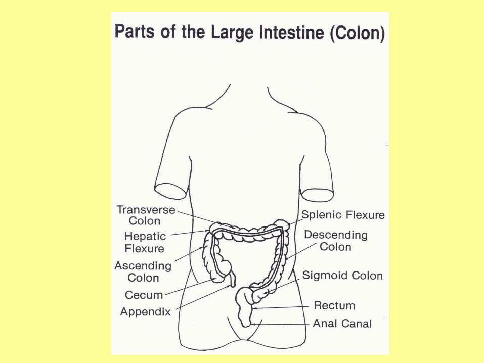
Small Intestine and Colon
• Small intestine– Begins at pyloric
sphincter– Duodenum– Jejunum – Ileum– Mesenteric small
intestine
• Colon– Cecum
• Appendix
– Ascending colon– Transverse colon– Descending colon– Rectum, anus
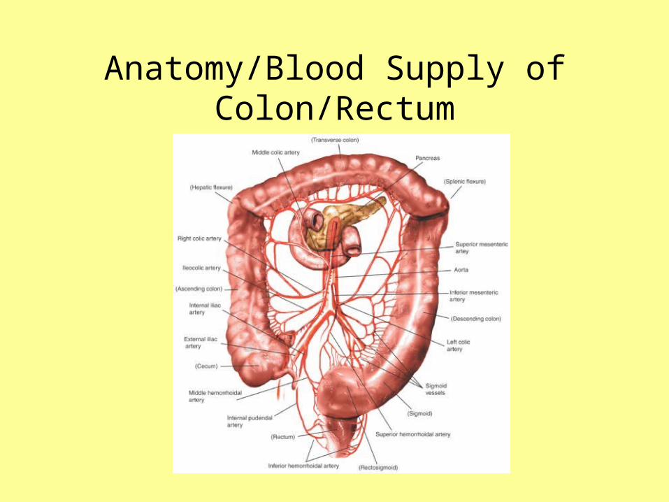
Anatomy/Blood Supply of Colon/Rectum
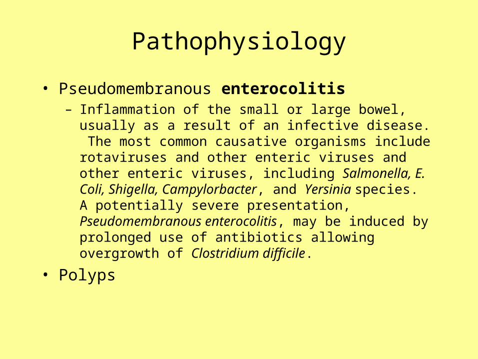
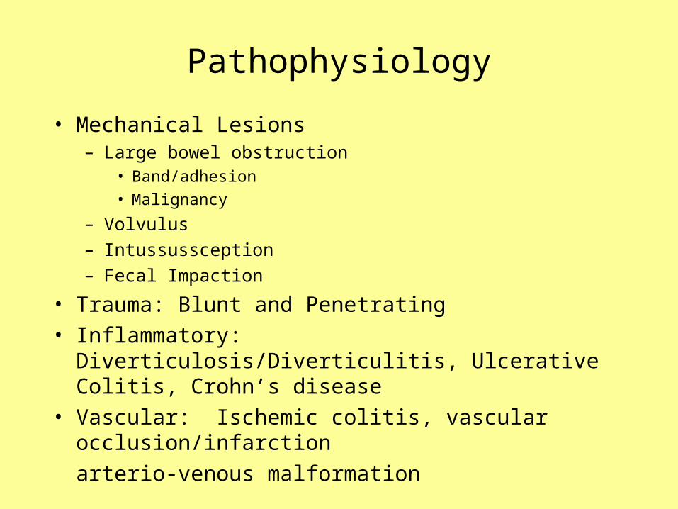
Pathophysiology
• Pseudomembranous enterocolitis– Inflammation of the small or large bowel, usually as a result of an
infective disease. The most common causative organisms include rotaviruses and other enteric viruses and other enteric viruses, including Salmonella, E. Coli, Shigella, Campylorbacter, and Yersinia species. A potentially severe presentation, Pseudomembranous enterocolitis, may be induced by prolonged use of antibiotics allowing overgrowth of Clostridium difficile.
• Polyps
Pathophysiology
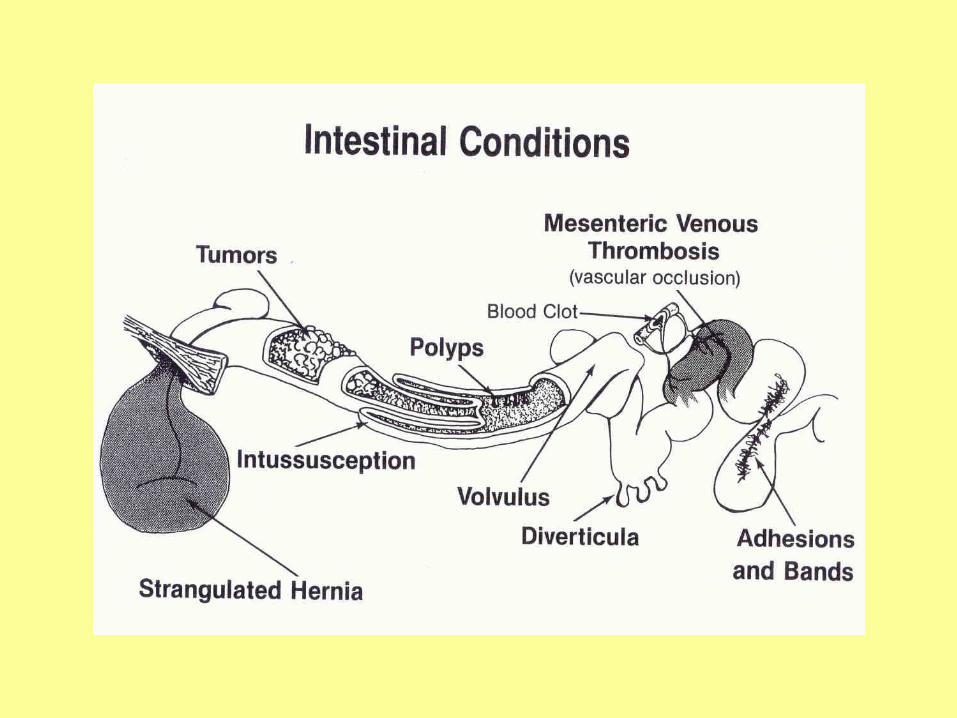
• Mechanical Lesions– Large bowel obstruction
• Band/adhesion
• Malignancy
– Volvulus
– Intussussception
– Fecal Impaction
• Trauma: Blunt and Penetrating
• Inflammatory: Diverticulosis/Diverticulitis, Ulcerative Colitis, Crohn’s disease
• Vascular: Ischemic colitis, vascular occlusion/infarction
arterio-venous malformation
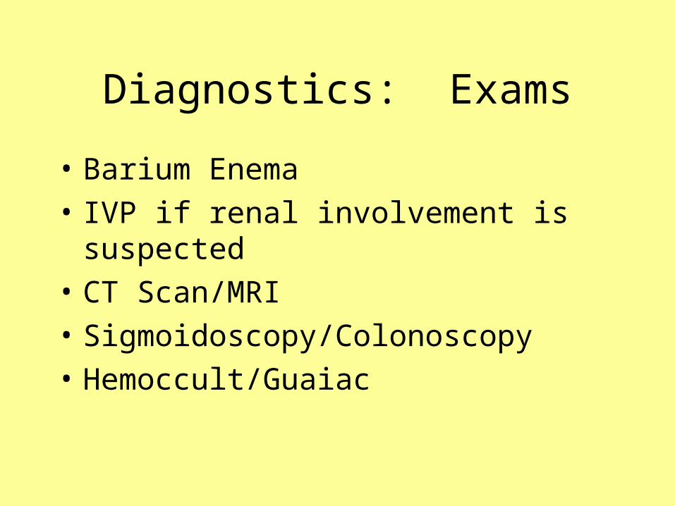
Diagnostics: Exams
• Barium Enema
• IVP if renal involvement is suspected
• CT Scan/MRI
• Sigmoidoscopy/Colonoscopy
• Hemoccult/Guaiac
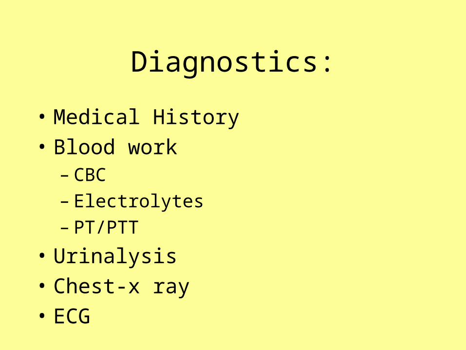
Diagnostics:
• Medical History
• Blood work– CBC– Electrolytes– PT/PTT
• Urinalysis
• Chest-x ray
• ECG

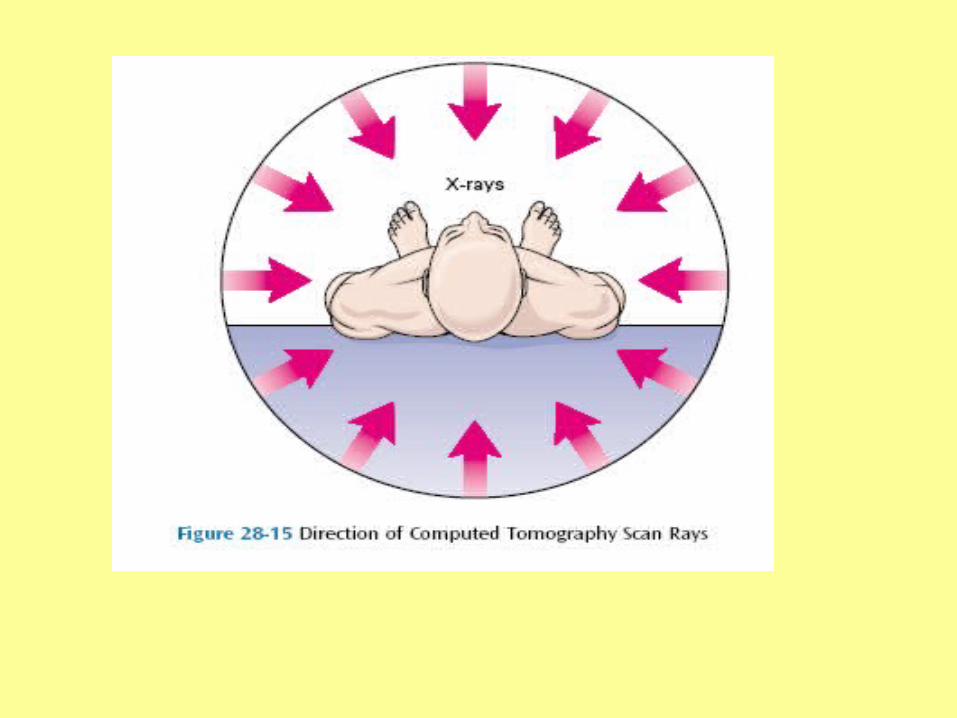
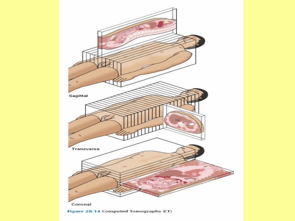
Computed Tomography (CT) or (CAT)
Painless, noninvasive x-ray procedure that has the unique capabilities of distinguishing minor differences in the density of tissues. It produce a three – dimensional image of the organ or structure.

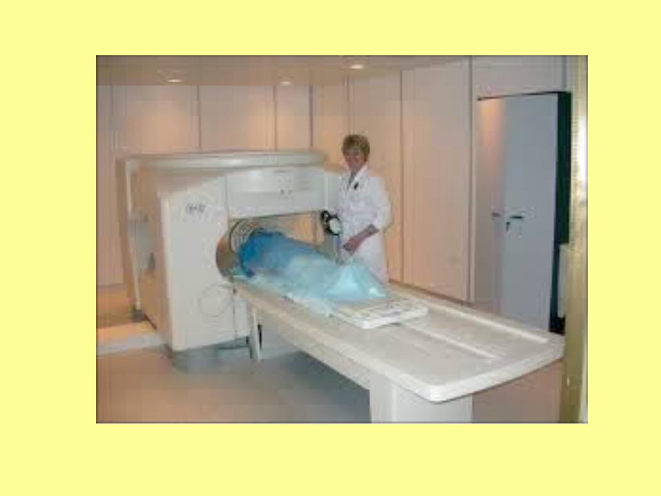
Magnetic Resonance Imaging (MRI)
Is a noninvasive diagnostic scanning technique in which the client is placed in a magnatic field. MRI provides a better contrast between normal and abnormal tissue than the CT scan. For visualization of the brain, spine, limbs, and joints, heart, blood vessels, abdomen and pelvis. The procedure lasts between 60 and 90 minutes.
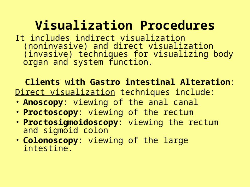
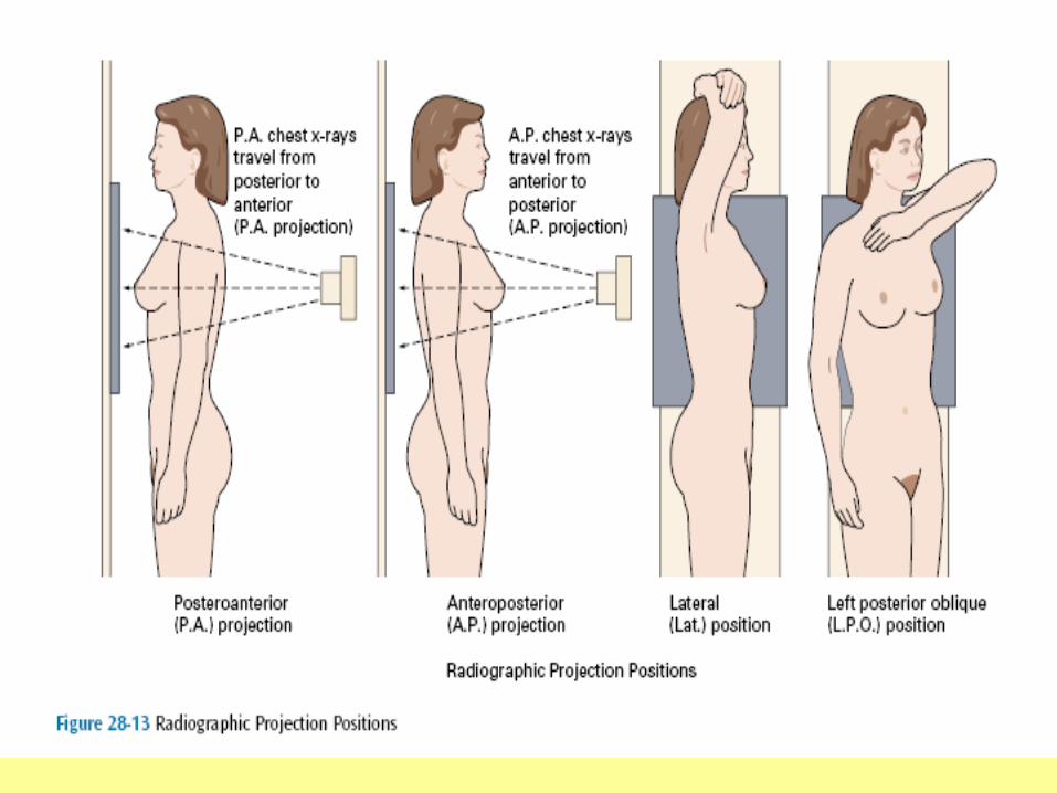
Visualization ProceduresIt includes indirect visualization (noninvasive) and
direct visualization (invasive) techniques for visualizing body organ and system function.
Clients with Gastro intestinal Alteration: Direct visualization techniques include:• Anoscopy: viewing of the anal canal• Proctoscopy: viewing of the rectum• Proctosigmoidoscopy: viewing the rectum and
sigmoid colon• Colonoscopy: viewing of the large intestine.

Indirect visualization of the gastrointestinal tract is achieved by:
X-rays of gastrointestinal tract can detect structure, obstructions, tumors, ulcers, inflammatory diseases or other structural changes such as hiatal hernias. Visualization of the tract is enhanced by the use of a barium. For examination of the upper gastrointestinal tract or small bowel, the client drinks the barium sulfate (barium swallow). For examination of the lower gastrointestinal tract, the client is given an enema containing the barium (Barium enema).
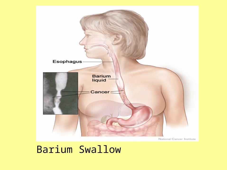
Barium Swallow
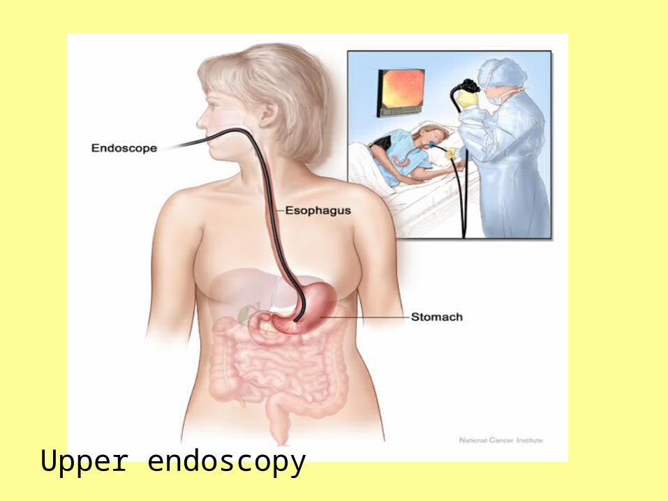
Upper endoscopy

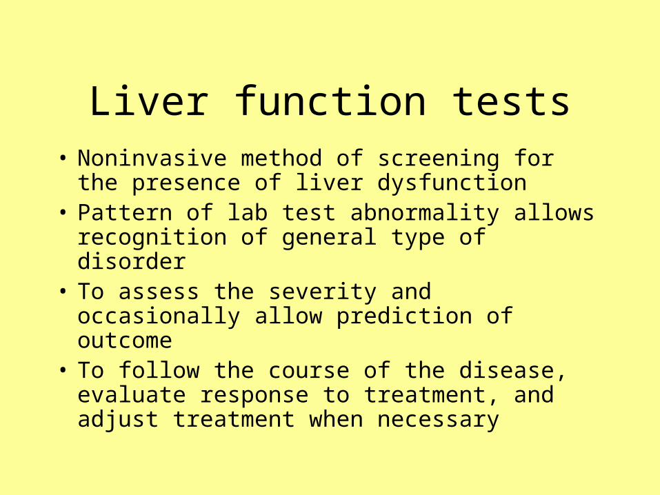
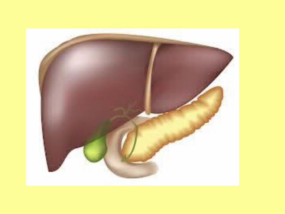
Liver function tests• Noninvasive method of screening for the
presence of liver dysfunction• Pattern of lab test abnormality allows
recognition of general type of disorder• To assess the severity and occasionally allow
prediction of outcome• To follow the course of the disease, evaluate
response to treatment, and adjust treatment when necessary
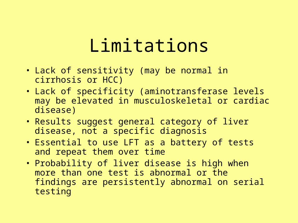
Limitations• Lack of sensitivity (may be normal in cirrhosis or HCC)• Lack of specificity (aminotransferase levels may be
elevated in musculoskeletal or cardiac disease)• Results suggest general category of liver disease, not a
specific diagnosis• Essential to use LFT as a battery of tests and repeat them
over time• Probability of liver disease is high when more than one
test is abnormal or the findings are persistently abnormal on serial testing

General categories of tests
• Tests of the capacity of the liver to transport organic anions and metabolize drugs
•Eg. S bilirubin, s bile acids, BSP etc
•Measures ability of the liver to clear endogenous or exogenous substances from the circulation
Tests to detect injury to hepatocytes•All the enzyme tests
•Most commonly done and most useful are aminotransferases and alkaline phosphatase
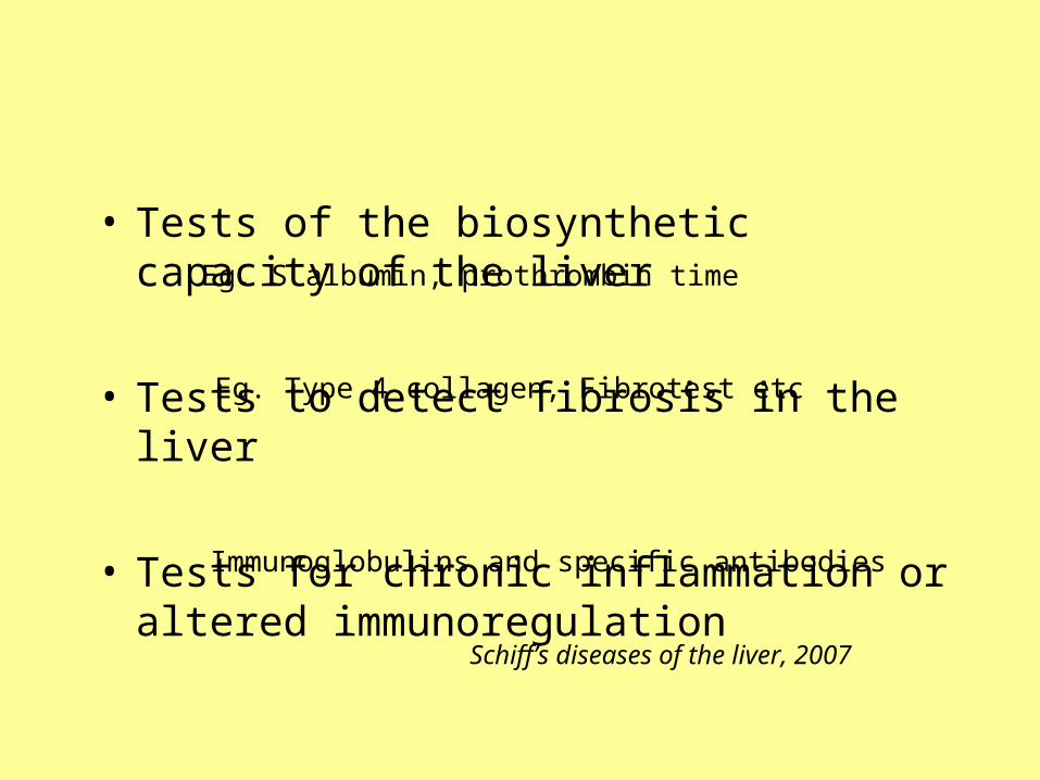
• Tests of the biosynthetic capacity of the liver
• Tests to detect fibrosis in the liver
• Tests for chronic inflammation or altered immunoregulation
Eg. S albumin, prothrombin time
Eg. Type 4 collagen, Fibrotest etc
Immunoglobulins and specific antibodies
Schiff’s diseases of the liver, 2007

Common serum liver chemistry tests
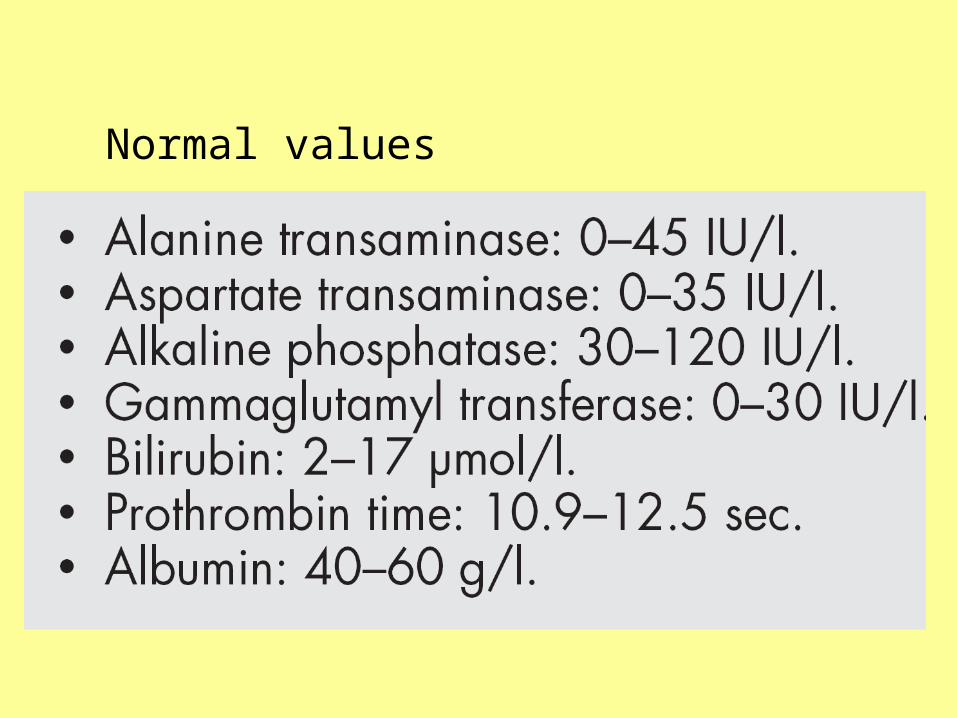
Normal values