THE LOCALIZATION OF BASIC FIBROBLAST GROWTH FACTOR …€¦ · THE LOCALIZATION OF BASIC FIBROBLAST...
10
Nagoya J. Med. Sci. 57. 143 - 152, 1994 THE LOCALIZATION OF BASIC FIBROBLAST GROWTH FACTOR (FGF-2) IN RAT SUBMANDIBULAR GLANDS YOSHIYUKI HIRAMATSU, HIDEAKI KAGAMI, KENICHI KOSAKI, TOSHIO SHIGETOMI, MINORU UEDA, SHIGERU KOBAYASHII and MASAHIRO SAKANAKA 2 Departments of Oral Surgery and I Anatomy, Nagoya University School of Medicine, Nagoya, and 2Department of Anatomy, Ehime University School of Medicine, Ehime, Japan ABSTRACT The immunohistochemical localization of basic fibroblast growth factor (FGF-2) in the submandibular glands of the rat was investigated by use of an antiserum to FGF-2. Nerve fiber bundles with FGF-2-immu- noreactivity were found in association with interlobular ducts and blood vessels; they dissociated into single immunoreactive nerve fibers perhaps to terminate in proximity to acinar cells, or to form a reticular fiber network within the tunica adventitia of blood vessels. The FGF-2-immunoreactive neurons were located in the submandibular ganglia, but not in the superior cervical ganglia; hence, at least some of these immunore- active nerve fibers probably come from the submandibular ganglia and are of parasympathetic origin. Most of the epithelial cells of the intercalated and collecting ducts showed notable FGF-2 immunoreactivity. The characteristic distribution of FGF-2 immunoreactivity in both the neuronal and epithelial tissues of the sali- vary glands suggests a role of this growth factor in complex physiological processes within the salivary glands. Key Words: Basic fibroblast growth factor, Submandibular gland, Submandibular ganglion, Superior cer- vical ganglion, Immunohistochemistry INTRODUCTION Basic fibroblast growth factor (FGF-2) stimulates the mitogenic activities of a variety of me- soderm- and neuroectoderm-derived cells,I,2) as well as to facilitate the survival and differentia- tion of these cells. 3 ,4) It has been detected biochemically in the retina,5) corpus luteum,6) adrenal gland,7) kidney,8) placenta,9) macrophages,IO) and prostate.J!) Recent immunohistochemical studies showed FGF-2 localized not only in peripheral tissues I2 ,14) but also in neuronal and neu- roglial elements of the brain. 15 - 20 ) In peripheral nerves, FGF- 2 is abundant within the somatic motor and sensory nervous systems. 14 ,21-23) However, there are few studies on FGF-2 in the pe- ripheral autonomic nervous system. The secretory activity of the salivary glands is controlled by the autonomic nerves. It has been proposed that parasympathetic nerve fibers regulate the viability of glandular cells, because their transection or interruption produces degeneration of the acinar cells,24-26) We sought to deter- mine whether FGF-2, like nerve growth factor, is localized to the salivary glands and/or to the parasympathetic nerves that innervate the glands. 27 ,28) Using a FGF-2 antiserum that has been characterized by immunoblot,14,16,19,29,30) we investigated the distribution of FGF-2-like immu- noreactivity in rat salivary glands with special attention given to the autonomic nerves. Correspondence: Dr. Yoshiyuki Hiramatsu, Departments of Oral Surgery, Nagoya University School of Me- dicine, 65 Tsurumai-Cho, Showa-Ku, Nagoya 466, Japan Accepted for Publication in August 8, 1994 143
Transcript of THE LOCALIZATION OF BASIC FIBROBLAST GROWTH FACTOR …€¦ · THE LOCALIZATION OF BASIC FIBROBLAST...

Nagoya J. Med. Sci. 57. 143 - 152, 1994
THE LOCALIZATION OF BASICFIBROBLAST GROWTH FACTOR (FGF-2)
IN RAT SUBMANDIBULAR GLANDS
YOSHIYUKI HIRAMATSU, HIDEAKI KAGAMI, KENICHI KOSAKI, TOSHIO SHIGETOMI,MINORU UEDA, SHIGERU KOBAYASHII and MASAHIRO SAKANAKA2
Departments of Oral Surgery and I Anatomy, Nagoya University School of Medicine, Nagoya, and2Department of Anatomy, Ehime University School of Medicine, Ehime, Japan
ABSTRACT
The immunohistochemical localization of basic fibroblast growth factor (FGF-2) in the submandibularglands of the rat was investigated by use of an antiserum to FGF-2. Nerve fiber bundles with FGF-2-immunoreactivity were found in association with interlobular ducts and blood vessels; they dissociated into singleimmunoreactive nerve fibers perhaps to terminate in proximity to acinar cells, or to form a reticular fibernetwork within the tunica adventitia of blood vessels. The FGF-2-immunoreactive neurons were located inthe submandibular ganglia, but not in the superior cervical ganglia; hence, at least some of these immunoreactive nerve fibers probably come from the submandibular ganglia and are of parasympathetic origin. Mostof the epithelial cells of the intercalated and collecting ducts showed notable FGF-2 immunoreactivity. Thecharacteristic distribution of FGF-2 immunoreactivity in both the neuronal and epithelial tissues of the salivary glands suggests a role of this growth factor in complex physiological processes within the salivaryglands.
Key Words: Basic fibroblast growth factor, Submandibular gland, Submandibular ganglion, Superior cervical ganglion, Immunohistochemistry
INTRODUCTION
Basic fibroblast growth factor (FGF-2) stimulates the mitogenic activities of a variety of me
soderm- and neuroectoderm-derived cells,I,2) as well as to facilitate the survival and differentia
tion of these cells. 3,4) It has been detected biochemically in the retina,5) corpus luteum,6) adrenal
gland,7) kidney,8) placenta,9) macrophages,IO) and prostate.J!) Recent immunohistochemical
studies showed FGF-2 localized not only in peripheral tissues I2,14) but also in neuronal and neu
roglial elements of the brain. 15- 20) In peripheral nerves, FGF-2 is abundant within the somatic
motor and sensory nervous systems. 14,21-23) However, there are few studies on FGF-2 in the pe
ripheral autonomic nervous system.
The secretory activity of the salivary glands is controlled by the autonomic nerves. It has been
proposed that parasympathetic nerve fibers regulate the viability of glandular cells, because their
transection or interruption produces degeneration of the acinar cells,24-26) We sought to deter
mine whether FGF-2, like nerve growth factor, is localized to the salivary glands and/or to the
parasympathetic nerves that innervate the glands.27,28) Using a FGF-2 antiserum that has beencharacterized by immunoblot,14,16,19,29,30) we investigated the distribution of FGF-2-like immu
noreactivity in rat salivary glands with special attention given to the autonomic nerves.
Correspondence: Dr. Yoshiyuki Hiramatsu, Departments of Oral Surgery, Nagoya University School of Medicine, 65 Tsurumai-Cho, Showa-Ku, Nagoya 466, JapanAccepted for Publication in August 8, 1994
143
144
Yoshiyuki Hiramatsu et al.
MATERIALS AND METHODS
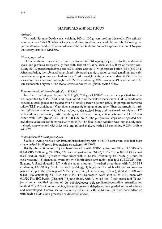
AnimalsTen male Sprague-Dawley rats weighing 100 to 150 g were used in this study. The animals
were kept on a 12h: 12h light-dark cycle, and given food and water ad libitum. The following experiments were conducted in accordance with the Guide for Animal Experimentation at NagoyaUniversity School of Medicine.
Tissue preparationThe animals were anesthetized with pentobarbital (40 mg/kg) injected into the abdominal
space and perfused transcardially, first with 100 ml of saline, then with 200 ml of fixative consisting of 4% paraformaldehyde and 0.2% picric acid in O.lM phosphate buffer (PB) (pH 7.4).After perfusion, the submandibular gland, sublingual gland, superior cervical ganglion, and submandibular ganglion were excised and postfixed overnight with the same fixative at 4T. The tissues were then immersed overnight in O.lM PB containing 30% sucrose at 4°C and cut into 10!-tm sections in a cryostat. The sections were mounted on gelatin-coated slides.
Preparation ofpolyclonal antibody to FGF-2In order to affinity-purify anti-FGF-2 IgG, 200 !-tg of FGF-2 in a partially purified fraction
was separated by SDS-PAGE and transblotted to nitrocellulose membranes. FGF-2 bands wereexcised as small pieces and treated with 5% bovine serum albumin (BSA) in phosphate bufferedsaline (PBS) overnight at 4T to block nonspecific binding of antibody. Then the protein A purified IgG fraction of anti-FGF-2 was added to the excised blots and incubated overnight at 4°Cwith end-over-end mixing. After washing with PBS ten times, antibody bound to FGF-2 waseluted with 0.2M glycine-HCI, pH 3.0, 0.15M NaCl. The purification steps were repeated several times using excised blots washed with PBS. The final eluted solution was immediately neutralized, supplemented with BSA to 1 mg/ml and dialyzed with PBS containing 0.02% sodiumazide.3!)
Immunohistochemical proceduresSections were processed for immunohistochemistry with a FGF-2 antiserum that had been
characterized by Western blot analysis elsewhere.!4.!6,!9,29,30)Briefly, the sections were 1) incubated for 48 h with FGF-2 antiserum, diluted 1:1000 with
O.lM PBS containing 5% BSA, 1% normal goat serum (NGS), 0.1% Triton X-100 (TX), and0.1 % sodium azide; 2) washed three times with O.lM PBS containing 1% NGS, (10 min foreach washing); 3) incubated overnight with biotinylated anti-rabbit goat IgG (VECTOR, Burlingame, U.S.A.) diluted 1:250 with the same solution; 4) washed three times with O.lM PBScontaining 1% NGS (10 min for each washing); 5) incubated for 24 h with peroxidase-conjugated streptavidin (Kirkegaard & Perry Lab, Inc., Gaithersburg, U.S.A.), diluted 1:300 withO.lM PBS containing 5% BSA and 0.1% TX; 6) washed twice with O.lM PBS, once with0.05M Tris-HCI buffer (TB) (pH 7.4) and finally with O.lM TB for 10 min each; and 7) subjected to a modified version of the cobalt-glucose oxidase-diaminobenzidine intensificationmethod.32,33) After immunostaining, the sections were dehydrated in a graded series of ethanoland coverslipped. Control sections were incubated with the antiserum that had been adsorbedwith bovine FGF-2 and processed as described above.
145
BASIC FIBROBLAST GROWTH FACTOR (FGF-2)
RESULTS
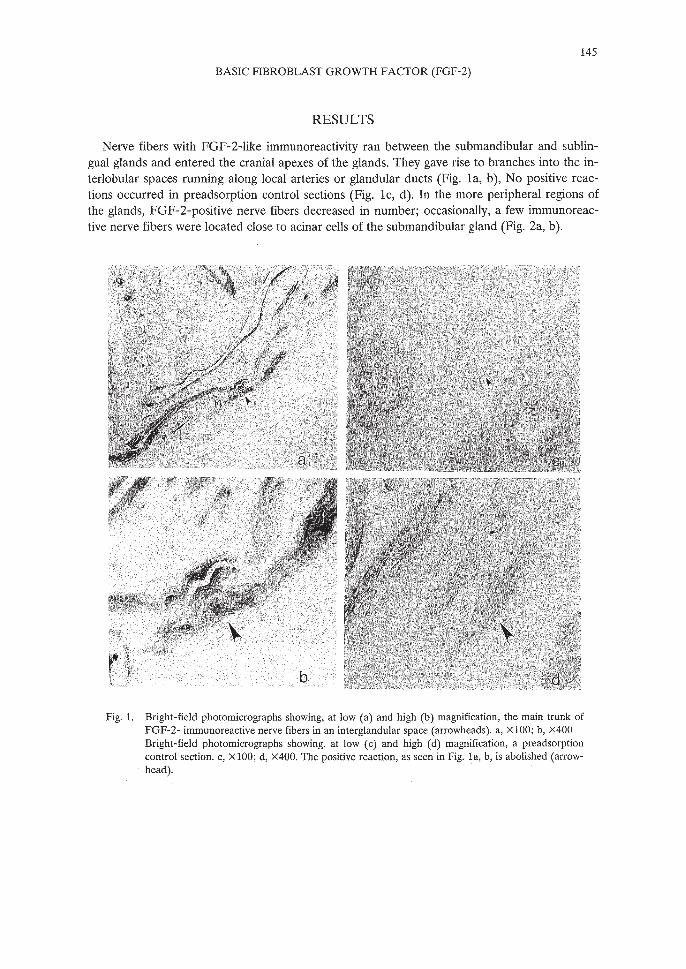
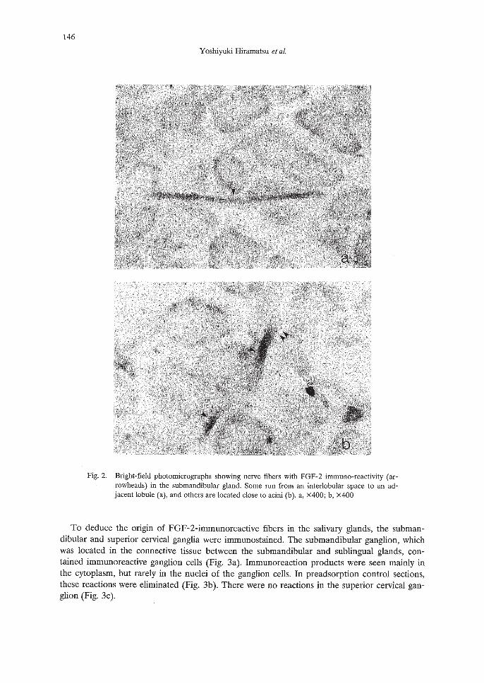
Nerve fibers with FGF-2-like immunoreactivity ran between the submandibular and sublingual glands and entered the cranial apexes of the glands. They gave rise to branches into the interlobular spaces running along local arteries or glandular ducts (Fig. la, b), No positive reactions occurred in preadsorption control sections (Fig. lc, d). In the more peripheral regions ofthe glands, FGF-2-positive nerve fibers decreased in number; occasionally, a few immunoreactive nerve fibers were located close to acinar cells of the submandibular gland (Fig. 2a, b).
Fig. 1. Bright-field photomicrographs showing, at low (a) and high (b) magnification, the main trunk ofFGF-2- immunoreactive nerve fibers in an interglandular space (arrowheads). a, XlOO; b, x400Bright-field photomicrographs showing, at low (c) and high (d) magnification, a preadsorptioncontrol section. c, XlOO; d, x400. The positive reaction, as seen in Fig. la, b, is abolished (arrowhead).
146
Yoshiyuki Hiramatsu et at.
Fig. 2. Bright-field photomicrographs showing nerve fibers with FGF-2 immuno-reactivity (arrowheads) in the submandibular gland. Some run from an interlobular space to an adjacent lobule (a), and others are located close to acini (b). a, X400; b, x400
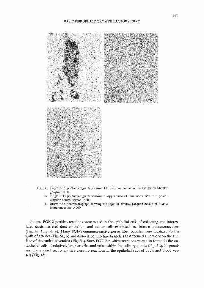
To deduce the origin of FGF-2-immunoreactive fibers in the salivary glands, the submandibular and superior cervical ganglia were immunostained. The submandibular ganglion, whichwas located in the connective tissue between the submandibular and sublingual glands, contained immunoreactive ganglion cells (Fig. 3a). Immunoreaction products were seen mainly inthe cytoplasm, but rarely in the nuclei of the ganglion cells. In preadsorption control sections,these reactions were eliminated (Fig. 3b). There were no reactions in the superior cervical ganglion (Fig. 3c).
147
BASIC FIBROBLAST GROWTH FACTOR (FGF-2)
Fig.3a. Bright-field photomicrograph showing FGF-2 immunoreaction in the submandibularganglion. x200
b. Bright-field photomicrograph showing disappearance of immunoreaction in a preadsorption control section. X 200
c. Bright-field photomicrograph showing the superior cervical ganglion devoid of FGF-2immunoreaction. x200
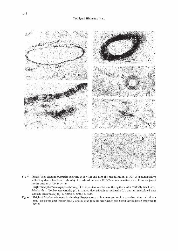
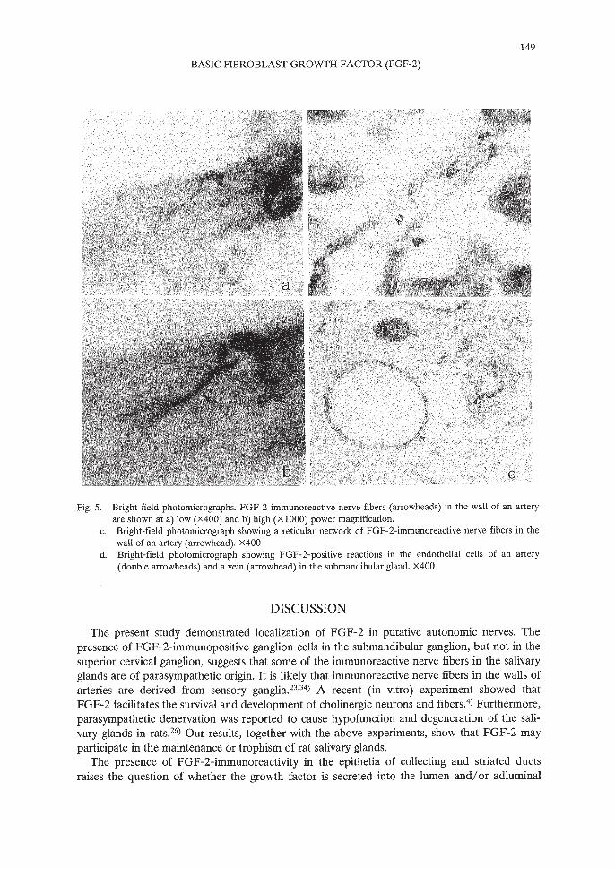
Intense FGF-2-positive reactions were noted in the epithelial cells of collecting and intercalated ducts; striated duct epithelium and acinar cells exhibited less intense immunoreactions(Fig. 4a, b, c, d, e). Many FGF-2-immunoreactive nerve fiber bundles were localized to thewalls of arteries (Fig. 5a, b) and dissociated into fine branches that formed a network on the surface of the tunica adventitia (Fig. 5c). Such FGF-2-positive reactions were also found in the endothelial cells of relatively large arteries and veins within the salivary glands (Fig. 5d). In preadsorption control sections, there were no reactions in the epithelial cells of ducts and blood vessels (Fig. 4f).
148
Yoshiyuki Hiramatsu et al.
Fig. 4. Bright-field photomicrographs showing, at low (a) and high (b) magnification, a FGF-2-immunopositivecollecting duct (double arrowheads). Arrowhead indicates FGF-2-immunoreactive nerve fibers subjacentto the duct. a, x 100; b, x400Bright-field photomicrographs showing FGF-2-positive reactions in the epithelia of a relatively small interlobular duct (double arrowheads) (c), a striated duct (double arrowheads) (d), and an intercalated duct(double arrowheads) (e). c, X400; d, X400; e, x200
Fig. 4f. Bright-field photomicrographs showing disappearance of immunoreaction in a preadsorption control section.: collecting duct (arrow head), striated duct (double arrowhead) and blood vessels (open arrowhead)x200
149
BASIC FIBROBLAST GROWTH FACTOR (FGF-2)
Fig. 5. Bright-field photomicrographs. FGF-2-immunoreactive nerve fibers (arrowheads) in the wall of an arteryare shown at a) low (X400) and b) high (XI000) power magnification.
c. Bright-field photomicrograph showing a reticular network of FGF-2-immunoreactive nerve fibers in thewall of an artery (arrowhead). X400
d. Bright-field photomicrograph showing FGF-2-positive reactions in the endothelial cells of an artery(double arrowheads) and a vein (arrowhead) in the submandibular gland. x400
DISCUSSION
The present study demonstrated localization of FGF-2 in putative autonomic nerves. Thepresence of FGF-2-immunopositive ganglion cells in the submandibular ganglion, but not in thesuperior cervical ganglion, suggests that some of the immunoreactive nerve fibers in the salivaryglands are of parasympathetic origin. It is likely that immunoreactive nerve fibers in the walls ofarteries are derived from sensory ganglia.23,34) A recent (in vitro) experiment showed thatFGF-2 facilitates the survival and development of cholinergic neurons and fibers. 4) Furthermore,parasympathetic denervation was reported to cause hypofunction and degeneration of the salivary glands in rats. 26
) Our results, together with the above experiments, show that FGF-2 mayparticipate in the maintenance or trophism of rat salivary glands.
The presence of FGF-2-immunoreactivity in the epithelia of collecting and striated ductsraises the question of whether the growth factor is secreted into the lumen and/or adluminal
150
Yoshiyuki Hiramatsu et al.
spaces, because most members of the FGF family including FGF-l and FGF-2 are devoid ofsignal sequence35). Mignatti et al. say that FGF-2 is released from the cell surface in a novelexocytic way independent of the classic endoplasmic reticulum-Golgi complex route andpossibly binds to heparan-sulfate proteoglycan in the extracellular matrix.36) Although theFGF-2 antiserum used in the present study may not have recognized FGF-2 molecules boundwith heparan-sulfate in the extracellular matrix, DiMario et alY) and Gonzalez et al,37) reportedthat the antisera they used reacted with them. Thus, we could not determine whether bFGF issecreted in the salivary glands. Use of antibodies that are directed to heparan-sulfate-boundFGF-2 may clarify this uncertainty.
The role of high concentration FGF-2 molecules in the intercalated ducts, as revealed by thepresent immunohistochemical study, is not clear. However, the characteristic distribution ofFGF-2 immunoreactivity suggests its important role in the physiological process.
ACKNOWLEDGEMENTS
The authors are grateful to Dr. Nobuaki Okumura (Osaka University) for the generous provision of FGF-2 antiserum and to Dr. Seiji Matsuda (Ehime University) for his fruitful suggestions through out this study. This work was supported, in part, by grants from the Senri LifeScience Foundation and the Uehara Memorial Foundation.
REFERENCES
1) Gospodarowicz, D.: Localization of a fibroblast growth factor and its effect alone and with hydrocortisone on3T3 cell growth. Nature, 249,123-127 (1974).
2) Gospodarowicz, D., Weseman, J. and Moran, J.: Presence in brain of a mitogenic agent promoting proliferation of myoblast in low density culture. Nature, 256, 216-219 (1975).
3) Gospodarowicz, D., Moran, J., Braun, D. and Birdwell, C.R.: Clonal growth of bovine vascular endothelialcells: Fibroblast growth factor as a survival agent. Proc. Natl. Acad. Sci. U.S.A., 73, 4120-4124 (1976).
4) Groth, C., Otto, D. and Unsicker, K: Basic fibroblast growth factor promotes in vitro survival and cholinergic development of rat septal neurons: Comparison with the effects of nerve growth factor. Neuroscience, 31,No.3, 649-661 (1989).
5) Baird, A., Esch, F., Gospodarowicz, D. and Guillemin, R.: Retina- and eye-derived endothelial cell growthfactor: Partial molecular characterization and identity with acidic and basic fibroblast growth factors. Biochemistry, 24, 7855-7859 (1985).
6) Gospodarowicz, D., Cheng, J., Lui, G.M., Baird, A., Esch, F. and Bohlen, P.: Corpus lutem angiogenic factor is related to fibroblast growth factor. Endocrinology, 117,2383-2391 (1985).
7) Gospodarowicz, D., Baird, A., Cheng, J., Lui, G.M., Esch, F. and Bohlen, P.: Isolation of fibroblast growthfactor from bovine adrenal gland: Physicochemical and biological characterization. Endocrinology, 118,82-90 (1986).
8) Baird, A., Esch, F., Ling, N. and Gospodarowicz, D.: Isolation and partial characterization of an endothelialcell growth factor from the bovine kidney: Homology with basic fibroblast growth factor. Regul. Pept., 12,201-213 (1985).
9) Gospodarowicz, D., Cheng, J., Lui, G.M., Fujii, D.K, Baird, A. and Bohlen, P.: Fibroblast growth factor inthe human placenta. Biochem. Biophys. Res. Commun., 30, 554-562 (1985).
10) Baird, A., Mormede, P. and Bohlen, P.: Immunoreactive fibroblast growth factor in cells of peritoneal exudate suggests its identity with macrophage-derived growth factor. Biochem. Biophys. Res. Commun., 126,358-364 (1985).
11) Nishi, N., Matsuo, Y., Muguruma, Y., Yoshitake, Y., Nishikawa, K and Wada, F.: A human prostaticgrowth factor (hPGF): Partial purification and characterization. Biochem. Biophys. Res. Commun., 132,1103-1109 (1985).
151
BASIC FIBROBLAST GROWTH FACTOR (FGF-2)
12) DiMario, J., Buffinger, N., Yamada, S. and Strohman, R.c.: Fibroblast growth factor in the extracellular matrix of dystrophic (mdx) mouse muscle. Science, 244, 753-765 (1989).
13) Grothe, C. and Unsicker, K: Immunocytochemical localization of basic fibroblast growth factor in bovineadrenal gland, ovary and pituitary. J. Histochem. Cytochem., 37,1877-1883 (1989).
14) Matsuda, S., Desaki, J., Fujita, H., Okumura, N. and Sakanaka, M.: Immuno-electron microscopic localization of basic fibroblast growth factor in the dystrophic mdx mouse masseter muscle. Cell Tissue Res., 270,569-576 (1992).
15) G6mez-Pinilla, F., Lee, J.W.-K and Cotman, C.W.: Basic FGF in adult rat brain: Cellular distribution andresponse to entorhinallesion and fimbria-formix transection. J. Neurosci., 12,345-355 (1992).
16) Iwata, H., Matsuyama, A., Okumura, N., Yoshida, S., Lee, Y, Imaizumi, K and Shiosaka, S.: Localization ofbasic FGF-like immunoreactivity in the hypothalamohypophyseal neuroendocrine axis. Brain Res., 550,No.2, 329-332 (1991).
17) Kumon, Y, Sasaki, S., Kadota, 0., Matsuda, S., Fujita, H., Yoshimura, H. and Sakanaka, M.: Transient increase in endogenous basic fibroblast growth factor in neurons of ischemic rat brains. Brain Res., 605,169-174 (1993).
18) Matsuda, S., Desaki, J., Okumura, N., Shiosaki, S., Imaoka, S. and Sakanaka, M.: Basic fibroblast growthfactor-like immunoreactivity in the trigeminal proprioceptive and motor systems. Brain Res., 577, No.1,92-100 (1992).
19) Matsuda, S., Okumura, N., Yoshimura, H., Koyama, Y. and Sakanaka, M.: Basic fibroblast growth factorlike immunoreactivity in Purkinje cells of the rat cerebellum. Neuroscience, 50, No.1, 99-106 (1992).
20) Pettmann, B., Labourdette, G., Weibel, M. and Sensenbrenner, M.: The brain fibroblast growth factor (FGF)is localized in neurons. Neurosci. Lett., 68, 175-180 (1986).
21) Eckenstein, F., Woodward, W.R. and Nishi, R.: Differential localization and possible function of aFGF andbFGF in the central and peripheral nervous systems. Ann. N. Y. Acad. Sci., 638, 348-360 (1991).
22) Logan, A. and Logan, S.D.: Distribution of fibroblast growth factor in the central and peripheral nervous systems of various mammals. Neurosci. Lett., 69, 162-165 (1986).
23) Weise, B., Unsicker, K. and Grothe, c.: Localization of basic fibroblast growth factor in a subpopulation ofrat sensory neurons. Cell Tissue Res., 267, 125-130 (1992).
24) Delfs, U. and Emmelin, N.: Parasympathetic degeneration secretion of saliva in rat. Q. J. Exp. Physiol., 64,109-117 (1979).
25) Emmelin, N.: 'Paralytic secretion' of saliva: An example of supersensitivity after denervation. Physiol. Rev.,32, 21-45 (1952).
26) Emmelin, N. and Trendelenburg, u.: Degeneration activity after parasympathetic or sympathetic denervation. Ergeb Physiol. Bioi. Chern. Exp. Pharmacol., 66, 147-211 (1972).
27) Cohen, S.: Purification of a nerve-growth promoting protein from the mouse salivary gland and its neuro-cytotoxic antiserum. Proc. Nac!. Acad. Sci. U.S.A., 46, 302-311 (1960).
28) Hendry, LA.: Developmental change in tissue and plasma concentrations of the biologically active species ofnerve growth factor in the mouse, by using a two-site radioimmunoassay. Biochem. J., 128, 1265-1272(1972).
29) Desaki, J., Matsuda, S., Okumura, N., Koyama, Y and Sakanaka, M.: Fine structure of nerve processes containing basic fibroblast growth factor in muscle spindles of the rat masseter muscle. Neurosci. Lett., 137,237-240 (1992).
30) Okumura, N., Takimoto, K, Okada, M. and Nakagawa, H.: C6 Glioma cells produce basic fibroblast growthfactor that can stimulate their own proliferation. J. Biochem., 106, 904-909 (1989).
31) Kozaki, K., Miyaishi, 0., Asai, N., Iida, K, Sakata, K, Hayashi, M., Nishida, T., Matsuyama, M., Shimizu,S., Kaneda, T. and Saga, S.: Tissue distribution of ERp61 and association of its increased expression withIgG production in hybridoma cells. Exp. Cell Res., in print.
32) Itoh, K, Konishi, A., Nomura, S., Mizuno, N., Nakamura, Y. and Sugimoto, T.: Application of coupled oxidation reaction to electron microscopic demonstration of horseradish peroxidase: Cobalt-glucose oxidasemethod., Brain Res., 175,341-346 (1979).
33) Sakanaka, M., Shibasaki, T. and Lederis, K: Improved fixation and cobalt-glucose oxidase-diaminobenzidineintensification for immunohistochemical demonstration of corticotropin-releasing factor in rat brain. J. Histochem. Cytochemi., 35, No.2, 207-212 (1987).
34) Okada, K., Matsuda, S., Ii, Y, Okumura, N., Uryu, K, Fujita, H. and Sakanaka, M.: Basic fibroblast growthfactor-like immunoreactivity in the rat trigeminal sensory system and perioral skin with vibrissae. Cell TissueRes., 272,417-427 (1993).
152
Yoshiyuki Hiramatsu et al.
35) Taira, M., Yoshida, T., Miyagawa, K., Sakamoto, H., Terada, M. and Sugimura, T.: cDNA sequence ofhuman transforming gene hst and identification of the coding sequence required for transformin activity.Proc. Nat!. Acad. Sci. USA, 84, 2980-2987 (1987).
36) Mignatti, P., Morimoto, T. and Rifkin, D. B.: Basic fibroblast growth factor, a protein devoid of secretorysignal sequence, is released by cells via a pathway independent of the endoplasmic reticulum-Golgi complex.J. Cell. Physiol., 151,81-93 (1992).
37) Gonzalez, A.-M., Buscaglia, M., Ong, M. and Baird, A.: Distribution of basic fibroblast growth factor in the18-day rat fetus: Localization in the basement membranes of diverse tissues. 1. Cell. BioI., 110, 753-765(1990).