
The LIM homeobox transcription factor Lhx2 is required … ·...
10
The LIM homeobox transcription factor Lhx2 is required to specify the retina field and synergistically cooperates with Pax6 for Six6 trans-activation Nicolas Tétreault a, 1 , Marie-Pier Champagne a, 1 , Gilbert Bernier a,b, ⁎ a Developmental Biology Laboratory, Maisonneuve-Rosemont Hospital, 5415 Boulevard l'Assomption, Montreal, Canada, H1T 2M4 b Department of Ophthalmology, University of Montreal, Montreal, Canada abstract article info Article history: Received for publication 2 October 2008 Revised 12 December 2008 Accepted 12 December 2008 Available online 30 December 2008 Keywords: Pax6 Lhx2 Six6 Homeobox gene Retina Stem cell Transcription Trans-activation Optic vesicle In mammals, a limited set of homeobox-containing transcription factors are expressed in the presumptive eye field and required to initiate eye development. How these factors interact together at the genetic and molecular level to coordinate this developmental process is poorly understood. We found that the Lhx2 and Pax6 transcription factors operate in a concerted manner during retinal development to promote transcriptional activation of the Six6 homeobox-gene in primitive and mature retinal progenitors. Lhx2 demarcates the presumptive retina field at the neural plate stage and Lhx2 inactivation delays initiation of Rx, Six3 and Pax6 expression in this domain. The later expressed Six6 is properly activated in the pituitary/hypothalamic axis of Lhx2 −/− embryos, but expression fails to be initiated in the optic vesicle. Lhx2 and Pax6 associate with the chromatin at several regions of Six6 in vivo and cooperate for trans-activation of Six6 regulatory elements in vitro. In retinal progenitor/stem cells, both Lhx2 and Pax6 are genetically required for proper Six6 expression and forced co-expression of Lhx2 and Pax6 can synergistically trans-activate the Six6 locus. Our work reveals how two master regulators of eye development coordinate their action to sequentially promote tissue-specific transcriptional initiation and full activation of a retinal determinant gene. © 2008 Elsevier Inc. All rights reserved. Introduction The optic vesicle of vertebrates originates from the neural tube and is first detected between embryonic stages (e) 8.25 and e8.5 as a bilateral evagination of the anterior neural plate, named the optic sulcus (Marquardt, 2003; Oliver and Gruss, 1997). Around e9.5, the intimate contact between the optic vesicle and the eye surface ectoderm induces the transformation of the ectoderm into a lens placode, which is revealed by the proliferation and thickening of the ectoderm (Oliver and Gruss, 1997). In turn, the lens placode induces the evagination of the optic vesicle into an optic cup around e10.0. The optic cup ultimately gives rise to the fully mature neural retina. How retinal specification is established at the neural plate stage and how definitive retinal identity is promoted and maintained in retinal progenitors remain important issues in developmental biology. The homeobox-containing transcription factors Pax6, Rx, Six3 and Lhx2 are genetically required for eye formation and over-expression of Pax6, Rx or Six3/Six6 can induce ectopic retinal tissues in frog and fish embryos (Bernier et al., 2000; Carl et al., 2002; Chow et al., 1999; Hill et al., 1991; Loosli et al., 1999; Mathers et al., 1997; Porter et al., 1997; Zuber et al., 1999). Although the genetic function of these factors has been highly studied, the molecular mechanisms by which they coordinate retinal development and establish definitive retinal identity remain poorly understood. Pax6 is a member of the paired-box and homeobox-containing gene family (PAX) of transcription factors and has been used as a prototype to study eye development in several model organisms (Gehring and Kazuho, 1999; Gehring, 2002). In mice, Pax6 is expressed starting at e8.0 in the eye surface ectoderm, and in the eye neural ectoderm, which gives rise to the optic vesicle (Walther and Gruss, 1991). Despite being anophthalmic at later stage of development, Pax6-null embryos form an optic vesicle that arrests in development prior to the optic cup stage (Grindley et al., 1995; Hogan et al., 1986). In the optic vesicle of Pax6 mutants, neuroepithelial (NE) progenitors over-proliferate and display an abnormal cell cycle kinetic, possibly owing to downregulation of cyclin-dependent kinase inhibitors (Duparc et al., 2007). Conditional mutagenesis of Pax6 in the e11.0 distal retina revealed that Pax6 is required at the time of retinogenesis to maintain progenitor cells proliferation and generate retinal cell types diversity, in part through transcriptional activation of pro-neural genes (Marquardt et al., 2001). Although Pax6 is being considered to operate at the apex of the genetic cascade governing eye and retinal development, expression of Lhx2, Rx, Otx2, Six3 and Six6 in the optic vesicle of Pax6 mutants is unaffected, revealing that early retinal specification does occur in the absence of Pax6 (Bernier et al., 2001; Jean et al., 1999). Developmental Biology 327 (2009) 541–550 ⁎ Corresponding author. Developmental Biology Laboratory, Maisonneuve-Rosemont Hospital, 5415 Boulevard l'Assomption, Montreal, Canada, H1T 2M4. Fax: +1 514 252 3569. E-mail address: [email protected] (G. Bernier). 1 These authors contributed equally to this work. 0012-1606/$ – see front matter © 2008 Elsevier Inc. All rights reserved. doi:10.1016/j.ydbio.2008.12.022 Contents lists available at ScienceDirect Developmental Biology journal homepage: www.elsevier.com/developmentalbiology Genomes & Developmental Control
Transcript of The LIM homeobox transcription factor Lhx2 is required … ·...

Developmental Biology 327 (2009) 541–550
Contents lists available at ScienceDirect
Developmental Biology
j ourna l homepage: www.e lsev ie r.com/deve lopmenta lb io logy
The LIM homeobox transcription factor Lhx2 is required to specify the retina field andsynergistically cooperates with Pax6 for Six6 trans-activation
Nicolas Tétreault a,1, Marie-Pier Champagne a,1, Gilbert Bernier a,b,⁎a Developmental Biology Laboratory, Maisonneuve-Rosemont Hospital, 5415 Boulevard l'Assomption, Montreal, Canada, H1T 2M4b Department of Ophthalmology, University of Montreal, Montreal, Canada
Genomes & Developmental Control
⁎ Corresponding author. Developmental Biology LaboHospital, 5415 Boulevard l'Assomption,Montreal, Canada,
E-mail address: [email protected] (G. Be1 These authors contributed equally to this work.
0012-1606/$ – see front matter © 2008 Elsevier Inc. Aldoi:10.1016/j.ydbio.2008.12.022
a b s t r a c t
a r t i c l e i n f oArticle history:
In mammals, a limited set of Received for publication 2 October 2008Revised 12 December 2008Accepted 12 December 2008Available online 30 December 2008Keywords:Pax6Lhx2Six6Homeobox geneRetinaStem cellTranscriptionTrans-activationOptic vesicle
homeobox-containing transcription factors are expressed in the presumptive eyefield and required to initiate eye development. How these factors interact together at the genetic andmolecularlevel to coordinate this developmental process is poorly understood. We found that the Lhx2 and Pax6transcription factors operate in a concerted manner during retinal development to promote transcriptionalactivation of the Six6 homeobox-gene in primitive and mature retinal progenitors. Lhx2 demarcates thepresumptive retina field at the neural plate stage and Lhx2 inactivation delays initiation of Rx, Six3 and Pax6expression in this domain. The later expressed Six6 is properly activated in the pituitary/hypothalamic axis ofLhx2−/− embryos, but expression fails to be initiated in the optic vesicle. Lhx2 and Pax6 associate with thechromatin at several regions of Six6 in vivo and cooperate for trans-activation of Six6 regulatory elements invitro. In retinal progenitor/stem cells, both Lhx2 and Pax6 are genetically required for proper Six6 expressionand forced co-expression of Lhx2 and Pax6 can synergistically trans-activate the Six6 locus. Our work revealshow twomaster regulators of eye development coordinate their action to sequentially promote tissue-specifictranscriptional initiation and full activation of a retinal determinant gene.
© 2008 Elsevier Inc. All rights reserved.
Introduction
The optic vesicle of vertebrates originates from the neural tube andis first detected between embryonic stages (e) 8.25 and e8.5 as abilateral evagination of the anterior neural plate, named the opticsulcus (Marquardt, 2003; Oliver and Gruss, 1997). Around e9.5, theintimate contact between the optic vesicle and the eye surfaceectoderm induces the transformation of the ectoderm into a lensplacode, which is revealed by the proliferation and thickening of theectoderm (Oliver and Gruss, 1997). In turn, the lens placode inducesthe evagination of the optic vesicle into an optic cup around e10.0. Theoptic cup ultimately gives rise to the fully mature neural retina. Howretinal specification is established at the neural plate stage and howdefinitive retinal identity is promoted and maintained in retinalprogenitors remain important issues in developmental biology. Thehomeobox-containing transcription factors Pax6, Rx, Six3 and Lhx2are genetically required for eye formation and over-expression ofPax6, Rx or Six3/Six6 can induce ectopic retinal tissues in frog and fishembryos (Bernier et al., 2000; Carl et al., 2002; Chow et al., 1999; Hillet al., 1991; Loosli et al., 1999; Mathers et al., 1997; Porter et al., 1997;
ratory, Maisonneuve-RosemontH1T 2M4. Fax: +1514 252 3569.rnier).
l rights reserved.
Zuber et al., 1999). Although the genetic function of these factors hasbeen highly studied, the molecular mechanisms by which theycoordinate retinal development and establish definitive retinalidentity remain poorly understood.
Pax6 is a member of the paired-box and homeobox-containinggene family (PAX) of transcription factors and has been used as aprototype to study eye development in several model organisms(Gehring and Kazuho,1999; Gehring, 2002). In mice, Pax6 is expressedstarting at e8.0 in the eye surface ectoderm, and in the eye neuralectoderm, which gives rise to the optic vesicle (Walther and Gruss,1991). Despite being anophthalmic at later stage of development,Pax6-null embryos form an optic vesicle that arrests in developmentprior to the optic cup stage (Grindley et al., 1995; Hogan et al., 1986). Inthe optic vesicle of Pax6 mutants, neuroepithelial (NE) progenitorsover-proliferate and display an abnormal cell cycle kinetic, possiblyowing to downregulation of cyclin-dependent kinase inhibitors(Duparc et al., 2007). Conditional mutagenesis of Pax6 in the e11.0distal retina revealed that Pax6 is required at the time of retinogenesisto maintain progenitor cells proliferation and generate retinal celltypes diversity, in part through transcriptional activation of pro-neuralgenes (Marquardt et al., 2001). Although Pax6 is being considered tooperate at the apex of the genetic cascade governing eye and retinaldevelopment, expression of Lhx2, Rx, Otx2, Six3 and Six6 in the opticvesicle of Pax6 mutants is unaffected, revealing that early retinalspecification does occur in the absence of Pax6 (Bernier et al., 2001;Jean et al., 1999).
542 N. Tétreault et al. / Developmental Biology 327 (2009) 541–550
Lhx2 is a transcription factor that plays an essential role inmammalian's eye development and that is conserved in lowervertebrate species (Porter et al., 1997; Zuber et al., 2003). Lhx2encodes a member of the LIM homeobox-containing transcriptionfactors family (LHX). LHX proteins can activate or repress genetranscription by direct DNA binding and associationwith co-activatorsor co-repressors through their LIM domain (Agulnick et al., 1996). Inmice, Lhx2 is required for the development of numerous organs,including the eye, the telencephalon and blood system (Porter et al.,1997). While little is known about how Lhx2 operates, experimentshave showed that red blood cell defects in Lhx2-null mice are non-cellautonomous and mediated by abnormalities that lie within the liver(Porter et al., 1997). In contrast, most abnormalities present in the CNSappear to be cell autonomous, as revealed by chimera aggregationstudies (Porter et al., 1997). Lhx2 is also expressed by immature B andT lymphocytes, but not by hematopoietic stem cells (Wu et al., 1996).Notably, some chromosomal translocations involved in humanleukemia's appear to include the LHX2 locus, and Lhx2 over-expression can immortalized human hematopoietic stem cells (Wuet al., 1996). Recent genetic studies have also revealed that Lhx2 isrequired for the self-renewal of epithelial stem cells, but the under-lying molecular mechanism remains elusive (Rhee et al., 2006).
Herein we report on the characterization of Lhx2 function duringthe earliest steps of retinal development. We found that Lhx2represents the first retinal determinant gene with Rx within thepresumptive retina field, and that Lhx2 mutation results in delayedinduction of Six3, Rx and Pax6 expression in this domain. Later on,Lhx2 is also required for Six6 expression initiation in the optic vesicle.In retinal tissue, Lhx2 and Pax6 proteins are bound to the chromatin atthe Six6 locus, and can cooperate to trans-activate Six6 regulatoryelements in vitro. In retinal progenitor/stem cells, Lhx2 and Pax6 aregenetically required for Six6 expression and can cooperate tosynergistically trans-activate the Six6 gene. Our work reveals thatLhx2 is required to establish primitive retinal identity at the neuralplate stage by allowing initiation of retinal-determinant genesexpression, and how later Lhx2 cooperates with Pax6 to establishdefinitive retinal identity and promote cellular proliferation.
Materials and methods
Animals
Adult mice from the albinos CD1 or 129sv strains were purchasedfrom Charles River (St-Constant, Qc., Canada). Pax6 mutant mice are agift from Peter Gruss (Max-Planck Institute, Goettingen) and Lhx2mutant mice from Heiner Westphal (National Institute of Health,Bethesda). Embryos stage was determined according to the time ofvaginal plug.
In situ hybridization
Embryos were dissected in PBS, fixed overnight in 4% paraformal-dehyde at 4 °C and embedded in Paraplast (Monoject Scientific).Sections (10 μm) were cut and dried onto super-frost glass slides(Fisher Scientific). 35S-labelled RNA probes using SP6, T3 or T7 RNApolymerase were done with Boehringer enzyme according to thedirective of the company. Exposure time for the radioactive RNA in situhybridization was 15 days. For in situ hybridizations on wholeembryos, preparations were hybridized with digoxigenin-labelledRNA probes and visualized with alkaline phosphatase-coupled anti-digoxigenin antibody (1/2000) (Boehringer) and NBT/BCIP substrate(Boehringer) at pH 9.5. For cryosection, embryos were cryoprotectedin 30% sucrose/PBS overnight at 4 °C, embedded in cryomatrixsolution and snap-frozen in liquid nitrogen. Specimens were cut usinga cryostat (Leica) at 10 μm and used for non-radioactive in situhybridization.
RT-PCR and quantitative real-time PCR
All primers were designed to flank individual exons and tested byPCR in RT+ and RT− control extracts. Total RNA was isolated usingTRIzol reagent (Invitrogen). Reverse transcription (RT) was performedusing 1 μg of total RNA and the MML-V reverse transcriptase(Invitrogen). PCR amplification was performed using the HotStarTAQ polymerase (Invitrogen). PCR was run as follow; 94 °C for 10 min,followed by 30 cycles of denaturing at 94 °C, annealing at 57 °C andextension at 72 °C in an Applied Biosystems thermal cycler. Real-timePCR was performed using the Platinum SYBRGreen SuperMix(Invitrogen) and a Real-Time PCR apparatus (BioRad).
Chromatin immunoprecipitation
ChIP was performed using the ChIP Assay kit (Upstate) accordingto the manufacturer's instructions. Briefly, 0.5×10⁶ cells weresonicated to shear the chromatin. Chromatin-associated proteinswere precipitated using goat anti-Lhx2 (Santa Cruz), rabbit anti-Pax6 (US Biological) or rabbit anti-IgG (Upstate) antibodies. Sampleswere heated to reverse the protein–DNA crosslinks and the DNArecovered by phenol/chloroform/isoamyl alcohol extraction. Geno-mic DNA was used as template for PCR amplification using primersto the Six6 loci.
Luciferase assay
Four distinct genomic DNA fragments from the Six6 locus (−3964to −2465, −2480 to −1263, −1284 to −75 and +638 to +1703) werecloned into the pGL3 Luciferase Reporter Vector (Promega). Pax6,Lhx2, Six3 and Otx2 cDNAs were cloned into the expression vectorpCS2+. Reporter vectors were transfected with either one expressionvector or a combination using Lipofectamine 2000 (Invitrogen). 48 hpost-transfection, cells were lysed using Passive Lysis Buffer (Pro-mega). Cells lysates were analyzed using the Dual Glow LuciferaseAssay System (Promega).
Cell culture
Optic vesicles of e9.5 embryos were dissected-out with tungstenneedles in HBSS, as described (Duparc et al., 2007). Optic vesicles weredirectly triturated in HBSS using needles (20G-10x; 22G-5x), in orderto obtain a suspension of single cells. After centrifugation, cells wereplaced in neural stem cell (NSC) media: DMEM/F12 (Invitrogen)containing 0.25% glucose, B27 supplement, Heparin (2 μg/ml; SIGMA),Gentamycin (25 μg/ml; Invitrogen) and human recombinant FGF2(10 ng/ml; Preprotech). Cells were cultured in 6 well plates (Sarstedt)for 3 to 10 days at 37 °C in 5% CO2 atmosphere. When applicable, BrdU(SIGMA) was added to the culture media at 10 μg/ml. For passage,single retinal spheres were dissociated with an enzyme-free solution(CHEMICON). After trituration, the single cell suspension washarvested at 300 g for 5 min and washed twice with HBSS. Cellswere plated at 2000 cells/ml in neural stem cell media. Cell viabilitywas evaluated using a hemacytometer and trypan blue exclusionstaining.
Immunofluorescence and immunohistochemistry
For BrdU-labeling experiments, retinal spheres were directlyfrozen in liquid nitrogen and post-fixed after sectioning using 100%ETOH for 30 min and 4% PFA/PBS for 10 min. Sections or cells weretreated with DNase I/0.05% HCl for 30 min in order to reveal BrdUepitopes. Samples were blocked in 1% BSA (Vector laboratories)/0.1%Tween 20/PBS solution and incubated with the primary antibodiesovernight at 4 °C. After washes with PBS, samples were incubatedwith appropriate secondary antibodies for 1 h at RT. Antibodies
543N. Tétreault et al. / Developmental Biology 327 (2009) 541–550
used: anti-BrdU (SIGMA), anti-P-H3 (Upstate), and anti-Ki67(Abcam). For immunohistochemistry labeling, 4% PFA/PBS fixedretinal spheres were equilibrated in sucrose and embed in OCTcompound. Frozen sections were analyzed by using the Vectastain®
ABC kit (Vector) according to the manufacturer instructions.Peroxidase substrates used is the Vector® DAB (Sigma). Observationswere made under microscope (Leica DMRE, Leica Microsystems) andimages were captured with a digital camera (Retiga EX; QIMAGING;with OpenLab, ver.3.1.1 software; Open-Lab, Canada). Primary anti-body used is the rabbit anti-cleaved caspase-3 (Cell signaling).
DNA micro-array
Total RNA was prepared using TRIzol reagent (Invitrogen) andpurified by the RNeasy MiniElute Cleanup kit (Qiagen) from 6WT and6 Lhx2−/− e9.0 embryos or 3WT and 3 Pax6−/− retinal stem cell culturesat passage 2. Microarray analysis using BeadChip Mouse Genome(Illumina) or GeneChip Mouse Expression Set 430 array, whichcontains ∼39,000 transcripts (Affimetrix) was performed at theCentre d'innovation at Genome Quebec (McGill university, Montreal,PQ). Data were analyzed using the FlexArray software.
DNA electroporation
Mouse Pax6 and Lhx2 cDNAs were cloned into the EF1α-CMV/GFPlentiviral vector (L. Cheng, Johns Hopkins University). Retinal stemcells were nucleofected with plasmids DNA using the Mouse NeuralStem Cell Nucleofector Kit according to manufacturer's instructions(Amaxa Biosystems), plated on matrigel (BD Bioscience) in NSC mediafor 36 h and sorted for GFP expression by fluorescence activated cellsorting.
Virus
Mouse Six6 cDNA (Jean et al., 1999) was cloned into EF.V.CMV.GFP(L. Cheng, Johns Hopkins University) and transfected in 293Tcells withhelper vectors (F. Boudreau, Sherbrooke University) using Lipofecta-mine 2000 (Invitrogen). Viral supernatants were ultra-centrifugedand exposed to single cell suspensions O/N. Aggregates were
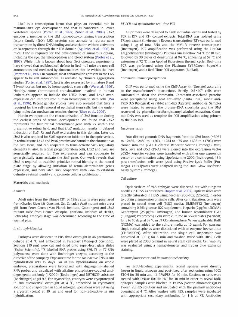
Fig. 1. Lhx2 is expressed in the visual system and demarcates the retina field (A–G). (A–C) Whneural plate of mouse embryos at the 2 somites stage (A), in the optic vesicle (arrowheads), tlimb bud at e12.5 (C). (D–F) On embryos sections, robust Lhx2 expression was detected in tcentral neural retina (nr) and ciliary marginal zone of the retina (cm) at e13.5 and e17.5. No ex(ls). Non-specific background signal is observed in the ocular mesenchyme in panel D. (G) Ianterior neural plate corresponding to the presumptive retina field, where Six3 and Pax6 ar
dissociated to single cells and plated at 2000 cells/ml in NSC mediafor 1 week. GFP positive retinal spheres were visualized using afluorescence-mounted inverted microscope (LEICA).
Statistical analysis
Statistical differences were analyzed using Student's t-test forunpaired samples. An analysis of variance (ANOVA) followed by theDunnett test was used for multiple comparisons with one controlgroup. In all cases, the criterion for significance (P value) was set asmentioned in the respective figures.
Results
Lhx2 demarcates the presumptive retina field
To characterize Lhx2 expression pattern during mouse eyedevelopment, we performed in situ hybridizations on wholeembryos (WISH). Lhx2 expression was first detected in the anteriorneural plate at the 2 somites stage in the region corresponding tothe prospective retina field (Fig. 1A) (Furukawa et al., 1997; Matherset al., 1997; Zuber et al., 2003). From e9.5 to e12.5, robust Lhx2expression was observed in the optic vesicle and eye, prospectivetelencephalon, and limb bud (Figs. 1B and C). On embryo sections,Lhx2 expression was detected at e10.5 in all components of theeye neuroectoderm i.e. optic stalk, optic cup and retinal-pigmentepithelium (RPE), and in the ventral diencephalon (Fig. 1D). In thee12.5 and e17.5 retina, Lhx2 expression is most intense at the retinalciliary margin and distal RPE, with strong expression also in theouter neural retina and RPE (Figs. 1E and F). Notably, comparativeexpression analysis at e8.25 revealed that Lhx2 is co-expressed withRx in the anterior neural plate and that Lhx2 overlap-with and iscontained within Rx expression domain (Fig. 1G). In contrast, Six3expression is predominant in the prospective pituitary/hypothalamicaxis and ventral forebrain, and nearly absent from the retina field(Fig. 1G) (Oliver et al., 1995). Pax6 has a broader and more diffuseexpression throughout the presumptive eye domain, consistent withits later expression in both epithelial and neuroepithelial derivatives(Fig. 1G) (Walther and Gruss, 1991).
ole-mount in situ hybridization shows that Lhx2 expression is detectable in the anteriorelencephalic vesicle and limb from e9.0 (B), and in the eye (arrowheads), neocortex andhe optic cup (oc) and optic stalk (os) at e10.5, in the retinal-pigment epithelium (rpe),pressionwas detectable in ectodermal derivatives such as the lens placode (lp) and lensn 2 somites stage embryos, Lhx2 and Rx expression demarcates a specific region of thee not yet expressed (arrowheads).
544 N. Tétreault et al. / Developmental Biology 327 (2009) 541–550
Lhx2 is required to initiate Six3, Rx and Pax6 expression within theretina field
Eye development in Lhx2 mutant embryos arrest at the opticvesicle stage around e9.0, allowing gene expression analysis at thisstage and earlier. To better understand Lhx2 function in eyedevelopment, we performed comparative DNA micro-array analysison e9.0 WT and Lhx2−/− forebrains. These experiments revealed thatseveral genes involved in eye/retinal development are downregu-lated in Lhx2−/− forebrains, including Mitf, Chx10, Vax2, Rx (Rax),Tbx5, Fzd5 and Six6 (Table 1). In sharp contrast, the Pax6 mutationis not associated with a downregulation of Lhx2, Rx, Six3, Chx10,
Table 1Comparative gene expression analysis between WT and Lhx2−/− embryos
Accessionnumber
Gene Description Foldchange
NM_011445.1 Sox6 SRY-box containing gene 6 −0,9082NM_011384.2 Six6 Sine oculis-related homeobox 6
homolog−0,9108
NM_001085495 Arfgef2 ADP-ribosylation factor guaninenucleotide-exchange
−0,9412
NM_178192.1 Hist1h4a Histone 1, H4a −0,9438AK084437 D230046H12Rik Unknown (Riken) −0,9641NM_013603.1 Mt3 Metallothionein −0,9667NM_029768.2 Use1 Unconventional SNARE in the ER 1
homolog−0,9724
AK018697 Ldlr Low density lipoprotein receptor −0,9785NM_011921 Aldh1a7 Aldehyde dehydrogenase family 1,
subfamily A7−0,9854
AK013968 3110001N23Rik Unknown (Riken) −0,98729530086O07Rik Unknown (Riken) −1,0055
NM_176841.2 Ccdc88a Coiled coil domain containing 88A −1,0313NM_001033193.1 Fzd5 Frizzled homolog 5 −1,0448NM_007740.2 Col9a1 Procollagen, type IX, alpha 1 −1,0536NM_175657 Hist1h4m Histone 1, H4m −1,0674NM_199065 Slitrk1 SLIT and NTRK-like family, member 1 −1,0994AK050619 P38ip-pending Transcription factor
(p38 interacting protein)−1,1068
B830012L14Rik Unknown (Riken) −1,1287AK084485 D330006D04Rik Unknown (Riken) −1,1665NM_009719.4 Neurog3 Neurogenin 3 −1,2109AK032066 Osp94 Osmotic stress protein 94 kDa −1,2355NM_024226 Rtn4 Reticulon 4, transcript variant 5 −1,2406AK054516 E330035H20Rik Unknown (Riken) −1,2491NM_153551.1 Dennd1c DENN/MADD domain containing 1C −1,3124NM_011537 Tbx5 T-box 5 −1,3659AK084692 Stk18 Serine/threonine kinase 18 −1,3736NM_008469.1 Krt1-15 Keratin complex 1, acidic, gene 15 −1,4316AK013410 Rab14 RAB14, member RAS oncogene family −1,4464NM_145463 Shisa2 Shisa homolog 2 −1,4861
C230053D17Rik Unknown (Riken) −1,4912AK054453 E330027G05Rik Unknown (Riken) −1,5453AK084661 D330026I07Rik Unknown (Riken) −1,5460NM_013833 Rax Retina and anterior neural fold
homeobox−1,6921
NM_011912.1 Vax2 Ventral anterior homeoboxcontaining gene 2
−1,7093
NM_001083587 Tsn3 Tensin 3 −1,7541AK042261 Sh3pxd2b SH3 and PX domains 2B −1,9207NM_013467 Aldh1a1 Aldehyde dehydrogenase family 1,
subfamily A1−1,9287
NM_007701.2 Chx10 C. elegans ceh-10 homolog −1,9390NM_010024.1 Dct Dopachrome tautomerase −2,0174NM_001009950 Slc38a8 Solute carrier family 38, member 8 −2,0912NM_007799 Ctse Cathepsin E −2,1450NM_008601 Mitf Microphthalmia-associated
transcription factor−2,2651
XM_122498.1 Tm7sf1 Transmembrane 7 superfamilymember 1
−2,6245
NM_010024 Dct Dopachrome tautomerase −3,2626
Six forebrains from each genotypes at e9.0 were analyzed using Illumina BeadArray™technology. Genes in bold have been involved in eye development and aredownregulated in Lhx2−/− embryos.
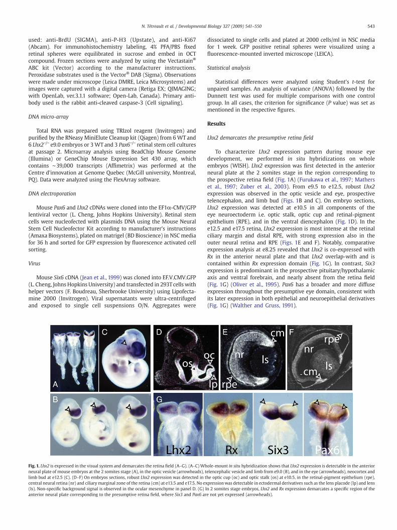
Fig. 2. Lhx2 is required for Six3, Rx and Pax6 expression in the retina field, asrevealed by in situ hybridization (A–D). (A and B) In Lhx2−/− embryos, Six3 expressionis detected in the pituitary/hypothalamic axis and ventral forebrain, but not in theretina field (arrowheads) at neural plate (A) and neural fold (B) stages. (C and D) InLhx2−/− embryos, Rx and Pax6 expression is absent or highly reduced in the retinafield (arrowheads).
Otx2 and Six6 expression in the forebrain and optic vesicle (Bernieret al., 2001; Duparc et al., 2006; Jean et al., 1999).
Based on the above result, we hypothesized that Lhx2 maycontrol the expression of eye genes at the earliest steps of retinalspecification. To test this, we performed WISH on e8.25/e8.5 WTand Lhx2−/− embryos, thus prior to any visible morphologicalabnormalities in Lhx2 mutants. In e8.25 WT embryos, Six3expression was robust in the presumptive pituitary/hypothalamicaxis and ventral forebrain region, but expression in the retina fieldwas only initiated. At e8.5, Six3 expression in the optic sulcus waswell established (Figs. 2A and B). In contrast, although Six3expression was present in the presumptive pituitary/hypothalamicaxis and ventral forebrain of Lhx2 mutants, expression failed to beinitiated in the retina field and optic sulcus (Figs. 2A and B).Similarly, Pax6 and Rx expression was absent or reduced in theanterior neural plate of Lhx2 mutants at e8.25 (Figs. 2C and D).These results show that Lhx2 is required at the earliest stage ofretina specification to initiate Six3, Rx and Pax6 expression withinthe retina field.
Lhx2 is required for Six6 expression in the optic vesicle
Although gene expression fails to be initiated at the neural platestage, Six3 expression was present (but reduced) at e9.0 in the opticvesicle of Lhx2 mutants (Fig. 3B and E). Six3 expression was alsoreduced in the prospective telencephalic vesicle of Lhx2 mutants (Fig.3B). In contrast to Six3, expression of the Six3-related homeobox geneSix6 is only initiated at 3–4 somites stage in the prospective pituitary/hypothalamic axis, and later spreads to the presumptive ventral opticstalk and optic vesicle at e9.0 (Jean et al., 1999). In Lhx2 mutants, weobserved normal Six6 expression pattern in the pituitary/hypothala-mic axis at e9.0, but expression in the optic vesicle was not detected(Figs. 3C and D). To further confirm this observation, we performedWISH using Rx as control for expression in the optic vesicle. Rxexpression was reduced but present in the optic vesicle of Lhx2−/−
embryos (Fig. 3A). To quantify these observations, we performed geneexpression analyses on forebrain extracts from e9.0 WT and Lhx2mutant embryos by Real-time PCR. Because the optic vesicle of Lhx2mutants is smaller even at this stage, we normalized for eye genesexpression using Rx as standard. Even after normalization, theexpression of Six3 and Six6 was still reduced by 75% and 85% in Lhx2mutant forebrains, respectively (Fig. 3E). These results suggest thatLhx2 is required to initiate Six6 and also possibly Six3 expression inthe optic vesicle.

Fig. 3. Lhx2 is required for Six6 expression in the optic vesicle. (A–D)Whole-mount in situhybridization shows that in e9.0 Lhx2−/− embryos, Rx (A) and Six3 (B) expression is presentbut reduced in the optic vesicle (arrowheads). Six3 expression is also reduced in thetelencephalic vesicle of Lhx2−/− embryos (black arrowhead in panel B). In Lhx2−/− embryos,Six6 expression is detected in the pituitary/hypothalamic axis (arrowhead in D) but not intheoptic vesicle (C). Real-timePCR analysis of e9.0 forebrains revealed thatwhen comparedtoWT littermates (n=4), Six3 and Six6 expression is highly reduced in Lhx2mutants (n=3),evenwhen normalized to Rx expression level (E), which was set to 1. ⁎⁎Pb0.01.
545N. Tétreault et al. / Developmental Biology 327 (2009) 541–550
Lhx2 and Pax6 can bind to the chromatin at the Six6 loci in vivoand trans-activate Six6 regulatory elements in vitro
Our results suggest that Lhx2 might bind to Six6 promoter regionsfor expression in the developing retina. To test this, we characterizedthe Six6 gene for putative Lhx2 DNA binding sites using the TRANS-FACT algorithm. Several putative sites were identified in the 5′promoter region and intron I of Six6 (Fig. 4B). Several putative DNAbinding sites were also identified for Pax6 (Fig. 4B). To test forpotential association of Lhx2 or Pax6 with the chromatin at theseregions in vivo, we performed chromatin immunoprecipitation(ChIP) experiments on e12.5 mouse retinas (Duparc et al., 2007).Based on the location of the putative DNA binding sites identified, wescanned 18 chromatin domains each covering ∼300 base pairs ofgenomic DNA. Lhx2 association with the chromatin was found ingenomic DNA regions corresponding to binding sites −3795, −1650and −290 of Six6 (Fig. 4A). Pax6 association with the chromatin wasfound in genomic DNA regions corresponding to binding sites −1650,−290, +685 and +4180 of Six6. Common and robust association ofLhx2 and Pax6 with the chromatin was found at sites −1650 and−290. Control ChIP with an anti-IgG antibody or amplification of theβ-globin promoter suggests that this association is specific (Fig. 4A-inset, and data not shown).
Based on the bioinformatics and ChIP results, we hypothesizedthat Lhx2 and Pax6 directly regulate Six6 transcription. To test this,we performed Luciferase assay in 293T cells using 4 distinct
genomic DNA fragments covering the 5′ promoter region (position−3964 to −75) and intron I (position +638 to +1703) of Six6 (Fig.4C). We found that Lhx2 alone was not sufficient to inducesignificant trans-activation of these DNA fragments. Pax6 alonecould trans-activate fragment 1 (position −3964 to −2465) and 3(position −1284 to −75) just above the baseline level, which wasestablished at 3 (Fig. 4C). Notably, adding Lhx2 and Pax6 togetherresulted in relatively strong trans-activation of fragments 1 and 2(position −2480 to −1263) (Fig. 4C). Robust activation of fragment 2using both factors correlated with co-association of Lhx2 and Pax6with the chromatin at position −1650, which is contained withinfragment 2. We also tested if adding additional factors, such as Six3and Otx2, could result in a more pronounced trans-activation ofSix6 regulatory elements (Zuber et al., 2003). Adding all factorstogether did not enhanced Pax6 and Lhx2 activity on Six6 DNAfragments. In contrast, it apparently interfered with the previouslyobserved activity on fragment 1 and 2 (Fig. 4C). These resultssuggest that Lhx2 and Pax6 are associated with the chromatin atthe Six6 locus in vivo and can trans-activate Six6 regulatoryelements in vitro.
Lhx2 is required for Six6 expression in retinal progenitor/stem cells
We previously reported on the isolation and characterization ofNE retinal progenitors present in the mouse optic vesicle (Duparcet al., 2007). These NE progenitors display all the characteristics ofretinal stem cells. We performed dissociated cultures of WT andLhx2-mutant optic vesicles at e9.0 in serum-free media to isolateNE progenitors in a neurosphere assay (Duparc et al., 2007). Retinalcolonies from Lhx2 mutants were smaller and less abundant thenfrom WT littermates (Fig. 5A). Considering the reduce size of theoptic vesicle in Lhx2 mutants, we performed secondary colonyformation assay (self-renewal assay) from single spheres, allowingnormalization of the number of cells plated/well. Single sphereswere dissociated to single cell suspensions and 2000 cells/ml werere-plated in the same media. Under these conditions, retinalcolonies were smaller and less abundant in the absence of Lhx2(Fig. 5B and data not shown). To identify the underlying cellulardefect, we first measured the mitotic index (PH3+/DAPI+ cells) inWT and Lhx2−/− colonies. No significant differences were foundbetween both genotypes (data not shown). The Ki67 antigen isexpressed at all phases of the cell cycle, but not in G0, and is usedas a marker for progenitors (Scholzen and Gerdes, 2000; Endl et al.,2001). To analyze the cell cycle, spheres from both genotypes werepulsed with Bromodeoxy-Uridine (BrdU) for 60 min and analyzedon sections with antibodies against BrdU and Ki67 (Chenn andWalsh, 2002; Klezovitch et al., 2004). BrdU incorporation assaysrevealed a marked reduction in the number of BrdU+/DAPI+ cells(Fig. 5C) and BrdU+/Ki67+ progenitor cells (Fig. 5D) in Lhx2−/−
colonies, suggesting that Lhx2−/− progenitors are partially arrestedat the G1 phase of the cell cycle. To further test this, we evaluatedthe frequency of progenitors not having entered the S phase (i.e.Ki67+/BdrU− cells) after chronic exposure to BrdU for 12 h. Wefound that the frequency of unlabeled progenitors was significantlyhigher in Lhx2−/− colonies, again suggesting that Lhx2−/− progenitorshave a tendency to be arrested in G1 (Fig. 5E). We also measuredthe frequency of apoptotic cells (activated caspase-3+/DAPI+ cells)and found a significant increase in the number of apoptotic cells inLhx2−/− retinal spheres (Fig. 5H).
To test if this system was relevant to study gene regulation, weperformed Real-time PCR analysis on WT and Lhx2−/− retinalcolonies. We found that Six3 and Six6 expression was reduced by50% and 70% in Lhx2−/− colonies, respectively, thus similarly as in theoptic vesicle (Fig. 5F). Work performed in medaka (Oryzias latipes)revealed that Six3 and Six6 proteins could promote cell cycle entryand DNA replication in part by preventing the physical interaction of

Fig. 4. Lhx2 and Pax6 can bind to the chromatin at the Six6 loci in vivo and trans-activate Six6 regulatory elements in vitro. (A) ChIP scanning experiment at the Six6 loci one12.5 mouse retinas using anti-Lhx2, anti-Pax6 and anti-IgG antibodies (n=2). (A and B) Based on the location of several putative Lhx2 or Pax6 DNA binding sites coveringabout 10 k base pairs of genomic DNA at the Six6 loci (B), PCR primers were designed to amplified 18 DNA fragments containing these sites. Quantitative analysis of theinput/ChIP ratio for each fragment reveals an enrichment of Lhx2 or Pax6 proteins on the chromatin at different regions of Six6 (A). ChIP enrichment was found significantwhen above the established arbitrary baseline level of 0.1. Control anti-IgG antibody and amplification of a fragment of the β-globin (βMajor) promoter suggest that theobserved associations are specific (inset). (B) Physical map of the Six6 loci showing the 4 DNA fragments used in Luciferase assays. The white boxes correspond to exon I andII of Six6. (C) Luciferase assays were performed in 293T cells using 4 genomic DNA fragments (n=3), and Luciferase activity was found significant when above the establishedarbitrary baseline level of 3.
546 N. Tétreault et al. / Developmental Biology 327 (2009) 541–550
Geminin with Cdt1, the main component for the assembly of thepre-replication complex (Del Bene et al., 2004). Six3 and Six6 mayalso promote cell proliferation by repressing the transcription ofcyclin-dependent kinase inhibitors (Gestri et al., 2005; Li et al.,2002). We compared cyclin-dependent kinase inhibitors (i.e. p21Cip1,p27Kip1 and p57Kip2) expression in WT and Lhx2−/− retinal colonies,micro-dissected e9.25 forebrains, and micro-dissected e9.0 opticvesicles by Real-time PCR. We found that the expression of all 3cyclin-dependent kinase inhibitors was increased in Lhx2−/− samplesin vitro and in vivo, with the most dramatic and consistent up-regulation observed for p27Kip (Figs. 5F and G).
Over-expression of Six3 or Six6 in Medaka or Xenopus embryosresulted in increased retinal progenitor cells proliferation andectopic retinal tissue formation (Bernier et al., 2000; Loosli et al.,1999; Zuber et al., 1999). We rationalized that reduced expression ofSix6 and Six3 in Lhx2−/− retinal colonies may explain the proliferationdefect. To test this, we over-expressed Six6/GFP or GFP alone in WT
and Lhx2−/− retinal colonies using lentiviruses. We observed thatcells infected with multiple Six6/GFP viral copies entered intoapoptosis within 48 h. Similar results were obtained with a Six3/GFPlentivirus. However, using lower viral concentrations, we couldachieve conditions where the Six6/GFP virus was non-toxic. Inclonal dissociation assays, we found that Lhx2−/− retinal coloniesinfected with the Six6/GFP virus were larger than those infectedwith the GFP virus after 1 or 2 passages (GFP virus: 194±14 μm;Six6/GFP virus: 230±24 μm, P=0.02). These results suggest thatrestoring Six6 expression in Lhx2−/− retinal colonies can partiallyrescue the growth defect, and reveal that Six3/Six6 gene-dosage isextremely sensitive in this particular cellular context. Collectively,our data show that Lhx2−/− retinal progenitor/stem cells generatesmaller colonies due to a proliferation defect and elevated apoptosis,and suggest that the reduced proliferation phenotype is link to Six6and Six3 down-regulation and increased cyclin-dependent kinaseinhibitors activity.

Fig. 5. Lhx2 is required for Six6 expression in retinal progenitor/stem cells. (A and B) Retinal spheres from Lhx2−/− embryos are smaller than WT littermates in primary cultures andafter serial passages of single spheres (Primary culture n=5, Secondary n=5, Tertiary n=3). (C and D) Immunofluorescence on sections of retinal spheres exposed to BrdU for 90 minrevealed a marked reduction in BrdU incorporation in Lhx2−/− spheres (n=3) compared toWT (n=3). BrdU saturation experiment reveals that most Ki67+ Lhx2−/− progenitors have notyet incorporate BrdU after12 h of BrdU exposition (E). Real-time PCR analysis of Six3 and Six6 (F), and p21Cip1, p27Kip1 and p57Kip2 expression levels in Lhx2−/− (n=5) and WT (n=5)retinal spheres (F), Lhx2−/− (n=2) and WT (n=2) e9.0 optic vesicles (O.V.), and Lhx2−/− (n=2) and WT (n=2) e9.25 forebrains (n=2) (G). Immunohistochemistry on Lhx2−/− and WTretinal sphere sections using an anti-activated caspase-3 antibody. Sections were mounted with DAPI to calculate the number of caspase-3+ cells over the total number of DAPI+ cells/section (H). ⁎Pb0.05; ⁎⁎Pb0.01.
547N. Tétreault et al. / Developmental Biology 327 (2009) 541–550
Pax6 is required for Six6 expression in retinal progenitor/stem cells
Pax6 is not required for Six6 expression in the optic vesicle(Jean et al., 1999). However, based on the ChIP and Luciferase assayresults, we hypothesized that Pax6 is required for Six6 expressionmaintenance. We performed global gene expression analysis usingDNA micro-arrays to compare WT and Pax6-null retinal coloniesisolated from e9.5 optic vesicle (Duparc et al., 2007). Theseexperiments revealed that in Pax6−/− retinal colonies, Six6 is themost down-regulated gene from the entire array (Fig. 6A). Toconfirm this, we performed gene expression analyses on individualWT and Pax6−/− retinal spheres by RT-PCR. We found that incontrast with Six3, Lhx2 and Otx2, which expression is apparently
normal, expression of Six6 is undetectable in Pax6−/− samples (Fig.6B). These results reveal that Pax6 is genetically required for Six6expression in retinal progenitor/stem cells.
Lhx2 and Pax6 can synergistically activate Six6 expression in retinalstem cells
To further explore the relationship between Lhx2, Pax6 and Six6gene activation, we forced Lhx2 or Pax6 expression in cultured retinalstem cells. Retinal colonies were dissociated to single cell suspensionsand electroporated with the Lhx2/GFP, Pax6/GFP or GFP-only DNAconstructs (Fig. 7). At the time of optimal GFP expression (i.e. 36 hpost-electroporation), cells were sorted by fluorescence activated cell
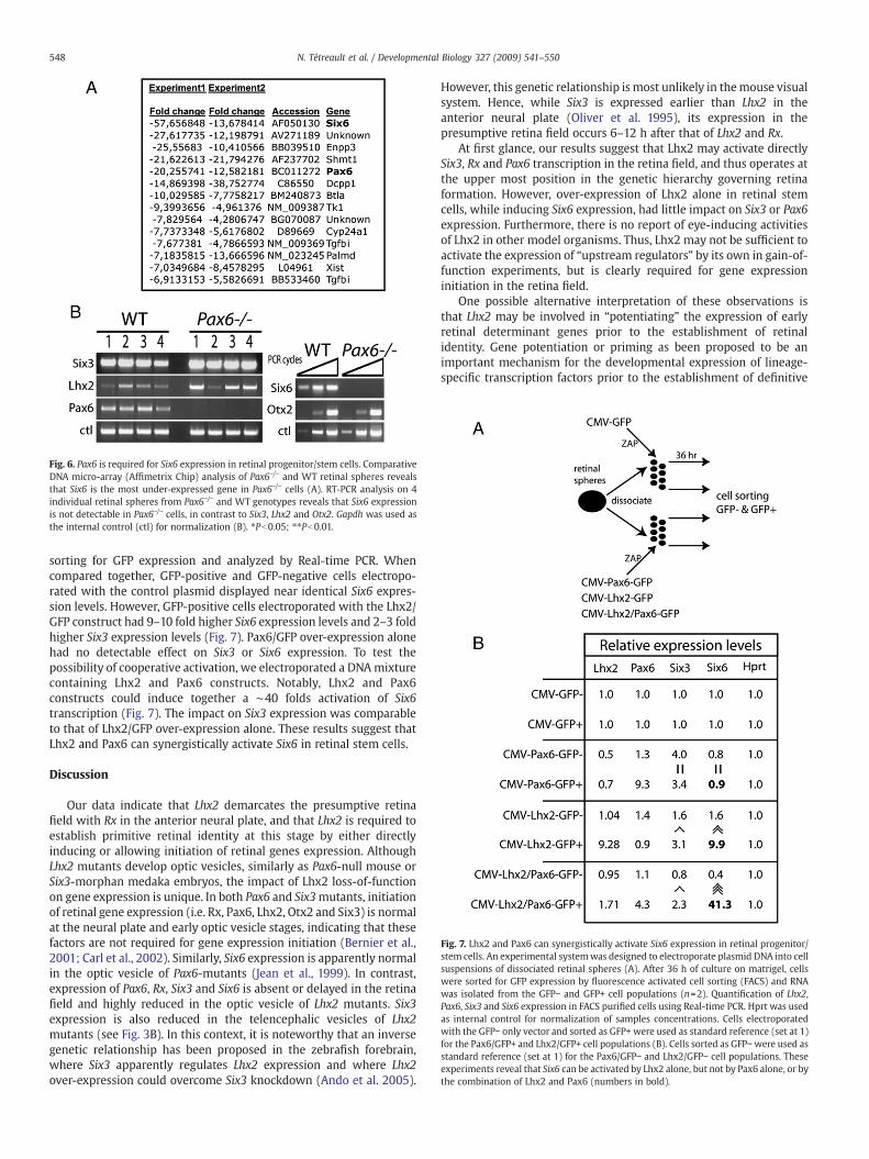
Fig. 7. Lhx2 and Pax6 can synergistically activate Six6 expression in retinal progenitor/stem cells. An experimental systemwas designed to electroporate plasmid DNA into cellsuspensions of dissociated retinal spheres (A). After 36 h of culture on matrigel, cellswere sorted for GFP expression by fluorescence activated cell sorting (FACS) and RNAwas isolated from the GFP− and GFP+ cell populations (n=2). Quantification of Lhx2,Pax6, Six3 and Six6 expression in FACS purified cells using Real-time PCR. Hprt was usedas internal control for normalization of samples concentrations. Cells electroporatedwith the GFP− only vector and sorted as GFP+ were used as standard reference (set at 1)for the Pax6/GFP+ and Lhx2/GFP+ cell populations (B). Cells sorted as GFP−were used asstandard reference (set at 1) for the Pax6/GFP− and Lhx2/GFP− cell populations. Theseexperiments reveal that Six6 can be activated by Lhx2 alone, but not by Pax6 alone, or bythe combination of Lhx2 and Pax6 (numbers in bold).
Fig. 6. Pax6 is required for Six6 expression in retinal progenitor/stem cells. ComparativeDNA micro-array (Affimetrix Chip) analysis of Pax6−/− and WT retinal spheres revealsthat Six6 is the most under-expressed gene in Pax6−/− cells (A). RT-PCR analysis on 4individual retinal spheres from Pax6−/− and WT genotypes reveals that Six6 expressionis not detectable in Pax6−/− cells, in contrast to Six3, Lhx2 and Otx2. Gapdh was used asthe internal control (ctl) for normalization (B). ⁎Pb0.05; ⁎⁎Pb0.01.
548 N. Tétreault et al. / Developmental Biology 327 (2009) 541–550
sorting for GFP expression and analyzed by Real-time PCR. Whencompared together, GFP-positive and GFP-negative cells electropo-rated with the control plasmid displayed near identical Six6 expres-sion levels. However, GFP-positive cells electroporated with the Lhx2/GFP construct had 9–10 fold higher Six6 expression levels and 2–3 foldhigher Six3 expression levels (Fig. 7). Pax6/GFP over-expression alonehad no detectable effect on Six3 or Six6 expression. To test thepossibility of cooperative activation, we electroporated a DNAmixturecontaining Lhx2 and Pax6 constructs. Notably, Lhx2 and Pax6constructs could induce together a ∼40 folds activation of Six6transcription (Fig. 7). The impact on Six3 expression was comparableto that of Lhx2/GFP over-expression alone. These results suggest thatLhx2 and Pax6 can synergistically activate Six6 in retinal stem cells.
Discussion
Our data indicate that Lhx2 demarcates the presumptive retinafield with Rx in the anterior neural plate, and that Lhx2 is required toestablish primitive retinal identity at this stage by either directlyinducing or allowing initiation of retinal genes expression. AlthoughLhx2 mutants develop optic vesicles, similarly as Pax6-null mouse orSix3-morphan medaka embryos, the impact of Lhx2 loss-of-functionon gene expression is unique. In both Pax6 and Six3mutants, initiationof retinal gene expression (i.e. Rx, Pax6, Lhx2, Otx2 and Six3) is normalat the neural plate and early optic vesicle stages, indicating that thesefactors are not required for gene expression initiation (Bernier et al.,2001; Carl et al., 2002). Similarly, Six6 expression is apparently normalin the optic vesicle of Pax6-mutants (Jean et al., 1999). In contrast,expression of Pax6, Rx, Six3 and Six6 is absent or delayed in the retinafield and highly reduced in the optic vesicle of Lhx2 mutants. Six3expression is also reduced in the telencephalic vesicles of Lhx2mutants (see Fig. 3B). In this context, it is noteworthy that an inversegenetic relationship has been proposed in the zebrafish forebrain,where Six3 apparently regulates Lhx2 expression and where Lhx2over-expression could overcome Six3 knockdown (Ando et al. 2005).
However, this genetic relationship is most unlikely in themouse visualsystem. Hence, while Six3 is expressed earlier than Lhx2 in theanterior neural plate (Oliver et al. 1995), its expression in thepresumptive retina field occurs 6–12 h after that of Lhx2 and Rx.
At first glance, our results suggest that Lhx2 may activate directlySix3, Rx and Pax6 transcription in the retina field, and thus operates atthe upper most position in the genetic hierarchy governing retinaformation. However, over-expression of Lhx2 alone in retinal stemcells, while inducing Six6 expression, had little impact on Six3 or Pax6expression. Furthermore, there is no report of eye-inducing activitiesof Lhx2 in other model organisms. Thus, Lhx2 may not be sufficient toactivate the expression of “upstream regulators” by its own in gain-of-function experiments, but is clearly required for gene expressioninitiation in the retina field.
One possible alternative interpretation of these observations isthat Lhx2 may be involved in “potentiating” the expression of earlyretinal determinant genes prior to the establishment of retinalidentity. Gene potentiation or priming as been proposed to be animportant mechanism for the developmental expression of lineage-specific transcription factors prior to the establishment of definitive
549N. Tétreault et al. / Developmental Biology 327 (2009) 541–550
identity in multi-potent hematopoietic stem cells (Bottardi et al.,2007). Gene potentiation involves the maintenance of an accessiblechromatin conformation in multi-potent stem cells, counterbalancingpossible epigenetic silencing at specific loci (Bottardi et al., 2007;Szutorisz et al., 2005). Whether Lhx2 has a function in modulatingchromatin organization or accessibility for other transcription factorsremains to be evaluated.
Once primitive retinal identity is established in the anteriorneural plate and optic sulcus, it is predicted that “master regulatorsof eye development” such as Rx, Pax6, Six3, Lhx2 and Otx2 wouldcooperate to establish definitive retinal identity by inducing highexpression levels of retinal determinant genes. The underlyingmechanisms to induce high gene expression levels could involvesynergistic activation and positive autoregulatory feedback loops.We found here that the concerted action of Lhx2 and Pax6 results inthe synergistic activation of Six6 transcription. Notably, thiscorrelates with association of both Pax6 and Lhx2 with thechromatin at the Six6 loci, sometime on the same chromatin region,suggesting the possible formation of a yet uncharacterized mole-cular complex. We also found that Lhx2 is required for Six6expression initiation in the optic vesicle, and that Pax6 is requiredfor Six6 expression maintenance in cultured retinal stem cellisolated from the optic vesicle. Interestingly, our results are inaccordance with studies performed in medaka and Xenopusembryos. In Xenopus, combined over-expression of Pax6, Six3 andOtx2 results in more robust ectopic eye induction than over-expression of Pax6 alone, suggesting either synergistic activationof retinal gene expression and/or more efficient re-programming ofretinal competence in non-retinal tissues (Zuber et al., 2003). Inmedaka, inactivation of Pax6 or Six3 is associated with normalretinal gene expression at early optic vesicle stages, but geneexpression is lost later on, showing that Pax6 and Six3 areindividually required for gene expression maintenance (Carl et al.,2002).
One of the most striking phenotype of Lhx2 mutants is thereduced optic vesicle and forebrain size owing to reduced cellproliferation (Porter et al. 1997). In Lhx2-null retinal progenitor/stem cell cultures, we found that proliferation was highly reducedand that progenitors were stalled at the G1 phase of the cell cycle athigher frequencies than normal. Apoptosis was also elevatedcompare to WT controls. The observed cell cycle phenotypecorrelated with reduced Six3 and Six6 expression and increasedexpression of cyclin-dependent kinase inhibitors in retinal progeni-tor/stem colonies, e9.25 forebrains and e9.0 optic vesicles of Lhx2mutants. These observations indirectly support the hypothesis thatSix3 and Six6 can repress cyclin-dependent kinase inhibitorsexpression (Gestri et al., 2005; Li et al., 2002). They also highlightthe unanticipated opposite function of Pax6 in repressing retinalprogenitor/stem cells proliferation through activation of cyclin-dependent kinase inhibitors expression (Duparc et al. 2007). Thecomplexity of the system is further revealed by the simultaneousrequirement for Pax6 in the maintenance of Six6 expression inretinal colonies (this study). The biological significance of theapparent genetic antagonism between Lhx2 and Pax6 in opticvesicle growth is unclear at the moment but could allow theestablishment of equilibrium in NE progenitors cell cycle kinetics.
In conclusion, our results suggest a model for early retinalspecification where Lhx2 delineates the retina field with Rx andallows induction of eye gene expression in this domain, possibly by“potentiating” the chromatin. Once primitive retinal identity isestablished, Lhx2 cooperates with Pax6 for robust trans-activation ofdownstream target genes such as Six6 in order to establish andmaintain definitive retinal identity. The herein proposed model likelyalso involves Six3, Rx and Otx2 in a much more complex molecularnetwork, and could represent a common strategy to establish andmaintain cell type identity in different regions of the developing CNS.
Acknowledgments
This work was supported by grants from the Natural Science andEngineering Research Council of Canada, Canadian Institutes of HealthResearch, Foundation Fighting Blindness Canada, and Antoine-TurmelFoundation for Macular Degeneration Research. N.T. is a Scholar fromthe Fonds de Recherche en Ophtalmologie de l'Université deMontréal.G.B is supported by the Fonds de Recherche en Santé du Québec.
References
Agulnick, A.D., Taira, M., Breen, J.J., Tanaka, T., Dawid, I.B., Westphal, H., 1996.Interactions of the LIM-domain-binding factor Ldb1 with LIM homeodomainproteins. Nature 384, 270–272.
Ando, H., Kobayashi, M., Tsubokawa, T., Uyemura, K., Furuta, T., Okamoto, H., 2005. Lhx2mediates the activity of Six3 in zebrafish forebrain growth. Dev. Biol. 287, 456–468.
Bernier, G., Panitz, F., Zhou, X., Hollemann, T., Gruss, P., Pieler, T., 2000. Expandedretina territory by midbrain transformation upon overexpression of Six6 (Optx2)in Xenopus embryos. Mech. Dev. 93, 59–69.
Bernier, G., Vukovich, W., Neidhardt, L., Herrmann, B.G., Gruss, P., 2001. Isolation andcharacterization of a downstream target of Pax6 in the mammalian retinalprimordium. Development 138, 3987–3994.
Bottardi, S., Ghiam, A.F., Bergeron, F., Milot, E., 2007. Lineage-specific transcriptionfactors in multipotent hematopoietic progenitors: a little bit goes a long way. CellCycle 6, 1035–1039.
Carl, M., Loosli, F., Wittbrodt, J., 2002. Six3 inactivation reveals its essential role for theformation and patterning of the vertebrate eye. Development 129, 4057–4063.
Chenn, A., Walsh, C.A., 2002. Regulation of cerebral cortical size by control of cell cycleexit in neural precursors. Science 297, 365–369.
Chow, R.L., Altmann, C.R., Lang, R.A., Hemmati-Brivanlou, A., 1999. Pax6 induces ectopiceyes in a vertebrate. Development 126, 4213–4222.
Del Bene, F., Tessmar-Raible, K., Wittbrodt, J., 2004. Direct interaction of geminin andSix3 in eye development. Nature 427, 745–749.
Duparc, R.H., Abdouh, M., David, J., Lepine, M., Tetreault, N., Bernier, G., 2007. Pax6controls the proliferation rate of neuroepithelial progenitors from the mouse opticvesicle. Dev. Biol. 301, 374–387.
Duparc, R.H., Boutemmine, D., Champagne, M.P., Tetreault, N., Bernier, G., 2006. Pax6 isrequired for delta-catenin/neurojugin expression during retinal, cerebellar andcortical development in mice. Dev. Biol. 300, 647–655.
Endl, E., Hollmann, C., Gerdes, J., 2001. Antibodies against the Ki-67 protein: assessment ofthe growth fraction and tools for cell cycle analysis. Methods Cell Biol. 63, 399–418.
Furukawa, T., Kozak, C.A., Cepko, C.L., 1997. rax, a novel paired-like homeobox gene,shows expression in the anterior neural fold and developing retina. Proc. Natl. Acad.Sci. U. S. A. 94, 3088–3093.
Gehring, W., Kazuho, I., 1999. Pax6 mastering eye morphogenesis and eye evolution.Trends Genet. 15, 371–377.
Gehring, W.J., 2002. The genetic control of eye development and its implications for theevolution of the various eye-types. Int. J. Dev. Biol. 46, 65–73.
Gestri, G., Carl, M., Appolloni, I., Wilson, S.W., Barsacchi, G., Andreazzoli, M., 2005. Six3functions in anterior neural plate specification by promoting cell proliferation andinhibiting Bmp4 expression. Development 132, 2401–2413.
Grindley, J.C., Davidson, D.R., Hill, R.E., 1995. The role of Pax-6 in eye and nasaldevelopment. Development 121, 1433–1442.
Hill, R.E., Favor, J., Hogan, B.L., Ton, C.C., Saunders, G.F., Hanson, I.M., Prosser, J., Jordan, T.,Hastie, N.D., van Heyningen, V., 1991. Mouse small eye results from mutations in apaired-like homeobox-containing gene. Nature 354, 522–525.
Hogan, B.L., Horsburgh, G., Cohen, J., Hetherington, C.M., Fisher, G., Lyon, M.F., 1986.Small eyes (Sey): a homozygous lethal mutation on chromosome 2 which affectsthe differentiation of both lens and nasal placodes in the mouse. J. Embryol. Exp.Morphol. 97, 95–110.
Jean, D., Bernier, G., Gruss, P., 1999. Six6 (Optx2) is a novel murine Six3-relatedhomeobox gene that demarcates the presumptive pituitary/hypothalamic axis andthe ventral optic stalk. Mech. Dev. 84, 31–40.
Klezovitch, O., Fernandez, T.E., Tapscott, S.J., Vasioukhin, V., 2004. Loss of cell polaritycauses severe brain dysplasia in Lgl1 knockout mice. Genes Dev. 18, 559–571.
Li, X., Perissi, V., Liu, F., Rose, D.W., Rosenfeld, M.G., 2002. Tissue-specific regulation ofretinal and pituitary precursor cell proliferation. Science 297, 1180–1183.
Loosli, F., Winkler, S., Wittbrodt, J., 1999. Six3 overexpression initiates the formation ofectopic retina. Genes Dev. 13, 649–654.
Marquardt, T., 2003. Transcriptional control of neuronal diversification in the retina.Prog. Retin. Eye Res. 22, 567–577.
Marquardt, T., Ashery-Padan, R., Andrejewski, N., Scardigli, R., Guillemot, F., Gruss, P., 2001.Pax6 is required for the multipotent state of retinal progenitor cells. Cell 105, 43–55.
Mathers, P.H., Grinberg, A., Mahon, K.A., Jamrich, M., 1997. The Rx homeobox gene isessential for vertebrate eye development. Nature 387, 603–607.
Oliver, G., Gruss, P.,1997. Current views on eye development. TrendsNeurosci. 20, 415–421.Oliver, G., Mailhos, A., Wehr, R., Copeland, N.G., Jenkins, N.A., Gruss, P., 1995. Six3, a
murine homologue of the sine oculis gene, demarcates the most anterior border ofthe developing neural plate and is expressed during eye development. Development121, 4045–4055.
Porter, F.D., Drago, J., Xu, Y., Cheema, S.S., Wassif, C., Huang, S.P., Lee, E., Grinberg, A.,Massalas, J.S., Bodine, D., et al., 1997. Lhx2, a LIM homeobox gene, is required for eye,forebrain, and definitive erythrocyte development. Development 124, 2935–2944.

550 N. Tétreault et al. / Developmental Biology 327 (2009) 541–550
Rhee, H., Polak, L., Fuchs, E., 2006. Lhx2 maintains stem cell character in hair follicles.Science 312, 1946–1949.
Scholzen, T., Gerdes, J., 2000. The Ki-67 protein: from the known and the unknown.J. Cell. Physiol. 182, 311–322.
Szutorisz, H., Canzonetta, C., Georgiou, A., Chow, C.M., Tora, L., Dillon, N., 2005.Formation of an active tissue-specific chromatin domain initiated by epigeneticmarking at the embryonic stem cell stage. Mol. Cell. Biol. 25, 1804–1820.
Walther, C., Gruss, P., 1991. Pax-6, a murine paired box gene, is expressed in thedeveloping CNS. Development 113, 1435–1449.
Wu, H.K., Heng, H.H., Siderovski, D.P., Dong, W.F., Okuno, Y., Shi, X.M., Tsui, L.C., Minden,M.D., 1996. Identification of a human LIM-Hox gene, hLH-2, aberrantly expressed inchronic myelogenous leukaemia and located on 9q33–34.1. Oncogene 12,1205–1212.
Zuber, M.E., Perron, M., Philpott, A., Bang, A., Harris, W.A., 1999. Giant eyes in Xenopuslaevis by overexpression of XOptx2. Cell 98, 341–352.
Zuber, M.E., Gestri, G., Viczian, A.S., Barsacchi, G., Harris, W.A., 2003. Specification of thevertebrate eye by a network of eye field transcription factors. Development 130,5155–5167.


