The late negative episodic memory effect: the effect of recapitulating study details at test
14
Research report The late negative episodic memory effect: the effect of recapitulating study details at test David Friedman * , Yael M. Cycowicz, Michael Bersick Cognitive Electrophysiology Laboratory, New York State Psychiatric Institute, Unit 6, 1051 Riverside Drive, New York City, NY 10032, United States Accepted 18 October 2004 Available online 21 November 2004 Abstract An hypothesis concerning mnemonic function suggests that perceptual details of previously experienced episodes are retrieved from the cortices that initially processed that information during the encoding phase. Cycowicz et al. [Cycowicz, Y.M., Friedman, D. and Snodgrass, J.G., Remembering the color of objects: an ERP investigation of source memory, Cereb Cortex, 11 (2001) 322–334.] have interpreted the presence of a late negative episodic memory (EM) effect, maximal over parieto-occipital scalp, as a brain signature of the search for and/or retrieval/evaluation of the specific perceptual source-specifying attributes (i.e., color) of pictures in the visual cortical regions that were recruited during the encoding of that information. The present study assessed the validity of this hypothesis. Twelve participants studied pictures outlined in red or green and were subsequently tested with inclusion (i.e., item; old or new regardless of color) and exclusion (i.e., source; same color, different color/new judgments) tasks. In both, old pictures were presented either in the same color as at study or in the alternate color. A late negative, parieto-occipital EM effect was of much larger amplitude in the source compared to the item task. It was of similar magnitude to correctly recognized pictures whose colors were identical at study and test relative to those whose colors changed, and was not modulated by the success or failure of the source retrieval. These data run counter to the initial hypothesis that the late negative EM effect reflects the search for and/or retrieval of specific perceptual attributes such as color. Rather, the late negative EM effect may reflect the search for and/or retrieval/evaluation of more general source-specifying information in the cortical regions that initially processed the stimuli. D 2004 Elsevier B.V. All rights reserved. Theme: Neural basis of behavior Topic: Learning and memory: physiology Keywords: Item memory; Source memory; ERP episodic memory (EM) effect 1. Introduction Investigators of memory function have often contrasted two processes, familiarity and recollection, which are thought to underlie recognition memory performance [18]. Whereas familiarity is relatively automatic and is hypothe- sized to underlie the retrieval of item or content information without the details, or context within which the event was embedded, recollection is effortful and is required when retrieving contextual information, such as the spatio- (where) temporal (when) attributes within which the initial episode was encountered. The retrieval of contextual attributes is labeled source memory. Compared to simple, old/new recognition memory paradigms, source memory paradigms, in addition to requiring old/new memory judgments, also solicit judg- ments concerning the context within which the original episode was experienced. For example, during a study phase participants might hear words presented in either a male or female voice. During the subsequent test phase, subjects would be asked to make old/new judgments to visually presented words. Then, for any word judged old, they would be asked to provide a source judgment concerning the original presentation, was it presented in either the male or 0926-6410/$ - see front matter D 2004 Elsevier B.V. All rights reserved. doi:10.1016/j.cogbrainres.2004.10.005 * Corresponding author. Fax: +1 212 543 6002. E-mail address: [email protected] (D. Friedman). Cognitive Brain Research 23 (2005) 185 – 198 www.elsevier.com/locate/cogbrainres
-
Upload
david-friedman -
Category
Documents
-
view
215 -
download
1
Transcript of The late negative episodic memory effect: the effect of recapitulating study details at test
www.elsevier.com/locate/cogbrainres
Cognitive Brain Research
Research report
The late negative episodic memory effect: the effect of recapitulating
study details at test
David Friedman*, Yael M. Cycowicz, Michael Bersick
Cognitive Electrophysiology Laboratory, New York State Psychiatric Institute, Unit 6, 1051 Riverside Drive, New York City, NY 10032, United States
Accepted 18 October 2004
Available online 21 November 2004
Abstract
An hypothesis concerning mnemonic function suggests that perceptual details of previously experienced episodes are retrieved from the
cortices that initially processed that information during the encoding phase. Cycowicz et al. [Cycowicz, Y.M., Friedman, D. and Snodgrass,
J.G., Remembering the color of objects: an ERP investigation of source memory, Cereb Cortex, 11 (2001) 322–334.] have interpreted the
presence of a late negative episodic memory (EM) effect, maximal over parieto-occipital scalp, as a brain signature of the search for and/or
retrieval/evaluation of the specific perceptual source-specifying attributes (i.e., color) of pictures in the visual cortical regions that were
recruited during the encoding of that information. The present study assessed the validity of this hypothesis. Twelve participants studied
pictures outlined in red or green and were subsequently tested with inclusion (i.e., item; old or new regardless of color) and exclusion (i.e.,
source; same color, different color/new judgments) tasks. In both, old pictures were presented either in the same color as at study or in the
alternate color. A late negative, parieto-occipital EM effect was of much larger amplitude in the source compared to the item task. It was of
similar magnitude to correctly recognized pictures whose colors were identical at study and test relative to those whose colors changed, and
was not modulated by the success or failure of the source retrieval. These data run counter to the initial hypothesis that the late negative EM
effect reflects the search for and/or retrieval of specific perceptual attributes such as color. Rather, the late negative EM effect may reflect the
search for and/or retrieval/evaluation of more general source-specifying information in the cortical regions that initially processed the stimuli.
D 2004 Elsevier B.V. All rights reserved.
Theme: Neural basis of behavior
Topic: Learning and memory: physiology
Keywords: Item memory; Source memory; ERP episodic memory (EM) effect
1. Introduction
Investigators of memory function have often contrasted
two processes, familiarity and recollection, which are
thought to underlie recognition memory performance [18].
Whereas familiarity is relatively automatic and is hypothe-
sized to underlie the retrieval of item or content information
without the details, or context within which the event was
embedded, recollection is effortful and is required when
retrieving contextual information, such as the spatio-
0926-6410/$ - see front matter D 2004 Elsevier B.V. All rights reserved.
doi:10.1016/j.cogbrainres.2004.10.005
* Corresponding author. Fax: +1 212 543 6002.
E-mail address: [email protected] (D. Friedman).
(where) temporal (when) attributes within which the initial
episode was encountered. The retrieval of contextual
attributes is labeled source memory.
Compared to simple, old/new recognition memory
paradigms, source memory paradigms, in addition to
requiring old/new memory judgments, also solicit judg-
ments concerning the context within which the original
episode was experienced. For example, during a study phase
participants might hear words presented in either a male or
female voice. During the subsequent test phase, subjects
would be asked to make old/new judgments to visually
presented words. Then, for any word judged old, they would
be asked to provide a source judgment concerning the
original presentation, was it presented in either the male or
23 (2005) 185–198
D. Friedman et al. / Cognitive Brain Research 23 (2005) 185–198186
female voice? [35]. In this type of memory experiment,
simply judging whether the item is old or new can be
accomplished without reference to contextual details, i.e.,
this judgment can be based solely on whether the item
seems familiar or not.
However, remembering whether the item was presented,
for example, by a male or female voice, requires active
recollection of such contextual information. In these kinds
of designs, a recollection-based response has been oper-
ationalized as a correctly recognized old item (i.e., a hit)
whose source has also been correctly retrieved. A familiar-
ity-based recognition response has been operationalized as a
correctly recognized old item whose source has been
incorrectly attributed. Presumably, this indicates that this
latter type of recognition judgment was not accompanied by
contextual detail. That is, an item correctly recognized as
old, but receiving an incorrect source judgment is assumed
to be based only on familiarity; an item correctly recognized
as old attracting a correct source judgment is assumed to be
based on familiarity as well as recollection.
This source memory paradigm has been very often
employed in event-related brain potential (ERP) studies of
recognition memory. The aim of these investigations has
been to obtain brain activity signatures corresponding to
familiarity and recollection hypothesized to operate in two-
process theories of recognition memory [18]. Investigators
using this type of paradigm have uncovered a series of old/
new or episodic memory (EM) effects that appear to reflect
unique mnemonic functions [7,13,24,25]. Typically, cor-
rectly recognized old items elicit greater positivity than
correctly rejected new items. The EM effect is then defined
as the ERP difference between correctly recognized old
and correctly rejected new items. The most consistently
reported EM effects have been labeled the medial
prefrontal EM effect (active between about 300 and 500
ms), the left parietal EM effect (500–900 ms), and the right
prefrontal EM effect (800–2000 ms). Some consensus as to
the functional roles of each of these EM effects exists. The
medial prefrontal EM effect has been associated with the
familiarity component of recognition memory [2,14,19]
(but see [37] and [29]). This conclusion is based on the
findings that the medial prefrontal EM effect is of
equivalent magnitude in the ERPs associated with pre-
viously studied, correctly recognized old items whether
those items were given a brememberQ (retrieval of contextbased on recollection) or bknowQ (retrieval of content
based on familiarity) judgment [28], according to the
paradigm originally described by Tulving [30]. In the same
vein, the medial prefrontal EM effect is generally of
equivalent magnitude in the ERPs associated with old
items and unstudied lures that are highly similar to one
another and thus generate a large familiarity signal [2,22].
The subsequent parietal EM effect has been associated
with recollection, based on a large number of findings
indicating that this EM effect is larger in association with
items whose sources are correctly attributed compared to
those that are not [28,34,35]. The functional role of the
right prefrontal EM effect is currently controversial,
although some investigators have advanced the hypothesis
that it reflects some kind of executive control function,
such as monitoring the products of retrieval in the service
of modifying ongoing memory performance (e.g., Refs.
[25,36]).
Relatively few types of contexts or sources have been
used in these investigations, which have included gender of
voice, spatial location and list membership (i.e., temporal
context). Despite the obvious differences, these source
types have yielded fairly similar EM effects. By contrast
with these types of source information, Cycowicz et al. [5]
used line drawings of common objects that were painted in
either red or green during the study phase. During two
ensuing test phases, pictures were outlined in black and, in
the inclusion or item test, subjects had to judge simply
whether the picture was old or new, regardless of its outline
color in the study phase; during the exclusion or source
test, subjects had to judge whether the item was initially
painted in a target color (for example, red) during study
(hereafter referred to as Targets), or was painted in the
alternate color (green in this example) or was new (these
latter responses were assigned to the same response hand).
Previously studied pictures painted in the alternate color are
hereafter designated as Nontargets to distinguish them from
new pictures.
Unlike previous investigations of source memory, Cyco-
wicz et al. recorded a large-amplitude negative EM effect,
maximal over parieto-occipital scalp, which was markedly
larger in the source than the item task. Due to three factors,
(1) the source in this paradigm was color, (2) the negative
EM was coincident with mean reaction time (RT), and (3)
the negative EM effect showed a topographic focus over
parieto-occipital scalp, Cycowicz and co-workers suggested
that the late negative EM effect could have reflected the
search for and/or retrieval of the color in which the picture
was painted during the study phase in the cortical regions
that originally processed color information.
As mentioned, the peak latency of the negative EM effect
(between ~800 and 1000 ms) occurred at about the same
time as mean reaction time. The negative activity onset as
the preceding parietal EM effect was returning to baseline.
On this basis, Cycowicz et al. [5] advanced the hypothesis
that sufficient time would have elapsed for the late negative
EM effect to have reflected brain activity related to an
attempt to reinstate the initial image along with its
associated color during the source retrieval task [6]. Further,
in the original Cycowicz et al. study [5], the negative EM
effect was as large during successful as it was during
unsuccessful source retrieval. Hence, the breinstatementQinterpretation offered by Cycowicz and co-workers is
consistent with the presence of negative-going activity in
the ERPs associated with trials on which an incorrect source
decision had been made, as subjects would have had to
attempt to retrieve the conjunction of attributes (the picture

D. Friedman et al. / Cognitive Brain Research 23 (2005) 185–198 187
and its color) regardless of the success of the retrieval
attempt.
Although other investigators had also observed similar
negative-going EM effects (review by Ref. [11]), Cycowicz
et al. [5] were, to our knowledge, the first to compare
directly this activity between item and source retrieval
tasks. Subsequent investigations have also assessed the
difference between item and source tasks. For example, in a
study by Johansson et al. [12], subjects viewed words
which were followed by the presentation of a rectangular
outline. In half the trials, a picture was presented within the
rectangle. In the remainder, a picture was not presented and
subjects were asked to imagine the object named by the
label and project it into the rectangular outline. For both
types of trials, subjects were asked to determine how well
the pictured (or imagined) object fit the verbal label. At test,
subjects performed two tasks, in which only the verbal
labels of previously studied and new items were intermixed.
One task was a simple, old/new recognition paradigm, the
other a source monitoring procedure. In the latter, partic-
ipants had to retrieve the action they performed during
study (either viewing the object or imagining it) in
association with the verbal label. By contrast with the old/
new recognition task, a late-onset, negative EM effect was
of much larger amplitude in the source task. It peaked
between 1000 and 1200 ms and was maximal over parieto-
occipital scalp, highly similar to the negative EM effect
recorded by Cycowicz et al. [5] (the scalp topography of the
Johansson et al. [12] negative EM effect can be seen in Fig.
2 of Johansson and Mecklinger [11]). Johansson et al. [12]
raised the possibility, consistent with the interpretation
offered by Cycowicz et al. [5], that the negativity reflected
the reinstatement of the original object (or imaged object)
along with the action that was performed (perceiving or
imagining).
Although lacking a comparison of item and source
tasks, Leynes et al. [16] also assessed source monitoring
by asking their subjects during study to perform an
action or to plan to perform the action in response to the
presentation of an action phrase. During test, the action
phrases (planned and performed) were re-presented
intermixed with new action phrases. Participants were
required to identify the phrase as performed, planned or
new. Leynes et al. recorded a large-amplitude, negative
EM effect that peaked between 1200 and 1800 ms and
displayed a parieto-occipital topography. Because both
planning to perform an action and actual performance of
an action phrase involve a high degree of visual
processing, the Leynes et al. data may also be interpreted
as consistent with the reinstatement of the representation
of the action and its contextual attribute, i.e., whether it
was planned or performed. In both the Johansson et al.
[12] and Leynes et al. [16] investigations, the parieto-
occipital topography of the negative EM effects is
consistent with these computations involving visual
cortical processing regions.
Johansson and Mecklinger [11] have reviewed these and
other studies in which late, negative EM effects have been
observed. The main conclusions from the review are that (1)
this negative activity can be recorded in old/new recognition
memory paradigms, provided baction monitoringQ is
required—typically engendered by difficult response
demands, as in false memory paradigms. In these tasks the
negativity is observed with a posterior scalp topography, but
only in reaction time (RT)-locked averages; (2) the late
negative EM effect occurs whether retrieval is successful or
not and (3) when attribute conjunctions may have to be
retrieved (as in source memory paradigms), the negativity is
observed in stimulus-locked averages. Johansson and
Mecklinger [11] proposed that, in source memory tasks,
the stimulus-locked, late negative EM effect could reflect
b. . . processes related to forming and holding a representa-
tion of a conjunction of attributes that specify the prior
episodeQ (p. 23), for example, the pictorial object and its
color as in previous investigations from this laboratory [3].
As stated earlier, both the original object and its associated
color might be reinstated for evaluation.
Based on the fact that the late negative EM effect is not
associated with successful retrieval, it is possible that,
similar to the functional role proposed for the right
prefrontal EM effect, the late negative EM effect might
reflect monitoring and/or evaluative operations that, due to
the perceptual nature of the source (e.g., color; visualizing
whether an object was imagined or viewed), take place in
posterior cortical regions associated with visual processing.
In previous investigations from this laboratory, pictures
were presented in red or green outline during the study
phase, but were presented in black outline during the test
phases. In the current investigation, by contrast, pictorial
objects were presented in red or green outline during the
encoding phases and then, during two test phases, were re-
presented either in the same color as during study or the
alternate color. During the item test, subjects were required
to judge the items as old or new regardless of the color in
which they were outlined during study and test. On the other
hand, during the source test subjects had to make Same (old
object, same color) or Different (old object, different color)/
New judgments, labeled, respectively, target old, nontarget
old and new items.
It was predicted, based on previous work from this [3,5]
and other [11] laboratories, that the amplitude of the
negative EM effect would not be modulated by whether or
not retrieval of source attributes was successful. Johansson
and Mecklinger [11] postulate that the late negative EM
effect is generated when attributes of the previously studied
item are not easily recovered upon presentation of the test
probe. In the current experimental design, this might occur
when the object presented at test is painted in a different
color than its studied counterpart, because the color cue does
not match the stored representation. On this view, one would
predict larger negative activity when the test cue and the
stored representation differ. Johansson and Mecklinger [11]
D. Friedman et al. / Cognitive Brain Research 23 (2005) 185–198188
have also suggested that the magnitude of the posterior
negative EM effect increases when response demands are
complex or difficult. On this basis, one could predict that
this negative EM effect would be larger to nontargets in the
exclusion or source task, as these items require a complex
hand to response mapping, i.e., nontarget old and new items
are both assigned to the same response hand, whereas target
old items require a different response hand.
1 For complete details and rationale for the use of the proportions of old
and new items used here, see Cycowicz et al. [5].
2. Materials and methods
For ease of exposition, the inclusion or old/new
recognition task will be referred to as the bitemQ task,
because the retrieval of contextual information is not
explicitly required. By contrast, the exclusion task, which
requires retrieval of context, will be referred to as the
bsourceQ task. We use these terms as convenient labels, even
though on a proportion of bitem taskQ trials recollection may
have been involved and on a proportion of bsource taskQtrials a familiarity process was undoubtedly employed.
2.1. Subjects
Nineteen young adults were recruited for this study. The
data of 7 could not be used, 5 due to excessive eye
movements, 1 due to very poor performance, and 1 due to
technical difficulties during the experimental run. The data
of the remaining 12 young adults (9 female; mean age=23)
are the subject of this report. All subjects were native
English speakers, reported themselves to be in good health
and to have no major medical, neurological, or psychiatric
problems. All participants signed informed consent and
received payment for their participation.
2.2. Stimuli
The experimental stimuli were 312 unambiguous line
drawings of common objects that were divided into 6
lists of 52 pictures each, with lists carefully constructed
so that they were equated on category membership,
concept agreement, name agreement, familiarity, and
visual complexity according to previously published
norms [1,4,27]. Statistical analysis of the variables
characterizing the picture sets revealed no significant
differences among lists (PN0.10). An additional 52
pictures from the same normative sources, not used in
the experimental phases, were used for a practice session
and as fillers. The experiment was divided into six
phases. Each phase consisted of one study and two test
blocks, item and source. In each phase, one of the six
lists of pictures was used, with the order of list
presentation randomized across phases separately for each
subject. Of the 52 pictures in a list, 32 were randomly
assigned to the study block, while the remaining 20 were
assigned as foils to the test block.
2.3. Procedure1
The order of the item and source tests was counter-
balanced across subjects. Each picture was displayed for
500 ms with an ISI of 2000 ms. Study Block. Subjects
viewed 32 pictures, half in red and half in green, and were
asked to press one button for red and another button for
green pictures. Subjects were instructed to memorize the
pictures and their associated colors for a subsequent
memory test. Test Block—Item Recognition. Subjects
viewed 26 pictures, half in red and half in green, of which
12 were old (seen during study) and 14 were new. Of the 12
old items, half were presented in the same color as during
study, while the remainder was presented in the alternate
color. Subjects were asked to press one button for old and
another button for new pictures, regardless of the color in
which they were presented at study. Test Block—Source
Recognition. Subjects viewed 26 pictures, half in red and
half in green, of which 20 were old and 6 were new. Of the
20 old items, half were presented in the same color as during
study, while the remainder was presented in the alternate
color. Half of the new pictures were randomly assigned to
red and half to green. Subjects were asked to press one
button for old pictures presented in the same color as during
study (SAME), and another button for old pictures
presented in a different color (DIFF) during study. The Diff
button was also used for new pictures.
In order to ensure that all subjects knew which button to
press, cues were presented on the computer screen during
the entire block. Hence, during the study block, small
rectangles were presented below and to the right and left of
the to-be-remembered pictures. In one rectangle the word
bREDQ appeared in red and in the other the word bGREENQappeared in green. The left/right positions of these cues
reflected the hand assigned to each color. Similarly, during
the item test blocks, bOLDQ and bNEWQ cues in black
lettering appeared in rectangles below and to the right and
left of the pictures, again consistent with the assigned hand
of response. During the source test blocks, the cues
contained the word bSAMEQ and bDIFFQ in black letters.
During study, item and source blocks, subjects made choice,
speeded and accurate, respectively, bredQ/bgreen,Q boldQ/bnew,Q and bSAMEQ (old)/bDIFFQ (old painted in a different
color or new) decisions to each picture. The hands assigned
during study to bredQ and bgreenQ buttons, during the item
task to boldQ and bnew,Q and during the source task to
bSAMEQ and bDIFFQ were counterbalanced across subjects.
The horizontal visual angles ranged from 0.858 to 4.818, andthe vertical visual angles from 0.568 to 3.408, respectively,for the smallest and largest pictures. To counterbalance
order effects, in half of the test phases, the item task
preceded the source task and, in the other half, source
testing preceded item testing. Subjects were not informed

D. Friedman et al. / Cognitive Brain Research 23 (2005) 185–198 189
prior to the study-block which test block would be
administered first. The sequence of stimuli was separately
randomized for each subject.
Hereafter, for the item task, old pictures presented in the
same color at study and test are referred to as bOld Same;Qold pictures presented in different colors at study and test are
referred to as bOld Diff.Q For the Source task, old pictures
presented in the same color at study and test (requiring a
response of bSameQ) are referred to as bTargetQ items; old
pictures presented in different colors at study and test
(requiring a response of bDifferentQ) are referred to as
bNontargetQ items. Correctly rejected new items are labeled
bNewQ (in the source test, these items also required a
response of bDifferentQ).
2.4. EEG recording
EEG (sintered Ag/AgCl electrodes; DC; 100 Hz upper
cutoff; 250 Hz digitization rate) was recorded continuously
with Synamp amplifiers (Neurosoft) using an Electrocap
with 62 locations (extended 10–20 system placements [23],
including left and right mastoids). All leads were referred to
nose tip. Vertical EOG was recorded bipolarly from
electrodes placed on the supraorbital and infraorbital ridges
of the right eye, and horizontal EOG was recorded bipolarly
from electrodes placed on the outer canthi of the two eyes.
Trials containing eye movement artifact were corrected off-
line using the procedure developed by Gratton et al. [8].
Trials were epoched off-line with 100 ms pre- and 1900 ms
post-stimulus periods.
2.5. Data analyses
ERPs were averaged to correctly recognized Old same and
Old diff and correctly rejected new pictures during the item
task and to correctly recognized Target and Nontarget old
trials and correctly rejected new items during the source task.
The SPSS V. 11.5 repeated measures ANOVA program was
Table 1
Behavioral data in the item (A) and source (B) tasks
(A)
Item task
%
Hits
RT
Hits
%
CR
RT
CR
%
FA
%
S
Mean 81.9 829.4 94.2 836.3 5.8 8
S.D. 7.4 152.2 4.5 153.9 4.5
(B)
Source task
%
Target
RT
Target
%
CR
RT
CR
% FA
New
%
T
Mean 73.1 956.2 94.6 841.6 5.0 6
S.D. 10.1 153.9 4.6 162.5 7.4 1
CR=correct rejection; FA=false alarm; Diff=different.
A target in the source task is a picture correctly recognized as having been present
correctly recognized during the test phase as having been presented in the alterna
Pr=measure of discrimination of old from new items; Br=measure of bias, both c
In the source task, Pr was computed in two ways: (1) by subtracting the false alarm
alarm rate to non-target items from the target old hit rate (z). Two Br indices wer
* Reliably different from zero as assessed by t-test.
used for all analyses. The Greenhouse–Geisser epsilon (e)correction [10] was used where appropriate. Uncorrected
degrees of freedom are reported along with the epsilon value;
the P values reflect the epsilon correction. Significant main
effects and interactions were followed-up, where appropriate,
with simple effects tests and/or post-hoc analyses using the
Tukey Honestly Significant Difference (HSD) test.
To be consistent with our previous work on the late
negative EM effect [5], and to capture any anterior/posterior
and/or left/right asymmetries, the main ANOVAs were
performed on the data recorded from 24 scalp sites along
lateral (left, midline, right) and anterior/posterior planes.
The 24 scalp sites included on the left, FP1, F3, FC3, C3,
CP3, P3, PO3, and O1; on the midline, FPz, FCz, Fz, Cz,
CPz, Pz, POz, and Oz; on the right, FP2, F4, FC4, C4, CP4,
P4, PO4, and O2. The ANOVAs always included the factors
of Laterality (left, midline, and right), and Anterior/Posterior
scalp location (Frontal Pole, Frontal, Fronto-Central, Cen-
tral, Centro-Parietal, Parietal, Parieto-Occipital, Occipital).
However, unless they interacted with the variables of
interest (e.g., item vs. source tasks), the main effects or
interactions of Laterality and Anterior/Posterior location are
not interpreted or reported as, by themselves, they do not
reflect memory-related differences.
3. Results
3.1. Behavioral data
Table 1 presents the behavioral data from the item (A)
and source (B) tasks.
3.1.1. Reaction time (RT)
RTs in the item and source tasks were compared in an
ANOVA that assessed the effects of Task (item, source) and
Study/Test color pairing (same, different). RTs were longer
in the source (1021 ms) than in the item (831 ms) task
ame
RT
Same
%
Diff
RT
Diff
Pr Br
4.5 812.0 79.2 849.0 0.75* 0.26
9.8 148.7 8.0 166.0 0.08 0.14
Non-
arget Diff
RT
Nontarget
Pr# Br# Prz Brz
1.9 1086.7 0.67* 0.17 0.35* 0.58
1.3 207.7 0.12 0.11 0.13 0.12
ed in the same color during study. A nontarget in the source task is a picture
te color during study.
omputed according to Snodgrass and Corwin [26].
rate to new items from the target hit rate (#); and (2) by subtracting the false
e also computed using the new and non-target false alarm rates.
D. Friedman et al. / Cognitive Brain Research 23 (2005) 185–198190
(F(1,11)=48.43, Pb0.0001), and Old same/Target items
were responded to faster (884 ms) than Old Diff/Nontarget
items (968 ms; F(1,11)=16.44, Pb0.002). However, Task
and Study/Test pairing interacted ( F (1,11)=17.06,
Pb0.002). Post-hoc testing indicated that the difference
between Old same and Old diff in the item task failed to
reach significance (although in the expected direction),
whereas the difference between Target and Nontarget RTs
was reliable in the source task.
A repeated measures ANOVA with the factors of Task
(item, source) and Old/New (target RTs were used for the
source task) revealed that RTs in the source test were longer
than in the item task (F(1,11)=20.60, Pb0.001) and that RTs
to new items were faster than those to old items
(F(1,11)=6.97, Pb0.02). However, the two main effects
interacted (F(1,11)=43.90, Pb0.0001). Post-hoc testing
indicated that old and new RTs did not differ significantly
for the item task, while target RTs were reliably slower than
their new counterparts in the source task.
3.1.2. Percent correct and sensitivity
This analysis assessed the effect of Task (item, source)
and Study/Test color pairing (same, different) on the
percentage of correct responses. The percentage of correct
responses was larger in the item than the source task
(F(1,11)=32.10, Pb0.0001). For both tasks, the percentage
of correct responses to Old same/Target items was reliably
greater than Old diff/Nontarget items (F(1,11)=5.50,
Pb0.04; interaction F(1,11)=1.90, PN0.10).
Estimates of the subjects’ ability to discriminate old from
new items (Pr) along with response bias (Br) were
computed according to the methods described by Snodgrass
and Corwin [26], and are shown in Table 1. In the source
task, these indices were computed in two ways: using the
false alarm rate associated with new and nontarget items.
The 3 Pr values listed in Table 1 were all reliably different
from zero, as assessed by t-tests (item Pr t(11)=33.8,
Pb0.0001; source Pr based on the new FA rate t(11)=19.6,
Pb0.0001; source Pr based on the nontarget FA rate
t(11)=9.3, Pb0.0001). Using the false alarm rate to new
items, Pr was larger in the item than the source task
(F(1,11)=5.40, Pb0.04), and responding was more liberal
in the source than the item task (F(1,11)=6.11, Pb0.03).
Using the false alarm rate to nontarget items, Pr was again
larger in the item than the source task (F(1,11)=98.70,
Pb0.0001), and responding was still more liberal in the
source task (F(1,11)=55.03, Pb0.0001). A comparison of
the two Pr values in the source task revealed greater
sensitivity in detecting targets relative to new items
compared to detecting targets relative to nontargets
(F(1,11)=93.02, Pb0.00001).
In sum, accuracy was lower and RTs were longer in the
source task. Regardless of which false alarm rate was used,
participants were able to discriminate among the three
classes of stimuli in the source task. Changing the picture’s
outline color between study and test led to lower accuracy
and longer RTs in both item and source tasks although, for
RTs, this effect was reliable only in the source task.
3.2. ERP Data
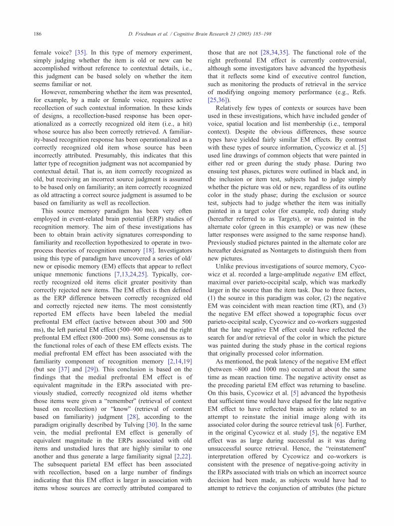
Fig. 1 depicts the grand mean ERP data for both the item
and source tasks, categorized according to the conjunction
of the color in which the item was presented at study and
test. For the item task, the ERPs associated with correctly
rejected new and correctly recognized Old same and Old
diff items are depicted in the left panel of Fig. 1. Similarly,
for the source task, in the right panel of Fig. 1, the ERPs
associated with correctly recognized Target and Nontarget
trials are depicted along with the ERPs elicited by correctly
rejected new items. The mean number (FS.D.) of trials
comprising each of the averages depicted in Fig. 1 are
presented in Table 2.
Fig. 1 indicates that the waveforms from both the item
and source tasks are characterized by parietal EM effects
(maximal at ~500–600 ms) that do not appear to differ in
magnitude. Second, the presence of late negative activity
appears to reduce the magnitude of the parietal EM effect,
resulting in a large magnitude late positivity (~700 ms)
associated with correctly rejected new items in both tasks.
Third, while a late negative EM effect (between about 800
and 1000 ms) appears to be present in the ERPs of the item
task, this activity is much larger in the source task.
For the item task, Fig. 1 suggests that neither the parietal
nor the late negative EM effects differs according to whether
the test picture was painted in the same or the alternate color
as at study. By contrast, there is a suggestion in the source
task data that, for the parietal EM effect, pictures painted in
the same color as at study elicit a somewhat larger EM effect
than those painted in the alternate color. In similar fashion to
the item task, the late negative EM effect does not appear to
differ according to the conjunction of study and test color
pairing. The mean RT marks at the top of the figure
(depicted below the FPz scalp site) demonstrate that, for
both the item and source tasks, RT activity occurs during the
latency window of the late negative EM effects for the two
classes of old items. There is also the suggestion of a right
prefrontal EM effect in the source task data only at the FP2
electrode site, which is absent at the FP1 site. At this
location, targets appear to elicit more positive-going activity
than either new or nontarget items, beginning quite early
(~300–400 ms) and lasting until the end of the recording
epoch.
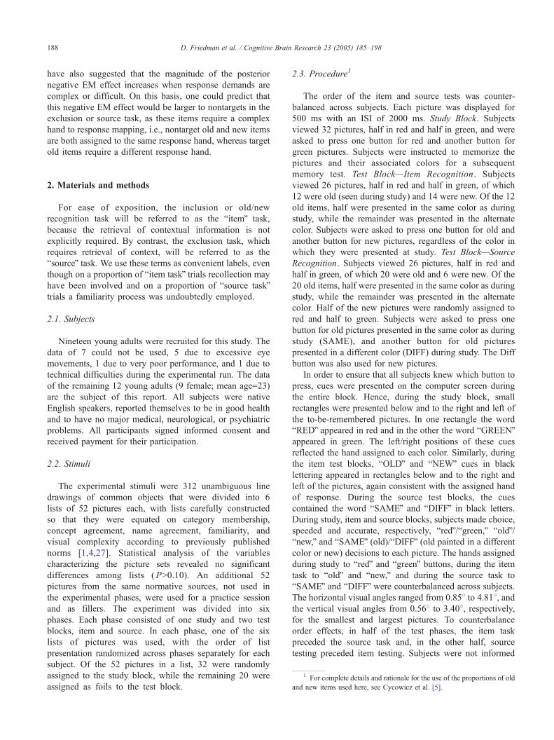
Fig. 2 depicts, from a reduced number of scalp sites, the
old–new difference waveforms for the item and source tasks
computed using the data depicted in Fig. 1. It is clear that
the parietal EM effects elicited in both tasks are highly
similar. Both item and source task waveforms are charac-
terized by late negative EM effects (~800–1000 ms), which
are larger in the source task.
The statistical analyses of effects identified in Fig. 1
were performed on a series of averaged voltages
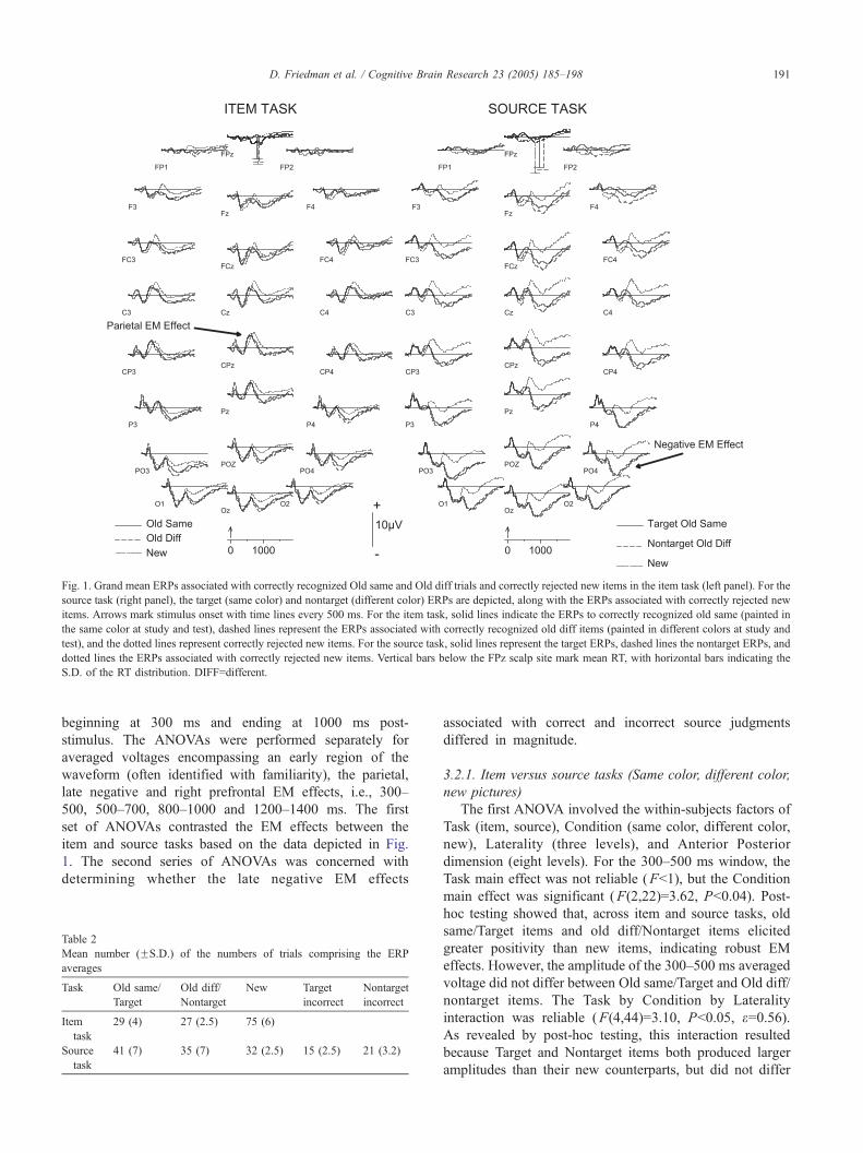
Fig. 1. Grand mean ERPs associated with correctly recognized Old same and Old diff trials and correctly rejected new items in the item task (left panel). For the
source task (right panel), the target (same color) and nontarget (different color) ERPs are depicted, along with the ERPs associated with correctly rejected new
items. Arrows mark stimulus onset with time lines every 500 ms. For the item task, solid lines indicate the ERPs to correctly recognized old same (painted in
the same color at study and test), dashed lines represent the ERPs associated with correctly recognized old diff items (painted in different colors at study and
test), and the dotted lines represent correctly rejected new items. For the source task, solid lines represent the target ERPs, dashed lines the nontarget ERPs, and
dotted lines the ERPs associated with correctly rejected new items. Vertical bars below the FPz scalp site mark mean RT, with horizontal bars indicating the
S.D. of the RT distribution. DIFF=different.
D. Friedman et al. / Cognitive Brain Research 23 (2005) 185–198 191
beginning at 300 ms and ending at 1000 ms post-
stimulus. The ANOVAs were performed separately for
averaged voltages encompassing an early region of the
waveform (often identified with familiarity), the parietal,
late negative and right prefrontal EM effects, i.e., 300–
500, 500–700, 800–1000 and 1200–1400 ms. The first
set of ANOVAs contrasted the EM effects between the
item and source tasks based on the data depicted in Fig.
1. The second series of ANOVAs was concerned with
determining whether the late negative EM effects
Table 2
Mean number (FS.D.) of the numbers of trials comprising the ERP
averages
Task Old same/
Target
Old diff/
Nontarget
New Target
incorrect
Nontarget
incorrect
Item
task
29 (4) 27 (2.5) 75 (6)
Source
task
41 (7) 35 (7) 32 (2.5) 15 (2.5) 21 (3.2)
associated with correct and incorrect source judgments
differed in magnitude.
3.2.1. Item versus source tasks (Same color, different color,
new pictures)
The first ANOVA involved the within-subjects factors of
Task (item, source), Condition (same color, different color,
new), Laterality (three levels), and Anterior Posterior
dimension (eight levels). For the 300–500 ms window, the
Task main effect was not reliable (Fb1), but the Condition
main effect was significant (F(2,22)=3.62, Pb0.04). Post-
hoc testing showed that, across item and source tasks, old
same/Target items and old diff/Nontarget items elicited
greater positivity than new items, indicating robust EM
effects. However, the amplitude of the 300–500 ms averaged
voltage did not differ between Old same/Target and Old diff/
nontarget items. The Task by Condition by Laterality
interaction was reliable (F(4,44)=3.10, Pb0.05, e=0.56).As revealed by post-hoc testing, this interaction resulted
because Target and Nontarget items both produced larger
amplitudes than their new counterparts, but did not differ
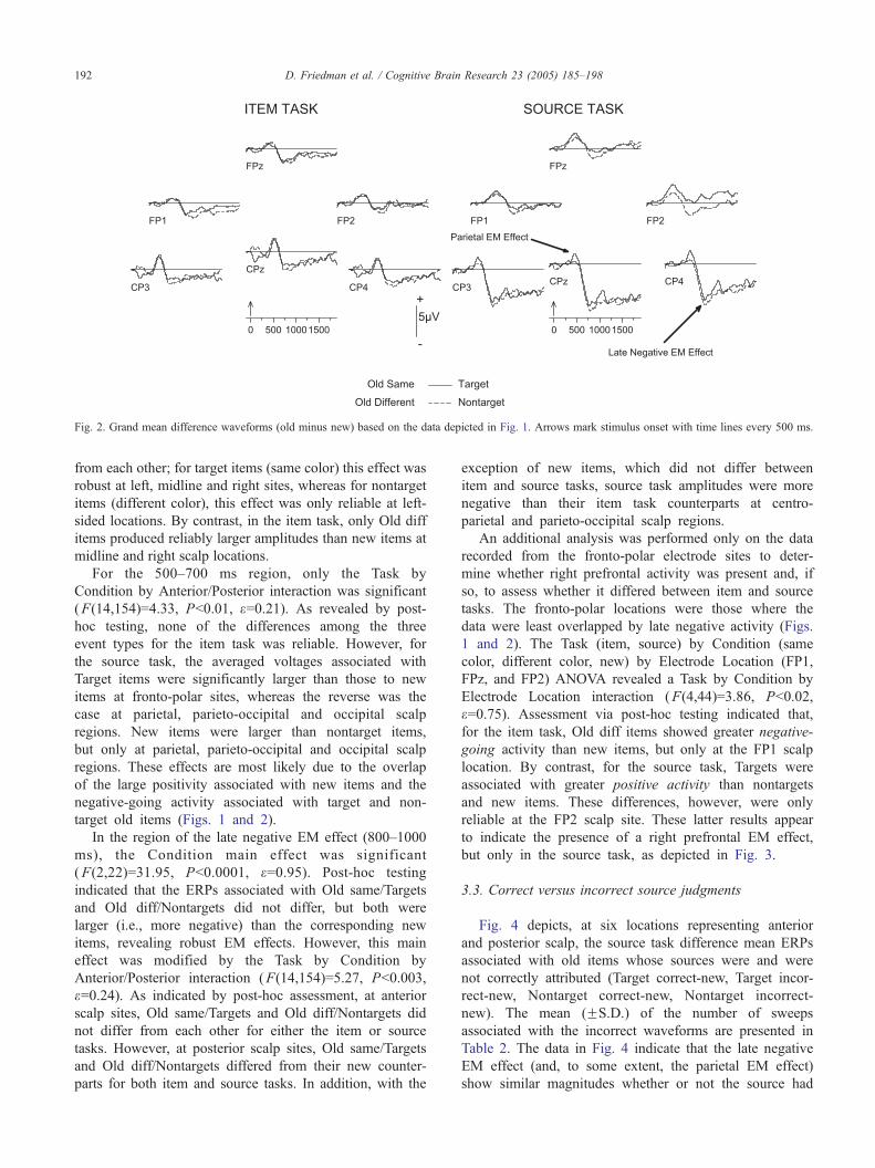
Fig. 2. Grand mean difference waveforms (old minus new) based on the data depicted in Fig. 1. Arrows mark stimulus onset with time lines every 500 ms.
D. Friedman et al. / Cognitive Brain Research 23 (2005) 185–198192
from each other; for target items (same color) this effect was
robust at left, midline and right sites, whereas for nontarget
items (different color), this effect was only reliable at left-
sided locations. By contrast, in the item task, only Old diff
items produced reliably larger amplitudes than new items at
midline and right scalp locations.
For the 500–700 ms region, only the Task by
Condition by Anterior/Posterior interaction was significant
(F(14,154)=4.33, Pb0.01, e=0.21). As revealed by post-
hoc testing, none of the differences among the three
event types for the item task was reliable. However, for
the source task, the averaged voltages associated with
Target items were significantly larger than those to new
items at fronto-polar sites, whereas the reverse was the
case at parietal, parieto-occipital and occipital scalp
regions. New items were larger than nontarget items,
but only at parietal, parieto-occipital and occipital scalp
regions. These effects are most likely due to the overlap
of the large positivity associated with new items and the
negative-going activity associated with target and non-
target old items (Figs. 1 and 2).
In the region of the late negative EM effect (800–1000
ms), the Condition main effect was significant
(F(2,22)=31.95, Pb0.0001, e=0.95). Post-hoc testing
indicated that the ERPs associated with Old same/Targets
and Old diff/Nontargets did not differ, but both were
larger (i.e., more negative) than the corresponding new
items, revealing robust EM effects. However, this main
effect was modified by the Task by Condition by
Anterior/Posterior interaction (F(14,154)=5.27, Pb0.003,
e=0.24). As indicated by post-hoc assessment, at anterior
scalp sites, Old same/Targets and Old diff/Nontargets did
not differ from each other for either the item or source
tasks. However, at posterior scalp sites, Old same/Targets
and Old diff/Nontargets differed from their new counter-
parts for both item and source tasks. In addition, with the
exception of new items, which did not differ between
item and source tasks, source task amplitudes were more
negative than their item task counterparts at centro-
parietal and parieto-occipital scalp regions.
An additional analysis was performed only on the data
recorded from the fronto-polar electrode sites to deter-
mine whether right prefrontal activity was present and, if
so, to assess whether it differed between item and source
tasks. The fronto-polar locations were those where the
data were least overlapped by late negative activity (Figs.
1 and 2). The Task (item, source) by Condition (same
color, different color, new) by Electrode Location (FP1,
FPz, and FP2) ANOVA revealed a Task by Condition by
Electrode Location interaction (F(4,44)=3.86, Pb0.02,
e=0.75). Assessment via post-hoc testing indicated that,
for the item task, Old diff items showed greater negative-
going activity than new items, but only at the FP1 scalp
location. By contrast, for the source task, Targets were
associated with greater positive activity than nontargets
and new items. These differences, however, were only
reliable at the FP2 scalp site. These latter results appear
to indicate the presence of a right prefrontal EM effect,
but only in the source task, as depicted in Fig. 3.
3.3. Correct versus incorrect source judgments
Fig. 4 depicts, at six locations representing anterior
and posterior scalp, the source task difference mean ERPs
associated with old items whose sources were and were
not correctly attributed (Target correct-new, Target incor-
rect-new, Nontarget correct-new, Nontarget incorrect-
new). The mean (FS.D.) of the number of sweeps
associated with the incorrect waveforms are presented in
Table 2. The data in Fig. 4 indicate that the late negative
EM effect (and, to some extent, the parietal EM effect)
show similar magnitudes whether or not the source had

Fig. 3. Grand mean averaged voltages at the FP1, FPz, and FP2 scalp sites computed on the ERPs associated with same study-test color pairings (Old same/
Target), different study-test color pairings (Old diff/Nontarget) and new items in both item and source tasks. Asterisks indicate the electrodes at which the
conditions within the brackets differed reliably. DIFF=different.
D. Friedman et al. / Cognitive Brain Research 23 (2005) 185–198 193
been judged correctly. By contrast, there is a suggestion
that, for targets, correct judgments lead to larger
amplitudes of the activity between 300 and 700 ms,
whereas for nontargets, incorrect judgments lead to larger
amplitudes than correct judgments.
To determine statistically if the magnitudes of the
parietal and late negative EM effects differed reliably for
successful compared to unsuccessful source retrieval, the
300–500, 500–700, 800–1000, and 1200–1400 ms
regions were analyzed in Target/Nontarget by Correctness
(correct, incorrect source) by Laterality by Anterior/
Posterior ANOVAs using the difference waveforms (Fig.
4). For all four averaged voltage windows, none of the
main or interaction effects involving the Target/Nontarget
or Correctness factors was reliable (Fsb2.50, PsN0.10).
Fig. 4. Grand mean difference waveforms in the Source Task associated with correc
every 500 ms. Solid lines indicate the ERPs associated with correct target (left pan
the ERPs associated with incorrect target (left panel) and incorrect nontarget (rig
3.4. Comparison of nose- and mastoid-referenced data
Fig. 5 depicts the Fig. 1 data re-referred to a linked
mastoid reference. The major difference between the
nose- and mastoid-referred data appears to be the
presence of a right prefrontal EM effect, which is larger
in the source compared to the item task, especially for
the ERPs associated with Target trials. As pointed out
earlier, re-inspection of Fig. 1 also demonstrates the
presence of this right prefrontal EM, although it does not
appear as prominent as in the mastoid-referenced ERP
waveforms. Fig. 6 illustrates a more direct comparison of
the two sets of data using the difference means at
prefrontal and centro-parietal electrode sites. Overall, the
mastoid-referred data are characterized by more positive-
t and incorrect source decisions. Arrows mark stimulus onset with time lines
el) and correct nontarget (right panel) old decisions. Dashed lines represent
ht panel) decisions.
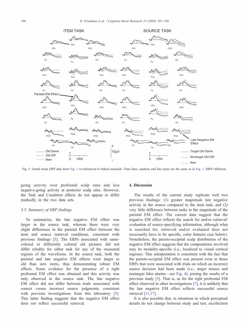
Fig. 5. Grand mean ERP data from Fig. 1 re-referenced to linked mastoids. Time lines, markers and line types are the same as in Fig. 1. DIFF=different.
D. Friedman et al. / Cognitive Brain Research 23 (2005) 185–198194
going activity over prefrontal scalp sites and less
negative-going activity at posterior scalp sites. However,
the Task and Condition effects do not appear to differ
markedly in the two data sets.
3.5. Summary of ERP findings
To summarize, the late negative EM effect was
larger in the source task, whereas there were very
slight differences in the parietal EM effect between the
item and source retrieval conditions, consistent with
previous findings [5]. The ERPs associated with same-
colored or differently colored old pictures did not
differ reliably for either task for any of the measured
regions of the waveforms. In the source task, both the
parietal and late negative EM effects were larger to
old than new items, thus demonstrating robust EM
effects. Some evidence for the presence of a right
prefrontal EM effect was obtained and this activity was
only observed in the source task. The late negative
EM effect did not differ between trials associated with
correct versus incorrect source judgments, consistent
with previous investigations from this laboratory [5].
This latter finding suggests that the negative EM effect
does not reflect successful retrieval.
4. Discussion
The results of the current study replicate well two
previous findings: (1) greater magnitude late negative
activity in the source compared to the item task; and (2)
very little difference between tasks in the magnitude of the
parietal EM effect. The current data suggest that the
negative EM effect reflects the search for and/or retrieval/
evaluation of source-specifying information, although what
is searched for, retrieved and/or evaluated does not
necessarily have to be specific, color features (see below).
Nonetheless, the parieto-occipital scalp distribution of the
negative EM effect suggests that the computations involved
may be modality-specific (i.e., localized to visual cortical
regions). This interpretation is consistent with the fact that
the parieto-occipital EM effect was present even in those
ERPs that were associated with trials on which an incorrect
source decision had been made (i.e., target misses and
nontarget false alarms—see Fig. 4), joining the results of a
previous study [5]. That is, as for the right prefrontal EM
effect observed in other investigations [7], it is unlikely that
the late negative EM effect reflects successful source
retrieval [11,17].
It is also possible that, in situations in which perceptual
details do not change between study and test, recollection
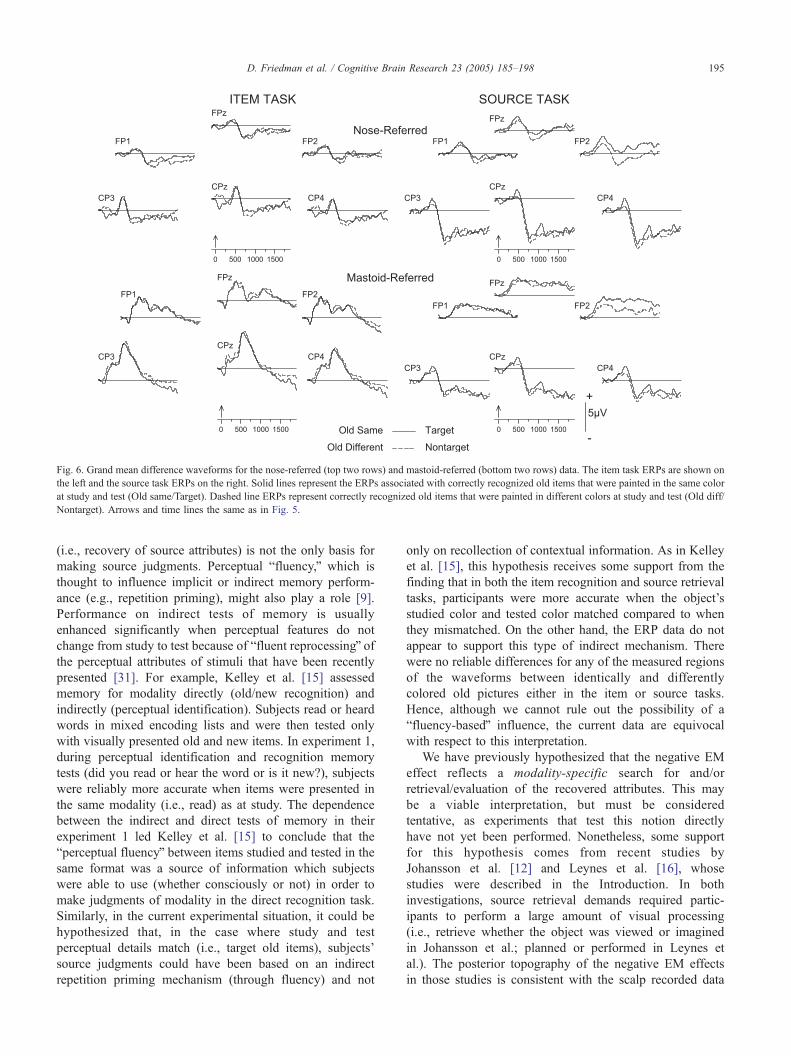
Fig. 6. Grand mean difference waveforms for the nose-referred (top two rows) and mastoid-referred (bottom two rows) data. The item task ERPs are shown on
the left and the source task ERPs on the right. Solid lines represent the ERPs associated with correctly recognized old items that were painted in the same color
at study and test (Old same/Target). Dashed line ERPs represent correctly recognized old items that were painted in different colors at study and test (Old diff/
Nontarget). Arrows and time lines the same as in Fig. 5.
D. Friedman et al. / Cognitive Brain Research 23 (2005) 185–198 195
(i.e., recovery of source attributes) is not the only basis for
making source judgments. Perceptual bfluency,Q which is
thought to influence implicit or indirect memory perform-
ance (e.g., repetition priming), might also play a role [9].
Performance on indirect tests of memory is usually
enhanced significantly when perceptual features do not
change from study to test because of bfluent reprocessingQ ofthe perceptual attributes of stimuli that have been recently
presented [31]. For example, Kelley et al. [15] assessed
memory for modality directly (old/new recognition) and
indirectly (perceptual identification). Subjects read or heard
words in mixed encoding lists and were then tested only
with visually presented old and new items. In experiment 1,
during perceptual identification and recognition memory
tests (did you read or hear the word or is it new?), subjects
were reliably more accurate when items were presented in
the same modality (i.e., read) as at study. The dependence
between the indirect and direct tests of memory in their
experiment 1 led Kelley et al. [15] to conclude that the
bperceptual fluencyQ between items studied and tested in the
same format was a source of information which subjects
were able to use (whether consciously or not) in order to
make judgments of modality in the direct recognition task.
Similarly, in the current experimental situation, it could be
hypothesized that, in the case where study and test
perceptual details match (i.e., target old items), subjects’
source judgments could have been based on an indirect
repetition priming mechanism (through fluency) and not
only on recollection of contextual information. As in Kelley
et al. [15], this hypothesis receives some support from the
finding that in both the item recognition and source retrieval
tasks, participants were more accurate when the object’s
studied color and tested color matched compared to when
they mismatched. On the other hand, the ERP data do not
appear to support this type of indirect mechanism. There
were no reliable differences for any of the measured regions
of the waveforms between identically and differently
colored old pictures either in the item or source tasks.
Hence, although we cannot rule out the possibility of a
bfluency-basedQ influence, the current data are equivocal
with respect to this interpretation.
We have previously hypothesized that the negative EM
effect reflects a modality-specific search for and/or
retrieval/evaluation of the recovered attributes. This may
be a viable interpretation, but must be considered
tentative, as experiments that test this notion directly
have not yet been performed. Nonetheless, some support
for this hypothesis comes from recent studies by
Johansson et al. [12] and Leynes et al. [16], whose
studies were described in the Introduction. In both
investigations, source retrieval demands required partic-
ipants to perform a large amount of visual processing
(i.e., retrieve whether the object was viewed or imagined
in Johansson et al.; planned or performed in Leynes et
al.). The posterior topography of the negative EM effects
in those studies is consistent with the scalp recorded data
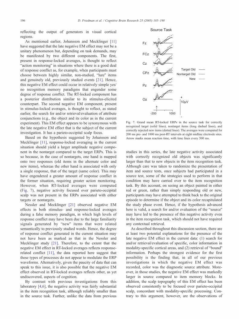
Fig. 7. Grand mean RT-locked ERPs in the source task for correctly
recognized target (solid lines), nontarget items (long dashed lines), and
correctly rejected new items (dotted lines). The averages were computed for
200 ms pre- and 1000 ms post-RT intervals at eight midline electrode sites.
Arrow marks mean reaction time, with time lines every 500 ms.
D. Friedman et al. / Cognitive Brain Research 23 (2005) 185–198196
reflecting the output of generators in visual cortical
regions.
As mentioned earlier, Johansson and Mecklinger [11]
have suggested that the late negative EM effect may not be a
unitary phenomenon but, depending on task demands, may
be manifested by two different components. The first,
present in response-locked averages, is thought to reflect
baction monitoringQ in situations where there is a good deal
of response conflict as, for example, when participants must
choose between highly similar, non-studied, blureQ items
and genuinely old, previously studied events [21]. Hence,
this negative EM effect could occur in relatively simple yes/
no recognition memory paradigms that engender some
degree of response conflict. The RT-locked component has
a posterior distribution similar to its stimulus-elicited
counterpart. The second negative EM component, present
in stimulus-locked averages, is thought to reflect, as stated
earlier, the search for and/or retrieval/evaluation of attribute
conjunctions (e.g., the object and its color as in the current
experiment). This EM effect appears to be synonymous with
the late negative EM effect that is the subject of the current
investigation. It has a parieto-occipital scalp focus.
Based on the hypothesis suggested by Johansson and
Mecklinger [11], response-locked averaging in the current
situation should yield a larger amplitude negative compo-
nent in the nontarget compared to the target ERPs. This is
so because, in the case of nontargets, one hand is mapped
onto two responses (old items in the alternate color and
new items), whereas the other hand is associated with only
a single response, that of the target (same color). This may
have engendered a greater amount of response conflict in
the former situation, requiring greater action monitoring.
However, when RT-locked averages were computed
(Fig. 7), negative activity focused over parieto-occipital
scalp was not present in the ERPs associated with either
targets or nontargets.
Nessler and Mecklinger [21] observed negative EM
effects in both stimulus- and response-locked averages
during a false memory paradigm, in which high levels of
response conflict may have been due to the large familiarity
signals generated by bnewQ items that were related
semantically to previously studied words. Hence, the degree
of response conflict generated in the current situation may
not have been as marked as that in the Nessler and
Mecklinger study [21]. Therefore, to the extent that the
negative EM effect in RT-locked averages reflects response-
related conflict [11], the data reported here suggest that
these types of processes do not appear to modulate the ERP
waveforms. Alternatively, given the paucity of data that can
speak to this issue, it is also possible that the negative EM
effect observed in RT-locked averages reflects other, as yet
undiscovered, aspects of cognition.
By contrast with previous investigations from this
laboratory [4,6], the negative activity was fairly substantial
in the item recognition task, although reliably smaller than
in the source task. Further, unlike the data from previous
studies in this series, the late negative activity associated
with correctly recognized old objects was significantly
larger than that to new objects in the item recognition task.
Although care was taken to randomize the presentation of
item and source tests, once subjects had participated in a
source test, some of the strategies used to perform in that
condition may have carried over to the item recognition
task. By this account, on seeing an object painted in either
red or green, rather than simply responding old or new,
participants may have attempted to think back to the original
episode to determine if the object and its color recapitulated
the study phase event. Hence, if the hypothesis advanced
here is valid, a search for and/or evaluation of the bsourceQmay have led to the presence of this negative activity even
in the item recognition task, which should not have required
any contextual retrieval.
As described throughout this discussion section, there are
at least two potential explanations for the presence of the
late negative EM effect in the current data: (1) search for
and/or retrieval/evaluation of specific, color information in
modality-specific cortical areas, and (2) retrieval of bboundQinformation. Perhaps the strongest evidence for the first
possibility is the finding that, in all of our previous
investigations in which the negative EM effect was
recorded, color was the diagnostic source attribute. More-
over, in those studies, the negative EM effect was markedly
larger in source compared to item memory blocks. In
addition, the scalp topography of this EM effect has been
observed consistently to be focused over parieto-occipital
scalp, concordant with modality-specific processing. Con-
trary to this argument, however, are the observations of
D. Friedman et al. / Cognitive Brain Research 23 (2005) 185–198 197
putatively similar late, parieto-occipital, negative EM effects
associated with non-pictorial stimuli whose source attributes
were strikingly different (and not necessarily bvisualQ)compared to the one used here, e.g., temporal list member-
ship, gender of voice and encoding task [32,33]. Similarly,
the finding that the amplitude of the negative EM effect did
not differ between the ERPs associated with colors that were
or were not identical between study and test appears to run
counter to the hypothesis that the negative EM effect reflects
the search for and/or retrieval of specific, perceptual
attributes (i.e., color). Rather, it may reflect the search for
and/or retrieval/evaluation of more general, source-specify-
ing information. Moreover, the search and/or retrieval/
evaluation might not necessarily occur for attributes
specified by the experimenter, i.e., bdiagnosticQ [20]. Thatis, for example, non-diagnostic, idiosyncratic attributes
might also be retrieved and evaluated.
However, as mentioned, the attributes retrieved would not
necessarily have to be the correct ones, as indicated by a lack
of amplitude difference between the ERPs associated with
correct and incorrect source judgments in the stimulus-locked
data. Presumably, whether or not these attributes matched, a
search and/or retrieval/evaluation would still be required to
support ongoing memory performance. However, the finding
that the correct and incorrect waveforms did not differ in
magnitude should be viewed with caution, as the trial counts
associated with the incorrect waveforms were low and may
have impacted negatively the power of this analysis.
The possibility that the negative EM effect could reflect
the retrieval of source-specifying attributes might be sup-
ported further by the temporal relation between the onset of
the negative EM effect (~500 ms) and mean reaction time
(~1000 ms). That is, sufficient time would have elapsed
between the onset of the negative EM effect and RTso that the
negative EM effect could be the brain event reflecting the
subject’s decision as to whether a target or nontarget had been
retrieved. Hence, the negative EM effect could have been
causally related to the reaction time response.
The second possibility, suggested by Johansson and
Mecklinger [11], is the retrieval of bound attributes when
the recovery of those attributes is difficult or continues to
require evaluation. In the current design, these bboundQfeatures might be the picture itself and the color in which it
was painted during the initial learning episode. Because the
design used here did not require the overt retrieval of
attribute conjunctions, the current data cannot speak directly
to this issue. On the other hand, we have previously
suggested that one strategy for retrieving the color of a
previously presented item might be to image the conjunction
of the object and its associated color to determine if a match
had occurred [5]. As suggested in the Introduction, it might
be more difficult to retrieve this information when the test
object’s color does not match its studied counterpart. In the
Cycowicz et al. [5] investigation, all test items were
presented in black outline; hence, no color cue was available
with which the participant could have attempted to match
the stored memory trace. In the current study, by contrast,
objects were presented either in the same or alternate color
as they were painted in the study phase, thereby providing,
at least in the case of a target, an additional cue for matching
the test item with a stored representation. However, the late,
negative EM effect was not larger in the condition that
ostensibly engendered greater retrieval and/or evaluative
demands, even though that condition resulted in lower
performance. These findings appear to run counter to the
suggestion made by Johansson and Mecklinger [11], that the
late negative EM effect could reflect the retrieval of attribute
conjunctions when they are difficult to recover or neces-
sitate continued evaluation. However, these differing inter-
pretations remain highly speculative in the absence of
experiments that are designed specifically to test them.
Johansson and Mecklinger [11] have further suggested
that in order for the attribute conjunctions to be formed
and stored, a sensory specific search function would most
likely be needed, the products of which would be
bboundQ to the recognized item. As previously discussed,
we have also suggested that the negative EM effect could
reflect a search for and/or retrieval/evaluation of the
attributes in modality-specific cortical regions that pro-
cessed the information during the study phase [3,5].
However, the modality specificity of the late negative EM
effect has, to our knowledge, never been assessed directly.
Such research is sorely needed before this hypothesis can
be entertained further.
Acknowledgments
The authors thank Mr. Charles L. Brown, III for
computer programming and technical assistance, Ms.
Letecia Latif for subject recruitment, and the volunteers
for generously giving their time. This study was supported
in part by grant HD14959 from NICHD, and by the New
York State Department of Mental Hygiene.
References
[1] S. Berman, D. Friedman, M. Hamberger, J.G. Snodgrass, Devel-
opmental picture norms: relationships between name agreement,
familiarity and visual complexity for child and adult ratings of two
sets of line drawings, Behavior Research Methods, Instruments, &
Computers 21371–382 (1989) 371–382.
[2] T. Curran, Brain potentials of recollection and familiarity, Memory &
Cognition 28 (2000) 923–938.
[3] Y.M. Cycowicz, D. Friedman, Source memory for the color of
pictures: event-related brain potentials (ERPs) reveal sensory-specific
retrieval-related activity, Psychophysiology 40 (2003) 455–464.
[4] Y.M. Cycowicz, D. Friedman, M. Rothstein, J.G. Snodgrass, Picture
naming by young children: norms for name agreement, familiarity,
and visual complexity, Journal of Experimental Child Psychology 65
(1997) 171–237.
[5] Y.M. Cycowicz, D. Friedman, J.G. Snodgrass, Remembering the color
of objects: an ERP investigation of source memory, Cerebral Cortex
11 (2001) 322–334.
D. Friedman et al. / Cognitive Brain Research 23 (2005) 185–198198
[6] M.J. Farah, F. Peronnet, M.A. Gonon, M.H. Giard, Electrophysio-
logical evidence for a shared representational medium for visual
images and visual percepts, Journal of Experimental Psychology.
General 117 (3) (1988) 248–257.
[7] D. Friedman, R. Johnson, Event-related potential (ERP) studies of
memory encoding and retrieval: a selective review, Microscopy
Research and Technique 51 (2000) 6–28.
[8] G. Gratton, M.G.H. Coles, E. Donchin, A new method for off-line
removal of ocular artifact, Electroencephalography and Clinical
Neurophysiology 55 (1983) 468–484.
[9] L.L. Jacoby, M. Dallas, On the relationship between autobiographical
memory and perceptual learning, Journal of Experimental Psychology.
General 110 (1981) 306–340.
[10] J.R. Jennings, C.C. Wood, The e-adjustment procedure for repeated
measures analyses of variance, Psychophysiology 13 (1976) 277–278.
[11] M. Johansson, A. Mecklinger, The late posterior negativity in ERP
studies of episodic memory: a selective review and a functional
account, Biological Psychology 64 (2003) 91–117.
[12] M. Johansson, G. Stenberg, M. Lindgren, I. Rosen, Memory for
perceived and imagined pictures—an event-related potential study,
Neuropsychologia 40 (2002) 986–1002.
[13] R. Johnson, Event-related potential insights into the neurobiology of
memory systems, in: F. Boller, J. Grafman (Eds.), Handbook of
Neuropsychology, vol. 10, Elsevier, Amsterdam, 1995, pp. 135–163.
[14] R. Johnson Jr., K. Kreiter, B. Russo, J. Zhu, A spatio-temporal
analysis of recognition-related event-related brain potentials, Interna-
tional Journal of Psychophysiology 29 (1998) 83–104.
[15] C.M. Kelley, L.L. Jacoby, A. Hollingshead, Direct versus indirect tests
of memory for source: judgements of modality, Journal of Experimental
Psychology. Learning, Memory, and Cognition 15 (1989) 1101–1108.
[16] P. Leynes, M. Bink, Did I do that? An ERP study of memory for
performed and planned actions, International Journal of Psychophy-
siology 45 (2002) 197.
[17] J. Li, A.M. Morcom, M.D. Rugg, The effects of age on the neural
correlates of successful episodic retrieval: an ERP study, Cognitive
and Affective Behavioral Neuroscience 4 (3) 279–293.
[18] G. Mandler, Recognizing: the judgement of previous occurrence,
Psychological Review 87 (1980) 252–271.
[19] A. Mecklinger, Interfacing mind and brain: a neurocognitive model of
recognition memory, Psychophysiology 37 (2000) 565–582.
[20] N.W. Mulligan, E. Hirshman, Measuring the bases of recognition
memory: an investigation of the process-dissociation framework,
Journal of Experimental Psychology. Learning, Memory, and Cogni-
tion 23 (1997) 280–304.
[21] D. Nessler, A. Mecklinger, ERP correlates of true and false
recognition after different retention delays: stimulus- and response-
related processes, Psychophysiology 40 (2003) 146–159.
[22] D. Nessler, A. Mecklinger, T.B. Penney, Event related brain potentials
and illusory memories: the effects of differential encoding, Cognitive
Brain Research 10 (2001) 283–301.
[23] M.R. Nuwer, D. Lehmann, F. Lopes da Silva, S. Matsuoka, M.
Sutherling, J.F. Vibert, IFCN guidelines for topographic and
frequency analysis of EEGs and EPs. Report of an IFCN
committee. International Federation of Clinical Neurophysiology,
Electroencephalography and Clinical Neurophysiology 91 (1994)
1–5.
[24] K.A. Paller, Neurocognitive foundations of human memory, in: D.L.
Medin (Ed.), The Psychology of Learning and Motivation, vol. 40,
Academic Press, San Diego, 2001, pp. 121–145.
[25] M.D. Rugg, E.L. Wilding, Retrieval processing and episodic memory,
Trends in Cognitive Sciences 4 (2000) 108–115.
[26] J.G. Snodgrass, J. Corwin, Pragmatics of measuring recognition
memory: applications to dementia and amnesia, Journal of Exper-
imental Psychology. General 117 (1988) 34–50.
[27] J.G. Snodgrass, M. Vanderwart, A standardized set of 260 pictures:
norms for name agreement, image agreement, familiarity and visual
complexity, Journal of Experimental Psychology. Human Learning
and Memory 6 (1980) 174–215.
[28] C.T. Trott, D. Friedman, W. Ritter, M. Fabiani, J.G. Snodgrass,
Episodic priming and memory for temporal source: event-related
potentials reveal age-related differences in prefrontal functioning,
Psychology and Aging 14 (1999) 390–413.
[29] D. Tsivilis, L.J. Otten, M.D. Rugg, Context effects on the neural
correlates of recognition memory. An electrophysiological study,
Neuron 31 (2001) 497–505.
[30] E. Tulving, Memory and consciousness, Canadian Psychologist 26
(1985) 1–12.
[31] A.D. Wagner, J.D. Gabrieli, On the relationship between
recognition familiarity and perceptual fluency: evidence for
distinct mnemonic processes, Acta Psychologica 98 (1998)
211–230.
[32] D.J. Wegesin, D. Friedman, N. Varughese, Y. Stern, Age-related
changes in source memory retrieval: an ERP replication and
extension, Cognitive Brain Research 13 (2002) 323–338.
[33] E.L. Wilding, Separating retrieval strategies from retrieval success: an
event-related potential study of source memory, Neuropsychologia 37
(1999) 441–454.
[34] E.L. Wilding, In what way does the parietal ERP old/new effect index
recollection? International Journal of Psychophysiology 35 (2000)
81–87.
[35] E.L. Wilding, M.D. Rugg, An event-related potential study of
recognition memory with and without retrieval of source, Brain 119
(1996) 889–905.
[36] E.L. Wilding, H. Sharpe, Episodic memory encoding and retrieval:
recent insights from event-related postentials, in: A. Zani, A.M.
Proverbio (Eds.), The Cognitive Electrophysiology of Mind and
Brain, Academic Press, New York, 2002, pp. 169–196.
[37] G. Yovel, K.A. Paller, The neural basis of the butcher-on-the-bus
phenomenon: when a face seems familiar but is not remembered,
NeuroImage 21 (2004) 789–800.


![[DL輪読会]Neural Episodic Control/Model-Free Episodic Control](https://static.fdocuments.net/doc/165x107/5a64790d7f8b9a57568b463b/dlneural-episodic-controlmodel-free-episodic-control.jpg)