The larynx Situated in the · 2015-01-21 · The larynx Situated in the midline ofthe neck opposite...
27
Transcript of The larynx Situated in the · 2015-01-21 · The larynx Situated in the midline ofthe neck opposite...


The larynx Situated in the midline of the neck opposite to the 3rd - 6th cervical vertebrae. It consists of framework of cartilages connected by ligaments & membranes. It is lined by a mucous membrane surrounded by muscles.

Cartilaginous Framework
Unpaired
1- Epiglottis
2- Thyroid
3- Cricoid
Paired
1- Arytenoid
2- Corniculate
3- Cuniform

Laryngeal cartilages:Form the main framework of the larynx & divided into:1. UNpaired cartilages: three in number:a. Thyroid cartilage: is the largest one & consists of 2parts, each one is called thyroid ala or lamina (square-shaped). The 2 alae meet in the midline forming an angle of about 90in men& about 120 in women ,thus the junction is more prominent in men (Adam's Apple). The thyroid cartilage articulates inferiorly with the cricoid
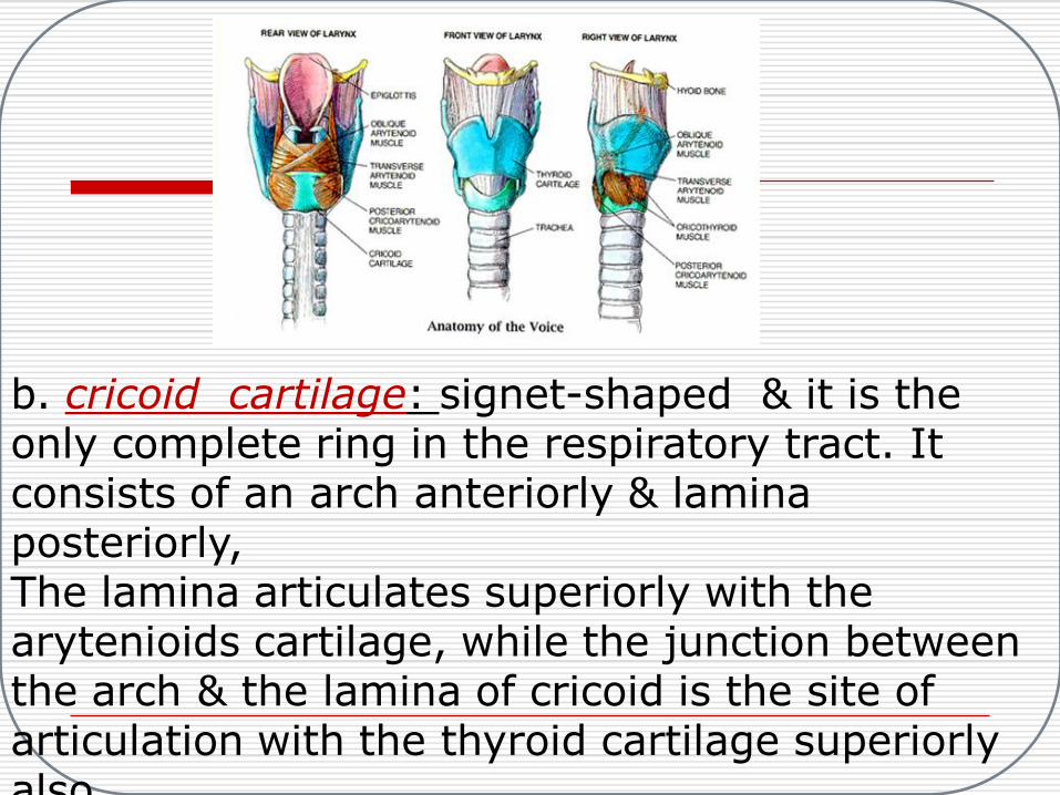
b. cricoid cartilage: signet-shaped & it is the only complete ring in the respiratory tract. It consists of an arch anteriorly & lamina posteriorly,The lamina articulates superiorly with the arytenioids cartilage, while the junction between the arch & the lamina of cricoid is the site of articulation with the thyroid cartilage superiorly also.

c. Epiglottis: rise up behind the tongue,itis a thin leaf like sheet of elastic fibrocartilage. It has free upper part & a thin long stem inferiorly attached to the inner surface of the thyroid alae at their junction.
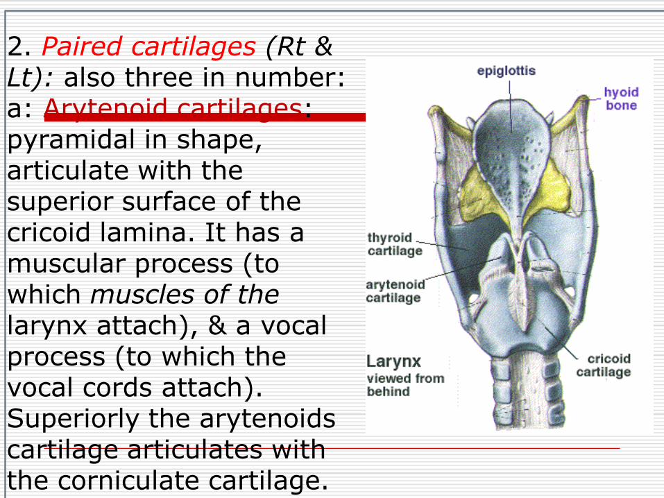
2. Paired cartilages (Rt & Lt): also three in number:a: Arytenoid cartilages: pyramidal in shape, articulate with the superior surface of the cricoid lamina. It has a muscular process (to which muscles of the larynx attach), & a vocal process (to which the vocal cords attach). Superiorly the arytenoids cartilage articulates with the corniculate cartilage.

B: corniculatecartilages: these are small & articulate with the apices of arytenoids cartilages.
C: Cuniformcartilages: small cartilages, each one situated in one aryepiglottic fold.


Laryngeal joints:twosynovial joint:1: Crico-Thyroid joint: between the inferior part of the thyroid cartilage & upper surface of the cricoid cartilage.
2: Crico-Arytenoid: between the arytenoid & cricoid cartilages.

Laryngeal ligaments & membranes: (two types) intrinsic & extrinsic:1.. Intrinsic: uniting the cartilages of the larynx to one another including:
A: The elastic membrane of the larynx, which lies beneath the mucosal layer & it is divided into upper & lower parts by the ventricle of the larynx. 'The upper part is called Quadrangular membrane, The vestibular ligament which forms the framework of the false vocal cord is the free lower edge (the strongest part) of theQuadrangular membrane ,while the lower one is called Conus elasticus,The vocal ligament which forms the framework of the true vocal cord is the free upper edge (the strongest

B: Cricothyroid membrane: between the cricoid cartilage (below) & the thyroid cartilage (above).


2. Extrinsic: uniting the larynx to the surrounding structures, including:A: Thyrohyoid membrane: between the thyroid cartilage below & the hyoid bone above.B:Cricotracheal membrane: is attached to the lower border of cricoid cartilage above & the upper part of the first tracheal ring,
below.C: hyoepiglottic ligament: attaches the epiglottis to the hyoid bone.

Laryngeal muscles: also intrinsic & extrinsic:1. Intrinsic: between one laryngeal cartilage & another, divided into:a. Abductors of the vocal cords: only one muscle on each side, that is the posterior crico-arytenoid muscle. It is the only muscle that opens the glottis. Paralysis of this muscle (if it is bilateral) resulting in emergency respiratory obstruction & stridor, tracheostomy might be required immediately.b. Adductors of the vocal cords: include:I: Lateral Cricoarytenoid muscle (Rt & Lt).II: Interarytenoid muscle (single muscle).III: External portion of thyroarytenoidmuscle (Rt &Lt).c. Tensors of the vocal cords:1.Cricothyroid muscle.II.Lateral portion of thyroarytenoidmuscle (Rt &Lt).

2. Extrinsic: between the larynx & neighboring structures,these are called the strap muscles & divided into infrahyoid & suprahyoid muscles:a. infrahyoid strap muscles:I.Sternothyroid muscle.II.Thyrohyoid muscle.III. Sternohyoid muscle.b. Suprahyoid strap muscles:1.Mylohyoid muscle.2.Geniohyoid muscle. 3.hyoglossus musele.4. Stylohyoid muscle


Cavity of the larynx:
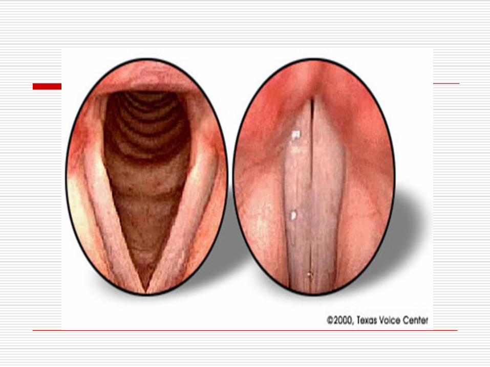
False vocal cords: formed by mucous membrane covering the vestibular ligament.True vocal cords: project further into the cavity than the false cords & lies at a lower level. It consists of epithelium closely bound to the underlying vocal ligament. The true vocal cords have poor blood supply& poor lymphatic drainage. The true & false vocal cords divide the laryngeal
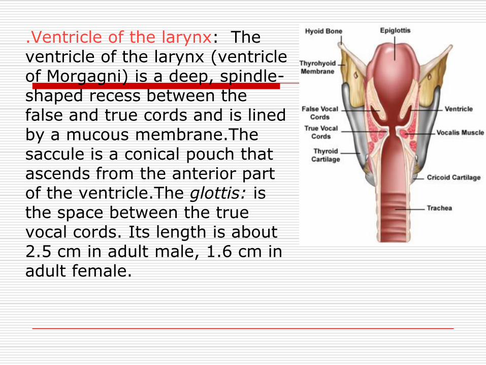
.Ventricle of the larynx: The ventricle of the larynx (ventricle of Morgagni) is a deep, spindle-shaped recess between the false and true cords and is lined by a mucous membrane.Thesaccule is a conical pouch that ascends from the anterior part of the ventricle.The glottis: is the space between the true vocal cords. Its length is about 2.5 cm in adult male, 1.6 cm in adult female.
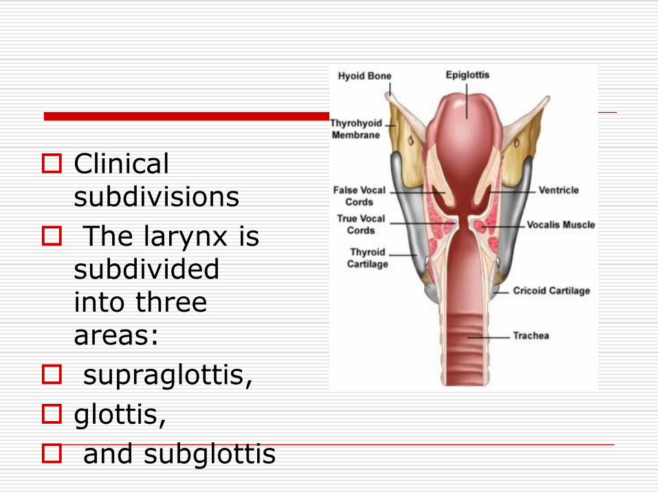
Clinical subdivisions
The larynx is subdivided into three areas:
supraglottis,
glottis,
and subglottis

Mucous membrane of the larynx:• Stratified squamous epithelium is found over the true vocal cords &over the upper part of the supraglottic space. • Ciliated columnar epithelium lines the reminder of the larynx.Blood supply of the larynx:• laryngeal branches of the: superior thyroid artery &
inferior thyroid artery.
• Vein drainage ultimately

Nerve supply of the larynx:Supplied by branches of the vagus (X) nerve:• Superior laryngeal nerve: has two branches:A: internal branch: entirely sensory, supplies the cavity of the larynx as far down as the vocalcords.B: External branch: mixed branch(motor&sensory) supplies the cricothyroid muscle.• Recurrent laryngeal nerve: has a much longer course on the left
side than the right side. On left side it turns round the arch of aorta. On right side it turns around the subclavian artery, then the nerve ascends up between the esophagus & trachea till it reaches the larynx. It supplies all the intrinsic muscles of the larynx except cricothyroidmuscle, & also sensory supply to the laryngeal cavity below the level of the vocal cords.


Lymphatic drainage of the larynx:• Supraglottic region: drains into the upper deep cervical lymph nodes.• Subglottic region- drains into the pre-laryngeal., pre-tracheal & lower deep cervical lymph nodes.• The vocal cords themselves have practically no lymphatic vessels, hence malignant tumors limited to them & rarely spread.

Functions of the larynx:1. Protection of lower air passages:The most important function of the larynx is to protect the lower airways from inhalation of food particles, saliva, other foreign bodies. Several mechanism is involved:A: Closure of laryngeal inlet: the aryepiglottic folds move toword one another& the epiglottis curves backword.B: Closure of glottis: by approximation of the vocal cords, this normal reflex present from the time of birth. C.Cessation of respiration during swallowing of food. d.Cough reflex: should any particles enter the trachea & bronchi the act of coughing will usually dislodge them.2: Phonation: voice is produced by vibrations of vocal cords, the sound so produced is modified by resonating chambers of the pharynx, mouth & nose.3: Respiration: the larynx plays a part in the mechanism of respiration by reflex adjustment of the glottic aperture.4: Fixation of the chest: when the larynx is closed, the thoracic cage becomes fixed during heavy weight lifting.