
The Ketel Gene Encodes a Drosophila Homologue of Importin-ß
12
Copyright 2000 by the Genetics Society of America The Ketel Gene Encodes a Drosophila Homologue of Importin- b Mo ´ nika Lippai,* La ´szlo ´ Tiria ´n,* Imre Boros, Jo ´ zsef Miha ´ly, Miklo ´s Erde ´lyi, Istva ´n Belecz,* Endre Ma ´the ´,* Ja ´nos Po ´ sfai, Adam Nagy,* Andor Udvardy, Efrosyni Paraskeva, ‡ Dirk Go ¨rlich ‡ and Ja ´nos Szabad* *Faculty of General Medicine, Department of Biology, University of Szeged, H-6720 Szeged, Hungary, Biological Research Center of the Hungarian Academy of Sciences, H-6701 Szeged, Hungary and ‡ Zentrum fu ¨r Molekulare Biologie, Universita ¨t Heidelberg, Heidelberg 69120, Germany Manuscript received January 27, 2000 Accepted for publication September 14, 2000 ABSTRACT The Drosophila melanogaster Ketel gene was identified via the Ketel D dominant female sterile mutations and their ketel r revertant alleles that are recessive zygotic lethals. The maternally acting Ketel D mutations inhibit cleavage nuclei formation. We cloned the Ketel gene on the basis of a common breakpoint in 38E1.2-3 in four ketel r alleles. The Ketel 1 transgenes rescue ketel r -associated zygotic lethality and slightly reduce Ketel D -associated dominant female sterility. Ketel is a single copy gene. It is transcribed to a single 3.6-kb mRNA, predicted to encode the 97-kD Ketel protein. The 884-amino-acid sequence of Ketel is 60% identical and 78% similar to that of human importin-b, the nuclear import receptor for proteins with a classical NLS. Indeed, Ketel supports import of appropriately designed substrates into nuclei of digitonin- permeabilized HeLa cells. As shown by a polyclonal anti-Ketel antibody, nurse cells synthesize and transfer Ketel protein into the oocyte cytoplasm from stage 11 of oogenesis. In cleavage embryos the Ketel protein is cytoplasmic. The Ketel gene appears to be ubiquitously expressed in embryonic cells. Western blot analysis revealed that the Ketel gene is not expressed in several larval cell types of late third instar larvae. A LONG a genetic dissection of maternal effects in The mutant phenotype suggests involvement of the Ketel gene in a NE-related function and motivated cloning Drosophila, we isolated 75 dominant female sterile (Fs) mutations (Erde ´lyi and Szabad 1989; Szabad et of the gene. As described in this article, the Ketel gene encodes for al. 1989). In 32 of the Fs mutations the Fs/1 females deposit normal-looking eggs, and although the eggs are the Drosophila homologue of importin-b, a key player in nuclear protein import. (For recent reviews on nuclear fertilized embryogenesis does not commence or ceases protein import see Corbett and Silver 1997; Go ¨ rlich after a few abnormal cleavage divisions. The 32 Fs muta- and Mattaj 1997; Mattaj and Englmeier 1998; Mel- tions identify 21 genes, suggesting that products of sev- chior and Gerace 1998; Pemberton et al. 1998; Weis eral genes are required for commencement and the 1998; Wozniak et al. 1998; Go ¨ rlich and Kutay 1999). initial steps of embryogenesis. This conclusion is sup- Briefly, importin-b, the founding member of the im- ported by the fact that very few, if any, of the zygotic portin-b superfamily, was originally described to partici- genes are expressed during early embryogenesis and pate in import of proteins that carry a classical nuclear evidently the initial steps of embryogenesis are under localization signal (cNLS) into the nucleus. The C-ter- maternal control (Wieschaus 1996). minal section of importin-b associates with importin-a, The Ketel gene, which was identified by four Fs(2)Ketel an adapter molecule, that binds to the cNLS-containing (5 Ketel D ) mutations, is one of the 21 genes mentioned import substrate. Importin-b forms 19 HEAT- and arma- above (Szabad et al. 1989; Erde ´lyi et al. 1997). As de- dillo-resembling repeats and wraps around the im- scribed in the accompanying article (Tiria ´n et al. 2000), portin-b-binding (IBB) domain of importin-a (Cingo- embryogenesis is terminated in Ketel D eggs, which are lani et al. 1999). The substrate-importin-a-importin-b deposited by the Ketel D /1 females, soon after fertiliza- complex docks, in an energy-independent manner, on tion due to the failure of cleavage nuclei formation. When the cytoplasmic side of the nuclear pore complexes injected into wild-type cleavage embryos, the Ketel D egg (NPCs). During translocation through the NPCs, im- cytoplasm is toxic: it hinders formation of cleavage nu- portin-b interacts with a number of nucleoporins with clei following mitosis most likely through the prevention its NPC binding domains located toward the N terminus of nuclear envelope (NE) assembly and/or function. (Kutay et al. 1997; Wozniak et al. 1998). Import of the cNLS-containing nuclear protein is completed on the nuclear surface of the NPCs, where following interaction Corresponding author: Ja ´nos Szabad, Faculty of General Medicine, of the transport complex with Ran-GTP, the substrate- Department of Biology, University of Szeged, H-6720 Szeged, Somogyi B. u. 4, Hungary. E-mail: [email protected] importin-a-importin-b complex disassembles. (Ran is a Genetics 156: 1889–1900 (December 2000)
Transcript of The Ketel Gene Encodes a Drosophila Homologue of Importin-ß

Copyright 2000 by the Genetics Society of America
The Ketel Gene Encodes a Drosophila Homologue of Importin-b
Monika Lippai,* Laszlo Tirian,* Imre Boros,† Jozsef Mihaly,† Miklos Erdelyi,† Istvan Belecz,*Endre Mathe,* Janos Posfai,† Adam Nagy,* Andor Udvardy,† Efrosyni Paraskeva,‡
Dirk Gorlich‡ and Janos Szabad*
*Faculty of General Medicine, Department of Biology, University of Szeged, H-6720 Szeged, Hungary, †Biological Research Center of theHungarian Academy of Sciences, H-6701 Szeged, Hungary and ‡Zentrum fur Molekulare Biologie,
Universitat Heidelberg, Heidelberg 69120, Germany
Manuscript received January 27, 2000Accepted for publication September 14, 2000
ABSTRACTThe Drosophila melanogaster Ketel gene was identified via the Ketel D dominant female sterile mutations
and their ketel r revertant alleles that are recessive zygotic lethals. The maternally acting Ketel D mutationsinhibit cleavage nuclei formation. We cloned the Ketel gene on the basis of a common breakpoint in38E1.2-3 in four ketel r alleles. The Ketel1 transgenes rescue ketel r-associated zygotic lethality and slightlyreduce Ketel D-associated dominant female sterility. Ketel is a single copy gene. It is transcribed to a single3.6-kb mRNA, predicted to encode the 97-kD Ketel protein. The 884-amino-acid sequence of Ketel is 60%identical and 78% similar to that of human importin-b, the nuclear import receptor for proteins with aclassical NLS. Indeed, Ketel supports import of appropriately designed substrates into nuclei of digitonin-permeabilized HeLa cells. As shown by a polyclonal anti-Ketel antibody, nurse cells synthesize and transferKetel protein into the oocyte cytoplasm from stage 11 of oogenesis. In cleavage embryos the Ketel proteinis cytoplasmic. The Ketel gene appears to be ubiquitously expressed in embryonic cells. Western blotanalysis revealed that the Ketel gene is not expressed in several larval cell types of late third instar larvae.
ALONG a genetic dissection of maternal effects in The mutant phenotype suggests involvement of the Ketelgene in a NE-related function and motivated cloningDrosophila, we isolated 75 dominant female sterile
(Fs) mutations (Erdelyi and Szabad 1989; Szabad et of the gene.As described in this article, the Ketel gene encodes foral. 1989). In 32 of the Fs mutations the Fs/1 females
deposit normal-looking eggs, and although the eggs are the Drosophila homologue of importin-b, a key player innuclear protein import. (For recent reviews on nuclearfertilized embryogenesis does not commence or ceasesprotein import see Corbett and Silver 1997; Gorlichafter a few abnormal cleavage divisions. The 32 Fs muta-and Mattaj 1997; Mattaj and Englmeier 1998; Mel-tions identify 21 genes, suggesting that products of sev-chior and Gerace 1998; Pemberton et al. 1998; Weiseral genes are required for commencement and the1998; Wozniak et al. 1998; Gorlich and Kutay 1999).initial steps of embryogenesis. This conclusion is sup-Briefly, importin-b, the founding member of the im-ported by the fact that very few, if any, of the zygoticportin-b superfamily, was originally described to partici-genes are expressed during early embryogenesis andpate in import of proteins that carry a classical nuclearevidently the initial steps of embryogenesis are underlocalization signal (cNLS) into the nucleus. The C-ter-maternal control (Wieschaus 1996).minal section of importin-b associates with importin-a,The Ketel gene, which was identified by four Fs(2)Ketelan adapter molecule, that binds to the cNLS-containing(5 Ketel D) mutations, is one of the 21 genes mentionedimport substrate. Importin-b forms 19 HEAT- and arma-above (Szabad et al. 1989; Erdelyi et al. 1997). As de-dillo-resembling repeats and wraps around the im-scribed in the accompanying article (Tirian et al. 2000),portin-b-binding (IBB) domain of importin-a (Cingo-embryogenesis is terminated in KetelD eggs, which arelani et al. 1999). The substrate-importin-a-importin-bdeposited by the KetelD/1 females, soon after fertiliza-complex docks, in an energy-independent manner, ontion due to the failure of cleavage nuclei formation. Whenthe cytoplasmic side of the nuclear pore complexesinjected into wild-type cleavage embryos, the KetelD egg(NPCs). During translocation through the NPCs, im-cytoplasm is toxic: it hinders formation of cleavage nu-portin-b interacts with a number of nucleoporins withclei following mitosis most likely through the preventionits NPC binding domains located toward the N terminusof nuclear envelope (NE) assembly and/or function.(Kutay et al. 1997; Wozniak et al. 1998). Import of thecNLS-containing nuclear protein is completed on thenuclear surface of the NPCs, where following interaction
Corresponding author: Janos Szabad, Faculty of General Medicine,of the transport complex with Ran-GTP, the substrate-Department of Biology, University of Szeged, H-6720 Szeged, Somogyi
B. u. 4, Hungary. E-mail: [email protected] importin-a-importin-b complex disassembles. (Ran is a
Genetics 156: 1889–1900 (December 2000)

1890 M. Lippai et al.
were cloned into pBluescriptII KS1 vector. We used the sub-Ras-related G protein without a membrane anchoringclones to precisely map the ketel rX-associated breakpoints forsite. For a recent review see Azuma and Dasso 2000.)sequencing and screening cDNA libraries.
The import substrate stays in the nucleus, while im- The Ketel cDNA clones were isolated from a lgt10 cDNAportin-a and importin-b are recycled to initiate a new library constructed from mRNAs of 0- to 4-hr-old Drosophila
embryos. (The cDNA library was a kind gift from Dr. J. Tam-import cycle: importin-b returns to the cytoplasm inkun.) The screening of z1.5 3 105 independent plaques re-a complex with Ran-GTP. In the cytoplasm Ran-GTPsulted in 17 cDNA clones that hybridized with at least one ofdissociates from importin-b and is converted to Ran-the subclones covering part of the Ketel gene. The overlapping
GDP by RanGTP-ase activating protein (RanGAP) and cDNA clones were identified and subcloned into the pBlue-the Ran binding protein 1 (RanBP1); thus importin-b scriptII KS1 vector. Sequencing of genomic and cDNA clones
were done by the dideoxy method in an IBI automated seque-can participate in a new transport cycle (Bischoff andnator on both strands. The 59 end of the mRNA was deter-Gorlich 1997; Azuma and Dasso 2000).mined by primer extension. The primer extension was doneThis article describes a combined genetic, molecular,by using total mRNA isolated from adult females and the
and cell biological approach and reveals novel features synthetic olygonucleotide 59GCTCTTTTGCTCCTATATGATof importin-b. The Ketel protein shows characteristic TTCTAC39, which hybridized close to the 59 end of the isolated
cDNA. The 39 end was present in some of the isolated cDNAsfeatures of importin-b: (i) it supports import of a cNLS-as revealed by the poly(A) tail. The intron-exon compositioncontaining substrate into nuclei of digitonin-permeabil-of the region that encodes the 3.6-kb Ketel mRNA was deter-ized HeLa cells and (ii) it is largely cytoplasmic withmined by sequencing and analyzing a 7870-bp genomic frag-
pronounced accumulation in the NE. Surprisingly, the ment. For developmental Northern analysis poly(A) mRNAshighly “toxic” KetelD egg cytoplasm, which prevents NE were purified, blotted, and probed with the cDNA that corre-
sponds to the 3.6-kb Ketel mRNA. Digoxigenin (DIG)-labeledassembly following mitosis, does not prevent nuclearKetel cDNA was used for the detection of Ketel mRNA duringprotein import. Unexpectedly, as revealed by Westernoogenesis and embryogenesis, according to standard proce-blot analysis, the Ketel gene is not expressed in mostdures.
cells of the larvae and adults, raising questions about Homology search and putative function of the Ketel gene:cellular functions of importin-b and nuclear import of Having the above-mentioned sequences and to establish possi-the cNLS-containing proteins. ble function of the Ketel protein, we screened databases with
the BLAST service of the National Center for BiotechnologyInformation for identifying sequences displaying homologywith the Ketel cDNA. Protein alignments were done using theMATERIALS AND METHODSMaxHom EMBL multiple sequence alignment program.
The Ketel 1 transgenes: We constructed three different typesThe Ketel mutant alleles: The EMS-induced Ketel D allelesof Ketel1 (K1) transgenes. The first type included the entirewere isolated following EMS mutagenesis in a screen for domi-22-kb fragment shown in Figure 1A. The second type coverednant female-sterile mutations (Szabad et al. 1989). The 27a 13.8-kb Xba genomic fragment (Figure 1B). In the third typerecessive ketel r alleles were generated through second muta-a 4.0-kb Xba-BamHI genomic fragment—including the Ketelgenesis of the Ketel D alleles (Erdelyi et al. 1997; Szabad etpromoter and the 59 segment of the Ketel coding region—wasal. 1989). The ketel r/2 and the Ketel D/2 hemizygotes werecombined with a 2.3-kb cDNA fragment that corresponded toproduced by crossing y/y; ketel rX32/y1CyO females with y/Y;the rest of the transcribed part of the Ketel gene (see Figureketel r/y1CyO and y/Y; Ketel D/y1CyO males, respectively. [The1B). The above sequences were cloned into the CaSpeR vectorketel rX32 allele, abbreviated as 2, is a small deficiency that re-with the mini-white marker gene and germline transformantsmoves the Ketel and a few adjacent loci (Erdelyi et al. 1997).were generated by standard procedures. The K1 transgene-The y1CyO balancer chromosome carries a y1 transgene (Tim-carrying flies have light to orange-yellowish eyes on the whitemons et al. 1993).] Head skeleton and ventral setae of thegenetic background. The K1 transgenes were used for thedescending ketel r/2 and the Ketel D/2 larvae are yellow andconstruction of K1; ketel r/2 and K1; Ketel D/2 as well as K1;allow their separation from the heterozygous nonyellowKetel D/1 and K1/K1; Ketel D/1 zygotes. Their viability and the(y1CyO) siblings that have dark chitinous structures. For anfertility of the females were tested.explanation of the genetic symbols see Lindsley and Zimm
Production of the Ketel protein in bacteria and the genera-(1992) and the FlyBase website (http://flybase.bio.indiana.tion of anti-Ketel polyclonal antibodies: A pGEX-Ketel plasmidedu). All experiments were carried out at 258.was constructed first by the insertion of the BamHI-EcoRI frag-Molecular cloning of the Ketel gene: DNA manipulations,ment of the Ketel cDNA (Figure 1A) into the correspondingplasmid constructions, restriction mapping, Southern andsites of a pGEX4T-1 vector and glutathione-S-transferaseNorthern hybridizations, and Western blotting were done ac-(GST)-Ketel fusion protein was produced in Escherichia coli.cording to standard procedures. For identification of theThe fusion protein consisted of the GST moiety fused in framebreakpoints in the four ketel rX-associated rearrangements, we
isolated DNA from ketel rX/1 adult flies. The DNA was digested with the 147–884 amino-acid encoding segment of the Ketelprotein. The GST-Ketel fusion protein was purified by affinitywith EcoRV and hybridized on Southern blots. The 32P-labeled
probes for Southern hybridizations were generated by random chromatography on a glutathion-agarose column and usedfor immunization of rabbits for the production of anti-Ketelprimer labeling of restriction fragments that had been isolated
from a lEMBL4 library and from CoSpeR clones identified in polyclonal antibodies following standard protocols. After sev-eral boosts, the crude sera were analyzed for the presence ofa chromosomal walk. The chromosomal walk initiated from
a clone that hybridized to the 38E1.2-3 cytological region and anti-Ketel antibody by Western blots. Two rabbits producedgood titers of anti-Ketel sera by virtue of their ability to recog-was kindly provided by Dr. P. Maroy. Cloning the Ketel gene
was also confirmed by in situ hybridizations on salivary gland nize the Ketel protein in E. coli extracts from strains withpGEX-Ketel but not in the control bacterial extracts.chromosomes of the ketel rX/1 larvae. A detailed restriction
map of the Ketel region was constructed and the subfragments For production of a nearly full-length Ketel protein, we

1891Drosophila Importin-b Homologue
made use of the pET-His3A expression system (Chen and Hai delineated the Ketel locus and allowed molecular clon-1994). The His-tagged Ketel protein, with amino acids 4–884, ing of the Ketel gene. To identify the breakpoints in thewas purified by a Ni-chelating column and used for prepara-
four ketel rX alleles, we initiated a genomic walk from ation of a Ketel protein affinity column.nearby genomic fragment. The genomic walk coveredThe anti-Ketel antibody was purified in two steps: first on
a protein-A and afterward on a Ketel protein affinity column. z60 kb and resulted in a 5.5-kb genomic BglII-BamHIThe affinity-purified anti-Ketel antibody was used both in West- fragment that included all four of the ketel rX-associatedern blots and in confocal microscopy for the detection of breakpoints (Figure 1A). The corresponding region ofKetel protein. For Western blots protein extracts were pre-
the Drosophila genome is included in a cosmid clonepared from embryos, larvae, and adults as well as from differ-that we isolated from a CoSpeR Not-Bam-Not cosmid li-ent organs of late third instar larvae. For laser scanning micros-
copy ovaries were dissected, fixed, and treated with antibodies. brary (Figure 1A). On Northern blots, the BglII-BamHIThe Ketel protein was detected by the affinity-purified poly- fragment strongly hybridized to a 3.6-kb mRNA speci-clonal anti-Ketel rabbit antibody that was made visible by a men that most likely represents the Ketel gene. The 3.6-goat anti-rabbit rhodamin-labeled secondary antibody (Jack-
kb mRNA is abundant in females and young embryosson Laboratories, West Grove, PA). The NE was made visibleand appears to be present, although in much reducedwith a primary monoclonal anti-lamin mouse antibody (Harel
et al. 1989; Paddy et al. 1996) and a fluorescein-labeled anti- concentrations, throughout development (data notmouse secondary antibody (Jackson Laboratories). Optical shown).sections were generated in a Zeiss (Thornwood, NY) LSM 410 To isolate cDNA clones that correspond to the 3.6-confocal microscope.
kb Ketel mRNA, overlapping genomic fragments cov-The in vitro nuclear protein import assay: Drosophila im-ering the Xba-EcoRV 10.7-kb region (Figure 1A) wereportin-b cDNA was cloned into the SphI-XmaI sites of pQE30
(QIAGEN, Valencia, CA), expressed with an NH2-terminal His used for the screening of a cDNA library prepared fromtag and purified, on nickel-NTA agarose, followed by chroma- 0- to 4-hr-old embryos. The longest isolated cDNA wastography on a Superdex 200 gel filtration column. 2858 bp long. However, it did not contain poly(A) tail.
The nuclear protein import assay was conducted as follows.Overlaps of the 2858-bp cDNA clone with poly(A)-con-Permeabilized HeLa cells were prepared by a modification oftaining cDNAs allowed the reconstruction of a 3378-bp-a published protocol (Adam et al. 1990). Briefly, HeLa cells
were grown on coverslips to 50–80% confluence, washed in long cDNA. In vitro extension was done on the basis ofice-cold permeabilization buffer (20 mm HEPES/KOH pH the mRNAs isolated from adult females and of a primer7.5, 110 mm potassium acetate, 5 mm magnesium acetate, 250 complementary to the 59 end of the 2858-bp-long cDNA.mm sucrose, 0.5 mm EGTA) and permeabilized for 15 min in
The primer extension indicated a major transcriptionthe same buffer containing 60 mg/ml digitonin. The coverslipsinitiation site 478 bp upstream from the 59 end of thewere washed three times in permeabilization buffer without
digitonin. Coverslips were incubated as indicated with each 2858-bp cDNA. We concluded, after finding out about20 ml of import reaction. The import buffer contained 2 mg/ the missing 39 and 59 ends of the 2858-bp cDNA, thatml nucleoplasmin core (to block nonspecific binding), 20 the encoded Ketel mRNA is 3656 nucleotides long andmm HEPES/KOH pH 7.5, 140 mm potassium acetate, 5 mm
corresponds to the 3.6-kb mRNA detected in Northernmagnesium acetate, 250 mm sucrose, 0.5 mm EGTA. Whereanalysis.indicated, reactions were supplemented with an energy-regen-
erating system (0.5 mm ATP, 0.5 mm GTP, 10 mm creatine To determine molecular organization of the Ketel lo-phosphate, 50 mg/ml creatine kinase) and Ran mix (3 mm cus, we sequenced a 7870-bp long genomic DNA regionRan-GDP, 150 nm Rna1p, 300 nm NTF2, 150 nm RanBP1). that corresponds to the encoded cDNA and the sur-Nuclear import of a fluorescent substrate was monitored in
rounding sequences (Figure 1A). The nucleotide se-optical sections. The substrate was the pentamer of a fusionquence is available in the EMBL nucleotide sequenceprotein in which the nucleoplasmin core domain was com-
bined with the importin-b-binding domain from importin-a database under the accession no. AJ002729. Compari-(IBB core pentamer). Import reaction samples contained 0.24 son of the cDNA and the genomic sequences revealedmm fluorescein-labeled IBB core pentamer. In the indicated that the Ketel gene contains 5 introns. The Ketel mRNAreactions 1.2 mm Drosophila importin-b, Ran, and an energy-
is composed from a 444-bp leader sequence, a 2652-bpregenerating system were added. Reactions were stopped afteropen reading frame (ORF), and a 560-nucleotide-long5 min by fixation in 3% paraformaldehyde (w/v) in PBS,
washed in PBS and water, and mounted with 2 ml of vec- trailer sequence (Figure 1A). To decide whether Keteltorshield mounting medium (Vector, Burlingame, CA). is a single copy gene or is present in multiple copies, we
The digitonin-permeabilized HeLa cell system was also used digested genomic DNA with three different restrictionto follow nuclear import of the cNLS-phycoerythrin (cNLS-
enzymes (EcoRI, Xba, and BglII) and carried out South-PE; Cserpan and Udvardy 1995) substrate in presence ofern analyses with 32P-labeled cDNA fragments. In everycytosol samples prepared from ovaries of wild-type and
Ketel D/1 females. case, the labeled cDNA fragments hybridized with asingle band, making it very likely that Ketel is a singlecopy gene (data not shown). The finding that under
RESULTShigh stringency conditions the Ketel cDNA hybridizedexclusively to the 38E1.2-3 region on salivary gland chro-Molecular cloning of the Ketel gene of Drosophila:
Four of the X-ray-induced ketel rX revertant alleles have mosomes supports the above conclusion.The Ketel 1 transgenes: To prove that the cloned genea common breakpoint in the 38E1.2-3 cytological region
(Erdelyi et al. 1997). The common breakpoint both is indeed Ketel, we generated three different types of

1892 M. Lippai et al.
Figure 1.—Restriction map of a 22-kb genomic segment from the 38E1.2-3 cytological region comprising the Ketel gene. (A)One breakpoint of each of the four ketel rX revertant alleles with visible chromosome rearrangements fall within the 5.5-kb BglII-BamHI genomic fragment delineated by the top thick line. The box on the line below represents the Ketel mRNA encodingregion. Detailed genomic map of the Ketel mRNA encoding region is shown below. Exons, introns, and the untranslated 59 and39 regions are indicated by solid, open, and dotted boxes, respectively. (B). Structure of two of the three K1 transgenes. TypeI comprises the entire 22-kb genomic fragment shown in A. Type II is the 13.8-kb Xba genomic fragment. Type III contains theXba-BamHI 4.0-kb genomic fragment combined in frame with a 2.3-kb BamHI–EcoRI segment of the cDNA. The indicatedrestriction sites are as follows: B, BglII; BH, BamHI; E, EcoRI; RV, EcoRV; X, Xba.
altogether 21 Ketel1 (K1) transgenes (Figure 1B and deposited by the K1; KetelD2/1 females embryogenesisprogressed to the stage of embryonic cuticle forma-Table 1) and analyzed their effects on both ketel r and
KetelD mutations. Nine of the 12 tested K1 transgenes tion and even a few offspring developed from the K1;KetelD2/1 females. However, the rate of offspring pro-brought about full rescue of lethality associated with the
loss-of-function ketel rX13/2 genotype: the K1; ketel rX13/2 duction was as low as 2–4 3 1023 offspring/(female 3day), as compared to the z50 offspring/(female 3 day)flies developed with the expected frequencies and were
fully fertile, showing that the cloned gene is Ketel (Table control value. (It should be noted that cuticle and off-spring did not develop from tens of thousands of eggs1; ketel rX13 is a null allele).
Type I and each of the three tested type III K1 trans- deposited by KetelD2/1 females.) One copy of the K1
transgenes had no effects on the other three KetelD muta-genes brought about slight rescue of the KetelD2-associ-ated dominant female sterility. In z1% of the eggs tions. However, two copies of the K1 transgenes brought
TABLE 1
The K1 transgenes and their rescue effects on ketel r/2-associated zygotic lethality
Chromosome linkage
Type and composition of the K1 transgenesa X 2nd 3rd 4th Total
I: 22-kb genomic segment 1 (0/1) 0 0 0 1II: 13.8-kb genomic segment 0 3 5 (4/5) 0 8III: 4.0-kb genomic combined with a 2.3-kb cDNA segment 1 (1/1) 4 5 (3/4) 2 (1/1) 12
In parentheses, number of rescuing/tested K1 transgenes.a See Figure 1 for composition of the K1 transgenes.

1893Drosophila Importin-b Homologue
about slight reduction of female sterility when sperm Expression pattern of the Ketel gene: To study theexpression pattern of the Ketel gene, we detected bothwith two normal Ketel gene copies fertilized the eggs
(Tirian et al. 2000). The slight rescue of the K1 trans- Ketel mRNA and Ketel protein during oogenesis andembryogenesis. Some Ketel mRNA, as detected with thegenes on KetelD-associated dominant female sterility
clearly shows that (i) the cloned gene is Ketel and (ii) DIG-labeled Ketel cDNA, is present in nurse cells of thestage 9 egg primordia. The concentration of Ketel mRNAthe normal and the KetelD-encoded gene products partic-
ipate in the same process and hence the KetelD alleles becomes rather high by stage 10 (Figure 5A) whendumping of the Ketel mRNA into the oocyte cytoplasmare strong dominant-negative mutations.
The Ketel gene encodes the Drosophila homologue of begins. Beyond stage 11 the Ketel mRNA is homoge-neously distributed in the oocyte cytoplasm (Figure 5B).importin-b: The Ketel cDNA contains a 2652-nucleotide-
long ORF encoding for a protein of 884 amino acids The Ketel gene appears to be ubiquitously expressed inevery blastoderm cell (Figure 5C) and, as far as it canwith a molecular mass of 97 kD. Comparison of the Ketel
protein amino acid sequence with known sequences be deduced from the staining patterns, also during laterstages of embryogenesis (Figure 5, D–F). The Ketel generevealed a 60.3% amino acid identity and a 78.2% simi-
larity with human importin-b, a known component of seems to be intensively expressed in the central nervoussystem and in the larval gonads (Figure 5, E and F).nuclear protein import (Figure 2; Adam and Adam
1994; Chi et al. 1995; Gorlich et al. 1995a,b; Imamoto The larval gonads include both ovaries and testes sincethe gonads possess intensive staining in each of theet al. 1995; Iovine et al. 1995; Radu et al. 1995). The
high level of homology suggests that Ketel encodes the embryos.We also followed Ketel gene expression through theDrosophila homologue of importin-b, the founding
member of the importin-b superfamily (Wozniak et al. detected Ketel protein with the affinity-purified poly-clonal anti-Ketel antibody. The anti-Ketel antibody de-1998, Gorlich and Kutay 1999).
The Ketel protein supports nuclear protein import: tected a single 97-kD protein band on Western blotswith extracts prepared from different developmentalTo decide whether the Ketel protein does indeed func-
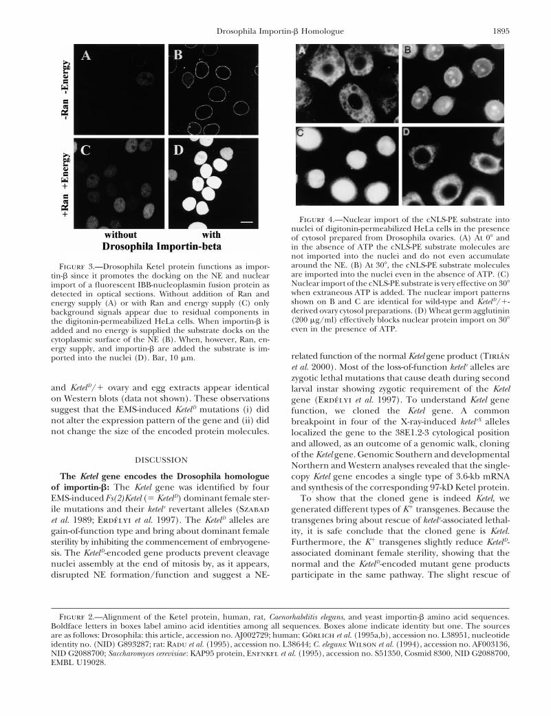
tion as importin-b we monitored (i) the docking on the stages (Figure 6). The Ketel protein is abundant in theovaries and in the newly deposited eggs throughoutcytoplasmic surface of the NE and (ii) import into nuclei
of digitonin-permeabilized HeLa cells of a fluorescent- embryogenesis and is present throughout all stages ofdevelopment. However, when compared, e.g., to ovaries,labeled nuclear substrate in the presence of the Ketel
protein and other components of the nuclear protein the relative Ketel protein concentration was rather lowin larvae and adult females from which the ovaries wereimport apparatus (see materials and methods). As
shown on Figure 3B, the substrate docked on the NE removed. To clarify the low Ketel protein content wedissected different organs from late third instar larvaein presence of the Ketel protein, and when Ran and an
energy source were added the substrate was imported and subjected them to Western blot analysis. As shownin Figure 6, while, e.g., the imaginal discs containedinto the nuclei (Figure 3, B and D). The permeabilized
HeLa cell experiments clearly showed that the Ketel significant amounts of the Ketel protein, there were nodetectable amounts of Ketel protein present in a num-protein molecules function as importin-b: they can assist
docking and import of nuclear proteins into the nuclei. ber of larval tissues including the salivary glands, gut,Malpighian tubules, or the larval epidermis with theTo understand effects of the KetelD mutations on nu-
clear protein import, we prepared cytosol from ovaries overlying larval musculature.Formation and localization of the Ketel protein wasof both KetelD/1 and wild-type females. All the four
KetelD mutations were included in this study. The cytosol also followed in the course of oogenesis and embryogen-esis by immunocytology and confocal microscopy. Thepreparations were used in the permeabilized HeLa cell
assay and import of the cNLS-PE substrate was moni- Ketel protein is first detectable in nurse cells duringstage 8 of oogenesis (not shown). By stage 10 the nursetored (see materials and methods). In presence of
the wild-type ovary cytosol the cNLS-PE substrate en- cells contain large quantities of the Ketel protein. Theprotein is cytoplasmic with pronounced accumulationtered the nuclei of HeLa cells within a few minutes
(Figure 4B). Surprisingly, the KetelD/1-derived cytosol in the NEs (Figure 7, A and C). Nurse cells dump theirKetel protein contents into the oocyte cytoplasm frompreparations just as efficiently supported nuclear import
of the cNLS-PE substrate as the wild-type ovary cytosol, stage 11 of oogenesis. The follicle cells also contain Ketelprotein (Figure 7A). Cytoplasm of a newly deposited eggshowing that the KetelD-encoded mutant molecules do
not interfere with import of the cNLS-PE substrate. Note contains stockpiles of the Ketel protein. During cleavagedivisions the Ketel protein is present throughout thethat when injected into wild-type cleavage embryos,
traces of the KetelD egg cytoplasm prevent the formation cleavage cycles. It is cytoplasmic and shows accumula-tion in the NE (Figure 7, D and F).of cleavage nuclei at the end of mitosis (Tirian et al.
2000). However, nuclei of the digitonin-permeabilized Immunoreactive features of the KetelD-encoded pro-tein molecules are not different from wild type: amountHeLa cells remained intact for at least 4 hr in presence
of the KetelD/1-derived ovary cytosol. and size of the immunoreactive components in wild-type

1894 M. Lippai et al.
1895Drosophila Importin-b Homologue
Figure 4.—Nuclear import of the cNLS-PE substrate intonuclei of digitonin-permeabilized HeLa cells in the presenceof cytosol prepared from Drosophila ovaries. (A) At 08 andin the absence of ATP the cNLS-PE substrate molecules arenot imported into the nuclei and do not even accumulatearound the NE. (B) At 308, the cNLS-PE substrate moleculesFigure 3.—Drosophila Ketel protein functions as impor-are imported into the nuclei even in the absence of ATP. (C)tin-b since it promotes the docking on the NE and nuclearNuclear import of the cNLS-PE substrate is very effective on 308import of a fluorescent IBB-nucleoplasmin fusion protein aswhen extraneous ATP is added. The nuclear import patternsdetected in optical sections. Without addition of Ran andshown on B and C are identical for wild-type and Ketel D/1-energy supply (A) or with Ran and energy supply (C) onlyderived ovary cytosol preparations. (D) Wheat germ agglutininbackground signals appear due to residual components in(200 mg/ml) effectively blocks nuclear protein import on 308the digitonin-permeabilized HeLa cells. When importin-b iseven in the presence of ATP.added and no energy is supplied the substrate docks on the
cytoplasmic surface of the NE (B). When, however, Ran, en-ergy supply, and importin-b are added the substrate is im-
related function of the normal Ketel gene product (Tirianported into the nuclei (D). Bar, 10 mm.et al. 2000). Most of the loss-of-function ketel r alleles arezygotic lethal mutations that cause death during second
and KetelD/1 ovary and egg extracts appear identical larval instar showing zygotic requirement of the Ketelon Western blots (data not shown). These observations gene (Erdelyi et al. 1997). To understand Ketel genesuggest that the EMS-induced KetelD mutations (i) did function, we cloned the Ketel gene. A commonnot alter the expression pattern of the gene and (ii) did breakpoint in four of the X-ray-induced ketel rX allelesnot change the size of the encoded protein molecules. localized the gene to the 38E1.2-3 cytological position
and allowed, as an outcome of a genomic walk, cloningof the Ketel gene. Genomic Southern and developmental
DISCUSSIONNorthern and Western analyses revealed that the single-
The Ketel gene encodes the Drosophila homologue copy Ketel gene encodes a single type of 3.6-kb mRNAof importin-b: The Ketel gene was identified by four and synthesis of the corresponding 97-kD Ketel protein.EMS-induced Fs(2)Ketel (5 KetelD) dominant female ster- To show that the cloned gene is indeed Ketel, weile mutations and their ketel r revertant alleles (Szabad generated different types of K1 transgenes. Because theet al. 1989; Erdelyi et al. 1997). The KetelD alleles are transgenes bring about rescue of ketel r-associated lethal-gain-of-function type and bring about dominant female ity, it is safe conclude that the cloned gene is Ketel.sterility by inhibiting the commencement of embryogene- Furthermore, the K1 transgenes slightly reduce KetelD-sis. The KetelD-encoded gene products prevent cleavage associated dominant female sterility, showing that thenuclei assembly at the end of mitosis by, as it appears, normal and the KetelD-encoded mutant gene products
participate in the same pathway. The slight rescue ofdisrupted NE formation/function and suggest a NE-
Figure 2.—Alignment of the Ketel protein, human, rat, Caenorhabditis elegans, and yeast importin-b amino acid sequences.Boldface letters in boxes label amino acid identities among all sequences. Boxes alone indicate identity but one. The sourcesare as follows: Drosophila: this article, accession no. AJ002729; human: Gorlich et al. (1995a,b), accession no. L38951, nucleotideidentity no. (NID) G893287; rat: Radu et al. (1995), accession no. L38644; C. elegans: Wilson et al. (1994), accession no. AF003136,NID G2088700; Saccharomyces cerevisiae : KAP95 protein, Enenkel et al. (1995), accession no. S51350, Cosmid 8300, NID G2088700,EMBL U19028.

1896 M. Lippai et al.
Ketel D-associated dominant female sterility implies adominant-negative nature of the KetelD mutations; i.e.,the KetelD-encoded molecules impede function of thenormal Ketel gene products (Tirian et al. 2000).
Comparison of nucleotide and amino acid sequencesof the Ketel gene and the Ketel protein revealed stronghomology with human importin-b, a component of nu-clear protein import: in the two protein sequences 60%of the amino acids are identical and 78% are of similarnature.
Importin-b (also called karyopherin-b) is a majorcomponent of nuclear protein import and has beenknown from biochemical studies in which componentsof nuclear protein import were identified (Adam andAdam 1994; Chi et al. 1995; Gorlich et al. 1995a,b;Imamoto et al. 1995; Radu et al. 1995). The Ketel genedoes indeed encode the Drosophila importin-b sincethe Ketel protein possesses characteristic features ofimportin-b. In absence of an energy source the Ketelprotein produced in bacteria supports docking of a IBB-nucleoplasmin core fusion protein on the NE of digito-nin-permeabilized HeLa cells (Figure 3B). When anenergy source is provided, the nuclear protein is im-ported into the nucleus.
Transport of macromolecules between the cytoplasmand the nucleus proceeds through the NPCs and ismediated by shuttling receptors of the importin-b super-family (for reviews see Mattaj and Englmeier 1998;Pemberton et al. 1998; Weis 1998; Wozniak et al. 1998;Gorlich and Kutay 1999). The importins bind theircargo, directly or through adapter molecules like im-portin-a, in the cytoplasm and release them in the nu-cleus. A RanGTP gradient provides the driving force fortransport: importins bind their cargo in the cytoplasmwhere RanGTP levels are low and release it upon en-countering high RanGTP concentration in the nucleus.The conversion of RanGTP to RanGDP in the cytoplasmis catalyzed by RanGAP1 and is further stimulated byRanBPs. RanGTP is generated from RanGDP in thenucleus by RCC1 (regulator of chromatin condensa-tion), a chromatin-associated nucleotide exchange fac-tor. Importin-b is engaged in nuclear import of proteinswith cNLS through importin-a, an adapter molecule.Importin-b can also operate as an autonomous receptorindependently of importin-a. A number of types of pro-
Figure 5.—In situ hybridizations for the detection of KetelmRNA during oogenesis (A and B) and different stages ofembryogenesis: cellular blastoderm, stage 5 (C), stage 8 (D),stage 14 (E), and stage 17 (F) embryos. Lateral (C, D, and F;anterior left and dorsal up) and dorsal (E) views. nc, nursecells; oc, oocyte; pc, pole cells; am, anterior midgut primor-dium; pm, posterior midgut primordium; m, mesoderm; lg,larval gonad; spg, supra oesophageal ganglion; vnc, ventralnerve cord (embryos were staged as described in Campos-Ortega and Hartenstein 1997). Bar, 50 mm.

1897Drosophila Importin-b Homologue
adapter molecules: snurportin1 for the import of m3G-capped UsnRNPs (Huber et al. 1998) and XRIPa for theimport of the Xenopus replication protein A (Jullien etal. 1999).
Several members of the importin-b superfamily havebeen identified mainly in yeast and vertebrates (Woz-niak et al. 1998; Gorlich and Kutay 1999). A searchin the Drosophila genome (at http://flybase.bio.indiana.edu) for homologues of vertebrate importin-b super-family members identified 10 genes (Table 2). It appearsthat most members of the human importin-b family arealso present in Drosophila (Table 2). However, there isno apparent homologue of exportin-t that is engagedin export of tRNAs from the nuclei to the cytoplasm.The closely related human importin-5 and RanBP6(Gorlich and Kutay 1999) have a single correspond-
Figure 6.—Western blot analysis to detect Ketel protein ing Drosophila homologue (Karybeta3). Similarly, hu-with a polyclonal anti-Ketel antibody. Equal amounts of pro- man importin-7 and RanBP8 have a single Drosophilatein samples (as measured by photometry and confirmed with relative called dim-7. As in humans, there are two trans-Ponceau-stained control gels) were loaded in the different
portin genes in Drosophila (Table 2). Of the Drosophilaslots. With respect to third instar larval organs, the centralimportin-b family members, functions have been as-nervous system (cns) did not include the ring gland and the
larval gonads were removed from the fat body (fb) sample. sg, signed thus far to transportin (Siomi et al. 1998) andsalivary glands; id, imaginal discs; g 1 Mt, gut and Malpighian to a homologue of human CRM1 embargoed (Fasken ettubules; le 1 lm, larval epidermis with the overlaying larval
al. 2000) and, as described in this article, to Ketel.musculature.The Ketel protein is cytoplasmic and is not present
in every cell type: As other members of the importin-bfamily, the Ketel protein is largely cytoplasmic (Gorlichteins have been identified that can directly bind to im-et al. 1995a) with pronounced accumulation in the NEportin-b and are imported into the nucleus, e.g., some(Figure 7). As predicted by features of the KetelD andribosomal proteins and the HIV Rev protein (Hender-ketel r mutant phenotypes (Tirian et al. 2000), the Ketelson and Percipalle 1997; Jakel and Gorlich 1998;protein is produced and is dumped into the oocyteTruant and Cullen 1999). Importin-b can also formcytoplasm during oogenesis and cleavage embryos makea complex with importin-7 and mediate histone H1 im-use of the Ketel maternal dowry. Surprisingly, however,port (Gorlich et al. 1997; Jakel et al. 1999). In addition,
apart from importin-a, importin-b also uses other the Ketel gene does not appear to be expressed in the
Figure 7.—Distribution ofthe Ketel protein, as detectedin optical sections, in a stage10 egg primordium and in aninterphase cleavage embryo. TheKetel protein is shown in red(A and D), the nuclear laminaappears in green (B and E).Merged signals are shown on Cand F where yellow colorationresults from superimpositionof green and red signals. nc,nurse cells; oc, oocyte; fc, folli-cle cells. Bar, 50 mm for A–Cand 5 mm for D–F. As shownby the lamin signal in C andD the oocyte nucleus containsuniformly distributed lamin mol-ecules inside (Ashery-Padan etal. 1997).

1898 M. Lippai et al.
TABLE 2
Members of the Drosophila importin-b family and their closest human homologues on the basis of amino acid sequence identities
Accession nos. IdentityDrosophila Cytological Closest human of the levelproteina localization homologue(s) human gene (%) References in FlyBase
Ketel(CG2637) 38 E1.2-3 Importin-b I52907 60 This article; Szabad et al. (1989);
Erdelyi et al. (1997); Kozlova et al. (1998)Transportin
(CG7398) 65 A6 Transportin-1 Q92973 71 Siomi et al. (1998); Norvell et al. (1999)Transportin-2 AF019039 68
CG 8219 65 A6 Transportin-1 Q92973 58Transportin-2 AF019039 56
CAS(CG13281) 36 B1-2 CAS P55060 51 Spradling et al. (1999)
Karybeta 3(CG1059) 82 D1 Importin-5 Y08890 51 Colas et al. (1999); Spradling et al. (1999)
Dim-7(CG7935) 66 B7-10 Importin-7 AF098799 52
CG 8212 52 C1-2 RanBP11 AK001696 35CG2848 23 B1 Mtr10a AJ133749 41CG12234 18 D10-11 KIAA1291 34Embargoed
(CG13387) 29 C1 CRM1 Y08614 69 Fasken et al. (2000)
a Code as available from FlyBase (http://flybase.bio.indiana.edu).
fully differentiated larval cells. Apparently the Ketel pro- ered by unidentified components of the ovary cytosol.The existence of parallel import pathways is well estab-tein appears to be present largely in mitotically active
cells. Genetic requirement of the Ketel gene is discussed lished. For example, the human ribosomal protein L25is imported through at least four pathways (Jakel andin the accompanying article by Tirian et al. (2000).
The possible mode of action of the KetelD-encoded Gorlich 1998). The KetelD-encoded molecules well maysupport nuclear protein import, a feature not knownproteins: When injected into wild-type cleavage em-
bryos, traces of the KetelD egg cytoplasm exert deleteri- at present. It is also possible that although the KetelD-encoded molecules do not participate in nuclear pro-ous effects through the prevention of cleavage nuclei
formation (Tirian et al. 2000). Toxic effects of the KetelD- tein import, they do not interfere with import functionof the normal Ketel molecules, and their toxic effectsencoded molecules are perhaps an outcome of arrested
nuclear protein import. To elaborate this possibility, we become apparent when the importin-b molecules per-form a function other than nuclear protein import.prepared cytosol from ovaries of the KetelD/1 females
and studied their effects on nuclear protein import. Indeed, the deleterious effects of the KetelD mutationsbecome apparent at the end of cleavage mitosis whenUnexpectedly, the KetelD cytosol preparations did not
prevent nuclear import of the cNLS-PE substrate (Fig- the NE reassemble and daughter nuclei form. Remark-ably, the KetelD cytosol did not disrupt HeLa cell nucleiure 4). In fact, the cNLS-PE molecules were equally
efficiently imported into the nuclei in the presence of and, along with this observation, Drosophila wild-typeinterphase cleavage nuclei remained intact in presencethe KetelD or wild-type ovary cytosol. Consistent with this
observation, the KetelD egg cytoplasm did not prevent of the KetelD egg cytoplasm. Because the digitonin-per-meabilized HeLa cells do not divide, they are inade-import of the cNLS-PE molecules into interphase nuclei
of wild-type cleavage embryos (Tirian et al. 2000). quate to detect defects associated with NE assembly. Itappears as if the KetelD mutations identify a novel func-Knowing that the KetelD alleles are strong dominant-
negative mutations, the above results may be surprising. tion of importin-b required during reassembly of theNE at the end of mitosis. Perhaps importin-b is notA number of possibilities may come to light to explain
the former observation. It is very unlikely that all four only engaged in nuclear protein import but is also astructural component of the NPCs, as Corbett andof the EMS-induced KetelD alleles altered expression of
the Ketel gene such that the cytosol preparations did not Silver (1997) proposed, and the KetelD mutations iden-tify the nucleoporin function of the gene.contain KetelD-encoded molecules. Although the KetelD-
encoded molecules block function of the normal ones, NE assembly is a stepwise process (Marshall andWilson 1997; Gant et al. 1998; Sutovsky et al. 1998;perhaps the cNSL-PE substrate is imported into the
nuclei via another nuclear protein import route pow- Zhang and Clarke 2000). First, every chromosome as-

1899Drosophila Importin-b Homologue
ture of importin-b bound to the IBB domain of importin-a. Na-sociates with Ran-GDP (Zhang et al. 1999). The chroma-ture 399: 221–229.
tin-associated Ran-GDP promotes binding to chromatin Colas, J., Y. Launay and L. Maroteaux, 1999 Maternal and zygoticcontrol of serotonin biosynthesis are both necessary for Drosoph-of membrane vesicles and recruits RCC1, the guanineila germband extension. Mech. Dev. 87: 67–76.nucleotide exchange factor for Ran, and promotes the
Corbett, A. H., and P. A. Silver, 1997 Nucleocytoplasmic transportassociation of nucleoporins (Goldberg et al. 1997; of macromolecules. Microbiol. Mol. Biol. Rev. 61: 193–211.
Cserpan, I., and A. Udvardy, 1995 The mechanism of nuclearGant et al. 1998). RCC1 generates Ran-GTP from Ran-transport of natural or artificial transport substances in digitoninGDP, and Ran-GTP causes fusion of the vesicles andpermeabilized cell. J. Cell Sci. 108: 1849–1861.
formation of double nuclear membrane (Gant et al. Enenkel, C., G. Blobel and M. Rexach, 1995 Identification of ayeast karyopherin heterodimer that targets import substrate to1998; Zhang and Clarke 2000). Formation of the NEmammalian nuclear pore complexes. J. Biol. Chem. 270: 16499–with NPCs establishes a condition for resumed nuclear16502.
protein import and the formation of functional nuclei. Erdelyi, M., and J. Szabad, 1989 Isolation and characterization ofdominant female sterile mutations of Drosophila melanogaster. I.The process takes place in vitro where NE forms fromMutations on the third chromosome. Genetics 122: 111–127.egg cytoplasm extract components over the demembra-
Erdelyi, M., E. Mathe and J. Szabad, 1997 Genetic and develop-nated sperm chromatin (Burke and Gerace 1986) in mental analysis of mutant Ketel alleles that identify the Drosophila
importin-b homologue. Acta Biol. Hung. 48: 323–338.a process that is similar to NE assembly around theFasken, M. B., R. Saunders, M. Rosenberg and D. W. Brighty, 2000sperm chromatin during male pronucleus formation
A leptomycin B-sensitive homologue of human CRM1 promotesfollowing fertilization (Sutovsky et al. 1998). As de- nuclear export of nuclear export sequence-containing proteins
in Drosophila cells. J. Biol. Chem. 275: 1878–1886.scribed recently by Zhang and Clarke (2000), func-Gant, T. M., M. W. Goldberg and T. D. Allen, 1998 Nucleartional NEs form over Sepharose beads loaded with Ran-
envelope and nuclear pore assembly: analysis of assembly interme-GDP in Xenopus egg extract in the absence of DNA or diates by electron microscopy. Curr. Opin. Cell Biol. 10: 409–415.
Goldberg, M. W., C. Wiese, T. D. Allen and K. L. Wilson, 1997chromatin. However, the role of importin-b in NE/NPCDimples, pores, star-rings, and thin rings on growing nuclearassembly waits to be elucidated.envelopes: evidence for structural intermediates in nuclear poreassembly. J. Cell Sci. 110: 409–420.We thank Dr. A. Shearn for the y1CyO chromosome, Drs. Y. Gruen-
Gorlich, D., and U. Kutay, 1999 Transport between the cell nu-baum and H. Saumweber for the anti-lamin antibody samples, J. Tam-cleus and the cytoplasm. Annu. Rev. Cell Dev. Biol. 15: 607–660.kun for the cosmid library, and P. Maroy for the clone to initiate the
Gorlich, D., and I. Mattaj, 1997 Nucleocytoplasmic transport.genomic walk. We thank the excellent technical help of Revesz KatiScience 271: 1513–1518.and Kissne Ani. We express our gratitude to Dr. David Glover, who
Gorlich, D., F. Vogel, A. D. Mills, E. Hartmann and R. A. Laskey,organized support through the Preadhesion pour les Dix Pays d’Eu- 1995a Distinct functions for the two importin-subunits in nu-rope Centrale et Orientale no. CEC ERB CIPD CT 94 0049 EC Cell clear protein import. Nature 377: 246–248.Cycle Network program. Support for the “Ketel project” came from Gorlich, D., S. Kostka, R. Kraft, C. Dingwall, R. A. Laskey et al.,the following additional sources: OTKA 922, OTKA T5537, and OTKA 1995b Two different subunits of importin-cooperate to recog-
nize nuclear localization signals and bind them to the nuclearT32540 from the Hungarian National Science Foundation, FKFP grantenvelope. Curr. Biol. 5: 383–392.1348/1997 from the Hungarian Education and Science Foundation
Gorlich, D., M. Dabrowski, F. R. Bischoff, U. Kutay, P. Bork etand the Poland and Hungary: Action for the Restructuring of theal., 1997 A novel class of RanGTP binding proteins. J. Cell Biol.Economy-ACCORD Program no. H-9112-0528.138: 65–80.
Harel, A., F. Zlotkin, S. Nainudel-Epszteyn, N. Feinstein, P. A.Fisher et al., 1989 Persistence of major nuclear envelope anti-gens in an envelope-like structure during mitosis in DrosophilaLITERATURE CITEDmelanogaster embryos. J. Cell Sci. 944: 463–470.
Henderson, B. R., and P. Percipalle, 1997 Interactions betweenAdam, E. J. H., and S. A. Adam, 1994 Identification of cytosolicfactors required for nuclear location sequence-mediated binding HIV Rev and nuclear import and export factors: the Rev nuclear
localisation signal mediates specific binding to human importin-to the nuclear envelope. J. Cell Biol. 125: 547–555.Adam, S. A., R. Sterne-Marr and L. Gerace, 1990 Nuclear import beta. J. Mol. Biol. 274: 693–707.
Huber, J., U. Cronshagen, M. Kadokura, C. Marschally, T. Wadain permeabilized mammalian cells requires soluble cytoplasmicfactors. J. Cell Biol. 111: 807–816. et al., 1998 Snurportin 1, an m3G-cap-specific nuclear import
receptor with a novel domain structure. EMBO J. 17: 4114–4126.Ashery-Padan, R., N. Ulitzur, A. Arbel, M. Goldberg, A. M. Weisset al., 1997 Localization and posttranslational modifications of Imamoto, N., T. Shimamoto, S. Kose, T. Takao, T. Tachibana et
al., 1995 The nuclear pore targeting complex binds to nuclearotefin, a protein required for vesicle attachment to chromatin,during Drosophila melanogaster development. Mol. Cell. Biol. 17: pores after association with a karyophile. FEBS Lett. 368: 415–419.
Iovine, M. K., J. L. Watkins and S. R. Wente, 1995 The GLFG4114–4123.Azuma, Y., and M. Dasso, 2000 The role of Ran in nuclear function. repetitive region of the nucleoporin Nup116p interacts with
Kap95p, an essential yeast nuclear import factor. J. Cell Biol. 131:Curr. Opin. Cell Biol. 12: 302–307.Bischoff, F. R., and D. Gorlich, 1997 RanBP1 is crucial for the 1699–1731.
Jakel, S., and D. Gorlich, 1998 Importin-b, transportin, RanBP5release of RanGTP from importin-beta-related nuclear transportfactors. FEBS Lett. 419: 249–254. and RanBp7 mediate nuclear import of ribosomal proteins in
mammalian cells. EMBO J. 17: 4491–4502.Burke, B., and L. Gerace, 1986 A cell free system to study reassemblyof the nuclear envelope at the end of mitosis. Cell 44: 639–652. Jakel, S., W. Albig, U. Kutay, F. R. Bischoff, K. Schwamborn et al.,
1999 The importin-b/importin-7 heterodimer is a functionalCampos-Ortega, J. A., and V. Hartenstein, 1997 The EmbryonicDevelopment of Drosophila melanogaster. Springer, Berlin. nuclear import receptor for histone H1. EMBO J. 18: 2411–2423.
Jullien, D., D. Gorlich, U. K. Laemmli and Y. Adachi, 1999 Nu-Chen, B. P. C., and T. Hai, 1994 Expression vectors for affinitypurification and radiolabeling of proteins using E. coli as host. clear import of RPA in Xenopus egg extracts requires a novel
protein XRIPa but not importin-a. EMBO J. 18: 4348–4358.Gene 139: 73–75.Chi, N. C., E. J. H. Adam and S. A. Adam, 1995 Sequence and Kozlova, T., G. V. Pokholkova, G. Tzertzinis, J. D. Sutherland,
I. F. Zhimulev et al., 1998 Drosophila hormone receptor 38characterization of cytoplasmic nuclear protein import factorp97. J. Cell Biol. 130: 265–274. functions in metamorphosis: a role in adult cuticle formation.
Genetics 149: 1465–1475.Cingolani, G., C. Petosa, K. Weis and C. W. Muller, 1999 Struc-

1900 M. Lippai et al.
Kutay, U., E. Izaurralde, F. R. Bischoff, I. W. Mattaj and D. Assembly of nuclear pore complexes and annulate lamellae pro-motes normal pronuclear development in fertilized mammalianGorlich, 1997 Dominant-negative mutants of importin-b blockoocytes. J. Cell Sci. 111: 2841–2854.multiple pathways of import and export through the nuclear
Szabad, J., M. Erdelyi, G. Hoffmann, J. Szidonya and T. R. F.pore complex. EMBO J. 16: 1153–1163.Wright, 1989 Isolation and characterization of dominant fe-Lindsley, D. L., and G. G. Zimm, 1992 The Genome of Drosophilamale sterile mutations of Drosophila melanogaster. II. Mutations onmelanogaster. Academic Press, San Diego and London.the second chromosome. Genetics 122: 823–835.Marshall, I. B., and K. L.Wilson, 1997 Nuclear envelope assembly
Timmons, L., E. Hersperger, E. Woodhouse, J. Xu, L. Z. Liu et al.,after mitosis. Trends Cell Biol. 7: 69–74.1993 The expression of the Drosophila awd gene during normalMattaj, I. W., and L. Englmeier, 1998 Nucleocytoplasmic trans-development and in neoplastic brain tumors caused by lgl muta-port: the soluble phase. Annu. Rev. Biochem. 67: 265–306.tions. Dev. Biol. 158: 364–379.Melchior, F., and L. Gerace, 1998 Two-trafficking with Ran.
Tirian, L., J. Puro, M. Erdelyi, I. Boros, B. Papp et al., 2000 TheTrends Cell Biol. 8: 175–179.Ketel D dominant-negative mutations identify maternal functionNorvell, A., R. L. Kelley, K. Wehr and T. Schupbach, 1999 Spe-of the Drosophila importin-b gene required for cleavage nucleicific isoforms of squid, a Drosophila hnRNP, perform distinctformation. Genetics 156: 1901–1912.roles in Gurken localization during oogenesis. Genes Dev. 13:
Truant, R., and B. R. Cullen, 1999 The arginine-rich domains864–876.present in human immunodeficiency virus type 1 tat and revPaddy, M. R., H. Saumweber, D. A. Agard and J. W. Sedat, 1996function as direct importin-beta-dependent nuclear localizationTime-resolved, in vivo studies of mitotic spindle formation andsignals. Mol. Cell. Biol. 19: 1210–1217.nuclear lamina breakdown in Drosophila early embryos. J. Cell
Weis, K., 1998 Importins and exportins. Trends Biol. Sci. 23: 185–Sci. 109: 591–607.189.Pemberton, L. F., G. Blobel and J. Rosenblum, 1998 Transport
Wieschaus, E., 1996 Embryonic transcription and the control ofthrough the nuclear pore complex. Curr. Opin. Cell Biol. 10: developmental pathways. Genetics 142: 5–10.392–399. Wilson, R., R. Ainscough, K. Anderson, C. Baynes, M. Berks et al.,Radu, A., G. Blobel and M. S. Moore, 1995 Identification of a 1994 2.2 Mb of contiguous nucleotide sequence from chromo-protein complex that is required for nuclear protein import and some III of C. elegans. Nature 368: 32–38.mediates docking of import substrate to distinct nucleoporin. Wozniak, R. W., M. P. Rout and J. D. Aitchison, 1998 KaryopherinsProc. Natl. Acad. Sci. USA 92: 1769–1773. and kissing cousins. Trends Cell Biol. 8: 184–188.
Siomi, M. C., M. Fromont, J. C. Rain, L. Wan, F. Wang et al., 1998 Zhang, C., M. Hughes and P. R. Clarke, 1999 Ran-GTP stabilizesFunctional conservation of the transportin-nuclear import path- microtubule asters and inhibits nuclear assembly in Xenopus eggway in divergent organisms. Mol. Cell. Biol. 18: 4141–4148. extracts. J. Cell Sci. 112: 2453–2461.
Spradling, A. C., D. Stern, A. Beaton, E. J. Rhem, T. Laverty et al., Zhang, C., and P. R. Clarke, 2000 Chromatin-independent nuclear1999 The Berkeley Drosophila genome project gene disruption envelope assembly induced by Ran GTPase Xenopus egg extracts.project: single P-element insertions mutating 25% of vital Dro- Science 288: 1429–1432.sophila genes. Genetics 153: 135–177.
Sutovsky, P., C. Simerly, L. Hewitson and G. Schatten, 1998 Communicating editor: T. C. Kaufman


![Importin β4 Mediates Nuclear Import of GRF-Interacting ... · Importin b4 Mediates Nuclear Import of GRF-Interacting Factors to Control Ovule Development in Arabidopsis1[OPEN] Hai-Hong](https://static.fdocuments.net/doc/165x107/5eb6fe816e293641ea00fcf3/importin-4-mediates-nuclear-import-of-grf-interacting-importin-b4-mediates.jpg)