The interplay between exosomes and autophagy – partners in ... · to the plasma membrane (Deretic...
11
REVIEW The interplay between exosomes and autophagy – partners in crime Jing Xu 1,2 , Robert Camfield 1 and Sharon M. Gorski 1,2,3, * ABSTRACT The eukaryotic endomembrane system is a complex series of interconnected membranous organelles that play important roles in responding to stress and maintaining cell homeostasis during health and disease. Two components of this system, exosome biogenesis and autophagy, are linked by the endolysosomal pathway. Exosomes are cargo-laden extracellular vesicles that arise from endosome-derived multivesicular bodies, and autophagy is a lysosomal-dependent degradation and recycling pathway. Recent studies have revealed shared molecular machinery between exosome biogenesis and autophagy, as well as substantial crosstalk between these two processes. In this Review, we first describe the classic view of exosome biogenesis and autophagy, including their links to the endolysosomal pathway. We then present the evidence for autophagy- related proteins in exosome biogenesis, the emerging roles of amphisomes and the evolving models of exosome-autophagy pathway interactions. Finally, we discuss the implications of exosome and autophagy interplay in the context of neurodegeneration and cancer. KEY WORDS: Autophagy, Exosome, Lysosome, Extracellular vesicle Introduction The eukaryotic endomembrane system (EMS) is a set of interrelated membrane-bound organelles that include the endoplasmic reticulum (ER), Golgi, lysosome and the plasma membrane, as well as the vesicles trafficking between them. It is responsible for numerous cellular processes, such as endocytosis and exocytosis. The components of the EMS are structurally and functionally intertwined. Delineating these intricate connections will improve our understanding of intracellular vesicular trafficking, the fate of vesicular cargos, and the contributions of membrane compartments to both intracellular and intercellular communication. This Review will discuss two modules of the EMS: exosome biogenesis, defined as the formation and release of vesicles of endosomal origin into the extracellular space, and macroautophagy, an intracellular lysosome-mediated pathway of self-digestion and recycling. Exosomes were originally identified as means of shedding receptors in reticulocytes (Pan and Johnstone, 1983; Harding et al., 1983), and have since attracted considerable attention due to their novel signaling capabilities and biomarker potential. Similarly, macroautophagy was initially considered to be merely a cellular waste removal program, until subsequent studies revealed additional roles, ranging from unconventional secretion to stress adaptation and cell–cell communication (Claude-Taupin et al., 2017; Deretic et al., 2013; Cadwell and Debnath, 2018). Recent studies have uncovered the molecular machinery and regulatory mechanisms shared between exosome biogenesis and macroautophagy, suggesting that the two processes are intimately linked. Emerging evidence from studies of normal development, as well as multiple disease contexts is beginning to reveal a coordinated exosome–macroautophagy response that functions to maintain homeostasis through lysosomal degradation and/or release of cellular cargo (Baixauli et al., 2014; Ojha et al., 2017). Here, we begin with a description of the classic view of exosome biogenesis and macroautophagy. We then describe non-canonical roles of macroautophagy and macroautophagy-related proteins, including the discovery that a subset of macroautophagy proteins function in exosome biogenesis. We discuss how the amphisome is emerging as an important organelle linking exosomes and macroautophagy, and also outline how studies of viruses have contributed to our understanding of how these processes interact. We conclude by considering the important implications of coordinated interactions between exosome biogenesis and macroautophagy in the context of disease, with a focus on neurodegeneration and cancer. Overview of exosomes Exosomes are nano-sized extracellular vesicles originating from the endocytic pathway. Endocytosis is the process by which cells internalize fluids, macromolecules, membranes and receptors via invaginations of the plasma membrane. These membrane invaginations, sometimes coated with clathrin or caveolin, become intracellular vesicles following membrane scission. Primary endocytic vesicles fuse with early endosomes, where cargo sorting is initiated. Through a process known as endosome maturation, early endosomes undergo a series of biochemical changes that give rise to late endosomes, which ultimately fuse with lysosomes (Huotari and Helenius, 2011; Scott et al., 2014) (Fig. 1). During maturation, some endosomes undergo another membrane invagination and fission event that produces intermediate organelles characterized by numerous intraluminal vesicles (ILVs). These intermediate organelles are termed multivesicular bodies (MVBs) because of this morphology. MVBs can fuse with the plasma membrane to release the ILVs to the extracellular space, creating exosomes (Théry et al., 2002; Hessvik and Llorente, 2018; Gould et al., 2003) (Fig. 1). Exosomes are a type of extracellular vesicle (EV), a collective term for all membrane-limited vesicles released from cells (Colombo et al., 2014). EVs also include larger vesicles, such as microvesicles and apoptotic bodies, as well as smaller vesicles, such as ectosomes, that originate from the plasma membrane (Yáñez-Mó et al., 2015). In this Review, the term exosomes will be used to indicate what are currently considered bona fide exosomes: EVs that are between 50 and 130 nM in diameter and enriched for a set of 1 Canada’s Michael Smith Genome Sciences Centre, BC Cancer Agency, Vancouver, BC, Canada V5Z 1L3. 2 Department of Molecular Biology and Biochemistry, Simon Fraser University, Burnaby, BC, Canada V5A 1S6. 3 Centre for Cell Biology, Development, and Disease, Simon Fraser University, Burnaby, BC, Canada V5A 1S6. *Author for correspondence ([email protected]) S.M.G., 0000-0002-3821-8289 1 © 2018. Published by The Company of Biologists Ltd | Journal of Cell Science (2018) 131, jcs215210. doi:10.1242/jcs.215210 Journal of Cell Science
Transcript of The interplay between exosomes and autophagy – partners in ... · to the plasma membrane (Deretic...

REVIEW
The interplay between exosomes and autophagy – partners incrimeJing Xu1,2, Robert Camfield1 and Sharon M. Gorski1,2,3,*
ABSTRACTThe eukaryotic endomembrane system is a complex series ofinterconnected membranous organelles that play important roles inresponding to stress andmaintaining cell homeostasis duringhealth anddisease. Two components of this system, exosome biogenesis andautophagy, are linked by the endolysosomal pathway. Exosomes arecargo-laden extracellular vesicles that arise from endosome-derivedmultivesicular bodies, and autophagy is a lysosomal-dependentdegradation and recycling pathway. Recent studies have revealedshared molecular machinery between exosome biogenesis andautophagy, as well as substantial crosstalk between these twoprocesses. In this Review, we first describe the classic view ofexosome biogenesis and autophagy, including their links to theendolysosomal pathway. We then present the evidence for autophagy-related proteins in exosome biogenesis, the emerging roles ofamphisomes and the evolving models of exosome-autophagypathway interactions. Finally, we discuss the implications of exosomeandautophagy interplay in thecontext of neurodegenerationandcancer.
KEY WORDS: Autophagy, Exosome, Lysosome, Extracellularvesicle
IntroductionThe eukaryotic endomembrane system (EMS) is a set of interrelatedmembrane-bound organelles that include the endoplasmic reticulum(ER), Golgi, lysosome and the plasma membrane, as well as thevesicles trafficking between them. It is responsible for numerouscellular processes, such as endocytosis and exocytosis. Thecomponents of the EMS are structurally and functionallyintertwined. Delineating these intricate connections will improveour understanding of intracellular vesicular trafficking, the fate ofvesicular cargos, and the contributions of membrane compartmentsto both intracellular and intercellular communication.This Review will discuss two modules of the EMS: exosome
biogenesis, defined as the formation and release of vesicles ofendosomal origin into the extracellular space, and macroautophagy,an intracellular lysosome-mediated pathway of self-digestion andrecycling. Exosomes were originally identified as means ofshedding receptors in reticulocytes (Pan and Johnstone, 1983;Harding et al., 1983), and have since attracted considerable attentiondue to their novel signaling capabilities and biomarker potential.Similarly, macroautophagy was initially considered to be merely acellular waste removal program, until subsequent studies revealed
additional roles, ranging from unconventional secretion to stressadaptation and cell–cell communication (Claude-Taupin et al.,2017; Deretic et al., 2013; Cadwell and Debnath, 2018).
Recent studies have uncovered the molecular machinery andregulatory mechanisms shared between exosome biogenesis andmacroautophagy, suggesting that the two processes are intimatelylinked. Emerging evidence from studies of normal development, aswell as multiple disease contexts is beginning to reveal acoordinated exosome–macroautophagy response that functions tomaintain homeostasis through lysosomal degradation and/or releaseof cellular cargo (Baixauli et al., 2014; Ojha et al., 2017). Here, webegin with a description of the classic view of exosome biogenesisand macroautophagy. We then describe non-canonical roles ofmacroautophagy and macroautophagy-related proteins, includingthe discovery that a subset of macroautophagy proteins function inexosome biogenesis.We discuss how the amphisome is emerging asan important organelle linking exosomes and macroautophagy, andalso outline how studies of viruses have contributed to ourunderstanding of how these processes interact. We conclude byconsidering the important implications of coordinated interactionsbetween exosome biogenesis and macroautophagy in the context ofdisease, with a focus on neurodegeneration and cancer.
Overview of exosomesExosomes are nano-sized extracellular vesicles originating fromthe endocytic pathway. Endocytosis is the process by whichcells internalize fluids, macromolecules, membranes and receptorsvia invaginations of the plasma membrane. These membraneinvaginations, sometimes coated with clathrin or caveolin, becomeintracellular vesicles following membrane scission. Primaryendocytic vesicles fuse with early endosomes, where cargo sortingis initiated. Through a process known as endosome maturation, earlyendosomes undergo a series of biochemical changes that give rise tolate endosomes, which ultimately fuse with lysosomes (Huotari andHelenius, 2011; Scott et al., 2014) (Fig. 1).
During maturation, some endosomes undergo another membraneinvagination and fission event that produces intermediate organellescharacterized by numerous intraluminal vesicles (ILVs). Theseintermediate organelles are termed multivesicular bodies (MVBs)because of this morphology. MVBs can fuse with the plasmamembrane to release the ILVs to the extracellular space, creatingexosomes (Théry et al., 2002; Hessvik and Llorente, 2018; Gouldet al., 2003) (Fig. 1).
Exosomes are a type of extracellular vesicle (EV), a collectiveterm for all membrane-limited vesicles released from cells(Colombo et al., 2014). EVs also include larger vesicles, such asmicrovesicles and apoptotic bodies, as well as smaller vesicles, suchas ectosomes, that originate from the plasma membrane (Yáñez-Móet al., 2015). In this Review, the term exosomes will be used toindicate what are currently considered bona fide exosomes: EVs thatare between 50 and 130 nM in diameter and enriched for a set of
1Canada’s Michael Smith Genome Sciences Centre, BC Cancer Agency,Vancouver, BC, Canada V5Z 1L3. 2Department of Molecular Biology andBiochemistry, Simon Fraser University, Burnaby, BC, Canada V5A 1S6. 3Centre forCell Biology, Development, and Disease, Simon Fraser University, Burnaby, BC,Canada V5A 1S6.
*Author for correspondence ([email protected])
S.M.G., 0000-0002-3821-8289
1
© 2018. Published by The Company of Biologists Ltd | Journal of Cell Science (2018) 131, jcs215210. doi:10.1242/jcs.215210
Journal
ofCe
llScience

molecular markers commonly associated with an endosomal origin(Yáñez-Mó et al., 2015). ‘Exosome-like’ vesicles or small EVs willbe used to denote EVs whose size and physical properties aresimilar to those of exosomes, yet have not demonstrated enrichmentof markers associated with classic exosomes (Bobrie et al., 2012;Lötvall et al., 2014). According to the most recent findings, theseclassical exosome markers include CD63, CD9, CD81, TSG101and syntenin-1 (Kowal et al., 2016).
Exosome biogenesis and cargo loadingThe process of exosome biogenesis and its known participatingproteins have been reviewed recently (Colombo et al., 2014). Briefly,the endosome sorting complexes required for transport (ESCRTs)and their accessory proteins have been shown to play a significant rolein the formation of ILVs and in exosome biogenesis (Hurley, 2015).Accordingly, silencing selected individual components of theESCRT machinery leads to changes in exosome size, quantity andprotein composition (Colombo et al., 2013). In some cell types,ceramide has been implicated in the inward budding of the MVBmembrane and subsequent exosome biogenesis in an ESCRT-independent manner (Trajkovic et al., 2008).Exosomes contain combinations of membrane-associated and
soluble proteins, DNA, mRNAs and species of small RNAs, such asmicroRNAs (Hessvik and Llorente, 2018). While nonselective bulkloading of exosomes is likely, there is evidence in some instancesfor selective loading, the mechanisms of which vary dependingon cell type and stimulus (Villarroya-Beltri et al., 2014).Oligonucleotides can enter into exosomes by association withRNA-binding proteins or lipid rafts in MVBs (Janas et al., 2015;Villarroya-Beltri et al., 2013). Ubiquitylation also plays a role in theselective incorporation of exosomal proteins (Smith et al., 2015;Katzmann et al., 2001; Buschow et al., 2005). Furthermore,membrane microdomains enriched in tetraspanins also participatein the recruitment of protein and oligonucleotide cargo intoexosomes (Andreu and Yáñez-Mó, 2014; Perez-Hernandez et al.,2013). Onemechanism of exosomal loading utilizes the electrostaticassociation of HSC70 (also known as HSPA8) with the MVB
membrane to facilitate exosomal loading of proteins possessingKFERQ motifs in mammals (Sahu et al., 2011). The heterogeneityof exosome composition and function suggest that othermechanisms of exosome loading exist; however, the extent oftheir individual contribution remains to be demonstrated(Villarroya-Beltri et al., 2014).
Exosome targeting and uptakeWhen an MVB fuses with the plasma membrane, exosomes arereleased into the extracellular space. Exosomes can then beinternalized by the secreting cell itself (autocrine) or by other cellsin a paracrine or endocrine fashion. The factors that determine therelease and uptake of exosomes are not completely understood.Examples of known components include tetherin, which is a tetheringfactor utilized by HIV viruses that also attaches exosomes to thesurfaces of releasing cells and affects the range of exosome signaling(Edgar et al., 2016). Surface integrins are known to facilitate exosomeattachment and intake into their intended recipient cells inmammalian systems (Clayton et al., 2004; Hoshino et al., 2015).Conversely, the presence of CD47 ‘do not eat me’ signals onexosomal membranes protects them from scavenging phagocytes andimproves their stability in circulation (Kamerkar et al., 2017).
Uptake of exosomes can occur through various pathwaysincluding, but not limited to, membrane receptor-mediatedendocytosis, phagocytosis and macropinocytosis (Feng et al., 2010;Fitzner et al., 2011; Christianson et al., 2013), some of which havebeen reviewed recently (Mulcahy et al., 2014). Interestingly,filopodia have also been found to serve as hotspots for exosomeinternalization, likely due to an elevated rate of endocytosis duringthe dynamic construction and deconstruction of these cell protrusions(Heusermann et al., 2016). These observations suggest that exosomesmay utilize entry pathways seen for other small extracellular entitiessuch as viral particles.
MacroautophagyMacroautophagy (hereafter referred to as autophagy) is a processubiquitous among almost all eukaryotes in which cytosolic proteins
Early endosome MVB/late endosome
Amphisome
Exosomes
Phagophore AutophagosomeIsolation membrane
ILVs
Lysosome
Plasma membrane
Fig. 1. The classic view of theendocytic pathway, autophagy andexosome biogenesis. The maturationof early endosomes gives rise tomultivesicular bodies (MVBs), lateendocytic compartments containingmany intraluminal vesicles (ILVs).Fusion of MVBs with the plasmamembrane results in the release ofILVs into the extracellular space asexosomes. Macroautophagy startswith the nucleation and expansion ofphagophores, which engulfcytoplasmic proteins and organelles.Sealing of the double-membranedphagophores results in the formation ofautophagosomes, which subsequentlyfuse with lysosomes to degradeengulfed contents. Alternatively,autophagosomes can fuse with MVBsto form hybrid organelles termedamphisomes, which are believed toeventually fuse with lysosomes.
2
REVIEW Journal of Cell Science (2018) 131, jcs215210. doi:10.1242/jcs.215210
Journal
ofCe
llScience

and organelles are captured by double-membraned vesicles, termedautophagosomes, and degraded through fusion with lysosomes(Klionsky, 2000) (Fig. 1). Autophagy serves to remove proteins,protein aggregates and damaged organelles, while the amino acids,lipids and sugars recycled from degradation can be used to sustaincell survival, especially under stress conditions such as starvation(Klionsky, 2000; Kroemer et al., 2010; Levine and Klionsky, 2004).The prefix ‘macro’ serves to differentiate macroautophagy fromother types of cellular self-digestion, namely microautophagyand chaperone-mediated autophagy. Microautophagy facilitates thelysosomal degradation of proteins through inward budding oflysosomes, whereas chaperone-mediated autophagy transportstarget proteins directly across the lysosomal membrane fordegradation; neither process involves autophagosomes, and willnot be the focus of this Review (Galluzzi et al., 2017).
Autophagy machinery and regulationThe genes necessary for autophagy, otherwise known as autophagy-related (ATG) genes, were first discovered through genetic screensin yeast (Tsukada and Ohsumi, 1993). More than 30 ATG geneshave now been identified that have well-conserved homologs acrosseukaryotes (Ohsumi, 2014), amidst rapid expansion of the field anda Nobel Prize awarded to Yoshinori Ohsumi in 2016 for hispioneering work in this area.The rate of autophagy turnover, or autophagy flux, is regulated by
various signaling pathways. Stressors, such as starvation, reactiveoxygen species (ROS) and hypoxia, are known to induce autophagy(He and Klionsky, 2009). The best-known regulator of autophagy isnutrient availability, which is mediated through the mechanistic targetof -rapamycin (mTOR) pathway. In the presence of abundant nutrientsand growth factors, the mTOR complex 1 (mTORC1) phosphorylatesand inactivates the autophagy-initiating kinase ULK1, therebyinhibiting autophagy. Conversely, inactivating mTORC1 throughnutrient starvation induces autophagy (Park et al., 2016; Kim et al.,2011). Upon induction of autophagy, a complex comprising ATG1and ULK1 initiates nucleation of the nascent phagophore and recruitsthe ATG6 (Beclin1 in mammals)-containing PI3K complex, whichsynthesizes phosphatidylinositol 3-phosphate [PI(3)P] to promotephagophore expansion (Matsuura et al., 1997). Two ubiquitin-likeconjugation systems consisting of ATG5–ATG12 and ATG7–ATG3complexes enable the covalent linkage of ATG8 [microtubule-associated protein 1 light chain 3B (MAP1LC3B) or LC3B inmammals] to phosphatidylethanolamine (PE) on the growingautophagosomal membrane (Mizushima et al., 1998; Ichimuraet al., 2000). The completion of autophagy is achieved when thecompleted autophagosomes fuse with lysosomes, where cargos aredegraded by acid hydrolases and their components recycled (Fig. 1).
Non-canonical functions of autophagyIn addition to its degradative functions, the autophagy machineryparticipates in the secretion of cytosolic proteins, in a manner that isdistinct from the conventional secretion pathway from the ER to theGolgi and then the plasma membrane (PM), which requires signalpeptide sequences. This autophagy-dependent, unconventionalsecretion pathway is gaining increasing interest. For example, anLC3B-positive carrier is thought to sequester the cytokineinterleukin 1β (IL-1β) from the cytosol and subsequently fusewith the plasma membrane to release the IL-1β contained through aprocess that is sometimes referred to as secretory autophagy(Ponpuak et al., 2015; Kimura et al., 2017; Zhang et al., 2015).Autophagy has also been shown to facilitate conventional andregulated secretion, as well as the movement of membrane proteins
to the plasma membrane (Deretic et al., 2012), demonstrating itsversatility in cellular functions and potential roles in intercellularcommunication.
LC3B has long served as a marker of autophagy flux owing to itsincorporation into autophagosome membranes. LC3B can also berecruited to single-membrane phagosomes and macropinosomes ina process termed LC3-associated phagocytosis (LAP), whichrequires the LC3 lipidation machinery, but not the formation ofdouble-membrane autophagosomes (Florey et al., 2011; Martinezet al., 2011). Here, the ATG5–ATG12–ATG16L1 complex plays asignificant role in targeting LC3B to the phagosome membrane(Fletcher et al., 2018; Fujita et al., 2008). Often referred to as a typeof non-canonical autophagy (Codogno et al., 2012), LAP has beenspeculated to expedite the degradation of phagosome content bymediating fusion with lysosomes. Recent observations of LC3Blipidation occurring at single-membrane endosomes, even in thepresence of lysosomal inhibition, raises exciting possibilities ofnon-degradative functions of a LAP-like machinery (Jacquin et al.,2017), and these involves exosomes as discussed below.
Crosstalk between autophagy and exosome biogenesisBeyond known interactions between autophagy and endocytosispreviously reviewed (Tooze et al., 2014), emerging evidencesuggests additional direct links between autophagy and exosomebiogenesis through shared molecular machinery or organelles, withimportant implications for normal physiology and disease states.
Autophagy-related proteins in exosome biogenesisSubsets of the autophagy machinery have been shown to contributeto exosome biogenesis (i.e. the formation and release of vesicles ofendosomal origin into the extracellular space), while the completionof the autophagic process itself appears dispensable (Guo et al.,2017; Murrow et al., 2015). A recent report highlighted crucial non-autophagic functions of ATG5 and ATG16L1 in exosomebiogenesis (Guo et al., 2017) (Fig. 2A). ATG5 has been shownmediate the dissociation of vacuolar proton pumps (V1Vo-ATPase)from MVBs, which prevents acidification of the MVB lumen andallows MVB–PM fusion and exosome release. Accordingly,knockout of ATG5 or ATG16L1 significantly reduces exosomerelease and attenuates the exosomal enrichment of lipidated LC3B.Moreover, treatment with lysosomal or V-ATPase inhibitors rescuesexosome release in ATG5-knockout cells, which further supportsthe role of luminal pH in controlling whether MVBs undergolysosomal degradation or plasma membrane fusion. Importantly,ATG7 knockout did not affect exosome release, suggesting that theformation of autophagosomes or LC3B lipidation was not required.This study thus provides a mechanism where autophagy-relatedproteins directly regulate the fate of MVBs and subsequent exosomebiogenesis. While the biological function of LC3B in exosomesremains unclear, its localization on the lumen side of ILVs as shown inthe Guo et al. study suggests a LAP-like lipidation event either at theMVB membrane or at membrane invaginations that subsequentlybecome ILVs. The eventual release of intact LC3B-positive exosomespoints to non-degradative functions of the LAP-like mechanism (Guoet al., 2017).
The ATG12–ATG3 complex that catalyzes LC3B conjugationhas also been found to regulate exosome biogenesis through itsinteraction with ALG-2-interacting protein X (ALIX, also known asPDCD6IP), an ESCRT-associated protein crucial to exosomebiogenesis (Murrow et al., 2015). Here, loss of ATG12–ATG3altered MVB morphology, impeded late endosome trafficking andreduced exosome biogenesis. ALIX knockdown also reduced basal
3
REVIEW Journal of Cell Science (2018) 131, jcs215210. doi:10.1242/jcs.215210
Journal
ofCe
llScience

autophagy flux, demonstrating a reciprocal regulation betweenautophagy and exosome biogenesis. Importantly, starvation-induced autophagy remained intact despite loss of ALIX ordisruption of the ATG12–ATG3 complex, implying that differentregulatory machineries control basal and stress-induced autophagy,as well as the interactions of these pathways with endocyticcompartments (Murrow et al., 2015). As elaborated below, contextdependency is a recurring theme in exosome–autophagy crosstalk,with both processes responsive to various forms of cellular stress.A past study has also implicated ATG9, the only transmembrane
ATG, as being involved in the formation of ILVs in Drosophila.Under basal conditions, loss of ATG9 impaired autophagy flux andreduced the number of ILVs in amphisomes and autolysosomes(Bader et al., 2015). However, whether the ILVs, in this case, werereleased as exosomes remains unknown.Although not designated as ATGs, the class III PI3K complex is
required for autophagy and endocytosis by phosphorylatingphosphatidylinositide to produce PI(3)P that regulates membranetrafficking. VPS34 (also known as PIK3C3), Beclin1 and p150 (alsoknown as PIK3R4) constitute the core mammalian PI3K complexthat is shared between the endocytosis and autophagy processes, andassociation with different regulatory proteins determines the functionof the complex. For example, association with ATG14L mediatesautophagosome expansion, whereas the association with UVRAGfacilitates endosome maturation (Kihara et al., 2001). Association ofthe PI3K complex with Run domain Beclin-1-interacting andcysteine-rich domain-containing protein (Rubicon), an effector ofRAB7, has been shown to suppress autophagy and endocytosis (Sun
et al., 2010), and to also be required for LC3-associated phagocytosis(Martinez et al., 2015). The destabilization of the PI3K complex thatoccurs upon suppressing Beclin1, either via siRNA-mediatedknockdown or Spautin-1 treatment, reduces both exosome releaseand autophagy flux in chronic myeloid leukemia (CML) cells (Liuet al., 2016). Given its role in endocytosis and autophagy, the impactof various components of the PI3K complex in exosome biogenesis isworthy of further investigation. While the utilization of the sameprotein complexes in both autophagy and exosome biogenesis is notentirely surprising, autophagymight also play amore direct role in themaking of exosomes, which will be discussed below.
The amphisome – a degradative compartment and novel secretoryorganelleHistorically, amphisomes have been defined as degradativecompartments in the cell. Nascent autophagosomes fuse withMVBs to produce hybrid organelles termed amphisomes, which cansubsequently fuse with lysosomes for content degradation (Gordonet al., 1992; Liou et al., 1997) (Fig. 1). Antagonistic interactionsbetween autophagy and exosome release in the form of amphisomedegradation have been well documented. In the erythroleukemic cellline K562, starvation or rapamycin treatment induces autophagy,increases autophagosome–MVB fusion and decreases exosomerelease (Fader et al., 2008), perhaps as cells attempt to recycleMVBs for energy instead. Failure to release exosomes can also leadto the redirection of MVBs to autophagic degradation. For example,in mammalian cell line and mouse models, conjugation of theubiquitin-like protein ISG15 (known as ISGylation) promotes
Amphisome
A Non-canonical LC3B lipidation at endosomal membrane
B Secretory amphisome
ATG5ATG16L
1
LC3B
V-ATPase
MVB
MVB
Autophagosome
Amphisome
Plasma membrane
RAB8ARAB27ARAB11
ANXA2
LC3B
?
Fig. 2. Emerging interplay between autophagyand exosomes. Existing data suggest that there aremultiple possible interactions between theautophagy machinery and exosome biogenesis.(A) Subsets of the autophagy machinery maycontribute to exosome biogenesis. As an example,shown here is the ATG5–ATG16 complex, whichlocalizes to MVBs and mediates non-canonicallipidation of LC3B. The ATG5–ATG16 complex alsofacilitates the dissociation of V-ATPase, preventingthe acidification of MVBs and their subsequentlysosomal degradation. MVBs then fuse with theplasma membrane (right) to release exosomes.(B) Amphisomes can fuse with the PM and secretetheir contents. Shown here is the autophagy-dependent secretion of annexin A2 (ANXA2), wherean amphisome intermediate is required for therelease of cytosolic ANXA2 in exosomes.
4
REVIEW Journal of Cell Science (2018) 131, jcs215210. doi:10.1242/jcs.215210
Journal
ofCe
llScience

protein aggregation and degradation, along with a decrease in thenumber of MVBs and reduced exosome release (Villarroya-Beltriet al., 2016). ISGylation of TSG101, an ESCRT-1 accessoryprotein, is sufficient to impair exosome biogenesis. Prevention ofendosome–lysosome fusion through the use of bafilomycin A1, adominant-negative mutant form of RAB7 or inhibition ofautophagy all rescue exosome release, which suggests thatautophagy is involved in the lysosomal degradation of MVBs thatcontain ISGylation-induced aggregates (Villarroya-Beltri et al.,2016). Another recent report has demonstrated autophagic clearanceof aberrant endocytic vacuoles caused by CD63 knockout, whereinhibition of autophagy partially rescued exosome biogenesis in theCD63-null cells (Hurwitz et al., 2018). These studies illustrate theprevalence of autophagic degradation of MVBs in diverse contexts.However, as discussed below, additional evidence indicates thatMVB and autophagosme fusion may perform additional functionsin some contexts.Recently, non-degradative functions of amphisomes have been
revealed. In mouse intestinal goblet cells, LC3B was found tocolocalize with the endosomal markers EEA1, RAB7 and RAB11 onamphisome-like organelles, which are vital to the production ofreactive oxygen species (ROS) that regulate the secretion of mucingranules (Patel et al., 2013). Another report further demonstrated thepossibility of amphisomes serving secretory functions in lungepithelial cells. Here, interferon-γ (IFN-γ)-induced autophagy-dependent exosome secretion of annexin A2 (ANXA2), whichlikely took place through amphisomes (Chen et al., 2017). IFN-γtreatment caused the colocalization of LC3B, CD63 and ANXA2 onamphisomes. This colocalization and subsequent exosome releasewere dependent upon ATG5, RAB11 and RAB27A, suggesting thatthe formation of autophagosomes, MVBs and the fusion ofamphisomes with the plasma membrane were vital to the process(Chen et al., 2017) (Fig. 2B). However, care must be taken todifferentiate the autophagy-dependent unconventional secretion fromexosomal secretion. For example, while functional MVBs arerequired for optimal autophagy-dependent secretion of IL-1β(Zhang et al., 2015), autophagosome–lysosome fusion isdispensable (Kimura et al., 2017), suggesting that LC3B-positiveIL-1β carrier vesicles fuse directly with the plasma membrane. Thereliance onMVB functionality could be due to the extensive crosstalkbetween autophagy and endocytosis (Tooze et al., 2014). Curiously,IFN-γ-induced exosomal secretion of ANXA2 requires RAB8A(Chen et al., 2017), a knownmediator of autophagy-dependent IL-1βsecretion (Dupont et al., 2011). These observations suggest apotential overlap between autophagy-mediated unconventionalsecretion and exosome release, but further studies are required todelineate the possible connections between these processes.
Exosome–autophagy crosstalk is exploited by virusesStudies of viral infections provide a fascinating perspective oninteractions between autophagy and exosome production. Virusesare known to hijack the exosomal pathway to evade the hostimmune system and increase infectivity (Gould et al., 2003).Increasing evidence suggests that viruses may also take advantageof the autophagy–exosome crosstalk to facilitate their replicationand release. The hepatitis C virus (HCV) offers a unique model todelineate the links between autophagy and exosome biogenesis.HCV infection has been shown to lead to the upregulation ofautophagy, as well as the release of virus-containing exosomes(Bukong et al., 2014; Liu et al., 2014). Knockdown of Beclin1 orATG7 decreases the level of extracellular exosome-associatedHCVs (Shrivastava et al., 2016), suggesting that the core autophagy
machinery plays a role in the packaging of HCV particles intoexosomes. Indeed, increased autophagosome–lysosome fusionreduced the release of HCV particles, suggesting that a portion ofHCV particles or its replication machinery could reside withinautophagosomes (Ren et al., 2016). Curiously, HCV infectiondifferentially regulates autophagy at different time points. In theearly stages of HCV infection, upregulation of Rubicon, a negativeregulator of autophagosome–lysosome fusion, suppressesautophagy flux, indicating that HCV viruses may exploit thebuild-up of autophagosomes for replication (Wang et al., 2015).Later on during the infection, UVRAG expression is induced (Wanget al., 2015). Given the role of UVRAG in enhancing endosomaltransport and endosome maturation (Liang et al., 2008), its delayedinduction in HCV infection may reflect altered endosomaltrafficking, which facilitates virus escape via exosomes.Delineating the egress route of HCV particles may thus providecrucial insights into the molecular links between autophagy andendocytic pathways in the context of infections.
Coordination of autophagy and exosome releaseThe crosstalk between autophagy and exosome biogenesis is largelycontext dependent. Autophagy and exosome release offer somefunctional redundancy in eliminating unwanted proteins wherebyeach route may compensate for a deficiency in the other. DefectiveMVBs and their contents may be subject to autophagic degradation,and inhibition of autophagy may rescue exosome release fromMVBs that would otherwise be degraded (Villarroya-Beltri et al.,2016). Alternatively, exosome release and autophagy may act inconcert to counter cellular stress (Kumar et al., 2014). Theseinteractions are best illustrated in the context of diseases, which willbe discussed below.
Autophagy–exosome crosstalk in neurodegenerationStudies focusing on amyloid transmission have unveiled manyinteresting links between autophagy, endocytosis and exosomebiogenesis (Borland and Vilhardt, 2017). Neuronal cells frequentlyutilize autophagic degradation and exosome secretion to eliminateprotein aggregates to reduce proteotoxicity (Fig. 3). α-Synuclein(SNCA) has been well studied because of its relevance inParkinson’s disease, where cell-to-cell transmission of SNCAfrom diseased to healthy neurons is believed to propagateneurodegeneration (Gitler et al., 2009). The ATPase ion pumpATP13A2 has been found to regulate both autophagic degradationof SNCA and its exosomal release (Bento et al., 2016). Depletion ofATP13A2 suppresses autophagy in multiple neuronal cell linesthrough downregulation of SYT11, which then impairs lysosomalfunction and hence SNCA degradation. Conversely, overexpressionof ATP13A2 in neurons alleviates the detrimental effect of highlevels of SNCA, presumably by inducing its autophagic degradation(Bento et al., 2016). ATP13A2 has also been found to closelyassociate with autophagosomes and MVBs; here, elevated levels ofATP13A2 enhances the externalization of SNCA throughexosomes, which is proposed to be accomplished throughATP13A2-mediated modulation of intraluminal zinc ion levels inMVBs (Kong et al., 2014).
When defects in autophagy or lysosomal function prevent theefficient degradation of intracellular protein aggregates, exosomerelease may be enhanced to alleviate the proteotoxic stress. Forinstance, overexpression of tubulin polymerization-promotingprotein (p25α, also known as TPPP) inhibits autophagosomematuration and promotes autophagy-dependent secretion of SNCAinstead (Ejlerskov et al., 2013). The secretory membrane carrier
5
REVIEW Journal of Cell Science (2018) 131, jcs215210. doi:10.1242/jcs.215210
Journal
ofCe
llScience
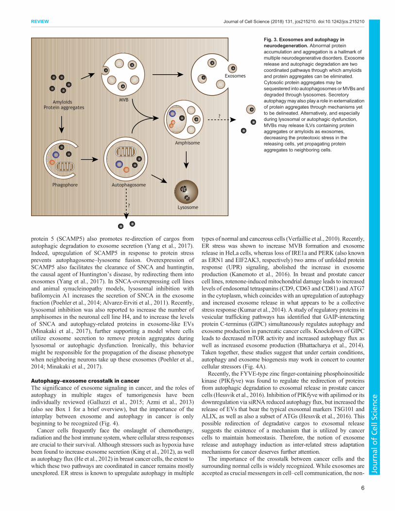
protein 5 (SCAMP5) also promotes re-direction of cargos fromautophagic degradation to exosome secretion (Yang et al., 2017).Indeed, upregulation of SCAMP5 in response to protein stressprevents autophagosome–lysosome fusion. Overexpression ofSCAMP5 also facilitates the clearance of SNCA and huntingtin,the causal agent of Huntington’s disease, by redirecting them intoexosomes (Yang et al., 2017). In SNCA-overexpressing cell linesand animal synucleinopathy models, lysosomal inhibition withbafilomycin A1 increases the secretion of SNCA in the exosomefraction (Poehler et al., 2014; Alvarez-Erviti et al., 2011). Recently,lysosomal inhibition was also reported to increase the number ofamphisomes in the neuronal cell line H4, and to increase the levelsof SNCA and autophagy-related proteins in exosome-like EVs(Minakaki et al., 2017), further supporting a model where cellsutilize exosome secretion to remove protein aggregates duringlysosomal or autophagic dysfunction. Ironically, this behaviormight be responsible for the propagation of the disease phenotypewhen neighboring neurons take up these exosomes (Poehler et al.,2014; Minakaki et al., 2017).
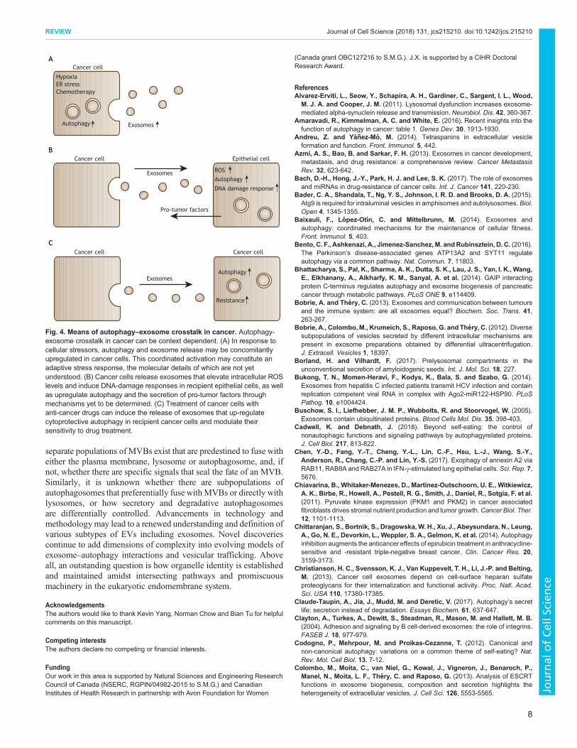
Autophagy–exosome crosstalk in cancerThe significance of exosome signaling in cancer, and the roles ofautophagy in multiple stages of tumorigenesis have beenindividually reviewed (Galluzzi et al., 2015; Azmi et al., 2013)(also see Box 1 for a brief overview), but the importance of theinterplay between exosome and autophagy in cancer is onlybeginning to be recognized (Fig. 4).Cancer cells frequently face the onslaught of chemotherapy,
radiation and the host immune system, where cellular stress responsesare crucial to their survival. Although stressors such as hypoxia havebeen found to increase exosome secretion (King et al., 2012), as wellas autophagy flux (He et al., 2012) in breast cancer cells, the extent towhich these two pathways are coordinated in cancer remains mostlyunexplored. ER stress is known to upregulate autophagy in multiple
types of normal and cancerous cells (Verfaillie et al., 2010). Recently,ER stress was shown to increase MVB formation and exosomerelease in HeLa cells, whereas loss of IRE1a and PERK (also knownas ERN1 and EIF2AK3, respectively) two arms of unfolded proteinresponse (UPR) signaling, abolished the increase in exosomeproduction (Kanemoto et al., 2016). In breast and prostate cancercell lines, rotenone-induced mitochondrial damage leads to increasedlevels of endosomal tetraspanins (CD9, CD63 and CD81) and ATG7in the cytoplasm, which coincides with an upregulation of autophagyand increased exosome release in what appears to be a collectivestress response (Kumar et al., 2014). A study of regulatory proteins invesicular trafficking pathways has identified that GAIP-interactingprotein C-terminus (GIPC) simultaneously regulates autophagy andexosome production in pancreatic cancer cells. Knockdown of GIPCleads to decreased mTOR activity and increased autophagy flux aswell as increased exosome production (Bhattacharya et al., 2014).Taken together, these studies suggest that under certain conditions,autophagy and exosome biogenesis may work in concert to countercellular stressors (Fig. 4A).
Recently, the FYVE-type zinc finger-containing phosphoinositidekinase (PIKfyve) was found to regulate the redirection of proteinsfrom autophagic degradation to exosomal release in prostate cancercells (Hessvik et al., 2016). Inhibition of PIKfyve with apilimod or itsdownregulation via siRNA reduced autophagy flux, but increased therelease of EVs that bear the typical exosomal markers TSG101 andALIX, as well as also a subset of ATGs (Hessvik et al., 2016). Thispossible redirection of degradative cargos to exosomal releasesuggests the existence of a mechanism that is utilized by cancercells to maintain homeostasis. Therefore, the notion of exosomerelease and autophagy induction as inter-related stress adaptationmechanisms for cancer deserves further attention.
The importance of the crosstalk between cancer cells and thesurrounding normal cells is widely recognized. While exosomes areaccepted as crucial messengers in cell–cell communication, the non-
MVB
Amphisome
Autophagosome
Lysosome
AmyloidsProtein aggregates
?
?
Phagophore
Exosomes
Fig. 3. Exosomes and autophagy inneurodegeneration. Abnormal proteinaccumulation and aggregation is a hallmark ofmultiple neurodegenerative disorders. Exosomerelease and autophagic degradation are twocoordinated pathways through which amyloidsand protein aggregates can be eliminated.Cytosolic protein aggregates may besequestered into autophagosomes orMVBs anddegraded through lysosomes. Secretoryautophagy may also play a role in externalizationof protein aggregates through mechanisms yetto be delineated. Alternatively, and especiallyduring lysosomal or autophagic dysfunction,MVBs may release ILVs containing proteinaggregates or amyloids as exosomes,decreasing the proteotoxic stress in thereleasing cells, yet propagating proteinaggregates to neighboring cells.
6
REVIEW Journal of Cell Science (2018) 131, jcs215210. doi:10.1242/jcs.215210
Journal
ofCe
llScience
cell-autonomous roles of autophagy are only beginning to emerge,such as its participation in interactions between tumor cells, stromalcells and immune cells (Maes et al., 2013). Given that autophagycan influence exosome release, it would be interesting to determinewhether non-cell autonomous roles of autophagy are accomplished,in part, via exosomal signaling.A recent study has found that breast cancer cells released
exosomes that alter autophagy flux in recipient breast epithelial cells(Dutta et al., 2014). Through mechanisms yet to be determined,human breast epithelial cells produce increased levels of ROS uponexosome uptake, which plays a role in the upregulation of
autophagy flux. Subsequently, the breast epithelial cells secretepro-tumor growth factors as the result of the uptake of exosomes thatwere derived from the cancer cells (Dutta et al., 2014) (Fig. 4B).Secretory autophagy in stromal cells has been reported to mediatethe release of nutrients or growth factors that promote cancer cellgrowth (Sousa et al., 2016; Chiavarina et al., 2011), so it would beinteresting to investigate whether exosome-mediated signaling isable to regulate autophagic secretion.
Acquired resistance to chemotherapies and targeted therapies isone of the major obstacles in combating cancers and remains a fieldof active investigation. Understanding how cancer cells withstandchemotherapy and develop resistance is crucial to successful cancercontrol. Upregulation of autophagy and exosome release have beendocumented following drug treatments (Ertmer et al., 2007; Sunet al., 2011), suggesting that they constitute a part of the cancer cellstress response or survival mechanism against chemotherapy. Insupport of this possibility, increases in the levels of autophagy fluxand exosome production in various types of chemotherapy-resistantcancers have been reported (Yang et al., 2011; Yu et al., 2015). Forexample, increased release of exosomes has been observed inplatinum-resistant ovarian cancer cell lines, as well as in serum frompatients with cisplatin-resistant tumors (Yin et al., 2012), whileincreased autophagy flux has also been found in platinum-resistantovarian cancer cells (Pasto et al., 2016). Although it is unknownwhether these changes are part of the resistance mechanism ormerely a consequence of the shifting cellular phenotype, theseobservations provide the basis to investigate the therapeutic value ofinhibiting autophagy and disrupting exosome release to counterchemotherapy resistance.
The downstream signaling effects of exosomes released fromchemotherapy-resistant cancer cells are also essential to consider. Ithas been proposed that exosomes might propagate drug-resistantphenotypes through the transfer of miRNA or multidrug-resistanttransporter (MDR) proteins (Azmi et al., 2013; Torreggiani et al.,2016; Bach et al., 2017). In another context, exosomes from gefitinib-treated EGFR-mutant PC-9 cells have been shown to increaseautophagy flux in recipient cancer cells that were subsequently lessresponsive to cisplatin treatment (Li et al., 2016) (Fig. 4C). Thesestudies illustrate the potential capacity of the crosstalk between tumor-derived exosomes and autophagy to influence tumor behavior and itsinteractions with the microenvironment.
Conclusions and future directionsThe interplay between exosome biogenesis and autophagy occurs inmultiple different ways. At the molecular level, there are examplesof autophagy-related proteins and protein complexes that function inexosome biogenesis. At the organelle level, the exosome andautophagy pathways intersect at amphisomes, the contents of whichhave multiple fates, including extracellular release or lysosomaldegradation. Both exosome biogenesis and autophagy play vitalroles in maintaining cellular homeostasis and mitigating cellularstress, with increasing evidence to indicate that these cellularresponses are accomplished through a crosstalk between autophagyand exosomes. What has become clear is that the dynamic andcontext-dependent nature of the interplay between exosomebiogenesis and autophagy has important implications not only fornormal physiology but also for disease – and thus perhaps alsorepresents therapeutic opportunities if we can better understand itsregulation and complexity.
Questions underlying the identity and heterogeneity of variousintermediate compartments in exosome–autophagy crosstalk andvesicular trafficking still remain. For instance, it is unclear whether
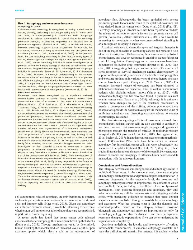
Box 1. Autophagy and exosomes in cancerAutophagy in cancerCell-autonomous autophagy is recognized as having a dual role incancer, typically, performing a tumor-suppressing role in normal cellsand acting as tumor-promoting in transformed cells. Autophagycontributes to cellular homeostasis in normal tissue by removingpotentially damaging proteins and organelles and therefore acts toprevent tumorigenesis (Galluzzi et al., 2015). In established tumors,however, autophagy supports tumor progression, for example, bymaintaining mitochondrial integrity in cancer cells with oncogenic Rasmutations (Guo et al., 2011; Amaravadi et al., 2016). At the genomiclevel, the core autophagy machinery is often intact in many types ofcancer, which supports its indispensability for tumorigenesis (Lebovitzet al., 2015). Hence, autophagy inhibition is under investigation as apotential anti-cancer therapy strategy. In vitro and animal studies havedemonstrated the growth-suppressing and cell-killing effect of autophagyinhibition in cancer cells (Sun et al., 2011; He et al., 2015; Chittaranjanet al., 2014). However, a thorough understanding of the context-dependent roles of autophagy in cancer is needed for more preciseand efficient autophagy modulation for therapeutic benefits (Levy et al.,2017). In comparison, cell non-autonomous roles of autophagy in cancerare less explored, although autophagy-dependent secretion has beenimplicated in some aspects of tumorigenesis (Keulers et al., 2016).Exosomes in cancerExosomes have been recognized as a significant cell–cellcommunication pathway in cancer. Many recent reviews havediscussed the roles of exosomes in the tumor microenvironment(Minciacchi et al., 2015; Azmi et al., 2013; Kharaziha et al., 2012;Tkach and Théry, 2016) and anti-tumor immune responses (Greeninget al., 2015; Bobrie and Théry, 2013; Filipazzi et al., 2012). Overall,tumor-derived exosomes are known to alter the microenvironment of apro-cancer phenotype, facilitate immunosurveillance evasion andpromote local invasion and distant metastases. In a metastatic breastcancer model, expression of different sets of surface integrins on tumor-derived exosomes has been shown to determine their target cells andthereby influence the organotropism of subsequent metastases(Hoshino et al., 2015). Exosomes from metastatic melanoma cells canalter the phenotype of bone marrow progenitor cells, leading to anincrease in the size of the primary tumor, as well as in the size andnumber of metastases (Peinado et al., 2012). Owing to their presence inbodily fluids, including blood and urine, circulating exosomes are underinvestigation for their potential to serve as biomarkers for cancerprogression or treatment response. Serum exosomes have beenshown to carry DNA with a mutation profile that is almost identical tothat of the primary tumor (Kahlert et al., 2014). Thus, the detection ofbiomarkers in exosomesmay reveal small, hidden tumors at early stagesof the disease (Melo et al., 2015). It may be possible in the future tosurvey the change in exosome composition as the disease progresses orresponds to treatment, and so obtain crucial insight into the change indisease status without the need for invasive sampling. Furthermore,engineered exosomes are promising carriers for drugs and nucleic acids.Tumors that actively scavenge nutrients through macropinocytosis, suchas Ras-transformed pancreatic cancer cells (Commisso et al., 2013),may be especially responsive to such an exosome-mediated drugdelivery.
7
REVIEW Journal of Cell Science (2018) 131, jcs215210. doi:10.1242/jcs.215210
Journal
ofCe
llScience
separate populations of MVBs exist that are predestined to fuse witheither the plasma membrane, lysosome or autophagosome, and, ifnot, whether there are specific signals that seal the fate of an MVB.Similarly, it is unknown whether there are subpopulations ofautophagosomes that preferentially fuse with MVBs or directly withlysosomes, or how secretory and degradative autophagosomesare differentially controlled. Advancements in technology andmethodologymay lead to a renewed understanding and definition ofvarious subtypes of EVs including exosomes. Novel discoveriescontinue to add dimensions of complexity into evolving models ofexosome–autophagy interactions and vesicular trafficking. Aboveall, an outstanding question is how organelle identity is establishedand maintained amidst intersecting pathways and promiscuousmachinery in the eukaryotic endomembrane system.
AcknowledgementsThe authors would like to thank Kevin Yang, Norman Chow and Bian Tu for helpfulcomments on this manuscript.
Competing interestsThe authors declare no competing or financial interests.
FundingOur work in this area is supported by Natural Sciences and Engineering ResearchCouncil of Canada (NSERC, RGPIN/04982-2015 to S.M.G.) and CanadianInstitutes of Health Research in partnership with Avon Foundation for Women
(Canada grant OBC127216 to S.M.G.). J.X. is supported by a CIHR DoctoralResearch Award.
ReferencesAlvarez-Erviti, L., Seow, Y., Schapira, A. H., Gardiner, C., Sargent, I. L., Wood,
M. J. A. and Cooper, J. M. (2011). Lysosomal dysfunction increases exosome-mediated alpha-synuclein release and transmission. Neurobiol. Dis. 42, 360-367.
Amaravadi, R., Kimmelman, A. C. and White, E. (2016). Recent insights into thefunction of autophagy in cancer: table 1. Genes Dev. 30, 1913-1930.
Andreu, Z. and Yan ez-Mo, M. (2014). Tetraspanins in extracellular vesicleformation and function. Front. Immunol. 5, 442.
Azmi, A. S., Bao, B. and Sarkar, F. H. (2013). Exosomes in cancer development,metastasis, and drug resistance: a comprehensive review. Cancer MetastasisRev. 32, 623-642.
Bach, D.-H., Hong, J.-Y., Park, H. J. and Lee, S. K. (2017). The role of exosomesand miRNAs in drug-resistance of cancer cells. Int. J. Cancer 141, 220-230.
Bader, C. A., Shandala, T., Ng, Y. S., Johnson, I. R. D. and Brooks, D. A. (2015).Atg9 is required for intraluminal vesicles in amphisomes and autolysosomes. Biol.Open 4, 1345-1355.
Baixauli, F., Lopez-Otın, C. and Mittelbrunn, M. (2014). Exosomes andautophagy: coordinated mechanisms for the maintenance of cellular fitness.Front. Immunol. 5, 403.
Bento, C. F., Ashkenazi, A., Jimenez-Sanchez,M. andRubinsztein, D. C. (2016).The Parkinson’s disease-associated genes ATP13A2 and SYT11 regulateautophagy via a common pathway. Nat. Commun. 7, 11803.
Bhattacharya, S., Pal, K., Sharma, A. K., Dutta, S. K., Lau, J. S., Yan, I. K., Wang,E., Elkhanany, A., Alkharfy, K. M., Sanyal, A. et al. (2014). GAIP interactingprotein C-terminus regulates autophagy and exosome biogenesis of pancreaticcancer through metabolic pathways. PLoS ONE 9, e114409.
Bobrie, A. and Thery, C. (2013). Exosomes and communication between tumoursand the immune system: are all exosomes equal? Biochem. Soc. Trans. 41,263-267.
Bobrie, A., Colombo,M., Krumeich, S., Raposo, G. and Thery, C. (2012). Diversesubpopulations of vesicles secreted by different intracellular mechanisms arepresent in exosome preparations obtained by differential ultracentrifugation.J. Extracell. Vesicles 1, 18397.
Borland, H. and Vilhardt, F. (2017). Prelysosomal compartments in theunconventional secretion of amyloidogenic seeds. Int. J. Mol. Sci. 18, 227.
Bukong, T. N., Momen-Heravi, F., Kodys, K., Bala, S. and Szabo, G. (2014).Exosomes from hepatitis C infected patients transmit HCV infection and containreplication competent viral RNA in complex with Ago2-miR122-HSP90. PLoSPathog. 10, e1004424.
Buschow, S. I., Liefhebber, J. M. P., Wubbolts, R. and Stoorvogel, W. (2005).Exosomes contain ubiquitinated proteins. Blood Cells Mol. Dis. 35, 398-403.
Cadwell, K. and Debnath, J. (2018). Beyond self-eating: the control ofnonautophagic functions and signaling pathways by autophagyrelated proteins.J. Cell Biol. 217, 813-822.
Chen, Y.-D., Fang, Y.-T., Cheng, Y.-L., Lin, C.-F., Hsu, L.-J., Wang, S.-Y.,Anderson, R., Chang, C.-P. and Lin, Y.-S. (2017). Exophagy of annexin A2 viaRAB11, RAB8A and RAB27A in IFN-γ-stimulated lung epithelial cells. Sci. Rep. 7,5676.
Chiavarina, B., Whitaker-Menezes, D., Martinez-Outschoorn, U. E., Witkiewicz,A. K., Birbe, R., Howell, A., Pestell, R. G., Smith, J., Daniel, R., Sotgia, F. et al.(2011). Pyruvate kinase expression (PKM1 and PKM2) in cancer associatedfibroblasts drives stromal nutrient production and tumor growth.Cancer Biol. Ther.12, 1101-1113.
Chittaranjan, S., Bortnik, S., Dragowska, W. H., Xu, J., Abeysundara, N., Leung,A., Go, N. E., Devorkin, L., Weppler, S. A., Gelmon, K. et al. (2014). Autophagyinhibition augments the anticancer effects of epirubicin treatment in anthracycline-sensitive and -resistant triple-negative breast cancer. Clin. Cancer Res. 20,3159-3173.
Christianson, H. C., Svensson, K. J., Van Kuppevelt, T. H., Li, J.-P. and Belting,M. (2013). Cancer cell exosomes depend on cell-surface heparan sulfateproteoglycans for their internalization and functional activity. Proc. Natl. Acad.Sci. USA 110, 17380-17385.
Claude-Taupin, A., Jia, J., Mudd, M. and Deretic, V. (2017). Autophagy’s secretlife: secretion instead of degradation. Essays Biochem. 61, 637-647.
Clayton, A., Turkes, A., Dewitt, S., Steadman, R., Mason, M. and Hallett, M. B.(2004). Adhesion and signaling by B cell-derived exosomes: the role of integrins.FASEB J. 18, 977-979.
Codogno, P., Mehrpour, M. and Proikas-Cezanne, T. (2012). Canonical andnon-canonical autophagy: variations on a common theme of self-eating? Nat.Rev. Mol. Cell Biol. 13, 7-12.
Colombo, M., Moita, C., van Niel, G., Kowal, J., Vigneron, J., Benaroch, P.,Manel, N., Moita, L. F., Thery, C. and Raposo, G. (2013). Analysis of ESCRTfunctions in exosome biogenesis, composition and secretion highlights theheterogeneity of extracellular vesicles. J. Cell Sci. 126, 5553-5565.
Autophagy
Exosomes
Exosomes
HypoxiaER stressChemotherapy
ROS
Cancer cell
Epithelial cell
Cancer cell
Autophagy
Pro-tumor factors
Cancer cell
Autophagy
Resistance
A
B
C
Cancer cell
DNA damage response
Exosomes
Fig. 4. Means of autophagy–exosome crosstalk in cancer. Autophagy-exosome crosstalk in cancer can be context dependent. (A) In response tocellular stressors, autophagy and exosome release may be concomitantlyupregulated in cancer cells. This coordinated activation may constitute anadaptive stress response, the molecular details of which are not yetunderstood. (B) Cancer cells release exosomes that elevate intracellular ROSlevels and induce DNA-damage responses in recipient epithelial cells, as wellas upregulate autophagy and the secretion of pro-tumor factors throughmechanisms yet to be determined. (C) Treatment of cancer cells withanti-cancer drugs can induce the release of exosomes that up-regulatecytoprotective autophagy in recipient cancer cells and modulate theirsensitivity to drug treatment.
8
REVIEW Journal of Cell Science (2018) 131, jcs215210. doi:10.1242/jcs.215210
Journal
ofCe
llScience

Colombo, M., Raposo, G. and Thery, C. (2014). Biogenesis, secretion, andintercellular interactions of exosomes and other extracellular vesicles. Annu. Rev.Cell Dev. Biol 30, 255-289.
Commisso, C., Davidson, S. M., Soydaner-Azeloglu, R. G., Parker, S. J.,Kamphorst, J. J., Hackett, S., Grabocka, E., Nofal, M., Drebin, J. A.,Thompson, C. B. et al. (2013). Macropinocytosis of protein is an amino acidsupply route in Ras-transformed cells. Nature 497, 633-637.
Deretic, V., Jiang, S. and Dupont, N. (2012). Autophagy intersections withconventional and unconventional secretion in tissue development, remodelingand inflammation. Trends Cell Biol. 22, 397-406.
Deretic, V., Saitoh, T. and Akira, S. (2013). Autophagy in infection, inflammationand immunity. Nat. Rev. Immunol. 13, 722-737.
Dupont, N., Jiang, S., Pilli, M., Ornatowski, W., Bhattacharya, D. and Deretic, V.(2011). Autophagy-based unconventional secretory pathway for extracellulardelivery of IL-1β. EMBO J. 30, 4701-4711.
Dutta, S., Warshall, C., Bandyopadhyay, C., Dutta, D. and Chandran, B. (2014).Interactions between exosomes from breast cancer cells and primary mammaryepithelial cells leads to generation of reactive oxygen species which induce DNAdamage response, stabilization of p53 and autophagy in epithelial cells. PLoSONE 9, e97580.
Edgar, J. R., Manna, P. T., Nishimura, S., Banting, G. and Robinson, M. S.(2016). Tetherin is an exosomal tether. eLife 5, e17180.
Ejlerskov, P., Rasmussen, I., Nielsen, T. T., Bergstrom, A.-L., Tohyama, Y.,Jensen, P. H. and Vilhardt, F. (2013). Tubulin polymerization-promoting protein(TPPP/p25α) promotes unconventional secretion of α-synuclein throughexophagy by impairing autophagosome-lysosome fusion. J. Biol. Chem. 288,17313-17335.
Ertmer, A., Huber, V.,Gilch, S., Yoshimori, T., Erfle, V., Duyster, J., Elsasser, H.-P.and Schatzl, H. M. (2007). The anticancer drug imatinib induces cellularautophagy. Leukemia 21, 936-942.
Fader, C. M., Sanchez, D., Furlan, M. and Colombo, M. I. (2008). Induction ofautophagy promotes fusion of multivesicular bodies with autophagic vacuoles ink562 cells. Traffic 9, 230-250.
Feng, D., Zhao, W.-L., Ye, Y.-Y., Bai, X.-C., Liu, R.-Q., Chang, L.-F., Zhou, Q. andSui, S.-F. (2010). Cellular internalization of exosomes occurs throughphagocytosis. Traffic 11, 675-687.
Filipazzi, P., Burdek, M., Villa, A., Rivoltini, L. and Huber, V. (2012). Recentadvances on the role of tumor exosomes in immunosuppression and diseaseprogression. Semin. Cancer Biol. 22, 342-349.
Fitzner, D., Schnaars, M., Van Rossum, D., Krishnamoorthy, G., Dibaj, P.,Bakhti, M., Regen, T., Hanisch, U.-K. and Simons, M. (2011). Selective transferof exosomes from oligodendrocytes to microglia by macropinocytosis. J. Cell Sci.124, 447-458.
Fletcher, K., Ulferts, R., Jacquin, E., Veith, T., Gammoh, N., Arasteh, J. M.,Mayer, U., Carding, S. R., Wileman, T., Beale, R. et al. (2018). The WD40domain of ATG16L1 is required for its non-canonical role in lipidation of LC3 atsingle membranes. EMBO J. 37, e97840.
Florey, O., Kim, S. E., Sandoval, C. P., Haynes, C. M. andOverholtzer, M. (2011).Autophagy machinery mediates macroendocytic processing and entotic celldeath by targeting single membranes. Nat. Cell Biol. 13, 1335-1343.
Fujita, N., Itoh, T., Omori, H., Fukuda, M., Noda, T., Yoshimori, T. and Riezman,H. (2008). The Atg16L complex specifies the site of LC3 lipidation for membranebiogenesis in autophagy. Mol. Biol. Cell 19, 2092-2100.
Galluzzi, L., Pietrocola, F., Bravo-San Pedro, J. M., Amaravadi, R. K.,Baehrecke, E. H., Cecconi, F., Codogno, P., Debnath, J., Gewirtz, D. A.,Karantza, V. et al. (2015). Autophagy in malignant transformation and cancerprogression. EMBO J. 34, 856-880.
Galluzzi, L., Baehrecke, E. H., Ballabio, A., Boya, P., Bravo-San Pedro, J. M.,Cecconi, F., Choi, A. M., Chu, C. T., Codogno, P., Colombo, M. I. et al. (2017).Molecular definitions of autophagy and related processes. EMBO J. 36,1811-1836.
Gitler, A. D., Chesi, A., Geddie, M. L., Strathearn, K. E., Hamamichi, S., Hill, K. J.,Caldwell, K. A., Caldwell, G. A., Cooper, A. A., Rochet, J.-C. et al. (2009).Alpha-synuclein is part of a diverse and highly conserved interaction network thatincludes PARK9 and manganese toxicity. Nat. Genet. 41, 308-315.
Gordon, P. B., Høyvik, H. and Seglen, P. O. (1992). Prelysosomal and lysosomalconnections between autophagy and endocytosis. Biochem. J. 283, 361-369.
Gould, S. J., Booth, A. M. and Hildreth, J. E. K. (2003). The Trojan exosomehypothesis. Proc. Natl. Acad. Sci. USA 100, 10592-10597.
Greening, D. W., Gopal, S. K., Xu, R., Simpson, R. J. and Chen, W. (2015).Exosomes and their roles in immune regulation and cancer. Semin. Cell Dev. Biol.40, 72-81.
Guo, J. Y., Chen, H.-Y., Mathew, R., Fan, J., Strohecker, A. M., Karsli-Uzunbas,G., Kamphorst, J. J., Chen, G., Lemons, J. M. S., Karantza, V. et al. (2011).Activated Ras requires autophagy to maintain oxidative metabolism andtumorigenesis. Genes Dev. 25, 460-470.
Guo, H., Chitiprolu, M., Roncevic, L., Javalet, C., Hemming, F. J., Trung, M. T.,Meng, L., Latreille, E., Tanese De Souza, C., McCulloch, D. et al. (2017).Atg5 disassociates the V 1 V 0 -ATPase to promote exosome production and
tumor metastasis independent of canonical macroautophagy. Dev. Cell 43,716-730.e7.
Harding, C., Heuser, J. and Stahl, P. (1983). Receptor-mediated endocytosis oftransferrin and recycling of the transferrin receptor in rat reticulocytes. J. Cell Biol.97, 329-339.
He, C. and Klionsky, D. J. (2009). Regulation mechanisms and signaling pathwaysof autophagy. Annu. Rev. Genet. 43, 67-93.
He, W.-S., Dai, X.-F., Jin, M., Liu, C.-W. and Ren, J.-H. (2012). Hypoxia-inducedautophagy confers resistance of breast cancer cells to ionizing radiation. Oncol.Res. 20, 251-258.
He, J., Yu, J.-J., Xu, Q., Wang, L., Zheng, J. Z., Liu, L.-Z. and Jiang, B.-H. (2015).Downregulation of ATG14 by EGR1-MIR152 sensitizes ovarian cancer cells tocisplatin-induced apoptosis by inhibiting cyto-protective autophagy. Autophagy11, 373-384.
Hessvik, N. P. and Llorente, A. (2018). Current knowledge on exosome biogenesisand release. Cell. Mol. Life Sci. 75, 193-208.
Hessvik, N. P., Øverbye, A., Brech, A., Torgersen, M. L., Jakobsen, I. S.,Sandvig, K. and Llorente, A. (2016). PIKfyve inhibition increases exosomerelease and induces secretory autophagy. Cell. Mol. Life Sci. 73, 4717-4737.
Heusermann, W., Hean, J., Trojer, D., Steib, E., Von Bueren, S., Graff-Meyer, A.,Genoud, C., Martin, K., Pizzato, N., Voshol, J. et al. (2016). Exosomes surf onfilopodia to enter cells at endocytic hot spots, traffic within endosomes, and aretargeted to the ER. J. Cell Biol. 213, 173-184.
Hoshino, A., Costa-Silva, B., Shen, T.-L., Rodrigues, G., Hashimoto, A., TesicMark, M., Molina, H., Kohsaka, S., Di Giannatale, A., Ceder, S. et al. (2015).Tumour exosome integrins determine organotropic metastasis. Nature 527,329-335.
Huotari, J. and Helenius, A. (2011). Endosome maturation. EMBO J. 30,3481-3500.
Hurley, J. H. (2015). ESCRTs are everywhere. EMBO J. 34, 2398-2407.Hurwitz, S. N., Cheerathodi, M. R., Nkosi, D., York, S. B. and Meckes, D. G.
(2018). Tetraspanin CD63 bridges autophagic and endosomal processes toregulate exosomal secretion and intracellular signaling of Epstein-Barr virusLMP1. J. Virol. 92, e01969-17.
Ichimura, Y., Kirisako, T., Takao, T., Satomi, Y., Shimonishi, Y., Ishihara, N.,Mizushima, N., Tanida, I., Kominami, E., Ohsumi, M. et al. (2000). A ubiquitin-like system mediates protein lipidation. Nature 408, 488-492.
Jacquin, E., Leclerc-Mercier, S., Judon, C., Blanchard, E., Fraitag, S. andFlorey, O. (2017). Pharmacological modulators of autophagy activate a parallelnoncanonical pathway driving unconventional LC3 lipidation. Autophagy 13,854-867.
Janas, T. T. T., Janas, M. M., Sapon, K. and Janas, T. (2015). Mechanisms of RNAloading into exosomes. FEBS Lett. 589, 1391-1398.
Kahlert, C., Melo, S. A., Protopopov, A., Tang, J., Seth, S., Koch, M., Zhang, J.,Weitz, J., Chin, L., Futreal, A. et al. (2014). Identification of doublestrandedgenomic DNA spanning all chromosomes with mutated KRAS and P53 DNA inthe serum exosomes of patients with pancreatic cancer. J. Biol. Chem. 289,3869-3875.
Kamerkar, S., Lebleu, V. S., Sugimoto, H., Yang, S., Ruivo, C. F., Melo, S. A.,Lee, J. J. and Kalluri, R. (2017). Exosomes facilitate therapeutic targeting ofoncogenic KRAS in pancreatic cancer. Nature 546, 498-503.
Kanemoto, S., Nitani, R., Murakami, T., Kaneko, M., Asada, R., Matsuhisa, K.,Saito, A. and Imaizumi, K. (2016). Multivesicular body formation enhancementand exosome release during endoplasmic reticulum stress. Biochem. Biophys.Res. Commun. 480, 166-172.
Katzmann, D. J., Babst, M. and Emr, S. D. (2001). Ubiquitin-dependent sorting intothe multivesicular body pathway requires the function of a conserved endosomalprotein sorting complex, ESCRT-I. Cell 106, 145-155.
Keulers, T. G., Schaaf, M. B. E. and Rouschop, K. M. A. (2016). Autophagy-dependent secretion: contribution to tumor progression. Front. Oncol. 6, 251.
Kharaziha, P., Ceder, S., Li, Q. and Panaretakis, T. (2012). Tumor cell-derivedexosomes: a message in a bottle. Biochim. Biophys. Acta 1826, 103-111.
Kihara, A., Noda, T., Ishihara, N. and Ohsumi, Y. (2001). Two distinct Vps34phosphatidylinositol 3-kinase complexes function in autophagy andcarboxypeptidase Y sorting in Saccharomyces cerevisiae. J. Cell Biol. 152,519-530.
Kim, J., Kundu, M., Viollet, B. and Guan, K.-L. (2011). AMPK and mTOR regulateautophagy through direct phosphorylation of Ulk1. Nat. Cell Biol. 13, 132-141.
Kimura, T., Jia, J., Kumar, S., Choi, S. W., Gu, Y., Mudd, M., Dupont, N., Jiang,S., Peters, R., Farzam, F. et al. (2017). Dedicated SNAREs and specialized TRIMcargo receptors mediate secretory autophagy. EMBO J. 36, 42-60.
King, H. W., Michael, M. Z. and Gleadle, J. M. (2012). Hypoxic enhancement ofexosome release by breast cancer cells. BMC Cancer 12, 421.
Klionsky, D. J. (2000). Autophagy as a regulated pathway of cellular degradation.Science 290, 1717-1721.
Kong, S. M. Y., Chan, B. K. K., Park, J.-S., Hill, K. J., Aitken, J. B., Cottle, L.,Farghaian, H., Cole, A. R., Lay, P. A., Sue, C. M. et al. (2014). Parkinson’sdisease-linked human PARK9/ATP13A2 maintains zinc homeostasis andpromotes α-Synuclein externalization via exosomes. Hum. Mol. Genet. 23,2816-2833.
9
REVIEW Journal of Cell Science (2018) 131, jcs215210. doi:10.1242/jcs.215210
Journal
ofCe
llScience
Kowal, J., Arras, G., Colombo, M., Jouve, M., Morath, J. P., Primdal-Bengtson,B., Dingli, F., Loew, D., Tkach, M. and Thery, C. (2016). Proteomic comparisondefines novel markers to characterize heterogeneous populations of extracellularvesicle subtypes. Proc. Natl. Acad. Sci. USA 113, E968-E977.
Kroemer, G., Marin o, G. and Levine, B. (2010). Autophagy and the integratedstress response. Mol. Cell 40, 280-293.
Kumar, D., Gupta, D., Shankar, S. and Srivastava, R. K., (2014). Biomolecularcharacterization of exosomes released from cancer stem cells: possibleimplications for biomarker and treatment of cancer. Oncotarget 6, 3280-3291.
Lebovitz, C. B., Robertson, A. G., Goya, R., Jones, S. J., Morin, R. D., Marra,M. A. and Gorski, S. M. (2015). Cross-cancer profiling of molecular alterationswithin the human autophagy interaction network. Autophagy 11, 1668-1687.
Levine, B. and Klionsky, D. J. (2004). Development by self-digestion: molecularmechanisms and biological functions of autophagy. Dev. Cell 6, 463-477.
Levy, J. M. M. M., Towers, C. G. and Thorburn, A. (2017). Targeting autophagy incancer. Nat. Rev. Cancer 17, 528-542.
Li, X.-Q., Liu, J.-T., Fan, L.-L., Liu, Y., Cheng, L., Wang, F., Yu, H.-Q., Gao, J., Wei,W.,Wang, H. et al. (2016). Exosomes derived from gefitinib-treated EGFR-mutantlung cancer cells alter cisplatin sensitivity via up-regulating autophagy.Oncotarget 7, 24585-24595.
Liang, C., Lee, J., Inn, K.-S., Gack, M. U., Li, Q., Roberts, E. A., Vergne, I.,Deretic, V., Feng, P., Akazawa, C. et al. (2008). Beclin1-binding UVRAG targetsthe class C Vps complex to coordinate autophagosome maturation and endocytictrafficking. Nat. Cell Biol. 10, 776-787.
Liou,W., Geuze, H. J., Geelen, M. J. H. and Slot, J. W. (1997). The autophagic andendocytic pathways converge at the nascent autophagic vacuoles. J. Cell Biol.136, 61-70.
Liu, Z., Zhang, X., Yu, Q. and He, J. J. (2014). Exosome-associated hepatitisC virus in cell cultures and patient plasma. Biochem. Biophys. Res. Commun.455, 218-222.
Liu, J., Zhang, Y., Liu, A., Wang, J., Li, L., Chen, X., Gao, X., Xue, Y., Zhang, X.and Liu, Y. (2016). Distinct dasatinib-induced mechanisms of apoptotic responseand exosome release in imatinib-resistant human chronic myeloid leukemia cells.Int. J. Mol. Sci. 17, 531.
Lotvall, J., Hill, A. F., Hochberg, F., Buzas, E. I., Di Vizio, D., Gardiner, C., Gho,Y. S., Kurochkin, I. V., Mathivanan, S., Quesenberry, P. et al. (2014). Minimalexperimental requirements for definition of extracellular vesicles and theirfunctions: a position statement from the International Society for ExtracellularVesicles. J. Extracell. Vesicles 3, 26913.
Maes, H., Rubio, N., Garg, A. D. and Agostinis, P. (2013). Autophagy: shaping thetumor microenvironment and therapeutic response. Trends Mol. Med. 19,428-446.
Martinez, J., Almendinger, J., Oberst, A., Ness, R., Dillon, C. P., Fitzgerald, P.,Hengartner, M. O. and Green, D. R. (2011). Microtubule-associated protein 1light chain 3 alpha (LC3)-associated phagocytosis is required for the efficientclearance of dead cells. Proc. Natl. Acad. Sci. USA 108, 17396-17401.
Martinez, J., Malireddi, R. K. S., Lu, Q., Cunha, L. D., Pelletier, S., Gingras, S.,Orchard, R., Guan, J.-L., Tan, H., Peng, J. et al. (2015). Molecularcharacterization of LC3-associated phagocytosis reveals distinct roles forRubicon, NOX2 and autophagy proteins. Nat. Cell Biol. 17, 893-906.
Matsuura, A., Tsukada, M., Wada, Y. and Ohsumi, Y. (1997). Apg1p, a novelprotein kinase required for the autophagic process in Saccharomyces cerevisiae.Gene 192, 245-250.
Melo, S. A., Luecke, L. B., Kahlert, C., Fernandez, A. F., Gammon, S. T., Kaye, J.,Lebleu, V. S., Mittendorf, E. A., Weitz, J., Rahbari, N. et al. (2015). Glypican-1identifies cancer exosomes and detects early pancreatic cancer. Nature 523,177-182.
Minakaki, G., Menges, S, Kittel, A, Emmanouilidou, E, Schaeffner, I, Barkovits,K, Bergmann, A, Rockenstein, E, Adame, A, Marxreiter, F et al. (2017).Autophagy inhibition promotes SNCA/alpha-synuclein release and transfer viaextracellular vesicles with a hybrid autophagosome-exosome-like phenotype.Autophagy 8627, 1-61.
Minciacchi, V. R., Freeman, M. R. and Di Vizio, D. (2015). Extracellular vesicles incancer: exosomes, microvesicles and the emerging role of large oncosomes.Semin. Cell Dev. Biol. 40, 41-51.
Mizushima, N., Noda, T., Yoshimori, T., Tanaka, Y., Ishii, T., George, M. D.,Klionsky, D. J., Ohsumi, M. and Ohsumi, Y. (1998). A protein conjugationsystem essential for autophagy. Nature 395, 395-398.
Mulcahy, L. A., Pink, R. C. and Carter, D. R. F. (2014). Routes and mechanisms ofextracellular vesicle uptake. J. Extracell. Vesicles 3, 24641.
Murrow, L., Malhotra, R. and Debnath, J. (2015). ATG12–ATG3 interacts with Alixto promote basal autophagic flux and late endosome function. Nat. Cell Biol. 17,300-310.
Ohsumi, Y. (2014). Historical landmarks of autophagy research. Nat. PublishingGroup 24, 9-23.
Ojha, C. R., Lapierre, J., Rodriguez, M., Dever, S., Zadeh, M., Demarino, C.,Pleet, M., Kashanchi, F. and El-Hage, N. (2017). Interplay between autophagy,exosomes and HIV-1 associated neurological disorders: new insights fordiagnosis and therapeutic applications. Viruses 9, 176.
Pan, B.-T. and Johnstone, R. M. (1983). Fate of the transferrin receptor duringmaturation of sheep reticulocytes in vitro: selective externalization of the receptor.Cell 33, 967-978.
Park, J.-M., Jung, C. H., Seo, M., Otto, N. M., Grunwald, D., Kim, K. H., Moriarity,B., Kim, Y.-M., Starker, C., Nho, R. S. et al. (2016). The ULK1 complex mediatesMTORC1 signaling to the autophagy initiation machinery via binding andphosphorylating ATG14. Autophagy 12, 547-564.
Pasto, A. Pagotto, A, Pilotto, G, De Paoli, A, De Salvo, GL, Baldoni, A, Nicoletto,MO, Ricci, F, Damia, G, Bellio, C et al. (2016). Resistance to glucose starvationas metabolic trait of platinum-resistant human epithelial ovarian cancer cells.Oncotarget 8, 6433-6445.
Patel, K. K., Miyoshi, H., Beatty, W. L., Head, R. D., Malvin, N. P., Cadwell, K.,Guan, J.-L., Saitoh, T., Akira, S., Seglen, P. O. et al. (2013). Autophagy proteinscontrol goblet cell function by potentiating reactive oxygen species production.EMBO J. 32, 3130-3144.
Peinado, H., Aleckovic, M., Lavotshkin, S., Matei, I., Costa-Silva, B., Moreno-Bueno, G., Hergueta-Redondo, M., Williams, C., Garcıa-Santos, G., Ghajar,C. M. et al. (2012). Melanoma exosomes educate bone marrow progenitor cellstoward a pro-metastatic phenotype through MET. Nat. Med. 18, 883-891.
Perez-Hernandez, D., Gutierrez-Vazquez, C., Jorge, I., Lopez-Martın, S., Ursa,A., Sanchez-Madrid, F., Vazquez, J. and Yan ez-Mo, M. (2013). The intracellularinteractome of tetraspanin-enriched microdomains reveals their function assorting machineries toward exosomes. J. Biol. Chem. 288, 11649-11661.
Poehler, A.-M., Xiang,W., Spitzer, P., May, V. E. L., Meixner, H., Rockenstein, E.,Chutna, O., Outeiro, T. F., Winkler, J., Masliah, E. et al. (2014). Autophagymodulates SNCA/α-synuclein release, thereby generating a hostilemicroenvironment. Autophagy 10, 2171-2192.
Ponpuak, M., Mandell, M. A., Kimura, T., Chauhan, S., Cleyrat, C. and Deretic, V.(2015). Secretory autophagy. Curr. Opin. Cell Biol. 35, 106-116.
Ren, H., Elgner, F., Jiang, B., Himmelsbach, K., Medvedev, R., Ploen, D. andHildt, E. (2016). The autophagosomal SNARE protein syntaxin 17 is an essentialfactor for the hepatitis C virus life cycle. J. Virol. 90, 5989-6000.
Sahu, R., Kaushik, S., Clement, C. C., Cannizzo, E. S., Scharf, B., Follenzi, A.,Potolicchio, I., Nieves, E., Cuervo, A. M. and Santambrogio, L. (2011).Microautophagy of cytosolic proteins by late endosomes. Dev. Cell 20, 131-139.
Scott, C. C., Vacca, F. and Gruenberg, J. (2014). Endosome maturation, transportand functions. Semin. Cell Dev. Biol. 31, 2-10.
Shrivastava, S., Devhare, P., Sujijantarat, N., Steele, R., Kwon, Y.-C., Ray, R.and Ray, R. B. (2016). Knockdown of autophagy inhibits infectious hepatitis Cvirus release by the exosomal pathway. J. Virol. 90, 1387-1396.
Smith, V. L., Jackson, L. andSchorey, J. S. (2015). Ubiquitination as amechanismto transport soluble mycobacterial and eukaryotic proteins to exosomes.J. Immunol. 195, 2722-2730.
Sousa, C. M., Biancur, D. E., Wang, X., Halbrook, C. J., Sherman, M. H., Zhang,L., Kremer, D., Hwang, R. F.,Witkiewicz, A. K., Ying, H. et al. (2016). Pancreaticstellate cells support tumour metabolism through autophagic alanine secretion.Nat. Publ. Group 536, 479-483.
Sun, Q., Westphal, W., Wong, K. N., Tan, I. and Zhong, Q. (2010). Rubiconcontrols endosomematuration as aRab7 effector.Proc. Natl. Acad. Sci. USA 107,19338-19343.
Sun,W.-L., Chen, J., Wang, Y.-P. and Zheng, H. (2011). Autophagy protects breastcancer cells from epirubicin-induced apoptosis and facilitates epirubicin-resistance development. Autophagy 7, 1035-1044.
Thery, C., Zitvogel, L. and Amigorena, S. (2002). Exosomes: composition,biogenesis and function. Nat. Rev. Immunol. 2, 569-579.
Tkach, M. and Thery, C. (2016). Communication by extracellular vesicles: whereweare and where we need to go. Cell 164, 1226-1232.
Tooze, S. A., Abada, A. and Elazar, Z. (2014). Endocytosis and autophagy:exploitation or cooperation? Cold Spring Harbor Perspect. Biol. 6, a018358.
Torreggiani, E., Roncuzzi, L., Perut, F., Zini, N. andBaldini, N. (2016). Multimodaltransfer of MDR by exosomes in human osteosarcoma. Int. J. Oncol. 49, 189-196.
Trajkovic, K., Hsu, C., Chiantia, S., Rajendran, L., Wenzel, D., Wieland, F.,Schwille, P., Brugger, B. and Simons, M. (2008). Ceramide triggers budding ofexosome vesicles into multivesicular endosomes. Science 319, 1244-1247.
Tsukada, M. and Ohsumi, Y. (1993). Isolation and characterization of autophagy-defective mutants of Saccharomyces cerevisiae. FEBS Lett. 333, 169-174.
Verfaillie, T., Salazar, M., Velasco, G. and Agostinis, P. (2010). Linking ER stressto autophagy: potential implications for cancer therapy. Int. J. Cell Biol. 2010,930509.
Villarroya-Beltri, C., Gutierrez-Vazquez, C., Sanchez-Cabo, F., Perez-Hernandez, D., Vazquez, J., Martin-Cofreces, N., Martinez-Herrera, D. J.,Pascual-Montano, A., Mittelbrunn, M. and Sanchez-Madrid, F. (2013).Sumoylated hnRNPA2B1 controls the sorting of miRNAs into exosomesthrough binding to specific motifs. Nat. Commun. 4, 2980.
Villarroya-Beltri, C., Baixauli, F., Gutierrez-Vazquez, C., Sanchez-Madrid, F.and Mittelbrunn, M. (2014). Sorting it out: regulation of exosome loading. Semin.Cancer Biol. 28, 3-13.
Villarroya-Beltri, C., Baixauli, F., Mittelbrunn, M., Fernandez-Delgado, I.,Torralba, D., Moreno-Gonzalo, O., Baldanta, S., Enrich, C., Guerra, S. and
10
REVIEW Journal of Cell Science (2018) 131, jcs215210. doi:10.1242/jcs.215210
Journal
ofCe
llScience

Sanchez-Madrid, F. (2016). ISGylation controls exosome secretion by promotinglysosomal degradation of MVB proteins. Nat. Commun. 7, 13588.
Wang, L., Tian, Y., Ou, J.-J. and Luo, G. G. (2015). HCV induces the expression ofRubicon and UVRAG to temporally regulate the maturation of autophagosomesand viral replication. PLoS Pathog. 11, e1004764.
Yan ez-Mo, M., Siljander, P. R.-M., Andreu, Z., Bedina Zavec, A., Borras, F. E.,Buzas, E. I., Buzas, K., Casal, E., Cappello, F., Carvalho, J. et al. (2015).Biological properties of extracellular vesicles and their physiological functions.J. Extracell. Vesicles 4, 27066.
Yang, Z. J., Chee, C. E., Huang, S. and Sinicrope, F. A. (2011). The role ofautophagy in cancer: therapeutic implications. Mol. Cancer Ther. 10, 1533-1541.
Yang, Y., Qin, M., Bao, P., Xu,W., Xu, J. and Trajkovic, V. (2017). Secretory carriermembrane protein 5 is an autophagy inhibitor that promotes the secretion ofα-synuclein via exosome. PLoS ONE 12, e0180892.
Yin, J., Yan, X., Yao, X., Zhang, Y., Shan, Y., Mao, N., Yang, Y. and Pan, L. (2012).Secretion of annexin A3 from ovarian cancer cells and its association withplatinum resistance in ovarian cancer patients. J. Cell. Mol. Med. 16, 337-348.
Yu, D.-D., Wu, Y., Shen, H.-Y., Lv, M.-M., Chen,W.-X., Zhang, X.-H., Zhong, S.-L.,Tang, J.-H. and Zhao, J.-H. (2015). Exosomes in development, metastasis anddrug resistance of breast cancer. Cancer Sci. 106, 959-964.
Zhang, M., Kenny, S. J., Ge, L., Xu, K. and Schekman, R. (2015). Translocation ofinterleukin-1β into a vesicle intermediate in autophagy-mediated secretion. eLife4, 1463.
11
REVIEW Journal of Cell Science (2018) 131, jcs215210. doi:10.1242/jcs.215210
Journal
ofCe
llScience