The international system for reporting serous fluid ...
62
The 2020 International System for reporting Serous Fluid Cytopathology Mark Chien-Chin Chen, MD MSc FIAC Department of Pathology, Chia-Yi Christian Hospital, TAIWAN Taiwan Society of Clinical Cytology Editorial, Cancer cytopathology (5.284) & Cytopathology (2.073) South Taiwan Cytology Education Course TNN 15:00-17:00 Aug 07, 2021
Transcript of The international system for reporting serous fluid ...
The 2020 International System for
reporting Serous Fluid Cytopathology
Mark Chien-Chin Chen, MD MSc FIAC
Department of Pathology, Chia-Yi Christian Hospital, TAIWAN
Taiwan Society of Clinical Cytology
Editorial, Cancer cytopathology (5.284) & Cytopathology (2.073)
South Taiwan Cytology Education Course
TNN 15:00-17:00 Aug 07, 2021

2

Disclosure of COI
No conflicts of interests to be disclosed.
3
Co-editors
• Dr Ashish Chandra
• Dr Barbara Crothers
• Dr Dan Kurtycz
• Dr. Fernando Schmitt

Co-editors
Ashish Chandra
Barbara Crothers
Daniel Kurtycz
Fernando Schmitt
Chapter lead authors
Mauro Saieg (ND)
Eva Wojcik (NFM)
Philippe Vielh (AUS)
Giota Mikou (SFM)
Claire Michael (MAL- Primary)
Ben Davidson (MAL - Secondary)
Lukas Bubendorf (Ancillary testing)
Stefan Pambuccian (Pathogenesis)
Donna Russell (Cytopreparatory methods)
Barbara Centeno (Quality Assurance)
Christopher VandenBussche (Peritoneal washings)
4

5

6

7
We need answers to practical clinical questions!
Evaluating evidence of adequacy (volume & cellularity)
Defining what is a true negative sample
The use of atypia and suspicious categories
Mesothelioma: revisiting the value of cytology in diagnosis
Peritoneal washings: how to report the presence of epithelial cells
8
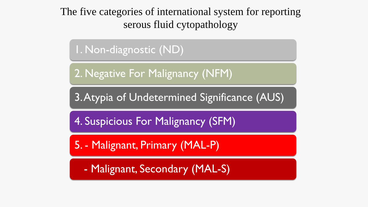
The five categories of international system for reporting
serous fluid cytopathology
1. Non-diagnostic (ND)
2. Negative For Malignancy (NFM)
3. Atypia of Undetermined Significance (AUS)
4. Suspicious For Malignancy (SFM)
5. - Malignant, Primary (MAL-P)
- Malignant, Secondary (MAL-S)

Three parts of a sample report
1. Adequacy statement
2. Diagnostic category
3. Clinical comment
9
10
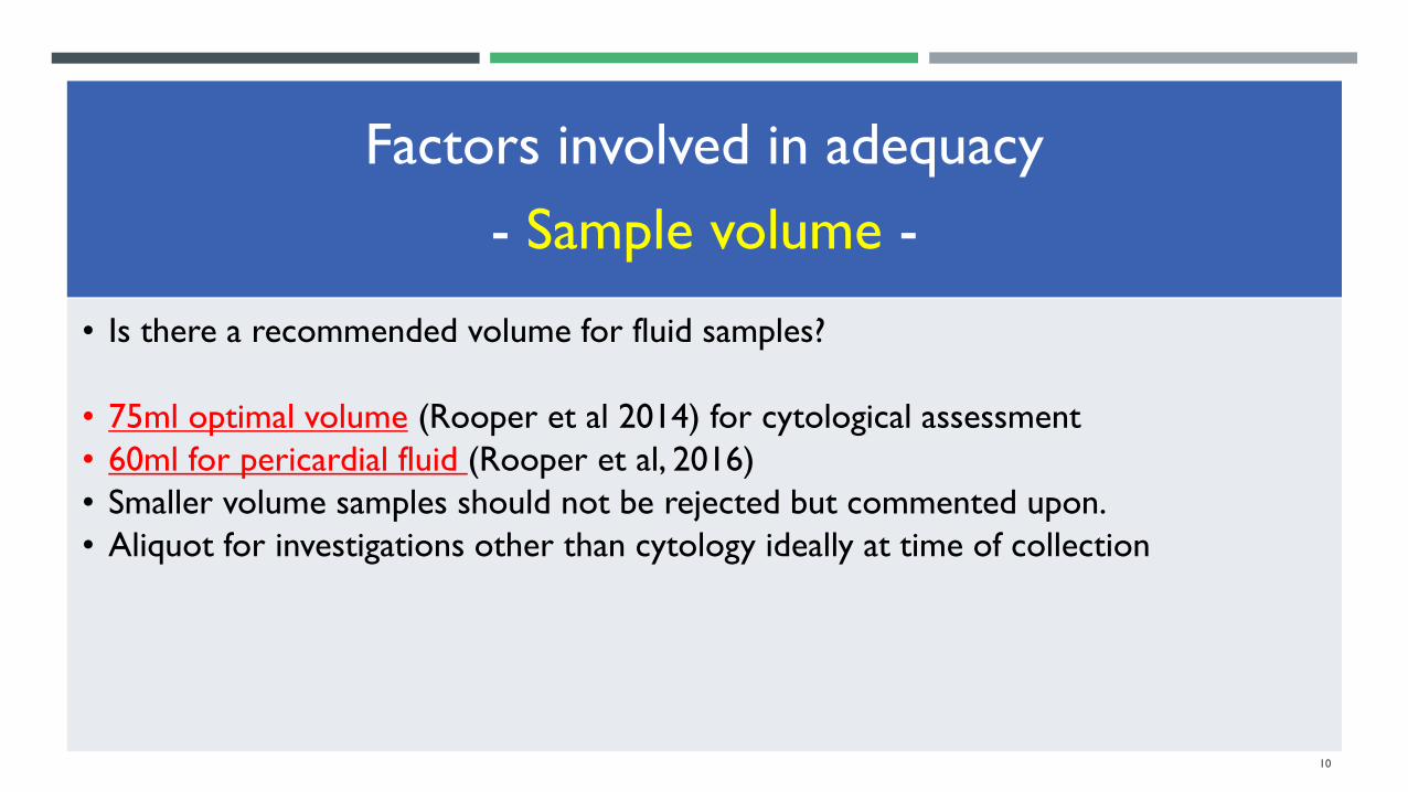
Factors involved in adequacy
- Sample volume -
• Is there a recommended volume for fluid samples?
• 75ml optimal volume (Rooper et al 2014) for cytological assessment
• 60ml for pericardial fluid (Rooper et al, 2016)
• Smaller volume samples should not be rejected but commented upon.
• Aliquot for investigations other than cytology ideally at time of collection

11
Factors involved in adequacy
- Cellular content -
• Do we need to see mesothelial cells?
• Acceptable to find only lymphocytes (TB, chylous effusion) or neutrophils (acute bacterial infections) in benign effusions without mesothelial cells
• Diagnosis of malignancies with a one cell population may be made without mesothelial cells

12
Factors involved in adequacy
-Cellular preservation-
• Can a sample be non-diagnostic in spite of being cellular?
• Loss of quality due to degenerative changes due to delay in reaching the lab, bacterial overgrowth, technical artefacts and contaminants

CASE 1
54-year-old male with left sided pleural effusion.
Smoker, cough and chest pain for one week.
Macro: 2ml of heavily blood-stained fluid
received. One ThinPrepand one DQ cytospin
prepared.
13

14

15
ThinPrep

16
DQ

Sample report for non-diagnostic category
Evaluation limited by heavy blood-staining, likely non-representative sample.
NON-DIAGNOSTIC
Repeat sampling advised (75ml volume if possible).
17

CASE 2
64-year-old male with liver cirrhosis
and ascites.
Macro: 60ml of straw colored fluid. Two cytospins, Pap and Giemsa, prepared.
18

19
Giemsa

20
Pap

Sample report for negative for malignancy category
Satisfactory for evaluation.
Neutrophils, mesothelial cells and a few lymphocytes are present.
NEGATIVE FOR MALIGNANCY
A high proportion of neutrophils is present and may represent spontaneous
bacterial peritonitis (SBP). Please correlate with clinical findings.
21

Negative for malignancy (NFM)
Normal (expected) cell populations in variable numbers
Lymphocytes
Macrophages
Mesothelial cells
Neutrophils
Eosinophils
22

Patterns of reactive effusions
If specific pattern of reactive effusion present such as eosinophilic or
lymphocytic, suggest possible causes in the clinical comment.
Eosinophilic effusion: Recent pleural fluid aspiration, allergic conditions
including hypereosinophilic syndrome etc
Lymphocytic effusion: Viral infections, TB
Neutrophilic effusion: Empyema (purulent fluid) usually indicative of
bacterial infection, occasionally malignant eg. lung squamous cell
carcinoma rupturing into pleural cavity
23

CASE 3
46-year-old female with history of breast
carcinoma 6 years ago. Now, cough and small pleural effusion.
Macro: 20ml straw-colored fluid.
Cytospins, Pap, MGG
24

25
MGG

26
Pap

Atypia of undetermined significance (AUS)
Occasional poorly preserved cells with nuclear enlargement and mild hyperchromasia
but no obvious chromatin or nuclear membrane abnormalities
Likely degenerated macrophages or mesothelial cells
Cell block made and IHC performed to detect any epithelial cells (BerEP4, MOC31,
Claudin-4) (Epithelial cell adhesion molecules)
Downgraded to NFM as epithelial markers negative
27

28
Cell block

29
BerEP4
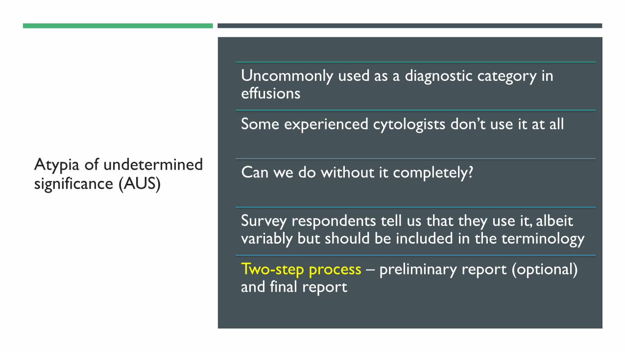
Atypia of undetermined significance (AUS)
Uncommonly used as a diagnostic category in effusions
Some experienced cytologists don’t use it at all
Can we do without it completely?
Survey respondents tell us that they use it, albeit variably but should be included in the terminology
Two-step process – preliminary report (optional) and final report
30

AUS Algorithm
Small number of atypical cells
macrophages? mesothelial cells? epithelial cells?
ICC demonstrates atypical cells to be macrophages or
mesothelial cells
Final report: NFM
ICC demonstrates atypical cells to be epithelial
Final report: SFM or Malignant (secondary)
Insufficient representative cells or ICC equivocal
Final report: AUS
Preliminary assessment: AUS
31

CASE 4
68-year-old man with ascitic fluid. History of lung
carcinoma.
Macro: 30ml of blood-tinged
fluid.
32

33
Giemsa

34
Pap

SFM Algorithm
Small number of cells on cytospins (and clot/cell block). Features favor epithelial or other malignancy
ICC confirms malignancy.
Final report: Malignant (secondary)
Insufficient representative cells or ICC equivocal
Final report: SFM
Preliminary assessment: SFM
35

36
Cell block

37
TTF-1

ICC: TTF-1 and Napsin A were positive
SFM upgraded to:
Malignant (secondary)-lung adenocarcinoma
38

Ancillary testing of lung adenocarcinoma is critical for future treatment.
Insufficient cells for PD-L1, ALK, ROS1 (IHC)
Insufficient cells for mutation analysis (NGS or just EGFR, KRAS)
Further sample may be needed for targeted chemotherapy
Restrict use of IHC (TTF1, Napsin A, P40) to a minimum to conserve material for
molecular testing.
39

Comparison of AUS and SFM categories: the international system for reporting serous fluid cytopathology
AUS SFM
Cytological features Only mild cytological abnormalities
such as nuclear enlargement and
hyperchromasia present usually as
small numbers of dispersed cells and
occasional small groups
Greater degree of cytological
abnormalities present usually as small
numbers of cells, including
architectural features such as
occasional 3 dimensional groups
Cell lineage Benign cell type favored, but epithelial
or other malignant cell of origin not
excluded
Epithelial or other malignant cell of
origin strongly favored
Immunochemistry Outcomes may be benign,
SFM/malignant or inconclusive
Outcomes usually malignant or
inconclusive.
Suggested Risk of Malignancy ~20% ~80%
40

CASE 5
68-year-old man. History of occupational exposure to asbestos. Unilateral hemorrhagic
pleural effusion.
MACRO: 80ml of blood-stained fluid with a clot. Cytospins, MGG,
Pap, HE
41

42
Cytology of normal mesothelial cell
- central round nuclei
- moderate amount of light purple cytoplasm
- Binucleated and multinucleated may be seen
if the cells are reactive.
- skirt" or "halo" at pale outer rim of cell
- Two or more mesothelial cells are often
separated by "window“.
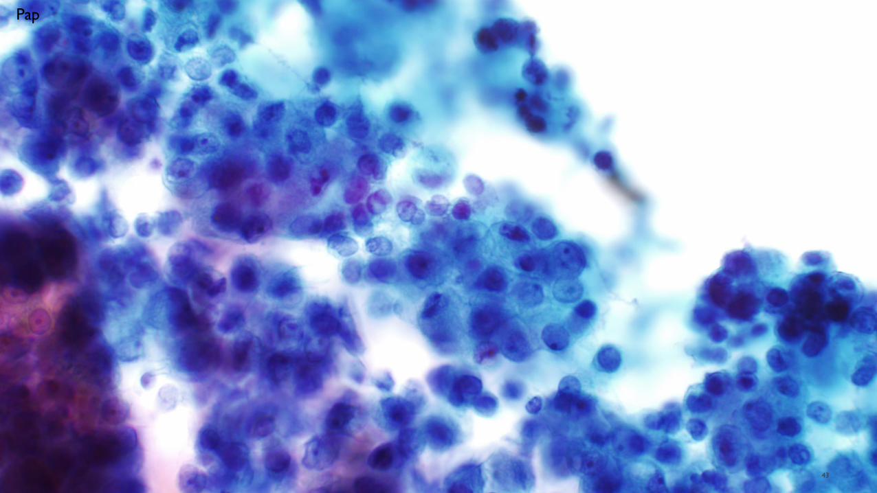
43
Pap

44
Pap

45
Cell block
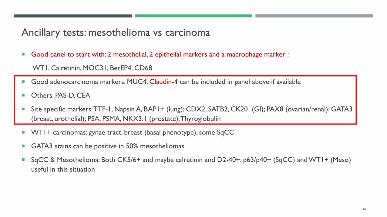
Ancillary tests: mesothelioma vs carcinoma
Good panel to start with: 2 mesothelial, 2 epithelial markers and a macrophage marker :
WT1, Calretinin, MOC31, BerEP4, CD68
Good adenocarcinoma markers: MUC4, Claudin-4 can be included in panel above if available
Others: PAS-D, CEA
Site specific markers: TTF-1, Napsin A, BAP1+ (lung); CDX2, SATB2, CK20 (GI); PAX8 (ovarian/renal); GATA3
(breast, urothelial); PSA, PSMA, NKX3.1 (prostate); Thyroglobulin
WT1+ carcinomas: gynae tract, breast (basal phenotype), some SqCC
GATA3 stains can be positive in 50% mesotheliomas
SqCC & Mesothelioma: Both CK5/6+ and maybe calretinin and D2-40+; p63/p40+ (SqCC) and WT1+ (Meso)
useful in this situation
46

Ancillary testing of mesothelial proliferations
NFM MESOTHELIOMA
Desmin (cytoplasmic) + -
EMA (membranous) - +
HEG1 (membranous) - + (epithelioid, not sarcomatoid)
BAP1 (nuclear) + -
MTAP (IHC nuclear / FISH) IHC + / FISH: No deletion IHC - / FISH: Deletion detected
5-hmC (IHC nuclear) + -
P16/CDKN2A (FISH) No deletion Deletion detected
47
*MTAP loss by IHC was 78% sensitive and 96% specific for CDKN2A homozygous deletion.

48
Calretinin MTAP BAP-1

49
Algorithm for establishing a diagnosis of malignant mesothelioma.
Mod Pathol 2019;32:376-86.

Sample reports for a mesothelial proliferation
Satisfactory for evaluation.
Small spherical groups and dispersed mesothelial cells with mild nuclear pleomorphism are present
suspicious for mesothelioma.
Immunostains requested for confirmation (on cell block or biopsy).
If immunostains confirmatory- MALIGNANT (PRIMARY): MESOTHELIOMA. Clinical correlation
essential.
If morphology classic but immunostains not confirmatory: SUSPICIOUS FOR MESOTHELIOMA
If morphology not classic and immunostains not confirmatory: ATYPICAL MESOTHELIAL
PROLIFERATION. Further investigation advised.
50

MALIGNANT (MAL)
Recognisable abnormal cell population present and adequate for robust diagnosis on which clinical management may be based.
Malignant cell type should be specified on morphology alone or supported by immunochemistry
Malignant- Primary: Mesothelioma
Malignant- Secondary:
Metastatic carcinoma – adenocarcinoma, small cell carcinoma, squamous cell carcinoma
Lymphoma, Melanoma, Other malignancies e.g. sarcoma, leukemias
Primary organ site may need to be investigated for adenocarcinomas
51

CASE 6
45-year-old female. Ascites.
MACRO: 35mlof blood-stained fluid. MGG, Pap
52

53
Pap

54
MGG

55
Cell block

56
PAX8

Ascertaining the primary origin
Site specific markers:
Lung: TTF1, Napsin A, (BAP1+)
Breast: GATA3, mammaglobin, GCDFP15
Thyroid: Thyroglobulin, PAX8
GI: CK20, CDX2
Ovarian: PAX8, WT1, CA125
Kidney (CCRCC): PAX8, CAIX, RCC antigen, Vimentin
Urothelial: GATA3, Uroplakins, p63, p40, 34BE12
Prostate: PSA, PRAP, PSMA, NKX3.1
57

Sample report for MALIGNANT (SECONDARY)
Satisfactory for evaluation.
Spherical groups of tightly cohesive large cells with vacuolated cytoplasm and nuclear pleomorphism
are present. Dispersed single cells are also present.
MALIGNANT (SECONDARY)
Immunostains requested to ascertain the primary, gynae and GI tracts being the most likely sites.
58

Diagnostic categories & clinical management
Cytospins/LBP
(+/- clot/cell block)
Non-diagnosticAtypia of
Undetermined
Significance
Suspicious for
malignancy
Negative for
malignancyMalignant
Repeat sample Discharge or
clinical follow up
Ancillary testing
Correlation with
biopsy
& clinical data
Ancillary testing to
establish primary site
& prognostic/predictive
markers
59

International System for Reporting Serous Fluid Cytopathology: Implied Risk of Malignancy (ROM)
Diagnostic Category % ROM (SE)
Non-Diagnostic (ND) 17% (± 8.9%)
Negative for Malignancy (NFM) 21% (± 0.3%)
Atypia of Undetermined Significance (AUS) 66% (± 10.6%)
Suspicious for Malignancy (SFM) 82% (± 4.8%)
Malignant (MAL) 99% (± 0.1%)
60

Practical Approach to Serous Effusions
Immunocytochemical studies, clinical and radiographic
correlations required. Slide courtesy Dr Eva Wojcik
Effusions
AdequateInadequate
Expected cellular findings (mesothelial cells, some inflammatory cells) Unexpected cellular and non-cellular findings
In regard to volume and
distribution:
Dx. Negative for malignancy
(NFM)
Mostly mesothelial cells arranged
singly and/or in small clusters.
No cellular atypia. Some histiocytes,
lymphocytes, neutrophils
Increased volume and/or cell
distribution:
Predominantly mesothelial cells
(single and/or numerous clusters)Dx. NFM
Dx. Mesothelioma
Predominantly histiocytes
(often appearing like a “second cell
population”)Dx. NFM
Predominantly lymphocytes
Dx. NFM
Dx. Lymphoma
Predominantly or increased
eosinophils
Predominantly neutrophils Dx. NFM
Dx. NFM
Second (malignant) cell population
Single cells Dx. Melanoma, lymphoma, breast (lobular) ca, sarcoma
Small clusters Dx. AdenoCa, breast, lung, small cell carcinoma
Large clusters Dx. AdenoCa, ovarian, pancreatic
Psammoma bodies
Collagen balls
Asbestos bodies
LE cells
Necrosis, spindle and giant cells
Detached ciliary tufts
Dx. NFM
Dx. NFM
Dx. NFM
Dx. NFM
Dx. NFM
Dx. NFM
Infectious organisms Dx. NFM61

REFERENCES
Farahani S, Baloch Z. Are we ready to develop a tiered scheme for the effusion cytology? A comprehensive review and analysis of the literature. Diagn Cytopathol. 2019:1-
19
Valerio E, Nunes W,Cardoso J, et al. A two year retrospective study on pleural effusions: A cancer center experience. Cytopathology 2019;30:607-613.
Sundling KE, Cibas ES. Ancillary studies in pleural, pericardial, and peritoneal effusion cytology. Cancer Cytopathol. 2018;126:590-598.
Pambuccian SE. What is atypia? Use, misuse, and overuse of the term of atypia in diagnostic cytopathology. J Am Soc Cytopathol. 2015;4:44-52.
Chandra A, Crothers B, Kurtycz D, Schmitt FS. Announcement: the international system for reporting serous fluid cytopathology. Acta Cytol. 2019;24:1-3.
Rooper LM, Ali SZ and Olson MT. Cancer Cytopathol. 2014;122:657-665.
Chapel DB, Schulte JJ, Berg K, et al. MTAP immunohistochemistry is an accurate and reproducible surrogate for CDKN2A fluorescence in situ hybridization in diagnosis of
malignant pleural mesothelioma. Mod Pathol. 2020;33(2):245-254.
Chapel DB, Schulte JJ, Husain AN, Krausz T. Application of immunohistochemistry in diagnosis and management of malignant mesothelioma. Transl Lung Cancer Res.
2020;9(Suppl 1):S3-S27.
Naso JR, Tsuji S, Churg A. HEG1 Is a Highly Specific and Sensitive Marker of Epithelioid Malignant Mesothelioma. Am J Surg Pathol. 2020;44(8):1143-1148.
Chapel DB, Husain AN, Krausz T. Immunohistochemical evaluation of nuclear 5-hydroxymethylcytosine (5-hmC) accurately distinguishes malignant pleural mesothelioma
from benign mesothelial proliferations. Mod Pathol. 2019;32(3):376-386.
62