
The Integumentary system. Skin Skin is regarded as our largest organ. It covers about 2 square...
32
The Integumentary system
-
Upload
camren-spivey -
Category
Documents
-
view
216 -
download
1
Transcript of The Integumentary system. Skin Skin is regarded as our largest organ. It covers about 2 square...

The Integumentary system


Skin Skin is regarded as our largest organ. It covers
about 2 square metres in a tall person and about 1.5 square metres in a short person. It is an amazing structure with many functions such as keeping infection out of our body, maintaining our internal environment and is used to keep us at the same temperature. Look carefully at this slide and note that skin is divided into the epidermis (outside layer) and dermis (inside layer)
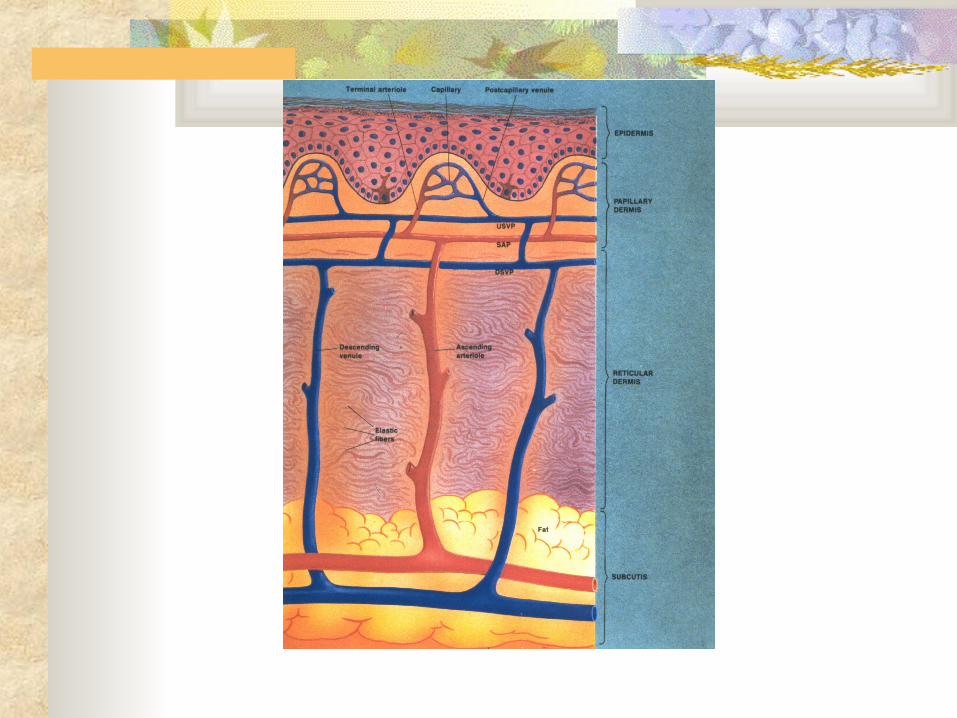

The epidermis We will concentrate on the epidermis but before
we do so you should note that elastic fibres are very prominent in the dermis. If you pinch you skin you will note that it quickly returns to the normal shape when you let go. This is because of the elastic fibres present. As people age their elastic fibres start to degenerate and this leads to wrinkles. Now look at this slide of the epidermis carefully and note the different layers in the epidermis. We are now going to look at each section of the epidermis


Basal layer The basal layer is single row of cuboidal cells
resting on basement membrane. You can see the basal layer in this slide as the bottom darker blue layer
These cuboidal cells divide by mitosis to produce the cells of the epidermis.
These cells move upwards and differentiate into squamous cells of the strata spinosum


melanocytes Melanocytes reside in the basal layer and show a pale
cytoplasm in fair skin. They make two different types of melanin and transfer it to the keratinocytes. People with fair skin have less melanin than people with darker skin. The melanin is produced to protect a person from UV light and UV light actually activates the cells to produce the pigment. Production is probably also under endocrine control as melanin synthesis can increase during pregnancy. You can see a melanocyte in this slide as the large cell with a pale cytoplasm on the basal layer towards the left hand side


Dark skin Look carefully at this slide and you will see
a darker pigment around the basal layer. This is from a person with dark skin and you will be able to see the pigmented layer


Strata spinosum The keratinocytes in the Strata spinosum
exhibit innumerable spines. It used to be called the prickle layer because the spines look like prickles. It is thought that the melanocytes transfer their melanin to the keratinocytes through these spines. The keratinocytes secrete keratin, which is the outside layer of your skin and this protein helps make you waterproof


Stratum corneum This is the outside layer of your skin. It
consists of dying cells and keratin. Skin cells are programmed to live about 30 days after which they die and disintegrate. But they are replaced by the cells that are produced when the basal cells divide. After 30 days these cells finally make their way to the top where they die and disintegrate.


Skin from finger Skin has different thicknesses across the
body. This slide shows skin from a finger and you can easily see the fingerprints. The following slide shows skin from the sole of a foot and you can see that is thicker than finger skin. The slide after that is from the face and you can see hair follicles


Sweat glands Skin is well endowed with sweat glands
and the sweat produced helps you to cool on a hot day. Sweat glands run through to the skin surface and start underneath the hair follicles


Arrector pilli muscle We can retain heat when we are cold or frightened by our
hairs sticking up. You can see in this slide that the arrector pili muscle sticks out at a slant from the hair follicle. This muscle contracts when a nerve impulse from the brain caused by fear or cold reaches the muscle. This causes goose bumps on the skin and these help with cold as they trap an insulating air bubble between the hair and the skin. Look at this model of skin and visualise what happens when the arrector pili muscle contracts.


Sebaceous glands Sebaceous glands are lobulated structures
filled with cells that secrete sebum, often into the canal of the hair follicle. The sole of foot and palm of hand do not contain sebaceous glands. Some people have skin that is more oily than others and the oil is sebum produced by the sebaceous glands. You can see them as white cell structures in this slide


Continuity of sebaceous gland with hair follicle If the canal is blocked then white spots can
appear on the skin because of accumulation of pigment or sebum
Cells at the sides of sebaceous glands are stimulated by sex hormones at puberty. This causes them to proliferate and secrete excessive amounts of sebum. Some bacteria can colonise the glands and cause acne.

Sebaceous cells These are filled with
lipid rich droplets Cells at the margins
are stimulated by sex hormones at puberty to proliferate causing excessive sebum production and acne


Sensory receptors Naked nerve endings act as pain and temperature
receptors (hot and cold). They are called nociceptors and generate a pain response if the signal is outside the normal range. They are located in most body tissue including epithelial cells


Other receptor cells Merkel cells are external receptors and respond to
light pressure. They are located in the basal layer of the skin and are mostly found in skin of lips and around hair follicles
Meissner’s corpuscle control the sense of light touch and low frequency vibration These encapsulated nerve endings are located in the skin’s dermis on the palms, soles, lips, eyelids, genitals and nipples. Ruffini corpuscle responds to deep touch and Pacinian corpuscle responds to vibration


