The Integumentary System Chapter 6. Organs are two or more tissues which together perform a...
52
The Integumentary System Chapter 6
-
date post
22-Dec-2015 -
Category
Documents
-
view
215 -
download
0
Transcript of The Integumentary System Chapter 6. Organs are two or more tissues which together perform a...

The Integumentary System
Chapter 6

• Organs are two or more tissues which together perform a specialized function.
• Epithelial membranes are thin structures that usually contain both epithelial and connective tissue.
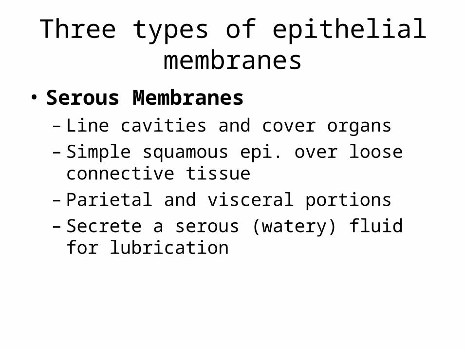
Three types of epithelial membranes
• Serous Membranes– Line cavities and cover organs– Simple squamous epi. over loose connective
tissue– Parietal and visceral portions– Secrete a serous (watery) fluid for lubrication
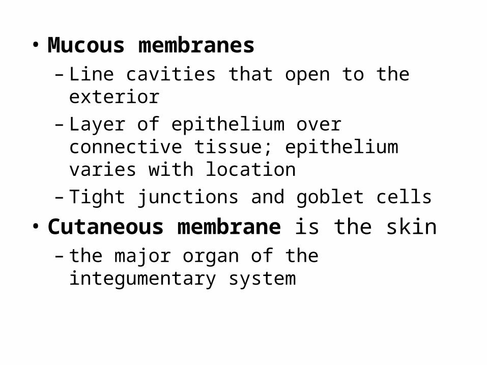
• Mucous membranes – Line cavities that open to the exterior– Layer of epithelium over connective tissue;
epithelium varies with location– Tight junctions and goblet cells
• Cutaneous membrane is the skin– the major organ of the integumentary system
• Integumentary system is the skin and the organs derived from it (hair, glands, nails)
• One of the largest organs– 2 square meters; 10-11 lbs.– Largest sense organ in the body
• The study of the skin is Dermatology

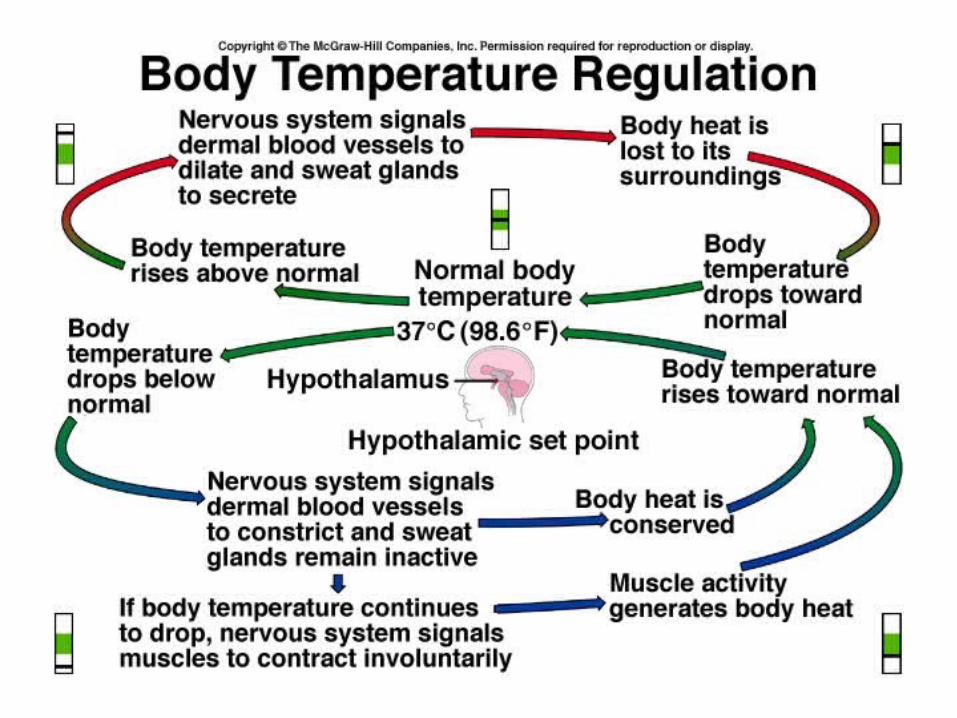
Functions:1. Regulation of body temperature
– Cellular metabolism produces heat as a waste product .
– High temperature• Dilate surface blood vessels• Sweating
– Low temperature• Surface vessels constrict• shivering
2. Protection
physical abrasion
dehydration
ultraviolet radiation
3. Sensation
touch
vibration
pain
temperature

4. Excretion
5. Immunity/ Resistance
6. Blood Reservoir
8-10 % in a resting adult
7. Synthesis of vitamin Duv light
aids absorption of calcium

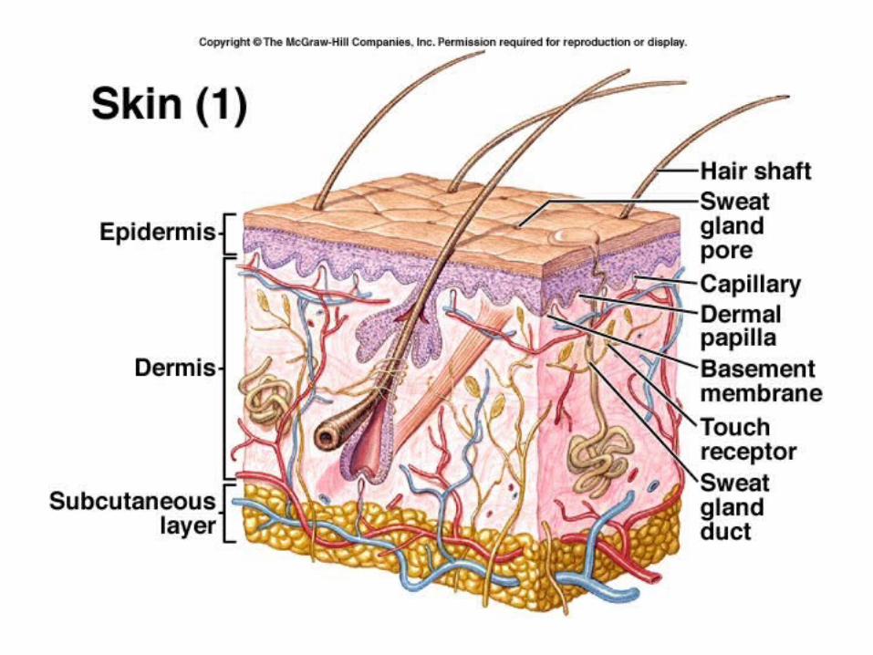
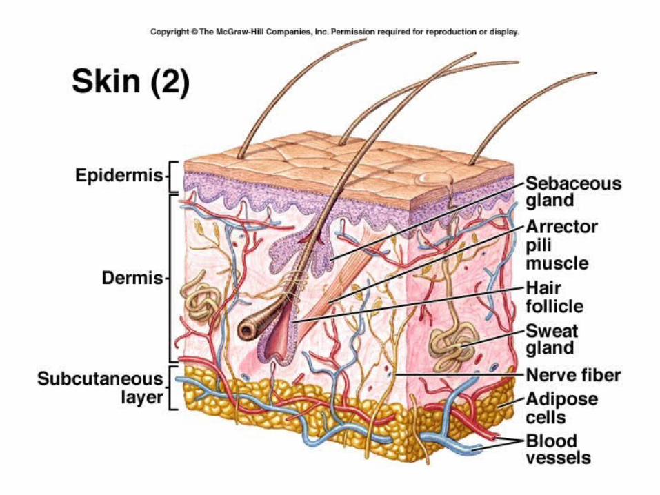
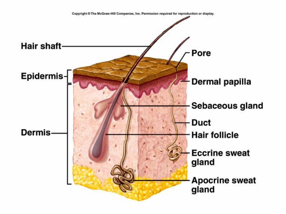
Anatomy
• Epidermis Skin
• Dermis
• Subcutaneous layer or hypodermis


Epidermis
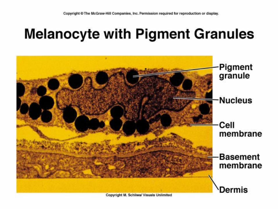
• Stratum basale (stratum germinativum)– Single layer of cuboidal to columnar cells– Stem cells that produce keratinocytes– Melanocytes - # the same for all races
• Melanin produced in a melanosome

• Stratum spinosum (thorn-like, prickly)– 8-10 layers attached by desmosomes– See spines when cell is stained for
microscopy– Keratinocytes take in melanin by cytocrine
secretion

• Stratum granulosum– 3-5 layers – Keratinization begins here– Keratohyalin found in granules– Cells beginning to die
• Stratum lucidum (lucid = clear)– More apparent in thick skin– 3-5 layers of clear cells– Eleidin
• Stratum corneum (corneum means horny)– Dead, flat cells full of keratin– Keratin is waterproof– Cells are shed
• Basal cell to surface – about 2-4 weeks
Dermis• Connective tissue layer
• Collagen and elastic fibers, nerves, blood vessels, muscle fibers, adipose cells, hair follicles and glands.
• Papillary layer – 1/5 of dermis – loose areolar connective
tissue– Highly vascular– Dermal papillae - fingerprints
• Reticular (net) layer– Dense irregular connective tissue– Sebaceous (oil) glands– Hair follicles– Ducts of sudoriferous (sweat) glands– Striae or stretch marks– Meissner’s corpuscles and Pacinian
corpuscles
Hypodermis
• Attaches the reticular layer to the underlying organs
• Loose connective tissue and adipose tissue
• Major blood vessels – rete cutaneum

Accessory organs or epidermal derivatives
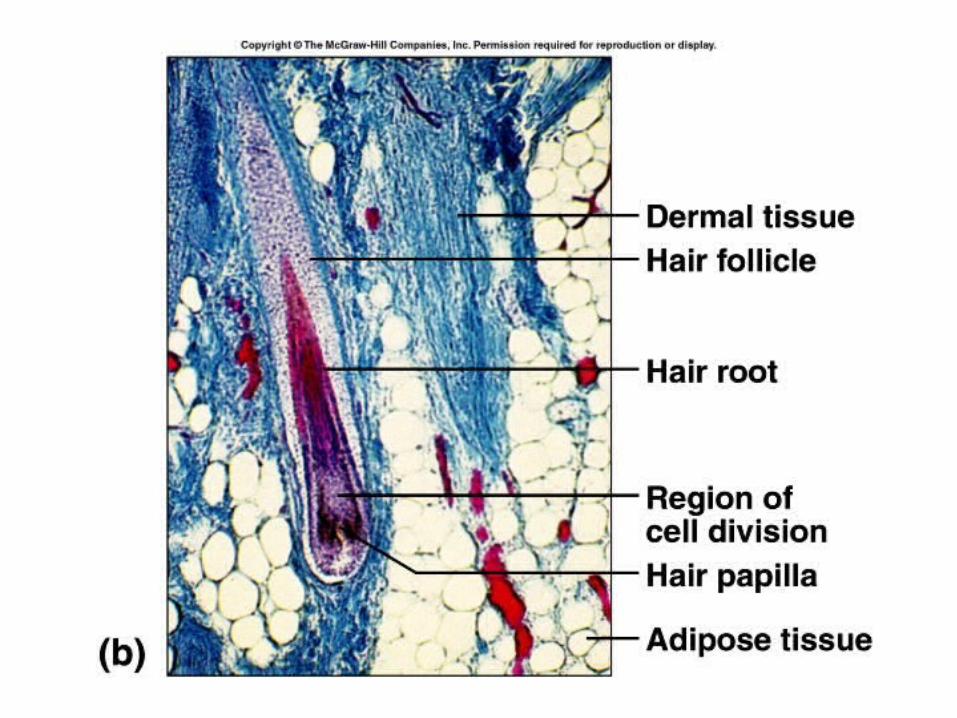
• Hairs– Epidermal growths that function in protection– Shaft, root, and folllicle– Sebaceous glands, arrector pili muscle, and
hair root plexus (touch)– Hair growth and replacement have a cyclical
pattern– ‘male-pattern’ baldness


Nails
• Plates of highly packed, keratinized cells
• Protection, scratching, & manipulation
• Formed by cells in nail bed called the matrix ( in area of lunula)
• 1 mm / week
• Eponychium - cuticle


Skin Glands
• Sebaceous (oil) glands– Usually connected to hair follicles– Holocrine glands– Fats, cholesterol, proteins, salts, and cell
debris– Moistens hair and waterproofs skin

• Sweat (sudoriferous) glands– Eccrine sweat glands
• Merocrine glands• Water, salt, wastes• Function is to cool the body (also nervous)
– Apocrine sweat glands• Larger, merocrine glands• Associated with hair follicles• More viscous – fatty acids and proteins• Odor occurs when broken down by bacteria

• Ceruminous glands– Modified sudoriferous glands – Secrete cerumen (ear wax)
• Mammary glands– Secrete milk


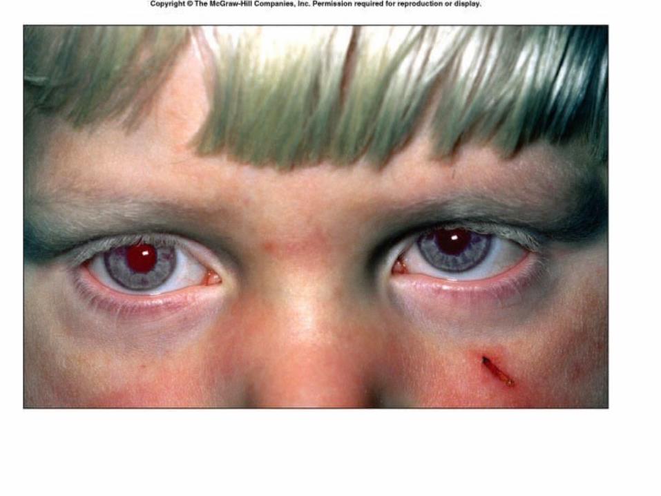
Skin color
• Genetic factors– Same number of melanocytes– Albinism
• Environmental factors– Uv light or x-rays

• Physiological factors– Amount of blood– Amount of oxygen
• Cyanosis• Carotene accumulation• Jaundice – liver disorder
Wound healing
• Inflammation– Blood vessels dilate and become permeable
• Heat, redness, swelling and pain
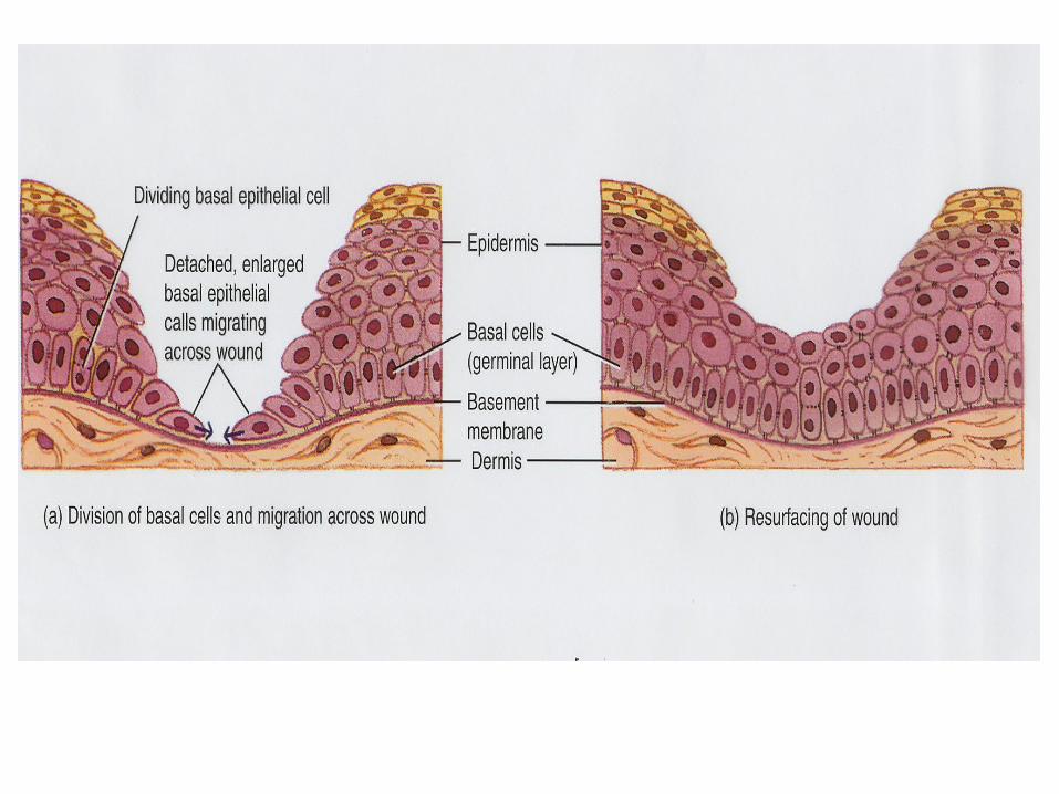
• Shallow cuts– Epithelial cells migrate– Contact inhibition
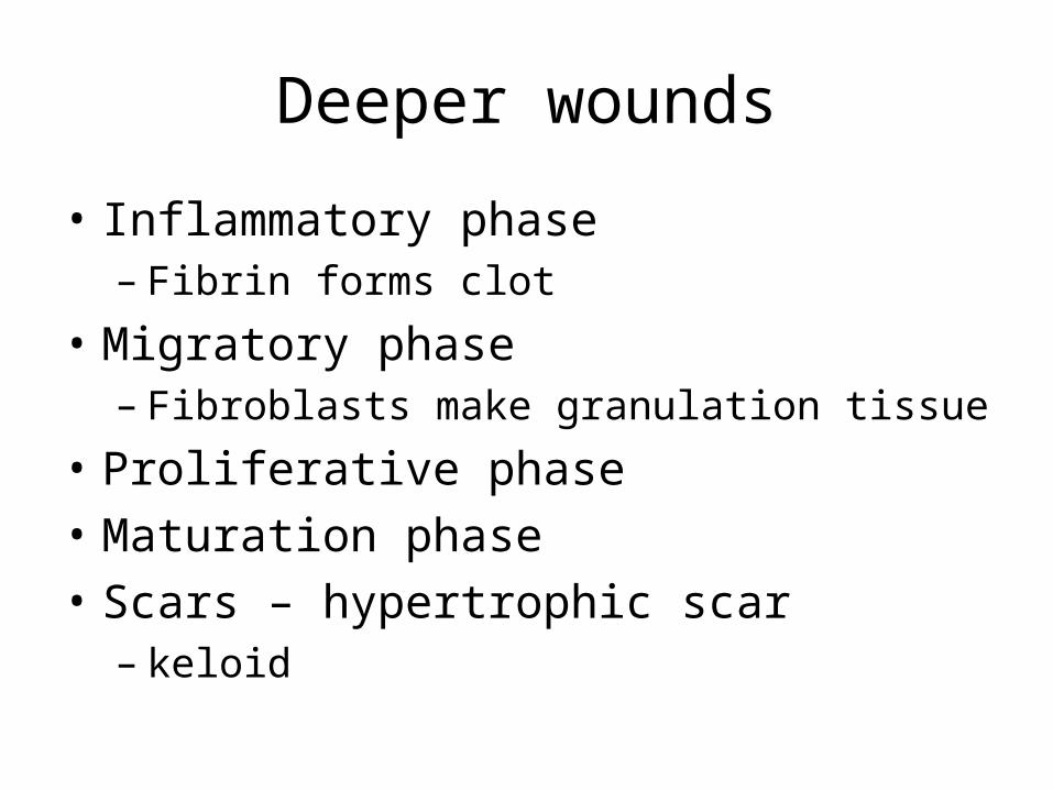
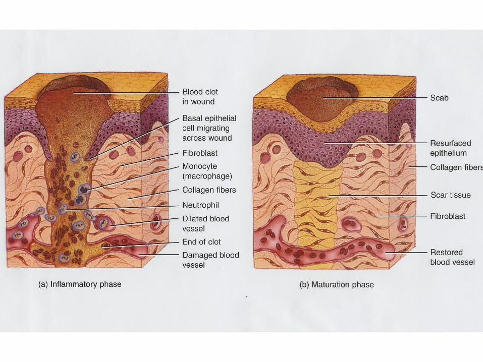
Deeper wounds
• Inflammatory phase– Fibrin forms clot
• Migratory phase– Fibroblasts make granulation tissue
• Proliferative phase
• Maturation phase
• Scars – hypertrophic scar – keloid


Burns
• First degree or partial thickness burn– Only epidermis is damaged– Erythema, mild edema, surface layer shed– Healing – a few days to two weeks– No scarring

• Second degree- deep partial-layer burn– Destroys epidermis– Blisters form – Healing depends on survival of accessory
organs– No scars unless infected
• Third degree or full-thickness burn– Destroys epidermis, dermis and accessory
organs of the skin– Healing occurs from margins inward– Skin grafting may be needed
• Autograft• Homograft
• Rule of Nines