The integrated response of the human cerebro-cerebellar and limbic
18
The integrated response of the human cerebro-cerebellar and limbic systems to acupuncture stimulation at ST 36 as evidenced by fMRI Kathleen K.S. Hui, a, * Jing Liu, a Ovidiu Marina, a Vitaly Napadow, a Christian Haselgrove, b Kenneth K. Kwong, a David N. Kennedy, b and Nikos Makris b a Athinoula A. Martinos Center for Biomedical Imaging, Department of Radiology, Massachusetts General Hospital and Harvard Medical School, Building 149, 13th Street, Room 2301, Charlestown, MA 02129, USA b Center for Morphometric Analysis, Department of Neurology, Massachusetts General Hospital and Harvard Medical School, Charlestown, MA 02129, USA Received 12 June 2004; revised 26 April 2005; accepted 28 April 2005 Available online 19 July 2005 Clinical and experimental data indicate that most acupuncture clinical results are mediated by the central nervous system, but the specific effects of acupuncture on the human brain remain unclear. Even less is known about its effects on the cerebellum. This fMRI study demonstrated that manual acupuncture at ST 36 (Stomach 36, Zusanli ), a main acupoint on the leg, modulated neural activity at multiple levels of the cerebro-cerebellar and limbic systems. The pattern of hemodynamic response depended on the psychophysical response to needle manipulation. Acupuncture stimulation typically elicited a composite of sensations termed deqi that is related to clinical efficacy according to traditional Chinese medicine. The limbic and paralimbic structures of cortical and subcortical regions in the telencephalon, diencephalon, brainstem and cerebellum demonstrated a concerted attenuation of signal intensity when the subjects experi- enced deqi . When deqi was mixed with sharp pain, the hemodynamic response was mixed, showing a predominance of signal increases instead. Tactile stimulation as control also elicited a predominance of signal increase in a subset of these regions. The study provides preliminary evidence for an integrated response of the human cerebro- cerebellar and limbic systems to acupuncture stimulation at ST 36 that correlates with the psychophysical response. D 2005 Elsevier Inc. All rights reserved. Keywords: Acupuncture; Negative BOLD; Limbic; Cerebrum; Cerebellum; Sensations; Pain; Affect; Cognition Introduction Acupuncture, an ancient healing technique that originated in China, is gaining popularity in the Western medical community for the treatment of diverse disorders (NIH, 1998). Traditional acupuncture involves stimulation with very fine needles inserted into defined sites on the body, eliciting a composite of sensations, termed deqi , which is considered to be related to clinical efficacy in traditional Chinese medicine (Cheng, 1997; Helms, 1995; Vincent et al., 1989). In clinical practice, different acupuncture points can be used to treat the same disorder, whereas one acupuncture point can be used to treat different disorders (Cheng, 1997; Stux and Hammerschlag, 2001). A body of clinical and experimental evidence indicates that most acupuncture effects are mediated via the brain (Han et al., 1982; Stux and Hammerschlag, 2001). However, the central effects of acupuncture and the neural correlates of deqi remain unclear. Modern neuroimaging has provided revolutionary tools to monitor the dynamic response of the whole brain to acupuncture with specific regional localization. Functional MRI and PET studies on acupuncture at commonly used acupuncture points have demonstrated significant modulatory effects on the limbic system, paralimbic and subcortical gray structures (Hsieh et al., 2001; Hui et al., 1997, 2000; Kong et al., 2002; Napadow et al., 2005; Wu et al., 1999, 2002; Zhang et al., 2003). The role of the cerebellum in autonomic control, cognition and affect, as well as in sensorimotor control is gaining increasing attention (Schmahmann, 1991, 2000a; Schmahmann and Sherman, 1998). Clinical and experimental data indicate that direct and indirect links connect the cerebellar vermis and deep nuclei with subcortical and cortical limbic/paralimbic regions in the cerebrum and brainstem, including the amygdala, hippocampus, septal nuclei, cingulate gyrus, hypothalamus, nucleus accumbens, prefrontal cortex, ventral tegmental area and reticular formation (Heath et al., 1978; Helmchen et al., 2003; Kelly and Strick, 2003; Leiner et al., 1989; Leroi et al., 2002; Paradiso et al., 1999; Price, 2000; Schmahmann and Pandya, 1997). Investigators are beginning to explore the effects of acupuncture on the cerebellum by fMRI (Hui et al., 2003; Yoo et al., 2004). Now that methods for specific 1053-8119/$ - see front matter D 2005 Elsevier Inc. All rights reserved. doi:10.1016/j.neuroimage.2005.04.037 * Corresponding author. Fax: +1 617 726 7422. E-mail address: [email protected] (K.K.S. Hui). Available online on ScienceDirect (www.sciencedirect.com). www.elsevier.com/locate/ynimg NeuroImage 27 (2005) 479 – 496
Transcript of The integrated response of the human cerebro-cerebellar and limbic

www.elsevier.com/locate/ynimg
NeuroImage 27 (2005) 479 – 496
The integrated response of the human cerebro-cerebellar and
limbic systems to acupuncture stimulation at ST 36 as
evidenced by fMRI
Kathleen K.S. Hui,a,* Jing Liu,a Ovidiu Marina,a Vitaly Napadow,a Christian Haselgrove,b
Kenneth K. Kwong,a David N. Kennedy,b and Nikos Makrisb
aAthinoula A. Martinos Center for Biomedical Imaging, Department of Radiology, Massachusetts General Hospital and Harvard Medical School,
Building 149, 13th Street, Room 2301, Charlestown, MA 02129, USAbCenter for Morphometric Analysis, Department of Neurology, Massachusetts General Hospital and Harvard Medical School,
Charlestown, MA 02129, USA
Received 12 June 2004; revised 26 April 2005; accepted 28 April 2005
Available online 19 July 2005
Clinical and experimental data indicate that most acupuncture clinical
results are mediated by the central nervous system, but the specific
effects of acupuncture on the human brain remain unclear. Even less is
known about its effects on the cerebellum. This fMRI study
demonstrated that manual acupuncture at ST 36 (Stomach 36,
Zusanli), a main acupoint on the leg, modulated neural activity at
multiple levels of the cerebro-cerebellar and limbic systems. The
pattern of hemodynamic response depended on the psychophysical
response to needle manipulation. Acupuncture stimulation typically
elicited a composite of sensations termed deqi that is related to clinical
efficacy according to traditional Chinese medicine. The limbic and
paralimbic structures of cortical and subcortical regions in the
telencephalon, diencephalon, brainstem and cerebellum demonstrated
a concerted attenuation of signal intensity when the subjects experi-
enced deqi. When deqi was mixed with sharp pain, the hemodynamic
response was mixed, showing a predominance of signal increases
instead. Tactile stimulation as control also elicited a predominance of
signal increase in a subset of these regions. The study provides
preliminary evidence for an integrated response of the human cerebro-
cerebellar and limbic systems to acupuncture stimulation at ST 36 that
correlates with the psychophysical response.
D 2005 Elsevier Inc. All rights reserved.
Keywords: Acupuncture; Negative BOLD; Limbic; Cerebrum; Cerebellum;
Sensations; Pain; Affect; Cognition
Introduction
Acupuncture, an ancient healing technique that originated in
China, is gaining popularity in the Western medical community for
the treatment of diverse disorders (NIH, 1998). Traditional
1053-8119/$ - see front matter D 2005 Elsevier Inc. All rights reserved.
doi:10.1016/j.neuroimage.2005.04.037
* Corresponding author. Fax: +1 617 726 7422.
E-mail address: [email protected] (K.K.S. Hui).
Available online on ScienceDirect (www.sciencedirect.com).
acupuncture involves stimulation with very fine needles inserted
into defined sites on the body, eliciting a composite of sensations,
termed deqi, which is considered to be related to clinical efficacy
in traditional Chinese medicine (Cheng, 1997; Helms, 1995;
Vincent et al., 1989). In clinical practice, different acupuncture
points can be used to treat the same disorder, whereas one
acupuncture point can be used to treat different disorders (Cheng,
1997; Stux and Hammerschlag, 2001). A body of clinical and
experimental evidence indicates that most acupuncture effects are
mediated via the brain (Han et al., 1982; Stux and Hammerschlag,
2001). However, the central effects of acupuncture and the neural
correlates of deqi remain unclear.
Modern neuroimaging has provided revolutionary tools to
monitor the dynamic response of the whole brain to acupuncture
with specific regional localization. Functional MRI and PET
studies on acupuncture at commonly used acupuncture points
have demonstrated significant modulatory effects on the limbic
system, paralimbic and subcortical gray structures (Hsieh et al.,
2001; Hui et al., 1997, 2000; Kong et al., 2002; Napadow et al.,
2005; Wu et al., 1999, 2002; Zhang et al., 2003). The role of the
cerebellum in autonomic control, cognition and affect, as well as in
sensorimotor control is gaining increasing attention (Schmahmann,
1991, 2000a; Schmahmann and Sherman, 1998). Clinical and
experimental data indicate that direct and indirect links connect the
cerebellar vermis and deep nuclei with subcortical and cortical
limbic/paralimbic regions in the cerebrum and brainstem, including
the amygdala, hippocampus, septal nuclei, cingulate gyrus,
hypothalamus, nucleus accumbens, prefrontal cortex, ventral
tegmental area and reticular formation (Heath et al., 1978;
Helmchen et al., 2003; Kelly and Strick, 2003; Leiner et al.,
1989; Leroi et al., 2002; Paradiso et al., 1999; Price, 2000;
Schmahmann and Pandya, 1997). Investigators are beginning to
explore the effects of acupuncture on the cerebellum by fMRI (Hui
et al., 2003; Yoo et al., 2004). Now that methods for specific

K.K.S. Hui et al. / NeuroImage 27 (2005) 479–496480
regional localizations of the cerebellum have become available
(Makris et al., 2003, 2005), whole brain imaging provides the
opportunity to explore the functional relationships and connectivity
between multiple levels of the human brain.
We hypothesize that the multifaceted effects of acupuncture
performed at main classical acupuncture points involve brain
circuits that can regulate and integrate diverse somatic and mental
functions in a coordinated manner, and that the limbic system may
play a central role. We further postulate that the limbic and
paralimbic structures in the cerebro-cerebellar system respond in a
concerted manner to acupuncture stimulation, and that the central
effects as evidenced by fMRI and psychophysical response are
interrelated. Our previous fMRI data collected during acupuncture
at classical acupuncture points, LI 4 (large intestine 4 or Hegu) on
the hand and ST 36 (stomach 36 or Zusanli) on the lower leg,
demonstrated a predominance of signal attenuation, particularly in
the limbic/paralimbic regions of the telencephalon, diencephalon
and brainstem (Hui et al., 1997, 2000, 2003; Napadow et al.,
2005). In this study acupuncture was performed at ST 36, a main
acupoint that is used for treating a variety of medical conditions.
The cerebellum was examined for region-specific and quantified
effects of acupuncture, using the new parcellation method
introduced by one of the authors (Makris et al., 2003, 2005).
The results demonstrated an integrated response of the human
cerebro-cerebellar and limbic systems.
Fig. 1. Acupuncture was performed at acupuncture point ST 36 on the right
leg (arrowhead). The acupuncture needle was inserted and the sensitivity of
the subject to manipulation was pre-tested and adjusted to tolerance prior to
each scanning run. After remaining at rest for 2 min, the needle was rotated
bidirectionally with even motion at the rate of 1 Hz for 2 min. After a rest
period of 3 min, needle manipulation was repeated in like manner. The
needle was removed at the end of the 10-min experimental run. The signal
intensities with the needle at rest before, between and after manipulation
served as the baseline for comparison with the signal intensities during
manipulation.
Materials and methods
Participants
The study was performed on 15 right-handed, acupuncture-
naı̈ve healthy adult volunteers with informed consent, as approved
by the Massachusetts General Hospital Subcommittee on Human
Studies. Subjects were screened and excluded for neurological,
mental and medical disorders, intake of conventional or alternative
medicine, head trauma with loss of consciousness and contra-
indications for exposure to high magnetic fields. Eight male and
seven female subjects, 22–47 years old (29.8 T 7.5 years), 1 Asian,2 Hispanic and 12 white, participated in the study. They were
acupuncture naı̈ve and were limited to participation in a single
session of the study.
Experimental paradigm
The investigation was conducted at the Athinoula A. Martinos
Center for Biomedical Imaging. The present report is derived from
data of a larger study that required approximately 2 h for data
acquisition. Anatomical scans of the brain and functional images of
sensory control stimulation were collected prior to acupuncture
imaging. The subjects were told acupuncture was to be performed
at different sites with different techniques that would generate
different sensations during the needling. They were not informed
of the order in which the sensory and the acupuncture stimulations
would be performed. They were instructed to lie still and keep their
eyes closed during the 10-min scan. At the end of each 10-min
scan, the subjects were questioned about the sensations that they
had felt during the stimulation and whether they were anxious or
relaxed during the procedure. They could not see their lower
extremities from their supine position in the enclosed scanner; and
being acupuncture-naı̈ve, they would be unable to discriminate the
tactile stimulation control from real acupuncture until they
experienced real acupuncture. Acupuncture was performed using
sterile disposable stainless steel needles at three acupuncture points
on the right extremity in separate runs: ST 36 on the leg, LI 4 on
the hand and LV 3 (Liver 3 or Taichong) on the foot. The order of
the acupoints used for stimulation was randomized. Only the
results of ST 36 from this cohort of subjects will be presented in
this report. Analysis on data from other acupoints is in progress.
The acupuncture point ST 36 is located in the tibialis anterior
muscle, 4 fingerbreadths below the kneecap and 1 fingerbreadth
lateral from the anterior crest of the tibia. Disposable sterile
stainless steel needles (KINGLI Medical Appliance Co., Ltd.,
Wuxi, China) of 0.22 mm in diameter and 40 mm in length were
used. The needle was inserted vertically to a depth of 2–3 cm. The
sensitivity of the subject to needle manipulation was tested and
adjusted to tolerance prior to scanning. In the event of a sharp
painful sensation, the needle position would be readjusted and the
pain would disappear within a few seconds. Stimulation consisted
of rotating the needle bidirectionally with an even motion to an
amplitude of approximately 180- at the rate of one cycle per
second. This approximates a technique used in clinical practice.
The total scanning time was 10 min per run. The needle was kept
in place for 2 min prior to needle manipulation. The two
stimulation blocks S1 and S2 were separated by an interval of 3
min with needle kept in place. Scanning continued for 1 min after
S2 (Fig. 1). Duplicate runs were performed at each acupuncture
point.
Sensory control stimulation consisted of tapping the skin gently
at the acupuncture point with a size 5.88 von Frey monofilament
using a paradigm matched to that of the acupuncture runs for the
subject. Due to time limitation, only one of the 3 acupoints to be
compared could be studied on a single subject in one session.
Imaging
Brain imaging was conducted on a 1.5-T Siemens Sonata MRI
system equipped for echo planar imaging (EPI) with a standard
head coil. Functional scans were collected with sagittal sections
parallel to the AC-PC plane, slice thickness 3.0 mm with 20% gap.
Imaging encompassed the entire brain, including the cerebellum
and brainstem. The functional data were acquired by a T2*-

K.K.S. Hui et al. / NeuroImage 27 (2005) 479–496 481
weighted gradient echo sequence (TE 30 ms, TR 4 s, matrix 64 �64, FOV 200 mm, flip angle 90-, in-plane resolution 3.125 �3.125 mm). A set of 3D MPRAGE (magnetization-prepared rapid
acquisition gradient echo) images, voxel size 1 mm3, 128 images
per set, and a set of T1-weighted high-resolution structural images
(TE 3.39 ms, TR 2.73 s, matrix 192 � 256, FOV 256 mm, flip
angle 7-, in-plane resolution 1 � 1 mm, slice thickness 1.33 mm)
were acquired prior to functional scans.
Psychophysical sensations
At the end of the 10-min scan, the subject was questioned
whether aching, pressure, soreness, heaviness, fullness, warmth,
coolness, numbness, tingling, dull pain, sharp pain or other
sensations had occurred during the stimulations, and, if present, to
rate the intensity of each sensation on a scale of 1 to 10 (0 = no
sensation, 1–3 = mild, 4–6 = moderate, 7–8 = strong, 9 = severe
and 10 = unbearable sensation). In order to minimize bias, the
subject was told prior to the test procedures that the type and
intensity of sensations would vary with different individuals and
different acupoints, and that a range from nil to multiple
sensations could be experienced for any given run. Although
the subject was to refrain from motion and to remain relaxed
during the scan, they were asked to signal to the acupuncturist by
raising one finger if deqi reached a score of 8 and raising 2
fingers in the event of sharp pain. When so signaled, the
acupuncturist would adjust the intensity of stimulation by
reducing the angle of rotation of the needle. In general, the
undue discomfort would disappear almost immediately, well
within the TR interval of 4 s.
Psychophysical data analysis
Deqi, or needling sensation, is the constellation of sensations a
person feels during acupuncture needle manipulation, such as
aching, pressure, soreness, fullness, distension, numbness, tingling,
local warm or cool sensations, pain and the spreading of these
sensations (Cheng, 1997; Park et al., 2002; Pomeranz, 1991;
Vincent et al., 1989). The needling technique adopted in this study
was gentle; the aim was to generate deqi with little or no sharp
pain. Whereas dull pain was considered as a component of deqi,
sharp pain was considered as inadvertent noxious stimulation. The
data sets were categorized for analysis according to the sensations
reported as follows: (1) deqi and (2) deqi plus sharp pain (mixed
sensations). In this cohort, none of the subjects experienced sharp
pain without deqi.
fMRI data analysis
The signal intensities with the needle at rest before, between
and after the needle manipulation periods served as the baseline for
the assessment of changes in signal intensity induced by needle
manipulation. Images were processed using the AFNI software
program (Cox, 1996). The data were first motion corrected; data
runs were excluded if gross motion exceeded 2 mm on any axis.
Statistical parametric mapping was completed via a generalized
linear model. The estimated response function was then com-
pared by a t test to the time series data in each brain voxel
(3dDeconvolve, AFNI). The spatial extent of our EPI images was
checked to ensure that the data from susceptibility-sensitive
regions were not corrupted by susceptibility-induced signal drop-
off. The statistics were color-coded and mapped onto the subject’s
own high-resolution 3D anatomical data set in Talairach space
(Talairach and Tournoux, 1988). No spatial or temporal pre-
processing smoothing was done on the data.
In individual analysis, data from duplicate runs for acupuncture
or for sensory control at an acupuncture point, if available, were
averaged. The data were then transformed into Talairach space,
normalized to average image intensity and blurred with a spatial
Gaussian filter (full-width half-max of 2 mm) to compensate for
any residual differences. The level of significance was thresholded
at P < 0.003 (t > 3.02) and a minimum cluster size of 3 voxels. The
time course of signal change was visually compared with the
experimental paradigm. Signal changes that failed to agree with the
paradigm were rejected as artifacts. The percent of signal change
was taken from the voxel with maximal change for each structure.
When signal increases and signal decreases coexisted in the same
structure, the predominant pattern was selected. In the absence of
predominance of any one pattern, both were listed. In order to
address the multiple comparison correction, a Monte Carlo
simulation was completed, the results of which demonstrated that
our combination of clustering and thresholding produced a false-
positive discovery rate, a, of less than 0.6% (AlphaSim, AFNI).
In group analysis, group-averaged functional statistical maps
were registered onto the group-averaged high-resolution anato-
mical maps of the subjects. Statistical significance was thresholded
at P < 0.001 (t > 3.37) and a minimum cluster size of 3 voxels,
with a less than 0.4% (AlphaSim, AFNI).
The anatomical definitions of the regions of interest in the
cerebral circuits were based on methods previously published
through the Center for Morphometric Analysis, Department of
Neurology, MGH (Caviness et al., 1996; Filipek et al., 1994).
These have been applied and adapted in a number of studies
(Breiter et al., 1997; Hui et al., 2000). The anatomical localization
and labeling of the functional data were determined by both
Talairach coordinates and direct inspection based on these
definitions.
The hemodynamic response of the cerebellum was derived
together with the response of the rest of the brain using AFNI for
data analysis as previously described (Napadow et al., 2005). The
AFNI data were imported into FreeSurfer, and the functional
activations were mapped onto the exterior surface of the cerebellum
that was reconstructed from the anatomical images (Dale et al.,
1999; Fischl et al., 1999). Only signal changes that were located at
or were contiguous with the exterior surface of the cerebellum were
projected onto the cerebellar surface, as shown in Fig. 8. Signal
changes located at the interface between white and gray matter that
did not reach the exterior surface were not included. The regions of
interest in the cerebellum were localized using cross sections in 3D
Talairach space and by the surface-assisted cortical parcellation
method (Makris et al., 2003, 2005). The anatomical definitions of
the regions of interest were based on this parcellation method and
on the MRI Atlas of the Human Cerebellum (Schmahmann et al.,
2000).
Results
Psychophysical response
Among the 15 subjects in the study, 11 experienced deqi
(designated as deqi) and 4 experienced deqi mixed with sharp

Fig. 2. Subjects were categorized based on whether they reported the
sensations listed for deqi but not sharp pain (deqi), or whether they reported
sharp pain as well as deqi sensations (mixed sensations). (A) The percentage
of subjects who reported having felt the given sensation, with binomial error
bars. The most frequently reported sensation was pressure, followed by
aching and numbness. (B) The average score, on a scale from 0 denoting no
sensation to 10 denoting an unbearable sensation, for each sensation, with
standard error bars. Spreading of any sensation was noted in a binary fashion
and coded as follows: 1—spreading reported; 0—spreading not reported.
K.K.S. Hui et al. / NeuroImage 27 (2005) 479–496482
pain (designated as mixed sensations). In subjects with mixed
sensations, deqi remained predominant. The prevalence of these
sensations is expressed as the percentage of individuals in the
Fig. 3. The influence of subjective sensations on fMRI signal changes of the brain
functional results were thresholded at P <0.001, cluster size of at least 3 voxels. A
space. Functional statistical maps (P values) were overlaid on the respective avera
increases. Regions: 1—frontal pole; 2—subgenual anterior cingulate, Brodmann a
posterior cingulate; 6—reticular formation; 7—cerebellar vermis (detailed in Fi
Acupuncture with deqi sensations (N = 11) but without sharp pain resulted in wide
cortex, hypothalamus, reticular formation and the cerebellar vermis. (Center) Ac
increases in several areas, including the frontal pole and the anterior, middle and po
in the frontal pole, posterior cingulate, thalamus and cerebellar vermis.
group that reported the given sensation (Fig. 2A). The intensity is
expressed as the average score T SE (Fig. 2B). For the deqi group,
the sensations in the order of decreasing frequency were as
follows: pressure (82%), aching, heaviness, fullness, numbness
and tingling (45–55%), dull pain, soreness, and warm or cool
feeling around the needling site (27%). With mixed sensations,
pressure also led the list (75%), followed by warm sensation
(50%), and soreness, numbness and tingling. Numbness and
tingling tended to be less common and weaker in mixed
sensations than in deqi (scores of 0.25 vs. 2.75 for numbness
and 0.25 vs. 1.63 for tingling). The sharp pain was of moderate
intensity, lasting at most a few seconds, and not more than 3 times
during each 2-min needle manipulation period. On the other hand,
the dull pain associated with deqi often persisted throughout the
needling manipulation and frequently occurred in the absence of
sharp pain. Importantly, dull pain did not activate the structures
related to noxious stimulation but caused deactivation as seen
with deqi. Interview records showed that the subjects remained
relaxed during the procedures, except when a procedure inadver-
tently produced sharp pain. They adapted well to the novel
environment although it was not relaxing as in acupuncture clinic
settings.
Hemodynamic response
Acupuncture stimulation produced distinct patterns of
hemodynamic response that differed between different catego-
ries of psychophysical response. As illustrated in parasagittal
sections in Fig. 3, extensive signal decreases occurred in the
cerebrum, brainstem and cerebellum in the deqi group. When
deqi was mixed with pain, the hemodynamic response also
became mixed, with signal increase becoming the predominant
pattern. In the sensory control, signal changes were sparse.
The signal change was also predominantly positive, except for
the ventro-medial prefrontal cortex that showed focal negative
activation. Interestingly, the secondary somatosensory cortex
(SII) demonstrated stronger activation in tactile stimulation
than in acupuncture needle stimulation with deqi (Fig. 4 and
Table 1).
during acupuncture and sensory control performed at ST 36. Group average
ll slices were taken sagittally at 2 mm to the right of the midline in Talairach
ge anatomical scans. Blue denotes signal decreases, and yellow to red signal
rea 24; 3—ventromedial prefrontal (VMPF) cortex; 4—hypothalamus; 5—
g. 6); 8—middle cingulate, Brodmann area 32; and 9—thalamus. (Left)
spread signal decreases, including the frontal pole, VMPF cortex, cingulate
upuncture with deqi and sharp pain sensations (N = 4) resulted in signal
sterior cingulate. (Right) Sensory control (N = 5) resulted in signal increases
Deqi + Pain Sensory
Y=-2mm
Y=-45mm
Y=-14mm
Y=-17mm
DeqiAcupuncture:
Am
ygd
ala
Hip
po
cam
pu
s &
SII
SII
Cer
ebel
lum
A
B
C
D
R L
10-4 10-4 10-810-8 p
(N=11) (N=4) (N=5)
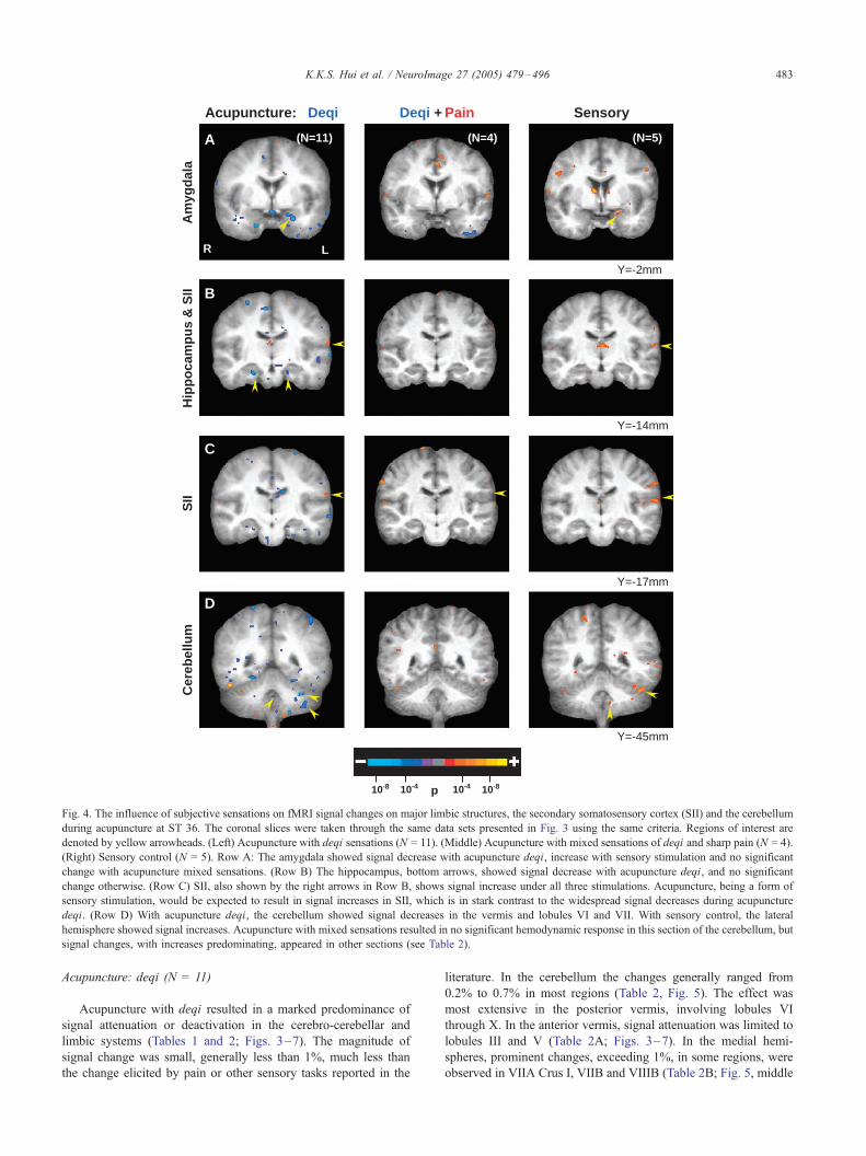
Fig. 4. The influence of subjective sensations on fMRI signal changes on major limbic structures, the secondary somatosensory cortex (SII) and the cerebellum
during acupuncture at ST 36. The coronal slices were taken through the same data sets presented in Fig. 3 using the same criteria. Regions of interest are
denoted by yellow arrowheads. (Left) Acupuncture with deqi sensations (N = 11). (Middle) Acupuncture with mixed sensations of deqi and sharp pain (N = 4).
(Right) Sensory control (N = 5). Row A: The amygdala showed signal decrease with acupuncture deqi, increase with sensory stimulation and no significant
change with acupuncture mixed sensations. (Row B) The hippocampus, bottom arrows, showed signal decrease with acupuncture deqi, and no significant
change otherwise. (Row C) SII, also shown by the right arrows in Row B, shows signal increase under all three stimulations. Acupuncture, being a form of
sensory stimulation, would be expected to result in signal increases in SII, which is in stark contrast to the widespread signal decreases during acupuncture
deqi. (Row D) With acupuncture deqi, the cerebellum showed signal decreases in the vermis and lobules VI and VII. With sensory control, the lateral
hemisphere showed signal increases. Acupuncture with mixed sensations resulted in no significant hemodynamic response in this section of the cerebellum, but
signal changes, with increases predominating, appeared in other sections (see Table 2).
K.K.S. Hui et al. / NeuroImage 27 (2005) 479–496 483
Acupuncture: deqi (N = 11)
Acupuncture with deqi resulted in a marked predominance of
signal attenuation or deactivation in the cerebro-cerebellar and
limbic systems (Tables 1 and 2; Figs. 3–7). The magnitude of
signal change was small, generally less than 1%, much less than
the change elicited by pain or other sensory tasks reported in the
literature. In the cerebellum the changes generally ranged from
0.2% to 0.7% in most regions (Table 2, Fig. 5). The effect was
most extensive in the posterior vermis, involving lobules VI
through X. In the anterior vermis, signal attenuation was limited to
lobules III and V (Table 2A; Figs. 3–7). In the medial hemi-
spheres, prominent changes, exceeding 1%, in some regions, were
observed in VIIA Crus I, VIIB and VIIIB (Table 2B; Fig. 5, middle

Table 1
FMRI signal changes in the cerebrum and brainstem � Deqi vs. Deqi + pain
K.K.S. Hui et al. / NeuroImage 27 (2005) 479–496484
The influence of subjective sensations on fMRI signal changes in the cerebrum and brainstem during acupuncture and sensory control at right. ST 36: The voxel
with the maximal signal change was selected for each region. Group data were thresholded at P < 0.001 with a cluster of at least 3 voxels. Values for regions
showing signal decreases are shown in blue, and increases in red. Acupuncture with deqi (N = 11) elicited a predominant pattern of signal attenuation in both
cerebrum and brainstem, in stark contrast to signal enhancement when deqi was mixed with sharp pain. Notably, the subcortical and cortical limbic regions were
deactivated while the somatosensory cortices and the dorsal raphe nucleus in the brainstem were activated during acupuncture deqi. When deqi was mixed with
sharp pain (N = 4), the result was predominantly activation, but fewer regions were affected. Sensory control (N = 5) resulted in activation of fewer regions.
Abbreviations: BA—Brodmann area; SCC—subcallosal cortex; VMPF—ventromedial prefrontal cortex; and SII—secondary somatosensory cortex.
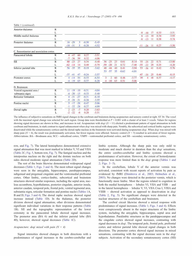
Table 1 (continued)
K.K.S. Hui et al. / NeuroImage 27 (2005) 479–496 485
row, and Fig. 7). The lateral hemispheres demonstrated extensive
signal attenuation that was most marked in lobules V, VI and VIIA
(Table 2C; Fig. 5, bottom row, Fig. 7). The fastigial nucleus and the
interpositus nucleus on the right and the dentate nucleus on both
sides showed moderate signal attenuation (Table 2D).
The rest of the brain likewise demonstrated widespread signal
decreases (Table 1; Figs. 3 and 4). The most robust signal changes
were seen in the amygdala, hippocampus, parahippocampus,
subgenual and pregenual cingulate and the ventromedial prefrontal
cortex. Other limbic, cortico-limbic, subcortical and brainstem
structures showed similar responses, including the septal area, nuc-
leus accumbens, hypothalamus, posterior cingulate, anterior insula,
anterior caudate, temporal pole, frontal pole, ventral tegmental area,
substantia nigra, reticular formation and pontine nuclei (Tables 1A,
B and D; Figs. 3 and 4). The dorsal raphe nucleus showed signal
increase instead (Table 1D). In the thalamus, the posterior
division showed signal attenuation; other divisions demonstrated
significant individual variations in their response patterns (Table
1B). SII and the topographic representation of the lower
extremity in the paracentral lobule showed signal increases.
The premotor area (BA 6) and the inferior parietal lobe (BA
40), however, showed signal decreases (Table 1C).
Acupuncture: deqi mixed with pain (N = 4)
Signal intensities showed changes in both directions with a
predominance of signal increases in the cerebro-cerebellar and
limbic systems. Although the sharp pain was only mild to
moderate and much shorter in duration than the deqi sensations,
the entire cerebro-cerebellar and limbic systems showed a
predominance of activation. However, the extent of hemodynamic
response was more limited than in the deqi group (Tables 1 and
2; Figs. 3–5).
In the cerebellum, lobule V of the anterior vermis was
activated, consistent with recent reports of activation by pain as
evidenced by fMRI (Dimitrova et al., 2003; Helmchen et al.,
2003). No changes were detected in the posterior vermis, which is
functionally more limbic. Most the regions related to cognition in
both the medial hemispheres – lobules VI, VIIA and VIIB – and
in the lateral hemispheres – lobules V, VI, VIIA Crus I, VIIIA and
VIIIB – showed activation as opposed to deactivation in deqi
(Table 2, Fig. 5). No significant changes were detected in the
nuclear structures of the cerebellum and brainstem.
The cerebral circuit likewise showed a mixed response with
predominance of signal increases. (Table 1; Figs. 3 and 4). Effects
were conspicuously absent in the major structures of the limbic
system, including the amygdala, hippocampus, septal area and
hypothalamus. Paralimbic structures as the parahippocampus and
the cingulate cortex showed signal increases, contrasting with
signal decreases in deqi. The temporal pole, frontal pole, prefrontal
cortex and inferior parietal lobe showed signal changes in both
directions. The premotor cortex showed signal increase in mixed
sensations, contrasting with the signal decrease seen in the deqi
subjects. Activation of the secondary somatosensory cortex (SII)

Table 2
FMRI signal changes in the cerebellum � Deqi vs. Deqi +pain
K.K.S. Hui et al. / NeuroImage 27 (2005) 479–496486

Quantified fMRI signal changes in the cerebellum, with comparisons, data sources and color coding the same as described for Table 1. The functions of
different regions of the cerebellum, as described in the literature, are listed on the right (Makris et al., 2003). The regions related to emotion and autonomic
control in the vermis and to cognition in the medial and lateral hemispheres showed significant changes in signal intensity. The predominant response of the
cerebellum correlated with those of the cerebrum and brainstem, in that signal attenuation was predominant with deqi and enhancement with sharp pain and
sensory control (see Table 1).
Table 2 (continued)
K.K.S. Hui et al. / NeuroImage 27 (2005) 479–496 487
was stronger than in the deqi group, but the signal change
remained under 1%, less than values reported in pain literature
(Table 1C). In concert with the stronger somatosensory cortex
activation in mixed sensations than in deqi, signal increase was
observed in a large subset of the cerebro-cerebellar and limbic
structures in mixed sensations instead of the signal decrease seen in
deqi (Hui et al., 2003).
Sensory control (N = 5)
Superficial tactile stimulation over the acupuncture point ST 36
also produced predominant signal enhancement in the cerebro-
cerebellar circuit. In the cerebellum signal increases were observed
in lobules VI and VIIA of the vermis (sensorimotor), lobules V–
IX of the medial hemispheres (sensory, cognition, balance) and
lobules V–VIIIB of the lateral hemispheres (cognition). The
response was mostly bilateral, being most prominent in VIIA Crus
I of the lateral hemispheres, with signal increase reaching 1.4%.
The dentate nucleus showed a slight signal increase on the left
(Table 2; Figs. 3–6). Interestingly, the ventromedial prefrontal
cortex demonstrated predominant signal attenuation. Changes
were conspicuously absent in other limbic regions such as the
amygdala, hippocampus, septal area and hypothalamus (Table 1;
Figs. 3–5).
Discussion
This is the first systematic study of the effects of traditional
Chinese acupuncture on the entire human cerebro-cerebellar
circuit that correlates the patterns of hemodynamic response
and psychophysical response with each other. The central effects
of acupuncture needle stimulation demonstrated in this study
differ markedly from those of conventional peripheral nerve
stimulation conducted on the tibial nerve of the leg or on the
median nerve of the hand that innervate the acupuncture points
ST 36 and LI 4, respectively. Studies using EEG, MEG and fMRI
reported that non-painful electrical or other sensory stimulation of
the tibial nerve in normal subjects activated the somatosensory,
motor, premotor, posterior parietal and cingulate cortices, the
thalamus and the cerebellum (Smith et al., 2003). The same
pattern of response has been described more extensively for the
median nerve at the wrist (Desmedt and Bourguet, 1985;
Korvenoja et al., 1999). The activation of the cingulate and
cerebellum by tactile stimulation (Smith et al., 2003) is in
agreement with our results on tactile control stimulation at the
acupoint. We are unaware of reports of deactivation by non-
acupuncture oriented stimulation.
Anatomical and electrophysiological studies of acupuncture
point structure and function show that both manual and electro-
acupuncture stimulate the same peripheral nerves as conventional
electrical stimulation. However, in marked contrast to the signal
enhancement in these non-acupuncture oriented studies, manual
acupuncture at ST 36 in the present study and at LI 4 in an earlier
study (Hui et al., 2000) elicited widespread and synchronized
signal decreases in the cerebro-cerebellar circuit, especially marked
in the limbic system. Except for conditions that require stronger
stimulation to achieve excitatory action, acupuncture at the main
points generally produces anti-stress and anti-anxiety effects
(Mann, 1992). These findings suggest that modulation of the
cerebro-cerebellar and limbic system activity may constitute an
important pathway of acupuncture action. Acupuncture action
involves the interplay between multiple neurotransmitters and
modulators. Correlation of the distribution and the known
functions of these mediators in the cerebro-cerebellar and limbic
systems with the hemodynamic response to acupuncture suggests
that the down-regulation of dopaminergic and norepinephrinergic
tone coupled with the up-regulation of the serotonergic tone during
the procedure may initiate a cascade of reactions that results in the
more delayed effects of acupuncture. Validation would require
investigation with methodologies that can probe into the funda-
mental processes underlying the BOLD response.

Fig. 5. Comparison of hemodynamic response of the cerebellum between different categories of psychophysical response. The magnitude of signal change in
the cerebellum for different stimulations at acupoint ST 36 as listed in Table 2. The known functionality of the different regions of the cerebellum are labeled
with a color code at the bottom of the chart. Acupuncture with deqi resulted in widespread signal decreases in the vermis and hemispheres. When deqi was
mixed with pain, response was more limited. A large subset of regions related to pain, cognition, affect and autonomic control were activated. Sensory control
demonstrated predominant signal increases, with minor signal decrease in the posterior medial hemisphere.
Fig. 6. Close-up of group average cerebellum from Fig. 3A, as described therein. The slices are taken sagittally at 2 mm to the right of the midline in Talairach
space. The time course shown in the lower left corner of each image is taken from the location denoted by the arrowhead in that image. The vertical axis of the
time course denotes percent signal change, and the horizontal axis the time within the experimental run, in seconds. Zero percent signal change is placed at the
average activity level for the three epochs with the needle at rest. The shaded columns represent the periods of needle manipulation. (Left) Acupuncture with
deqi sensations (N = 11) resulted in widespread signal decreases, including lobules VI, VIIB, VIIIA and B, IX and X. (Center) Acupuncture with deqi plus
sharp pain (N = 4) resulted in signal increase in lobules V, VI. (Right) Sensory control (N = 5) demonstrated signal increase in lobules VI, VIIA and VIIIB.
K.K.S. Hui et al. / NeuroImage 27 (2005) 479–496488
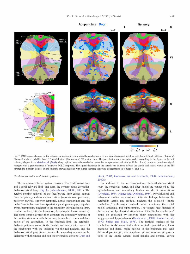
Fig. 7. fMRI signal changes on the exterior surface are overlaid onto the cerebellum overlaid onto its reconstructed surface, both 3D and flattened. (Top row)
Flattened surface. (Middle Row) 3D caudal view. (Bottom row) 3D rostral view. The parcellation units are color coded according to the figure in the left
column, adapted from Makris et al. (2003). Gray regions denote the cerebellar peduncles. Acupuncture with deqi (middle column) produced prominent signal
changes with a predominance of negative BOLD response. The signal decreases in the vermis can be seen in both the caudal and rostral views of the 3D
cerebellum. Sensory control (right column) showed regions with signal increase that were concentrated in lobules VI and VII.
K.K.S. Hui et al. / NeuroImage 27 (2005) 479–496 489
Cerebro-cerebellar and limbic systems
The cerebro-cerebellar system consists of a feedforward limb
and a feedbackward limb that form the cerebro-ponto-cerebellar-
thalamo-cortical loop (Fig. 8) (Schmahmann, 2000b, 2001). The
cerebro-pontine pathway of the feedforward limb carries outputs
from the primary and association cortices (sensorimotor, prefrontal,
posterior parietal, superior temporal, dorsal extrastriate) and the
limbic/paralimbic structures (posterior parahippocampus, cingulate
gyrus, mammillary nucleus) to the brainstem (periaqueductal gray,
pontine nucleus, reticular formation, dorsal raphe, locus caeruleus).
The ponto-cerebellar tract then connects the secondary neurons of
the pontine structures with the vermis, hemispheric zones and deep
nuclei of the cerebellum. In the feedback limb, the cerebello-
thalamic pathway connects the dentate and interpositus nuclei of
the cerebellum with the thalamus via the red nucleus, and the
thalamo-cortical projection connects the secondary neurons in the
thalamus with the motor and non-motor cerebral cortices (Dum and
Strick, 2003; Gonzalo-Ruiz and Leichnetz, 1990; Schmahmann,
2000a).
In addition to the cerebro-ponto-cerebellar-thalamo-cortical
loop, the cerebellar cortex and deep nuclei are connected to the
hypothalamus and mamillary bodies via direct connections
(Dietrichs, 1984; Haines and Dietrichs, 1984). Physiological and
behavioral studies demonstrated intimate linkage between the
cerebellar vermis and fastigial nucleus, the so-called Flimbic
cerebellum,_ with major cerebral limbic structures, the septal
nuclei, amygdala and hippocampus. The violent rage induced in
the cat and rat by electrical stimulation of the Flimbic cerebellum_could be abolished by severing their connections with the
amygdala and hypothalamus (Heath et al., 1978; Rasheed et al.,
1970; Snider and Maiti, 1976). The fastigial nucleus of the
cerebellum is also connected with he ventral tegmental area, locus
caeruleus and dorsal raphe nucleus in the brainstem that send
diffuse dopaminergic, norepinephrinergic and serotonergic projec-
tions to the limbic system, basal ganglia and cerebral cortex
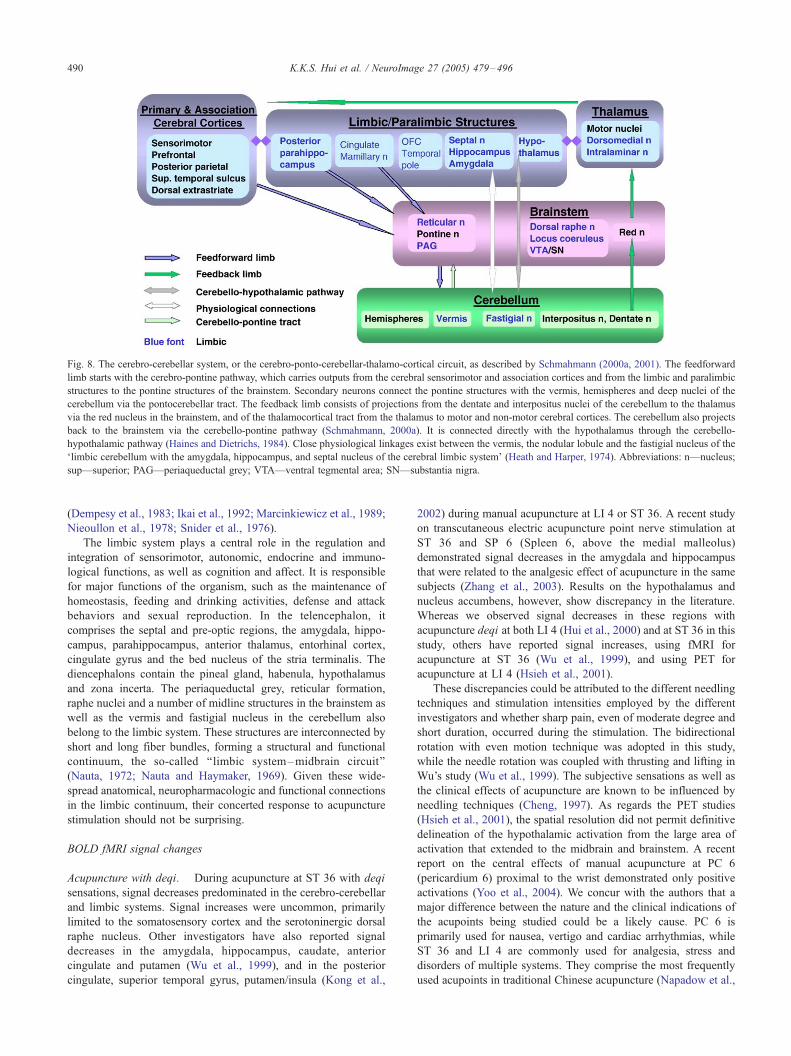
Fig. 8. The cerebro-cerebellar system, or the cerebro-ponto-cerebellar-thalamo-cortical circuit, as described by Schmahmann (2000a, 2001). The feedforward
limb starts with the cerebro-pontine pathway, which carries outputs from the cerebral sensorimotor and association cortices and from the limbic and paralimbic
structures to the pontine structures of the brainstem. Secondary neurons connect the pontine structures with the vermis, hemispheres and deep nuclei of the
cerebellum via the pontocerebellar tract. The feedback limb consists of projections from the dentate and interpositus nuclei of the cerebellum to the thalamus
via the red nucleus in the brainstem, and of the thalamocortical tract from the thalamus to motor and non-motor cerebral cortices. The cerebellum also projects
back to the brainstem via the cerebello-pontine pathway (Schmahmann, 2000a). It is connected directly with the hypothalamus through the cerebello-
hypothalamic pathway (Haines and Dietrichs, 1984). Close physiological linkages exist between the vermis, the nodular lobule and the fastigial nucleus of the
Flimbic cerebellum with the amygdala, hippocampus, and septal nucleus of the cerebral limbic system_ (Heath and Harper, 1974). Abbreviations: n—nucleus;
sup—superior; PAG—periaqueductal grey; VTA—ventral tegmental area; SN—substantia nigra.
K.K.S. Hui et al. / NeuroImage 27 (2005) 479–496490
(Dempesy et al., 1983; Ikai et al., 1992; Marcinkiewicz et al., 1989;
Nieoullon et al., 1978; Snider et al., 1976).
The limbic system plays a central role in the regulation and
integration of sensorimotor, autonomic, endocrine and immuno-
logical functions, as well as cognition and affect. It is responsible
for major functions of the organism, such as the maintenance of
homeostasis, feeding and drinking activities, defense and attack
behaviors and sexual reproduction. In the telencephalon, it
comprises the septal and pre-optic regions, the amygdala, hippo-
campus, parahippocampus, anterior thalamus, entorhinal cortex,
cingulate gyrus and the bed nucleus of the stria terminalis. The
diencephalons contain the pineal gland, habenula, hypothalamus
and zona incerta. The periaqueductal grey, reticular formation,
raphe nuclei and a number of midline structures in the brainstem as
well as the vermis and fastigial nucleus in the cerebellum also
belong to the limbic system. These structures are interconnected by
short and long fiber bundles, forming a structural and functional
continuum, the so-called ‘‘limbic system–midbrain circuit’’
(Nauta, 1972; Nauta and Haymaker, 1969). Given these wide-
spread anatomical, neuropharmacologic and functional connections
in the limbic continuum, their concerted response to acupuncture
stimulation should not be surprising.
BOLD fMRI signal changes
Acupuncture with deqi. During acupuncture at ST 36 with deqi
sensations, signal decreases predominated in the cerebro-cerebellar
and limbic systems. Signal increases were uncommon, primarily
limited to the somatosensory cortex and the serotoninergic dorsal
raphe nucleus. Other investigators have also reported signal
decreases in the amygdala, hippocampus, caudate, anterior
cingulate and putamen (Wu et al., 1999), and in the posterior
cingulate, superior temporal gyrus, putamen/insula (Kong et al.,
2002) during manual acupuncture at LI 4 or ST 36. A recent study
on transcutaneous electric acupuncture point nerve stimulation at
ST 36 and SP 6 (Spleen 6, above the medial malleolus)
demonstrated signal decreases in the amygdala and hippocampus
that were related to the analgesic effect of acupuncture in the same
subjects (Zhang et al., 2003). Results on the hypothalamus and
nucleus accumbens, however, show discrepancy in the literature.
Whereas we observed signal decreases in these regions with
acupuncture deqi at both LI 4 (Hui et al., 2000) and at ST 36 in this
study, others have reported signal increases, using fMRI for
acupuncture at ST 36 (Wu et al., 1999), and using PET for
acupuncture at LI 4 (Hsieh et al., 2001).
These discrepancies could be attributed to the different needling
techniques and stimulation intensities employed by the different
investigators and whether sharp pain, even of moderate degree and
short duration, occurred during the stimulation. The bidirectional
rotation with even motion technique was adopted in this study,
while the needle rotation was coupled with thrusting and lifting in
Wu’s study (Wu et al., 1999). The subjective sensations as well as
the clinical effects of acupuncture are known to be influenced by
needling techniques (Cheng, 1997). As regards the PET studies
(Hsieh et al., 2001), the spatial resolution did not permit definitive
delineation of the hypothalamic activation from the large area of
activation that extended to the midbrain and brainstem. A recent
report on the central effects of manual acupuncture at PC 6
(pericardium 6) proximal to the wrist demonstrated only positive
activations (Yoo et al., 2004). We concur with the authors that a
major difference between the nature and the clinical indications of
the acupoints being studied could be a likely cause. PC 6 is
primarily used for nausea, vertigo and cardiac arrhythmias, while
ST 36 and LI 4 are commonly used for analgesia, stress and
disorders of multiple systems. They comprise the most frequently
used acupoints in traditional Chinese acupuncture (Napadow et al.,
K.K.S. Hui et al. / NeuroImage 27 (2005) 479–496 491
2005). Moreover, PC 6 is located in tendinous structures just above
the wrist while LI 4 and ST 36 are located in muscles. PC 6 is more
superficial and more prone to cause overt pain. The distribution of
receptors and afferent neural fibers may differ with tissue type.
The down-regulation of limbic system activity by acupuncture
was demonstrated by electro-physiological studies in non-human
primates as early as 3 decades ago. Using chronically implanted
microelectrodes in the squirrel monkey, both manual and electro-
acupuncture at LI 4 and ST 36 decreased the cell firing rate and
increased the interspike interval in the septal area, amygdala and
anterior cingulate, while thalamic activity in the parafascicular and
ventral posteromedial nuclei remained unaffected. It was proposed
that the limbic response rather than the thalamic response could be
the neurophysiologic correlate of acupuncture analgesia (Jacobs et
al., 1977). However, most acupuncture research to date has been
devoted to acupuncture analgesia, using rats as animal models and
focusing on sensory perception and opioid peptides rather than on
the affective dimension of pain and the limbic system. Perhaps the
modulatory effects of acupuncture as evidenced by modern in vivo
neuroimaging in humans may serve to revive this important old
concept.
The magnitude of signal change observed in acupuncture deqi
was small, generally less than 1%, compared with the 2–4%
activation by visual stimulation or other sensory tasks reported in
the literature (Kwong et al., 1992). The smaller response suggests
that acupuncture, unlike noxious insults and pharmacological
agents, may act within physiological limits. This could explain in
part why acupuncture treatment generally causes fewer side effects
than medications, particularly potent analgesics.
Acupuncture with mixed sensations (deqi and pain). When
acupuncture deqi is mixed with sharp pain, the hemodynamic
response associated with the sensations may oppose or neutralize
each other. The net outcome may depend on which sensation
prevails. Structures that show signal attenuation in acupuncture
deqi such as the amygdala, hippocampus, cingulate cortex,
temporal pole, frontal pole are known to show signal enhancement
by pain and noxious insults (Becerra et al., 1999; Casey et al.,
1994; Coghill et al., 1994; Craig et al., 1996; Davis, 2000; Jones
et al., 1991; Price, 2000; Talbot et al., 1991). In this study, the
stronger activation of the somatosensory cortices in the mixed
sensations than in the deqi group can be attributed to the presence
of pain. The limbic cortices, including the temporal pole, frontal
pole, ventromedial prefrontal cortex, however, showed mixed
responses. Importantly hemodynamic response was conspicuously
absent in a large subset of the limbic structures, including the
amygdala, hippocampus, the subgenual, pregenual and posterior
cingulate cortex, the septal area and the anterior insula. The two
opposing effects, deactivation elicited by deqi and activation
elicited by pain, might have neutralized each other. An alternative
explanation could be the failure of acupuncture to deactivate these
limbic structures and suppress the pain. The predominant effect of
pain was manifest in the activation of the parahippocampus,
middle cingulate cortex (Table 1) and cerebellum (Table 2).
Recent fMRI studies on pain have demonstrated extensive
activation of the anterior vermis (Dimitrova et al., 2003;
Helmchen et al., 2003). Consistent with these reports, activation
was observed in lobule V of the anterior vermis in mixed
sensations, in contrast to deactivation of multiple anterior vermal
lobules in deqi. Signal increases were also widespread in the
hemispheric regions related to cognition. Overall, the results
suggest that in mixed sensations, the central effects of pain
prevailed, exhibiting an integrated response with predominance of
activation over deactivation in the cerebro-cerebellar and limbic
systems (Table 2, Fig. 5). The contrast in the direction of signal
change between different types of psychophysical response was
even more striking when sharp pain occurred in the absence of
deqi. In earlier studies when the sensitivity to needling was not
pre-tested prior to scanning, one subject experienced pain without
deqi during acupuncture at ST36. Multiple lobules of the anterior
vermis and hemispheric zones exhibited robust activation with
signal reaching 1–5% in the cerebro-cerebellar and limbic
systems (Hui et al., 2003).
Control stimulation. Tactile stimulation over the acupoint using
the same paradigm elicited a predominance of signal increase in
the entire brain, in marked contrast to signal decrease observed in
acupuncture deqi (Tables 1 and 2). The deactivation of the
paralimbic ventromedial prefrontal cortex and adjoining subge-
nual cingulate is interesting (Table 1A). It is reported that
attention and anticipatory anxiety could induce deactivation of the
ventro-medial prefrontal cortex (Gusnard et al., 2001; Simpson et
al., 2001a,b). The subjects were instructed to pay attention to the
sensations that they might experience during the stimulation so
that they could describe them during the interview at the end of
the scan. This could constitute an attention task and contribute to
the deactivation of these regions. Another possible explanation
could be the down-regulation of the brain activity from its default
state by the soothing rhythmic tactile stimulation (Raichle et al.,
2001). The activation of the dentate nucleus of the cerebellum by
tactile stimulation was in agreement with reports that the deep
cerebellar nuclei were activated by sensory stimulation in the
absence of any motor task (Gao et al., 1996). Activation of the
hemispheric zones of the cerebellum that participate in cognition
and emotional processing was also in agreement with an
increasing number of clinical and neuroimaging reports that
relate the cerebellum to thought and emotional control in health
and in disease (Ritvo et al., 1986; Schmahmann and Sherman,
1998; Townsend et al., 2001; Wallesch and Horn, 1990; Weis et
al., 2004).
Negative BOLD response. The signal intensity in BOLD fMRI is
the net result of the neuronal activity that determines oxygen
consumption and the hemodynamic response to the changes in O2
demand. Most fMRI studies are based on the detection of a positive
BOLD response. Here, we have demonstrated a robust negative
BOLD response to acupuncture that correlates with deqi sensa-
tions. The redistribution of blood to activated regions with a greater
demand, known as ‘‘blood stealing’’ or ‘‘physiological steal’’, has
been proposed for negative activations that are coupled to positive
responses in the proximity (Shmuel et al., 2001, 2002). However,
the absence of positive response in adjacent regions in acupuncture
rules out such an explanation for the negative response in this task.
Although hemodynamic changes independent of the local changes
in neuronal activity cannot be completely ruled out, we favor
reduction of neuronal activity due to a task-specific deactivation of
these brain regions. Specifically the task is acupuncture stimulation
that induces deqi sensation. This proposal is based on an increasing
number of reports that cognitive processes, emotion, attention and
goal-directed behaviors can induce functional deactivations as
measured by BOLD fMRI, PET and electrophysiological methods
(Ferris et al., 2004; Friston et al., 1991; Frith et al., 1991; Ghatan et
K.K.S. Hui et al. / NeuroImage 27 (2005) 479–496492
al., 1998; Goldapple et al., 2004; Hutchinson et al., 1999; Liotti et
al., 2000; Lustig et al., 2003; Raichle et al., 2001; Shulman et al.,
1997; Simpson et al., 2001a,b). Various causes have been
proposed, including regional inhibition (Frith et al., 1991), task-
specific inhibition (Ghatan et al., 1998), task-independent sup-
pression of tonic activity (Shulman et al., 1997), an overactive
default state (Lustig et al., 2003; Raichle et al., 2001) or intrinsic to
the activation state (Hutchinson et al., 1999). Regions in the medial
aspect of the brain, including the ventromedial prefrontal cortex,
cingulate cortex (subgenual and posterior divisions), the medial
parietal lobe and the amygdala, are prone to negative activation by
tasks that involve attention and cognition (Gusnard et al., 2001;
Lustig et al., 2003; Raichle et al., 2001; Simpson et al., 2001a,b).
These regions converge with the ones that demonstrate negative
activation in acupuncture.
Evidence is emerging that establishes the negative BOLD
response as a marker of neuronal deactivation rather than a local
phenomenon attributed to ‘‘blood stealing’’. A visual stimulus that
stimulated primary visual cortex in one hemisphere caused
extensive suppression in the other hemisphere (Shmuel et al.,
2003a,b; Smith et al., 2004). The BOLD response, cerebral blood
flow (CBF) and cerebral metabolic rate of oxygen consumption
(CMRO2) signals increased in the contralateral and decreased in the
ipsilateral cortex in response to a hand motor task. The relative
changes in the CBF and CMRO2 were linearly related. The findings
characterized the hemodynamic and metabolic down-regulation
accompanying neuronal inhibition and indicated neuronal deacti-
vation as the cause of the negative BOLD response (Stefanovic et
al., 2004). The negative BOLD response during human sleep
correlated with EEG changes that suggest true cortical deactivation
(Czisch et al., 2004). It has also been shown by neuroimaging that
sedation attenuates cerebral blood oxygenation in contrast to
augmentation by stimulants (Kleinschmidt et al., 1999). Among
several possible interpretations, neuronal deactivation is the most
plausible neurocorrelate of a sustained negative BOLD response.
Whether the deactivation results from down-regulation of excitatory
or up-regulation of inhibitory forces remains to be explored. This
would require other experimental approaches to probe into the
neurochemical processes that underlie the hemodynamic response.
Neurotransmitters/neuromodulators. Of the multiple mediators
that may participate in acupuncture action, the endogenous opioid
peptides are best known, but the latency that is required for a
peptidergic neurotransmitter to respond to a stimulus cannot
explain the rapid rise and fall of fMRI signals during the 10-min
scan. Among different possible interpretations, we propose that the
diffuse neuromodulators widely distributed in the cerebro-cerebel-
lar and limbic systems, especially dopamine, may play an
important role. Dopamine is the neuromodulator of highest
concentration in the limbic system (Cooper et al., 2002), and its
transporters occur in abundance in the posterior– inferior lobules of
the cerebellar vermis (Melchitzky and Lewis, 2000). Animal data
acquired by different experimental approaches indicated that
acupuncture suppressed the synthesis and/or release of dopamine
induced by pain (Shi et al., 1986), cocaine (Yang et al., 2001) or
alcohol (Yoon et al., 2004). A recent fMRI study demonstrated that
electroacupuncture at the forepaw of rats reversed the activation of
the nucleus accumbens, striatum, cingulate and amygdala induced
by amphetamine. The normalization of the hemodynamic response
was accompanied by a return of the elevated dopamine content to
the baseline levels as evidenced by microdialysate assays (Chen et
al., 2001, and private communications). Convergence of these
animal data with the prominent deactivation of the richly
dopaminergic regions in the limbic network in human brains
suggests that regulation of the dopaminergic tone could be an
important mechanism of acupuncture action. Serotonin, another
monoaminergic neuromodulator, is reported to play an important
role in acupuncture analgesia (Han, 1998). It participates in the
control of Purkinje cell glutamate release in the cerebellum
(Darrow et al., 1990; Kitzman and Bishop, 1997) and its deficiency
is implicated in cerebellar ataxia (Trouillas et al., 1997). The signal
increase of the median raphe nuclear groups observed in the
present and an earlier study (Napadow et al., 2005) is consistent
with serotonergic activation. The deactivation of the subgenual
cingulate and the reticular formation observed by fMRI is also
consistent with reports of sympathetic tone down-regulation by
acupuncture (Cao et al., 1983; Haker and Bjerring, 2000). Thus,
the patterns of hemodynamic response are in accord with the
current knowledge of acupuncture effects on the monoaminergic
mediators and autonomic system function, a finding that requires
validation with studies of larger sample size.
Acupuncture and the limbic aspects of the cerebro-cerebellar
system. The negative BOLD response to acupuncture with deqi
is among the most robust deactivations reported for a task-specific
stimulus. In addition to signal decrease in the multiple limbic/
paralimbic structures and limbic cortices in the cerebrum (such as
the amygdala, hippocampus, parahippocampus, cingulate, septal
area, temporal pole, frontal pole and ventromedial prefrontal
cortex), the same direction of signal change was observed in the
ventral tegmental area and reticular formation of the brainstem, and
in the vermis, deep nuclear groups and hemispheric zones of the
cerebellum. The vermis and fastigial nucleus are known as the
‘‘limbic cerebellum’’ and the hemispheric regions are involved in
cognition and emotional control.
The modulatory effects of acupuncture with deqi may bear
resemblance to that of cognitive behavioral therapy (CBT) in bipolar
disorder patients. In patients who responded to CBT, resting-state18F-deoxyglucose PET demonstrated signal attenuation in the
cortico-limbic regions, while in patients who received conventional
medication treatment, signal enhancement was observed instead
(Goldapple et al., 2004). The authors proposed that CBT might
mobilize the neurophysiological system to self-regulate and restore
the homeostasis of cognitive and affective functions. Acupuncture
could likewise mobilize the neurophysiological system to self-
regulate and restore the balance of multisystem functions.
Psychophysical sensations. Sensations of numbness, pressure,
aching and fullness are conducted by type II and III (Ah, Ag,Ay) myelinated nerve fibers with a faster conducting rate, while
temperature, soreness and pain are conducted by slower
conducting type III Ay or type IV (C) fibers (Gardner et al.,
2000; Lu et al., 1979; Wang et al., 1985). The high prevalence
and rating of pressure, aching, numbness and tingling in the
acupuncture deqi group are consistent with the major involve-
ment of the myelinated and fast-conducting type II and III
afferents in muscle tissues. As the stimulation increases in
intensity, more type III Ay and some type IV or C fibers become
involved, giving rise to sensations of warmth, coolness, soreness
and pain. The dull pain induced by noxious insults and carried by
fine C fibers is generally preceded by sharp pain (Basbaun and
Jessell, 2000). The pain-related structures are activated as

K.K.S. Hui et al. / NeuroImage 27 (2005) 479–496 493
demonstrated by PET or fMRI (Casey et al., 1994; Coghill et al.,
1994; Davis et al., 1997; Jones et al., 1991). In marked contrast,
the dull pain in acupuncture deqi often occurs in the absence of
sharp pain or precedes the sharp pain. It is typically associated
with deactivation of the limbic and pain-related structures. This
interesting dichotomy raises the question whether the fiber bun-
dles carrying dull pain in acupuncture differ from those carrying
classical second pain. Acupuncture may predominantly stimulate
type II and type III fibers to elicit the typical psychophysical and
hemodynamic responses that are distinct from responses to
noxious insults. There is a spectrum of type IV or C fibers with
gradations in size and impulse transmission rate that transmit
different sensations in addition to pain perception (Craig, 2004).
It is possible that the dull pain in deqi may involve some C fibers
which are not as small and not as slow conducting as the C fibers
that carry second pain. Involvement of the very fine fibers in
noxious stimulations would activate the so-called diffuse noxious
inhibitory system and elicit a positive BOLD response.
Limitations
Sample size. The data sets used in this report are derived from an
ongoing investigation that is of much larger sample size. Thus, the
preliminary results from this study will require validation with
additional data. However, acupuncture-imaging studies of similar
or smaller size have yielded interesting and innovative findings and
provided impetus to further investigations. We are the first to
examine in detail the effects of acupuncture on the entire brain, and
correlate the visualized central effects with the psychophysical
response; the novel findings warrant further investigation. As part
of a large study, acupuncture was administered to ST36, LI4 and
LV3 in a randomized order in the same subject. Possible
interference among the points cannot be established or ruled out
with this small sample size.
Confounding factors when deqi is mixed with pain. To compare
the central effects of acupuncture deqi and noxious stimulation, it
would be desirable to have a group of subjects who respond with
overt pain in the absence of deqi. However, the study was not
designed to use matched groups for comparison. The specific aim
of the investigation was to study the effect of typical acupuncture
with deqi. Sharp pain was caused only unintentionally. The
subjects were grouped in retrospect according to the sensations
that they experienced during the procedures. None of the subjects
experienced pain without deqi and only a small number had deqi
mixed with pain. In the mixed sensations group, the effects of deqi
and sharp pain confound each other, but we can still glean the
differences between them. The overwhelmingly negative activation
with deqi is in stark contrast to the predominant activation with
mixed sensations. One could design an experiment that blocks the
conductance of type II and type III fibers related to deqi
(Pomeranz, 2001) and administer forceful needle manipulation
that would stimulate type IVor C fibers to produce overt pain. This
would require a separate study. In this retrospective study, we can
only draw on the abundant evidence in the literature on the positive
activation of the cerebro-cerebellar and limbic systems by noxious
insults as a basis for comparison.
Experimental control. The design of an optimal control for
acupuncture stimulation remains unresolved (Ernst and White,
1997). It is known that acupuncture performed to sites that are not
located on meridians can have varying degrees of physiological
and clinical effects. Therefore, we chose to deliver superficial
tactile stimulation to the skin surface as a non-invasive control at
the acupoint rather than invasive needling at a non-acupoint. Lying
supine in the magnet bore, the subjects could not see the devices
being used to conduct the tests on their extremity. They were not
aware which test was being performed, the sensory control or the
real acupuncture. When control stimulation is given in an
environment where the subject’s view of the site of stimulation
is not blocked, a retractable Fplacebo_ acupuncture needle would bemore appropriate (Streitberger and Kleinhenz, 1998).
Temporal effects and acupuncture techniques. The neural cir-
cuits and neurotransmitter systems involved in acupuncture action
may be time dependent. How the early events demonstrated in this
study translate into the cumulative effects and health benefits of
acupuncture require further investigation. The findings reported
here apply to the commonly used manual acupuncture technique of
needle rotation that is Fbalanced in reinforcing and reducing action_in terms of traditional Chinese medicine (Cheng, 1997). Studies on
other techniques of needle manipulation are in progress.
Artifactual activation. Although modern neuroimaging has
opened up avenues for the non-invasive monitoring of the human
brain, susceptibility artifacts and motion artifacts limit the inter-
pretation of the hemodynamic response. The structures at the base of
the brain that border the nasal cavity are particularly subject to
susceptibility artifacts. Cerebrospinal fluid movement due to cardiac
pulsations affect the brainstem. In addition, residual intersubject
registration errors of the functional data can cause confounds of
signal detection, particularly in the brainstem and cerebellum.
Although our findings are in large part consistent across the studies
conducted to-date, special experimental designs and methods are
being devised to address these problems in our investigation.
Conclusions
This is the first detailed study of the cerebellum in conjunction
with the rest of the brain that maps the extensive effects of
acupuncture on the cerebro-cerebellar and limbic systems. Results
indicate that acupuncture elicits an integrated response from
multiple levels of the brain that is dependent on the psychophysical
response. Negative activation of the cerebrocerebellar and limbic
systems is typically seen with deqi. The hemodynamic response
correlates with the current knowledge of acupuncture effects on the
monoaminergic systems. We propose that these neuromodulators,
in particular dopamine, may play an important role in acupuncture
action, a hypothesis that warrants investigation.
Acknowledgments
This work was supported in part by the National Institutes of
Health National Center for Complementary and Alternative
Medicine (R21AT00978) (1-P01-002048-01) (K01-AT-002166-
01), the National Center for Research Resources (P41RR14075),
the Mental Illness and Neuroscience Discovery (MIND) Institute
and the Human Brain Project Grant NS 34189.
The authors thank Dr. Suk-tak Chan for help with data analysis,
John E. Schlerf for help with cerebellum figure preparation, Dr.

K.K.S. Hui et al. / NeuroImage 27 (2005) 479–496494
Mark Vangel for consultation on statistics and Dr. Bruce R. Rosen
for advice and support.
References
Basbaun, A.E., Jessell, T.M., 2000. The perception of pain. In: Kandel, E.P.,
Schwartz, J.H., Jessell, T.M. (Eds.), Principles of Neuroscience, 4th edR,
McGraw-Hill, New York, pp. 472–491.
Becerra, L.R., Breiter, H.C., Stojanovic, M., Fishman, S., Edwards, A.,
Comite, A.R., Gonzalez, R.G., Borsook, D., 1999. Human brain
activation under controlled thermal stimulation and habituation to
noxious heat: an fMRI study. Magn. Reson. Med. 41, 1044–1057.
Breiter, H.C., Gollub, R.L., Weisskoff, R.M., Kennedy, D.N., Makris, N.,
Berke, J.D., Goodman, J.M., Kantor, H.L., Gastfriend, D.R., Riorden,
J.P., Mathew, R.T., Rosen, B.R., Hyman, S.E., 1997. Acute effects of
cocaine on human brain activity and emotion. Neuron 19, 591–611.
Cao, X.D., Xu, S.F., Lu, W.X., 1983. Inhibition of sympathetic nervous
system by acupuncture. Acupunct. Electro-Ther. Res. 8, 25–35.
Casey, K.L., Minoshima, S., Berger, K.L., Koeppe, R.A., Morrow, T.J.,
Frey, K.A., 1994. Positron emission tomographic analysis of
cerebral structures activated specifically by repetitive noxious heat
stimuli. J. Neurophysiol. 71, 802–807.
Caviness, V.S., Meyer, J., Makris, N., Kennedy, D.N., 1996. MRI-based
topographic parcellation of human neocortex: an anatomically specified
method with estimate of reliability. J. Cogn. Neurosci. 8, 566–587.
Chen, Y., Hui, K., Kong, J., Kwong, K., 2001. Forepaw stimulation at a
classical acupuncture point modulates cerebral dopaminergic tone—A
phMRI study. NeuroImage 13, S976.
Cheng, X., 1997. Chinese Acupuncture and Moxibustion. Foreign
Languages Press, Beijing.
Coghill, R.C., Talbot, J.D., Evans, A.C., Meyer, E., Gjedde, A., Bushnell,
M.C., Duncan, G.H., 1994. Distributed processing of pain and vibration
by the human brain. J. Neurosci. 14, 4095–4108.
Cooper, T.R., Bloom, F.E., Roth, R.H., 2002. Dopamine. In: Cooper, T.R.,
Bloom, F.E., Roth, R.H. (Eds.), The Biochemical Basis of Neuro-
pharmacology. Oxford Academic Press, New York.
Cox, R.W., 1996. AFNI: software for analysis and visualization of
functional magnetic resonance neuroimages. Comput. Biomed. Res.
29, 162–173.
Craig, A.D., 2004. C-fibres are not just for pain anymore. Symposium, Soc
for Neurosc 34th Annual Meeting, San Diego. Program 128.
Craig, A.D., Reiman, E.M., Evans, A., Bushnell, M.C., 1996. Functional
imaging of an illusion of pain. Nature 384, 258–260.
Czisch, M., Wehrle, R., Kaufman, C., Wetter, T.C., Holzboer, F., Auer, D.P.,
2004. Functional MRI during sleep: BOLD signal decreases and their
electrophysiological correlates. Eur. J. Neurosci. 20, 566–574.
Dale, A.M., Fischl, B., Sereno, M.I., 1999. Cortical surface-based
analysis: I. Segmentation and surface reconstruction. NeuroImage 9,
179–194.
Darrow, E.J., Strahlendorf, H.K., Strahlendorf, J.C., 1990. Response of
cerebellar Purkinje cells to serotonin and the 5-HT1A agonists 8-OH-
DPAT and ipsapirone in vitro. Eur. J. Pharmacol. 175, 145–153.
Davis, K.D., 2000. The neural circuitry of pain as explored with functional
MRI. Neurol. Res. 22, 313–317.
Davis, K.D., Taylor, S., Crawley, A., Wood, M., 1997. Functional MRI
of pain and attention related activations in the human cingulate
cortex. J. Neurophysiol. 77.
Dempesy, C.W., Tootle, D.M., Fontana, C.J., Fitzjarrell, A.T., Garey, R.E.,
Heath, R.G., 1983. Stimulation of the paleocerebellar cortex of the cat:
increased rate of synthesis and release of catecholamines at limbic sites.
Biol. Psychiatry 18, 127–132.
Desmedt, J.E., Bourguet, M., 1985. Color imaging of parietal and frontal
somatosensory potential fields evoked by stimulation of median or
posterior tibial nerve in man. Electroencephalogr. Clin. Neurophysiol.
62, 1–17.
Dietrichs, E., 1984. Cerebellar autonomic function: direct hypothalamoce-
rebellar pathway. Science 223, 591–593.
Dimitrova, A., Kolb, F.P., Elles, H.G., Maschke, M., Forsting, M., Diener,
H.C., Timmann, D., 2003. Cerebellar responses evoked by nociceptive
leg withdrawal reflex as revealed by event-related FMRI. J. Neuro-
physiol. 90, 1877–1886.
Dum, R.P., Strick, P.L., 2003. An unfolded map of the cerebellar dentate
nucleus and its projections to the cerebral cortex. J. Neurophysiol. 89,
634–639.
Ernst, E., White, A.R., 1997. A review of problems in clinical acupuncture
research. Am. J. Chin. Med. 25, 3–11.
Ferris, C.F., Snowdon, C.T., King, J.A., Duong, T.Q., 2004. Activation of
neural pathways associated with sexual arousal in non-human primates.
J. Magn. Reson. Imaging 19, 168–175.
Filipek, P.A., Richelme, C., Kennedy, D.N., Caviness Jr., V.S., 1994. The
young adult human brain: an MRI-based morphometric analysis. Cereb.
Cortex 4, 344–360.
Fischl, B., Sereno, M.I., Dale, A.M., 1999. Cortical surface-based analysis:
II. Inflation, flattening, and a surface-based coordinate system. Neuro-
Image 9, 195–207.
Friston, K.J., Frith, C.D., Liddle, P.E., Frackowski, R.S.J., 1991.
Investigating a network model of word generation with positron
emission tomography. Proc. R. Soc. Lond., B Biol. Sci. 244,
101–106.
Frith, C.D., Friston, K.J., Liddle, P.F., Frackowiak, R.S., 1991. A PET study
of word finding. Neuropsychologia 29, 1137–1148.
Gao, J.H., Parsons, L.M., Bower, J.M., Xiong, J., Li, J., Fox, P.T., 1996.
Cerebellum implicated in sensory acquisition and discrimination rather
than motor control. Science 272, 545–547.
Gardner, E., Martin, J., Jessell, T., 2000. The bodily senses. In: Kandel,
E.R., Schwartz, J.H., Jessel, T.M. (Eds.), Principles of Neuroscience.
McGraw, New York, pp. 430–450.
Ghatan, P.H., Hsieh, J.C., Petersson, K.M., Stone-Elander, S., Ingvar, M.,
1998. Coexistence of attention-based facilitation and inhibition in the
human cortex. NeuroImage 7, 23–29.
Goldapple, K., Segal, Z., Garson, C., Lau, M., Bieling, P., Kennedy, S.,
Mayberg, H., 2004. Modulation of cortical-limbic pathways in major
depression: treatment-specific effects of cognitive behavior therapy.
Arch. Gen. Psychiatry 61, 34–41.
Gonzalo-Ruiz, A., Leichnetz, G.R., 1990. Connections of the caudal
cerebellar interpositus complex in a new world monkey (Cebus apella).
Brain Res. Bull. 25, 919–927.
Gusnard, D.A., Akbudak, E., Shulman, G.L., Raichle, M.E., 2001. Medial
prefrontal cortex and self-referential mental activity: relation to a
default mode of brain function. Proc. Natl. Acad. Sci. U. S. A. 98 (7),
4259–4264.
Haines, D.E., Dietrichs, E., 1984. An HRP study of hypothalamo-cerebellar
and cerebello-hypothalamic connections in squirrel monkey (Saimiri
sciureus). J. Comp. Neurol. 229, 559–575.
Haker, E.H.E., Bjerring, P., 2000. Effect of sensory stimulation (acupunc-
ture) on sympathetic and parasympathetic activities in healthy subjects.
J. Auton. Nerv. Syst. 79, 52–59.
Han, J.S., 1998. Electroacupuncture accelerated the expression of c-fos
protooncogene in serotonergic neurons of nucleus raphe dorsalis. The
Neurochemical Basis of Pain Relief by Acupuncture, vol. 2. Hubei
Science and Technology Press, Wuhan, China, pp. 230–236.
Han, J.S., Xie, G.X., Zhou, Z.F., Folkesson, R., Terenius, L., 1982.
Enkephalin and beta-endorphin as mediators of electro-acupuncture
analgesia in rabbits: an antiserum microinjection study. Adv. Biochem.
Psychopharmacol. 33, 369–377.
Heath, R.G., Harper, J.W., 1974. Ascending projections of the cerebellar
fastigial nucleus to the hippocampus, amygdala, and other temporal
lobe sites: evoked potential and histological studies in monkeys and
cats. Exp. Neurol. 45 (2), 268–287.
Heath, R.G., Dempesy, C.W., Fontana, C.J., Myers, W.A., 1978. Cerebellar
stimulation: effects on septal region, hippocampus and amygdala of cats
and rats. Biol. Psychiatry 13, 501–529.

K.K.S. Hui et al. / NeuroImage 27 (2005) 479–496 495
Helmchen, C., Mohr, C., Erdmann, C., Petersen, D., Nitschke, M.F., 2003.
Differential cerebellar activation related to perceived pain intensity
during noxious thermal stimulation in humans: a functional magnetic
resonance imaging study. Neurosci. Lett. 335, 202–206.
Helms, J.M., 1995. Acupuncture energetics. Aclinical Approach for
Physicians. Medical Acupuncture Publishers, Berkeley.
Hsieh, J.C., Tu, C.H., Chen, F.P., Chen, M.C., Yeh, T.C., Cheng, H.C., Wu,
Y.T., Liu, R.S., Ho, L.T., 2001. Activation of the hypothalamus
characterizes the acupuncture stimulation at the analgesic point in
human: a positron emission tomography study. Neurosci. Lett. 307,
105–108.
Hui, K.K., Liu, J., Chen, A.J.W., Wu, M.T., Rosen, B.R., Kwong, K.K.,
1997. Effects of acupuncture on human limbic system and basal ganglia
measured by fMRI. NeuroImage 5.
Hui, K.K., Liu, J., Makris, N., Gollub, R.L., Chen, A.J., Moore, C.I.,
Kennedy, D.N., Rosen, B.R., Kwong, K.K., 2000. Acupuncture
modulates the limbic system and subcortical gray structures of the
human brain: evidence from fMRI studies in normal subjects. Hum.
Brain Mapp. 9, 13–25.
Hui, K.K.S., Liu, J., An, H., Napadow, V., Kwong, K.K., Makris, N., 2003.
Effects of acupuncture on the cerebellum by fMRI: contrast between
deqi and pain. NeuroImage 19 (2) (Pt. 2), S62 (#1466).
Hutchinson, M., Schiffer, W., Joseffer, S., Liu, A., Schlosser, R., Dikshit,
S., Goldberg, E., Brodie, J.D., 1999. Task-specific deactivation patterns
in functional magnetic resonance imaging. Magn. Reson. Imaging 17,
1427–1436.
Ikai, Y., Takada, M., Shinonaga, Y., Mizuno, N., 1992. Dopaminergic and
non-dopaminergic neurons in the ventral tegmental area of the rat
project, respectively, to the cerebellar cortex and deep cerebellar nuclei.
Neuroscience 51, 719–728.
Jacobs, S., Anderson, G., Bailey, S., Ottaviano, J., McCarthy, V., 1977.
Neurophysiological correlates of acupuncture: limbic and thalamic
responses to analgesic studies in non-human primates. TIT J. Life Sci.
7, 37–42.
Jones, A.K., Brown, W.D., Friston, K.J., Qi, L.Y., Frackowiak, R.S., 1991.
Cortical and subcortical localization of response to pain in man using
positron emission tomography. Proc. R. Soc. Lond., B Biol. Sci. 244,
39–44.
Kelly, R.M., Strick, P.L., 2003. Cerebellar loops with motor cortex
and prefrontal cortex of nonhuman primate. J. Neurosci. 23,
8432–8444.
Kitzman, P.H., Bishop, G.A., 1997. The physiological effects of serotonin
on spontaneous and amino acid-induced activation of cerebellar nuclear
cells: an in vivo study in the cat. Prog. Brain Res. 114, 209–223.
Kleinschmidt, A., Bruhn, H., Kruger, G., Merboldt, K., Stoppe, G., Frahm,
J., 1999. Effects of sedation, stimulation, and placebo on cerebral blood
oxygenation: a magnetic resonance neuroimaging study of psychotropic
drug action. NMR Biomed. 12, 286–292.
Kong, J., Ma, L., Gollub, R.L., Wei, J., Yang, X., Li, D., Weng, X., Jia, F.,
Wang, C., Li, F., Li, R., Zhuang, D., 2002. A pilot study of functional
magnetic resonance imaging of the brain during manual and electro-
acupuncture stimulation of acupuncture point (LI-4 Hegu) in normal
subjects reveals differential brain activation between methods. J. Altern.
Complement. Med. 8, 411–419.
Korvenoja, A., Huttunen, J., Salli, E., Pohjonen, H., Martinkauppi, S.,
Palva, J.M., Lauronen, L., Virtanen, J., Ilmoniemi, R.J., Aronen, H.J.,
1999. Activation of multiple cortical areas in response to somatosensory
stimulation: combined magnetoencephalographic and functional mag-
netic resonance imaging. Hum. Brain Mapp. 8, 13–27.
Kwong, K.K., Belliveau, J.W., Chesler, D.A., Goldberg, I.E., Weisskoff,
R.M., Poncelet, B., Kennedy, D.N., Hoppel, B.E., Cohen, M.S.,
Turner, R., et al., 1992. Dynamic magnetic resonance imaging of
human brain activity during primary sensory stimulation. Proc. Natl.
Acad. Sci. U. S. A. 89 (12), 5675–5679.
Leiner, H.C., Leiner, A.L., Dow, R.S., 1989. Reappraising the cerebellum:
what does the hindbrain contribute to the forebrain? Behav. Neurosci.
103, 998–1008.
Leroi, I., O’Hearn, E., Marsh, L., Lyketsos, C.G., Rosenblatt, A., Ross,
C.A., Brandt, J., Margolis, R.L., 2002. Psychopathology in patients
with degenerative cerebellar diseases: a comparison to Huntington’s
disease. Am. J. Psychiatry 159, 1306–1314.
Liotti, M., Mayberg, H.S., Brannan, S.K., McGinnis, S., Jerabek, P., Fox,
P.T., 2000. Differential limbic-cortical correlates of sadness and anxiety
in healthy subjects: implications for affective disorders. Biol. Psychiatry
48, 30–42.
Lu, G., Liang, R., Xie, J.Q., 1979. Characteristics of afferent fiber
innervation on acupoint Zusanli (Chin). Sci. Sin. 22, 495–503.
Lustig, C., Snyder, A.Z., Bhakta, M., O’Brien, K.C., McAvoy, M., Raichle,
M.E., Morris, J.C., Buckner, R.L., 2003. Functional deactivations:
change with age and dementia of the Alzheimer type. Proc. Natl. Acad.
Sci. U. S. A. 100, 14504–14509.
Makris, N., Hodge, S.M., Haselgrove, C., Kennedy, D.N., Dale, A., Fischl,
B., Rosen, B.R., Harris, G., Caviness Jr., V.S., Schmahmann, J.D.,
2003. Human cerebellum: surface-assisted cortical parcellation and
volumetry with magnetic resonance imaging. J. Cogn. Neurosci. 15,
584–599.
Makris, N., Schlerf, J.E., Hodge, S.M., Haselgrove, C., Albaugh, M.D.,
Seidman, L.J., Rauch, S.L., Harris, G., Siederman, J., Caviness Jr., V.S.,
Kennedy, D.N., Schmahmann, J.D., 2005. MRI-based surface-assisted
parcellation of human cerebellar cortex: an anatomically specified
method with estimate of reliability. Neuroimage 25 (4), 1146–1160.
Mann, F., 1992. Reinventing Acupuncture: A New Concept of Ancient
Medicine. Biddles Ltd., Great Britain.
Marcinkiewicz, M., Morcos, R., Chretien, M., 1989. CNS connections with
the median raphe nucleus: retrograde tracing with WGA-apoHRP-Gold
complex in the rat. J. Comp. Neurol. 289, 11–35.
Melchitzky, D.S., Lewis, D.A., 2000. Tyrosine hydroxylase- and dopamine
transporter-immunoreactive axons in the primate cerebellum. Evidence
for a lobular- and laminar-specific dopamine innervation. Neuro-
psychopharmacology 22, 466–472.
Napadow, V., Makris, N., Liu, J., Kettner, N.W., Kwong, K.K., Hui,
K.K.S., 2005. Effects of electro-acupuncture vs. manual acupuncture
on the human brain as measured by fMRI. Hum. Brain Mapp. 24 (3),
193–205.
Nauta, W.J.H., 1972. The central visceromotor system: a general survey. In:
Hockman, C.H. (Ed.), Limbic System Mechanisms and Autonomic
Function. Thomas, Springfield, pp. 21–38.
Nauta, W.J.H., Haymaker, W., 1969. Hypothalamic nuclei and fiber
connections. In: Haymaker, W., A.E., Nauta, W.J.H. (Eds.), The
Hypothalamus. Thomas, Springfield, pp. 136–209.
Nieoullon, A., Cheramy, A., Glowinski, J., 1978. Release of dopamine in
both caudate nuclei and both substantia nigrae in response to unilateral
stimulation of cerebellar nuclei in the cat. Brain Res. 148, 143–152.
NIH, 1998. NIH consensus conference statement acupuncture. JAMA 280,
1518–1524.
Paradiso, S., Johnson, D.L., Andreason, N.C., Hichwa, R.D., 1999.
Cerebral blood flow changes associated with attribution of emotional
valence to pleasant, unpleasant, and neutral visual stimuli in a PET
study of normal subjects. Am. J. Psychiatry 156, 1518–1629.
Park, H., Park, J., Lee, H., 2002. Does deqi (needle sensations) exist? Am.
J. Chin. Med. 30, 45–50.
Pomeranz, B., 1991. Scientific basis of acupuncture. In: Stux, G.,
Pomeranz, B. (Eds.), Basics of Acupuncture. Springer-Verlag, Berlin.
Pomeranz, B., 2001. Acupuncture analgesia-basic research. In: Stux, G.,
Hammerschlag, R. (Eds.), Clinical Acupuncture: Scientific Basis.
Springer, Heidelberg, Germany.
Price, D.D., 2000. Psychological and neural mechanisms of the affective
dimension of pain. Science 288, 1769–1772.
Raichle, M.E., MacLeod, A.M., Snyder, A.Z., Powers, W.J., Gusnard, D.A.,
Shulman, G.L., 2001. A default mode of brain function. Proc. Natl.
Acad. Sci. U. S. A. 98, 676–682.
Rasheed, B.M., Manchanda, S.K., Anand, B.K., 1970. Effects of the
stimulation of paleocerebellum on certain vegetative functions in the
cat. Brain Res. 20, 293–308.

K.K.S. Hui et al. / NeuroImage 27 (2005) 479–496496
Ritvo, E.R., Freeman, B.J., Scheibel, A.B., Duong, T., Robinson, H.,
Guthrie, D., Ritvo, A., 1986. Lower Purkinje cell counts in the cerebella
of four autistic subjects: initial findings of the UCLA-NSAC autopsy
research report. Am. J. Psychiatry 143, 862–866.
Schmahmann, J.D., 1991. An emerging concept. The cerebellar contribu-
tion to higher function. Arch. Neurol. 48, 1178–1187.
Schmahmann, J.D., 2000a. Cerebellum and brainstem. In: Mazziotta, J.C.
(Ed.), Brain Mapping—The Systems. Academic Press, pp. 207–251.
Schmahmann, J.D., 2000b. The role of the cerebellum in affect and
psychosis. J. Neurolinguist. 4, 1–25.
Schmahmann, J.D., 2001. The cerebrocerebellar system: anatomic sub-
strates of the cerebellar contribution to cognition and emotion. Int. Rev.
Psych. 13, 247–260.
Schmahmann, J.D., Pandya, D.N., 1997. The cerebro-cerebellar system. Int.
Rev. Neurobiol. 41, 31–60.
Schmahmann, J.D., Sherman, J.C., 1998. The cerebellar cognitive affective
syndrome. Brain 121, 561–579.
Schmahmann, J.D., Doyon, J., Toga, A., Petrides, M., Evans, A., 2000.
MRI Atlas of the Human Cerebellum. Academic Press, New York.
Shi, M.T., Li, L., He, G.R., Lu, G.W., 1986. Effect of somatosensory
stimulation at specific points on levels of monoamines in cerebrospinal
fluid. Acta Beijing Second Med. Coll. 7, 15–18.
Shmuel, A., Yacoub, E., Pfeuffer, J., Van de Moortele, P.F., Adriany, G.,
Ugurbil, K., Hu, X., 2001. Negative BOLD Response and its
coupling to the positive response in the human brain. NeuroImage,
S1004 (Part 2 of 2 Parts).
Shmuel, A., Yacoub, E., Pfeuffer, J., Van de Moortele, P.F., Adriany, G.,
Hu, X., Ugurbil, K., 2002. Sustained negative BOLD, blood flow and
oxygen consumption response and its coupling to the positive response
in the human brain. Neuron 36 (6), 1195–1210.
Shmuel, A., Augath, M., Oeltermann, A., Pauls, J., Murayama, Y.,
Logothetis, N.K., 2003a. The negative BOLD response in monkey V1
is associated with decreases in neuronal activity. NeuroImage 19, 308
(Suppl.).
Shmuel, A., Augath, M., Rounis, E., Logothetis, N.K., Smirmarkis, S.,
2003b. Negative BOLD response ipsilateral to the visual stimulus origin
is not blood stealing. NeuroImage 19, 309 (Suppl.).
Shulman, G.L., Fiez, J.A., Corbettea, M., Buckner, R., Mieza, F., Raichel,
M., Petersen, S., 1997. Common blood flow changes across visual
tasks: II. Decreases in cerebral cortex. J. Cogn. Neurosci. 9, 648–663.
Simpson Jr., J.R., Drevets, W.C., Snyder, A.Z., Gusnard, D.A., Raichle,
M.E., 2001a. Emotion-induced changes in human medial prefrontal
cortex: II. During anticipatory anxiety. Proc. Natl. Acad. Sci. U. S. A.
98, 688–693.
Simpson Jr., J.R., Snyder, A.Z., Gusnard, D.A., Raichle, M.E., 2001b.
Emotion-induced changes in human medial prefrontal cortex: I.
During cognitive task performance. Proc. Natl. Acad. Sci. U. S. A.
98, 683–687.
Smith, G.V., Alon, G., Roys, S.R., Gullapalli, R.P., 2003. Functional MRI
determination of a dose– response relationship to lower extremity
neuromuscular electrical stimulation in healthy subjects. Exp. Brain
Res. 150, 33–39.
Smith, A.T., Williams, A.L., Singh, K.D., 2004. Negative BOLD in the
visual cortex: evidence against blood stealing. Hum. Brain Mapp. 21,
213–220.
Snider, R.S., Maiti, A., 1976. Cerebellar contributions to the Papez circuit.
J. Neurosci. Res. 2, 133–146.
Snider, R.S., Maiti, A., Snider, S.R., 1976. Cerebellar connections to
catecholamine systems: anatomical and biochemical studies. Trans. Am.
Neurol. Assoc. 101, 295–297.
Stefanovic, B., Warnking, J.M., Pike, G.B., 2004. Hemodynamic and
metabolic responses to neuronal inhibition. NeuroImage 22, 771–778.
Streitberger, K., Kleinhenz, J., 1998. Introducing placebo needle into
acupuncture research. Lancet 352 (9125), 364–365.
Stux, G., Hammerschlag, R., 2001. Clinical Acupuncture: Scientific Basis.
Springer, Heidelberg, Germany.
Talairach, J., Tournoux, P., 1988. Co-Planar Stereotactic Atlas of the
Human Brain. Thieme Medical Publishers, New York.
Talbot, J.D., Marrett, S., Evans, A.C., Meyer, E., Bushnell, M.C., Duncan,
G.H., 1991. Multiple representations of pain in human cerebral cortex.
Science 251, 1355–1358.
Townsend, J., Westerfield, M., Leaver, E., Makeig, S., Jung, T., Pierce, K.,
Courchesne, E., 2001. Event-related brain response abnormalities in
autism: evidence for impaired cerebello-frontal spatial attention net-
works. Brain Res. Cogn. Brain Res. 11, 127–145.
Trouillas, P., Xie, J., Adeleine, P., 1997. Buspirone, a serotonergic 5-HT1A
agonist, is active in cerebellar ataxia. A new fact in favor of the
serotonergic theory of ataxia. Prog. Brain Res. 114, 589–599.
Vincent, C.A., Richardson, P.H., Black, J.J., Pither, C.E., 1989. The
significance of needle placement site in acupuncture. J. Psychosom.
Res. 33, 489–496.
Wallesch, W., Horn, A., 1990. A long-term effect of cerebellar pathology on
cognitive functions. Brain Cogn. 14, 19–25.
Wang, K., Yao, S., Xian, Y., Hou, Z.L., 1985. A study on the receptive
field of acupoints and the relationship between characteristics if
needling sensation and groups of afferent fibres. Sci. Sin., Ser. B 28
(9), 963–971.
Weis, S., Klaver, P., Reul, J., Elger, C.E., Fernandez, G., 2004. Temporal
and cerebellar brain regions that support both declarative memory
formation and retrieval. Cereb. Cortex 14, 256–267.
Wu, M.T., Hsieh, J.C., Xiong, J., Yang, C.F., Pan, H.B., Chen, Y.C., Tsai,
G., Rosen, B.R., Kwong, K.K., 1999. Central nervous pathway for
acupuncture stimulation: localization of processing with functional
MR imaging of the brain-preliminary experience. Radiology 212,
133–141.
Wu, M.T., Sheen, J.M., Chuang, K.H., Yang, P., Chin, S.L., Tsai, C.Y.,
Chen, C.J., Liao, J.R., Lai, P.H., Chu, K.A., Pan, H.B., Yang, C.F.,
2002. Neuronal specificity of acupuncture response: a fMRI study with
electroacupuncture. NeuroImage 16, 1028–1037.
Yang, C.H., Shim, I., Kim, S.Y., 2001. Acupuncture attenuates the
neurochemical and behavioral sensitization to repeated cocaine treat-
ment. Abstr. -Soc. Neurosci. 27 (1), 228 (#440.15).
Yoo, S.S., Teh, E.K., Blinder, R.A., Jolesz, F.S., 2004. Modulation of
cerebellar activities by acupuncture stimulation: evidence from fMRI
study. NeuroImage 22 (2), 932–940.
Yoon, S.S., Kwon, Y.K., Kim, M.R., Shim, I., Kim, K.J., Lee, M.H., Lee,
Y.S., Golden, G.T., 2004. Acupuncture-mediated inhibition of ethanol-
induced dopamine release in the rat nucleus accumbens through the
GABA receptor. Neurosci. Lett. 369 (3), 234–238.
Zhang, W.T., Jin, Z., Cui, G.H., Zhang, K.L., Zhang, L., Zeng, Y.W., Luo,
F., Chen, A.C., Han, J.S., 2003. Relations between brain network
activation and analgesic effect induced by low vs. high frequency
electrical acupoint stimulation in different subjects: a functional
magnetic resonance imaging study. Brain Res. 982, 168–178.