The influence of macrolide antibiotics on the uptake...
33
DMD #14407 1 The influence of macrolide antibiotics on the uptake of organic anions and drugs mediated by OATP1B1 and OATP1B3 Annick Seithel 1 , Sonja Eberl 1 , Katrin Singer, Daniel Auge, Georg Heinkele, Nadine B. Wolf, Frank Dörje, Martin F. Fromm, Jörg König 1 contributed equally Institute of Experimental and Clinical Pharmacology and Toxicology, University of Erlangen-Nuremberg, Fahrstr. 17, 91054 Erlangen, Germany (AS, SE, KS, DA, NBW, MFF, JK) Dr. Margarete Fischer-Bosch-Institute of Clinical Pharmacology, Auerbachstr. 112, 70376 Stuttgart, Germany (GH) Pharmacy Department, Erlangen University Hospital, Palmsanlage 3, 91054 Erlangen, Germany (SE, FD) DMD Fast Forward. Published on February 12, 2007 as doi:10.1124/dmd.106.014407 Copyright 2007 by the American Society for Pharmacology and Experimental Therapeutics. This article has not been copyedited and formatted. The final version may differ from this version. DMD Fast Forward. Published on February 12, 2007 as DOI: 10.1124/dmd.106.014407 at ASPET Journals on January 24, 2020 dmd.aspetjournals.org Downloaded from
Transcript of The influence of macrolide antibiotics on the uptake...
DMD #14407
1
The influence of macrolide antibiotics on the uptake of
organic anions and drugs mediated by OATP1B1 and
OATP1B3
Annick Seithel1, Sonja Eberl1, Katrin Singer, Daniel Auge, Georg Heinkele, Nadine B.
Wolf, Frank Dörje, Martin F. Fromm, Jörg König
1contributed equally
Institute of Experimental and Clinical Pharmacology and Toxicology, University of
Erlangen-Nuremberg, Fahrstr. 17, 91054 Erlangen, Germany (AS, SE, KS, DA,
NBW, MFF, JK)
Dr. Margarete Fischer-Bosch-Institute of Clinical Pharmacology, Auerbachstr. 112,
70376 Stuttgart, Germany (GH)
Pharmacy Department, Erlangen University Hospital, Palmsanlage 3, 91054
Erlangen, Germany (SE, FD)
DMD Fast Forward. Published on February 12, 2007 as doi:10.1124/dmd.106.014407
Copyright 2007 by the American Society for Pharmacology and Experimental Therapeutics.
This article has not been copyedited and formatted. The final version may differ from this version.DMD Fast Forward. Published on February 12, 2007 as DOI: 10.1124/dmd.106.014407
at ASPE
T Journals on January 24, 2020
dmd.aspetjournals.org
Dow
nloaded from
DMD #14407
2
Running title: Inhibition of OATP-mediated uptake by macrolides
Address for correspondence:
Dr. Jörg König
Institute of Experimental and Clinical Pharmacology and Toxicology
Friedrich-Alexander-University Erlangen-Nuremberg
Fahrstraße 17
91054 Erlangen
Germany
phone +49 (0)9131 85-22077
Fax +49 (0)9131 85-22773
E-mail: [email protected]
Text pages: 28
Figures: 5
References: 45
Words in Abstract: 250
Words in Introduction: 482
Words in Discussion: 1049
Abbreviations: n.d., not determined; BSP, sulfobromophthalein; SLC, solute carrier;
OATP, organic anion transporting polypeptide; HMG-CoA, 3-hydroxy-3-
methylglutaryl-coenzym A; Km, Michaelis-Menten constant
This article has not been copyedited and formatted. The final version may differ from this version.DMD Fast Forward. Published on February 12, 2007 as DOI: 10.1124/dmd.106.014407
at ASPE
T Journals on January 24, 2020
dmd.aspetjournals.org
Dow
nloaded from
DMD #14407
3
Abstract
Macrolides may cause severe drug-interactions due to the inhibition of metabolizing
enzymes. Transporter-mediated uptake of drugs into cells [e.g. by members of the
human organic anion transporting polypeptide (OATP) family] is a determinant of
drug disposition and a prerequisite for subsequent metabolism. However, it has not
been systematically studied, whether macrolides are also inhibitors of uptake
transporters thereby providing an additional mechanism of drug-interactions.
The human OATP family members OATP1B1 and OATP1B3 mediate the uptake of
endogenous substances and drugs like antibiotics and HMG-CoA reductase
inhibitors (statins) into hepatocytes. In this study we investigated the potential role of
these uptake transporters on macrolide-induced drug-interactions. Using
sulfobromophthalein (BSP) and the HMG-CoA reductase inhibitor pravastatin as
substrates, the effect of the macrolides azithromycin, clarithromycin, erythromycin,
roxithromycin, and of the ketolide telithromycin on the OATP1B1- and OATP1B3-
mediated uptake was analyzed. These experiments demonstrated that the
OATP1B1- and OATP1B3-mediated uptake of BSP and pravastatin can be inhibited
by increasing concentrations of all macrolides except azithromycin. The IC50 values
for the inhibition of OATP1B3-mediated BSP uptake were 11 µM for telithromycin,
32 µM for clarithromycin, 34 µM for erythromycin, and 37 µM for roxithromycin. These
IC50 values were lower than the IC50 values for inhibition of OATP1B1-mediated BSP
uptake (96 – 217 µM). These macrolides also inhibited in a concentration-dependent
manner the OATP1B1- and OATP1B3-mediated uptake of pravastatin. In summary,
these results indicate that alterations of uptake transporter function by certain
macrolides / ketolides have to be considered as a potential additional mechanism
underlying drug-drug interactions.
This article has not been copyedited and formatted. The final version may differ from this version.DMD Fast Forward. Published on February 12, 2007 as DOI: 10.1124/dmd.106.014407
at ASPE
T Journals on January 24, 2020
dmd.aspetjournals.org
Dow
nloaded from
DMD #14407
4
Introduction
Macrolide antibiotics (e.g. erythromycin, clarithromycin) can cause severe drug
interactions by increasing plasma concentrations of simultaneously administered
compounds. The major mechanism underlying these drug interactions is believed to
be inhibition of the major drug metabolizing enzyme CYP3A4 in small intestine and
liver (Ito et al., 2003; Polasek and Miners, 2006; Wrington and E.Thummel, 2000).
Published data indicate that certain macrolides are also inhibitors of the apically /
luminally localized drug efflux pump P-glycoprotein (Eberl et al., 2005; Kim et al.,
1999; Marzolini et al., 2004). By inhibition of P-glycoprotein function they increase
drug absorption from the gut lumen and decrease biliary elimination and renal
secretion of concomitantly administered drugs such as the cardiac glycoside digoxin
(Rengelshausen et al., 2003). This in turn leads to increased drug concentrations and
drug toxicity.
A newly recognized, additional determinant of drug disposition are uptake
transporters of the OATP (SLCO) family (Hagenbuch and Meier, 2004; König et al.,
2006). Members of the OATP family transport a wide range of drugs including HMG-
CoA reductase inhibitors (cerivastatin, fluvastatin, pitavastatin, pravastatin,
rosuvastatin), benzylpenicillin, digoxin, fexofenadine, methotrexate, and rifampicin
(Hagenbuch and Meier, 2003; König et al., 2006). OATP1B1 and OATP1B3 are
expressed in the basolateral membrane of hepatocytes and mediate the uptake of
endogenous substances and drugs from the portal venous blood into the liver. The
importance of uptake transporters for drug disposition has been demonstrated
analyzing genetic alterations in the SLCO1B1 gene encoding human OATP1B1.
Several polymorphisms or haplotypes have been associated with reduced drug
uptake activity in vitro (Iwai et al., 2004; Kameyama et al., 2005; Michalski et al.,
2002; Tirona et al., 2001). Furthermore, it has been shown in vivo that the basepair-
This article has not been copyedited and formatted. The final version may differ from this version.DMD Fast Forward. Published on February 12, 2007 as DOI: 10.1124/dmd.106.014407
at ASPE
T Journals on January 24, 2020
dmd.aspetjournals.org
Dow
nloaded from
DMD #14407
5
exchange T521C, resulting in an amino acid exchange Val174Ala, was related to
increased drug concentrations [e.g. for atrasentan (Katz et al., 2006), fexofenadine
(Niemi et al., 2005b), pitavastatin (Chung et al., 2005), pravastatin (Niemi et al.,
2006; Nishizato et al., 2003), simvastatin acid (Pasanen et al., 2006), repaglinide
(Niemi et al., 2005a), and rosuvastatin (Lee et al., 2005)].
Since alterations in the OATP1B1 protein can be associated with a change in
transport activity for certain drugs, uptake transporters may also be a mechanism for
drug-drug interactions. For instance, it has been demonstrated that the macrolides
clarithromycin and erythromycin significantly increase pravastatin plasma
concentrations (Jacobson, 2004; Product-information, Pravasin® protect, 2005).
Since pravastatin is not metabolized by cytochrome P450 enzymes, uptake
transporters may account for this drug-drug interaction. In spite of the increasingly
recognized role of OATP uptake transporters for drug disposition, it has not been
systematically studied whether macrolides are inhibitors of the uptake of
concomitantly administered drugs mediated by OATPs and thereby providing a new
additional mechanism of macrolide-induced drug interactions.
Therefore, using HEK293 cells stably expressing the human uptake transporters
OATP1B1 or OATP1B3, we tested in the present study the influence of macrolide
antibiotics on the OATP1B1- and OATP1B3-mediated uptake of organic anions and
drugs.
This article has not been copyedited and formatted. The final version may differ from this version.DMD Fast Forward. Published on February 12, 2007 as DOI: 10.1124/dmd.106.014407
at ASPE
T Journals on January 24, 2020
dmd.aspetjournals.org
Dow
nloaded from
DMD #14407
6
Materials and Methods
Chemicals and antibodies
[3H]Sulfobromophthalein ([3H]BSP; 7585 GBq/mmol) was obtained from Hartmann
Analytic (Braunschweig, Germany). Unlabeled sulfobromophthalein, erythromycin,
and poly-D-lysine hydrobromide were purchased from Sigma-Aldrich Chemie GmbH
(Taufkirchen, Germany). Unlabeled pravastatin sodium salt was obtained from Tocris
bioscience (Tocris Cookson Inc., Missouri, USA). Unlabeled azithromycin,
clarithromycin, and roxithromycin were obtained from Chemos GmbH (Regenstauf,
Germany). Unlabeled telithromycin was obtained after extraction of Ketek® tablets
(Sanofi-Aventis Deutschland GmbH, Bad Soden, Germany) using ethyl acetate and
crystallization from ethyl acetate : hexane 8:2 (v/v). Purity was assayed by HPLC-UV
to be > 99 %.
The polyclonal antibodies pESL (König et al., 2000b) and pSKT (König et al., 2000a)
were raised in rabbits against human OATP1B1 and OATP1B3, respectively. Both
were kind gifts of Professor Dr. D. Keppler (German Cancer Research Center,
Heidelberg, Germany). The horseradish peroxidase-conjugated goat anti-rabbit IgG
was obtained from Amersham (GE Healthcare Europe GmbH, Munich, Germany).
Methanol (hypergrade quality), n-hexane (p.a.), acetonitrile (hypergrade quality), and
acetic acid (supra pure quality) were purchased from Merck KGaA (Darmstadt,
Germany). Diethyl ether (99.8 % purity), ammonium acetate (p.a.), and ibuprofen
were obtained from Sigma-Aldrich Chemie GmbH (Taufkirchen, Germany).
Cell culture and transfection
Human embryonic kidney (HEK293) cells were cultured in minimum essential
medium; containing 10 % heat inactivated fetal bovine serum, 100 U/ml penicillin and
100 µg/ml streptomycin, at 37 °C and 5 % CO2. The cells were routinely sub-
cultivated by trypsinization using trypsin (0.05 %) - EDTA (0.02 %) solution. All cell
This article has not been copyedited and formatted. The final version may differ from this version.DMD Fast Forward. Published on February 12, 2007 as DOI: 10.1124/dmd.106.014407
at ASPE
T Journals on January 24, 2020
dmd.aspetjournals.org
Dow
nloaded from
DMD #14407
7
culture media supplements were obtained from Invitrogen GmbH (Karlsruhe,
Germany). HEK293 cells were transfected with the respective plasmid pcDNA3.1(+)-
OATP1B1 (König et al., 2000b) and pcDNA3.1/Hygro(-)-OATP1B3 (Cui et al., 2001a)
using Effectene transfection reagent (Qiagen GmbH, Hilden, Germany). Plasmids
were a generous gift of Professor Dr. D. Keppler (Heidelberg, Germany). After
geneticin (for OATP1B1, 800 µg/ml) or hygromycin (for OATP1B3, 250 µg/ml)
selection, single colonies were characterized for SLCO1B1 (encoding human
OATP1B1) and SLCO1B3 (encoding human OATP1B3) mRNA and OATP1B1 or
OATP1B3 protein expression by real-time PCR and immunoblot analysis. Vector
transfected HEK-control cells were established by the same method using the
respective expression plasmid without insert for transfection.
For BSP uptake and immunoblot experiments HEK cells were seeded in petri dishes
or 6-well plates (PS plate, 6 well, Greiner Bio-One, Frickenhausen, Germany; coated
with 0.1 mg/ml poly-D-lysine), respectively, at an initial density of 125 000 cells
(OATP1B1) and 80 000 cells (OATP1B3) per cm2 growth area.
For pravastatin uptake HEK cells were seeded in poly-D-lysine (0.1 mg/ml)-coated
12-well plates (Cell Culture Multiwell Plate CELLSTAR®, Greiner Bio-One,
Frickenhausen, Germany) at an initial density of 700 000 cells per well.
The cells (HEK-control, HEK-OATP1B1, and HEK-OATP1B3) were grown to
confluence for 3 days and induced with 10 mM sodium butyrate (Merck KGaA,
Darmstadt, Germany) for 24 h prior the uptake and immunoblot experiments to obtain
higher levels of the recombinant proteins (Cui et al., 1999).
Immunoblot Analysis
Pelleted HEK293 cells expressing the respective protein were resuspended in protein
storage buffer (100 mM Tris-HCl, 1 mM EDTA, pH 7.4) containing protease inhibitors
(mini complete protease inhibitor cocktail tablets, Roche Diagnostics-Applied
This article has not been copyedited and formatted. The final version may differ from this version.DMD Fast Forward. Published on February 12, 2007 as DOI: 10.1124/dmd.106.014407
at ASPE
T Journals on January 24, 2020
dmd.aspetjournals.org
Dow
nloaded from
DMD #14407
8
Science, Mannheim, Germany) homogenized and sonificated. Protein concentrations
were determined by bicinchonic acid assay (BCATM Protein Assay Kit, Pierce,
Rockford, USA). 20 µg of total protein was diluted with Laemmli buffer and incubated
at 95 °C for 5 min before their separation on 4 % stacking and 10 % resolving SDS-
polyacrylamide gels. Immunoblotting was performed using a tank blotting system
from Bio-Rad (Munich, Germany) and enhanced chemiluminescence detection
(PerkinElmer Life Sciences, Rodgau-Jügesheim, Germany). The primary antibodies
pESL and pSKT were diluted 1:5 000 in TPBS (Dulbecco’s phosphate-buffered
saline, pH 7.4, 0.1 % Tween 20), respectively. The secondary antibody was a
horseradish peroxidase-conjugated goat anti-rabbit IgG from Amersham (GE
Healthcare Europe GmbH, Munich, Germany) used at a 1:10 000 dilution. Human
liver samples and vector transfected HEK293 cells served as positive and negative
controls, respectively.
Uptake assays
Before starting the uptake experiments the cells were washed with pre-warmed
(37 °C) uptake buffer (142 mM NaCl, 5 mM KCl, 1 mM K2HPO4, 1.2 mM MgSO4,
1.5 mM CaCl2, 5 mM glucose, and 12.5 mM HEPES, pH 7.3).
The [3H]BSP was dissolved in uptake buffer and unlabeled BSP was added to the
final concentration of 0.05 µM and 1 µM BSP for studies with HEK-OATP1B1 and
HEK-OATP1B3, respectively. To characterize the macrolides as inhibitors they were
added in increasing concentrations (up to 500 µM). The cells were incubated with the
test solution at 37 °C for 10 minutes as described previously (Letschert et al., 2004;
Michalski et al., 2002). Subsequently, the cells were washed three times with ice-cold
uptake buffer. After lysing the cells with 0.2 % sodium dodecyl sulfate (SDS) the
intracellular accumulation of radioactivity was calculated by liquid scintillation
counting (Perkin Elmer Life Sciences GmbH, Rodgau, Jügesheim, Germany) and the
This article has not been copyedited and formatted. The final version may differ from this version.DMD Fast Forward. Published on February 12, 2007 as DOI: 10.1124/dmd.106.014407
at ASPE
T Journals on January 24, 2020
dmd.aspetjournals.org
Dow
nloaded from
DMD #14407
9
appropriate protein concentration was determined by bicinchonic acid assay (BCATM
Protein Assay Kit, Pierce, Rockford, USA).
For experiments with pravastatin as substrate, 50 µM pravastatin was dissolved in
the uptake buffer. In addition, 10 µM or 100 µM of each macrolide was added. The
uptake assay was performed as described above except for the determination of the
intracellular pravastatin accumulation: after lysing the cells with 0.2 % SDS the
amount of intracellular pravastatin was determined by LC/MS/MS.
LC/MS/MS assay for pravastatin
Samples were prepared by adding 100 µl internal standard solution (200 ng/ml
ibuprofen in eluent) to 100 µl of the cell lysates. The injected volume was 30 µl.
LC/MS/MS analysis was performed using a Sciex API 4000TM (Applied Biosystems,
Foster City, USA) triple quadrupole mass spectrometer equipped with an
atmospheric pressure ionization (API) Turbo Ion Spray® interface coupled with a two
position actuator control module (VICI Valco Instruments Co. Inc., Houston, USA) to
separate the cell lysate salts. The HPLC system was an Agilent Series 1100 (Agilent
Technologies Deutschland GmbH, Böblingen, Germany). The HPLC column used
was a Luna 3 µ CN 100 Å (100 X 2.0 mm) with a pre-column VS (Cyano, 4 mm L X 2
mm ID) purchased from Phenomenex Inc. (Phenomenex Ltd. Deutschland,
Aschaffenburg, Germany). A mixture of 12 mM ammonium acetate and methanol
(50/50 v/v) was used as the mobile phase. The flow rate was set at 0.2 ml/min. The
retention time of pravastatin was 1.2 min and 1.4 min for the internal standard. The
peak area ratio of pravastatin to the internal standard was calculated using
Analyst 1.4.2 (Applied Biosystems, Foster City, USA) software. The lower limit of
quantification was 0.5 ng/ml. A calibration curve was constructed using 1/X-weighted
linear regression between spiked cell lysate concentrations and the measured ratios.
The calibration curves were linear over the range 0.5 – 30 ng/ml with the mean
This article has not been copyedited and formatted. The final version may differ from this version.DMD Fast Forward. Published on February 12, 2007 as DOI: 10.1124/dmd.106.014407
at ASPE
T Journals on January 24, 2020
dmd.aspetjournals.org
Dow
nloaded from
DMD #14407
10
correlation coefficients (n = 7 analytical runs) between 0.9967 and 0.9989. Cell lysate
calibration standards (0.5, 1.0, 2.5, 5.0, 10.0, 15.0, 20.0, 25.0, and 30.0 ng/ml),
quality controls, blank, and double blank samples were prepared in the same
manner. The intra-day coefficient of variation was 2.14 % at 0.5 ng/ml, 1.60 % at
1 ng/ml, 6.93 % at 10 ng/ml, 5.32 % at 30 ng/ml (n = 4 to 5).
Data Analysis
The OATP1B1- and OATP1B3-mediated net uptake was obtained by subtracting the
uptake in vector-transfected cells from that in OATP1B1 and OATP1B3 expressing
cells. The percentage of uptake inhibition was calculated from control experiments in
the absence of macrolides (100 % uptake). The corresponding IC50 values for
inhibition of OATP1B1- and OATP1B3-mediated BSP uptake were calculated by
fitting the data to a sigmoidal dose response regression curve (Prism 4.01 2004,
GraphPad Software, San Diego, USA). The IC50 value is the concentration at which
half of the substrate uptake was inhibited.
Statistical Analysis
The experiments were repeated at least 4 times. All data are presented as mean ±
standard error. Multiple comparisons were analyzed by ANOVA with subsequent
Dunett’s or Tukey’s multiple comparison test by using Prism 4.01 2004 (GraphPad
Software, San Diego, USA). A value of P < 0.05 was required for statistical
significance.
This article has not been copyedited and formatted. The final version may differ from this version.DMD Fast Forward. Published on February 12, 2007 as DOI: 10.1124/dmd.106.014407
at ASPE
T Journals on January 24, 2020
dmd.aspetjournals.org
Dow
nloaded from
DMD #14407
11
Results
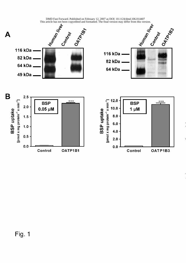
BSP uptake in HEK-OATP1B1 and HEK-OATP1B3 cells
The prerequisite for analyzing the inhibitory potency of drugs on OATP1B1- and
OATP1B3-mediated uptake is the availability of stably transfected cells expressing
the recombinant protein in high amounts. Therefore, HEK293 cells were stably
transfected with the SLCO1B1 cDNA and the SLCO1B3 cDNA and selected for a
high expression of the respective uptake transporter. The protein expression of the
selected cell clones has been analyzed using the OATP1B1-specific antibody pESL
(König et al., 2000a) and the OATP1B3-specific antibody pSKT (König et al., 2000b).
This analysis demonstrated a high protein expression in the HEK-OATP1B1 and
HEK-OATP1B3 cells (Fig. 1A).
Uptake mediated by OATP1B1 or OATP1B3 was analyzed using the prototypic
tritium-labeled substrate sulfobromophthalein (BSP). BSP was shown to be a high
affinity substrate for both OATP1B1 and OATP1B3 with Km values of 140 nM (Cui et
al., 2001b) and 3.3 µM (König et al., 2000a), respectively. The uptake experiments
(Fig. 1B) demonstrated that HEK-OATP1B1 cells as well as HEK-OATP1B3 cells
were able to mediate BSP uptake into cells. The net uptake rates were 2.1 pmol x mg
protein-1 x min-1 for HEK-OATP1B1 cells and 10.8 pmol x mg protein-1 x min-1 for
HEK-OATP1B3 cells.
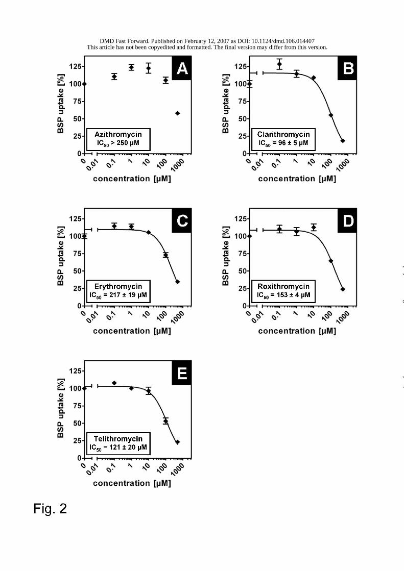
Inhibition of OATP1B1-mediated BSP uptake by macrolides
Uptake experiments have been carried out as described with adding different
concentrations of the respective macrolide. Interestingly, all investigated macrolides
except azithromycin showed a clear doses-dependent inhibition of OATP1B1-
mediated BSP uptake into HEK-OATP1B1 cells (Fig. 2). Azithromycin has been
analyzed up to a concentration of 500 µM and only at this high concentration a slight
decrease in BSP uptake was observed (Fig. 2A). Erythromycin also has a high IC50
This article has not been copyedited and formatted. The final version may differ from this version.DMD Fast Forward. Published on February 12, 2007 as DOI: 10.1124/dmd.106.014407
at ASPE
T Journals on January 24, 2020
dmd.aspetjournals.org
Dow
nloaded from
DMD #14407
12
value of 217 ± 19 µM (Fig. 2C) whereas clarithromycin, telithromycin, and
roxithromycin have IC50 values of 96 ± 5 µM, 121 ± 19 µM, and 153 ± 4 µM,
respectively (Fig. 2B, D, and E). Taken together, clarithromycin, erythromycin,
roxithromycin, and telithromycin were identified to inhibit the OATP1B1-mediated
BSP transport.
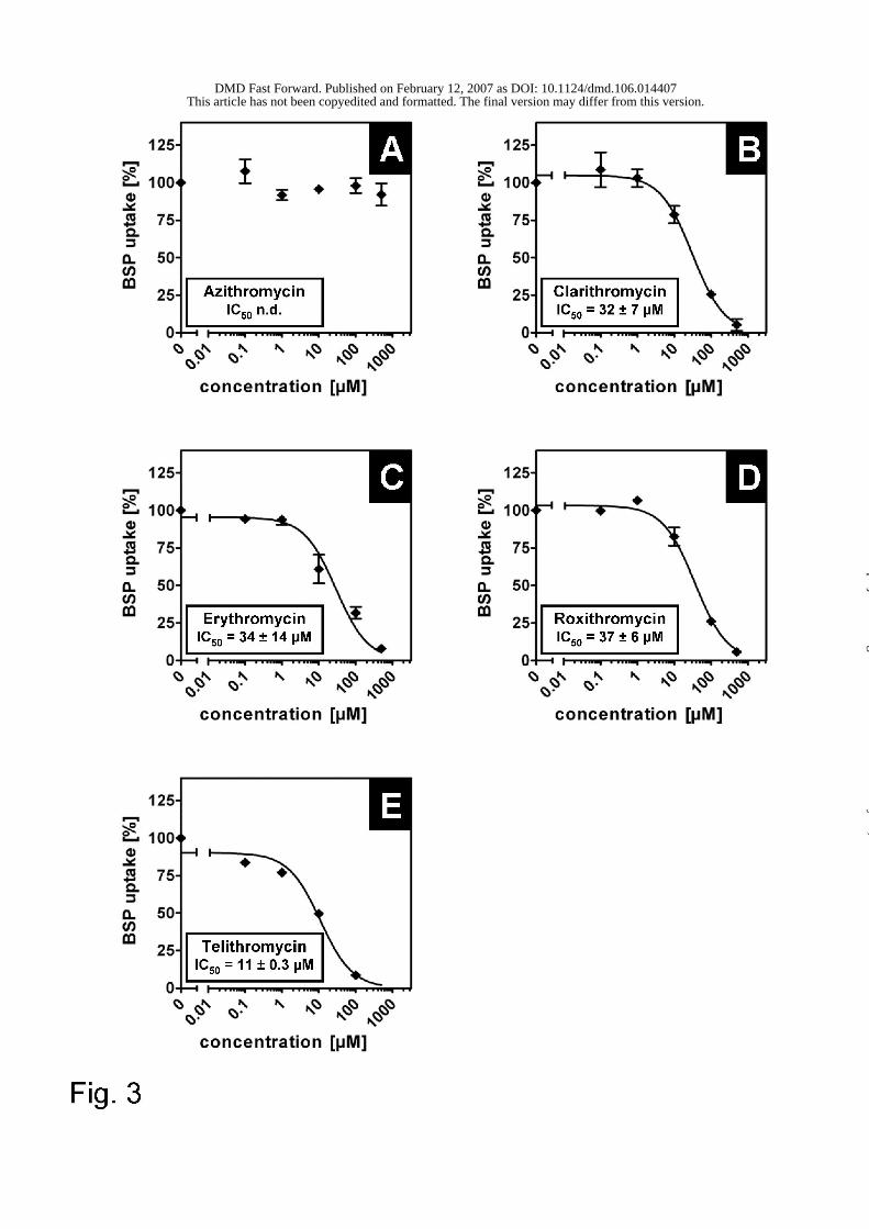
Inhibition of OATP1B3-mediated BSP uptake by macrolides
A similar experimental setup was used to analyze the inhibitory effect of macrolides
on OATP1B3-mediated BSP uptake. As shown for the inhibition of OATP1B1-
mediated uptake azithromycin did not inhibit the uptake mediated by the OATP1B3
protein (Fig. 3A). All other investigated macrolides inhibited the OATP1B3-mediated
BSP uptake (Fig. 3B – E). Telithromycin was a potent inhibitor for OATP1B3-
mediated uptake with an IC50 value of 11 ± 0.3 µM (Fig. 3E). The macrolides
erythromycin, clarithromycin, and roxithromycin showed inhibitory potency with IC50
values of 34 ± 14 µM, 32 ± 7 µM, and 37 ± 6 µM, respectively (Fig. 3B – D).
Interestingly, the calculated IC50 values for clarithromycin, erythromycin,
roxithromycin, and telithromycin were determined to be lower than the respective IC50
values for OATP1B1-mediated uptake.
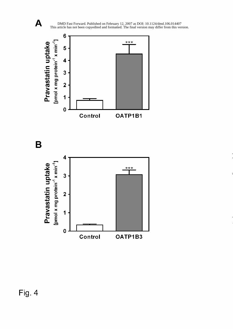
OATP1B1- and OATP1B3-mediated pravastatin uptake and inhibition by
macrolides
To test whether macrolides are also inhibitors for the OATP1B1- and OATP1B3-
mediated uptake of pravastatin, we performed pravastatin uptake and inhibition
experiments. Pravastatin is a known substrate for OATP1B1 (Km value of 34 µM;
(Hsiang et al., 1999). We confirmed that pravastatin is transported by OATP1B1 with
a significantly higher uptake in HEK-OATP1B1 cells (4.5 pmol x mg protein-1 x min-1)
compared to HEK-control cells (0.8 pmol x mg protein-1 x min-1) (Fig. 4A).
Furthermore, we could demonstrate for the first time that pravastatin is also a
This article has not been copyedited and formatted. The final version may differ from this version.DMD Fast Forward. Published on February 12, 2007 as DOI: 10.1124/dmd.106.014407
at ASPE
T Journals on January 24, 2020
dmd.aspetjournals.org
Dow
nloaded from
DMD #14407
13
substrate for OATP1B3 (Fig. 4B). The uptake experiments demonstrated also a
significantly higher uptake in HEK-OATP1B3 cells in comparison to HEK-control cells
(3.1 pmol x mg protein-1 x min-1 vs. 0.3 pmol x mg protein-1 x min-1) (Fig. 4B).
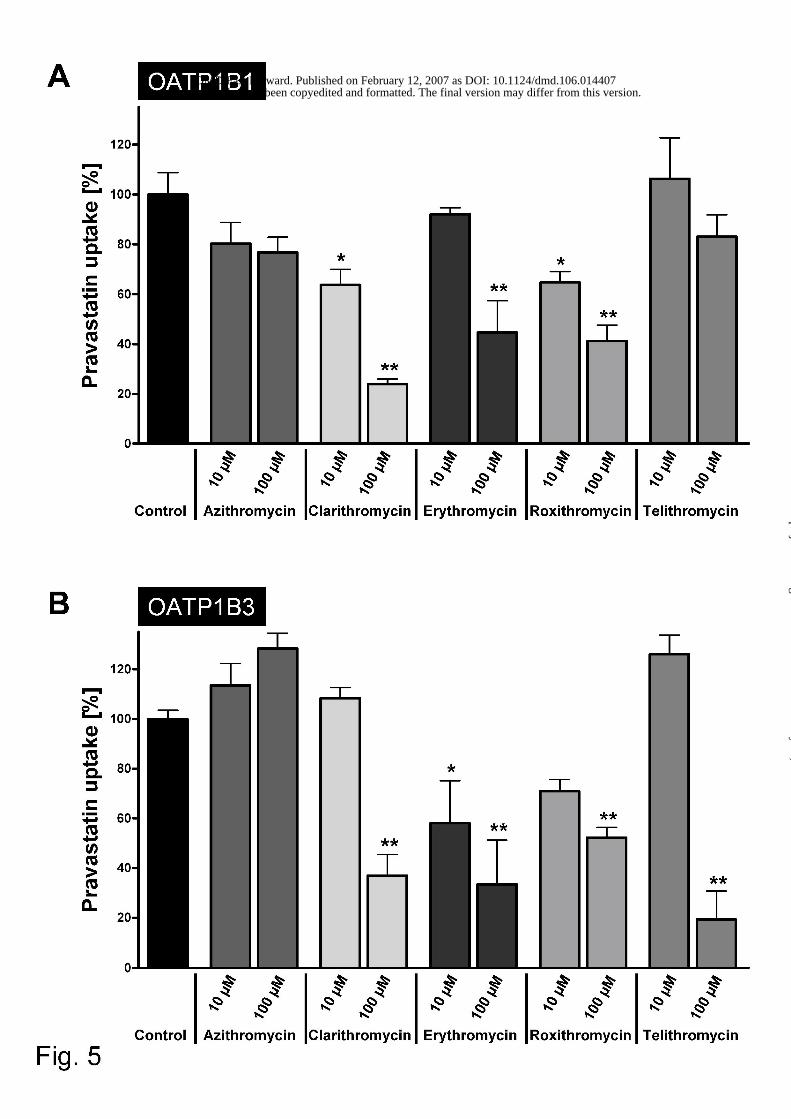
Clarithromycin, erythromycin, and roxithromycin significantly inhibited the uptake of
pravastatin in HEK-OATP1B1 cells (Fig. 5A). Addition of 10 µM of clarithromycin or
roxithromycin resulted in a reduced intracellular accumulation of pravastatin to 64 %
and 65 % compared to the control experiments without macrolides (Fig. 5A).
Clarithromycin (100 µM), a potent inhibitor of the OATP1B1-mediated BSP uptake,
led to a reduction to 24 % intracellular accumulation of pravastatin compared to the
control experiments. As for BSP, azithromycin did not inhibit the transporter-mediated
uptake of pravastatin. In contrast, telithromycin which was a moderate inhibitor for
BSP uptake was not significantly affecting the uptake of pravastatin (Fig. 5A) and
showed a moderate uptake inhibition only at the high concentration of 100 µM.
At low macrolide concentrations (10 µM) erythromycin and roxithromycin inhibited
OATP1B3-mediated pravastatin uptake. The addition of 100 µM clarithromycin,
erythromycin, roxithromycin, and telithromycin reduced the pravastatin uptake to
37 %, 36 %, 52 %, and 19 %, respectively (Fig. 5B). Interestingly, telithromycin did
not inhibit the uptake of pravastatin at the low concentration whereas the higher
concentration significantly inhibited the uptake. Moreover, a slight but not significant
transport activation by low clarithromycin and telithromycin concentrations could be
observed. In accordance with the BSP inhibition assay azithromycin was the only
macrolide showing no inhibition among the tested macrolides (Fig. 5B).
This article has not been copyedited and formatted. The final version may differ from this version.DMD Fast Forward. Published on February 12, 2007 as DOI: 10.1124/dmd.106.014407
at ASPE
T Journals on January 24, 2020
dmd.aspetjournals.org
Dow
nloaded from
DMD #14407
14
Discussion
In this study we focused on the analysis of the interaction of several macrolide /
ketolide antibiotics with the transport of the organic anion BSP and the HMG-CoA
reductase inhibitor pravastatin mediated by the hepatocellular uptake transporters
OATP1B1 and OATP1B3. Using newly established HEK cells recombinantly
expressing human OATP1B1 and OATP1B3 (Fig. 1) we found a concentration-
dependent inhibition of BSP uptake both in HEK-OATP1B1 and HEK-OATP1B3 cells
for all macrolides (except from azithromycin) and for the ketolide telithromycin. IC50
values were considerably smaller for the uptake inhibition of OATP1B3 than for
OATP1B1 (Figs. 2 and 3). Additionally to the inhibition studies with the prototypic
substrate BSP we have investigated the influence of macrolides and the ketolide
telithromycin on the uptake of the HMG-CoA reductase inhibitor pravastatin. We
demonstrated OATP1B1-mediated pravastatin uptake into HEK-OATP1B1 cells as
described earlier (Hsiang et al., 1999). Furthermore to the best of our knowledge, we
determined for the first time that pravastatin uptake is mediated by the second major
hepatocyte OATP family member OATP1B3 (Fig. 4). This transporter-mediated
pravastatin uptake could be inhibited by co-administration of clarithromycin,
erythromycin, and roxithromycin. In the case of OATP1B3, telithromycin was a potent
inhibitor for BSP uptake, however, a strong inhibition of pravastatin uptake was
observed only at high concentration (100 µM) of telithromycin. Clarithromycin,
erythromycin, and roxithromycin inhibited both OATP1B1- and OATP1B3-mediated
BSP and pravastatin uptake. On the other hand, azithromycin had no effect on BSP
or on pravastatin uptake.
OATP1B1 and OATP1B3 are expressed predominantly in the basolateral membrane
of human hepatocytes mediating the uptake of endogenous substances as well as
several xenobiotics and drugs. Both transporters share an overlapping substrate
This article has not been copyedited and formatted. The final version may differ from this version.DMD Fast Forward. Published on February 12, 2007 as DOI: 10.1124/dmd.106.014407
at ASPE
T Journals on January 24, 2020
dmd.aspetjournals.org
Dow
nloaded from
DMD #14407
15
spectrum. Important drugs which are taken up by OATP1B1 and OATP1B3 are
several HMG-CoA reductase inhibitors like fluvastatin and pitavastatin, the antibiotic
rifampicin, and the endothelin receptor antagonist BQ123 (König et al., 2006).
Furthermore, transport of cerivastatin (Shitara et al., 2003), pravastatin (Hsiang et al.,
1999), and rosuvastatin (Schneck et al., 2004) has been shown for OATP1B1
whereas OATP1B3 is able to mediate the uptake of digoxin (Kullak-Ublick et al.,
2001). Due to these substrate spectra and their localization between portal venous
blood and important drug metabolizing enzymes (e.g. CYP3A4) expressed in
hepatocytes, uptake transporters are increasingly recognized as important factors in
the directed elimination of drugs out of the body. Their presence can be a
prerequisite for substances to enter hepatocytes and getting metabolized prior their
elimination over the canalicular membrane into bile. Modification of uptake rates e.g.
by drug competition, therefore, may cause drug-drug interactions by lowering the
uptake rate of one drug followed by increased blood concentrations due to reduced
hepatic metabolism and / or decreased biliary elimination.
Inhibition of cytochrome P450 isoenzymes is one established mechanism of drug-
drug interactions. Multiple studies have demonstrated that macrolides are potent
inhibitors of CYP3A4 and therefore can increase the plasma concentration of co-
administered drugs that are CYP3A4 substrates (Niemi et al., 2001). Drug-
interactions have also been reported between macrolides and some HMG-CoA
reductase inhibitors. Clarithromycin for example increases the plasma concentration
of concomitantly administered simvastatin, atorvastatin, and pravastatin (Jacobson,
2004). In the case of simvastatin and atorvastatin this drug-interaction can be
explained by the inhibition of CYP3A4 which predominantly metabolizes these
statins. Interestingly, pravastatin is one of the statins which is not metabolized by
cytochromes in humans and which is excreted almost unchanged into bile or to a
This article has not been copyedited and formatted. The final version may differ from this version.DMD Fast Forward. Published on February 12, 2007 as DOI: 10.1124/dmd.106.014407
at ASPE
T Journals on January 24, 2020
dmd.aspetjournals.org
Dow
nloaded from
DMD #14407
16
small extent into urine (Jacobson, 2004). In this case an interaction of drug
transporting proteins, located in the basolateral hepatocyte membrane may account
for the increased plasma concentration. The data presented in this manuscript
confirmed these in vivo analyses of an interaction between clarithromycin and
pravastatin. Clarithromycin inhibited dose-dependent the OATP1B1- as well as the
OATP1B3-mediated pravastatin uptake in vitro. Therefore transporter-inhibition could
be the underlying mechanism of this pharmacokinetical drug-drug interaction. In
accordance with our findings, Hirano and coworkers very recently demonstrated that
both clarithromycin and erythromycin were inhibitors for the uptake of pitavastatin, an
established OATP1B1 substrate (Hirano et al., 2006) with Ki values of 8.3 and
11.4 µM, respectively.
Interestingly, an in vivo interaction between rosuvastatin, a recently established
HMG-CoA reductase inhibitor, and erythromycin does not appear (Cooper et al.,
2003). Rosuvastatin is a substrate of several OATP-family members (OATP1B1,
OATP1B3, OATP2B1, OATP1A2) and also of the sodium dependent bile salt
transporter NTCP (Ho et al., 2006; Schneck et al., 2004) and therefore, the uptake
inhibition of OATP1B1 or OATP1B3 could be compensated by transport via
alternative transporting proteins.
Published Ki values for inhibition of the metabolizing enzyme CYP3A4 by
clarithromycin, erythromycin, roxithromycin, and telithromycin are 30 µM, 13 µM,
72 µM, and 58 µM, respectively (Aventis-Pharmaceuticals, 2001; Polasek and
Miners, 2006). Interestingly, the determined IC50 values for macrolide-induced
OATP1B3 inhibition are in the same concentration range. In addition, azithromycin,
which has a high Ki value for CYP3A4 (Polasek and Miners, 2006) is the only
macrolide showing neither uptake inhibition of OATP1B1- nor OATP1B3-mediated
uptake.
This article has not been copyedited and formatted. The final version may differ from this version.DMD Fast Forward. Published on February 12, 2007 as DOI: 10.1124/dmd.106.014407
at ASPE
T Journals on January 24, 2020
dmd.aspetjournals.org
Dow
nloaded from
DMD #14407
17
As drugs reach the portal vein directly after intestinal absorption, the drug
concentration in portal venous blood is higher than in the systemic circulation. For
calculation of the predicted maximum drug concentration at the inlet to the liver we
used the method of Ito et al. (1998) taking into account the maximum plasma
concentration in the systemic circulation, the single dosage, the absorbed fraction of
the macrolide, the absorption rate and the hepatic blood flow rate [Table 1 (Ito et al.,
1998)]. For clarithromycin, erythromycin, roxithromycin, and telithromycin the
predicted portal venous concentrations are in the same range as the determined IC50
values for inhibition of OATP1B3-mediated uptake. We therefore conclude that
inhibition of drug transporters by macrolides / ketolides could be an additional
mechanism for clinical relevant drug-drug interactions. Further studies are necessary
to gain more inside into the molecular nature of this inhibition mechanism.
Taken together our data demonstrate that macrolides / ketolides can inhibit uptake of
organic anions and drugs mediated by the OATP family members OATP1B1 and
OATP1B3. This modification of uptake rates is a new mechanism of drug-drug
interactions in addition to the hitherto known mechanism of drug-drug interactions
due to the modification of metabolizing enzymes and efflux transporters. Based on
our findings it is therefore of importance to gain more knowledge on the modification
of uptake transporter function as additional mechanism underlying drug-drug
interactions.
This article has not been copyedited and formatted. The final version may differ from this version.DMD Fast Forward. Published on February 12, 2007 as DOI: 10.1124/dmd.106.014407
at ASPE
T Journals on January 24, 2020
dmd.aspetjournals.org
Dow
nloaded from
DMD #14407
18
Acknowledgements
We thank Mrs. C. Hoffmann and B. Endress for excellent technical assistance. We
would like to thank Professor Dr. D. Keppler (German Cancer Research Center,
Heidelberg) for providing the polyclonal antibodies pESL and pSKT and the plasmids
pcDNA3.1(+)-OATP1B1 and pcDNA3.1/Hygro(-)-OATP1B3.
This article has not been copyedited and formatted. The final version may differ from this version.DMD Fast Forward. Published on February 12, 2007 as DOI: 10.1124/dmd.106.014407
at ASPE
T Journals on January 24, 2020
dmd.aspetjournals.org
Dow
nloaded from
DMD #14407
19
References
Aventis-Pharmaceuticals (2001) KETEK(R) (telithromycin), in Briefing document for
the FDA anti-infective drug products advisory committee meeting, March 2001,
available from:http://www.fda.gov/ohrms/dockets/ac/01/briefing/3746b_01_
aventis.pdf.
Chung J-Y, Cho J-Y, Yu K-S, Kim J-R, Oh D-S, Jung H-R, Lim K-S, Moon K-H, Shin
S-G and Jang I-J (2005) Effect of OATP1B1 (SLCO1B1) variant alleles on the
pharmacokinetics of pitavastatin in healthy volunteers. Clin Pharmacol Ther
78:342-350.
Cooper KJ, Martin PD, Dane AL, Warwick MJ, Raza A and Schneck DW (2003) The
effect of erythromycin on the pharmacokinetics of rosuvastatin. Eur J Clin
Pharmacol 59:51-56.
Cui Y, König J, Buchholz JK, Spring H, Leier I and Keppler D (1999) Drug resistance
and ATP-dependent conjugate transport mediated by the apical multidrug
resistance protein, MRP2, permanently expressed in human and canine cells.
Mol Pharmacol 55:929-937.
Cui Y, König J and Keppler D (2001a) Vectorial transport by double-transfected cells
expressing the human uptake transporter SLC21A8 and the apical export
pump ABCC2. Mol Pharmacol 60:934-943.
Cui Y, König J, Leier I, Buchholz U and Keppler D (2001b) Hepatic uptake of bilirubin
and its conjugates by the human organic anion transporter SLC21A6. J Biol
Chem 276:9626-9630.
Eberl S, Bachmakov I, Dörje F and Fromm MF (2005) The effect of macrolide
antibiotics on the function of the drug transporter P-glycoprotein. Naunyn-
Schmiedeberg's Arch Pharmacol 371 Suppl. 1:R145 (abstract).
This article has not been copyedited and formatted. The final version may differ from this version.DMD Fast Forward. Published on February 12, 2007 as DOI: 10.1124/dmd.106.014407
at ASPE
T Journals on January 24, 2020
dmd.aspetjournals.org
Dow
nloaded from
DMD #14407
20
Hagenbuch B and Meier PJ (2003) The superfamily of organic anion transporting
polypeptides. Biochim Biophys Acta - Biomembranes 1609:1-18.
Hagenbuch B and Meier PJ (2004) Organic anion transporting polypeptides of the
OATP/SLC21 family: phylogenetic classification as OATP/SLCO superfamily,
new nomenclature and molecular/functional properties. Pfluegers Arch Eur J
Physiol 447:653-665.
Hirano M, Maeda K, Shitara Y and Sugiyama Y (2006) Drug-drug interaction between
pitavastatin and various drugs via OATP1B1. Drug Metab Dispos 34:1229-
1236.
Ho RH, Tirona RG, Leake BF, Glaeser H, Lee W, Lemke CJ, Wang Y and Kim RB
(2006) Drug and Bile Acid Transporters in Rosuvastatin Hepatic Uptake:
Function, Expression, and Pharmacogenetics. Gastroenterology 130:1793-
1806.
Hsiang B, Zhu Y, Wang Z, Wu Y, Sasseville V, Yang W-P and Kirchgessner TG
(1999) A novel human hepatic organic anion transporting polypeptide
(OATP2). Identification of a liver-specific human organic anion transporting
polypeptide and indentification of rat and human hydroxymethylglutaryl-CoA
reductase inhibitor transporters. J Biol Chem 274:37161-37168.
Ito K, Iwatsubo T, Kanamitsu S, Ueda K, Suzuki H and Sugiyama Y (1998) Prediction
of pharmacokinetic alterations caused by drug-drug interactions: metabolic
interaction in the liver. Pharmacol Rev 50:387-412.
Ito K, Ogihara K, Kanamitsu S and Itoh T (2003) Prediction of the in vivo interaction
between midazolam and macrolides based on in vitro studies using human
liver microsomes. Drug Metab Dispos 31:945-954.
This article has not been copyedited and formatted. The final version may differ from this version.DMD Fast Forward. Published on February 12, 2007 as DOI: 10.1124/dmd.106.014407
at ASPE
T Journals on January 24, 2020
dmd.aspetjournals.org
Dow
nloaded from
DMD #14407
21
Iwai M, Suzuki H, Ieiri I, Otsubo K and Sugiyama Y (2004) Functional analysis of
single nucleotide polymorphisms of hepatic organic anion transporter
OATP1B1 (OATP-C). Pharmacogenetics 14:749-757.
Jacobson TA (2004) Comparative pharmacokinetic interaction profiles of pravastatin,
simvastatin, and atorvastatin when coadministered with cytochrome P450
inhibitors. Am J Cardiol 94:1140-1146.
Kameyama Y, Yamashita K, Kobayashi K, Hosokawa M and Chiba K (2005)
Functional characterization of SLCO1B1 (OATP-C) variants, SLCO1B1*5,
SLCO1B1*15 and SLCO1B1*15+C1007G, by using transient expression
systems of HeLa and HEK293 cells. Pharmacogenet Genomics 15:513-522.
Katz DA, Carr R, Grimm DR, Xiong H, Holley-Shanks R, Mueller T, Leake B, Wang
Q, Han L and Wang PG (2006) Organic anion transporting polypeptide 1B1
activity classified by SLCO1B1 genotype influences atrasentan
pharmacokinetics. Clin Pharmacol Ther 79:186-196.
Kim RB, Wandel C, Leake B, Cvetkovic M, Fromm MF, Dempsey PJ, Roden MM,
Belas F, Chaudhary AK, Roden DM, Wood AJ and Wilkinson GR (1999)
Interrelationship between substrates and inhibitors of human CYP3A and P-
glycoprotein. Pharm Res 16:408-414.
König J, Cui Y, Nies AT and Keppler D (2000a) Localization and genomic
organization of a new hepatocellular organic anion transporting polypeptide. J
Biol Chem 275:23161-23168.
König J, Cui Y, Nies AT and Keppler D (2000b) A novel human organic anion
transporting polypeptide localized to the basolateral hepatocyte membrane.
Am J Physiol Gastrointest Liver Physiol 278:G156-G164.
König J, Seithel A, Gradhand U and Fromm MF (2006) Pharmacogenomics of human
OATP transporters. Naunyn-Schmiedeberg's Arch Pharmacol 372:432-443.
This article has not been copyedited and formatted. The final version may differ from this version.DMD Fast Forward. Published on February 12, 2007 as DOI: 10.1124/dmd.106.014407
at ASPE
T Journals on January 24, 2020
dmd.aspetjournals.org
Dow
nloaded from
DMD #14407
22
Kullak-Ublick G, Ismair M, Stieger B, Landmann L, Huber R, Pizzagalli F, Fattinger K,
Meier P and Hagenbuch B (2001) Organic anion-transporting polypeptide B
(OATP-B) and its functional comparison with three other OATPs of human
liver. Gastroenterology 120:525-533.
Lee E, Ryan S, Birmingham B, Zalikowski J, March R, Ambrose H, Moore R, Lee C,
Chen Y and Schneck D (2005) Rosuvastatin pharmacokinetics and
pharmacogenetics in white and Asian subjects residing in the same
environment. Clin Pharmacol Ther 78:330-341.
Letschert K, Keppler D and König J (2004) Mutations in the SLCO1B3 gene affecting
the substrate specificity of the hepatocellular uptake transporter OATP1B3
(OATP8). Pharmacogenetics 14:441-452.
Marzolini C, Paus E, Buclin T and Kim RB (2004) Polymorphisms in human MDR1
(P-glycoprotein): recent advances and clinical relevance. Clin Pharmacol Ther
75:13-33.
Michalski C, Cui Y, Nies AT, Nuessler AK, Neuhaus P, Zanger UM, Klein K,
Eichelbaum M, Keppler D and König J (2002) A naturally occurring mutation in
the SLC21A6 gene causing impaired membrane localization of the hepatocyte
uptake transporter. J Biol Chem 277:43058-43063.
Niemi M, Backman JT, Kajosaari LI, Leathart JB, Neuvonen M, Daly AK, Eichelbaum
M, Kivistö KT and Neuvonen PJ (2005a) Polymorphic organic anion
transporting polypeptide 1B1 is a major determinant of repaglinide
pharmacokinetics. Clin Pharmacol Ther 77:468-478.
Niemi M, Kivistö KT, Hofmann U, Schwab M, Eichelbaum M and Fromm MF (2005b)
Fexofenadine pharmacokinetics are associated with a polymorphism of the
SLCO1B1 gene (encoding OATP1B1). Br J Clin Pharmacol 59:602-604.
This article has not been copyedited and formatted. The final version may differ from this version.DMD Fast Forward. Published on February 12, 2007 as DOI: 10.1124/dmd.106.014407
at ASPE
T Journals on January 24, 2020
dmd.aspetjournals.org
Dow
nloaded from
DMD #14407
23
Niemi M, Neuvonen PJ and Kivistö KT (2001) The cytochrome P4503A4 inhibitor
clarithromycin increases the plasma concentrations and effects of repaglinide.
Clin Pharmacol Ther 70:58-65.
Niemi M, Pasanen MK and Neuvonen PJ (2006) SLCO1B1 polymorphism and sex
affect the pharmacokinetics of pravastatin but not fluvastatin. Clin Pharmacol
Ther 80:356-366.
Nishizato Y, Ieiri I, Suzuki H, Kimura M, Kawabata K, Hirota T, Takane H, Irie S,
Kusuhara H and Urasaki Y (2003) Polymorphisms of OATP-C (SLC21A6) and
OAT3 (SLC22A8) genes: Consequences for pravastatin pharmacokinetics.
Clin Pharmacol Ther 73:554-565.
Pasanen MK, Neuvonen M, Neuvonen PJ and Niemi M (2006) SLCO1B1
polymorphism markedly affects the pharmacokinetics of simvastatin acid.
Pharmacogenet Genomics 16:873-879.
Polasek T and Miners J (2006) Quantitative prediction of macrolide drug-drug
interaction potential from in vitro studies using testosterone as the human
cytochrome P4503A substrate. Eur J Clin Pharmacol 62:203-208.
Product information: Erythrocin® [Erythromycin], Wiesbaden, Germany; Abbott GmbH
& Co. KG; January 2001
Product information: Ketek® [Telithromycin], Antony, France; Aventis Pharma S.A.;
June 2005
Product information: Klacid® [Clarithromycin], Wiesbaden, Germany; Abbott GmbH &
Co. KG; July 2004
Product information: Pravasin® protect [Pravastatin], Munich, Germany; Bristol-Myers
Squibb; October 2005
Product information: Rulid® [Roxithromycin], Frankfurt a.M., Germany; Aventis
Pharma Deutschland GmbH; March 2004
This article has not been copyedited and formatted. The final version may differ from this version.DMD Fast Forward. Published on February 12, 2007 as DOI: 10.1124/dmd.106.014407
at ASPE
T Journals on January 24, 2020
dmd.aspetjournals.org
Dow
nloaded from
DMD #14407
24
Product information: Zithromax® [Azithromycin], Karlsruhe, Germany; Pfizer Pharma
GmbH; December 2004
Rengelshausen J, Goggelmann C, Burhenne J, Riedel KD, Ludwig J, Weiss J, Mikus
G, Walter-Sack I and Haefeli WE (2003) Contribution of increased oral
bioavailability and reduced nonglomerular renal clearance of digoxin to the
digoxin-clarithromycin interaction. Br J Clin Pharmacol 56:32-38.
Schneck DW, Birmingham BK, Zalikowski JA, Mitchell PD, Wang Y, Martin PD,
Lasseter KC, Brown CD, Windass AS and Raza A (2004) The effect of
gemfibrozil on the pharmacokinetics of rosuvastatin. Clin Pharmacol Ther
75:455-463.
Shitara Y, Itoh T, Sato H, Li AP and Sugiyama Y (2003) Inhibition of transporter-
mediated hepatic uptake as a mechanism for drug-drug interaction between
cerivastatin and cyclosporin A. J Pharmacol Exp Ther 304:610-616.
Tirona RG, Leake BF, Merino G and Kim RB (2001) Polymorphisms in OATP-C:
identification of multiple allelic variants associated with altered transport
activity among European- and African-Americans. J Biol Chem 276:35669-
35675.
Wrington SA and E.Thummel K (2000) CYP3A, in Metabolic Drug Interactions (Levy
RH, E.Thummel K, Trager WF, Hansten PD and Eichelbaum M eds) pp 115-
133, Lippincott Williams & Wilkins, Philadelphia.
This article has not been copyedited and formatted. The final version may differ from this version.DMD Fast Forward. Published on February 12, 2007 as DOI: 10.1124/dmd.106.014407
at ASPE
T Journals on January 24, 2020
dmd.aspetjournals.org
Dow
nloaded from
DMD #14407
25
Footnotes
This work was supported by grant DFG Ko 2120/1-3 of the Deutsche
Forschungsgemeinschaft.
This article has not been copyedited and formatted. The final version may differ from this version.DMD Fast Forward. Published on February 12, 2007 as DOI: 10.1124/dmd.106.014407
at ASPE
T Journals on January 24, 2020
dmd.aspetjournals.org
Dow
nloaded from
DMD #14407
26
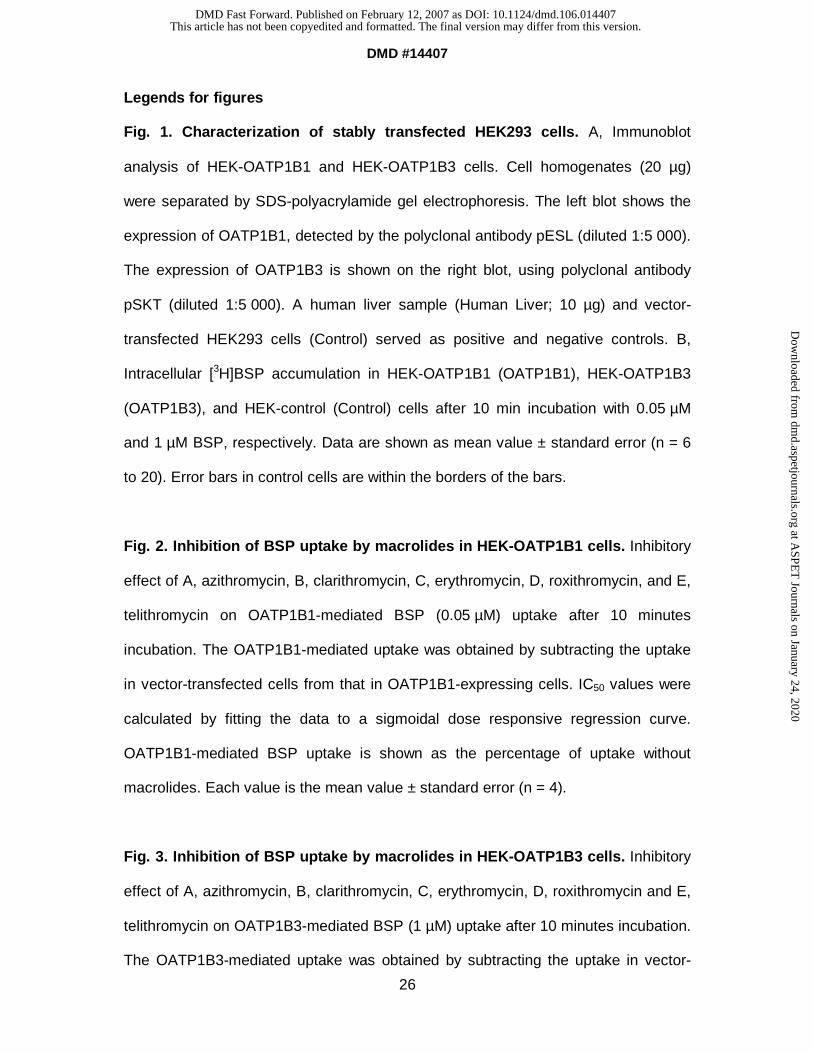
Legends for figures
Fig. 1. Characterization of stably transfected HEK293 cells. A, Immunoblot
analysis of HEK-OATP1B1 and HEK-OATP1B3 cells. Cell homogenates (20 µg)
were separated by SDS-polyacrylamide gel electrophoresis. The left blot shows the
expression of OATP1B1, detected by the polyclonal antibody pESL (diluted 1:5 000).
The expression of OATP1B3 is shown on the right blot, using polyclonal antibody
pSKT (diluted 1:5 000). A human liver sample (Human Liver; 10 µg) and vector-
transfected HEK293 cells (Control) served as positive and negative controls. B,
Intracellular [3H]BSP accumulation in HEK-OATP1B1 (OATP1B1), HEK-OATP1B3
(OATP1B3), and HEK-control (Control) cells after 10 min incubation with 0.05 µM
and 1 µM BSP, respectively. Data are shown as mean value ± standard error (n = 6
to 20). Error bars in control cells are within the borders of the bars.
Fig. 2. Inhibition of BSP uptake by macrolides in HEK-OATP1B1 cells. Inhibitory
effect of A, azithromycin, B, clarithromycin, C, erythromycin, D, roxithromycin, and E,
telithromycin on OATP1B1-mediated BSP (0.05 µM) uptake after 10 minutes
incubation. The OATP1B1-mediated uptake was obtained by subtracting the uptake
in vector-transfected cells from that in OATP1B1-expressing cells. IC50 values were
calculated by fitting the data to a sigmoidal dose responsive regression curve.
OATP1B1-mediated BSP uptake is shown as the percentage of uptake without
macrolides. Each value is the mean value ± standard error (n = 4).
Fig. 3. Inhibition of BSP uptake by macrolides in HEK-OATP1B3 cells. Inhibitory
effect of A, azithromycin, B, clarithromycin, C, erythromycin, D, roxithromycin and E,
telithromycin on OATP1B3-mediated BSP (1 µM) uptake after 10 minutes incubation.
The OATP1B3-mediated uptake was obtained by subtracting the uptake in vector-
This article has not been copyedited and formatted. The final version may differ from this version.DMD Fast Forward. Published on February 12, 2007 as DOI: 10.1124/dmd.106.014407
at ASPE
T Journals on January 24, 2020
dmd.aspetjournals.org
Dow
nloaded from
DMD #14407
27
transfected cells from that in OATP1B3-expressing cells. IC50 values were calculated
by fitting the data to a sigmoidal dose responsive regression curve. OATP1B3-
mediated BSP uptake is shown as the percentage of uptake without macrolides.
Each value is the mean value ± standard error (n = 4).
Fig. 4. Pravastatin uptake by HEK-OATP1B1 and HEK-OATP1B3 cells. A,
Intracellular pravastatin accumulation in HEK-OATP1B1 and HEK-control (Control)
cells after 10 min incubation with pravastatin (50 µM). B, Intracellular pravastatin
accumulation in HEK-OATP1B3 and HEK-control (Control) cells after 10 min
incubation with pravastatin (50 µM). Each value is the mean value ± standard error
(n = 4 to 6). *** P < 0.001 vs control.
Fig. 5. Inhibition of the pravastatin uptake by macrolides. Inhibitory effect using
10 µM and 100 µM of the macrolides azithromycin, clarithromycin, erythromycin,
roxithromycin, and telithromycin on A, OATP1B1- and B, OATP1B3-mediated
pravastatin (50 µM) uptake after 10 minutes incubation. The transporter-mediated
uptake was obtained by subtracting the uptake in vector-transfected cells from that in
transporter-expressing cells. OATP-mediated pravastatin uptake is shown as the
percentage of uptake without macrolides. Each value is the mean value ± standard
error (n = 4 to 6). * P < 0.05; ** P < 0.01 vs control.
This article has not been copyedited and formatted. The final version may differ from this version.DMD Fast Forward. Published on February 12, 2007 as DOI: 10.1124/dmd.106.014407
at ASPE
T Journals on January 24, 2020
dmd.aspetjournals.org
Dow
nloaded from
DMD #14407
28
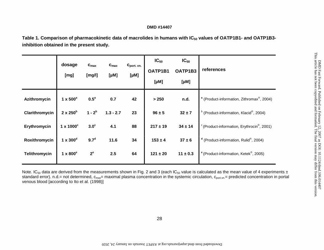
Table 1. Comparison of pharmacokinetic data of macrolides in humans with IC50 values of OATP1B1- and OATP1B3-
inhibition obtained in the present study.
dosage
[mg]
cmax
[mg/l]
cmax
[µM]
cport. vn.
[µM]
IC50
OATP1B1
[µM]
IC50
OATP1B3
[µM]
references
Azithromycin 1 x 500a 0.5a 0.7 42 > 250 n.d. a (Product-information, Zithromax®, 2004)
Clarithromycin 2 x 250b 1 - 2b 1.3 - 2.7 23 96 ± 5 32 ± 7 b (Product-information, Klacid®, 2004)
Erythromycin 1 x 1000c 3.0c 4.1 88 217 ± 19 34 ± 14 c (Product-information, Erythrocin®, 2001)
Roxithromycin 1 x 300d 9.7d 11.6 34 153 ± 4 37 ± 6 d (Product-information, Rulid®, 2004)
Telithromycin 1 x 800e 2e 2.5 64 121 ± 20 11 ± 0.3 e (Product-information, Ketek®, 2005)
Note. IC50 data are derived from the measurements shown in Fig. 2 and 3 (each IC50 value is calculated as the mean value of 4 experiments ± standard error). n.d.= not determined, cmax= maximal plasma concentration in the systemic circulation, cport.vn.= predicted concentration in portal venous blood [according to Ito et al. (1998)]
This article has not been copyedited and form
atted. The final version m
ay differ from this version.
DM
D Fast Forw
ard. Published on February 12, 2007 as DO
I: 10.1124/dmd.106.014407
at ASPET Journals on January 24, 2020 dmd.aspetjournals.org Downloaded from
This article has not been copyedited and formatted. The final version may differ from this version.DMD Fast Forward. Published on February 12, 2007 as DOI: 10.1124/dmd.106.014407
at ASPE
T Journals on January 24, 2020
dmd.aspetjournals.org
Dow
nloaded from
This article has not been copyedited and formatted. The final version may differ from this version.DMD Fast Forward. Published on February 12, 2007 as DOI: 10.1124/dmd.106.014407
at ASPE
T Journals on January 24, 2020
dmd.aspetjournals.org
Dow
nloaded from
This article has not been copyedited and formatted. The final version may differ from this version.DMD Fast Forward. Published on February 12, 2007 as DOI: 10.1124/dmd.106.014407
at ASPE
T Journals on January 24, 2020
dmd.aspetjournals.org
Dow
nloaded from
This article has not been copyedited and formatted. The final version may differ from this version.DMD Fast Forward. Published on February 12, 2007 as DOI: 10.1124/dmd.106.014407
at ASPE
T Journals on January 24, 2020
dmd.aspetjournals.org
Dow
nloaded from
This article has not been copyedited and formatted. The final version may differ from this version.DMD Fast Forward. Published on February 12, 2007 as DOI: 10.1124/dmd.106.014407
at ASPE
T Journals on January 24, 2020
dmd.aspetjournals.org
Dow
nloaded from