The incidence rates of endometrial hyperplasia and ... · The incidence rates of endometrial...
9
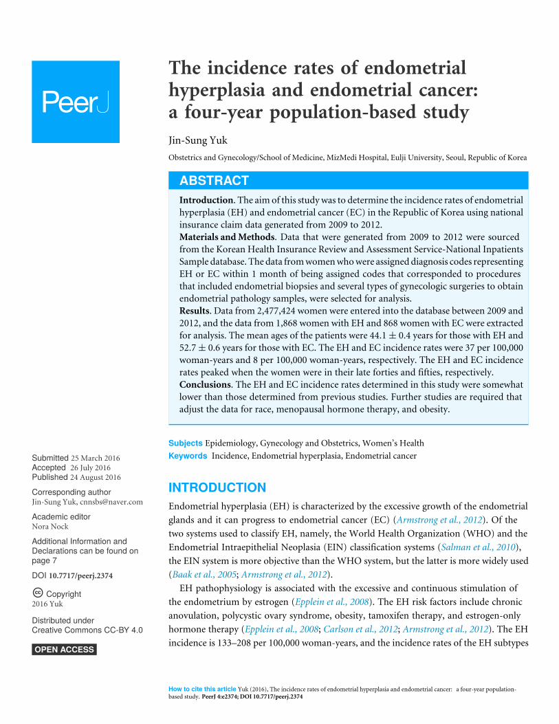
Submitted 25 March 2016 Accepted 26 July 2016 Published 24 August 2016 Corresponding author Jin-Sung Yuk, [email protected] Academic editor Nora Nock Additional Information and Declarations can be found on page 7 DOI 10.7717/peerj.2374 Copyright 2016 Yuk Distributed under Creative Commons CC-BY 4.0 OPEN ACCESS The incidence rates of endometrial hyperplasia and endometrial cancer: a four-year population-based study Jin-Sung Yuk Obstetrics and Gynecology/School of Medicine, MizMedi Hospital, Eulji University, Seoul, Republic of Korea ABSTRACT Introduction. The aim of this study was to determine the incidence rates of endometrial hyperplasia (EH) and endometrial cancer (EC) in the Republic of Korea using national insurance claim data generated from 2009 to 2012. Materials and Methods. Data that were generated from 2009 to 2012 were sourced from the Korean Health Insurance Review and Assessment Service-National Inpatients Sample database. The data from women who were assigned diagnosis codes representing EH or EC within 1 month of being assigned codes that corresponded to procedures that included endometrial biopsies and several types of gynecologic surgeries to obtain endometrial pathology samples, were selected for analysis. Results. Data from 2,477,424 women were entered into the database between 2009 and 2012, and the data from 1,868 women with EH and 868 women with EC were extracted for analysis. The mean ages of the patients were 44.1 ± 0.4 years for those with EH and 52.7 ± 0.6 years for those with EC. The EH and EC incidence rates were 37 per 100,000 woman-years and 8 per 100,000 woman-years, respectively. The EH and EC incidence rates peaked when the women were in their late forties and fifties, respectively. Conclusions. The EH and EC incidence rates determined in this study were somewhat lower than those determined from previous studies. Further studies are required that adjust the data for race, menopausal hormone therapy, and obesity. Subjects Epidemiology, Gynecology and Obstetrics, Women’s Health Keywords Incidence, Endometrial hyperplasia, Endometrial cancer INTRODUCTION Endometrial hyperplasia (EH) is characterized by the excessive growth of the endometrial glands and it can progress to endometrial cancer (EC) (Armstrong et al., 2012). Of the two systems used to classify EH, namely, the World Health Organization (WHO) and the Endometrial Intraepithelial Neoplasia (EIN) classification systems (Salman et al., 2010), the EIN system is more objective than the WHO system, but the latter is more widely used (Baak et al., 2005; Armstrong et al., 2012). EH pathophysiology is associated with the excessive and continuous stimulation of the endometrium by estrogen (Epplein et al., 2008). The EH risk factors include chronic anovulation, polycystic ovary syndrome, obesity, tamoxifen therapy, and estrogen-only hormone therapy (Epplein et al., 2008; Carlson et al., 2012; Armstrong et al., 2012). The EH incidence is 133–208 per 100,000 woman-years, and the incidence rates of the EH subtypes How to cite this article Yuk (2016), The incidence rates of endometrial hyperplasia and endometrial cancer: a four-year population- based study. PeerJ 4:e2374; DOI 10.7717/peerj.2374
-
Upload
vuongkhanh -
Category
Documents
-
view
221 -
download
0
Transcript of The incidence rates of endometrial hyperplasia and ... · The incidence rates of endometrial...
Submitted 25 March 2016Accepted 26 July 2016Published 24 August 2016
Corresponding authorJin-Sung Yuk, [email protected]
Academic editorNora Nock
Additional Information andDeclarations can be found onpage 7
DOI 10.7717/peerj.2374
Copyright2016 Yuk
Distributed underCreative Commons CC-BY 4.0
OPEN ACCESS
The incidence rates of endometrialhyperplasia and endometrial cancer:a four-year population-based studyJin-Sung YukObstetrics and Gynecology/School of Medicine, MizMedi Hospital, Eulji University, Seoul, Republic of Korea
ABSTRACTIntroduction. The aim of this studywas to determine the incidence rates of endometrialhyperplasia (EH) and endometrial cancer (EC) in the Republic of Korea using nationalinsurance claim data generated from 2009 to 2012.Materials andMethods. Data that were generated from 2009 to 2012 were sourcedfrom the Korean Health Insurance Review and Assessment Service-National InpatientsSample database. The data fromwomenwhowere assigneddiagnosis codes representingEH or EC within 1 month of being assigned codes that corresponded to proceduresthat included endometrial biopsies and several types of gynecologic surgeries to obtainendometrial pathology samples, were selected for analysis.Results. Data from 2,477,424 women were entered into the database between 2009 and2012, and the data from 1,868 women with EH and 868 women with EC were extractedfor analysis. The mean ages of the patients were 44.1 ± 0.4 years for those with EH and52.7 ± 0.6 years for those with EC. The EH and EC incidence rates were 37 per 100,000woman-years and 8 per 100,000 woman-years, respectively. The EH and EC incidencerates peaked when the women were in their late forties and fifties, respectively.Conclusions. The EH and EC incidence rates determined in this study were somewhatlower than those determined from previous studies. Further studies are required thatadjust the data for race, menopausal hormone therapy, and obesity.
Subjects Epidemiology, Gynecology and Obstetrics, Women’s HealthKeywords Incidence, Endometrial hyperplasia, Endometrial cancer
INTRODUCTIONEndometrial hyperplasia (EH) is characterized by the excessive growth of the endometrialglands and it can progress to endometrial cancer (EC) (Armstrong et al., 2012). Of thetwo systems used to classify EH, namely, the World Health Organization (WHO) and theEndometrial Intraepithelial Neoplasia (EIN) classification systems (Salman et al., 2010),the EIN system is more objective than the WHO system, but the latter is more widely used(Baak et al., 2005; Armstrong et al., 2012).
EH pathophysiology is associated with the excessive and continuous stimulation ofthe endometrium by estrogen (Epplein et al., 2008). The EH risk factors include chronicanovulation, polycystic ovary syndrome, obesity, tamoxifen therapy, and estrogen-onlyhormone therapy (Epplein et al., 2008; Carlson et al., 2012; Armstrong et al., 2012). The EHincidence is 133–208 per 100,000 woman-years, and the incidence rates of the EH subtypes
How to cite this article Yuk (2016), The incidence rates of endometrial hyperplasia and endometrial cancer: a four-year population-based study. PeerJ 4:e2374; DOI 10.7717/peerj.2374
are 121 per 100,000 woman-years for non-atypical EH and 16.8 per 100,000 woman-yearsfor atypical EH (Reed et al., 2009; Lacey et al., 2012).
Previous studies analyzed data that were generated between 1980 and 2003, andmenopausal hormone therapy (MHT) was actively used during this time (Reed et al.,2009; Lacey et al., 2012). Furthermore, estrogen-only hormone therapy, which is a typeof MHT, is a risk factor for EH (Epplein et al., 2008; Carlson et al., 2012; Armstrong et al.,2012). The use of MHT declined globally following the Women’s Health Initiative (WHI)study that investigated and described the MHT risks in 2002 (Rossouw et al., 2002; Haaset al., 2004; Anderson et al., 2004; Guay et al., 2007). Hence, new studies are necessary todetermine the EH and EC incidence rates following the WHI study, and to compare theserates with those reported before the WHI study.
The aim of this study was to determine the EH and EC incidence rates in the Republicof Korea using national insurance claim data generated between 2009 and 2012.
MATERIALS AND METHODSSampleKorean national health insurance covers the provision of a broad range of medical careservices to over 98% of the residents of South Korea, which has a population of about49 million people (Kim, Kim & Kim, 2014). The Health Insurance Review Agency is aneutral entity that is positioned between the Korean National Health Insurance ServiceCorporation and themedical institutions, and it assesses almost all medical payments (Kim,Kim & Kim, 2014). The Korean Health Insurance Review and Assessment Service-NationalInpatients Sample (HIRA-NIS) is a representative annual sample of one million patients’data that is extracted from the entire South Korean population. A probabilistic weightedsample extraction method is used to extract the samples for the HIRA-NIS (inpatientgroup: 13% extraction of the total inpatient population; outpatient group: 1% extractionof the total outpatient population), and the samples are stratified according to sex and age(Kim, Kim & Kim, 2014).
Yuk et al. (2015) and Yuk et al. (2016) recently reported two studies that describedthe incidence of unexpected uterine malignancies in women who had undergonemyomectomies: one of these studies used the HIRA-NIS, while the other study usedwhole claims data (Yuk et al., 2015; Yuk et al., 2016). The results from the two studies wereconsistent, which indicated that the HIRA-NIS represented the incidence of unexpecteduterine malignancies excellently.
Subject selectionWe used the HIRA-NIS data that were generated between January 1, 2009 and December31, 2012 (serial numbers: HIRA-NIS-2009-0066, 2010-0084, 2011-0063, and 2012-0058)(Kim, Kim & Kim, 2014). The diagnosis codes in the 10th revision of the InternationalClassification of Diseases were used to obtain data from women who had been diagnosedwith EH (N85.0: endometrial glandular hyperplasia and N85.1: endometrial adenomatoushyperplasia [atypia]) or with EC (C54.1: malignant neoplasm of the endometrium). Thepatients had endometrial disease if they had been assigned diagnosis codes for EH or EC
Yuk (2016), PeerJ, DOI 10.7717/peerj.2374 2/9
within 1month of undergoing procedures that included dilatation and curettage (procedurecode: R4521), aspiration biopsies (procedure code: C8573), simple curettage (procedurecode: C8574), simple abdominal hysterectomy (procedure code: R4145), complexabdominal hysterectomy (procedure code: R4146), subtotal hysterectomy (procedure code:R4130), vaginal hysterectomy (procedure code: R4202), and vaginal hysterectomy withanterior and posterior repairs (procedure code: R4203); the procedure codes were derivedfrom health insurance medical care expense claim forms. Patients with EH diagnosiscodes had been additionally assigned drug codes that included medroxyprogesteroneacetate, levonorgestrel-releasing intrauterine system, and gonadotropin-releasing hormoneagonists.
Statistical analysisR version 3.1.3 (The R Foundation for Statistical Computing, Vienna, Austria;http://www.R-project.org/) was used to manage and analyze the data. A weighted Pearson’schi-square test was used to analyze the categorical data. Weighted analyses were used tocalculate the means of the continuous data. The results were considered statisticallysignificant when p was <0.05. All of the statistical tests were two tailed.
Ethical statementSince the HIRA-NIS dataset uses anonymous identification codes to protect patients’personal information, institutional review board approval was not required.
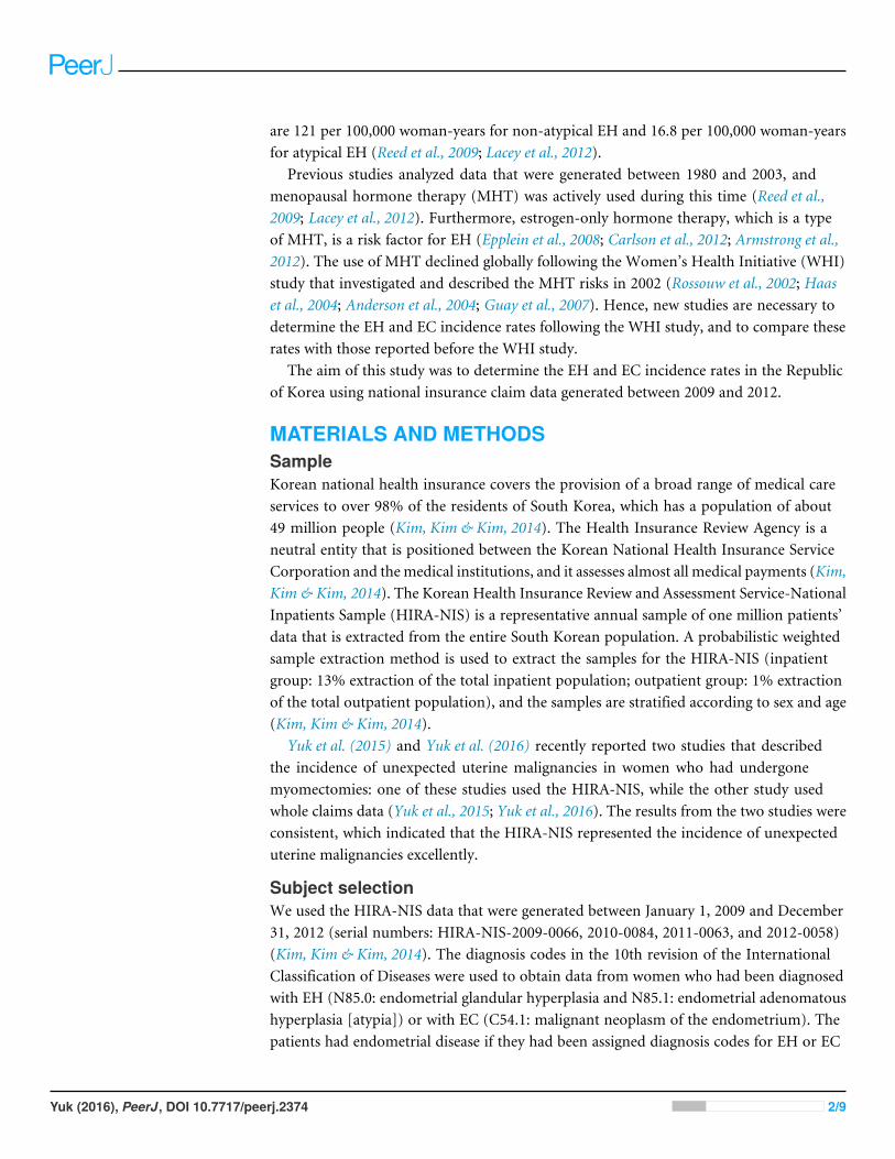
RESULTSData from 2,477,424 women were entered into the database from 2009 to 2012, and thedata from 1,868 women with EH and 868 women with EC were extracted for analysis(Table 1). The mean ages of the patients were 44.1 ± 0.4 years and 52.7 ± 0.6 years forthose with EH and EC, respectively. The incidence rates were 37 per 100,000 woman-years(95% confidence interval (CI) 34–40 per 100,000 woman-years) for EH and 7 per 100,000woman-years (95% CI (7–8) per 100,000 woman-years) for EC. Table 1 presents thecharacteristics of the women with EH or EC. Figure 1 presents the annual trends in theEH and EC incidence rates. Table 2 and Fig. 2 present the EH and EC incidence ratesstratified into 5-year age increments. The EH incidence, which included non-atypical EHand atypical EH, peaked when the women were in their late forties and it declined whenthe women were older than their late forties (Table 2; Fig. 2). The EC incidence peakedwhen the women were in their fifties (Table 2; Fig. 2).
DISCUSSIONOur study’s findings show that the EH and EC incidence rates were 37 per 100,000woman-years (non-atypical EH: 30 per 100,000 woman-years; atypical EH: 7 per 100,000woman-years) and 7 per 100,000 woman-years, respectively, and were somewhat lowerthan those determined from earlier studies. Previous studies that used data generated from1985 to 2003, determined that the EH and EC incidence rates were between 133 and 208per 100,000 woman-years (non-atypical EH: 121 per 100,000 woman-years; atypical EH: 17
Yuk (2016), PeerJ, DOI 10.7717/peerj.2374 3/9
Table 1 Characteristics of the women with endometrial hyperplasia or endometrial cancer from 2009 to 2012.
2009 2010 2011 2012 Total p value Mean age(years)
Number of patients 598,917 614,451 630,083 633,973 2,477,424Numbers of patients with each disease
Non-atypical EH 314 (21.4) 368 (25.1) 324 (22.1) 461 (31.4) 1,467 (100) 0.33a 44.1 ± 0.5b
Atypical EH 105 (26.1) 101 (25.1) 102 (25.3) 95 (23.6) 403 (100) 0.83a 44.1 ± 1.0b
EH 419 (22.4) 468 (25.1)c 425 (22.8)c 556 (29.8) 1,868 (100) 0.49a 44.1 ± 0.4b
Endometrial cancer 192 (22.1) 228 (26.3) 210 (24.2) 238 (27.4) 868 (100) 0.33a 52.7 ± 0.6b
Incidence per 100,000 woman-yearsd
Non-atypical EH 28 (22–33) 33 (27–39) 27 (22–33) 33 (27–39) 30 (27–33)Atypical EH 6 (4–8) 6 (4–9) 8 (5–10) 6 (4–9) 7 (5–8)EH 34 (28–40) 39 (33–46) 35 (29–41) 39 (33–46) 37 (34–40)Endometrial cancer 7 (5–8) 8 (7–9) 7 (6–8) 8 (7–9) 7 (7–8)
Notes.The data are presented as the numbers (%), the numbers (95% confidence intervals), or the means ± standard deviations.EH, endometrial hyperplasia.
aA weighted Pearson’s chi-square test was used to analyze the categorical data according to the year.bThe mean age was calculated using a weighted analysis method.cOne case had non-atypical endometrial hyperplasia and atypical endometrial hyperplasia simultaneously.dThe incidence rates were calculated using a weighted analysis method.
Figure 1 The annual trends in the endometrial hyperplasia and endometrial cancer incidence ratesfrom 2009 to 2010. EH, endometrial hyperplasia.
Yuk (2016), PeerJ, DOI 10.7717/peerj.2374 4/9
Table 2 The endometrial hyperplasia and endometrial cancer incidence rates from 2009 to 2012 strati-fied according to 5-year age increments.
Age (years) Incidence per 100,000 woman-years (95% confidence interval)
Non-atypical EH Atypical EH EH Endometrial cancer
<15 0 0 0 0 0 0.0 0 016–20 4 (−1–8) 0 0 4 (−1–8) 0 021–25 11 (3–18) 2 (−1–6) 13 (4–22) 1 (0–1)26–30 22 (12–32) 4 (0–8) 26 (15–37) 2 (1–3)31–35 30 (20–41) 11 (4–17) 41 (28–54) 6 (3–9)36–40 39 (27–51) 11 (5–17) 50 (36–63) 7 (4–9)41–45 74 (59–89) 15 (8–21) 89 (72–105) 7 (5–8)46–50 102 (84–120) 19 (13–26) 121 (102–141) 12 (9–14)51–55 61 (47–74) 9 (5–13) 69 (55–84) 20 (16–24)56–60 19 (11–27) 8 (2–15) 27 (17–37) 20 (17–23)61–65 8 (3–13) 2 (1–3) 9 (4–14) 15 (12–18)66–70 7 (2–12) 1 (0–2) 8 (3–13) 12 (9–15)71–75 7 (1–13) 4 (−2–10) 11 (3–19) 9 (6–12)>76 3 (0–7) 1 (0–2) 4 (0–8) 7 (3–11)Total 30 (27–33) 7 (5–8) 37 (34–40) 7 (7–8)
Notes.EH, endometrial hyperplasia.
Figure 2 The trends in the endometrial hyperplasia and endometrial cancer incidence rates from 2009to 2012 stratified according to 5-year age increments. EH, endometrial hyperplasia.
Yuk (2016), PeerJ, DOI 10.7717/peerj.2374 5/9
per 100,000 woman-years) and 78 per 100,000 woman-years, respectively (Reed et al., 2009;Lacey et al., 2012). While the reasons for these differences are unknown, the times at whichthe studies were performed might have affected the incidence rates. Previous studies werebased on data generated before 2002 when MHT was actively administered, but our studyanalyzed data that were generated after the WHI study (Rossouw et al., 2002; Haas et al.,2004; Anderson et al., 2004;Guay et al., 2007). The use of MHT has gradually declined sincethe WHI study’s findings provided evidence for the risks associated with MHT, and thismay have affected the EH incidence. The incidence of EH among women in their thirtiesin the present study (41–50 per 100,000 woman-years) is similar to that reported by Reedet al. (2009) (23–63 per 100,000 woman-years). However, the ratio of the perimenopausalincidence of EH among women aged 45–54 years to the incidence of EH among womenin their thirties was lower in our study (2.1) than that reported by Reed et al. (2009) (7.6).The ratio of the EH incidence among women aged around 60 years to the EH incidenceamong women who were in their thirties in the current study (0.4) showed a more distinctdifference compared with that reported by Reed et al. (2009) (6.1). The mean menopausalages are 51 years and 49 years in the United States and South Korea, respectively, and theEH incidence after perimenopause reported by Reed et al. was higher than that determinedin the current study (Kato et al., 1998; Reed et al., 2009; Cho et al., 2014). Differences in theuse of MHT may explain the disparities among the ratios, but the raw data analyzed in thestudies differed and they were not adjusted for MHT usage. Unlike unopposed estrogen,estrogen and progestogen in combination do not increase the EH incidence (Furness etal., 2012), indicating that further studies are needed. Furthermore, like breast cancer, theage-related EH or EC rates might vary among countries. While breast cancer rates increaseamong women until they are in their late forties or early fifties in countries in the FarEast, which include South Korea, Japan, and Taiwan, breast cancer rates in westernizedcountries increase consistently among women aged over 50 years (MacMahon, 2006; Leeet al., 2007). Obesity, which is a risk factor for EH and EC (Epplein et al., 2008; Carlsonet al., 2012; Armstrong et al., 2012), may be another factor that underlies the disparitiesbetween the studies’ data. Unfortunately, our study and previous studies did not includethe patients’ weights or body mass indices; hence, we were unable to analyze associationsbetween the EH and EC incidence rates and obesity, or to compare our study’s data withthose from previous studies. Further studies that adjust for obesity are needed.
Our study has several limitations. First, the data used in this study were based ondiagnosis codes and we did not analyze the histological data. Hence, the EH and ECdiagnoses were confirmed indirectly, and some EH or EC cases may have been wronglydiagnosed because of erroneous coding. However, patients with EH or EC were onlyselected when histological examinations had been carried out, so few wrongly diagnosedcases were analyzed in this study. Second, direct comparisons between our study andprevious studies are limited, because the current study did not adjust for race, the bodymass index, and MHT. The significance of this study’s data is that they verify the EH andEC incidence rates since 2009 and subsequent to the WHI study.
Yuk (2016), PeerJ, DOI 10.7717/peerj.2374 6/9
CONCLUSIONIn conclusion, the EH and EC incidence rates in South Korea from 2009 to 2012 were37 per 100,000 woman-years and 8 per 100,000 woman-years, respectively. The EH andEC diagnosis frequencies peaked when the women were in their late forties and fifties,respectively. The EH and EC rates were somewhat lower in our study than those reportedpreviously. Further studies are needed that adjust for race, MHT, and obesity.
ADDITIONAL INFORMATION AND DECLARATIONS
FundingThe author received no funding for this work.
Competing InterestsThe author declares there are no competing interests.
Author Contributions• Jin-Sung Yuk conceived and designed the experiments, performed the experiments,analyzed the data, contributed reagents/materials/analysis tools, wrote the paper,prepared figures and/or tables, reviewed drafts of the paper.
Data AvailabilityThe following information was supplied regarding data availability:
Yuk, Jin-Sung (2016): EH_incidence_2009_2012.sav.figshare.https://dx.doi.org/10.6084/m9.figshare.3124015.v1.
REFERENCESAnderson GL, Limacher M, Assaf AR, Bassford T, Beresford SAA, Black H, Bonds
D, Brunner R, Brzyski R, Caan B, Chlebowski R, Curb D, Gass M, Hays J, HeissG, Hendrix S, Howard BV, Hsia J, Hubbell A, Jackson R, Johnson KC, Judd H,Kotchen JM, Kuller L, LaCroix AZ, Lane D, Langer RD, Lasser N, Lewis CE, Man-son J, Margolis K, Ockene J, O’SullivanMJ, Phillips L, Prentice RL, RitenbaughC, Robbins J, Rossouw JE, Sarto G, StefanickML, Van Horn L,Wactawski-WendeJ, Wallace R,Wassertheil-Smoller S. 2004. Effects of conjugated equine estrogenin postmenopausal women with hysterectomy: the Women’s Health Initiativerandomized controlled trial. JAMA 291:1701–1712 DOI 10.1001/jama.291.14.1701.
Armstrong AJ, HurdWW, Elguero S, Barker NM, Zanotti KM. 2012. Diagnosis andmanagement of endometrial hyperplasia. Journal of Minimally Invasive Gynecology19:562–571 DOI 10.1016/j.jmig.2012.05.009.
Baak JP, Mutter GL, Robboy S, Van Diest PJ, Uyterlinde AM, Orbo A, Palazzo J, FianeB, Løvslett K, Burger C, Voorhorst F, Verheijen RH. 2005. The molecular geneticsand morphometry-based endometrial intraepithelial neoplasia classification systempredicts disease progression in endometrial hyperplasia more accurately than the
Yuk (2016), PeerJ, DOI 10.7717/peerj.2374 7/9
1994 World Health Organization classification system. Cancer 103:2304–2312DOI 10.1002/cncr.21058.
CarlsonMJ, Thiel KW, Yang S, Leslie KK. 2012. Catch it before it kills: progesterone,obesity, and the prevention of endometrial cancer. Discovery Medicine 14:215–222.
Cho B-J, Heo JW, Shin JP, Ahn J, Kim TW, Chung H. 2014. Association betweenreproductive factors and age-related macular degeneration in postmenopausalwomen: the Korea National Health and Nutrition Examination Survey 2010–2012.PLoS ONE 9:e102816 DOI 10.1371/journal.pone.0102816.
EppleinM, Reed SD, Voigt LF, Newton KM, Holt VL,Weiss NS. 2008. Risk of complexand atypical endometrial hyperplasia in relation to anthropometric measuresand reproductive history. American Journal of Epidemiology 168:563–570DOI 10.1093/aje/kwn168.
Furness S, Roberts H, Marjoribanks J, Lethaby A. 2012.Hormone therapy in post-menopausal women and risk of endometrial hyperplasia. The Cochrane Database ofSystematic Reviews 8:CD000402 DOI 10.1002/14651858.CD000402.pub4.
GuayM-P, Dragomir A, Pilon D, Moride Y, Perreault S. 2007. Changes in pattern ofuse, clinical characteristics and persistence rate of hormone replacement therapyamong postmenopausal women after the WHI publication. Pharmacoepidemiologyand Drug Safety 16:17–27 DOI 10.1002/pds.1273.
Haas JS, Kaplan CP, Gerstenberger EP, Kerlikowske K. 2004. Changes in the use ofpostmenopausal hormone therapy after the publication of clinical trial results.Annals of Internal Medicine 140:184–188DOI 10.7326/0003-4819-140-3-200402030-00009.
Kato I, Toniolo P, Akhmedkhanov A, Koenig KL, Shore R, Zeleniuch-Jacquotte A.1998. Prospective study of factors influencing the onset of natural menopauseJournal of Clinical Epidemiology 51:1271–1276 DOI 10.1016/S0895-4356(98)00119-X.
Kim L, Kim J-A, Kim S. 2014. A guide for the utilization of Health Insurance Review andAssessment Service National Patient Samples. Epidemiology and Health 36:e2014008DOI 10.4178/epih/e2014008.
Lacey JV, Chia VM, Rush BB, Carreon DJ, Richesson DA, Ioffe OB, Ronnett BM,Chatterjee N, Langholz B, ShermanME, Glass AG. 2012. Incidence rates ofendometrial hyperplasia, endometrial cancer and hysterectomy from 1980 to 2003within a large prepaid health plan. International Journal of Cancer 131:1921–1929DOI 10.1002/ijc.27457.
Lee JH, Yim SH,Won YJ, Jung KW, Son BH, Lee HD, Lee ES, Yoo KY, Ahn SH, ShinHR, Members of Korean Breast Cancer Society(KBCS). 2007. Population-basedbreast cancer statistics in Korea during 1993–2002: incidence, mortality, and survival.Journal of Korean Medical Science 22(Suppl):S11–S16DOI 10.3346/jkms.2007.22.S.S11.
MacMahon B. 2006. Epidemiology and the causes of breast cancer. International Journalof Cancer 118:2373–2378 DOI 10.1002/ijc.21404.
Yuk (2016), PeerJ, DOI 10.7717/peerj.2374 8/9
Reed SD, Newton KM, ClintonWL, EppleinM, Garcia R, Allison K, Voigt LF, WeissNS. 2009. Incidence of endometrial hyperplasia. American Journal of Obstetrics andGynecology 200:678.e1–6 DOI 10.1016/j.ajog.2009.02.032.
Rossouw JE, Anderson GL, Prentice RL, LaCroix AZ, Kooperberg C, StefanickML,Jackson RD, Beresford SAA, Howard BV, Johnson KC, Kotchen JM, Ockene J.2002. Risks and benefits of estrogen plus progestin in healthy postmenopausalwomen: principal results From the Women’s Health Initiative randomized con-trolled trial. The Journal of the American Medical Association 288:321–333DOI 10.1001/jama.288.3.321.
SalmanMC, Usubutun A, Boynukalin K, Yuce K. 2010. Comparison of WHO andendometrial intraepithelial neoplasia classifications in predicting the presence ofcoexistent malignancy in endometrial hyperplasia. Journal of Gynecologic Oncology21:97–101 DOI 10.3802/jgo.2010.21.2.97.
Yuk J-S, Ji HY, Lee SH, Park YS, Lee JH. 2015. Unexpected uterine malignancy inwomen who have undergone myomectomy. International Journal of Gynaecology andObstetrics 129:270–271 DOI 10.1016/j.ijgo.2014.12.004.
Yuk J-S, Ji HY, Shin J-Y, Kim LY, Kim S-H, Lee JH. 2016. Comparison of survival out-comes in women with unsuspected uterine malignancy diagnosed after laparotomicversus laparoscopic myomectomy: a national, population-based study. Annals ofSurgical Oncology 23:1287–1293 DOI 10.1245/s10434-015-4976-3.
Yuk (2016), PeerJ, DOI 10.7717/peerj.2374 9/9