The Immune System - Recently Added Immune System.pdf · Internal defenses Cells of the Immune...
68
Innate immunity Acquired immunity Antibody Classes Examples of immune system disorders The Immune System
Transcript of The Immune System - Recently Added Immune System.pdf · Internal defenses Cells of the Immune...

Innate immunity
Acquired immunity
Antibody Classes
Examples of immune system disorders
The Immune System

Overview
An animal must defend itself from the many
dangerous pathogens it may encounter in the
environment.
Two major kinds of defense have evolved that
counter these threats:
Innate immunity
Acquired immunity, also called adaptive
immunity
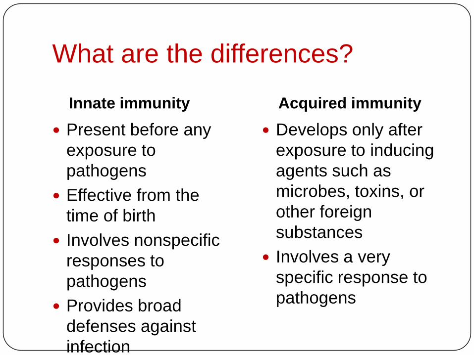
What are the differences?
Innate immunity Acquired immunity
Present before any
exposure to
pathogens
Effective from the
time of birth
Involves nonspecific
responses to
pathogens
Provides broad
defenses against
infection
Develops only after
exposure to inducing
agents such as
microbes, toxins, or
other foreign
substances
Involves a very
specific response to
pathogens

INNATE IMMUNITY
Recognition of traitsshared by broad rangesof pathogens, using asmall set of receptors
•
•Rapid (ready) response
•Recognition of traitsspecific to particularpathogens, using a vastarray of receptors
•Slower (unready) response
ACQUIRED IMMUNITY
Pathogens(microorganisms
and viruses)
Barrier defenses:SkinMucous membranesSecretions
Internal defenses:Phagocytic cellsAntimicrobial proteinsInflammatory responseNatural killer cells
Humoral response:Antibodies defend againstinfection in body fluids.
Cell-mediated response:Cytotoxic lymphocytes defendagainst infection in body cells.
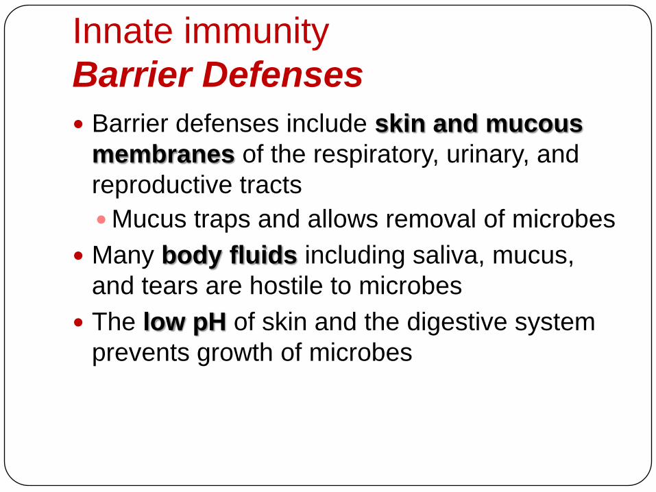
Innate immunity
Barrier Defenses
Barrier defenses include skin and mucous
membranes of the respiratory, urinary, and
reproductive tracts
Mucus traps and allows removal of microbes
Many body fluids including saliva, mucus,
and tears are hostile to microbes
The low pH of skin and the digestive system
prevents growth of microbes
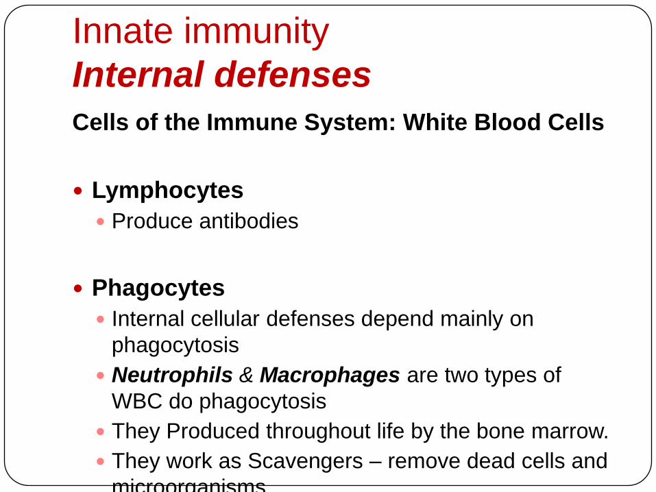
Innate immunity
Internal defenses
Cells of the Immune System: White Blood Cells
Lymphocytes
Produce antibodies
Phagocytes
Internal cellular defenses depend mainly on
phagocytosis
Neutrophils & Macrophages are two types of
WBC do phagocytosis
They Produced throughout life by the bone marrow.
They work as Scavengers – remove dead cells and
microorganisms.

Macrophages
Neutrophils Larger than neutrophils.
Found in the organs, not the blood.
Made in bone marrow as monocytes, called macrophages once they reach organs.
Long lived
Initiate immune responses as they display antigens from the pathogens to the lymphocytes.
60% of WBCs
„Patrol tissues‟ as they squeeze out of the capillaries.
Large numbers are released during infections
Short lived – die after digesting bacteria
Dead neutrophils make up a large proportion of

There are different types of phagocytic cells:
• Neutrophils engulf and destroy microbes
• Macrophages are part of the lymphatic system and are
found throughout the body
• Eosinophils discharge destructive enzymes
• Dendritic cells stimulate development of acquired immunity
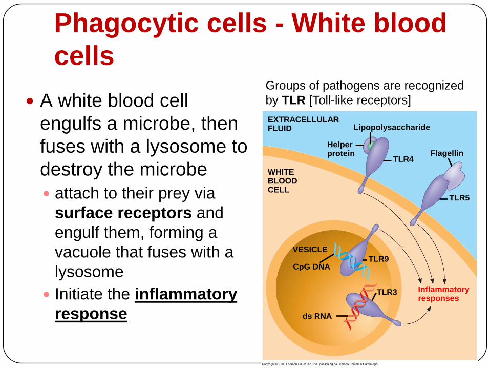
Phagocytic cells - White blood
cells
A white blood cell
engulfs a microbe, then
fuses with a lysosome to
destroy the microbe
attach to their prey via
surface receptors and
engulf them, forming a
vacuole that fuses with a
lysosome
Initiate the inflammatory
response
EXTRACELLULARFLUID Lipopolysaccharide
FlagellinTLR4
TLR5
Helperprotein
TLR9
TLR3
WHITEBLOODCELL
VESICLE
CpG DNA
ds RNA
Inflammatoryresponses
Groups of pathogens are recognized
by TLR [Toll-like receptors]

Phagocytosis
If cells are under attack they release
histamine.
Histamine plus chemicals from pathogens
mean neutrophils are attracted to the site of
attack.
Pathogens are attached to antibodies and
neutrophils have antibody receptors.
Enodcytosis of neutrophil membrane
phagocytic vacuole Lysosomes attach to
phagocytic vacuole pathogen digested by
proteases
Internal defenses
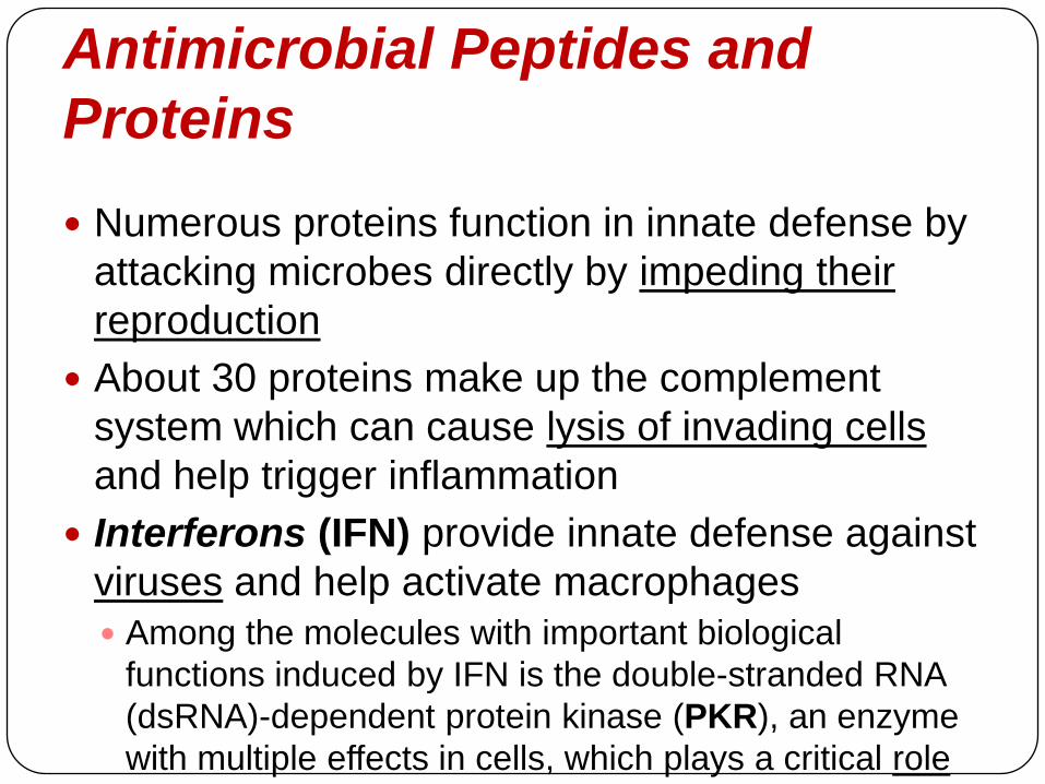
Antimicrobial Peptides and
Proteins
Numerous proteins function in innate defense by
attacking microbes directly by impeding their
reproduction
About 30 proteins make up the complement
system which can cause lysis of invading cells
and help trigger inflammation
Interferons (IFN) provide innate defense against
viruses and help activate macrophages
Among the molecules with important biological
functions induced by IFN is the double-stranded RNA
(dsRNA)-dependent protein kinase (PKR), an enzyme
with multiple effects in cells, which plays a critical role
in the antiviral defense mechanism of the host

Internal defenses
Inflammatory Responses
Following an injury, mast cells release histamine, which
promotes changes in blood vessels; this is part of
inflammatory response
Inflammatory responses are these changes which
increase local blood supply and allow more phagocytes
and antimicrobial proteins to enter tissues
Pus, a fluid rich in white blood cells, dead microbes, and cell
debris, accumulates at the site of inflammation
Inflammation can be either local or systemic (throughout
the body)
Fever is a systemic inflammatory response triggered by
pyrogens released by macrophages, and toxins from
pathogens
Septic shock is a life-threatening condition caused by an overwhelming
inflammatory response

Major events in the local inflammatory
responsePathogen Pin
Macrophage
Chemical signals
CapillaryPhagocytic cells
Red blood cell
Blood
clotting
elements
Blood clot
Phagocytosis
Fluid, antimicrobial
proteins, and clotting
elements move from
the blood to the site.
Clotting begins.
2Chemical signals
released by activated
macrophages
and mast cells at the
injury site cause
nearby capillaries
to widen and become
more permeable.
1 Chemokines released
by various kinds of cells
attract more phagocytic
cells from the blood to
the injury site.
3Neutrophils and
macrophages
phagocytose
pathogens and cell
debris at the site, and
the tissue heals.
4

Internal defenses
Natural Killer Cells
Natural killer (NK) cells
Patrol the body and attack virus-infected body cells
and cancer cells
Trigger apoptosis in the cells they attack
How do Natural killer (NK) cells work?
All cells in the body (except red blood cells) have a
class-I of MHC (Major Histocompatibility Complex)
protein on their surface
Cancerous or infected cells no longer express this
protein; causing natural killer (NK) cells attack these
damaged cells
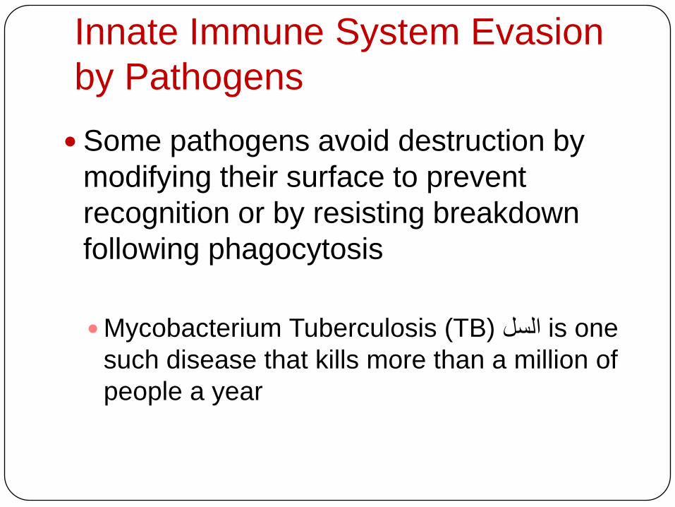
Innate Immune System Evasion
by Pathogens
Some pathogens avoid destruction by
modifying their surface to prevent
recognition or by resisting breakdown
following phagocytosis
Mycobacterium Tuberculosis (TB) السل is one
such disease that kills more than a million of
people a year

INNATE IMMUNITY
Recognition of traitsshared by broad rangesof pathogens, using asmall set of receptors
•
•Rapid response
•Recognition of traitsspecific to particularpathogens, using a vastarray of receptors
•Slower response
ACQUIRED IMMUNITY
Pathogens(microorganisms
and viruses)
Barrier defenses:SkinMucous membranesSecretions
Internal defenses:Phagocytic cellsAntimicrobial proteinsInflammatory responseNatural killer cells
Humoral response:Antibodies defend againstinfection in body fluids.
Cell-mediated response:Cytotoxic lymphocytes defendagainst infection in body cells.

Acquired immunity
Lymphocytes the white blood cells recognize and
respond to antigens, foreign molecules
Lymphocytes contribute to immunological memory, an
enhanced response to a foreign molecule
encountered previously
Two types of lymphocytic cells:
T cells, mature in Thymus
B cells, mature in Bone marrow then concentrate in
lymph nodes and spleen
Mature (B) and (T) cells then circulate in the blood

Acquired immunity Acquired immunity has two responses where Helper T
cells aid both:
1. Humoral immune response involves activation and
clonal selection of B cells, resulting in production of
secreted antibodies
2. Cell-mediated immune response involves activation
and clonal selection of cytotoxic T cellsAntigen-presentingcell
Peptide antigen
Cell-mediated
immunity
(attack on
infected cells)
Class II MHC molecule
CD4
TCR (T cell receptor)
Helper T cell
Humoral
immunity
(secretion of
antibodies by
plasma cells) Cytotoxic T cell
Cytokines
B cell
Bacterium
+
+ +
+
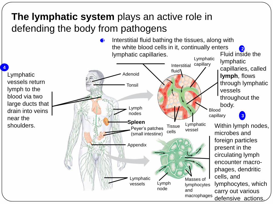
The lymphatic system plays an active role in
defending the body from pathogens
Adenoid
Tonsil
Lymph
nodes
SpleenPeyer‟s patches
(small intestine)
Appendix
Lymphatic
vesselsMasses of
lymphocytes
and
macrophages
Tissue
cells
Lymphatic
vessel
Blood
capillary
Lymphatic
capillaryInterstitial
fluid
Lymph
node
Interstitial fluid bathing the tissues, along with
the white blood cells in it, continually enters
lymphatic capillaries.
1
Fluid inside the
lymphatic
capillaries, called
lymph, flows
through lymphatic
vessels
throughout the
body.
2
Within lymph nodes,
microbes and
foreign particles
present in the
circulating lymph
encounter macro-
phages, dendritic
cells, and
lymphocytes, which
carry out various
defensive actions.
3
Lymphatic
vessels return
lymph to the
blood via two
large ducts that
drain into veins
near the
shoulders.
4

Antigen Recognition by
Lymphocytes Lymphocytes are activated by Cytokines which are
secreted by macrophages and dendritic cells
Lymphocyte receptors provide pathogen-specific
recognition
B cells and T cells have receptor proteins that can
bind to foreign molecules
Each individual lymphocyte is specialized to
recognize a specific type of molecule
An antigen is any foreign molecule to which a
lymphocyte responds
A single B cell or T cell has about 100,000 identical
antigen receptors
Antigen-bindingsite
Antigen-binding site
Antigen-bindingsite
Disulfidebridge
Variableregions
Constantregions
Transmembraneregion
Plasmamembrane
Lightchain
Heavy chains
T cell
chain chain
Disulfide bridge
Cytoplasm of T cell
(b) T cell receptor
Cytoplasm of B cell
(a) B cell receptor
B cell
C C C C
VV
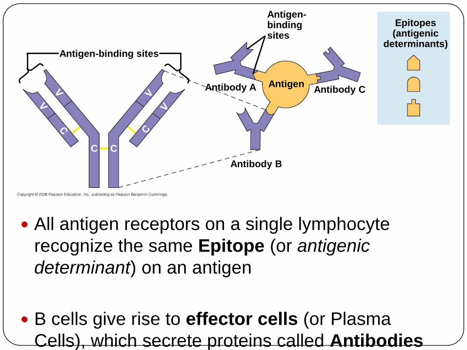
Antigen-binding sites
Antigen-bindingsites
Epitopes(antigenic
determinants)
Antigen
Antibody B
Antibody CAntibody A
CC
All antigen receptors on a single lymphocyte
recognize the same Epitope (or antigenic
determinant) on an antigen
B cells give rise to effector cells (or Plasma
Cells), which secrete proteins called Antibodies
(or immunoglobulins)

The Antigen Receptors of B Cells
B cell receptors bind to specific, intact antigens
The B cell receptor consists of two identical
heavy chains and two identical light chains:
The tips of the chains form a constant (C) region, and
each chain contains a variable (V) region, so named
because its amino acid sequence varies extensively
from one B cell to another
Secreted antibodies, or immunoglobulins, are
structurally similar to B cell receptors but lack
transmembrane regions that anchor receptors in
the plasma membrane

The Antigen Receptors of T Cells
Each T cell receptor consists of two
different polypeptide chains:
The tips of the chain form a variable (V) region;
the rest is a constant (C) region
T cells can bind to an antigen that is free
or on the surface of a pathogen
T cells bind to antigen fragments
presented on a host cell

Generation of Lymphocyte Diversity by
Gene Rearrangement
Differences in the variable region account for
specificity of antigen receptors
The immunoglobulin (Ig) gene encodes one
chain of the B cell receptor
Many different chains can be produced from the
same Ig chain gene by rearrangement of the
DNA
Rearranged DNA is transcribed and translated
and the antigen receptor formed

Immunoglobulin (antibody) gene rearrangement
The capacity to generate diversity is built into the
structure of the Ig light-chain gene.
A receptor light chain is encoded by three gene
segments:
a variable (V) segment,
a joining (J) segment
a constant (C) segment.
The V and J segments together encode the
variable region of the receptor chain, while the C
segment encodes the entire constant region.
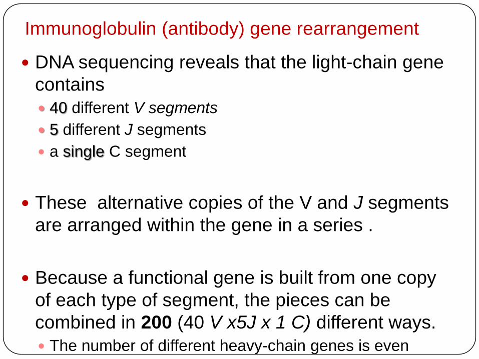
Immunoglobulin (antibody) gene rearrangement
DNA sequencing reveals that the light-chain gene
contains
40 different V segments
5 different J segments
a single C segment
These alternative copies of the V and J segments
are arranged within the gene in a series .
Because a functional gene is built from one copy
of each type of segment, the pieces can be
combined in 200 (40 V x5J x 1 C) different ways.
The number of different heavy-chain genes is even
greater.

DNA of undifferentiated B cell
1
DNA of differentiated B cell
pre-mRNA
mRNA
Light-chain polypeptide
Variableregion
Constantregion
Translation
B cell
B cell receptor
RNA processing
Transcription
DNA deleted between randomly selected V and J
segments
Functional gene
V37 V38 V39 V40 J1 J2 J3 J4 CJ5 Intron
V37 V38 V39 CJ5 Intron
V39 CJ5 Intron
V39 CJ5 Poly-A tailCap
CV
V
V
V
V
C C
C C
2
3
4

Origin of Self-Tolerance
Antigen receptors are generated by
random rearrangement of DNA
As lymphocytes mature in bone marrow
or the thymus, they are tested for self-
reactivity
Lymphocytes with receptors specific for the
body‟s own molecules are destroyed by
apoptosis, or rendered nonfunctional

Stem cell
Cell division and gene rearrangement
Antigen
Clonal selection
Elimination of
self-reactive
B cells
Formation of activated cell populationsAntibody
Microbe
Memory cells Effector B cells
Receptors bind to antigens
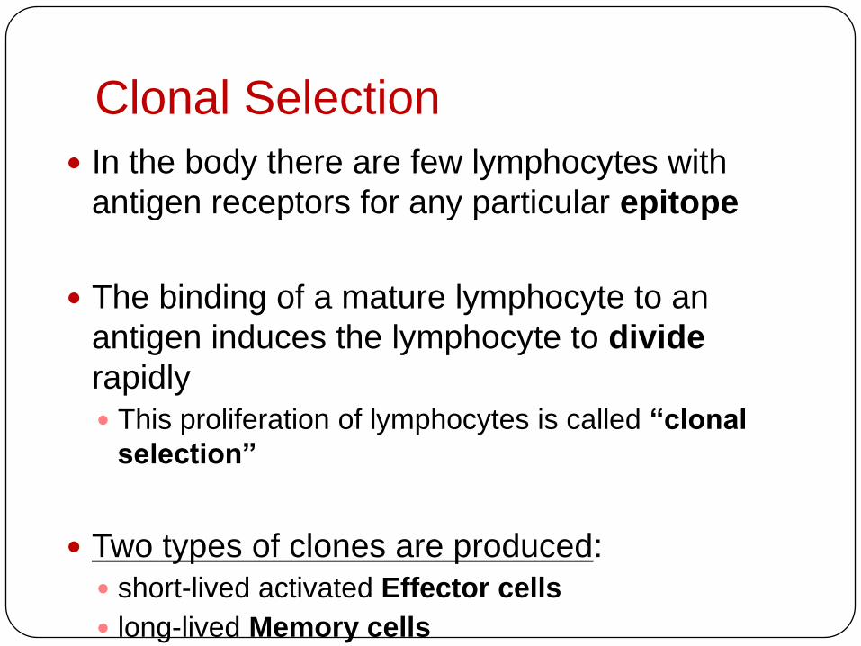
Clonal Selection
In the body there are few lymphocytes with
antigen receptors for any particular epitope
The binding of a mature lymphocyte to an
antigen induces the lymphocyte to divide
rapidly
This proliferation of lymphocytes is called “clonal
selection”
Two types of clones are produced:
short-lived activated Effector cells
long-lived Memory cells
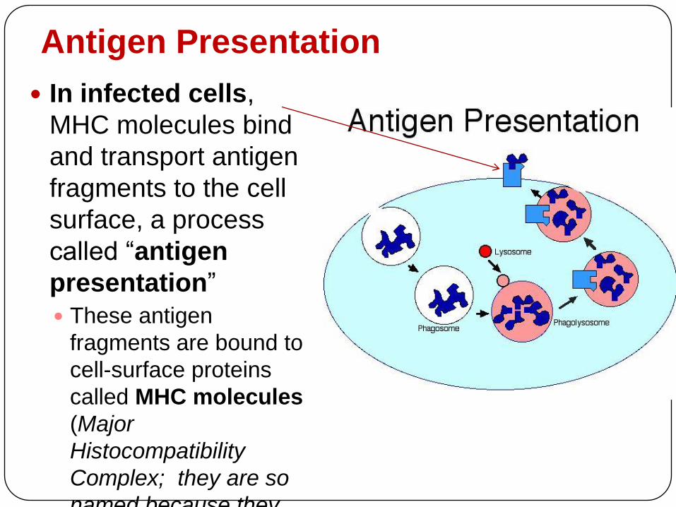
Antigen Presentation
In infected cells,
MHC molecules bind
and transport antigen
fragments to the cell
surface, a process
called “antigen
presentation”
These antigen
fragments are bound to
cell-surface proteins
called MHC molecules
(Major
Histocompatibility
Complex; they are so
named because they

Antigen Presentation
Different classes of MHC molecules:
Class I MHC molecules: are found on almost all nucleated cells of
the body and display antigens to cytotoxic T cells
Class II MHC molecules are located mainly on dendritic cells,
macrophages, and B cells that display to cytotoxic T cells and
helper T cellsInfected cell
Antigenfragment
Class I MHCmolecule
T cellreceptor
(a)
Antigenassociateswith MHCmolecule
T cellrecognizescombination
Cytotoxic T cell (b) Helper T cell
T cellreceptor
Class II MHCmolecule
Antigenfragment
Antigen-presentingcell
Microbe
1
11
2
22

Cytotoxic T Cells related to class I MHC
Cytotoxic T cells are the effector cells in cell-
mediated immune response
Cytotoxic T cells make CD8, a surface protein that
greatly enhances interaction between a target cell and a
cytotoxic T cell
Binding to a class I MHC complex on an infected cell
activates a cytotoxic T cell and makes it an active killer
The activated cytotoxic T cell secretes proteins
that destroy the infected target cell :
1. they attach to the cells to be killed and release
proteins called perforins that create holes in the cell
membrane of the target cell, with consequent cell
lysis.
2. they attach to a cell and kill it by triggering
mechanisms that induce programmed cell death, or

Cytotoxic T cell
Perforin
Granzymes
TCRCD8
Class I MHCmolecule
Targetcell
Peptideantigen
Pore
Released cytotoxic T cell
Dying target cell
The killing action of cytotoxic T cells

Antigen Presentation
Different classes of MHC molecules:
Class I MHC molecules: are found on almost all nucleated cells of
the body and display antigens to cytotoxic T cells
Class II MHC molecules are located mainly on dendritic cells,
macrophages, and B cells that display to cytotoxic T cells and
helper T cellsInfected cell
Antigenfragment
Class I MHCmolecule
T cellreceptor
(a)
Antigenassociateswith MHCmolecule
T cellrecognizescombination
Cytotoxic T cell (b) Helper T cell
T cellreceptor
Class II MHCmolecule
Antigenfragment
Antigen-presentingcell
Microbe
1
11
2
22
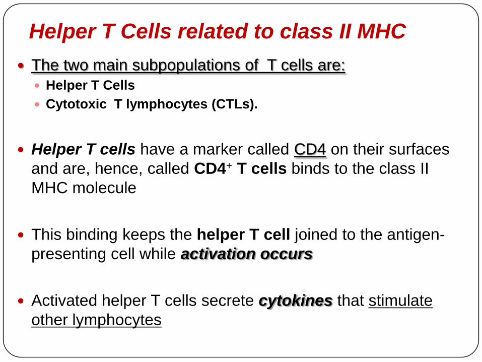
Helper T Cells related to class II MHC
The two main subpopulations of T cells are:
Helper T Cells
Cytotoxic T lymphocytes (CTLs).
Helper T cells have a marker called CD4 on their surfaces
and are, hence, called CD4+ T cells binds to the class II
MHC molecule
This binding keeps the helper T cell joined to the antigen-
presenting cell while activation occurs
Activated helper T cells secrete cytokines that stimulate
other lymphocytes

Acquired immunity Acquired immunity has two responses where Helper T
cells aid both:
1. Humoral immune response involves activation and
clonal selection of B cells, resulting in production of
secreted antibodies
2. Cell-mediated immune response involves activation
and clonal selection of cytotoxic T cellsAntigen-presentingcell
Peptide antigen
Cell-mediated
immunity
(attack on
infected cells)
Class II MHC molecule
CD4
TCR (T cell receptor)
Helper T cell
Humoral
immunity
(secretion of
antibodies by
plasma cells) Cytotoxic T cell
Cytokines
B cell
Bacterium
+
+ +
+
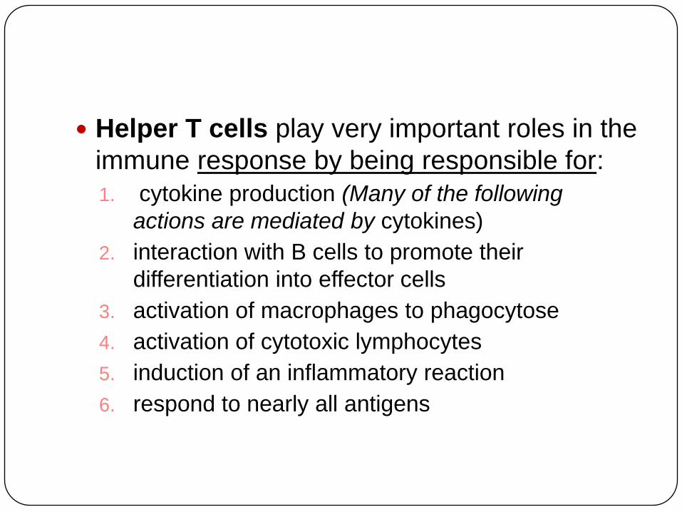
Helper T cells play very important roles in the
immune response by being responsible for:
1. cytokine production (Many of the following
actions are mediated by cytokines)
2. interaction with B cells to promote their
differentiation into effector cells
3. activation of macrophages to phagocytose
4. activation of cytotoxic lymphocytes
5. induction of an inflammatory reaction
6. respond to nearly all antigens

The first encounter of a CD4+ or CD8+ T cell
with its specific epitope is followed by
amplification of that clone
some of the cells of this increased
population become effector cells and some
remain memory helper or memory
cytotoxic T cells, reacting rapidly to the
next presentation of the same epitope

B Cells:
A Response to Extracellular
Pathogens
The humoral response is characterized by
secretion of antibodies by B cells
Activation of B cells is aided by cytokines
and antigen binding to helper T cells
Clonal selection of B cells generates antibody-
secreting effector cells (plasma cells), the
effector cells of humoral immunity

Antigen-presenting cell
Endoplasmicreticulum ofplasma cell
Secretedantibodymolecules
Bacterium
B cellPeptideantigen
Class II MHCmolecule
TCR CD4
Helper T cellActivated
helper T cell
Cytokines
Clone of memory
B cells
Clone of plasma cells
2 µm
+
B cell activation in the humoral immune response

The Role of Antibodies in
Immunity Neutralization: occurs when a pathogen can no longer
infect a host because it is bound to an antibody
Opsonization: occurs when antibodies bound to antigens
increase phagocytosis
Activation of complement system and pore formation:
Antibodies together with proteins of the complement
system generate a membrane attack complex and cell
lysis.Viral neutralization
Virus
Opsonization
Bacterium
Macrophage
Activation of complement system and pore formation
Complement proteins
Formation of
membrane
attack complex
Flow of water
and ions
Pore
Foreign
cell

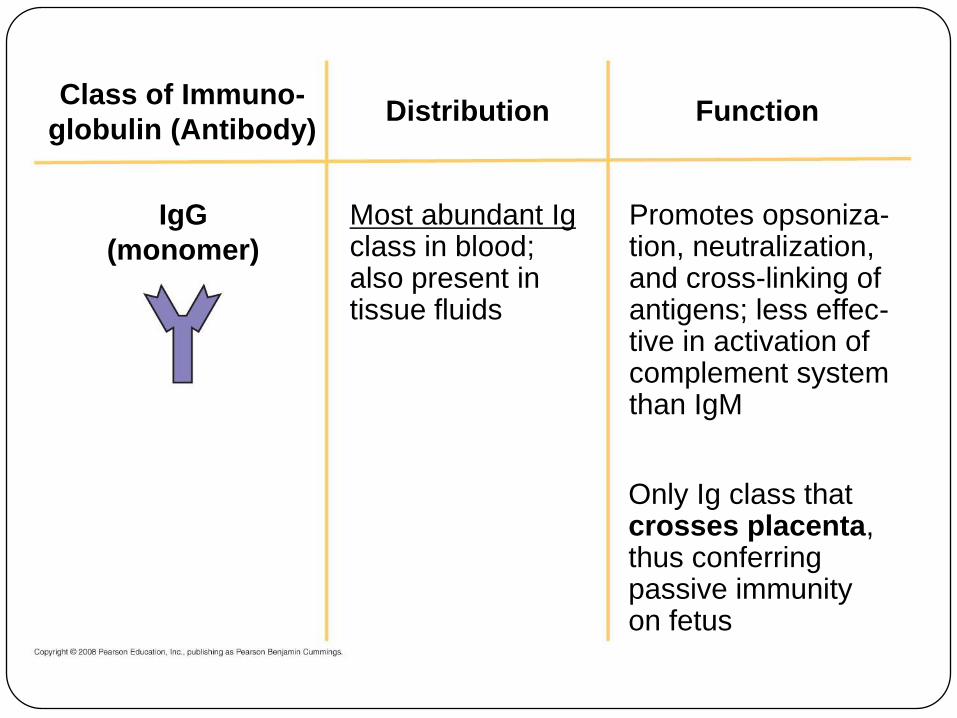
Antibody Classes
The five major classes of antibodies, or
immunoglobulins, differ in distribution and
function
Polyclonal antibodies are the products of many
different clones of B cells following exposure to a
microbial antigen
Monoclonal antibodies are prepared from a
single clone of B cells grown in culture

DistributionClass of Immuno-
globulin (Antibody)
IgM
(pentamer)
J chain
First Ig classproduced afterinitial exposure toantigen; then itsconcentration inthe blood declines
Promotes neutraliza-tion and cross-linking of antigens;very effective incomplement systemactivation
Function
Distribution FunctionClass of Immuno-
globulin (Antibody)
IgG
(monomer)
Most abundant Igclass in blood;also present intissue fluids
Promotes opsoniza-tion, neutralization,and cross-linking ofantigens; less effec-tive in activation ofcomplement systemthan IgM
Only Ig class thatcrosses placenta,thus conferringpassive immunityon fetus

Distribution FunctionClass of Immuno-
globulin (Antibody)
IgA
(dimer)
J chain
Secretory
component
Present insecretions suchas tears, saliva,mucus, andbreast milk
Provides localizeddefense of mucousmembranes bycross-linking andneutralization ofantigens
Presence in breastmilk conferspassive immunityon nursing infant

Distribution FunctionClass of Immuno-
globulin (Antibody)
IgE
(monomer)Present in bloodat lowconcentrations
Triggers release frommast cells andbasophils of hista-mine and otherchemicals that causeallergic reactions

Distribution FunctionClass of Immuno-
globulin (Antibody)
IgD
(monomer)
Trans-membraneregion
Present primarilyon surface ofB cells that havenot been exposedto antigens
Acts as antigenreceptor in theantigen-stimulatedproliferation anddifferentiation ofB cells (clonalselection)

Active and Passive Immunization
Active immunity develops naturally in response
to an infection
It can also develop following immunization, also called
vaccination
In immunization, a nonpathogenic form of a microbe or
part of a microbe elicits an immune response to an
immunological memory
Passive immunity provides immediate, short-
term protection
It is conferred naturally when IgG crosses the placenta
from mother to fetus or when IgA passes from mother to
infant in breast milk
It can be conferred artificially by injecting antibodies into

The exposure to a specific
antigen
The first exposure to a specific antigen
represents the “primary immune
response”
During this time, effector B cells (or plasma
cells) are generated, and T cells are
activated to their effector forms
In the “secondary immune response”,
memory cells facilitate a faster, more
efficient response

Antibodiesto A
Antibodiesto B
Secondary immune response toantigen A produces antibodies to A;
primary immune response to antigen
B produces antibodies to B.
Primary immune responseto antigen A produces
antibodies to A.
Antibody c
oncentr
ation
(arb
itra
ry u
nits)
Exposureto antigen A
Exposure toantigens A and B
Time (days)
104
103
102
101
100
0 7 14 21 28 35 42 49 56
The specificity of
immunological memory

Examples of immune system
disorders
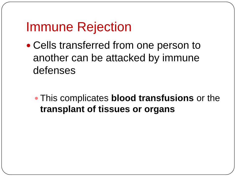
Immune Rejection
Cells transferred from one person to
another can be attacked by immune
defenses
This complicates blood transfusions or the
transplant of tissues or organs

Blood Groups
Antigens on red blood cells determine
whether a person has blood type A (A antigen),
B (B antigen), AB (both A and B antigens), or
O (neither antigen)
Antibodies to nonself blood types exist in the
body
Transfusion with incompatible blood leads to
destruction of the transfused cells
Recipient-donor combinations can be fatal
or safe

Tissue and Organ Transplants
MHC molecules are different among
genetically nonidentical individuals
Differences in MHC molecules stimulate
rejection of tissue grafts and organ transplants
Chances of successful transplantation
increase if donor and recipient MHC tissue
types are well matched
Immunosuppressive drugs facilitate
transplantation
Lymphocytes in bone marrow transplants may
cause the donor tissue to reject the recipient

Exaggerated, self-directed, or diminished
immune responses can cause disease
Disruption in immune system function can
elicit or exacerbate disease
Some pathogens have evolved to diminish the
effectiveness of host immune responses
If the delicate balance of the immune system is
disrupted the effects on the individual can range
from minor to often fatal consequences
Allergies
Autoimmune Diseases
Immunodeficiency Diseases
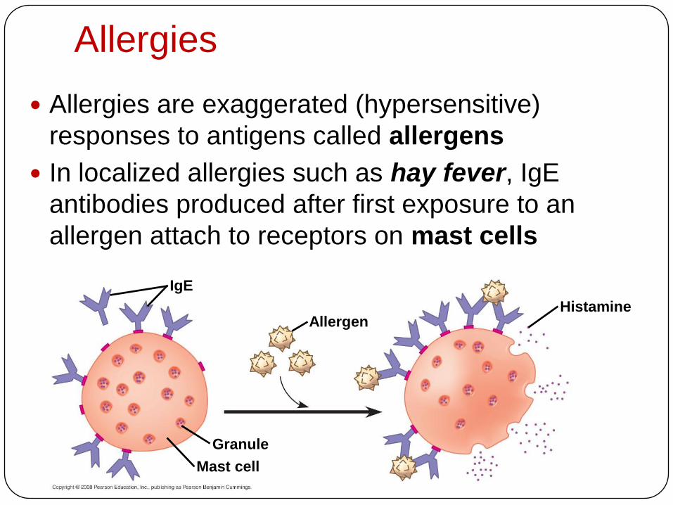
Allergies
Allergies are exaggerated (hypersensitive)
responses to antigens called allergens
In localized allergies such as hay fever, IgE
antibodies produced after first exposure to an
allergen attach to receptors on mast cells
Allergen
IgE
Granule
Mast cell
Histamine

Allergies
The next time the allergen enters the body, it
binds to mast cell–associated IgE molecules
Mast cells release histamine and other
mediators that cause vascular changes
leading to typical allergy symptoms
An acute allergic response can lead to
anaphylactic shock, a life-threatening
reaction that can occur within seconds of
allergen exposure
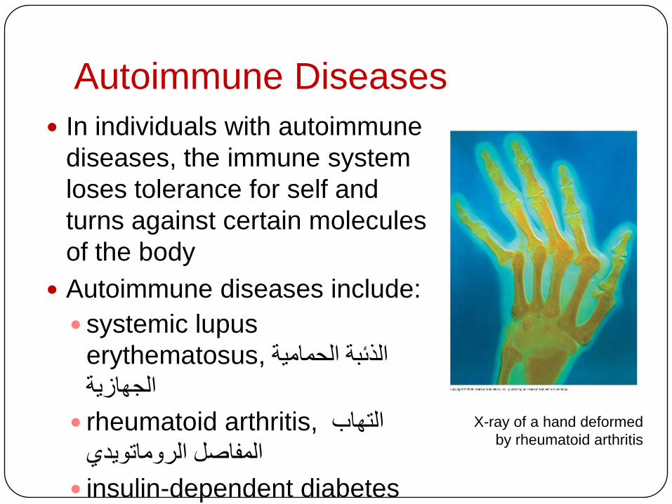
Autoimmune Diseases
In individuals with autoimmune
diseases, the immune system
loses tolerance for self and
turns against certain molecules
of the body
Autoimmune diseases include:
systemic lupus
erythematosus, الذئبة الحمامية
الجهازية
rheumatoid arthritis, التهاب
المفاصل الروماتىيدي
insulin-dependent diabetes
mellitus,
X-ray of a hand deformed
by rheumatoid arthritis
Exertion, Stress, and the Immune
System
Moderate exercise improves immune system
function
Psychological stress has been shown to
disrupt hormonal, nervous, and immune
systems

Antigenic Variation
Through antigenic variation, some pathogens
are able to change epitope expression and
prevent recognition
The human influenza virus mutates rapidly,
and new flu vaccines must be made each year
Human viruses occasionally exchange genes
with the viruses of domesticated animals
This poses a danger as human immune systems
are unable to recognize the new viral strain

Weeks after infection
Millio
ns
of
para
sit
es
per
mL
of
blo
od
Antibodies tovariant 1appear
Antibodies tovariant 2appear
Antibodies tovariant 3appear
Variant 3Variant 2Variant 1
25 26 27 280
0.5
1.0
1.5
Latency
Some viruses may remain in a host in an
inactive state called latency
Herpes simplex viruses can be present in a
human host without causing symptoms

Immunodeficiency Diseases
Inborn immunodeficiency results from
hereditary or developmental defects that
prevent proper functioning of innate, humoral,
and/or cell-mediated defenses
Acquired immunodeficiency results from
exposure to chemical and biological agents
Acquired immunodeficiency syndrome (AIDS) is
caused by a virus
Acquired immune system evasion by
Pathogens
Pathogens have evolved mechanisms to attack
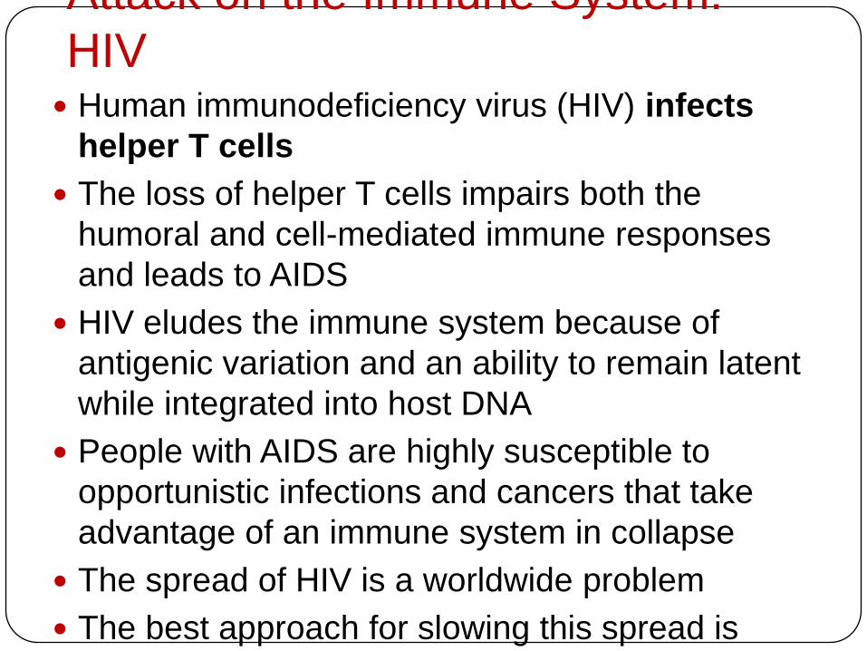
Attack on the Immune System:
HIV Human immunodeficiency virus (HIV) infects
helper T cells
The loss of helper T cells impairs both the
humoral and cell-mediated immune responses
and leads to AIDS
HIV eludes the immune system because of
antigenic variation and an ability to remain latent
while integrated into host DNA
People with AIDS are highly susceptible to
opportunistic infections and cancers that take
advantage of an immune system in collapse
The spread of HIV is a worldwide problem
The best approach for slowing this spread is
education about practices that transmit the virus

Latency
Relative antibodyconcentration
AIDSH
elp
er
T c
ell
co
ncen
trati
on
in b
loo
d (
cell
s/m
m3)
Helper T cellconcentration
Relative HIVconcentration
Years after untreated infection0 1 2 3 4 5 6 7 8 9 10
0
200
400
600
800
The progress of an untreated HIV infection

Cancer and Immunity
The frequency of certain cancers
increases when the immune response is
impaired
Two suggested explanations are
Immune system normally suppresses
cancerous cells
Increased inflammation increases the risk of
cancer