The Immediate Placement of Dental Implants Into …albest/DENS580/pdfs/Bell2011.pdf · Periapical...
5
IMPLANTS J Oral Maxillofac Surg 69:1623-1627, 2011 The Immediate Placement of Dental Implants Into Extraction Sites With Periapical Lesions: A Retrospective Chart Review Christopher Lincoln Bell,* David Diehl,† Brian Michael Bell,‡ and Robert E. Bell, DDS§ Purpose: The purpose of this study was to evaluate the success of dental implants placed immediately into extraction sites in the presence of chronic periapical pathology. Materials and Methods: The charts of 655 patients who had implants immediately placed into fresh extraction sites were reviewed for the presence or absence of periapical radiolucencies. A total of 922 implants were included. Of the 922 implants, 285 were immediately placed into sockets that had chronic periapical infections. The remaining 637 implants, without signs of periapical pathology, were used as the control group. Success of the implants was defined as successful osseointegration, successful restoration, and absence of evidence of bone loss or peri-implantitis. Other variables such as age, gender, smoking, diabetes, bisphosphonate use, lucencies of adjacent teeth, and implant stability at the time of placement were also evaluated. Results: Of the 922 implants, 285 were placed into sockets with periapical radiolucencies. The success rate of implants placed in the study group was 97.5%, whereas the success rate of the control group was 98.7%. The difference was not found to be statistically significant. The mean follow-up was 19.75 months, with a maximum of 93 months and a minimum of 3 months. A statistically higher failure rate was found for implants placed adjacent to retained teeth with periapical pathology. Conclusions: The placement of implants in sockets affected by chronic periapical pathology can be considered a safe and viable treatment option. There is a risk of implant failure when placing implants adjacent to teeth with periapical radiolucencies. © 2011 American Association of Oral and Maxillofacial Surgeons J Oral Maxillofac Surg 69:1623-1627, 2011 Dental implants are a well-established treatment op- tion to restore function in edentulous and partially edentulous patients. The original procedure for en- dosseous cylindrical implants as described by Bråne- mark and colleagues 1 called for implants placed in healed bone and submersion of the implants for ex- tended unloaded healing periods. Over time, implant surgery has evolved, and currently, implant place- ment into fresh extraction sites in a 1-stage fashion has proven to be a viable surgical option. 2,3 There are some obvious advantages to the place- ment of implants at the time of extraction including decreased number of surgical interventions, de- creased healing time, and possible improved mainte- nance of alveolar architecture. 4,5 Gordon L. Douglas stated that immediate implants ought to have 3- and 4-walled sockets, minimal periodontal bone resorp- tion, sufficient bone to stabilize the implant, and min- imal circumferential defects. As long as these prereq- uisites were met, immediate placement could be considered a safe and viable procedure. 6 In a retro- spective study performed by Schwartz-Arad and Chaushu, 2 95 immediately placed implants were stud- ied over a period of 5 years, having a success rate of approximately 95%. Schwartz-Arad and Chaushu con- *Student, Brigham Young University, Provo, UT. †Student, Brigham Young University, Provo, UT. ‡Dental Student, University of California, Los Angeles, Los Angeles, CA. §Oral and Maxillofacial Surgeon, Private Practice, Tulare, CA. Address correspondence and reprint requests to Dr Bell: 1080 N Cherry, Tulare, CA 93274; e-mail: [email protected] © 2011 American Association of Oral and Maxillofacial Surgeons 0278-2391/11/6906-0027$36.00/0 doi:10.1016/j.joms.2011.01.022 1623
Transcript of The Immediate Placement of Dental Implants Into …albest/DENS580/pdfs/Bell2011.pdf · Periapical...

Dtedm
0
d
IMPLANTS
J Oral Maxillofac Surg69:1623-1627, 2011
The Immediate Placement of DentalImplants Into Extraction Sites WithPeriapical Lesions: A Retrospective
Chart ReviewChristopher Lincoln Bell,* David Diehl,† Brian Michael Bell,‡
and Robert E. Bell, DDS§
Purpose: The purpose of this study was to evaluate the success of dental implants placed immediatelyinto extraction sites in the presence of chronic periapical pathology.
Materials and Methods: The charts of 655 patients who had implants immediately placed into freshextraction sites were reviewed for the presence or absence of periapical radiolucencies. A total of 922implants were included. Of the 922 implants, 285 were immediately placed into sockets that had chronicperiapical infections. The remaining 637 implants, without signs of periapical pathology, were used asthe control group. Success of the implants was defined as successful osseointegration, successfulrestoration, and absence of evidence of bone loss or peri-implantitis. Other variables such as age, gender,smoking, diabetes, bisphosphonate use, lucencies of adjacent teeth, and implant stability at the time ofplacement were also evaluated.
Results: Of the 922 implants, 285 were placed into sockets with periapical radiolucencies. The successrate of implants placed in the study group was 97.5%, whereas the success rate of the control group was98.7%. The difference was not found to be statistically significant. The mean follow-up was 19.75 months,with a maximum of 93 months and a minimum of 3 months. A statistically higher failure rate was foundfor implants placed adjacent to retained teeth with periapical pathology.
Conclusions: The placement of implants in sockets affected by chronic periapical pathology can beconsidered a safe and viable treatment option. There is a risk of implant failure when placing implantsadjacent to teeth with periapical radiolucencies.© 2011 American Association of Oral and Maxillofacial Surgeons
J Oral Maxillofac Surg 69:1623-1627, 2011s4tiuc
i
ental implants are a well-established treatment op-ion to restore function in edentulous and partiallydentulous patients. The original procedure for en-osseous cylindrical implants as described by Bråne-ark and colleagues1 called for implants placed in
healed bone and submersion of the implants for ex-tended unloaded healing periods. Over time, implant
*Student, Brigham Young University, Provo, UT.
†Student, Brigham Young University, Provo, UT.
‡Dental Student, University of California, Los Angeles, Los
Angeles, CA.
§Oral and Maxillofacial Surgeon, Private Practice, Tulare, CA.
Address correspondence and reprint requests to Dr Bell: 1080 N
Cherry, Tulare, CA 93274; e-mail: [email protected]
© 2011 American Association of Oral and Maxillofacial Surgeons
278-2391/11/6906-0027$36.00/0
aoi:10.1016/j.joms.2011.01.022
1623
surgery has evolved, and currently, implant place-ment into fresh extraction sites in a 1-stage fashionhas proven to be a viable surgical option.2,3
There are some obvious advantages to the place-ment of implants at the time of extraction includingdecreased number of surgical interventions, de-creased healing time, and possible improved mainte-nance of alveolar architecture.4,5 Gordon L. Douglastated that immediate implants ought to have 3- and-walled sockets, minimal periodontal bone resorp-ion, sufficient bone to stabilize the implant, and min-mal circumferential defects. As long as these prereq-isites were met, immediate placement could beonsidered a safe and viable procedure.6 In a retro-
spective study performed by Schwartz-Arad andChaushu,2 95 immediately placed implants were stud-ed over a period of 5 years, having a success rate of
pproximately 95%. Schwartz-Arad and Chaushu con-
iaa
iovtp
B
1624 DENTAL IMPLANTS AND PERIAPICAL LESIONS
cluded that, following specific protocols, immediateplacement of implants was a viable option.
It has been shown that implants can be placedsuccessfully into fresh extraction sites. However, canimplants be safely placed in chronically infected ex-traction sockets? Several authors have stated that ex-traction sites should be infection free to place imme-diate implants.7,8 In a study of 30 partially edentulouspatients, 61 transmucosal implants were placed im-mediately in sites with chronic periapical lesions. Ofthose implants, only 1 failed, for a success rate of98.4%. Del Fabbro et al9 concluded that immediatemplant placement in the presence of chronic peri-pical infection could be considered a safe, effective,nd predictable treatment option. Crespi et al10 stud-
ied 15 patients with periapical lesions who under-went immediate implant placement. Fifteen patientswith single-tooth extractions and no periapical pa-thology served as the control. Clinical parameterssuch as probing depth, modified plaque index, mod-ified bleeding index, marginal gingiva level, keratin-ized mucosa, and marginal bone levels were evaluatedat baseline and at 12 and 24 months after implantplacement. Both study groups had a survival rate of100%. Several other studies have been performedwith similar results.11-14 The sample size of thesenvestigations has been relatively small. The purposef this retrospective chart review is to explore theiability of immediately placed implants into extrac-ion sites with radiolucencies in a large cohort ofatients.
Materials and Methods
Patient records from a private oral surgery officewere reviewed for all patients having extraction andimmediate implant placement. Two independentevaluators reviewed radiographs and chart notes ofpatients dating from January 2001 to February 2009.While examining radiographs, the evaluators wereblinded as to whether implants had failed. Parametersthat were evaluated included the following: the pres-ence or absence of periapical radiolucencies on pre-operative Panorex or CT radiographs, preoperativesymptoms, the presence of preoperative soft tissueswelling, implant stability, tooth number and type,graft type, radiolucencies present on adjacent teeth,healing time, patients’ esthetic complaints, pocketdepth, postoperative pain or infections, bisphospho-nate use, smoking habits, age, gender, and other ex-isting medical conditions such as diabetes. Chronicperiapical pathology was defined for the purpose ofthis study as a radiographically visible periapical lu-cency on a tooth with carious pulpal exposure orevidence of a failed root canal that was not, per our
records, associated with acute pain or soft tissue lswelling. These data were collected and stripped ofidentifiers before evaluation. Major variables weresubjected to a �2 or z score statistical analysis. Onlyvariables with P � .05 were considered statisticallysignificant. The internal firewalls used exempt thisstudy from institutional review.
Implant procedures were performed by a singleoperator in a private practice setting. Implants werenot placed in patients with soft tissue swelling oracute pain. The implant placement protocol consistedof a preoperative Peridex oral rinse (3M ESPE DentalProducts, St Paul, MN) and 600 mg of intravenous (IV)clindamycin. In clindamycin-allergic patients, 2 g ofIV ampicillin was substituted. Teeth were typicallyextracted with forceps or were sectioned and carefullyremoved with elevators, with care taken to preservesurrounding bone. The periapical area was then de-brided with a curette and irrigated. Osteotomieswere performed via standard protocols in all cases,including surgical stents, slow-speed sequentialdrills, and copious irrigation. Strauman tissue orbone-level SLA (Strauman Dental Implants, Basel,Switzerland) implants were placed into the pre-pared osteotomies. Implant stability was monitored andnoted upon placement. Residual sockets were graftedwith platelet-rich plasma (prp) and 100% bone retrievedfrom a bone filter used during the osteotomies or with acombination of bone, xenograft, and prp. Implants wereleft exposed. If a residual socket greater than 1 mmremained after the suturing, Collatape (Zimmer Dental,Carlsbad, CA) soaked in prp was placed over the bonegraft to promote soft tissue healing. Implants wereloaded after a 3-month healing period.
FIGURE 1. The upper right lateral incisor postoperatively.
ell et al. Dental Implants and Periapical Lesions. J Oral Maxil-
ofac Surg 2011.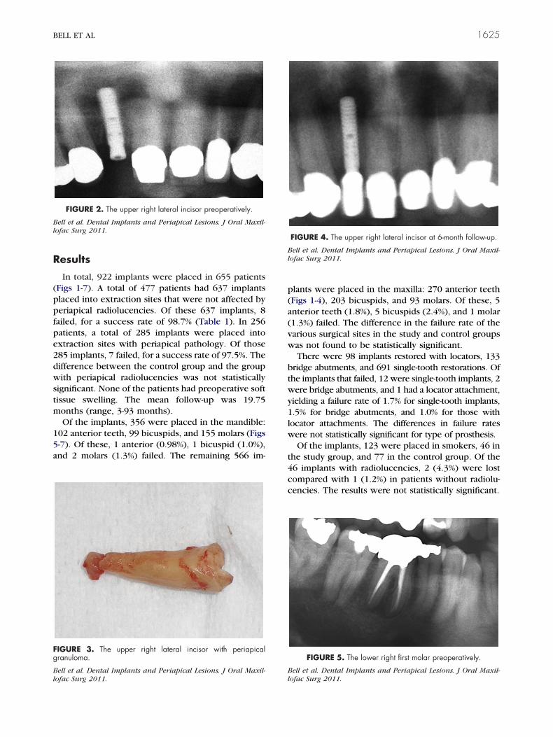
Bl
B
Bl
BELL ET AL 1625
Results
In total, 922 implants were placed in 655 patients(Figs 1-7). A total of 477 patients had 637 implantsplaced into extraction sites that were not affected byperiapical radiolucencies. Of these 637 implants, 8failed, for a success rate of 98.7% (Table 1). In 256patients, a total of 285 implants were placed intoextraction sites with periapical pathology. Of those285 implants, 7 failed, for a success rate of 97.5%. Thedifference between the control group and the groupwith periapical radiolucencies was not statisticallysignificant. None of the patients had preoperative softtissue swelling. The mean follow-up was 19.75months (range, 3-93 months).
Of the implants, 356 were placed in the mandible:102 anterior teeth, 99 bicuspids, and 155 molars (Figs5-7). Of these, 1 anterior (0.98%), 1 bicuspid (1.0%),and 2 molars (1.3%) failed. The remaining 566 im-
FIGURE 2. The upper right lateral incisor preoperatively.
ell et al. Dental Implants and Periapical Lesions. J Oral Maxil-ofac Surg 2011.
FIGURE 3. The upper right lateral incisor with periapicalgranuloma.
Bell et al. Dental Implants and Periapical Lesions. J Oral Maxil-
llofac Surg 2011.plants were placed in the maxilla: 270 anterior teeth(Figs 1-4), 203 bicuspids, and 93 molars. Of these, 5anterior teeth (1.8%), 5 bicuspids (2.4%), and 1 molar(1.3%) failed. The difference in the failure rate of thevarious surgical sites in the study and control groupswas not found to be statistically significant.
There were 98 implants restored with locators, 133bridge abutments, and 691 single-tooth restorations. Ofthe implants that failed, 12 were single-tooth implants, 2were bridge abutments, and 1 had a locator attachment,yielding a failure rate of 1.7% for single-tooth implants,1.5% for bridge abutments, and 1.0% for those withlocator attachments. The differences in failure rateswere not statistically significant for type of prosthesis.
Of the implants, 123 were placed in smokers, 46 inthe study group, and 77 in the control group. Of the46 implants with radiolucencies, 2 (4.3%) were lostcompared with 1 (1.2%) in patients without radiolu-cencies. The results were not statistically significant.
FIGURE 5. The lower right first molar preoperatively.
ell et al. Dental Implants and Periapical Lesions. J Oral Maxil-
FIGURE 4. The upper right lateral incisor at 6-month follow-up.
ell et al. Dental Implants and Periapical Lesions. J Oral Maxil-ofac Surg 2011.
ofac Surg 2011.

Bl
NPMMNN
SS
T
B
D
Bl
1626 DENTAL IMPLANTS AND PERIAPICAL LESIONS
Twenty-four of the implants were placed in pa-tients taking bisphosphonates; of those, six had peri-apical radiolucencies. No implants failed in these pa-tients.
Placement of 83 implants was performed in dia-betic patients, 23 with radiolucencies and 60 withoutradiolucencies. All of the implants were successful.
The initial stability of the implants was evaluated,and the mean stability of the study and control groupswas subjected to a z score analysis and was not foundto be significant. Torque values ranged from 5 to 45N-cm, with mean values of 30.5 N-cm and 35 N-cm inthe study and control groups, respectively. Interest-ingly, the failed implants in the control group had amean initial torque of 23.3 N-cm compared with 28.6N-cm in failed implants in the study group. However,this was not found to be statistically significant, pos-sibly because of the small sample size.
The mean size of lucencies was 2.5 mm. The meansize of radiolucencies within sockets where implantsfailed was 2.4 mm; the difference between the twowas not statistically significant.
FIGURE 7. The lower right first molar at 6-month follow-up.
ell et al. Dental Implants and Periapical Lesions. J Oral Maxil-
FIGURE 6. The lower right first molar postoperatively.
ell et al. Dental Implants and Periapical Lesions. J Oral Maxil-ofac Surg 2011.
ofac Surg 2011.
In total, 51 implants were placed adjacent to re-tained teeth with periapical radiolucencies, 21 in thestudy group and 30 in the control group. Of the 51implants placed, 4 failed. All 4 of the failed implantswere in the lucency group, giving an 81% success ratein the study group compared with 100% in the con-trol group. Thus implants placed in sockets with lu-cencies adjacent to retained teeth with lucencies hada significantly higher failure rate.
We found that 2 patients (0.7%) in the study grouphad postoperative infections compared with 6 pa-tients (0.1%) in the control group, which was notsignificant. No instances of implant periapical lesionswere detected.
There were 125 implants placed in men and 160placed in women in the study group and 258 implantsplaced in men and 279 placed in women in thecontrol group. The mean age was 58.4 years in thestudy group and 60.1 years in the control group.There was no correlation between age or gender andimplant failure.
The mean time between implant placement andimplant failure was 2.9 months (range, 1-10 months).
A review of the chart notes showed no estheticcomplaints.
Discussion
This study involved 655 patients and 922 implants.Each implant was placed immediately after the extrac-tion of the tooth. Every implant was placed by asimilar procedure. Each variable mentioned was also
Table 1. RESULTS
VariableNo. of
ImplantsNo. of
FailuresSuccess
Rate
o radiolucency 637 8 98.7%eriapical radiolucency 285 7 97.5%olars with lucency 90 2 97.8%olars without lucency 248 1 99.6%on-molars with lucency 194 5 97.4%on-molars withoutlucency
390 7 98.2%
mokers with lucency 46 2 95.7%mokers withoutlucency
77 1 98.8%
ooth with lucencyadjacent to tooth withlucency
21 4 81.0%
isphosphonate-takingpatients with lucency
6 0 100%
iabetic patients withlucency
23 0 100%
Bell et al. Dental Implants and Periapical Lesions. J Oral Maxil-lofac Surg 2011.
tested with either a �2 test or z score test to determine

tcs
thfhbt
fhIsc
BELL ET AL 1627
statistical significance. It was considered statisticallysignificant only if the P value was less than .05.
All patients received IV antibiotics, and the extrac-tion site for every implant was debrided and irrigated.It is unknown whether these antibiotics had an effecton implant survival in this study because all studypatients received them. Implants were not placed inpatients with soft tissue swelling or acute pain, andthis study does not, therefore, support placement ofimplants in patients with acute symptoms.
The only variable that significantly affected the out-come in this study was the presence of periapicalpathology in retained teeth adjacent to the implantbeing placed. Adjacent lucencies have previouslybeen found to increase implant failure.15,16 Endodon-ic treatment of teeth adjacent to implant sites, espe-ially if those implant sites have teeth with lucencies,hould be seriously considered.
The vast majority of implants had an insertionorque value at or above 15 N-cm. Only 72 implantsad an insertion value between 5 and 14 N-cm. Theailure rate of these implants was not significantlyigher than that in those with high insertion values,ut the number of less stable implants may have beenoo low to show significance.
In this study the question of implant placement intoresh extraction sites affected by periapical pathologyas been evaluated with several confounding variables.t seems that the immediate placement of implants intoockets affected by chronic periapical pathology can beonsidered a safe and viable treatment option.
References1. Adell R, Brånemark PI, Lekholm U: A 15-year study of osseointe-
grated implants in the treatment of the edentulous jaw. J Oral
Surg 10:387, 19812. Schwartz-Arad D, Chaushu G: Placement of implants into freshextraction sites: 4 to 7 years retrospective evaluation of 95immediate implants. J Periodontol 68:1110, 1997
3. Collaert B, De-Bruyn H: Comparison of Branemark fixture inte-gration and short-term survival using one or two stage surgeryin completely and partially edentulous mandibles. Clin OralImplants Res 9:131, 1998
4. Paolantonio M, Dolci M, Scarano A, et al: Immediate implanta-tion in fresh extraction sockets. J Periodontol 72:1560, 2001
5. Amler MH: The time sequence of tissue regeneration in humanextraction wounds. J Oral Surg 27:309, 1969
6. Douglass GL, Merin RL: The immediate dental implant. J CalifDent Assoc 30:362, 2002
7. Schwartz-Arad D, Chaushu G: The ways and wherefores ofimmediate placement of implants into fresh extraction sites: Aliterature review. J Periodontol 68:915, 1997
8. Becker W, Becker BE: Guided tissue regeneration for implantsplaced into extraction sockets and for implant dehiscences:Surgical techniques and case report. Int J Periodontics Restor-ative Dent 10:376, 1990
9. Del Fabbro M, Boggian C, Taschieri S: Immediate implantplacement into fresh extraction sites with chronic periapicalpathologic features combined with plasma rich in growth fac-tors: Preliminary results of single-cohort study. J Oral Maxillo-fac Surg 67:2476, 2009
10. Crespi R, Capparè P, Gherlone E: Fresh-socket implants inperiapical infected sites in humans. J Periodontol 81:378, 2010
11. Bataglion C, Gomes F, Horbylon BZ, et al: Immediate implantsplaced into infected sockets: A case report with 3-year follow-up. Braz Dent J 20:254, 2009
12. Novaes AB, Novaes AB Jr: Immediate implants placed intoinfected sites: A clinical report. Int J Oral Maxillofac Implants10:609, 1995
13. Waasdorp JA, Evian CI, Mandracchia M: Immediate placementof implants into infected sites: A systematic review of theliterature. J Periodontol 81:801, 2010
14. Siegenthaler DW, Jung RE, Holderegger C, et al: Replacementof teeth exhibiting periapical pathology by immediate im-plants. A prospective, controlled clinical trial. Clin Oral Im-plants Res 18:727, 2007
15. Brisman DL, Brisman AS, Moses MS: Implant failures associatedwith asymptomatic endodontically treated teeth. J Am DentAssoc 132:191, 2001
16. Tehemar SH: Factors affecting heat generation during implantsite drilling: A review of biologic observations and future con-
siderations. Int J Oral Maxillofac Implants 14:127, 1999