The HOTHEAD Protein: Assessing Enzymatic Activity using ...
116
The HOTHEAD Protein: Assessing Enzymatic Activity using Computational and Recombinant Protein Expression Approaches By Eric Le Dreff-Kerwin A thesis Presented to the University of Waterloo In fulfillment of the Thesis requirement for the degree of Master of Science In Biology Waterloo, Ontario, Canada, 2019 © Eric Le Dreff-Kerwin 2019
Transcript of The HOTHEAD Protein: Assessing Enzymatic Activity using ...

The HOTHEAD Protein Assessing
Enzymatic Activity using Computational and
Recombinant Protein Expression Approaches
By
Eric Le Dreff-Kerwin
A thesis
Presented to the University of Waterloo
In fulfillment of the
Thesis requirement for the degree of
Master of Science
In
Biology
Waterloo Ontario Canada 2019
copy Eric Le Dreff-Kerwin 2019
ii
Authorrsquos Declaration
I hereby declare that I am the sole author of this thesis
This is a true copy of the thesis including any required final revisions as accepted by my
examiners
I understand that my thesis may be made electronically available to the public
iii
Abstract
In Arabidopsis thaliana a number of genes regulating cuticle synthesis have been
identified by virtue of organ fusion phenotype One such gene HOTHEAD (HTH) was among
those originally identified by this phenotype but its exact role in cuticle formation has proven
challenging to determine Previous bioinformatic work has identified that the HTH protein is a
member of the GMC oxidoreductase family and shares peptide sequence identity with a
mandelonitrile lyase and an alcohol dehydrogenase that are within the same protein family This
thesis work investigated the potential enzymatic function of HTH by comparing a structural
model to two structural analogs The structure model of HTH as determined in this study shows
that HTH shares certain conserved features of GMC proteins The aim of this research also
included isolating a recombinantly expressed HTH protein from Escherichia coli and initial work
in Pichia pastoris Protein isolation attempts in Escherichia coli failed to yield active HTH
protein potentially due to the lack of post-translational modifications Continuing the work using
the eukaryote Pichia pastoris should solve the issue Finally crude extracts from Arabidopsis
thaliana floral tissue were obtained to test for the presence of HTH enzymatic activity but
showed no significant mandelonitrile lyase enzymatic activity potentially due to the inability to
successfully extract the HTH protein from the floral tissue
iv
Acknowledgements
I would like to thank my supervisors Dr Susan Lolle and Dr Todd Holyoak for all of
their help continuous support and being great examples of great scientists ones that I always
tried to live up to throughout my degree Even during the most challenging of times they were
always ready to get me back on my feet
Thank you to my committee members Dr Barbara Moffatt and Dr David Rose for their
valuable comments and for being continuously willing to help advance this project To the
current and past members of the Lolle and Holyoak labs Dr Pearl Chang Therese Francom
Serena Thamilselvan Saima Hossain Saeer Adeel Jackline Ponomariov and Christina Doherty
from the Lolle lab and Matt McLeod Iain Wallace Ben Morrow Norman Tran Sarah Barwell
Julia Solonenka and Dr Bidyadhar Das from the Holyoak lab as well as Nardo Nava-Rodriguez
Nicole Fraser and the other Rose lab members Thank you all for your support even if it was one
of the many late nights working in the lab
Thank you to Quinn Abram and Dr Tania Rodriguez-Ramos of the Dixon lab for
constantly putting up with my random visits to their lab in search for answers and tips on how to
improve my technique
v
Table of Contents
Chapter 1 1
General Introduction 1
11 The Plant Cuticle 2 111 Cuticle Function 2 112 Cuticle Composition 2 113 Biosynthesis Pathway 3
12 HOTHEAD 4 121 The HOTHEAD Gene 4 122 HOTHEAD Mutant Alleles 4 123 The HOTHEAD Protein 7 124 HOTHEAD Protein Localization 8
13 Alcohol Dehydrogenase 10
14 Mandelonitrile Lyase 11 141 The Role of Hydrogen Cyanide in Plants 13 142 HCN amp Plant Stress Response 14 143 HCN and Seed Dormancy 15 144 HCN amp Nitrate Assimilation 16 145 Moonlighting Proteins 18
15 Objectives of the Thesis 19
Chapter 2 20
Structural Analysis of the HOTHEAD Protein Model 20
21 Introduction 21
22 Methods 23 221 HOTHEAD Protein Structural Model Analysis 23
23 Results amp Discussion 24 231 Hypothesized Enzymatic Function of HTH 24 232 Structural Analogs to the HTH Protein Model 24 233 Conserved Active Site Residues in FAD-Dependent Hydroxynitrle Lyase 27 234 Conserved Active Site Residues in GMC Proteins 28 235 Predicted Ligand Binding Site on the HTH Protein Model 29 236 Comparing Hydroxynitrile Lyase N-Linked Glycosylation to the HOTHEAD Model 32
24 Conclusions 35
Chapter 3 37
Recombinant HOTHEAD Protein Isolation amp Enzymatic Assays 37
31 Introduction 38
32 Materials 39
vi
33 Methods 40 331 Synthetic HTH cDNA Sequence Design 40 332 Heat Shock Bacteria Transformation 40 333 Bacterial Plasmid Minipreparation 41 334 HTH_SUMO Restriction Endonuclease Digestion 41 335 HTH Protein Autoinduction Set up 42 336 Nickel Chromatography of HTH_SUMO Protein 43 337 Anion Exchange Chromatography 44 338 Cation Exchange Chromatography 45 339 Protein Solubilization via Arginine Treatment 46 3310 Mass Spectrometry Analysis 46 3311 Nickel Chromatography using HTH_pET24a+ 47 3312 Chemical Competent Saccharomyces cerevisiae Transformation 48 3313 HTH cDNA Q5 Site-Directed Mutagenesis 49 3314 DNA Sequence Analysis 49 3315 Plant material and Growth Conditions 50 3316 Floral Tissue Crude Protein Extraction 50 3317 In vitro Mandelonitrile Lyase Enzymatic Assay 51 3318 Western Blot Analysis of Floral Bud Crude Protein Extracts 52
34 Results amp Discussion 53 341 HTH Protein Construct Design 53 342 Protein Isolation in E coli 57 343 Protein Solubilization via Arginine Treatment 57 344 Potential Factors Inhibiting Protein Isolation from E coli 63 345 Eukaryotic Expression System 68 346 HTH Mutant Constructs 71 347 MDL Enzymatic Assay 74
35 Concluding remarks 78
References 93
vii
List of Figures
Figure 1 The HTH gene model 5 Figure 2 Schematic representation of the HTH protein 8 Figure 3 Schematic of the potential enzymatic activity of HTH 13 Figure 4 Cyanide-mediated assimilation of nitrate 17 Figure 5 Ribbon diagram of the predicted HTH model 25 Figure 6 Ribbon diagrams comparing the putative active site residues of the HTH protein to
conserved active site residues of an FAD-HNL (1ju2) amp a GMC protein (3nne) 31 Figure 7 Ribbon diagrams of the HTH model-ligand complexes 32 Figure 8 Ribbon diagrams of the HTH model amp FAD-HNL (1ju2) N-linked Glycosylation sites
34 Figure 9 HTH signal peptide prediction 55 Figure 10 HTH peptide sequence alignment 56 Figure 11 Restriction enzyme digestion of the HTH_SUMO vector 59 Figure 12 Autoinduction protein profile 60 Figure 13 HTH_SUMO protein isolations 61 Figure 14 Mass spectrometry analysis of the protein bands expressed in the pE_SUMOstar
vector 62 Figure 15 Restriction enzyme digestion of the HTH_pET24a vector 67 Figure 16 HTH_pET24a protein isolation 68 Figure 17 PichiaPink expression system 71 Figure 18 Restriction enzyme digestion of the HTH_SUMO vector 73 Figure 19 HTHproHTH-FP constructs 75 Figure 20 Western blot analysis of HTH protein using crude protein extract from HTHproHTH-
G3GFP cloned into the pGWB650 vector 77
viii
List of Tables
Table 1 Structural analogs to the HTH protein model 26 Table 2 Most prominent protein in each band sent for mass spectrometry analysis 63 Table 3 Spectrophotometric mandelonitrile lyase activity assay 76
ix
List of Appendices
Appendix A HTH_pESUMO vector sequence 80
Appendix B HTH_pET24a+ vector sequence 82
Appendix C Peptide sequence alignment between HOTHEAD and alcohol dehydrogenase from
Candida albicans 84
Appendix D Peptide sequence alignment between HOTHEAD and (R)-hydroxynitrile lyase
from Prunus mume 85
Appendix E Peptide sequence alignment between HOTHEAD and choline oxidase from A
globiformis 86
Appendix F Peptide sequence alignment between HOTHEAD and mandelonitrile lyase from P
dulcis 87
Appendix G Arginine treatment 88
Appendix H List of primer sets used for site-directed mutagenesis 89
Appendix I List of sequencing primers 90
Appendix J Sequencing primer locations on the synthetic HTH cDNA sequence cloned into
pE_SUMOstar 91
Appendix K Crude protein extraction absorbance trace from MDN enzymatic assays 92
x
List of Abbreviations and Acronyms
5rsquoUTR 5rsquo Untranslated region
ACS6 1-Aminocyclopropane-1-carboxyllate synthase 6
ADE2 Adenine deaminase 2
ADH Alcohol dehydrogenase
AmSO4 Ammonium sulfate
AOX1 Alcohol oxidase 1
bp Base pairs
cDNA Complementary DNA
CYP Cytochrome P450
Ds Dissociation element
DCA Dicarboxylic acid
DNA Deoxyribonucleic acid
ECL Enhanced chemiluminescent
eda-17 Embryo development arrest 17
EDTA Ethylenediaminetetraacetic acid
EMS Ethyl methanesulfonate
EnSpm Enhancersuppressor mutator
ER Endoplasmic reticulum
EV Empty vector
FA Fatty acid
FAD flavin adenine dinucleotide
FAD-HNL Flavin adenine dinucleotide dependent hydroxynitrile lyase
FG1 Female gametophyte development stage 1
FG2 Female gametophyte development stage 2
FG3 Female gametophyte development stage 3
FG8 Female gametophyte development stage 8
g Grams
GFP Green fluorescent protein
GPAT Glycerol-3 phosphate acyltransferase
HCN Hydrogen cyanide
HTH HOTHEAD
HTH-FP Fluorescent protein-tagged HTH construct
HTH_SUMO HOTHEAD_pE_SUMOstar
IDEN Sequence identity
IgG Immunoglobulin G
IPTG Isopropyl β-D-1-thiogalactopyranoside
kd Kilobase pair
kDa Kilo Dalton
LACS Long-chain acyl-coenzyme A synthase
LB Luria broth
MDL Mandelonitrile lyase
MEB1 Membrane of ER Body 1
MEB2 Membrane of ER Body 2
xi
mg Milligram
min Minutes
ml Millilitre
mRNA Messenger ribonucleic acid
ng Nanogram
nm Nanometer
OD Optical density
P-6 DG Polyacrylamide desalting gel with 6000 molecular weight exclusion limit
PCR Polymerase chain reaction
PAGE Polyacrylamide gel electrophoresis
RMSD Root-mean-square deviation
SDS Sodium dodecyl sulfate
TBS-T Tris-buffer saline with Tween 20
TEV Tobacco etch virus
TM-Score Template modeling score
UV Ultraviolet
VLCFA Very-long-chain fatty acid
WT Wild type
YFP Yellow fluorescent protein
∆A275 Change of absorbance of light measure at wavelength 275 nm
μg Microgram
μL Microlitre
μM Micromolar
μm Micrometer
α Alpha
β Beta
ω Omega
DNA Code
A Adenine
C Cytosine
G Guanine
T Thymine
Protein Code
Ala (A) Alanine
Arg (R) Arginine
Asn (N) Asparagine
Asp (D) Aspartic acid
Cys (C) Cysteine
Gln (Q) Glutamine
Glu (E) Glutamic acid
Gly (G) Glycine
His (H) Histidine
Ile (I) Isoleucine
Leu (L) Leucine
xii
Lys (K) Lysine
Met (M) Methionine
Phe (F) Phenylalanine
Pro (P) Proline
Ser (S) Serine
Thr (T) Threonine
Trp (W) Tryptophan
Tyr (Y) Tyrosine
Val (V) Valine
1
Chapter 1
General Introduction
2
11 The Plant Cuticle
111 Cuticle Function
Terrestrial plants have acquired a variety of adaptations to aid in their survival on dry
land One such adaptation is the plant cuticle The cuticle is a hydrophobic membrane that acts as
an extra layer separating the aerial tissue of the plant from its dynamic environment The cuticle
protects against ultraviolet radiation minimizes surface dirt buildup regulates the flow of gases
nutrients and molecules and protects against most pathogens (Fich Segerson amp Rose 2016
Yeats amp Rose 2013) An interesting exception is a pathogenic attack from Botrytis cinereal
Arabidopsis thaliana exhibiting a loss in cuticle integrity will be more effective at protecting
itself from B cinereal when compared to A thaliana with an intact cuticle (Fich et al 2016)
The cuticle is also important for proper organ partitioning in mutants with perturbed
cuticle function (or during normal carpel development of some plant species) epidermal cells
have been shown to fuse together when they come into direct contact with each other A water-
soluble signaling molecule identified in Catharanthus roseus travels between carpel cells
separated by a permeable barrier causing cell dedifferentiation (Verbeke amp Walker 1986) In
these carpels altered cuticle integrity appears to allow this signalling molecule to travel between
the cells it separates and is a natural process
112 Cuticle Composition
The cuticle is a thin multilayered membrane typically varying from 01-10 μm in
thickness although it has been observed to be thinner in A thaliana ranging from 002-01 μm
(Riederer amp Schreiber 2001 Shumborski Samuels amp Bird 2016) The cuticle is divided into
the cuticular layer and the cuticle proper The cuticle proper is further divided into the
intracuticular wax the epicuticular wax film and the epicuticular wax crystals Each layer is
3
made up of a mix of cutin monomers embedded in wax Higher levels of cutin can be found in
the cuticular layer while higher levels of wax are in the cuticle proper (Yeats amp Rose 2013)
The composition varies between species making the exact structure of the cuticle difficult to
determine For example the A thaliana cuticle is known to be dicarboxylic fatty acid (DCA)
rich with higher levels of glycerol compared to other species (Kurdyukov et al 2006) This leads
to the belief that the composition of this particular cuticle is different from most other plants
(Yang et al 2016)
113 Biosynthesis Pathway
Cuticle synthesis occurs in the underlying epidermal cells The formation of cutin and
wax monomers begin in the plastids with de novo biosynthesis of C16 and C18 fatty acids (FA)
These are converted to CoA thioesters by long-chain acyl-coenzyme A synthase (LACS) and
transported into the endoplasmic reticulum (ER) lumen via an unknown mechanism (Yeats amp
Rose 2013) Once in the ER the cuticle biosynthesis pathway divides into the cutin and the wax
biosynthesis pathways
Typical wax monomers include very-long-chain fatty acids (VLCFA) ranging in size
from C20 to C34 which include alkanes alcohols ketones and wax esters (Yeats amp Rose 2013)
To form the wax monomer the FAs are elongated by fatty acid elongase then reduced to fatty
aldehydes and primary alcohols They can also be reduced and decarbonylated to alkanes then
converted into ketones and secondary alcohols (Yeats amp Rose 2013) The wax monomers are
then transported out of the ER across the cell wall to the plant tissue surface
Typical cutin monomers include hydroxy FAs glycerol dicarboxylic acids and
phenylpropanoids Terminal and mid-chain oxidation will occur once the C16 and C18 FAs are
converted to CoA thioesters by LACS (Yeats amp Rose 2013) Terminal oxidation is believed to
4
be performed by CYP68A enzymes coded for by the cytochrome P450 (CYP) gene while mid-
chain oxidation is believed to be performed by CYP77A enzymes Finally the CoA is replaced
with a glycerol-3-phosphate by glycerol-3-phosphate acyltransferase (GPAT) (Fich et al 2016
Yeats amp Rose 2013)
Various putative mechanisms for cutin and wax monomer export and assembly have been
proposed (Pollard Beisson Li amp Ohlrogge 2008) The most recent research suggests that the
cutin monomers are transported through plasma membrane embedded ABC transporters while
wax monomers exit the cell at sites of close contact between the ER and the plasma membrane
(Fich et al 2016)
12 HOTHEAD
121 The HOTHEAD Gene
A list of known genes involved in various aspects of cuticle formation has been compiled
through molecular analyses of A thaliana mutants (Yeats amp Rose 2013) Most genes noted in
this list have a known function in cuticle formation while others have yet to be determined One
of those genes with an unknown function identified in A thaliana is HOTHEAD (HTH) (locus
At1g72970) (Figure 1) This gene maps to chromosome 1 of A thaliana and contains 6 exons
and 5 introns In the Columbia ecotype the HTH gene is 4843 base pairs (bp) in length This
accounts for the 2834 bp transcribed region and its 2009 bp promoter and 5rsquo-untranslated region
(5rsquoUTR) (Krolikowski et al 2003)
122 HOTHEAD Mutant Alleles
HTH was initially identified through forward genetic screens (Lolle Hsu amp Pruitt 1998)
A thaliana seeds treated with ethyl methanesulfonate (EMS) were visually screened at various
life stages for the presence of organ fusion This phenotype suggests a loss of cuticle integrity
5
This study identified eleven mutant hth alleles hth-1 through hth-11 with varying degrees of
floral bud fusion The relative position of the mutation in each hth allele is shown in Figure 1
(Krolikowski et al 2003) Two of these mutant alleles hth-8 and hth-10 also showed evidence
of ovule abnormalities (Lolle et al 1998) These 11 alleles are a result of a substitution mutation
due to the EMS treatment Most of these mutations alter amino acids in the peptide sequence
Two of these alleles hth-1 and hth-9 translate into truncated proteins due to the introduction of a
stop codon and the alteration of a splice junction sequence respectively (Lolle et al 1998)
Figure 1 The HTH gene model The HTH gene location on chromosome 1 of A thaliana and the
relative position of the various hth mutant alleles The large boxes represent the six exons and
the solid lines represent the introns The position of the single point mutations (hth-1 to hth-11)
transposon insertion sites (hth-12 and eda-17) and T-DNA insertion sites (hth-13 to hth-15) are
shown (Krolikowski et al 2003 Kurdyukov et al 2006) Based on Figure 18 from Chang
2016
In another study performed by Kurdyokov et al mutant A thaliana seeds were generated
by enhancersuppressor mutator (EnSpm) transposon mutagenesis (Kurdyukov et al 2006) The
hth-12 mutant was identified via gel blot and hybridization (Figure 1) A thaliana plants
harboring the hth-12 mutant allele showed floral bud organ fusion believed to be due to a loss of
6
cuticle integrity Transmission electron microscopy images showed that the cuticle of the hth-12
mutant was not continuous as seen in wild type (WT) A thaliana cuticle (Kurdyukov et al
2006) The composition of the hth-12 mutant cuticle was compared to the WT Arabidopsis
cuticle The mutant plant cuticle showed an increase in ω-hydroxy FAs and a decrease in ω-oxo
FAs Both of these molecules are precursors to α-ω-dicarboxylic acids a known cutin monomer
in the plant cuticle The cuticular wax composition did not appear to be affected by the hth-12
mutation (Kurdyukov et al 2006) This change in cutin composition due to the hth-12 mutant
allele links HTH to the cutin biosynthesis pathway
Pagnussat et al transformed A thaliana plants with a dissociation element (Ds)
transposon (Pagnussat et al 2005) and identified a novel hth mutant allele embryo sac
development arrest-17 (eda-17) containing a Ds transposon with the first intro of the HTH gene
(Figure 1) This is the only hth allele with a mutation located within this region This screen
identified numerous mutants with female gametophyte defects Plant lines containing the Ds
transposon were tested for segregation ratio distortion in kanamycin resistance since the Ds
element included a kanamycin resistance gene (Pagnussat et al 2005) Female gametophyte
development can be divided into two phases megasporogenesis and megagametogensis (Drews
amp Koltunow 2011) One megaspore is present at the end of megasporogensis and enters
megagametogenesis This process is divided into eight stages starting with female gametophyte
stage 1 (FG1) and finishing with an embryo sac at the end of female gametophyte stage 8 (FG8)
(Christensen King Jordan amp Drews 1997) An A thaliana harboring the eda-17 allele will
arrest at female gametophyte stage 2 (FG2) of megagametogenesis a stage where the nucleus
undergoes mitosis without cell division (Christensen et al 1997) This suggests that the HTH
gene is important for embryo sac development
7
Most hth mutants share a similar phenotype postgenital organ fusion which is due to a
loss in cuticle integrity This fusion is most prominent in the floral buds once the plant has
reached sexual maturity It was this phenotype that lead to the suggestion that HTH is involved in
cuticle synthesis most probably in the cutin biosynthesis pathway But other phenotypes suggest
that HTH is likely involved in more than just cuticle synthesis For example the eda-17 mutant
causes female gametophyte development arrest at the two-nuclear stage (Lolle et al 1998
Pagnussat et al 2005)
123 The HOTHEAD Protein
The HTH protein contains 594 amino acid residues with a predicted molecular mass of
653 kDa and an inferred isoelectric point of 975 (locus At1g72970) (Figure 2) (Krolikowski et
al 2003) The protein is predicted to be globular and contain a cleavable N-terminal signal
peptide spanning residues 1 to 19 (Chang 2016)
HTH is considered to be a part of the glucose-methanol-choline (GMC) oxidoreductase
protein family due to the presence of conserved N-terminal and C-terminal domains typical of
the family This family contains a variety of proteins with oxidoreductase enzymatic activity
(Cavener 1992) The GMC oxidoreductase N-terminal domain contains a Rossmann fold
indicating the presence of a flavin adenine dinucleotide (FAD) binding site (Bannwarth et al
2004) Based on previous bioinformatic work the HTH protein has a putative FAD binding site
between residues 64 to 91 which is found within the predicted GMC N-terminal domain (Chang
2016) The GMC oxidoreductase C-terminal domain is known as the substrate binding domain
All GMC proteins are predicted to have a conserved histidine active site residue (His466 based
on choline oxidase from Arthrobacter globiformis (PDB ID 3nne EC 11317)) while most
will have a second active site residue asparagine (Asn510 in cholesterol oxidase from A
8
globiformis) (Dreveny et al 2001) Both of these residues are found within this conserved C-
terminal domain (Li et al 1993) Based on previous bioinformatic work the HTH protein has
three putative active site residues His529 Ile527 and Asn567 all of which are found within this
conserved C-terminal domain (Chang 2016)
The HTH protein is also predicted to contain certain post-translational modifications 4
putative phosphorylation sites and 5 putative N-linked glycosylation sites (Chang 2016) None
of the known hth mutants are found at the locations of these putative post-translational
modifications HTH has been experimentally shown to be a glycoprotein based on size shifts
following endoglycosidase treatment of a HTHGFP protein extracted from transgenic plant
lines (Chang 2016) Whether these modifications are important for protein structure andor
function has yet to be determined
Figure 2 Schematic representation of the HTH protein The red arrows indicate the six exons of
the HTH gene The first 19 residues are predicted to be a part of the signal peptide (green
rectangular box) The two conserved GMC oxidoreductase domains are represented by grey
rectangular boxes The conserved N-terminal domain contains the FAD-binding site while the C-
terminal domain contains the three putative active site residues (red markers) The relative
position of the N-linked glycosylation sites (yellow markers) and phosphorylation sites (purple
markers) are indicated Based on Figure 41 from Chang 2016
124 HOTHEAD Protein Localization
Chang analyzed HTH localization in A thaliana transgenic plants transformed with a
fluorescent protein tagged HTH (Chang 2016) HTH is first observed in A thaliana ovules at
9
female gametophyte stage 3 (FG3) of megagametogenesis prior to fertilization (Chang 2016)
At this stage two nuclei are present and travel to opposite ends of the embryo sac while a
vacuole forms in the middle (Christensen et al 1997) It is at this stage that HTH is present at
the chalazal pole and remains there until anthesis the point where floral buds open after which
HTH becomes more diffuse (Chang 2016) Three days after anthesis HTH localizes in the
integument and persists until seed maturity (Chang 2016)
On a gross level the HTH mRNA appears to be ubiquitously distributed in A thaliana
epidermal and subepidermal cells (Krolikowski et al 2003) It has been observed in leaves
stems inflorescences siliques and roots (Krolikowski et al 2003) This does not necessarily
mean that the HTH protein also localizes in all these tissues since it may move to nearby cells
HTH protein has been detected in seedlings flowers and siliques (Chang 2016)
At the cellular level HTH localizes in the endoplasmic reticulum (ER) within epidermal
cells even though it does not have a canonical KDEL retention signal (Chang 2016) Instead it
has three KDEL-like sequences in areas predicted to be found on the protein surface KDEK at
position 270 KKEL at position 310 and KNEL at position 387 (Chang 2016) None of the
known hth mutants are found within these KDEL-like sequences
HTH has also been observed in ER-derived structures known as ER bodies (Chang
2016) These predominantly exist in young A thaliana plants within cotyledons hypocotyls and
roots The number of ER bodies decreases during senescence from the base to the tip of the
cotyledons Hypocotyl and root ER body levels do not appear to decrease (Matsushima et al
2003) Rosette leaves of mature plants typically do not have any ER bodies unless the tissue is
wounded where upon a large accumulation of ER bodies can be found localized around the
damaged tissue
10
Besides HTH ER bodies contain various proteins PYK10 NAI2 membrane of ER body
1 (MEB1) and membrane of ER body 2 (MEB2) (Yamada et al 2013) PYK10 is a β-
glucosidase which belongs to a group of enzymes known to produce compounds that deter pests
activate glycosylated hormones and break down the cell wall (Matsushima et al 2003) The
exact role of the PYK10 protein in ER bodies is undetermined but it appears to be important for
plant defence (Vinther et al 2008) The MEB1 and MEB2 proteins embed themselves in the ER
body membrane and are believed to be involved in metal ion homeostasis (Yamada et al 2013)
This could explain why ER bodies are constitutively expressed in roots a tissue constantly
exposed to high levels of metal ions Finally NAI2 is responsible for the accumulation of
PYK10 MEB1 and MEB2 in ER bodies (Yamada et al 2008) A nai2 mutant will have reduced
and elongated ER bodies PYK10 MEB1 and MEB2 levels will not be affected in this mutant
but they will not localize to ER bodies (Yamada et al 2008)
Based on localization and composition ER bodies are believed to be important for plant
defence (Matsushima et al 2003) PYK10 most likely aids in deterring pests and breaking down
cell walls while MEB1 and MEB2 ensure metal ion levels do not reach toxic levels The role of
HTH in these ER-derived structures is currently unknown
13 Alcohol Dehydrogenase
The potential enzymatic function of the HTH protein has not been confirmed As
previously mentioned Kurdyukov et al demonstrated that the cuticle of a hth-12 mutant A
thaliana has an increase in ω-hydroxy FAs and a decrease in ω-oxo FA a precursor to the α-ω-
dicarboxylic acid cutin monomer (Kurdyukov et al 2006) This appears to be due to the loss of
alcohol dehydrogenase (ADH) enzymatic activity (Figure 3A) Knowing that most hth mutants
cause a loss in cuticle integrity it is logical that the HTH protein acts as an ADH in the cutin
11
biosynthesis pathway Based on the overall peptide sequence HTH does share 25 peptide
sequence identity to long chain ω-fatty alcohol dehydrogenase from Candida (Kurdyukov et al
2006) Candida metabolizes carbon dioxide via a membrane-bound ω-oxidation pathway that
involves a flavin-dependent aldehyde dehydrogenase (Vanhanen et al 2000) The substrate
specificity of this enzyme varies For example in C tropicalis the enzyme is most active on
long-chain fatty alcohols but can also accept other substrates such as ω-hydroxy FA secondary
alcohols and dodecan-2-ol (Vanhanen et al 2000)
Most GMC proteins catalyze a redox reaction which involves FAD The N5 atoms in the
flavin interacts with the substrate to donate or accept a hydride anion (Dreveny Andryushkova
Glieder Gruber amp Kratky 2009) The exact conformation of the active site is not strictly
conserved due to the variability in substrate size ranging from cholesterol to small lactates
(Fraaije amp Mattevi 2000) GMC proteins have two conserved residues within the active site
His466 and Asn510 (numbering based on choline oxidase from A globiformis) (Yue Kass
Sampson amp Vrielink 1999) These residues form a hydrogen bond network to ensure that the
substrate is properly oriented and in proximity with the N5 atom of FAD (Dreveny et al 2009)
14 Mandelonitrile Lyase
On the other hand the overall peptide sequence of HTH shares 41 peptide sequence
identity to mandelonitrile lyase (MDL) from Prunus (Kurdyukov et al 2006) Wu amp Poulton
identified the localization of MDL in Prunus seeds (Wu amp Poulton 1991) MDL was primarily
localized to the cell wall with lower amounts present in protein bodies (Wu amp Poulton 1991) In
plants protein bodies are important for protein storage in seeds (Saberianfar et al 2016
Schmidt 2013) Thayer amp Conn also looked at MDL localization in Sorghum leaves and
observed it in the cytoplasm of mesophyll cells (Thayer amp Conn 1981)
12
MDL is an FAD-dependent hydroxynitrile lyase (FAD-HNL) that is a part of the same
protein family as HTH the GMC oxidoreductase protein family (Dreveny et al 2009) Most
proteins in this family perform some sort of oxidation or reduction reaction aided by the FAD
co-factor specifically the N5 atom in the flavin (Dreveny et al 2009) FAD-HNLs are an
exception since they perform cyanogenesis and their FAD co-factor does not appear to play an
enzymatic role Rather the FAD seems to be important for the proteinrsquos structural integrity as an
apo-HNL is found to be inactive unstable and degrade within minutes (Dreveny et al 2001) As
its name suggests MDL uses mandelonitrile as its substrate to produce benzaldehyde and
hydrogen cyanide (HCN) (Figure 3B) FAD-HNLs are believed to have evolved from a common
ancestor shared with aryl-alcohol oxidases since both proteins are a part of the GMC
oxidoreductase protein family and share a similar product benzaldehyde (Dreveny et al 2009)
As previously stated the FAD co-factor in most GMC proteins is used for an
oxidoreduction reaction resulting in the hydroxyl group of the substrate being in proximity of the
N5 atom of FAD FAD-HNLs are typically (R)-selective meaning they exclusively use
substrates which are (R)-stereoisomers (Andexer et al 2012) Due to this substrate specificity in
MDL the hydroxyl group of mandelonitrile will be oriented away from the flavin while the
cyano group will be oriented towards the flavin Therefore the hydroxyl group is positioned at a
distance that is too far to allow hydride transfer with the N5 of the FAD cofactor
In contrast to the alcohol dehydrogenase members of the GMC family FAD-HNLs have
two conserved histidine residues to facilitate chemistry His459 and His497 (numbering based on
the FAD-HNL peptide from Prunus dulcis (PDB ID 1ju2 EC 41210)) (Dreveny et al 2009)
These residues are found within the active site that is connected to the surface of the protein via a
hydrophobic tunnel The active site is observed to possess a negative electrostatic potential that
13
is hypothesized to assist in bringing the substrate in proximity to the active site residues
(Dreveny et al 2001) His497 is considered a general base that hydrogen bonds with the
hydroxyl group of mandelonitrile to help stabilize the substrate (Dreveny et al 2001) His459 on
the other hand mediates the protonation of the cyanide ion allowing for the production of
hydrogen cyanide (Dreveny et al 2009)
Figure 3 Schematic of the potential enzymatic activity of HTH (A) Dehydrogenation of ω-
dicarboxylic FA to ω-oxo FA by a ω-hydroxy fatty acyl dehydrogenase (ADH) (B)
Cyanogenesis of mandelonitrile to benzaldehyde and hydrogen cyanide by a mandelonitrile lyase
(MDL)
141 The Role of Hydrogen Cyanide in Plants
If HTH has a similar enzymatic activity to FAD-HNLs and produces HCN it is important
to consider the role of HCN in plants This molecule is highly regulated and is crucial for various
metabolic physiological and developmental processes be they direct or indirect (Siegień amp
Bogatek 2006) HCN is a small simple gaseous molecule that can easily diffuse to non-cyanide
producing tissues Plants can produce HCN multiple ways catabolism of cyanogenic glycosides
as a side product to the ethylene biosynthesis pathway or as a product of glyoxylates in
photorespiration and hydroxylamine (Siegień amp Bogatek 2006) A hydroxynitrile lyase will
catabolise an aliphatic or an aromatic containing hydroxynitrile to produce hydrogen cyanide and
ADH
MDL
14
aldehyde or ketone Mandelonitrile lyase will use mandelonitrile as its substrate to produce
benzaldehyde and HCN (Figure 3B)
142 HCN amp Plant Stress Response
Cyanide can be very toxic and is typically produced in response to an environmental
stress along with ethylene HCN levels must be tightly regulated in plants to prevent self-harm
Even at very low concentrations it is known to stunt growth reduce plant size and decrease
chlorophyll content (J M Smith amp Arteca 2000) When produced the tissue is quick to
metabolise the cyanide Cyanoalanine synthase uses HCN and L-cysteine to produce 3-cyano-L-
alanine which is then converted into aspartic acid or asparagine by A thaliana Nitrilase 4
(Piotrowski amp Volmer 2006) Certain areas of the plant may still have non-lethal levels of HCN
which is believed to trigger the plantrsquos response to stress (J M Smith amp Arteca 2000) This is
seen in tobacco plants whereby a non-lethal concentration of HCN enhances the resistance to the
tobacco mosaic virus (Chivasa amp Carr 1998) It is believed that cyanide is also important for
stress relief as non-lethal concentrations decrease the growth rate of the infected cells to
conserve energy and decrease the risk of heritable damage (May Vernoux Leaver Van
Montagu amp Inzeacute 1998)
ER bodies are also produced in response to environmental stress Matsushima et al
demonstrated that ER bodies were induced in rosette leaves treated with methyl jasmonate
Rosette leaves do not appear to contain ER bodies when the plant is not under stress
(Matsushima et al 2003) The induced ER bodies are limited to the peripheral regions exposed
to the stressor showing a localization of ER bodies in that area (Chang 2016 Matsushima et al
2003) ER bodies have been linked to plant defence based on their composition and localization
(Matsushima et al 2003) The HTH protein has been observed in these structures suggesting
15
that HTH may be involved in stress response (Chang 2016) If HTH were to have MDL
enzymatic activity this may explain its presence in ER bodies MDL enzymes have not been
previously shown to localize to ER bodies
A comparison between HTH and MDL has shown sequence similarity overall although
differences in amino acid conservation has been noted between residues 552 and 577 (numbering
based on the HTH peptide sequence from A thaliana) a region within the conserved C-terminal
substrate binding domain (Krolikowski et al 2003) One of the HTH putative active site
residues Asn567 (numbering based on the HTH peptide sequence from A thaliana) is found
within this region This may indicate that these proteins have distinct enzymatic function A
subsequent comparison of Arabidopsis and rice HTH to black cherry and almond MDL was also
performed and an unrooted phylogenetic tree showed that all the HTH sequences cluster in a
distinct group from MDL (Krolikowski et al 2003) This further suggests that although the
enzymatic function of these two proteins may be related to each other they are most likely
distinct
143 HCN and Seed Dormancy
HCN has also shown concentration dependent effects on dormant seeds HCN is
produced during the pre-germination period even in seeds that do not contain cyanogenic
glycosides It is believe that the source of HCN in non-cyanogenic seeds originates from
cyanogenic storage substances (Esashi et al 1991) Seed germination is stimulated when low
amounts of HCN are present but the effects are only observed once HCN is removed If HCN
remains present germination is blocked (Maruyama et al 1996) The mode of cyanide action in
pre-germinated seeds is still unclear Many putative modes of cyanide action have been
16
proposed forming a complex web of potential interactions but its importance during
germination could be further indicative of HTHrsquos hydroxynitrile lyase activity
A transgenic plant expressing GFP or yellow fluorescent protein (YFP) tagged HTH
construct (HTH-FP) has been observed as early as the FG3 stage of megagametogenesis and
remains present through seed maturation (Chang 2016) Cutin monomers and certain genes
previously identified to be important in cuticle synthesis have been identified in the seed coat
(Molina Bonaventure Ohlrogge amp Pollard 2006 Molina Ohlrogge amp Pollard 2008) Many
hth mutants have bigger seeds than WT due to seed coat defects (Chang 2016) but this does not
explain HTHrsquos role during megagametogenesis The HTH protein has also been linked to female
gametophyte development The eda-17 mutant will arrest at the FG2 stage of
megagametogenesis causing ovule defects and female sterility (Pagnussat et al 2005)
Interestingly eda17 is the only hth allele with a mutation within the first 1kb of the 5rsquo end of the
gene (Lolle unpublished results) This likely indicates that this region of the HTH gene may be
essential for female gametophyte development although that role is still unclear This link
between HCN production and seed dormancy may explain HTHrsquos presence during
megagametogenesis
144 HCN amp Nitrate Assimilation
In addition to pathogen defence and germination HCN is proposed to be involved in
nitrate assimilation (Solomonson amp Barber 1990) (Figure 4) Nitrogen reductase is a key
enzyme for nitrate assimilation When oxidized nitrate reductase can convert nitrate to nitrite
then complex with HCN and become inactivated (Siegień amp Bogatek 2006) Nitrite is used to
form hydroxyloamine which is an intermediate used to form amino acids (Solomonson amp
Spehar 1977) HCN can be dissociated from nitrogen reductase by exposing the complex to light
17
or oxygen linking this process to photorespiration (Solomonson amp Barber 1990) Crude extracts
from Chlorella have indicated that HCN is formed from glyoxylate and hydroxyloamine a
product of photorespiration and an intermediate in nitrate assimilation respectively More
glyoxylate is produced under high intracellular oxygen levels ultimately producing more HCN
When intracellular carbon dioxide levels are low HCN can inactivate nitrogen reductase and
prevent the accumulation of toxic molecules (Solomonson amp Spehar 1977)
HCNrsquos role in plants seems widespread and diverse It appears to be able to act in a
concentration dependent manner (stress response and germination) or act directly with enzymes
(nitrate assimilation) Since it has direct and indirect effects on various pathways it is unclear
how involved HCN is throughout the entire plant Further research is needed to gain a better
understanding of its role in plants If HTH has hydroxynitrile lyase enzymatic activity the
current knowledge on HCN could be an explanation as to why it is found in various tissues
Figure 4 Cyanide-mediated assimilation of nitrate Hydrogen cyanide (HCN) will bind and
inactivate nitrate reductase HCN levels will increase as hydroxyloamine and oxygen levels
increase Based on Figure 4 from Siegień amp Bogatek 2006
18
145 Moonlighting Proteins
Based on the current knowledge it is possible that HTH may be multifunctional and
could therefore be a moonlighting protein Moonlighting is a term used to describe proteins that
perform more than one activity These types of proteins are found in many species including
plants (Jeffery 2017) For example arogenate dehydratase 2 from A thaliana has arogenate
dehydratase activity but it has also been suggested that this protein is also important for
chloroplast division playing a non-enzymatic role (Rad et al 2018) Another example is
plastidial NAD+-dependent malate dehydrogenase from A thaliana This protein is a part of a
large protease complex important for protein processing during plastid biogenesis It has also
been reported that this protein also converts malate to oxaloacetate (Schreier et al 2018 S M
Smith 2018) Very few plant moonlighting proteins have been characterized opening up the
possibility that many have yet to be discovered Gaining a better understanding of the HTH
protein could help elucidate its potential enzymatic activity and give a better insight into its
potential moonlighting function
The most direct way to determine if a protein has a particular enzymatic function is to
perform enzymatic assays Although this may be easy to perform the challenge lies in obtaining
an active isolate of the protein of interest A previous attempt has been made to isolate and test a
bacterially expressed recombinant HTH protein for MDL and ADH enzymatic activity (Chang
2016) These tests showed no enzymatic activity from the isolated HTH protein This could
indicate that HTH does not perform either enzymatic function but it is more likely that the
isolated protein was not active In this previous attempt the HTH gene was cloned into the pMal-
c4x vector and expressed in E coli a common recombinant protein expression system (Chang
19
2016) Once HTH protein production was induced and cells were lysed very little HTH protein
was obtained in the soluble fraction
15 Objectives of the Thesis
The objective of the research outlined in this thesis was to expand the current knowledge
of the HTH protein function using two approaches The first approach focused on computational
analysis The second approach directly tested HTH protein for MDL activity using recombinant
HTH protein or by isolating crude protein from plant lines expressing HTH translationally fused
to GFP A model of the HTH protein was compared to two structural analogs to gain a better
understanding of the HTH protein structure and potential enzymatic function with a focus on
conserved active site residues N-linked glycosylation sites conserved in hydroxynitrile lyases
were compared to the putative sites found in HTH using a predicted structural model Ligands
predicted to bind to the HTH model were also analyzed A recombinant HTH protein with a C-
terminal His tag was expressed in E coli then isolated using nickel chromatography
Adjustments to the isolation protocol were made to optimize the isolation of the recombinant
protein with current work focusing on using Pichia pastoris to express HTH Finally crude
extracts from A thaliana floral tissue were collected to test for mandelonitrile lyase enzymatic
activity
20
Chapter 2
Structural Analysis of the HOTHEAD Protein Model
21
21 Introduction
The cuticle is an important adaptation that helps terrestrial plants survive on land A loss
of cuticle integrity has proven to cause negative effects on the plant (Fich et al 2016 Yeats amp
Rose 2013) To date many genes have been linked to cuticle formation but the exact role of
some of these genes has yet to be determined one of them being the HTH gene (Yeats amp Rose
2013)
The potential enzymatic function of the HTH protein has not been determined It does
contain three KDEL-like sequences (Chang 2016) and localizes to the ER the organelle where
the majority of the cuticle biosynthesis pathway occurs (Chang 2016 Yeats amp Rose 2013) A
role for the HTH protein in cuticle synthesis has been proposed an alcohol dehydrogenase that
leads to the production of a cutin monomer α-ω-dicarboxylic acid (Kurdyukov et al 2006)
HTH does share sequence identity to an ADH from Candida but it has higher sequence identity
to an MDL from Prunus (Kurdyukov et al 2006) MDL is an FAD-HNL that produces HCN a
small gaseous molecule known for being toxic and linked to plant defence (Siegień amp Bogatek
2006) The HTH protein appears to localize to ER bodies structures believed to be involved in
plant defence (Chang 2016) If HTH is a protein involved in cyanogenesis its presence in ER
bodies could be further justified
The structure of the HTH protein is currently unknown due to the inability to isolate it in
the active form making it quite challenging to determine its role in A thaliana Until a protein
isolation protocol is developed other approaches must be considered to elucidate on HTHrsquos
enzymatic function Thanks to technological advancements protein models can be easily
developed and have proven to be useful tools when the actual protein structure is unknown
Homology modeling also known as comparative modeling allows for the production of a
22
protein structural model using known protein structures (Yang amp Zhang 2015) I-TASSER is
one of those software programs which uses multiple-threading alignments (Yang amp Zhang
2015) This means that multiple proteins templates are aligned to a single protein sequence to
gain an overall more accurate prediction of the unknown protein structure (Peng amp Xu 2011)
In this chapter a model of the HTH protein was developed using the I-TASSER software
(Roy Kucukural amp Zhang 2010 Yang et al 2015 Yang amp Zhang 2015 Zhang 2008) Using
Chimera (Pettersen et al 2004) an interactive molecule visualization program the HTH protein
model was compared to a choline oxidase from A globiformis (PDB 3nne) as well as an FAD-
HNL from P dulcis (PDB 1ju2) The location of known key active site residues in both of these
proteins were compared to the residues in the HTH protein model to gain a better understanding
on HTHrsquos potential enzymatic function The binding site of potential ligands were also analyzed
using COACH (Yang Roy amp Zhang 2013b 2013a) and SwissDock (Grosdidier Zoete amp
Michielin 2011 Grosdidier 2011)
23
22 Methods
221 HOTHEAD Protein Structural Model Analysis
The complete HTH peptide sequence was input into the I-TASSER server
(httpszhanglabccmbmedumicheduI-TASSER) (Roy Kucukural amp Zhang 2010 Yang et
al 2015 Yang amp Zhang 2015 Zhang 2008) The HTH structural model was compared to two
of the structural analogs hydroxynitrile lyase from P dulcis (PDB ID 1ju2) and choline oxidase
from A globiformis (PDB 3nne) using Chimera (Pettersen et al 2004) The two structures
were superimposed based on peptide sequence and secondary structures Various ligands were
predicted using the COACH server and analyzed using Chimera (Yang Roy amp Zhang 2013b
2013a) Potential ligand docking sites were further investigated using the SwissDock server
(Grosdidier Zoete amp Michielin 2011 Grosdidier 2011) and analyzed using Chimera
24
23 Results amp Discussion
231 Hypothesized Enzymatic Function of HTH
As it stands the HTH protein is hypothesized to either have alcohol dehydrogenase
activity or hydoxynitrile lyase activity The HTH peptide sequence has 25 peptide sequence
identity to an alcohol dehydrogenase from Candida (Kurdyukov et al 2006) Work with the hth-
12 mutant has shown increased levels of ω-hydroxy FAs and decreased levels of ω-oxo FAs in
A thaliana cuticle ω-oxo FA is a precursor to α-ω-dicarboxilic FA one of the major cutin
monomers present in the cuticle (Kurdyukov et al 2006) This gives a clear link to HTHrsquos
involvement in cuticle synthesis and potentially seed coat development but it does not answer
why HTH localizes in ER bodies or appears to be involved in embryogenesis
In comparison to the Candida alcohol dehydrogenase HTH has higher peptide sequence
identity to hydroxynitrile lyase from Prunus at 41 (Kurdyukov et al 2006) This enzyme is a
part of the GMC oxidoreductase protein family like HTH (Krolikowski et al 2003)
Phylogenetic analysis of the HTH gene and hydroxynitrile lyases from Prunus showed that the
HTH sequences cluster in a distinct group from the HNL sequences suggesting that these
enzymes have similar but distinct enzymatic functions (Krolikowski et al 2003) Whether HTH
shares the same enzymatic function as HNLs is yet to be determined
232 Structural Analogs to the HTH Protein Model
Knowing the structure of a protein can be useful to better understand its function Since
the HTH protein has yet to be successfully isolated the structure has not been elucidated
However due to advancements in computational structure prediction algorithms this research
was able to generate a model of the HTH protein using the I-TASSER software that offers some
possible functional insights (Figure 5) This protein model contains the complete HTH peptide
25
sequence Structural analogs to the HTH model have been compiled all of which are members of
the GMC oxidoreductase protein family (Table 1)
Figure 5 Ribbon diagram of the predicted HTH model The HTH model was generated with I-
TASSER and visualized in Chimera The portion of the HTH model that is a part of the GMC N-
terminal domain (residues 63-335) is represented in blue The portion of the HTH model that is a
part of the HMC C-terminal domain (residues 431-577) is represented in orange The three
putative active site residues (Ile527 His529 and Asn567) are represented in magenta
These analogs are ranked based on their template modeling score (TM-Score) root-
mean-square deviation (RMSD) percent identity (IDEN) and coverage The RMSD is a
quantitative measure of the similarity between two proteins This value relies on a distance
measurement between equivalent residues in each structure to a refence point Proteins with a
lower RMSD are more similar than protein with a higher RMSD (Kufareva amp Abagyan 2012)
The TM-Score is another quantitative measure of the similarity between two proteins Where the
26
RMSD measurement weighs the distance between each residue evenly the TM-Score will weigh
shorter distances between equivalent residues more strongly than equivalent residues with longer
distances making it a more accurate measurement (Zhang amp Skolnick 2004) The TM-Score is a
value from 0-1 with 1 indicating two completely identical protein structures (Kufareva amp
Abagyan 2012) The IDEN is the percentage of identical residues in the aligned protein
sequences The coverage is the percentage of residues structurally aligned
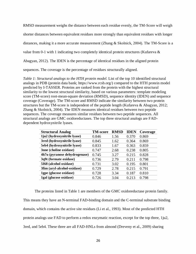
Table 1 Structural analogs to the HTH protein model List of the top 10 identified structural
analogs in PDB (protein data bank httpswwwrcsborg) compared to the HTH protein model
predicted by I-TASSER Proteins are ranked from the protein with the highest structural
similarity to the lowest structural similarity based on various parameters template modeling
score (TM-score) root-mean-square deviation (RMSD) sequence identity (IDEN) and sequence
coverage (Coverage) The TM-score and RMSD indicate the similarity between two protein
structures but the TM-score is independent of the peptide length (Kufareva amp Abagyan 2012
Zhang amp Skolnick 2004) The IDEN measures identical residues between two peptide
sequences The coverage measures similar residues between two peptide sequences All
structural analogs are GMC oxidoreductases The top three structural analogs are FAD-
dependent hydroxynitrile lyases
Structural Analog TM-score RMSD IDEN Coverage 1ju2 (hydroxynitrile lyase) 0846 156 0370 0869 3red (hydroxynitrile lyase) 0845 162 0364 0869 5eb4 (hydroxynitrile lyase) 0833 167 0363 0859 3nne (choline oxidase) 0747 268 0238 0805 4h7u (pyranose dehydrogenase) 0745 327 0215 0828 3q9t (formate oxidase) 0736 279 0211 0798 5I68 (alcohol oxidase) 0731 302 0195 0801 3fim (aryl-alcohol-oxidase) 0729 278 0215 0791 1gpe (glucose oxidase) 0728 334 0187 0810 1gal (glucose oxidase) 0726 304 0213 0798
The proteins listed in Table 1 are members of the GMC oxidoreductase protein family
This means they have an N-terminal FAD-binding domain and the C-terminal substrate binding
domain which contains the active site residues (Li et al 1993) Most of the predicted HTH
protein analogs use FAD to perform a redox enzymatic reaction except for the top three 1ju2
3red and 5eb4 These three are all FAD-HNLs from almond (Dreveny et al 2009) sharing
27
common features to other GMC oxidoreductase proteins but do not appear to use their FAD
cofactor for enzymatic purposes at least not for a redox reaction (Dreveny et al 2001)
233 Conserved Active Site Residues in FAD-Dependent Hydroxynitrle Lyase
It is predicted that all FAD-HLNs have two conserved active site residues His459 and
His497 (numbering based on the P dulcis HNL peptide sequence Figure 6B) HTH has three
putative active site residues Ile527 His529 and Asn567 (numbering based on the A thalian
HTH peptide sequence) (Chang 2016) By using Chimera the HTH protein model was
superimposed over 1ju2 the FAD-HNL from P dulcis (Figure 6D) HTH His529 overlaps with
1ju2 His459 The His459 in 1ju2 causes the protonation of the cyanide ion allowing for the
production of hydrogen cyanide (Dreveny et al 2009) Since HTH has the same residues in this
position it could have the same function HTH Asn567 overlaps with 1ju2 His497 The His497
in 1ju2 is considered a general base that hydrogen bonds with the hydroxyl group of
mandelonitrile to help stabilize the substrate (Dreveny et al 2001)
1ju2 also has a tyrosine at position 457 which could hydrogen bond with the substrate
(Dreveny et al 2009) but based on pKa values of the residue R-group His497 is more likely to
play that role (Dreveny et al 2001) HTH has an asparagine in the same position as 1ju2 His497
which is not ionizable under physiological conditions This means that it cannot be as strong of a
general base like histidine or tyrosine Histidine may be conserved in FAD-HNLs to ensure a
strong general base is present to properly orient the substrate within the active site Since HTH
does not contain this key conserved residue it is reasonable to suggest that HTH will not possess
MDL enzymatic activity
Neither of the conserved active site residues in 1ju2 (His459 and His497) overlap with
HTH Ile527 It is in the same position as Tyr457 in 1ju2 Isoleucine is an aliphatic residue and it
28
likely not an active site residue It could potentially hydrogen bond with the substrate to help
hold it in place or create a favourable environment within the active site Once further research
on potential HTH substrates is conducted the involvement of Ile527 in the binding process
should be clarified
234 Conserved Active Site Residues in GMC Proteins
It is predicted that GMC oxidoreductases have two conserved active site residues His466
and Asn510 (numbering based on the Choline Oxidase peptide from A globiformis) (Figure 6C)
(Dreveny et al 2009) These residues form a well-defined hydrogen bond network that ensures
the substrate is in the correct position and reacts with the N5 molecule of the FAD co-factor
(Fraaije amp Mattevi 2000 Yue et al 1999) As described previously HTH has three putative
active site residues Ile527 His529 and Asn567 (Chang 2016) Chimera was used to
superimpose 3nne over the HTH protein model (Figure 6E) HTH His529 overlaps with 3nne
His466 and HTH Asn567 overlaps with 3nne Asn510 Neither of the conserved active site
residues in 3nne overlap with HTH Ile527 Since HTH contains an asparagine and histidine in
the same location as the conserved residues of 3nne they could be important for proper
enzymatic function by forming hydrogen bonds with the substrate Since HTH does contain both
conserved redox required active site residues the structural model is more consistent with the
proposal that HTH has ADH enzymatic activity
If HTH were to have ADH enzymatic activity this could explain why it localizes in the
ER of epidermal cells (Chang 2016) As it has been previously proposed HTH appears to be
involved in cuticle synthesis a process that primarily takes places in the ER of epidermal cells
(Yeats amp Rose 2013) But this protein has been found in areas of the plant where cuticle
synthesis does not occur such as ER bodies in A thaliana hypocotyl cells (Chang 2016) These
29
are structures that have been linked to plant defence (Matsushima et al 2003) One of the known
hth mutant alleles eda-17 causes embryo sac development arrest linking HTH to early events in
female gametophyte development These results raise many questions about HTHrsquos role in A
thaliana one of which being Is HTH a moonlighting protein Moonlighting proteins are
proteins that have more than one function and typically arise from some sort of evolutionary split
and resource allocation (Jeffery 2017) Very few moonlighting proteins have been identified in
plants (Rad et al 2018) meaning that many have yet to be discovered HTH may be one of them
235 Predicted Ligand Binding Site on the HTH Protein Model
Potential ligands to the HTH protein were predicted using COACH software (Yang Roy
amp Zhang 2013b 2013a) This generated a list of ligands their predicted binding site and a
coinciding C-score The C-score is a confidence score of the prediction based on the threading
template alignments and convergence parameters of the structure assembly simulations (Roy
Kucukural amp Zhang 2010 Yang et al 2015 Yang amp Zhang 2015 Zhang 2008) This score
ranges from -5 to 2 with a higher value signifying a higher confidence
The cofactor with the highest C-score was FAD at 078 (Figure 7A) This further supports
the belief that HTH is an FAD-dependent protein and a member of the GMC oxidoreductase
protein family Whether HTH uses FAD for enzymatic purposes or simply for structural integrity
us unclear Taking a closer look at the predicted binding site of FAD it is possible to see that the
flavin portion of the molecule comes in close proximity to the three putative active site residues
in HTH (Figure 7A) No portion of the FAD molecule comes into contact with the predicted
FAD-binding site (residues 64-91 in HTH) This is likely due to the inaccuracies in this simple
docking experiment
30
Benzaldehyde and mandelonitrile were the two other ligands predicted to complex with
the HTH protein model Although benzaldehyde and mandelonitrile are fairly similar and close
in overall size their predicted binding sites are in different areas (Figure 7B-C) Benzaldehyde
was predicted to bind in proximity to the putative active site residues 43 Å from His529 in the
HTH protein model while mandelonitrile is predicted to bind to the surface of the HTH protein
12 Å from Ile527 in the HTH protein model the closest putative active site residue
Unfortunately benzaldehyde and mandelonitrile have very low C-scores at 004 and 001
respectively Due to the low C-score it is not possible to say with confidence that either ligands
bind to the HTH protein No other ligands were predicted to bind to the HTH protein model
An issue with the ligand docking predictions is that the FAD co-factor is not included
when the model is input into the software This generates a large empty space in the predicted
location of FAD within the HTH model (data not shown) This was further exemplified with the
SwissDock software (Grosdidier Zoete amp Michielin 2011 Grosdidier 2011) When
mandelonitrile was input as a potential ligand it was predicted to bind to the surface of the HTH
model as well as within the FAD binding site A similar issue could be happening within the
active site Since no ligands are included when the protein model is being created the residues
may be oriented differently to accommodate that empty space This could be preventing the
software algorithms to predict certain ligands bind within this site To account for this the
residues around the empty spaces could be manually adjusted in Chimera to see if ligands can fill
the space although this may not be an accurate representation of the model
These simple docking experiments were largely a failure The results should not be
strongly interpreted due to the reasons discussed above A more conplex docking study could be
undertaken to provide stronger evidence for or against the binding of these ligands
31
Figure 6 Ribbon diagrams comparing the putative active site residues of the HTH protein to
conserved active site residues of an FAD-HNL (1ju2) amp a GMC protein (3nne) The HTH model
was generated using I-TASSER and visualized in Chimera (A B C) The active site residues of
HTH 1ju2 and 3nne (A) The HTH protein model is represented in beige The three putative
active site residues (Ile527 His529 Asn567) are represented in magenta (B) The 1ju2 protein is
represented in blue The two conserved active site residues (His459 and His497) are represented
in red (C) The 3nne protein is represented in orange The two conserved active site residues
(His466 and Asn510) are represented in yellow (D E) The HTH protein model superimposed
over the 1ju2 and 3nne based on secondary structure using Chimera (D)The HTH protein model
(beige) superimposed over 1ju2 (blue) HTH His529 overlaps with 1ju2 His459 while HTH
Asn567 overlaps with 1ju2 His497 (E) The HTH protein model (beige) superimposed over 3nne
(orange) HTH His529 overlaps with 3nne His466 while HTH Asn567 overlaps with 3nne
Asn510
32
Figure 7 Ribbon diagrams of the HTH model-ligand complexes The HTH model complexes
were generated with COACH and visualized in Chimera The HTH protein model is represented
in beige The putative active site residues are purple (A) The most probable binding site of FAD
(green) within the HTH protein model The ligand looks to bind nicely within the FAD binding
domain with the N5 molecule located in proximity of the active site (B) The most probable
binding site of benzaldehyde (blue) within the HTH protein model The ligand is found within
the active site Its hydroxyl group is shown to be ~42Å from the closest putative active site
residue His529 (C) The most probable binding site of mandelonitrile (black) within the HTH
protein model The ligand is found binding to the surface of the protein structure
236 Comparing Hydroxynitrile Lyase N-Linked Glycosylation to the HOTHEAD
Model
N-linked protein glycosylation is a highly conserved process in eukaryotes (Burda amp
Aebi 1999) It begins in the ER with the formation of a glycan This oligosaccharide is found
within the ER lumen and bound to the membrane It is transferred to the protein by
oligosaccharyl transferase (Burda amp Aebi 1999) The glycan will bond with the proteinrsquos
asparagine with the proper recognition sequence (Burda amp Aebi 1999) At this point chaperone
proteins will recognize the glucose residues on the glycan and aid in protein folding (Helenius et
al 1997) Glucosidase will cleave off the glucose molecules allowing the folded protein to
dissociate from the chaperone proteins and travel to the Golgi apparatus (Helenius et al 1997)
Once in the Golgi apparatus the oligosaccharide will be subjected to further modifications
(Burda amp Aebi 1999 Schwarz amp Aebi 2011)
All known FAD-HNLs are predicted to have three conserved N-linked glycosylation
sites Asn118 Asn135 and Ans352 (numbering based on the P dulcis HNL peptide sequence)
33
(Dreveny et al 2001) These sites all have the same recognition sequences of N-X-ST (ie
Asp any amino acid Ser or Thr) (Dreveny et al 2001) Based on previous bioinformatic work
the HTH protein is predicted to have 5 putative N-linked glycosylation sites all with the same
recognition sequences as FAD-HNLs Asn84 Asn101 Asn420 Asn482 and Asn511 (Chang
2016) These glycosylation sites are all found on the surface of the protein (Figure 8) It is
interesting to note however that when the HTH protein model is superimposed with the top
structural analog based on secondary structures none of the putative glycosylation sites align
with the three conserved FAD-HNL N-linked glycosylation sites The alignment of these
glycosylation sites should be further investigated once the structure of the HTH protein is
confirmed
HTH has been experimentally shown to be a glycoprotein based on size shifts following
endoglycosidase treatment of a HTHGFP protein extracted from transgenic plant lines (Chang
2016) FAD-HNLs have varying amounts of glycosylation which may not be important to the
enzymersquos function as de-glycosylation of the purified enzyme from P dulcis did not affect
enzymatic activity (Dreveny et al 2001) This de-glycosylation occurred after the protein had
been folded so it is likely that the N-linked glycosylation was important for proper protein
folding It is unknown whether glycosylation is important for HTH protein function
34
Figure 8 Ribbon diagrams of the HTH model amp FAD-HNL (1ju2) N-linked Glycosylation sites
The HTH model complexes were generated with COACH and visualized in Chimera The HTH
protein model is represented in beige The 5 putative N-linked glycosylation sites (Asn84
Asn101 Asn420 Asn482 and Asn511) are represented in green The 1ju2 protein is represented
in blue The 3 conserved N-linked glycosylation sites (Asn118 Asn135 and Ans352) are
represented in purple All putative N-linked glycosylation sites are found on the surface of the
HTH protein model None of the glycosylation sites in the HTH model overlap with the
glycosylation sites in 1ju2
35
24 Conclusions
The protein model predicted by I-TASSER has been useful to learn more about the HTH
protein This model was built by aligning the HTH peptide sequence to proteins with known
structures Developing a model of a protein has proven to be an effective method to determine
the overall structure of a protein especially proteins with sequence identities ge 25 (Giorgetti
Raimondo Miele amp Tramontano 2005) Although not as precise as the actual HTH structure
since the model is based on the structure of other proteins it is a useful tool for structural and
computational analysis The fact that FAD was omitted from the model did create inaccuracies
that could have effects on the overall structure Once a protein isolation protocol is developed for
the HTH protein its structure can be determined and used to validate the findings presented in
this chapter
The software used to predict potential ligand docking sites omitted FAD causing
inaccurate predictions Residues likely changed orientation to fill in the empty space typically
occupied by FAD Due to these slight changes in the protein FAD was unable to bind to its
predicted binding site (residues 64-91 in HTH) Even though MDL and benzaldehyde two
molecules that bind to FAD-HNLs were predicted to bind to the HTH model their confidence
scores were too low to state that they truly bind to HTH These results could not be used to
further validate the conclusions drawn from the conserved active site residue analysis A more
robust docking prediction software should be used in the future to determine potential HTH
protein ligands
The HTH protein shares sequence identity to a GMC protein with redox activity and an
FAD-HNL (Kurdyukov et al 2006) Based on the results presented in Sections 233 and 234
the HTH protein model does not contain the conserved active site residues of FAD-HNLs and it
36
does contain the conserved active site residues of GMC oxidoreductase proteins (Figure 6)
These residues are important for forming a hydrogen bond network with the substrate to ensure
that it is properly positioned in proximity to the N5 atom of the FAD (Yue et al 1999) This
could mean that HTH has ADH enzymatic activity
N-linked glycosylation occurs within the ER and the Golgi apparatus This post-
translational modification is important for proper protein folding (Schwarz amp Aebi 2011) If a
glycoprotein is lacking its required N-linked glycosylation it most likely will not fold properly
It was shown that FAD-HNLs have three conserved N-linked glycosylation sites (Dreveny et al
2001) that do not overlap with the putative N-linked glycosylation sites in HTH If an FAD-HNL
is de-glycosylated it can still remain active (Dreveny et al 2001) The importance of N-linked
glycosylation in the HTH protein remains to be determined
HTH has been implicated in several different processes cuticle embryogenesis and plant
defence It may have more than one function which would classify it as a moonlighting protein
such as arogenate dehydratase 2 (Rad et al 2018) and plastidial NAD+-dependent malate
dehydrogenase (Schreier et al 2018 S M Smith 2018) To date very few moonlighting
proteins have been identified in plants Hopefully once the actual HTH protein structure is
determined further research can be done to gain a better understanding of its potential enzymatic
activity and its role in A thaliana
37
Chapter 3
Recombinant HOTHEAD Protein Isolation amp
Enzymatic Assays
38
31 Introduction
A significant bottleneck in assigning a definitive role for HTH in the cuticle synthetic
pathway has been the inability to isolate and characterize the HTH protein Being able to develop
an isolation protocol would be beneficial and bring new insight into the HTHrsquos role in A
thaliana growth and development
A recombinant protein isolation protocol can be very challenging to develop since many
variables must be considered to optimize the protocol and ensure that enough active protein is
isolated (Lee 2017) Once optimized this opens the door for further research opportunities A
proteinrsquos structure can be determined via x-ray crystallography and its potential enzymatic
function can be assessed with various enzymatic assays An attempt has been previously made to
isolate the HTH protein (Chang 2016) In this case HTH was expressed in E coli using the
pMal-c4x vector but the strategy yielded little to no protein
This chapter documents various attempts to isolate a recombinant HTH protein
expressed in E coli The construct design induction trials initial isolation attempts and all
troubleshooting steps are discussed Further optimization is still required with current work
focusing on inducing HTH in a yeast expression system In an alternate strategy to obtain active
HTH crude protein extracts were obtained from transgenic A thaliana plant lines These extracts
were tested for potential MDL enzymatic activity
39
32 Materials
The HTH gene was synthesized and cloned into the SUMOstar_Kan vector by GenScript
(GenScript Biotech Corporation Piscataway New Jersey United States) and inserted into
BL21(DE3) competent cells creating pE_SUMOstar_Kan The SUMOstar_Kan vector was
obtained from LifeSensors (LifeSensors Devault Pennsylvania United States) The GeneJET
Plasmid Miniprep Kit was purchased from Thermo Fisher Scientific (Thermo Fisher Scientific
Waltham Massachusetts Unites States) All restriction enzymes were purchased from New
England Biolabs (New England Biolabs Ipswish Massachusetts United States) The 1 kilobase
pair (kd) DNA size ladder was purchased from GeneDireX (GeneDireX Inc Taiwan) The
Protease Arrest and EDTA were purchased from G Biosciences (G Biosciences St Louis
Missouri United States) The Ultracel 10 kDa Ultrafiltration Disc was purchased from
MilliporeSigma (Millipore Sigma Burlington Massachusetts Unites States) The Ni-NTA
Agarose was purchased from Qiagen (Qiagen Germany) The Bio-gel P-6 DG (polyacrylamide
desalting gel with 6000 molecular weight exclusion limit) media was purchased from Bio-Rad
(Bio-Rad Hercules California United States) The UNOsphere Q and the Bio-Scale Mini
UNOsphere S 5 mL cartridge were purchased from Bio-Rad (Bio-Rad Hercules California
United States) The Q5 Site-Directed mutagenesis kit was purchased from New England Biolabs
(New England Biolabs Ipswish Massachusetts United States) All primers were synthesized by
Sigma (Sigma-Aldrich Life Science St Louis Missouri United States) unless otherwise
specified The LC1 Grower Mix was purchased from SunGro Sunshine (SunGro Sunshine
Alberta Ontario) The mandelonitrile lyase from Almonds was purchased from Sigma (Sigma-
Aldrich Life Science St Louis Missouri United States) The mandelonitrile was purchased
from Sigma (Sigma-Aldrich St Louis Missouri United States) The anti-GFP antibody was
40
purchased from Abcam (Abcam Biotechnology Company Cambridge United Kingdom) The
anti-rabbit IgG antibody conjugated to horseradish peroxidase was purchased from Sigma
(Sigma-Aldrich St Louis Missouri United States) All other general lab chemicals were of the
highest grade available and purchased from various companies
33 Methods
331 Synthetic HTH cDNA Sequence Design
The HTH peptide sequence was input into the SignalP 41 server to determine if HTH has
a signal peptide (httpwwwcbsdtudkservicesSignalP-41) The output graph indicated the C-
score S-score and Y-score of the first 70 residues in the peptide sequence (Petersen Brunak
Heijne amp Nielsen 2011) The first 19 residues are predicted to comprise the signal peptide The
predicted peptide sequence of the synthetic cDNA was aligned with the WT HTH peptide
sequence to ensure proper design The nucleotide bases that code for this region of the HTH
protein were therefore omitted in the synthetic HTH cDNA sequence to optimize for protein
expression in bacteria and facilitate isolation The HTH protein without this signal peptide has a
predicted mass of 63 kDa The synthetic cDNA was synthesized and cloned by GenScript
(GenScript Biotech Corporation Piscataway New Jersey United States) into the
pE_SUMOstar_Kan vector (HTH_SUMO) (Appendix A) and the pET24a+ vector
(HTH_pET24a+) (Appendix B)
332 Heat Shock Bacteria Transformation
A 100 μL aliquot of BL21(DE3) competent cells was gently mixed with 5 ng of
HTH_SUMO vector DNA and incubated on ice for 30 minutes Cells were heat shocked for 30
seconds at 42degC and added to 400 μL Luria broth (LB) (1 mgmL tryptone 1 mgmL NaCl and
05 mgmL yeast extract) then incubated at 37degC for 1 hour Cells were spread on LB agar plates
41
supplemented with 50 μgmL kanamycin 40 μgmL X-gal and 50 μgmL isopropyl β-D-1-
thiogalactopyranoside (IPTG) then incubated overnight at 37degC White colonies were picked and
added to separate flasks containing 50 mL LB supplemented with 50 mgmL kanamycin then
incubated overnight at 37degC A 25 glycerol stock was prepared by mixing 250 μL of the cell
resuspension with 250 μL 50 glycerol The glycerol stock was stored at -80degC The same heat
shock protocol was repeated to transform BL21(DE3) with HTH_pET24a+
333 Bacterial Plasmid Minipreparation
Bacterial plasmid DNA was isolated from transformed BL21(DE3) cells using the
GeneJET Plasmid Miniprep Kit (ThermoFisher Scientific Waltham Massachusetts United
States) 5-10 mL of BL21(DE3) transformed with HTH_SUMO was resuspended via vortex in
250 μL resuspension solution supplemented with 25 μL RNaseA 250 μL lysis solution was
added and mixed by inversion and 350 μL neutralizing solution was immediately added to the
lysate The cells were mixed by inversion and incubated on ice for 5 minutes before
centrifugation for 5 minutes at 13000 times g Up to 600 μL of the supernatant was applied to a
Thermo Scientific GeneJET Spin column 185 μL wash solution supplemented with 315 μL
100 EtOH was added to the spin column and centrifuged for 1 minutes at 13000 times g The wash
step was repeated a second time 50 μL elution buffer was added directly to the spin column
membrane and incubated at room temperature for 2 minutes before centrifugation for 2 minutes
at 13000 times g The final column flow through containing the plasmid was stored at -20degC
334 HTH_SUMO Restriction Endonuclease Digestion
HTH_SUMO plasmid isolated from transformed BL21(DE3) was digested with
restriction endonucleases to ensure that the HTH_SUMO vector taken up by the competent cells
was unaltered One sample of plasmid was incubated with NcoI (New England Biolabs Ipswich
42
Massachusetts United States) another sample was incubated with NruI (New England Biolabs)
and a final sample was incubated with both restriction endonucleases All samples as well as an
undigested HTH_SUMO plasmid were run on a 08 agarose gel casted with 01 μgmL
ethidium bromide (Thermo Fisher Scientific) DNA bands were visualized under UV light A 1
Kb DNA size ladder (GeneDireX Inc Taiwan) run concurrently on the same gel was used to
determine the apparent band sizes Colonies with plasmids yielding digested bands of the
appropriate size were kept and store at -80degC
The same diagnostic digest protocol was used on HTH_pET24A+ plasmid isolated from
BL21(DE3) using endonucleases XhoI (New England Biolabs) and NdeI (New England Biolabs)
This protocol was also used on hth mutant alleles generated using the Q5 Site-Direted
Mutagenesis kit (New England Biolabs) cloned into the pE_SUMOstar_Kan vector using
endonucleases ClaI (New England Biolabs) and XhoI (New England Biolabs)
335 HTH Protein Autoinduction Set up
50 mL of autoinduction media (1 mgmL tryptone 05 mgmL yeast extract 1M MgSo4
01 (vv) 1000 times TRACE elements (50 mM FeCl3 20 mMCaCl2 10 mM MnCl2 10 mM
ZnSO4 2 mM CoCl2 2mM CuCl2 2mM NiCl2 2mM Na2MoO4 2mM Na2SeO3 2mM H3BO3)
5 (vv) 20 times NPS (66 mgmL (NH4)2SO4 136 mgmL KH2PO4 142 mgmL Na2HPO4) 1
(vv) 50 glycerol 02 (vv) 25 glucose 2 (vv) 10 lactose) was supplemented with 50
mgmL kanamycin and 1 mL of a 50 mL BL21(DE3) starter culture resuspension successfully
transformed with HTH_SUMO (Fox amp Blommel 2017) Induction media inoculated with
transformed cells was incubated at 20degC for 24 hours Media was centrifuged for 10 minutes at
5100 times g Cells were resuspended and sonicated using the QSonica Q500 sonicator fitted with
the standard frac12 inch diameter probe (QSonica LLC Newtown Connecticut Unites States) The
43
cells were sonicated for a total of 2 minutes at 75 amplitude This 2-minute length of time was
divided into 5 second pulses with a 55 second interval between each one Therefore the total
sonication procedure took 24 minutes The cell lysate was subsequently centrifuged for 45
minutes at 12000 times g and a sample of the supernatant and the pellet were run on a 12 SDS-
PAGE gel to determine the protein induction profile
336 Nickel Chromatography of HTH_SUMO Protein
The induction protocol outlined in Section 335 was scaled up by increasing the
autoinduction media volume to 4 L and the HTH_SUMO transformed BL21(DE3) starter culture
volume to 100 mL Cell pellets were stored at -80degC prior to lysis and purification when
necessary and thawed on ice before use Cells were resuspended in 100 mL Buffer 1 (25 mM
HEPES 10 glycerol 200 mM NaCl 10 mM imidazole pH 75) supplemented with 1 times
BioScience Protease Arrest and 5 mM ethylenediaminetetraacetic acid (EDTA) (G Biosciences
St Louis Missouri United States) Resuspended cells were sonicated using the same protocol
outlined in 328 and subsequently centrifuged for 45 minutes at 20000 times g 360 mgmL
ammonium sulfate ((NH4)2SO4) was added to the supernatant and mixed for 30 minutes before
centrifugation for 30 minutes at 20000 times g The pellet was resuspended in 50 mL Buffer 1
20 mL Ni-NTA Agarose was added to a column (5 cm times 20 cm) (Qiagen Germany)
Before use the nickel resin was stripped and recharge The column was washed with 200 mL 02
M acetic acid and 6 M Guanidine hydrochloride and rinsed with 500 mL dH2O The column was
then washed with 300 mL 2 SDS solution and rinsed with 400 mL dH2O The column was then
washed with 500 mL 70 EtOH and rinsed with 500 mL dH2O The column was then washed
with 500 mL 100mM EDTA and rinsed with 500 mL dH2O The column was washed with 500
44
mL NiSO4 and rinsed with 500 mL dH2O Finally 300 mL of Buffer 1 was passed through the
column
The pellet resuspension was mixed with the nickel resin for 30 minutes and applied to a
column (5 cm times 20 cm) Buffer 1 was washed through the nickel column until the eluate reached
an OD280 below 01 2 mL eluate fractions were collected as Buffer 2 (25 mM HEPES 300 mM
imidazole pH 75) was washed through the nickel column All fractions with an OD280 above 01
were mixed together and concentrated to 10 mL using an Amicon stir cell fitted with an Ultracel
10 kDa Ultrafiltration Disc (Millipore Sigma Burlington Massachusetts Unites States)
pressurized under compressed nitrogen gas The concentrated protein sample was added to a
column (15 cm times 30 cm) filled with Bio-Gel P-6 DG Media (Bio-Rad) 2 mL eluate fractions
were collected as Buffer 3 (25 mM HEPES pH 75) was washed through the nickel column All
fractions with an OD280 above 01 were pooled 1 mL of isolated SUMO protease was mixed
overnight at 4degC with the pooled fractions to cleave off the 6 times His tag The nickel column was
washed with 300 mL Buffer 3 The protein solution was mixed with the nickel resin on column
for 30 minutes 2 mL eluate fractions were collected as Buffer 3 was washed through the nickel
column All fractions with an OD280 above 01 were mixed together and concentrated down to 10
mL using a same Amicon stir cell fitted with an Ultracel 10 kDa Ultrafiltration Disc (Millipore
Sigma) pressurized under compressed nitrogen gas The sample was concentrated down to 1 mL
using a 30 kDa Amicon Ultra-15 Centrifugal Filter Unit centrifuged for 10 minutes at 2800 times g
Samples were flash frozen in 20 μL aliquots using liquid nitrogen and stored at -80degC All steps
were performed at 4degC or on ice unless otherwise specified
45
337 Anion Exchange Chromatography
Protein fractions isolated via nickel chromatography were thawed on ice The P-6 DG
column was washed with 300 mL Buffer A (25 mM HEPES (pH 75)) The thawed protein
fractions were added to the column 2 mL eluate fractions were collected as Buffer A was
washed through the column All fractions with an OD280 above 01 were mixed together Up to 1
mL of pooled eluate was injected into the BioLogic Duo Flow Chromatography System (FPLC)
set up with the UNOsphere Q cartridge (Bio-Rad Hercules California United States) 35 mL of
Buffer A was pumped through the FPLC at a rate of 15 mLmin The protein was injected onto
the UNOsphere cartridge at a rate of 15 mLmin 15 mL of Buffer A was pumped through the
FPLC at a rate of 025 mLmin A gradient change from Buffer A to Buffer B (25 mM HEPES
05 M NaCl pH 75) was set at a rate of 15 mLmin for 100 mL until only Buffer B was
pumping through the FPLC for another 35 mL 2 mL fractions were collected throughout the
protocol A UV sensor was zeroed and used to measure the presence of protein as the solution
was pumped out of the machine All aforementioned cation exchange chromatography steps
were performed at 4degC or on ice unless otherwise specified The fractions containing protein
were run on a 12 SDS-PAGE gel
338 Cation Exchange Chromatography
Protein fractions isolated via nickel chromatography were thawed on ice The P-6 DG
column was washed with 300 mL Buffer A (25 mM HEPES (pH 75)) The thawed protein
fractions were added to the column 2 mL eluate fractions were collected as Buffer A was
washed through the column All fractions with an OD280 above 01 were mixed together Up to 1
mL of pooled eluate was injected into the BioLogic Duo Flow Chromatography System (FPLC)
set up with the Bio-Scale Mini UNOsphere S 5 mL cartridge (Bio-Rad) 35 mL of Buffer A was
46
pumped through the FPLC at a rate of 05 mLmin The protein was injected onto the
UNOsphere cartridge at a rate of 025 mLmin 15 mL of Buffer A was pumped through the
FPLC at a rate of 025 mLmin A gradient change from Buffer A to Buffer B (25 mM HEPES
05 M NaCl pH 75) was set at a rate of 025 mLmin for 100 mL until only Buffer B was
pumping through the FPLC for another 35 mL 2 mL fractions were collected throughout the
protocol A UV sensor was zeroed and used to measure the presence of protein as the solution
was pumped out of the machine All aforementioned cation exchange chromatography steps
were performed at 4degC or on ice unless otherwise specified The fractions containing protein
were run on a 12 SDS-PAGE gel
The protein fractions were also passed through the same cation exchange column at pH 6
The same protocol was used with the exception of the buffer A 25 mM MES buffer (pH 6) was
used for buffer A and a 25 mM MES buffer (pH 6) with 05 M NaCl was used for Buffer B
339 Protein Solubilization via Arginine Treatment
BL21(DE3) cells transformed with HTH_SUMO were induced using the protocol
outlined in Section 332 Six aliquots of 1 mL induced started culture were used Once the cells
were lysed and the supernatant was removed 500 μL PBS (pH 75) was added to the pellets
Arginine was added to each aliquot at increasing concentrations 0M 025 M 05 M 1M 15M
and 2M Solutions were mixed via pipette and incubated at 4degC overnight The solution was
centrifuged for 30 minutes at 12000 times g and a sample of the supernatant and the pellet were run
on a 12 SDS-PAGE gel to determine the protein profile
3310 Mass Spectrometry Analysis
Mass spectrometry analysis was performed on samples of the final fraction obtained from
nickel chromatography to determine the protein profile The protein sample was run on a 4
47
SDS-PAGE gel Five bands around the 70 kDa range were cut out of the acrylamide gel and sent
to the Sick Kidsrsquo SPARC BioCentre Molecular Analysis facility (Toronto Hospital for Sick
Children Ontario Canada) The protein profile of each band was visualized on the Scaffold
Viewer software (Proteome software Portland Oregon United States)
3311 Nickel Chromatography using HTH_pET24a+
The induction protocol outlined in Section 335 was scaled up by increasing the
autoinduction media volume to 4 L and the HTH_pET24a+ transformed BL21(DE3) starter
culture volume to 100 mL Cell pellets were stored at -80degC prior to lysis and purification when
necessary and thawed on ice before use Cells were resuspended in 100 mL Buffer 1 (25 mM
HEPES 10 glycerol 200 mM NaCl 10 mM imidazole pH 75) supplemented with 1 times
BioScience Protease Arrest and 5 mM ethylenediaminetetraacetic acid (EDTA) (G Biosciences)
Resuspended cells were sonicated using the same protocol outlined in 318 The cell lysate was
subsequently centrifuged for 45 minutes at 20000 times g 360 mgmL (NH4)2SO4 was added to the
supernatant and mixed for 30 minutes before centrifugation for 30 minutes at 20000 times g The
pellet was resuspended in 50 mL Buffer 1 The nickel column was prepared as previously
described in Section 336
The resuspended pellet was mixed with the nickel resin for 30 minutes and applied to a
column (5 cm times 20 cm) Buffer 1 was washed through the nickel column until the eluate reached
an OD280 below 01 Two mL eluate fractions were collected as Buffer 2 (25 mM HEPES 300
mM imidazole pH 75) was washed through the nickel column All fractions with an OD280
above 01 were mixed together and concentrated to 10 mL using an Amicon stir cell fitted with
an Ultracel 10 kDa Ultrafiltration Disc pressurized compressed nitrogen gas The concentrated
protein sample was added to a P-6DG column 2 mL eluate fractions were collected as Buffer 3
48
(25 mM HEPES pH 75) was washed through the nickel column All fractions with an OD280
above 01 were mixed together and concentrated down to 10 mL using a same Amicon stir cell
fitted with an Ultracel 10 kDa Ultrafiltration Disc pressurized under gaseous nitrogen from a
compressed nitrogen tank The sample was concentrated down to 1 mL using a 30 kDa Amicon
Ultra-15 Centrifugal Filter Unit centrifuged for 10 minutes at 2800 times g Samples were flash
frozen in 20 μL aliquots using liquid nitrogen and stored at -80degC All steps were performed at
4degC or on ice unless otherwise specified
3312 Chemical Competent Saccharomyces cerevisiae Transformation
HTH cDNA without the nucleotides coding for the signal peptide obtained from
Athalian mRNA reverse transcription (Chang 2016) was PCR amplified using the forward 5rsquo-
ATTCGCAGCATCCTCCGCATTAGCTGGGGGATCCACTAGTTCCACTGCCTCTAAAGG
TAAAG-3rsquo and reverse 5rsquo- GGTGCTCGAGGCCCTGAAAATACAGGTTTTCTGCGGCCGC
ACTAAACACCAGCTTTGTTTC-3rsquo primers (Sigma-Aldrich Life Science St Louis Missouri
United States) 15 μL PCR product was mixed with 5 μL fish sperm DNA 5 μL linearized and
dephosphorylated pPinkNN vector (Nava amp Amidi unpublished results) and 200 μL chemical
competent S cerevisiae (Amidi amp Heinisch unpublished results) The cell mixture was gently
mixed and incubated at 37degC for 10 minutes 1 mL Solution B (40 PEG 1500 100 mM Bicine
pH 84) was added to the suspension gently mixed and incubated at 30degC for 1 hour The
suspension was centrifuged and excess supernatant was removed The pellet was resuspended in
the remaining 200 μL supernatant then plated on Ura3- agar plates and incubated at 30degC for 3
days
49
3313 HTH cDNA Q5 Site-Directed Mutagenesis
The Q5 Site-Directed Mutagenesis kit (New England Biolabs) was used to introduce
mutations in the synthetic HTH DNA vector (HTH_SUMO) described in Section 331 Primer
sets were designed using the NEBaseChanger software (httpsnebasechangernebcom)
Mutations were introduced following the mutagenesis kit protocol and the mutated HTH_SUMO
vectors transformed via heat shock into DH 5-α competent cells Cell mixture was plated on LB
agar plates supplemented with 50 μgmL kanamycin 40 μgmL X-gal and 50 μgmL IPTG and
incubated at 37degC overnight White colonies were picked and added to separate flasks containing
50 mL LB supplemented with 50 mgmL kanamycin then incubated overnight at 37degC A 25
glycerol stock was prepared by mixing 250 μL of the cell resuspension with 250 μL 50 filtered
sterilized glycerol The glycerol stock was stored at -80degC
3314 DNA Sequence Analysis
Six primers were designed specifically for sequencing HTH_SUMO vectors (Sigma-
Aldrich) 5rsquo-AAGAAGGCTGAT GGAAGCGT-3rsquo 5rsquo- CTGACCCTGTGGCAAAAAGC-3rsquo
and 5rsquo-TTTTGGTCAGAGCCCGG AAA-3rsquo anneal to the DNA in the forward direction 5-
TTTCACCAGCTTCGGATCCC-3rsquo 5rsquo-AATACCGCTCAGCATCAGCA-3rsquo and 5rsquo-
CCCATTCGCCAATCCGGATA-3rsquo anneal to the DNA in the reverse direction (Appendix I)
Primers were spaced no more than 1000 bp apart to obtain a full coverage of the cDNA
sequence Sequencing was performed by The Centre for Applied Genomics (Toronto Hospital
for Sick Children Ontario Canada) Sequencing results were analysed to ensure correct
mutations were introduced into the HTH cDNA sequence
50
3315 Plant material and Growth Conditions
Previously the HTHproHTH-G3GFP construct (Chang 2016) was cloned into the
pGWB650 vector (Akamura et al 2010) and the pCAMBIA vector (Sinclair et al 2009) then
transformed into hth-9 mutant plants to create the P650 and P103 plant lines respectively
(Figure 19) hth-9 A thaliana seeds in the Wassilewskija ecotype were previously described by
Lolle et al (1998) The pGWB650 vector contains the 2834 bp HTH gene and its 2009 bp
putative promoter with a C-terminal green fluorescent protein tag a modified GFP protein
(Chang 2016 Kawakami amp Watanabe 1997) The pCAMBIA vector contained the HTH gene
under the control of the CaMV 35S promoter with a C-terminal G3 green fluorescent protein tag
(Chang 2016) Plant lines were also transformed with the empty vectors and denoted as P650EV
and P103EV respectively
Seeds from these four plant lines as well as untransformed hth-9 seeds were sown onto
moistened Sun Gro Sunshine LC1 Grower Mix (Sun Gro Sunshine Alberta Canada) and
stratified at 4degC for 4 days Plants were maintained in a Conviron PGW36 growth chamber at 20-
23degC and illuminated with a mixture of incandescent and fluorescent lights (140 ndash 170 μmol m-2
sec-1 at pot level) with a 24-hour photoperiod Plants were watered as needed typically three
times a week
3316 Floral Tissue Crude Protein Extraction
Plants were collected from the growth chamber once floral buds were formed and ready
to open which occurred approximately 4 weeks after being sown Fresh floral bud tissue was
homogenized in a small glass homogenizer using a 21 Extraction buffer (100 mM Tris pH75
150 mM NaCl 10 glycerol 01 Triton-X 1X BioScience ProteaseArrest) to floral tissue
ratio (2 mL extraction buffer to 1 mg tissue) to help extract the protein from the tissue The tissue
51
homogenate was centrifuged twice for 30 minutes at 12000 times g to remove insoluble plant
material 180 mgmL (NH4)2SO4 was mixed with the supernatant and incubated for 30 minutes at
4degC and the supernatant collected after centrifugation 30 minutes at 12000 times g 420 mgmL
(NH4)2SO4 was mixed with the supernatant incubated for 30 minutes at 4degC and the pellet
collected by centrifugation (30 minutes at 12000 times g) The final pellet fraction was resuspended
in 200 μL extraction buffer
3317 In vitro Mandelonitrile Lyase Enzymatic Assay
To determine if HTH had hydroxynitrile lyase enzymatic activity the final pellet fraction
from the crude floral protein extract was tested for activity using an in vitro enzymatic assay
(Jorns 1979) Crude extracts from the P650 P650EV P103 P103EV and untransformed hth-9
plant lines were used for the assay A positive control using a commercially available MDL
extracted from Almond (pH 54 Sigma-Aldrich Life Sciences) was also included in the assay
Recombinant HTH protein induced in E coli and isolated via nickel chromatography were also
tested for MDL enzymatic activity
Enzymatic activity was determined by monitoring cleavage of mandelonitrile into
benzaldehyde and HCN at 25degC by following the protocol developed by Jorns (1979) The assay
contained 95 mM sodium acetate (pH 54) 28 mM mandelonitrile 16 (vv) EtOH and 02
unit MDL The absorbance of the assay solution was monitored at 275 nm in a plastic cuvette
with a 1 cm light path until readings stabilized roughly 3 minutes The reaction was initiated by
addition of mandelonitrile (Sigma-Aldrich Life Sciences) The OD275 of the reaction was
continuously monitored for 15 minutes using the Cary Win UV Kinetics software (Agilent
Technologies Mississauga Ontario Canada) and a Cary 100 UV-Vis spectrometer (Agilent
Technologies) Enzymatic activity was calculated using the slope of ∆OD275nm The change in
52
absorbance from the moment mandelonitrile was added to the assay until 15 minutes after was
measured by using the ldquorule of least squaredrdquo cursor option provided in the UV Kinetics
software (Agilent Technologies)
3318 Western Blot Analysis of Floral Bud Crude Protein Extracts
The homogenate and final pellet fraction of the P650 crude protein extracts and the
homogenate of the hth9 crude protein extracts were used for protein immune-detection via
Western blotting The total protein concentration was determined using the Quick Start Bradford
1 times Dye Reagent (BioRad) according to the protocol (Bio-Rad 2012) 30 μg of protein was run
on a 10 SDS-PAGE gel then transferred to a polyvinylidene fluoride transfer membrane
soaked in transfer buffer (25 mM Tris 192 mM Glycine 10 methanol) supplemented with
01 SDS The transfer was performed on a Trans-Blot SD Semi-Dry Transfer cell (Bio-Rad)
The membrane was stained with Ponceau-S (01 (wv) in 5 acetic acid) to visualize protein
bands and then washed with milliQ water before soaking in blocking buffer (TBS-T (20 mM Tris
pH 75 300 mM NaCl 01 (vv) Tween-20) plus 5 skimmed milk) The membrane was
sealed in an airtight bag and incubated overnight at 4degC in 2 mL blocking buffer supplemented
with 13000 anti-GFP antibody (Biotechnology Company Cambridge United Kingdom
ab6556) The membrane was washed with TBS-T then incubated in blocking buffer
supplemented with 130000 anti-rabbit IgG antibody conjugated to horseradish peroxidase
(Sigma-Aldrich Life Sciences A0545) for 1 hour at room temperature The membrane was
washed with TBS-T Clarity Western ECL Substrates (BioRad) were mixed and added to the
membrane and chemiluminescence visualized on the ChemiDoc MP Imaging System (BioRad)
running the Image Lab 521 software
53
34 Results amp Discussion
This research attempted to improve recombinant HTH protein yield compared to previous
attempts which used a maltose-binding protein tag (Chang 2016) through transformation of a
synthetic HTH cDNA sequence designed for optimal bacterial expression using the
pE_SUMOstar vector While the attempts to isolate the HTH protein were unsuccessful
important research implications including the potential factors inhibiting the isolation of HTH
and corresponding implications for HTH functionality were revealed Overall the findings from
this research indicate that the most likely reason for unsuccessful HTH isolation stems from the
use of E coli as an expression system E coli is an organism commonly used for recombinant
protein production isolation and activity assays due to it having well characterized genetics as
well as being inexpensive easy to work with and efficient at protein production in comparison to
other systems (Baneyx 1999) This research aimed to improve protein yields induced in this
bacterial expression system This research has created a foundation for work in a eukaryotic
system and has identified concrete next steps for future research
341 HTH Protein Construct Design
A significant bottleneck in assigning a definitive role for HTH in the cuticle synthetic
pathway is the inability to isolate and characterize the HTH protein Previous attempts to express
HTH in E coli using the pMal-c4x vector yielded little to no protein (Chang 2016)
To try and improve yields a synthetic HTH cDNA sequence was designed for optimal
expression in bacteria omitting the nucleotides coding for the predicted signal peptide as
determined by the SignalP 41 data (Figure 9) (Petersen et al 2011) SignalP uses an artificial
neural network and a hidden Marvol model prediction to determine the signal peptide of a
peptide sequence (Nielsen 2017) SignalP 41 generated a C-score S-score and Y-score for the
54
HTH cDNA sequence to predict the location of the signal peptide and its cleavage site The C-
score indicates the location of the signal peptide cleavage site It is expected to be high at the
position immediately after the cleavage site The S-score determines if an amino acid is a part of
the signal peptide An amino acid with an S-score close to 1 is predicted to be a part of the signal
peptide A sharp drop in the S-score is expected after the cleavage site The Y-score uses the C-
score and S-score values to give a better prediction of the signal peptide cleavage site It
considers the high peaks from the C-score and finds the one located in the steep slope generated
by the S-score to determine the cleavage site (Petersen et al 2011) Based on the SignalP results
presented in Figure 9 it is highly likely that the HTH protein has a signal peptide spanning
residues 1-19
Synonymous nucleotide changes were introduced to optimize codons for bacterial
expression while preserving the amino acid sequence of the HTH protein These changes take
many factors into account to ensure biased codons are used and key sequences that cause
translational pausing are removed (Kudla Murray David amp Plotkin 2009 Li Oh amp Weissman
2012) A sequence alignment between the HTH peptide sequence and the synthetic peptide
shows perfect amino acid sequence alignment with the exception of the first 19 amino acids at
the N-terminus of the WT sequence the predicted signal peptide (Figure 10) The removal of
these amino acids should have no effect on the protein structure or expression in E coli since
under native condition it is predicted to be cleaved off in plant cells once it is localized to the
ER It should be noted that the HTH protein with the signal peptide removed has a predicted
mass of 63 kDa instead of 65 kDa
The pE_SUMOstar vector from GenScript was chosen in hopes that it would help with
protein induction and yield active soluble protein (Zuo et al 2005) This vector is fully
55
compatible with the pET expression system and has also been shown to aid with protein
expression and solubility (Malakhov et al 2004 Marblestone et al 2006 Zuo et al 2005) This
system uses a vector that has been engineered to work with mutated E coli strains denoted as
BL21(DE3) and ensures that the protein of interest is only expressed when induced with lactose
or a lactose mimic such as IPTG allowing for a highly regulated protein production system
(Soslashrensen amp Mortensen 2005) The vector is designed to insert a 12 kDa N-terminal SUMO
fusion protein (Figure 13A) that acts as a folding chaperone containing a 6 times His tag to facilitate
protein purification The SUMO fusion can be cleaved off during the purification with a SUMO
protease creating a final protein product with no additional N-terminal amino acids The SUMO
fusion protein and SUMO protease are removed during a secondary pass through a nickel resin
The protein of interest will elute because it no longer has an affinity for the nickel resin
Figure 9 HTH signal peptide prediction The HTH peptide sequence was inputted into the
SignalP 41 Server to predict the presence of a signal peptide sequence The C-score S-score and
Y-score of the first 70 residues are graphed C-score is highest at residue 20 and indicates a
cleavage site between residues 19 and 20 The S-score indicates the presence of a signal peptide
spanning the first 19 residues with a steep slope downwards starting around residue 20 The Y-
score which is at its highest at residue 20 further validate the results obtained from the C-score
and S-score It appears as though the HTH protein has a cleavable signal peptide spanning the
first 19 residues
56
Figure 10 HTH peptide sequence alignment The peptide sequence translated from the synthetic
HTH cDNA sequence (bottom sequence) was compared to the WT HTH plant peptide sequence
(top sequence) The two sequences have perfect alignment except for the residues predicted to
code for the signal sequence (lower dashed line)
57
342 Protein Isolation in E coli
HTH_SUMO plasmids from BL21(DE3) cells were analyzed via diagnostic restriction
enzyme digest to ensure the cells were transformed with the plasmid insert (Figure 11) Positive
transformants were subsequently used for HTH protein production Initial induction attempts
showed that the pE_SUMOstar system is capable of inducing large quantities of the HTH protein
although only a small portion was soluble (Figure 12) By using the cleavable N-terminal 6 times His
tag nickel chromatography was used to isolate the HTH protein from the soluble fraction (Figure
13A) A few non-specific bands still remained after the final elution off the nickel column
(Figure 13B) The final concentrated fraction was used to test for MDL activity (Jorns 1979) and
showed no enzymatic activity
Ion exchange chromatography was used to try isolating HTH from the non-specific
bands Anion exchange was performed at pH 75 using the UNOsphere Q cartridge (Bio-Rad)
Cation exchange was performed at pH 75 and 6 using the UNOsphere S cartridge (Bio-Rad)
None of the ion exchange trials appeared to successfully remove contaminating bands as
exemplified with the cation exchange chromatography performed in MES (Figure 13C) An
attempt was made to buffer exchange the recombinant HTH into a pH 5 buffer but the protein
appeared to pellet out of solution during the buffer exchange in the Bio-Gel P-6 DG resin
343 Protein Solubilization via Arginine Treatment
Based on the initial induction pattern shown in Figure 12 a significant amount of protein
was present in the pellet however very little seemed to be soluble To try and bring more protein
into solution pellets of induced BL21(DE3) cells transformed with HTH_SUMO were treated
with arginine It has been previously shown that arginine can help with protein refolding
allowing the protien to become soluble (Tischer Lilie Rudolph amp Lange 2010) Once
58
BL21(DE3) cells transformed with HTH_SUMO were lysed the pellet was mixed with varying
amounts of arginine to attempt solubilization of the protein stuck in the pellet (Appendix G) The
arginine was not removed from the pellet before running the sample on the SDS-PAGE creating
inaccurate patterns in the lanes containing those samples A bit of protein may have come into
solution when the pellet was treated with 025 M and 1 M arginine but this was not further
investigated It was decided that other avenues would be explored instead of further pursuing
arginine treatment This could be something to further investigate in the future
343 Mass Spectrometry Analysis
Protein bands from the final nickel chromatography fraction were resolved via SDS-
PAGE gel (Figure 14A) and sent for mass spectrometry analysis at the Sick Kidsrsquo SPARC
BioCentre Molecular Analysis facility (Toronto Hospital for Sick Children Ontario Canada)
The normalized spectral counts for each band are shown in Figure 14B A normal spectral count
is synonymous with the quantitative value of each protein in the individual samples (Lundgren amp
Hwang 2010) HTH is present in all samples but is most prominent in band 5 (Table 2) The
other proteins are native to E coli including high levels of chaperone proteins Considering the
initial induction pattern (Figure 12) it is evident that the majority of the induced HTH protein
appears to remain in inclusion bodies and does not go into solution ie is soluble Inclusion
bodies form when misfolded protein aggregates together making it insoluble (Baneyx amp
Mujacic 2004) The low levels of HTH protein found in the soluble fraction likely have many
chaperone proteins bound to them to aid in their solubilization Unfortunately the presence of
these chaperones indicates that HTH was largely misfolded even with the inclusion of the SUMO
fusion protein meaning that it was inactive and unusable for structure analysis or for performing
59
enzymatic activity assays The isolated protein fractions were also tested for MDL enzymatic
activity and showed no evidence of activity
Figure 11 Restriction enzyme digestion of the HTH_SUMO vector HTH cloned into
pE_SUMOstar was isolated from BL21(DE3) competent cells The banding pattern of the DNA
run on a 08 agarose gel cast with ethidium bromide was used to determine if transformation
was successful Bands were observed under UV light The undigested vector (N) will not be
linearized NcoI (Nc) and NruI (Nr) both linearize the vector to create a single 7356 band
Mixing both restriction enzymes with the vector (DD) will create a 3829 bp band and a 3527 bp
band All band sizes were determined by comparing to the 1 Kb DNA size ladder (L)
60
Figure 12 Autoinduction protein profile BL21(DE3) competent cells transformed with the
synthetic HTH cDNA cloned into pE_SUMOstar were induced via autoinduction for 24 hours at
20degC Cells were lysed A sample of the soluble fraction (S) and the pellet (P) were run on a 12
SDS-PAGE gel The HTH protein fused to the SUMO fusion protein has an expected size of 75
kDa (arrowhead) A prominent 75 kDa band can be seen in the pellet fraction A less prominent
band of equal size can be seen in the soluble fraction Based on this gel it appears as though the
SUMO expression system and autoinduction protocol successfully induces the production of
HTH protein but most is not soluble
61
Figure 13 HTH_SUMO protein isolations (A) Schematic of the synthetic HTH cDNA sequence
cloned into the pE_SUMOstar vector The 6 times His tag and SUMO fusion protein are located at
the N-terminus of the HTH protein (B) Protein fraction samples collected during nickel
chromatography run on a 12 SDS-PAGE gel The HTH protein has an expected size of 75 kDa
when fused with the SUMO fusion protein and 63 kDa once the SUMO fusion protein is cleaved
off (arrowhead) Bands around the 70 kDa size can be seen eluting off the nickel column (1)
These bands remain present when the sample is concentrated down to ~10 mL (2) exchanged
into a 25 mM HEPES buffer (3) and after cleavage of the SUMO fusion protein (4) Protein is
eluted off the final nickle column with a majority of the protein being around 63 kDa (5) It
appears as though the HTH protein was successfully isolated although contaminating bands are
also present in the final fraction All band sizes were determined by comparing to the ladder (L)
(C) Protein fraction samples collected during cation exchange in MES pH 6 run on a 12 SDS-
PAGE gel Four fractions (11 22 29 and 43) were selected because the UV sensor indicated that
they contained higher amounts of protein The 63 kDa band (arrowhead) can be seen in all
fractions and appears to be continuously eluting off the column never strongly binding to the
resin
A)
B)
C)
62
Figure 14 Mass spectrometry analysis of the protein bands expressed in the pE_SUMOstar
vector (A) The final protein fraction after nickel chromatography resolved on a 4 SDS-PAGE
gel (HTH) Bands around the 70 kDa range were isolated and sent for mass spectrometry
analysis at the Sick Kids SPARC Molecular Analysis facility Band sizes were determined by
comparing to the protein size ladder (L) A total of five bands (1-5) were sent for analysis (B)
Normalized spectral counts of the four most prominent proteins in each band HTH (green)
appears to be present in all bands and most prominent in the fifth band GroL (light grey) DnaK
(dark grey) and HtpG (black) are ArnA (stripped lines) are all proteins native to Escherichia coli
and appear to be in each sample at varying amounts
0
500
1000
1500
2000
2500
3000
1 2 3 4 5
SP
EC
TR
AL C
OU
NT
S
Normalized Spectral Counts
HTH
GroL
DnaK
HtpG
ArnA
A)
B)
63
Table 2 Most prominent protein in each band sent for mass spectrometry analysis
Bands Most Prominent Protein Weight
1 Chaperone protein (DnaK) 70 kDa
2 Bifunctioanl polymyxin resistance protein (ArnA) 75 kDa
3 Chaperone protein (HtpG) 71 kDa
4 60 kDa chaperone (GroL) 57 kDa
5 HOTHEAD (HTH) 65 kDa
344 Potential Factors Inhibiting Protein Isolation from E coli
Although the methods used in this research were purposefully chosen with the goal of
maximizing protein expression one or more factors caused the protein to not isolate in its active
form Therefore it is necessary to explore other factors in the protein isolation protocol as many
elements may influence the likelihood of successful protein isolation One such factor that may
influence the ability of a particular construct to yield soluble protein is temperature Proteins can
be induced at various temperatures and ideally temperature is optimized at each step of a protein
isolation protocol Warmer temperatures will accelerate the induction step while a colder
temperatures will decelerate the process (Baneyx 1999) If induction occurs too quickly it is
possible that large amounts of protein may stick together and go into inclusion bodies thus
preventing the protein from going into the soluble fraction Although induction of the HTH
protein was performed at 20degC it is possible that this temperature was still too high It is clear
that substantial protein was being induced but very little was going into the soluble fraction
Performing induction at a temperature as low as 10degC could be considered in future research
This low-temperature induction method has been successful for aggregation-prone proteins
(Vasina amp Baneyx 1997) The high levels of chaperone proteins however suggest that HTH
was not correctly folded This indicates that there likely are other factors that inhibit the
production of soluble properly folded and active HTH protein
64
The components of the induction media could also be a factor affecting protein isolation
The HTH protein is predicted to be a member of the GMC oxidoreductase protein family these
proteins all have a conserved N-terminal domain containing an FAD binding site suggesting that
HTH is an FAD-dependent protein The lack of FAD in the induction media most likely means
that the HTH protein will not fold properly and be insoluble While FAD may be added to the
induction media in attempt to mitigate this issue the approach used in this research excluded it
as isolation of other FAD dependent proteins with well-established isolation protocols do not
include FAD in their induction media (Doubayashi et al 2011 Maeda et al 2009 2010) It is
believed that the native levels of FAD in the expression system are sufficient enough to ensure
the protein folds properly remains stable and is enzymatically active Thus the induction media
seems less likely to be the cause preventing HTH from folding properly
The peptide sequence of HTH contains a stretch of consecutive serine residues from
residues 47 to 56 located close to the N-terminus of the HTH protein It is possible that this
represents an intrinsically disordered domain Often shown to play important biological roles for
proteins such an unstructured region can bind to other proteins leading to the formation of
inclusion bodies These protein aggregates are insoluble and prevent the protein from going into
the soluble fraction (Baneyx amp Mujacic 2004) The first 56 residues could be removed from the
current HTH construct to investigate if this unstructured region is a factor causing low soluble
protein yields It is worth restating that this recombinant protein does not contain the predicted
HTH signal peptide It may be of interest to express the complete HTH peptide sequence in E
coli using the pE_SUMO vector expression system to see if results vary
Another possible explanation for the lack of soluble protein may be the presence of a
histidine tag on the N-terminus of the HTH protein Previous work where hth-9 mutant A
65
thaliana were transformed with a N-terminus tagged gene construct failed to rescue the mutant
phenotype Once a C-terminal tagged gateway cassette was used the rescue was successful
restoring mutant plants to wild type phenotypes (Chang 2016) High levels of chaperone
proteins co-purifying with the recombinant HTH protein (Figure 14 Table 2) and the high levels
of protein in the pellet during induction (Figure 12) suggests that HTH is not folded properly
The low levels of HTH protein found in the soluble fraction likely have many chaperone proteins
bound to them to aid in their solubilization This suggests that HTH is highly unfolded The lack
of MDL enzymatic activity also suggests that HTH is not active Thus it was hypothesized that
the pE_SUMOstar vector may be an inappropriate expression vector for isolating HTH since it
adds a 6 times His Tag and the SUMO fusion protein to the N-terminus of the HTH protein As
discussed below another vector was therefore chosen that only adds tags to the C-terminal end
of the protein
The pET24a+ vector uses the pET expression system (Studier amp August 1991) This is
the same induction system used when HTH was cloned into the pE_SUMOstar vector This
means that the same induction protocol can be used This vector adds a 6 times His Tag on the C-
terminal end of the protein of interest but lacks the SUMO fusion protein (Figure 16A Appendix
B) removing the capability to cleave off the 6 times His Tag The HTH protein without the signal
peptide and the addition of the 6 times His tag should have a mass of 64 kDa Plasmids from
BL21(DE3) competent cells transformed with HTH cloned into the pET24a+ were analyzed via
diagnostic restriction enzyme digest to ensure the plasmids isolated from transformed cells
contained the desired recombinant HTH construct (Figure 15) HTH protein production was
induced in HTH_pET24a+ transformed cells and HTH was isolated via nickel chromatography
Once the final fraction was run on an SDS-PAGE gel (Figure16B) the banding pattern was
66
compared to the final fraction obtained from the HTH_SUMO protein isolation (Figure 13B) It
is apparent that more protein bands are present in the HTH_pET24a+ protein isolate This is due
to the fact that this vector does not contain the cleavable SUMO fusion protein the 6 times His tag
cannot be cleaved off and HTH cannot be negatively selected for with the nickel column Taking
a closer look around the expected size range of HTH in the SDS-PAGE (Figure 16B) it appears
as though the few co-purifying proteins are still present solely based on the protein band
patterns The presence of these co-purifying proteins suggests that the chaperone proteins are
most likely still present and that HTH protein is not active The final HTH_pET24a+ protein
fraction was not tested for MDL enzymatic activity In order to have an accurate comparison
between the banding pattern of the final HTH_SUMO fraction and HTH_pET24a+ both should
be run on the same gel and compared directly Protein fractions should also be tested for MDL
enzymatic activity
Although E coli is commonly used for protein expression it does have limitations one
of which being that bacterial cells cannot perform many types of post-translational
modifications Bioinformatic work on HTH has shown that it potentially has 4 phosphorylation
sites and 5 N-linked glycosylation sites (Chang 2016) It is possible that these post-translational
modifications are essential for the proper folding of the HTH protein Therefore expressing the
HTH protein in a prokaryotic system may prevent it from folding properly and being isolated in
its active form
67
Figure 15 Restriction enzyme digestion of the HTH_pET24a vector HTH cloned into pET24a+
was isolated from BL21(DE3) competent cells The banding pattern of the DNA run on a 08
agarose gel cast with ethidium bromide was used to determine if transformation was successful
Bands were observed under UV light The undigested vector (N) will not be linearized XhoI (X)
and NdeI (Nd) both linearize the vector to create a single 8962 bp band Mixing both restriction
enzymes with the vector (DD) will create a 5230 bp band and a 1732 bp band All band sizes
were determined by comparing to the 1 Kb DNA size ladder (L)
68
Figure 16 HTH_pET24a protein isolation (A) Schematic representation of the synthetic HTH
cDNA sequence cloned into the pET24a+ vector The 6 times His tag is located at the C-terminus of
the HTH protein (B) Protein fraction samples collected during nickel chromatography run on a
12 SDS-PAGE gel The HTH protein has an expected size of 64 kDa (arrowhead) A
significant 64 kDa band can be seen eluting off the nickel column (1) A 64 kDa band remains
present when the sample is concentrated down to ~10 mL (2) and exchanged into a 25 mM
HEPES buffer containing to imidazole (3) Based on the presence of a 64 kDa band it appears as
though the HTH protein was successfully isolated although contaminating bands are also present
in the final fraction All band sizes were determined by comparing to the protein size ladder (L)
345 Eukaryotic Expression System
Multiple expression systems exist for protein purification They each have their
advantages and disadvantages which should be considered before deciding on one to use for
protein isolation One of the expression systems is Pichia pastoris a yeast species This
eukaryote is commonly used for protein expression due to simplicity of use and cost
effectiveness It also allows for a variety of post-translational modifications the most significant
for this research being N-linked glycosylation (Maeda et al 2009 Wegner 1990) HTH is
predicted to have N-linked glycosylation sites (Chang 2016) This post-translational
modification is known for being important for protein folding (Helenius et al 1997) Although
A)
B)
69
FAD-HNLs also have N-linked glycosylation sites they are not essential for protein integrity
once the protein is folded It is unclear if HTH will remain active if unglycosylated
Initial steps have been taken to work with the pPinkNN expression vector within P
pastoris (Nava amp Amidi unpublished results) This vector has been designed to add a 6 times His tag
to the C-terminus of the protein of interest which is cleavable by the tobacco etch virus (TEV)
site The α-mating factor secretory signal on the N-terminus of the protein of interest facilitates
isolation (Figure 17A) (Cereghino Cereghino Ilgen amp Cregg 2002) by targeting the protein for
secretion into the media S cerevisiae is used to clone the cDNA sequence of the protein of
interest into the pPinkNN vector via homologous recombination The HTH cDNA sequence
from A thalinana has been PCR amplified with the appropriate homologous recombination
recognition sequences flanking HTH cDNA sequences (Figure 17B) Once HTH has been cloned
into the pPinkNN vector the plasmid can be transformed into competent P pastoris cells
P pastoris is methylotropic meaning it is capable of using methanol as a sole carbon
source through the oxidation of methanol to formaldehyde (Faber Harder Ab amp Veenhuis
1995) Alcohol oxidase performs this reaction using molecular oxygen Large amounts of alcohol
oxidase are typically translated since it does not have high affinity to oxygen The alcohol
oxidase (AOX1) gene is the major producer of this enzyme and its expression is regulated and
induced by methanol (Ellis et al 1985) The PichiaPink expression system takes advantage of
this by putting the expression of the protein of interest under the control of the AOX1 promoter
so that the protein is only produced when methanol is added to the media
A mutant strain of P pastoris denoted as PichiaPink is used for this expression system
These cells have a mutated adenine deaminase 2 (ADE2) gene that encodes a carboxylase
involved in the synthesis of purine PichiaPink cells will appear pink due to an accumulation of
70
purine precursors and will not be able to grow on media lacking adenine (Gedvilaite amp
Sasnauskas 1994 Zonneveld amp Van Der Zanden 1995) Cells that successfully maintain the
pPinkNN vector will obtain a functional ADE2 gene allowing them to grow on media lacking
adenine Initial steps have been taken to work with this system (Figure 17) Primers have been
designed to introduce the homologous recombination sequence on either end of the HTH cDNA
sequence As of yet HTH has not been successfully cloned into the pPinkNN vector Future
research will continue to investigate if successful isolation of the active HTH protein is possible
using this expression system
Plants can also be used as a recombinant expression system They will also allow for
post-translational modifications It takes longer to obtain protein in plants when compared to E
coli and yeast It was considered to collaborate with PlantForm Corporation (Guelph Ontario) to
try and induce HTH in plants Ultimately the decision was made to attempt expressing
recombinant HTH in yeast instead of plants
71
Figure 17 PichiaPink expression system (A) Schematic representation of the HTH cDNA
sequence cloned into the pPinkNN vector The 6 times His tag and TEV site are located at the C-
terminus of the HTH protein The secretory signal is located at the N-terminus of the HTH
protein (B) PCR amplification product of the HTH cDNA sequence The cDNA sequence was
amplified using primers that added base pairs on either end to allow for homologous
recombination with the pPinkNN vector once transformed into S cerevisiae PCR products were
run on a 08 agarose gel caster with ethidium bromide Bands were visualized under UV light
Three different PCR conditions were tested (1-3) Each reaction included a negative control (-)
and the PCR product (+) PCR condition 3 yielded the most product 10 DMSO was added to
the PCR mix and used at an annealing temperature set to 52degC
346 HTH Mutant Constructs
The HTH gene has eleven known mutant alleles created through substitution mutations
Most mutants encode for a different amino acid residue in the peptide sequence with the
exception of hth-1 and hth-9 These mutant alleles are predicted to translate into truncated
proteins due to the introduction of a stop codon and the alteration of a splice junction sequence
B)
A)
72
respectively These eleven mutation alleles have substitutions located in exons 3 4 and 5 with
the exception of hth-9 which is located in the second intron (Figure 1)
A prominent phenotype of mutations in the hth gene is the fusion between floral organs
This is believed to be due to a loss of cuticle integrity This phenotype is evident in all hth
mutants although to varying degrees providing genetic evidence for the functional importance
of each of these target residues as well as the importance of the HTH protein in cuticle
formation Once a protein isolation protocol has been developed working with HTH mutants
will be an efficient method to compare differences to the WT protein in terms of structure and
enzymatic function and will allow more to be learned about key residues
Versions of HTH_SUMO were created containing the different hth mutations (hth-1 to
hth-11) excluding hth-9 using the Q5 Site Directed Mutagenesis Kit (New England Biolabs)
Once made and transformed into competent DH 5-α competent E coli cells these mutations
were confirmed via restriction endonuclease diagnostic digest (Figure 18) and further validated
through DNA sequencing with the aid of custom primers (Appendix H-J) Successfully
generated mutant constructs were store at -80degC in glycerol stocks
Unfortunately results from protein isolation attempts in E coli indicate that this system
is not a suitable expression system and thus the synthesized HTH cDNA sequence cannot be
used in a eukaryotic system because it is optimized for bacterial protein expression Most of
these mutant proteins would not be expected to have fully wild type levels of enzymatic activity
The amount of HTH activity can be correlated to the severity in the floral bud fusion phenotype
For example the hth-9 mutant has a predicted truncated protein (Krolikowski et al 2003) which
means that it is likely to be inactive An A thaliana plant harboring this allele has a very severe
floral organ fusion (Krolikowski et al 2003) On the other hand an A thaliana plant harboring
73
the hth-5 allele has a less severe floral organ fusion (Krolikowski et al 2003) meaning that the
protein still has some activity although not as much as the WT The hth-5 mutant causes Pro564
to be replaced by a serine (Krolikowski et al 2003) This is only 3 resides away from Asn567
one of the putative active site residues (Chang 2016) It is interesting that the fusion phenotype
for this mutant is weak
Figure 18 Restriction enzyme digestion of the HTH_SUMO vector HTH mutant sequences
cloned into the pE_SUMOstar vector were isolated from XL1-Blue competent cells The banding
pattern of the DNA run on a 08 agarose gel cast with ethidium bromide was used to determine
successful mutagenesis Bands were observed under UV light ClaI (C) and XhoI (X) both
linearize the vector to create a single 7356 bp band Mixing both restriction enzymes with the
vector (DD) will create a 5944 bp band and a 1412 bp band The undigested vector (N) will not
be linearized All band sizes were determined by comparing to the 1 Kb DNA size ladder (L)
74
347 MDL Enzymatic Assay
P650 P650EV P103 P103EV and an untransformed hth-9 A thaliana plant line (Chang
2016) were used to perform floral bud crude protein extractions Mandelonitrile lyase enzymatic
activity assays were performed on these extracts to test the potential enzymatic function of HTH
P650 plant lines appear WT and should have native levels of HTH protein since the HTH gene is
under the control of its native promoter HTH mRNA expression levels from this construct have
not been measured and it appears to indicate that there may be a slightly higher level of protein
(Chang unpublished results) Chang also verified HTH protein expression by immunoblotting
(Chang 2016) and detected immunoreactive protein species were detected in seedlings flower
buds and siliques (Chang 2016) P103 plant lines appear WT and should have higher levels of
the HTH protein since the HTH gene is under the control of the 35S promoter Sunikumar et al
looked at tissue-specific expression of CaMV35S promoter linked to GFP in cotton (Sunilkumar
et al 2002) This study demonstrated GFP expression in all tissue types although levels were
much lower in floral tissue (Sunilkumar et al 2002) P650EV P103EV and untransformed hth-9
plant lines retain floral fusion phenotype presumably due to a loss of cuticle integrity because
these lines lack a fully functional HTH protein
The in vitro mandelonitrile lyase enzymatic assays were used to test extracts obtained
from these plant lines All samples showed a change in absorbance when the substrate and the
enzyme sample were mixed together (Appendix K) The specific activity of each sample was
determined based on the absorbance reads (Table 3) (Jorns 1979) It was expected that the P103
plant line would have greater enzymatic activity due to the HTH gene being under the control of
the 35S promoter however 35S expression appears to be fairly low in floral tissue (Sunilkumar
et al 2002) The specific activity for all the assays were very low and comparable to one
75
another The fact that no differences were observed between transgenic plant lines indicate that
HTH is unlikely to be driving cleavage This assay was performed at pH 54 while the crude
extraction buffer was at pH 75 A previous attempt was made to exchange HTH into a pH 5
solution during the ion exchange chromatography trials This appeared to cause HTH to
precipitate out of solution This change in pH when HTH is added to the MDL assay solution
could have a negative effect on protein activity and create an environment where HTH may not
be active
Figure 19 HTHproHTH-FP constructs (A) Schematic representation of the HTH gene cloned
into the pGWB650 vector The HTH gene was cloned into the vector along with its 5rsquo upstream
region which contains the genersquos promoter A G3 GFP tag is located downstream of the HTH
gene to place it at the C-terminus of the HTH protein (B) Schematic representation of the HTH
gene cloned into the pCAMBIA vector The HTH gene was cloned into the vector along with the
CaMV 35S promoter A G3 GFP tag is located downstream of the HTH gene to place it at the C-
terminus of the HTH protein
Since the assay was performed on crude protein extracts it is difficult to claim that the
change in absorbance was due to the presence of HTH A thaliana does have a hydroxynitrile
lyase that is not FAD-dependent (locus At5g10300) (Andexer et al 2012) The hydroxynitrile
lyase can be found at low levels in various tissues including the sepals of mature flowers (Winter
et al 2007) a tissue present in the plant homogenate used for the crude protein extraction When
referring to the chirality center of mandelonitrile the non-FAD-dependent enzyme present in
floral tissue is (S)-selective meaning that it exclusively uses substrates which are (S)-
B)
A)
76
stereoisomer On the other hand FAD-dependent hydroxynitrile lyases are typically (R)-
selective meaning it exclusively uses substrates which are (R)-stereoisomer (Andexer et al
2012) It is possible that this non-FAD dependent hydroxynitrile lyase is present in solution
Since a racemic mixture of mandelonitrile was used as the substrate for the enzymatic assay it is
not possible to say what drove the change in absorbance during the MDL assay
Table 3 Spectrophotometric mandelonitrile lyase activity assay Final crude protein fractions
were directly mixed with mandelonitrile and continuously monitored for the production of
benzaldehyde to determine enzymatic activity at an absorbance 275 nm The ∆275min was
determined by absorbance detected over a period of 15 minutes Values are the mean
Experiments were repeated three times
To detect the presence of HTH the initial homogenate and final pellet fractions from the
P650 plant line crude extracts were tested for the presence of the green fluorescent protein (GFP)
tag via Western blot (Figure 20) These fractions were chosen to see if HTH is present when the
plant tissue is homogenized and at the end of the extraction process The Western blot showed no
detectable recombinant HTHGFP in any of the extracts It is therefore possible that insufficient
HTH protein may have been isolated since it could not be detected in the extracts This is likely
due to the less harsh extraction method used to ensure HTH was not denatured Triton-X a mild
detergent was used to ensure that the proteins were not denatured A stronger detergent could be
considered but it must not cause denaturation Since HTH has been shown to localize in the ER
membrane it proves to be challenging to isolate the protein from a homogenized crude extract
MDL P650 P650EV P103 P103EV hth9
∆A275min 00104 00083 00085 00041 00079 00100
Specific Activity
(Uminmg)
6930
plusmn6348773
00061
plusmn00003
00026
plusmn00002
00037
plusmn00007
00025
plusmn00006
00076
plusmn00005
77
Figure 20 Western blot analysis of HTH protein using crude protein extract from
HTHproHTH-G3GFP cloned into the pGWB650 vector Probed with anti-GFP antibody
isolated GFP (lane 1) was detected at ~30 kDa No band corresponding to HTHGFP was
detected in the P650 crude extract homogenate (lane 2) and the P650 final fraction (lane 3) A
negative control lacking the HTH construct (lane 4) also did not detect HTHGFP
78
35 Concluding remarks
Attempting to isolate the HTH protein has proven to be challenging Even though protein
induction was successful in E coli isolation attempts indicated that HTH was not active even
when stabilized by the SUMO fusion protein Mass spectrometry analysis did confirm that HTH
was present in the final fraction after nickel chromatography although other chaperone proteins
appear to be co-purifying This provides insight into why the isolated protein was inactive
namely that recombinant HTH while soluble was likely misfolded The current hypothesis is
that HTH recombinantly expressed in E coli may lack post-translational modifications that are
essential to HTH folding and enzymatic activity Initial steps have been taken to test this
hypothesis through work in a yeast expression system that may address this final hurdle and
allow for successful isolation of recombinant active HTH If this hypothesis proves true
available hth mutant constructs will not be useful as they were generated in the synthetic cDNA
sequence optimized for bacterial expression
A change in absorbance was observed in the crude protein extracts from A thaliana floral
tissue when tested for mandelonitrile lyase enzymatic activity although it was very low
Considering that A thaliana has an endogenous hydroxynitrile lyase expressed in mature floral
tissue it is not possible to say which protein caused the change in absorbance if any Based on
the Western blot analysis no detectable amount of HTHGFP was observed indicating that HTH
was not successfully extracted from the floral tissue or it was extracted at a very low amount It
is unclear what enzyme drove MDL activity but it may not be due to HTHGFP A new
extraction protocol may need to be considered to successfully isolate HTH and test for enzymatic
activity such as the removal or chaperone proteins recently discussed by Morales et al (2019)
79
Overall this research will contribute to the development of a new HTH protein isolation
protocol Using the limited existing research of the HTH protein this work developed and tested
hypotheses to investigate the possible factors that contribute to the unsuccessful isolation While
the protocol is still being developed this research has reduced the list of potential factors that
may hinder protein isolation and has informed possible directions for future work
80
Appendices
Appendix A HTH_pESUMO vector sequence Nucleotide sequence coding for the synthesized
HTH cDNA cloned into pE_SUMOstar_Kan provided by GenScript (GenScript Biotech
Corporation Piscataway New Jersey United States) The nucleotides that code for the
pE_SUMOstar_Kan vector are in black The nucleotides that code for the HTH peptide sequence
are in blue
AGATCTCGATCCCGCGAAATTAATACGACTCACTATAGGGGAATTGTGAGCGGATAACAATTCCCCTCTAGAAATAATTTTGTTTA
ACTTTAAGAAGGAGATATACCATGGGTCATCACCATCATCATCACGGGTCCCTGCAGGACTCAGAAGTCAATCAAGAAGCTAAGC
CAGAGGTCAAGCCAGAAGTCAAGCCTGAGACTCACATCAATTTAAAGGTGTCCGATGGATCTTCAGAGATCTTCTTCAAGATCAAAAAGACCACTCCTTTAAGAAGGCTGATGGAAGCGTTCGCTAAAAGACAGGGTAAGGAAATGGACTCCTTAACGTTCTTGTACGAC
GGTATTGAAATTCAAGCTGATCAGACCCCTGAAGATTTGGACATGGAGGATAACGATATTATTGAGGCTCACCGCGAACAGATTG
GAGGTTGAGACCACTAGTGGTACCGGTCTCAGCACCGCGAGCAAGGGCAAAGAAAAGAAAAGCAAGTTCAACCCGTATCGTTAC
ACCTTCATCGACAAAGCGAGCACCTTCAGCAGCAGCAGCAGCAGCAGCTTCAGCAGCAACGGTCAGGACAGCAGCTACGATTAT
ATCGTGATTGGTGGCGGTACCGCGGGTTGCCCGCTGGCGGCGACCCTGAGCCAAAACTTCAGCGTGCTGGTTCTGGAGCGTGGCGGTGTTCCGTTTACCAACGCGAACGTGAGCTTCCTGCGTAACTTTCACATCGGTCTGGCGGACATTAGCGCGAGCAGCGCGAGCCA
GGCGTTTGTGAGCACCGATGGTGTTTACAACGCGCGTGCGCGTGTTCTGGGCGGTGGCAGCTGCATTAACGCGGGTTTCTATAGCC
GTGCGGACGCGGCGTTTGTTAAGCGTGCGGGCTGGGATCCGAAGCTGGTGAAAGAGAGCTACCCGTGGGTGGAGCGTGAAATCG
TTCACCAGCCGAAGCTGACCCTGTGGCAAAAAGCGCTGCGTGACAGCCTGCTGGAAGTGGGTGTTCGTCCGTTCAACGGCTTTAC
CTATGACCACGTGAGCGGTACCAAAATCGGTGGCACCATCTTCGATCGTTTTGGTCGTCGTCATACCGCGGCGGAACTGCTGGCGTATGCGAACCCGCAGAAGCTGCGTGTTCTGATTTATGCGACCGTGCAAAAAATCGTTTTCGACACCAGCGGTACCCGTCCGCGTGT
GACCGGCGTTATTTTTAAGGATGAGAAAGGTAACCAGCACCAAGCGCTGCTGAGCAACCGTAAGGGCAGCGAAGTGATTCTGAG
CAGCGGTGCGATTGGCAGCCCGCAGATGCTGATGCTGAGCGGTATTGGCCCGAAGAAAGAGCTGCAACGTCTGAAGATCCCGGT
GGTTCTGGAGAACGAACACGTTGGTAAAGGCATGGCGGATAACCCGATGAACACCATCCTGGTGCCGAGCAAGGCGCCGATCGA
ACAGAGCCTGATTCAAACCGTTGGTATCACCAAAATGGGCGTGTACGTTGAAGCGAGCACCGGTTTTGGTCAGAGCCCGGAAAGCATTCACACCCACTATGGTATCATGAGCAACAAGAACGAGCTGTTTAGCACCATTCCGGCGAAACAGCGTCGTCCGGAAGCGACCC
AAGCGTACATCACCCGTAACAAGTATCAACTGCACGAGGCGTTCAACGGTAGCTTTATTCTGGAAAAACTGGCGTACCCGATCAG
CCGTGGCCACCTGAGCCTGGTGAACACCAACGTTGACGATAACCCGAGCGTTACCTTCAACTATTTTAAGCACCCGGTGGACCTG
CAGCGTTGCGTTGAGGCGATCCGTCTGGTGAGCAAAGTGGTTACCAGCAACCGTTTCCTGAACTACACCCAGTGCGATAAGCAAAACGTTCACAAAATGCTGAGCCTGAGCGTGAAGGCGAACATTAACCTGCGTCCGAAGCAGCTGAACGACACCAAAAGCATGGCGC
AATTTTGCAAAGATACCGTGGTTACCATCTGGCACTACCACGGTGGCTGCCTGGTGGGCAAGGTGGTTAGCCCGAACCGTAAAGT
GCTGGGCGTTGACCGTCTGCGTGTTATCGACGGTAGCACCTTCGATGAGAGCCCGGGTACCAACCCGCAGGCGACCATGATGATG
ATGGGTCGTTATATGGGCGTGAAAATCCTGCGTGAGCGTCTGGGCAACAAGGCGGGTGTGTAACTCGAGCACCACCACCACCACC
ACTGAGATCCGGCTGCTAACAAAGCCCGAAAGGAAGCTGAGTTGGCTGCTGCCACCGCTGAGCAATAACTAGCATAACCCCTTGGGGCCTCTAAACGGGTCTTGAGGGGTTTTTTGCTGAAAGGAGGAACTATATCCGGATTGGCGAATGGGACGCGCCCTGTAGCGGCG
CATTAAGCGCGGCGGGTGTGGTGGTTACGCGCAGCGTGACCGCTACACTTGCCAGCGCCCTAGCGCCCGCTCCTTTCGCTTTCTTC
CCTTCCTTTCTCGCCACGTTCGCCGGCTTTCCCCGTCAAGCTCTAAATCGGGGGCTCCCTTTAGGGTTCCGATTTAGTGCTTTACGG
CACCTCGACCCCAAAAAACTTGATTAGGGTGATGGTTCACGTAGTGGGCCATCGCCCTGATAGACGGTTTTTCGCCCTTTGACGTT
GGAGTCCACGTTCTTTAATAGTGGACTCTTGTTCCAAACTGGAACAACACTCAACCCTATCTCGGTCTATTCTTTTGATTTATAAGGGATTTTGCCGATTTCGGCCTATTGGTTAAAAAATGAGCTGATTTAACAAAAATTTAACGCGAATTTTAACAAAATATTAACGTTTA
CAATTTCAGGTGGCACTTTTCGGGGAAATGTGCGCGGAACCCCTATTTGTTTATTTTTCTAAATACATTCAAATATGTATCCGCTCA
TGAATTAATTCTTAGAAAAACTCATCGAGCATCAAATGAAACTGCAATTTATTCATATCAGGATTATCAATACCATATTTTTGAAA
AAGCCGTTTCTGTAATGAAGGAGAAAACTCACCGAGGCAGTTCCATAGGATGGCAAGATCCTGGTATCGGTCTGCGATTCCGACT
CGTCCAACATCAATACAACCTATTAATTTCCCCTCGTCAAAAATAAGGTTATCAAGTGAGAAATCACCATGAGTGACGACTGAATCCGGTGAGAATGGCAAAAGTTTATGCATTTCTTTCCAGACTTGTTCAACAGGCCAGCCATTACGCTCGTCATCAAAATCACTCGCA
TCAACCAAACCGTTATTCATTCGTGATTGCGCCTGAGCGAGACGAAATACGCGATCGCTGTTAAAAGGACAATTACAAACAGGAA
TCGAATGCAACCGGCGCAGGAACACTGCCAGCGCATCAACAATATTTTCACCTGAATCAGGATATTCTTCTAATACCTGGAATGCT
GTTTTCCCGGGGATCGCAGTGGTGAGTAACCATGCATCATCAGGAGTACGGATAAAATGCTTGATGGTCGGAAGAGGCATAAATT
CCGTCAGCCAGTTTAGTCTGACCATCTCATCTGTAACATCATTGGCAACGCTACCTTTGCCATGTTTCAGAAACAACTCTGGCGCATCGGGCTTCCCATACAATCGATAGATTGTCGCACCTGATTGCCCGACATTATCGCGAGCCCATTTATACCCATATAAATCAGCATC
CATGTTGGAATTTAATCGCGGCCTAGAGCAAGACGTTTCCCGTTGAATATGGCTCATAACACCCCTTGTATTACTGTTTATGTAAG
CAGACAGTTTTATTGTTCATGACCAAAATCCCTTAACGTGAGTTTTCGTTCCACTGAGCGTCAGACCCCGTAGAAAAGATCAAAGG
ATCTTCTTGAGATCCTTTTTTTCTGCGCGTAATCTGCTGCTTGCAAACAAAAAAACCACCGCTACCAGCGGTGGTTTGTTTGCCGGA
TCAAGAGCTACCAACTCTTTTTCCGAAGGTAACTGGCTTCAGCAGAGCGCAGATACCAAATACTGTCCTTCTAGTGTAGCCGTAGTTAGGCCACCACTTCAAGAACTCTGTAGCACCGCCTACATACCTCGCTCTGCTAATCCTGTTACCAGTGGCTGCTGCCAGTGGCGAT
AAGTCGTGTCTTACCGGGTTGGACTCAAGACGATAGTTACCGGATAAGGCGCAGCGGTCGGGCTGAACGGGGGGTTCGTGCACAC
AGCCCAGCTTGGAGCGAACGACCTACACCGAACTGAGATACCTACAGCGTGAGCTATGAGAAAGCGCCACGCTTCCCGAAGGGA
GAAAGGCGGACAGGTATCCGGTAAGCGGCAGGGTCGGAACAGGAGAGCGCACGAGGGAGCTTCCAGGGGGAAACGCCTGGTAT
CTTTATAGTCCTGTCGGGTTTCGCCACCTCTGACTTGAGCGTCGATTTTTGTGATGCTCGTCAGGGGGGCGGAGCCTATGGAAAAACGCCAGCAACGCGGCCTTTTTACGGTTCCTGGCCTTTTGCTGGCCTTTTGCTCACATGTTCTTTCCTGCGTTATCCCCTGATTCTGTG
GATAACCGTATTACCGCCTTTGAGTGAGCTGATACCGCTCGCCGCAGCCGAACGACCGAGCGCAGCGAGTCAGTGAGCGAGGAA
GCGGAAGAGCGCCTGATGCGGTATTTTCTCCTTACGCATCTGTGCGGTATTTCACACCGCATATATGGTGCACTCTCAGTACAATC
TGCTCTGATGCCGCATAGTTAAGCCAGTATACACTCCGCTATCGCTACGTGACTGGGTCATGGCTGCGCCCCGACACCCGCCAACA
81
CCCGCTGACGCGCCCTGACGGGCTTGTCTGCTCCCGGCATCCGCTTACAGACAAGCTGTGACCGTCTCCGGGAGCTGCATGTGTCA
GAGGTTTTCACCGTCATCACCGAAACGCGCGAGGCAGCTGCGGTAAAGCTCATCAGCGTGGTCGTGAAGCGATTCACAGATGTCTGCCTGTTCATCCGCGTCCAGCTCGTTGAGTTTCTCCAGAAGCGTTAATGTCTGGCTTCTGATAAAGCGGGCCATGTTAAGGGCGGT
TTTTTCCTGTTTGGTCACTGATGCCTCCGTGTAAGGGGGATTTCTGTTCATGGGGGTAATGATACCGATGAAACGAGAGAGGATGC
TCACGATACGGGTTACTGATGATGAACATGCCCGGTTACTGGAACGTTGTGAGGGTAAACAACTGGCGGTATGGATGCGGCGGGA
CCAGAGAAAAATCACTCAGGGTCAATGCCAGCGCTTCGTTAATACAGATGTAGGTGTTCCACAGGGTAGCCAGCAGCATCCTGCG
ATGCAGATCCGGAACATAATGGTGCAGGGCGCTGACTTCCGCGTTTCCAGACTTTACGAAACACGGAAACCGAAGACCATTCATGTTGTTGCTCAGGTCGCAGACGTTTTGCAGCAGCAGTCGCTTCACGTTCGCTCGCGTATCGGTGATTCATTCTGCTAACCAGTAAGG
CAACCCCGCCAGCCTAGCCGGGTCCTCAACGACAGGAGCACGATCATGCGCACCCGTGGGGCCGCCATGCCGGCGATAATGGCCT
GCTTCTCGCCGAAACGTTTGGTGGCGGGACCAGTGACGAAGGCTTGAGCGAGGGCGTGCAAGATTCCGAATACCGCAAGCGACA
GGCCGATCATCGTCGCGCTCCAGCGAAAGCGGTCCTCGCCGAAAATGACCCAGAGCGCTGCCGGCACCTGTCCTACGAGTTGCAT
GATAAAGAAGACAGTCATAAGTGCGGCGACGATAGTCATGCCCCGCGCCCACCGGAAGGAGCTGACTGGGTTGAAGGCTCTCAAGGGCATCGGTCGAGATCCCGGTGCCTAATGAGTGAGCTAACTTACATTAATTGCGTTGCGCTCACTGCCCGCTTTCCAGTCGGGAA
ACCTGTCGTGCCAGCTGCATTAATGAATCGGCCAACGCGCGGGGAGAGGCGGTTTGCGTATTGGGCGCCAGGGTGGTTTTTCTTTT
CACCAGTGAGACGGGCAACAGCTGATTGCCCTTCACCGCCTGGCCCTGAGAGAGTTGCAGCAAGCGGTCCACGCTGGTTTGCCCC
AGCAGGCGAAAATCCTGTTTGATGGTGGTTAACGGCGGGATATAACATGAGCTGTCTTCGGTATCGTCGTATCCCACTACCGAGA
TATCCGCACCAACGCGCAGCCCGGACTCGGTAATGGCGCGCATTGCGCCCAGCGCCATCTGATCGTTGGCAACCAGCATCGCAGTGGGAACGATGCCCTCATTCAGCATTTGCATGGTTTGTTGAAAACCGGACATGGCACTCCAGTCGCCTTCCCGTTCCGCTATCGGCT
GAATTTGATTGCGAGTGAGATATTTATGCCAGCCAGCCAGACGCAGACGCGCCGAGACAGAACTTAATGGGCCCGCTAACAGCG
CGATTTGCTGGTGACCCAATGCGACCAGATGCTCCACGCCCAGTCGCGTACCGTCTTCATGGGAGAAAATAATACTGTTGATGGG
TGTCTGGTCAGAGACATCAAGAAATAACGCCGGAACATTAGTGCAGGCAGCTTCCACAGCAATGGCATCCTGGTCATCCAGCGGA
TAGTTAATGATCAGCCCACTGACGCGTTGCGCGAGAAGATTGTGCACCGCCGCTTTACAGGCTTCGACGCCGCTTCGTTCTACCATCGACACCACCACGCTGGCACCCAGTTGATCGGCGCGAGATTTAATCGCCGCGACAATTTGCGACGGCGCGTGCAGGGCCAGACTG
GAGGTGGCAACGCCAATCAGCAACGACTGTTTGCCCGCCAGTTGTTGTGCCACGCGGTTGGGAATGTAATTCAGCTCCGCCATCG
CCGCTTCCACTTTTTCCCGCGTTTTCGCAGAAACGTGGCTGGCCTGGTTCACCACGCGGGAAACGGTCTGATAAGAGACACCGGC
ATACTCTGCGACATCGTATAACGTTACTGGTTTCACATTCACCACCCTGAATTGACTCTCTTCCGGGCGCTATCATGCCATACCGC
GAAAGGTTTTGCGCCATTCGATGGTGTCCGGGATCTCGACGCTCTCCCTTATGCGACTCCTGCATTAGGAAGCAGCCCAGTAGTAGGTTGAGGCCGTTGAGCACCGCCGCCGCAAGGAATGGTGCATGCAAGGAGATGGCGCCCAACAGTCCCCCGGCCACGGGGCCTGC
CACCATACCCACGCCGAAACAAGCGCTCATGAGCCCGAAGTGGCGAGCCCGATCTTCCCCATCGGTGATGTCGGCGATATAGGCG
CCAGCAACCGCACCTGTGGCGCCGGTGATGCCGGCCACGATGCGTCCGGCGTAGAGGATCG
82
Appendix B HTH_pET24a+ vector sequence Nucleotide sequence coding for the synthesized
HTH cDNA cloned into pET24a+ provided by GenScript (GenScript Biotech Corporation
Piscataway New Jersey United States) The nucleotides that code for the pET24a+ vector are in
black The nucleotides that code for the HTH peptide sequence are in blue
ATCCGGATATAGTTCCTCCTTTCAGCAAAAAACCCCTCAAGACCCGTTTAGAGGCCCCAAGGGGTTATGCTAGTTATTGCTCAGCG
GTGGCAGCAGCCAACTCAGCTTCCTTTCGGGCTTTGTTAGCAGCCGGATCTCAGTGGTGGTGGTGGTGGTGCTCGAGCACACCCGCCTTGTTGCCCAGACGCTCACGCAGGATTTTCACGCCCATATAACGACCCATCATCATCATGGTCGCCTGCGGGTTGGTACCCGGGC
TCTCATCGAAGGTGCTACCGTCGATAACACGCAGACGGTCAACGCCCAGCACTTTACGGTTCGGGCTAACCACCTTGCCCACCAG
GCAGCCACCGTGGTAGTGCCAGATGGTAACCACGGTATCTTTGCAAAATTGCGCCATGCTTTTGGTGTCGTTCAGCTGCTTCGGAC
GCAGGTTAATGTTCGCCTTCACGCTCAGGCTCAGCATTTTGTGAACGTTTTGCTTATCGCACTGGGTGTAGTTCAGGAAACGGTTG
CTGGTAACCACTTTGCTCACCAGACGGATCGCCTCAACGCAACGCTGCAGGTCCACCGGGTGCTTAAAATAGTTGAAGGTAACGCTCGGGTTATCGTCAACGTTGGTGTTCACCAGGCTCAGGTGGCCACGGCTGATCGGGTACGCCAGTTTTTCCAGAATAAAGCTACCG
TTGAACGCCTCGTGCAGTTGATACTTGTTACGGGTGATGTACGCTTGGGTCGCTTCCGGACGACGCTGTTTCGCCGGAATGGTGCT
AAACAGCTCGTTCTTGTTGCTCATGATACCATAGTGGGTGTGAATGCTTTCCGGGCTCTGACCAAAACCGGTGCTCGCTTCAACGT
ACACGCCCATTTTGGTGATACCAACGGTTTGAATCAGGCTCTGTTCGATCGGCGCCTTGCTCGGCACCAGGATGGTGTTCATCGGG
TTATCCGCCATGCCTTTACCAACGTGTTCGTTCTCCAGAACCACCGGGATCTTCAGACGTTGCAGCTCTTTCTTCGGGCCAATACCGCTCAGCATCAGCATCTGCGGGCTGCCAATCGCACCGCTGCTCAGAATCACTTCGCTGCCCTTACGGTTGCTCAGCAGCGCTTGGTG
CTGGTTACCTTTCTCATCCTTAAAAATAACGCCGGTCACACGCGGACGGGTACCGCTGGTGTCGAAAACGATTTTTTGCACGGTCG
CATAAATCAGAACACGCAGCTTCTGCGGGTTCGCATACGCCAGCAGTTCCGCCGCGGTATGACGACGACCAAAACGATCGAAGAT
GGTGCCACCGATTTTGGTACCGCTCACGTGGTCATAGGTAAAGCCGTTGAACGGACGAACACCCACTTCCAGCAGGCTGTCACGC
AGCGCTTTTTGCCACAGGGTCAGCTTCGGCTGGTGAACGATTTCACGCTCCACCCACGGGTAGCTCTCTTTCACCAGCTTCGGATCCCAGCCCGCACGCTTAACAAACGCCGCGTCCGCACGGCTATAGAAACCCGCGTTAATGCAGCTGCCACCGCCCAGAACACGCGCA
CGCGCGTTGTAAACACCATCGGTGCTCACAAACGCCTGGCTCGCGCTGCTCGCGCTAATGTCCGCCAGACCGATGTGAAAGTTAC
GCAGGAAGCTCACGTTCGCGTTGGTAAACGGAACACCGCCACGCTCCAGAACCAGCACGCTGAAGTTTTGGCTCAGGGTCGCCGC
CAGCGGGCAACCCGCGGTACCGCCACCAATCACGATATAATCGTAGCTGCTGTCCTGACCGTTGCTGCTGAAGCTGCTGCTGCTG
CTGCTGCTGAAGGTGCTCGCTTTGTCGATGAAGGTGTAACGATACGGGTTGAACTTGCTTTTCTTTTCTTTGCCCTTGCTCGCGGTGCTCATATGTATATCTCCTTCTTAAAGTTAAACAAAATTATTTCTAGAGGGGAATTGTTATCCGCTCACAATTCCCCTATAGTGAGTC
GTATTAATTTCGCGGGATCGAGATCTCGATCCTCTACGCCGGACGCATCGTGGCCGGCATCACCGGCGCCACAGGTGCGGTTGCT
GGCGCCTATATCGCCGACATCACCGATGGGGAAGATCGGGCTCGCCACTTCGGGCTCATGAGCGCTTGTTTCGGCGTGGGTATGG
TGGCAGGCCCCGTGGCCGGGGGACTGTTGGGCGCCATCTCCTTGCATGCACCATTCCTTGCGGCGGCGGTGCTCAACGGCCTCAACCTACTACTGGGCTGCTTCCTAATGCAGGAGTCGCATAAGGGAGAGCGTCGAGATCCCGGACACCATCGAATGGCGCAAAACCTT
TCGCGGTATGGCATGATAGCGCCCGGAAGAGAGTCAATTCAGGGTGGTGAATGTGAAACCAGTAACGTTATACGATGTCGCAGA
GTATGCCGGTGTCTCTTATCAGACCGTTTCCCGCGTGGTGAACCAGGCCAGCCACGTTTCTGCGAAAACGCGGGAAAAAGTGGAA
GCGGCGATGGCGGAGCTGAATTACATTCCCAACCGCGTGGCACAACAACTGGCGGGCAAACAGTCGTTGCTGATTGGCGTTGCCA
CCTCCAGTCTGGCCCTGCACGCGCCGTCGCAAATTGTCGCGGCGATTAAATCTCGCGCCGATCAACTGGGTGCCAGCGTGGTGGTGTCGATGGTAGAACGAAGCGGCGTCGAAGCCTGTAAAGCGGCGGTGCACAATCTTCTCGCGCAACGCGTCAGTGGGCTGATCATT
AACTATCCGCTGGATGACCAGGATGCCATTGCTGTGGAAGCTGCCTGCACTAATGTTCCGGCGTTATTTCTTGATGTCTCTGACCA
GACACCCATCAACAGTATTATTTTCTCCCATGAAGACGGTACGCGACTGGGCGTGGAGCATCTGGTCGCATTGGGTCACCAGCAA
ATCGCGCTGTTAGCGGGCCCATTAAGTTCTGTCTCGGCGCGTCTGCGTCTGGCTGGCTGGCATAAATATCTCACTCGCAATCAAAT
TCAGCCGATAGCGGAACGGGAAGGCGACTGGAGTGCCATGTCCGGTTTTCAACAAACCATGCAAATGCTGAATGAGGGCATCGTTCCCACTGCGATGCTGGTTGCCAACGATCAGATGGCGCTGGGCGCAATGCGCGCCATTACCGAGTCCGGGCTGCGCGTTGGTGCGG
ATATCTCGGTAGTGGGATACGACGATACCGAAGACAGCTCATGTTATATCCCGCCGTTAACCACCATCAAACAGGATTTTCGCCTG
CTGGGGCAAACCAGCGTGGACCGCTTGCTGCAACTCTCTCAGGGCCAGGCGGTGAAGGGCAATCAGCTGTTGCCCGTCTCACTGG
TGAAAAGAAAAACCACCCTGGCGCCCAATACGCAAACCGCCTCTCCCCGCGCGTTGGCCGATTCATTAATGCAGCTGGCACGACA
GGTTTCCCGACTGGAAAGCGGGCAGTGAGCGCAACGCAATTAATGTAAGTTAGCTCACTCATTAGGCACCGGGATCTCGACCGATGCCCTTGAGAGCCTTCAACCCAGTCAGCTCCTTCCGGTGGGCGCGGGGCATGACTATCGTCGCCGCACTTATGACTGTCTTCTTTA
TCATGCAACTCGTAGGACAGGTGCCGGCAGCGCTCTGGGTCATTTTCGGCGAGGACCGCTTTCGCTGGAGCGCGACGATGATCGG
CCTGTCGCTTGCGGTATTCGGAATCTTGCACGCCCTCGCTCAAGCCTTCGTCACTGGTCCCGCCACCAAACGTTTCGGCGAGAAGC
AGGCCATTATCGCCGGCATGGCGGCCCCACGGGTGCGCATGATCGTGCTCCTGTCGTTGAGGACCCGGCTAGGCTGGCGGGGTTG
CCTTACTGGTTAGCAGAATGAATCACCGATACGCGAGCGAACGTGAAGCGACTGCTGCTGCAAAACGTCTGCGACCTGAGCAACAACATGAATGGTCTTCGGTTTCCGTGTTTCGTAAAGTCTGGAAACGCGGAAGTCAGCGCCCTGCACCATTATGTTCCGGATCTGCAT
CGCAGGATGCTGCTGGCTACCCTGTGGAACACCTACATCTGTATTAACGAAGCGCTGGCATTGACCCTGAGTGATTTTTCTCTGGT
CCCGCCGCATCCATACCGCCAGTTGTTTACCCTCACAACGTTCCAGTAACCGGGCATGTTCATCATCAGTAACCCGTATCGTGAGC
ATCCTCTCTCGTTTCATCGGTATCATTACCCCCATGAACAGAAATCCCCCTTACACGGAGGCATCAGTGACCAAACAGGAAAAAA
CCGCCCTTAACATGGCCCGCTTTATCAGAAGCCAGACATTAACGCTTCTGGAGAAACTCAACGAGCTGGACGCGGATGAACAGGCAGACATCTGTGAATCGCTTCACGACCACGCTGATGAGCTTTACCGCAGCTGCCTCGCGCGTTTCGGTGATGACGGTGAAAACCTCT
GACACATGCAGCTCCCGGAGACGGTCACAGCTTGTCTGTAAGCGGATGCCGGGAGCAGACAAGCCCGTCAGGGCGCGTCAGCGG
GTGTTGGCGGGTGTCGGGGCGCAGCCATGACCCAGTCACGTAGCGATAGCGGAGTGTATACTGGCTTAACTATGCGGCATCAGAG
CAGATTGTACTGAGAGTGCACCATATATGCGGTGTGAAATACCGCACAGATGCGTAAGGAGAAAATACCGCATCAGGCGCTCTTC
CGCTTCCTCGCTCACTGACTCGCTGCGCTCGGTCGTTCGGCTGCGGCGAGCGGTATCAGCTCACTCAAAGGCGGTAATACGGTTATCCACAGAATCAGGGGATAACGCAGGAAAGAACATGTGAGCAAAAGGCCAGCAAAAGGCCAGGAACCGTAAAAAGGCCGCGTTG
CTGGCGTTTTTCCATAGGCTCCGCCCCCCTGACGAGCATCACAAAAATCGACGCTCAAGTCAGAGGTGGCGAAACCCGACAGGAC
TATAAAGATACCAGGCGTTTCCCCCTGGAAGCTCCCTCGTGCGCTCTCCTGTTCCGACCCTGCCGCTTACCGGATACCTGTCCGCC
TTTCTCCCTTCGGGAAGCGTGGCGCTTTCTCATAGCTCACGCTGTAGGTATCTCAGTTCGGTGTAGGTCGTTCGCTCCAAGCTGGGC
TGTGTGCACGAACCCCCCGTTCAGCCCGACCGCTGCGCCTTATCCGGTAACTATCGTCTTGAGTCCAACCCGGTAAGACACGACTTATCGCCACTGGCAGCAGCCACTGGTAACAGGATTAGCAGAGCGAGGTATGTAGGCGGTGCTACAGAGTTCTTGAAGTGGTGGCCT
83
AACTACGGCTACACTAGAAGGACAGTATTTGGTATCTGCGCTCTGCTGAAGCCAGTTACCTTCGGAAAAAGAGTTGGTAGCTCTT
GATCCGGCAAACAAACCACCGCTGGTAGCGGTGGTTTTTTTGTTTGCAAGCAGCAGATTACGCGCAGAAAAAAAGGATCTCAAGAAGATCCTTTGATCTTTTCTACGGGGTCTGACGCTCAGTGGAACGAAAACTCACGTTAAGGGATTTTGGTCATGAACAATAAAACTG
TCTGCTTACATAAACAGTAATACAAGGGGTGTTATGAGCCATATTCAACGGGAAACGTCTTGCTCTAGGCCGCGATTAAATTCCA
ACATGGATGCTGATTTATATGGGTATAAATGGGCTCGCGATAATGTCGGGCAATCAGGTGCGACAATCTATCGATTGTATGGGAA
GCCCGATGCGCCAGAGTTGTTTCTGAAACATGGCAAAGGTAGCGTTGCCAATGATGTTACAGATGAGATGGTCAGACTAAACTGG
CTGACGGAATTTATGCCTCTTCCGACCATCAAGCATTTTATCCGTACTCCTGATGATGCATGGTTACTCACCACTGCGATCCCCGGGAAAACAGCATTCCAGGTATTAGAAGAATATCCTGATTCAGGTGAAAATATTGTTGATGCGCTGGCAGTGTTCCTGCGCCGGTTG
CATTCGATTCCTGTTTGTAATTGTCCTTTTAACAGCGATCGCGTATTTCGTCTCGCTCAGGCGCAATCACGAATGAATAACGGTTTG
GTTGATGCGAGTGATTTTGATGACGAGCGTAATGGCTGGCCTGTTGAACAAGTCTGGAAAGAAATGCATAAACTTTTGCCATTCTC
ACCGGATTCAGTCGTCACTCATGGTGATTTCTCACTTGATAACCTTATTTTTGACGAGGGGAAATTAATAGGTTGTATTGATGTTG
GACGAGTCGGAATCGCAGACCGATACCAGGATCTTGCCATCCTATGGAACTGCCTCGGTGAGTTTTCTCCTTCATTACAGAAACGGCTTTTTCAAAAATATGGTATTGATAATCCTGATATGAATAAATTGCAGTTTCATTTGATGCTCGATGAGTTTTTCTAAGAATTAATT
CATGAGCGGATACATATTTGAATGTATTTAGAAAAATAAACAAATAGGGGTTCCGCGCACATTTCCCCGAAAAGTGCCACCTGAA
ATTGTAAACGTTAATATTTTGTTAAAATTCGCGTTAAATTTTTGTTAAATCAGCTCATTTTTTAACCAATAGGCCGAAATCGGCAAA
ATCCCTTATAAATCAAAAGAATAGACCGAGATAGGGTTGAGTGTTGTTCCAGTTTGGAACAAGAGTCCACTATTAAAGAACGTGG
ACTCCAACGTCAAAGGGCGAAAAACCGTCTATCAGGGCGATGGCCCACTACGTGAACCATCACCCTAATCAAGTTTTTTGGGGTCGAGGTGCCGTAAAGCACTAAATCGGAACCCTAAAGGGAGCCCCCGATTTAGAGCTTGACGGGGAAAGCCGGCGAACGTGGCGAG
AAAGGAAGGGAAGAAAGCGAAAGGAGCGGGCGCTAGGGCGCTGGCAAGTGTAGCGGTCACGCTGCGCGTAACCACCACACCCG
CCGCGCTTAATGCGCCGCTACAGGGCGCGTCCCATTCGCCA
84
Appendix C Peptide sequence alignment between HOTHEAD and alcohol dehydrogenase from
Candida albicans The sequence alignment was performed using Clustal Omega The Asterix ()
indicates position with a conserved residue The colon () indicates the position of two residues
with strongly similar properties The period () indicates the position of two residues with weakly
similar properties
85
Appendix D Peptide sequence alignment between HOTHEAD and (R)-hydroxynitrile lyase from
Prunus mume The sequence alignment was performed using Clustal Omega The Asterix ()
indicates position with a conserved residue The colon () indicates the position of two residues
with strongly similar properties The period () indicates the position of two residues with weakly
similar properties
86
Appendix E Peptide sequence alignment between HOTHEAD and choline oxidase from A
globiformis The sequence alignment was performed using Clustal Omega The Asterix ()
indicates position with a conserved residue The colon () indicates the position of two residues
with strongly similar properties The period () indicates the position of two residues with weakly
similar properties
87
Appendix F Peptide sequence alignment between HOTHEAD and mandelonitrile lyase from P
dulcis The sequence alignment was performed using Clustal Omega The Asterix () indicates
position with a conserved residue The colon () indicates the position of two residues with
strongly similar properties The period () indicates the position of two residues with weakly
similar properties
88
Appendix G Arginine treatment Various concentrations of arginine were added to the pellet of
lysed Bl21(DE3) transformed with HTH_SUMO and induced via autoinduction The soluble (S)
and pellet (P) fractions were run on a 12 SDS-PAGE for each concentration of Arginine 0M
025M 05M 1M 15M and 2M
89
Appendix H List of primer sets used for site-directed mutagenesis List of mutants and the
primers designed for site-directed mutagenesis Primers were designed using NEBaseChanger
Mutant Direction Sequence
hth-1 Forward GAGCCTGATTTAAACCGTTGGTATC
Reverse TGTTCGATCGGCGCCTTG
htt-2 Forward CTGAGCAGCGATGCGATTGGC
Reverse AATCACTTCGCTGCCCTTAC
hth-3 Forward GATCAGCCGTAGCCACCTGAG
Reverse GGGTACGCCAGTTTTTCC
hth-4 Forward TTTTGGTCGTTGTCATACCGC
Reverse CGATCGAAGATGGTGCCA
hth-511 Forward ATTAACCTGCGTCCGAAGCA
Reverse AGACGCTCACGCAGGATTTT
hth-6 Forward CAAAATCGGTAGCACCATCTTCGATC
Reverse GTACCGCTCACGTGGTCA
hth-7 Forward ATTAACCTGCGTCCGAAGCA
Reverse AGACGCTCACGCAGGATTTT
hth-8 Forward TGAGAGCCCGAGTACCAACCC
Reverse AGACGCTCACGCAGGATTTT
hth-10 Forward CAAACCGTTGATATCACCAAAATG
Reverse AATCAGGCTCTGTTCGATC
Exon5 Deletion Forward CCAAGCGTACATCACCCGTA
Reverse TTCGGGCTAACCACCTTGC
90
Appendix I List of sequencing primers Primers were designed to sequence the synthetic cDNA
sequence cloned into pE_SUMOstar The primers denoted with an asterisk () were used to
sequence HTH mutants at the Centre for Applied Genomics (Toronto Hospital for Sick Children
Ontario Canada)
Primer Sequence
SUMOHTH_L1 AAGAAGGCTGATGGAAGCGT
SUMOHTH_L2 TTATTGAGGCTCACCGCGAA
SUMOHTH_L3 GCGAGCAAGGGCAAAGAAAA
SUMOHTH_L4 GTCAGGACAGCAGCTACGAT
SUMOHTH_L5 TGCTGGTTCTGGAGCGTG
SUMOHTH_L6 CATCGGTCTGGCGGACATTA
SUMOHTH_L7 CTGACCCTGTGGCAAAAAGC
SUMOHTH_L8 TCGATCGTTTTGGTCGTCGT
SUMOHTH_L9 CCGAAGAAAGAGCTGCAACG
SUMOHTH_L10 TTTTGGTCAGAGCCCGGAAA
SUMOHTH_L11 CCAAGCGTACATCACCCGTA
SUMOHTH_L12 TCCGTCTGGTGAGCAAAGTG
SUMOHTH_L13 AACGACACCAAAAGCATGGC
SUMOHTH_R1 CACGCTCCAGAACCAGCA
SUMOHTH_R2 TTTCACCAGCTTCGGATCCC
SUMOHTH_R3 CTTTTTGCCACAGGGTCAGC
SUMOHTH_R4 ACGACGACCAAAACGATCGA
SUMOHTH_R5 CGCTGGTGTCGAAAACGATT
SUMOHTH_R6 AATACCGCTCAGCATCAGCA
SUMOHTH_R7 CGTACACGCCCATTTTGGTG
SUMOHTH_R8 AACGCCTCGTGCAGTTGATA
SUMOHTH_R9 GACGCAGGTTAATGTTCGCC
SUMOHTH_R10 GCCATGCTTTTGGTGTCGTT
SUMOHTH_R11 CACGCCCATATAACGACCCA
SUMOHTH_R12 CAAGACCCGTTTAGAGGCCC
SUMOHTH_R13 CCCATTCGCCAATCCGGATA
91
Appendix J Sequencing primer locations on the synthetic HTH cDNA sequence cloned into
pE_SUMOstar Six primers were used to completely sequence the synthetically synthesized
HTH cDNA cloned in pE_SUMOstar Three primers sequenced in the forward direction
(HTHSUMO_L1 HTHSUMO_L7 and HTHSUMO_L10) Three primers sequenced in the
reverse direction (HTHSUMO_R2 HTHSUMO_R6 HTHSUMO_R11) Primers were spaced no
more than 1000 bp apart to ensure complete coverage of the sequence
Appendix C MDL Enzymatic Assay Traces All assays were performed on the Cary Win UV
Kinetics software and a Cary 100 UV-Vis spectrometer (Agilent Technologies) Six fractions
were tested (A) MDL (B) hth-9 (C) P560 (D) P650EV (E) P103 and (F) P103EV
92
Appendix K Crude protein extraction absorbance trace from MDN enzymatic assays The
change in absorbance was determined using Cary Win UV Kinetics software and a Cary 100
UV-Vis spectrometer (Agilent Technologies) Six samples were tested (A) Prunus MDL
(Sigma-Aldrich Life Sciences) (B) untransformed hth-9 A thaliana (C) P650 (D) P650EV (E)
P103 and (F) P103EV
A B
C D
E F
93
References
Akamura S N Ano S M Anaka Y T Hnishi M O Akamori C N Raki M A hellip
Shiguro S I (2010) Gateway Binary Vectors with the Bialaphos Resistance Gene bar as
a Selection Marker for Plant Transformation Bioscience Biotechnology and Biochemistry
74(6) 1315ndash1319 httpsdoiorg101271bbb100184
Andexer J N Staunig N Eggert T Kratky C Pohl M amp Gruber K (2012)
Hydroxynitrile Lyases with αβ-Hydrolase Fold Two Enzymes with Almost Identical 3D
Structures but Opposite Enantioselectivities and Different Reaction Mechanisms
ChemBioChem 13(13) 1932ndash1939 httpsdoiorg101002cbic201200239
Baneyx F (1999) Recombinant protein expression in Escherichia coli Current Opinion in
Biotechnology 10(5) 411ndash421 httpsdoiorg101016S0958-1669(99)00003-8
Baneyx F amp Mujacic M (2004) Recombinant protein folding and misfolding in Escherichia
coli Nature Biotechnology 22(11) 1399ndash1408 httpsdoiorg101038nbt1029
Bannwarth M Bastian S Heckmann-pohl D Giffhorn F amp Schulz G E (2004) Crystal
Structure of Pyranose 2-Oxidase from the White-Rot Fungus Biochemistry 43(37) 11683ndash
11690 httpsdoiorg101021bi048609q
Bio-Rad (2012) Quick StartTM Bradform Protein Assay Instruction Manual
httpsdoiorg10108000173139309428968
Burda P amp Aebi M (1999) The dolichol pathway of N-linked glycosylation Biochemica et
Biophysica Acta 1426(2) 239ndash257
Cavener D R (1992) GMC Oxidoreductases A Newly Defined Family of Homologous
94
Catalytic Activities Journal of Molecular Biology 223(2) 811ndash814
Cereghino G P L Cereghino J L Ilgen C amp Cregg J M (2002) Production of
recombinant proteins in fermenter cultures of the yeast Pichia pastoris Current Opinion in
Biotechnology 13(4) 329ndash332 httpsdoiorg101016S0958166902003300
Chang P P (2016) Localization and possible functions of Arabidopsis HOTHEAD protein
University of Waterloo
Chivasa S amp Carr J P (1998) Cyanide Restores N Gene-Mediated Resistance to Tobacco
Mosaic Virus in Transgenic Tobacco Xxpressing Salicylic Acid Hydroxylase The Plant
Cell 10(9) 1489ndash1498 httpsdoiorg101105tpc1091489
Christensen C A King E J Jordan J R amp Drews G N (1997) Megagametogenesis in
Arabidopsis wild type and the Gf mutant Sex Plant Reproduction 10(1) 49ndash64
Doubayashi D Ootake T Maeda Y Oki M Tokunaga Y Sakurai A hellip Uchida H
(2011) Formate Oxidase an Enzyme of the Glucose-Methanol-Choline Oxidoreductase
Family Has a His-Arg Pair and 8-Formyl-FAD at the Catalytic Site Bioscience
Biotechnology and Biochemistry 75(9) 1662ndash1667 httpsdoiorg101271bbb110153
Dreveny I Andryushkova A S Glieder A Gruber K amp Kratky C (2009) Substrate
Binding in the FAD-Dependent Hydroxynitrile Lyase from Almond Provides Insight into
the Mechanism of Cyanohydrin Formation and Explains the Absence of Dehydrogenation
Activity Biochemistry 48(15) 3370ndash3377 httpsdoiorg101021bi802162s
Dreveny I Gruber K Glieder A Thompson A amp Kratky C (2001) The Hydroxynitrile
Lyase from Almond A Lyase that Looks Like an Oxidoreductase Structure 9(9) 803ndash815
httpsdoiorg101016S0969-2126(01)00639-6
Drews A G N amp Koltunow A M G (2011) The Female Gametophyte The Arabidopsis
95
Book 2011(9) httpsdoiorg101199tab0155
Ellis S B Brust P F Koutz P J Waters A N N F Harpold M M Gingeras T R amp Al
E E T (1985) Isolation of Alcohol Oxidase and Two Other Methanol Regulatable Genes
from the Yeast Pichia pastoris Molecular and Cellular Biology 5(5) 1111ndash1121
Esashi Y Isuzugawa K Matsuyama S Ashino H amp Hasegawa R (1991) Endogenous
evolution of HCN during pre-germination periods in many seed species Physiologia
Plantarum 83(1) 27ndash33
Faber K N Harder W I M Ab G amp Veenhuis M (1995) Review Methylotrophic Yeasts
as Factories for the Production of Foreign Proteins Yeast 11(4) 1331ndash1344
Fich E A Segerson N A amp Rose J K C (2016) The Plant Polyester Cutin Biosynthesis
Structure and Biological Roles Annual Review of Plant Biology 67 207ndash233
httpsdoiorg101146annurev-arplant-043015-111929
Fox B G amp Blommel P G (2017) Autoinduction of Protein Expression Current Protocols in
Protein Science Chapter 5(Unit 5) 23
httpsdoiorg1010020471140864ps0523s56Autoinduction
Fraaije M W amp Mattevi A (2000) Flavoenzymes diverse catalysts with recurrent features
Trends in Biochemical Science 25(3) 126ndash132
Gedvilaite A amp Sasnauskas K (1994) Control of the expression of the ADE2 gene of the
yeast Saccharomyces cerevisiae Current Genetics 25(6) 475ndash479
Giorgetti A Raimondo D Miele A E amp Tramontano A (2005) Evaluating the usefulness
of protein structure models for molecular replacement Bioinformatics 21(2) 72ndash76
httpsdoiorg101093bioinformaticsbti1112
Grosdidier A Zoete V amp Michielin O (2011) SwissDock a protein-small molecule docking
96
web service based on EADock DSS Nucleic Acids Research 39 270ndash277
httpsdoiorg101093nargkr366
Helenius A Trombetta E S Hebert D N amp Simons J F (1997) Calnexin calreticulin and
the folding of glycoproteins Trends in Cell Biology 7(5) 193ndash200
Jeffery C J (2017) Protein moonlighting what is it and why is it important Phylosophical
Transactions of the Royal Society of London 19(373) 1738
Jorns M S (1979) Mechanism of Catalysis by the Flavoenzyme Oxynitrilase The Journal of
Biological Chemistry 254(23) 12145ndash12152
Kawakami S amp Watanabe Y (1997) Use of Green Fluorescent Protein as a Molecular Tag of
Protein Movement in vivo Plant Biology 14(2) 127ndash130
Krolikowski K A Victor J L Wagler T N Lolle S J amp Pruitt R E (2003) Isolation and
characterization of the Arabidopsis organ fusion gene HOTHEAD The Plant Journal
35(4) 501ndash511 httpsdoiorg101046j1365-313X200301824x
Kudla G Murray A W David T amp Plotkin J B (2009) Coding-Sequence Determinants of
Gene Expression in Escherichia coli Science 324(5924) 255ndash258
httpsdoiorg101126science1170160
Kufareva I amp Abagyan R (2012) Methods of protein structure comparison Methods of
Molecular Biology 857 231ndash257 httpsdoiorg101007978-1-61779-588-6
Kurdyukov S Faust A Trenkamp S Baumlr S Franke R Efremova N hellip Yephremov A
(2006) Genetic and biochemical evidence for involvement of HOTHEAD in the
biosynthesis of long-chain α-ω-dicarboxylic fatty acids and formation of extracellular
matrix Planta 224(2) 315ndash329 httpsdoiorg101007s00425-005-0215-7
Lee C (2017) A Simple Outline of Methods for Protein Isolation and Purification
97
Endocrinology and Metabolism 32(1) 18ndash22
Li G W Oh E amp Weissman J S (2012) The anti-Shine-Dalgarno sequence drives
translational pausing and codon choice in bacteria Nature 484(7395) 538ndash541
httpsdoiorg101038nature10965
Li J Vrielink A Brick P amp Blow D M (1993) Crystal Structure of Cholesterol Oxidase
Complexed with a Steroid Substrate Implications for Flavin Adenine Dinucleotide
Dependent Alcohol Oxidases Biochemistry 32(43) 11507ndash11515
httpsdoiorg101021bi00094a006
Lolle S J Hsu W amp Pruitt R E (1998) Genetic Analysis of Organ Fusion in Arabidopsis
thaliana Genetics 149(2) 607ndash619
Lundgren D H amp Hwang S (2010) Role of spectral counting in quantitative proteomics
Expert Review of Proteomics 7(1) 39ndash53
Maeda Y Doubayashi D Oki M Nose H Sakurai A Isa K hellip Uchida H (2009)
Expression in Escherichia coli of an unnamed protein gene from Aspergillus oryzae RIB40
and cofactor analyses of the gene product as formate oxidase Bioscience Biotechnology
and Biochemistry 73(12) 2645ndash2649 httpsdoiorg101271bbb90497
Maeda Y Doubayashi D Ootake T Oki M Mikami B amp Uchida H (2010)
Crystallization and preliminary X-ray analysis of formate oxidase an enzyme of the
glucose-methanol-choline oxidoreductase family Structural Biology and Crystallization
Communications 66(9) 1064ndash1066 httpsdoiorg101107S1744309110028605
Malakhov M P Mattern M R Malakhova O A Drinker M Weeks S D amp Butt T R
(2004) SUMO fusions and SUMO-specific protease for efficient expression and
purification of proteins Journal of Structural and Functional Genomics 5(1ndash2) 75ndash86
98
httpsdoiorg101023BJSFG00000292377031652
Marblestone J G Edavettal S C Lim Y Lim P Zuo X amp Butt T R (2006) Comparison
of SUMO fusion technology with traditional gene fusion systems Enhanced expression and
solubility with SUMO Protein Science 15(1) 182ndash189
httpsdoiorg101110ps051812706for
Maruyama A Yoshiyama M Adachi Y Tani A Hasegawa R amp Esashi Y (1996)
Promotion of Cocklebur Seed Germination by Allyl Sulfur and Cyanogenic Compounds
Plant and Cell Physiology 37(8) 1054ndash1058
httpsdoiorg101093oxfordjournalspcpa029053
Matsushima R Hayashi Y Yamada K Shimada T Nishimura M amp Hara-Nishimura I
(2003) The ER Body a Novel Endoplasmic Reticulum-Derived Structure in Arabidopsis
Plant and Cell Physiology 44(7) 661ndash666 httpsdoiorg101093pcppcg089
May M J Vernoux T Leaver C Van Montagu M amp Inzeacute D (1998) Glutathione
homeostasis in plants implications for environmental sensing and plant development
Journal of Experimental Botany 49(321) 649ndash667 Retrieved from
httpjxboxfordjournalsorgcgicontentabstract49321649
Molina I Bonaventure G Ohlrogge J amp Pollard M (2006) The lipid polyester composition
of Arabidopsis thaliana and Brassica napus seeds Phytochemistry 67(23) 2597ndash2610
httpsdoiorg101016jphytochem200609011
Molina I Ohlrogge J B amp Pollard M (2008) Deposition and localization of lipid polyester
in developing seeds of Brassica napus and Arabidopsis thaliana The Plant Journal 53(3)
437ndash449 httpsdoiorg101111j1365-313X200703348x
Morales E S Parcerisa I L amp Ceccarelli E A (2019) A novel method for removing
99
contaminant Hsp70 molecular chaperones from recombinant proteins Protein Science
28(4) 800ndash807 httpsdoiorg101002pro3574
Nielsen H (2017) Predicting secretory proteins with SignalP Methods of Molecular Biology
1611 59ndash73 httpsdoiorg101007978-1-4939-7015-5
Pagnussat G C Yu H-J Ngo Q A Rajani S Mayalagu S Johnson C S hellip Sundaresan
V (2005) Genetic and molecular identification of genes required for female gametophyte
development and function in Arabidopsis Development 132(3) 603ndash614
httpsdoiorg101242dev01595
Peng J amp Xu J (2011) A multiple-template approach to protein threading Proteins 79(6)
1930ndash1939 httpsdoiorg101002prot23016A
Petersen T N Brunak S Heijne G Von amp Nielsen H (2011) SignalP 40 discriminating
signal peptides from transmembrane regions Nature Methods 8(10) 785ndash786
httpsdoiorg101038nmeth1701
Pettersen E F Goddard T D Huang C C Couch G S Greenblatt D M Meng E C amp
Ferrin T E (2004) UCSF Chimera - A Visualization System for Exploratory Research and
Analysis Journal of Computational Chemistry 25(13) 1605ndash1612
httpsdoiorg101002jcc20084
Piotrowski M amp Volmer J J (2006) Cyanide metabolism in higher plants cyanoalanine
hydratase is a NIT4 homolog Plant Molecular Biology 61(1ndash2) 111ndash122
httpsdoiorg101007s11103-005-6217-9
Pollard M Beisson F Li Y amp Ohlrogge J B (2008) Building lipid barriers biosynthesis
of cutin and suberin Trends in Plant Science 13(5) 236ndash246
httpsdoiorg101016jtplants200803003
100
Rad S A Clayton E J Cornelius E J Howes T R amp Kohalmi S E (2018) Moonlighting
proteins putting the spotlight on enzymes Plant Signaling amp Behavior 13(10) 1ndash6
httpsdoiorg1010801559232420181517075
Riederer M amp Schreiber L (2001) Protecting against water loss analysis of the barrier
properties of plant cuticles Journal of Experimental Botany 52(363) 2023ndash2032
Roy A Kucukural A amp Zhang Y (2010) I-TASSER A unified platform for automated
protein structure and function prediction Nature Protocols 5(4) 725ndash738
httpsdoiorg101038nprot20105
Saberianfar R Sattarzadeh A Joensuu J J amp Kohalmi S E (2016) Protein Bodies in
Leaves Exchange Contents through the Endoplasmic Reticulum Frontiers in Plant Science
7 1ndash16 httpsdoiorg103389fpls201600693
Schmidt S R (2013) Protein Bodies in Nature and Biotechnology Molecular Biology 54(2)
257ndash268 httpsdoiorg101007s12033-012-9563-2
Schreier T B Cleacutery A Schlaumlfli M Galbier F Stadler M Demarsy E hellip Zeeman S C
(2018) Plastidial NAD-Dependent Malate Dehydrogenase A Moonlighting Protein
Involved in Early Chloroplast Development through Its Interaction with an FtsH12-FtsHi
Protease Complex Plant Cell 30(8) 1745ndash1769 httpsdoiorg101105tpc1800121
Schwarz F amp Aebi M (2011) Mechanisms and principles of N-linked protein glycosylation
Current Opinion in Structural Biology 21(5) 576ndash582
httpsdoiorg101016jsbi201108005
Shumborski S J Samuels A L amp Bird D A (2016) Fine structure of the Arabidopsis stem
cuticle effects of fixation and changes over development Planta 244(4) 843ndash851
httpsdoiorg101007s00425-016-2549-8
101
Siegień I amp Bogatek R (2006) Cyanide action in plants - From toxic to regulatory Acta
Physiologiae Plantarum 28(5) 483ndash497 httpsdoiorg101007BF02706632
Sinclair A M Trobacher C P Mathur N Greenwood J S amp Mathur J (2009) Peroxule
extension over ER-defined paths constitutes a rapid subcellular response to hydroxyl stress
The Plant Journal 59(2) 231ndash242 httpsdoiorg101111j1365-313X200903863x
Smith J M amp Arteca R N (2000) Molecular control of ethylene production by cyanide in
Arabidopsis thaliana Physiologia Plantarum 109(2) 180ndash187
httpsdoiorg101034j1399-30542000100210x
Smith S M (2018) Moonlighting NAD + Malate Dehydrogenase Is Essential for Chloroplast
Biogenesis Plant Cell 30 1663ndash1664 httpsdoiorg101105tpc1800474
Solomonson L P amp Barber M J (1990) Assimilatory Nitrate Reductase Functional
Properties and Regulation Annual Review of Plant Biology 41(1) 225ndash253
Solomonson L P amp Spehar A M (1977) Model for the regulation of nitrate assimilation
Nature 265(5592) 373ndash375 httpsdoiorg101038266309a0
Soslashrensen H P amp Mortensen K K (2005) Advanced genetic strategies for recombinant
protein expression in Escherichia coli Journal of Biotechnology 115(2) 113ndash128
httpsdoiorg101016jjbiotec200408004
Studier F W amp August R (1991) Use of Bacteriophage T7 Lysozyme to Improve an
Inducible T7 Expression System Journal of Molecular Biology 219(1) 37ndash44
Sunilkumar G Mohr L Lopata-finch E Emani C amp Keerti S (2002) Developmental and
tissue-specific expression of CaMV 35S promoter in cotton as revealed by GFP Plant
Molecular Biology 50(3) 463ndash474
Thayer S S amp Conn E E (1981) Subcellular Localization of Dhurrin fl-Glucosidase and
102
Hydroxynitrile Lyase in the Mesophyll Cells of Sorghum Leaf Blades rsquo Plant Physiology
67(4) 617ndash622
Tischer A Lilie H Rudolph R amp Lange C (2010) L-Arginine hydrochloride increases the
solubility of folded and unfolded recombinant plasminogen activator rPA Protein Science
19(9) 1783ndash1795 httpsdoiorg101002pro465
Vanhanen S West M Kroon J T M Lindner N Casey J Cheng Q hellip Slabas A R
(2000) A consensus Sequence for Long-chain Fatty-acid Alcohol Oxidases from Candida
Identifies a Family of Genes Involved in Lipid ω-Oxidation in Yeast with Homologues in
Plants and Bacteria The Journal of Biological Chemistry 275(6) 4445ndash4452
httpsdoiorg101074jbc27564445
Vasina J A amp Baneyx F (1997) Expression of Aggregation-Prone Recombinant Proteins at
Low Temperatures A Comparative Study of the Escherichia coli cspA and tac Promoter
Systems Protein Expression and Purification 9(2) 211ndash218
httpsdoiorg101006prep19960678
Verbeke J A amp Walker D B (1986) Morphogenetic factors controlling differentiation and
dedifferentiation of epidermal cells in the gynoecium of Catharanthus roseus Planta
168(1) 43ndash49 Retrieved from papers2publicationuuid5E4EEDCC-9AAA-4C5C-BF16-
3038C7A34563
Vinther A Joslashrgensen K Joslashrgensen C Michelle S Saacutenchez-peacuterez R Lindberg B amp Bak
S (2008) β-Glucosidases as detonators of plant chemical defense Phytochemistry 69(9)
1795ndash1813 httpsdoiorg101016jphytochem200803006
Wegner G H (1990) Emerging applications of the methylotrophic yeasts FEMS Microbiology
Reviews 87(3ndash4) 279ndash284 httpsdoiorg1010160378-1097(90)90467-5
103
Winter D Vinegar B Nahal H Ammar R Wilson G V amp Provart N J (2007) An
ldquoElectronic Fluorescent Pictographrdquo Browser for Exploring and Analyzing Large-Scale
Biological Data Sets PLoS ONE 2(8) 718 httpsdoiorg101371journalpone0000718
Wu H amp Poulton J E (1991) Immunocytochemical Localization of Mandelonitrile Lyase in
Mature Black Cherry ( Prunus serotina Ehrh) Seeds Plant Physiology 96(4) 1329ndash1337
Yamada K Nagano A J Nishina M Hara-nishimura I amp Nishimura M (2008) NAI2 Is
an Endoplasmic Reticulum Body Component That Enables ER Body Formation in
Arabidopsis thaliana Plant Cell 20(9) 2529ndash2540 httpsdoiorg101105tpc108059345
Yamada K Nagano A J Nishina M Hara-nishimura I amp Nishimura M (2013)
Identification of Two Novel Endoplasmic Reticulum Body-Speci fi c Integral Membrane
Proteins Plant Physiology 161(January) 108ndash120 httpsdoiorg101104pp112207654
Yang J Roy A amp Zhang Y (2013a) BioLiP A semi-manually curated database for
biologically relevant ligand-protein interactions Nucleic Acids Research 41(D1) D1096ndash
D1103 httpsdoiorg101093nargks966
Yang J Roy A amp Zhang Y (2013b) Protein-ligand binding site recognition using
complementary binding-specific substructure comparison and sequence profile alignment
Bioinformatics 29(20) 2588ndash2595 httpsdoiorg101093bioinformaticsbtt447
Yang J Yan R Roy A Xu D Poisson J amp Zhang Y (2015) The I-TASSER Suite
protein structure and function prediction Nature Methods 12(1) 7ndash8
httpsdoiorg101038nmeth3213
Yang J amp Zhang Y (2015) I-TASSER server new development for protein structure and
function predictions Nucleic Acids Research 43(W1) W174ndashW181
httpsdoiorg101093nargkv342
104
Yang W Pollard M Li-Beisson Y amp Ohlrogge J (2016) Quantitative analysis of glycerol
in dicarboxylic acid-rich cutins provides insights into Arabidopsis cutin structure
Phytochemistry 130 159ndash169 httpsdoiorg101016jphytochem201603017
Yeats T H amp Rose J K C (2013) The Formation and Function of Plant Cuticles Plant
Physiology 163(1) 5ndash20 httpsdoiorg101104pp113222737
Yue Q K Kass I J Sampson N S amp Vrielink A (1999) Crystal Structure Determination
of Cholesterol Oxidase from Streptomyces and Structural Characterization of Key Active
Site Mutants Biochemistry 38(14) 4277ndash4286 httpsdoiorg101021bi982497j
Zhang Y (2008) I-TASSER server for protein 3D structure prediction BMC Bioinformatics
9(40) 1ndash8 httpsdoiorg1011861471-2105-9-40
Zhang Y amp Skolnick J (2004) Scoring Function for Automated Assessment of Protein
Structure Template Quality Proteins Structure Function and Bioinformatics 57(4) 702ndash
710 httpsdoiorg101002prot20264
Zonneveld B J M amp Van Der Zanden A L (1995) The Red ade Mutants of Kluyveromyces
lactis and their Classification by Complementation with Cloned A DE7 or ADE2 Genes
from Saccharomyces cerevisiae Yeast 11(9) 823ndash827
Zuo X Li S Hall J Mattern M R Tran H Shoo J hellip Butt T R (2005) Enhanced
expression and purification of membrane proteins by SUMO fusion in escherichia coli
Journal of Structural and Functional Genomics 6(2ndash3) 103ndash111
httpsdoiorg101007s10969-005-2664-4
- Chapter 1
- General Introduction
-
- 11 The Plant Cuticle
-
- 111 Cuticle Function
- 112 Cuticle Composition
- 113 Biosynthesis Pathway
-
- 12 HOTHEAD
-
- 121 The HOTHEAD Gene
- 122 HOTHEAD Mutant Alleles
- 123 The HOTHEAD Protein
- 124 HOTHEAD Protein Localization
-
- 13 Alcohol Dehydrogenase
- 14 Mandelonitrile Lyase
-
- 141 The Role of Hydrogen Cyanide in Plants
- 142 HCN amp Plant Stress Response
- 143 HCN and Seed Dormancy
- 144 HCN amp Nitrate Assimilation
- 145 Moonlighting Proteins
-
- 15 Objectives of the Thesis
-
- Chapter 2
- Structural Analysis of the HOTHEAD Protein Model
-
- 21 Introduction
- 22 Methods
-
- 221 HOTHEAD Protein Structural Model Analysis
-
- 23 Results amp Discussion
-
- 231 Hypothesized Enzymatic Function of HTH
- 232 Structural Analogs to the HTH Protein Model
- 233 Conserved Active Site Residues in FAD-Dependent Hydroxynitrle Lyase
- 234 Conserved Active Site Residues in GMC Proteins
- 235 Predicted Ligand Binding Site on the HTH Protein Model
- 236 Comparing Hydroxynitrile Lyase N-Linked Glycosylation to the HOTHEAD Model
-
- 24 Conclusions
-
- Chapter 3
- Recombinant HOTHEAD Protein Isolation amp Enzymatic Assays
-
- 31 Introduction
- 32 Materials
- 33 Methods
-
- 331 Synthetic HTH cDNA Sequence Design
- 332 Heat Shock Bacteria Transformation
- 333 Bacterial Plasmid Minipreparation
- 334 HTH_SUMO Restriction Endonuclease Digestion
- 335 HTH Protein Autoinduction Set up
- 336 Nickel Chromatography of HTH_SUMO Protein
- 337 Anion Exchange Chromatography
- 338 Cation Exchange Chromatography
- 339 Protein Solubilization via Arginine Treatment
- 3310 Mass Spectrometry Analysis
- 3311 Nickel Chromatography using HTH_pET24a+
- 3312 Chemical Competent Saccharomyces cerevisiae Transformation
- 3313 HTH cDNA Q5 Site-Directed Mutagenesis
- 3314 DNA Sequence Analysis
- 3315 Plant material and Growth Conditions
- 3316 Floral Tissue Crude Protein Extraction
- 3317 In vitro Mandelonitrile Lyase Enzymatic Assay
- 3318 Western Blot Analysis of Floral Bud Crude Protein Extracts
-
- 34 Results amp Discussion
-
- 341 HTH Protein Construct Design
- 342 Protein Isolation in E coli
- 343 Protein Solubilization via Arginine Treatment
-
- Table 2 Most prominent protein in each band sent for mass spectrometry analysis
-
- 344 Potential Factors Inhibiting Protein Isolation from E coli
- 345 Eukaryotic Expression System
- 346 HTH Mutant Constructs
- 347 MDL Enzymatic Assay
-
- 35 Concluding remarks
-
- References
-

ii
Authorrsquos Declaration
I hereby declare that I am the sole author of this thesis
This is a true copy of the thesis including any required final revisions as accepted by my
examiners
I understand that my thesis may be made electronically available to the public
iii
Abstract
In Arabidopsis thaliana a number of genes regulating cuticle synthesis have been
identified by virtue of organ fusion phenotype One such gene HOTHEAD (HTH) was among
those originally identified by this phenotype but its exact role in cuticle formation has proven
challenging to determine Previous bioinformatic work has identified that the HTH protein is a
member of the GMC oxidoreductase family and shares peptide sequence identity with a
mandelonitrile lyase and an alcohol dehydrogenase that are within the same protein family This
thesis work investigated the potential enzymatic function of HTH by comparing a structural
model to two structural analogs The structure model of HTH as determined in this study shows
that HTH shares certain conserved features of GMC proteins The aim of this research also
included isolating a recombinantly expressed HTH protein from Escherichia coli and initial work
in Pichia pastoris Protein isolation attempts in Escherichia coli failed to yield active HTH
protein potentially due to the lack of post-translational modifications Continuing the work using
the eukaryote Pichia pastoris should solve the issue Finally crude extracts from Arabidopsis
thaliana floral tissue were obtained to test for the presence of HTH enzymatic activity but
showed no significant mandelonitrile lyase enzymatic activity potentially due to the inability to
successfully extract the HTH protein from the floral tissue
iv
Acknowledgements
I would like to thank my supervisors Dr Susan Lolle and Dr Todd Holyoak for all of
their help continuous support and being great examples of great scientists ones that I always
tried to live up to throughout my degree Even during the most challenging of times they were
always ready to get me back on my feet
Thank you to my committee members Dr Barbara Moffatt and Dr David Rose for their
valuable comments and for being continuously willing to help advance this project To the
current and past members of the Lolle and Holyoak labs Dr Pearl Chang Therese Francom
Serena Thamilselvan Saima Hossain Saeer Adeel Jackline Ponomariov and Christina Doherty
from the Lolle lab and Matt McLeod Iain Wallace Ben Morrow Norman Tran Sarah Barwell
Julia Solonenka and Dr Bidyadhar Das from the Holyoak lab as well as Nardo Nava-Rodriguez
Nicole Fraser and the other Rose lab members Thank you all for your support even if it was one
of the many late nights working in the lab
Thank you to Quinn Abram and Dr Tania Rodriguez-Ramos of the Dixon lab for
constantly putting up with my random visits to their lab in search for answers and tips on how to
improve my technique
v
Table of Contents
Chapter 1 1
General Introduction 1
11 The Plant Cuticle 2 111 Cuticle Function 2 112 Cuticle Composition 2 113 Biosynthesis Pathway 3
12 HOTHEAD 4 121 The HOTHEAD Gene 4 122 HOTHEAD Mutant Alleles 4 123 The HOTHEAD Protein 7 124 HOTHEAD Protein Localization 8
13 Alcohol Dehydrogenase 10
14 Mandelonitrile Lyase 11 141 The Role of Hydrogen Cyanide in Plants 13 142 HCN amp Plant Stress Response 14 143 HCN and Seed Dormancy 15 144 HCN amp Nitrate Assimilation 16 145 Moonlighting Proteins 18
15 Objectives of the Thesis 19
Chapter 2 20
Structural Analysis of the HOTHEAD Protein Model 20
21 Introduction 21
22 Methods 23 221 HOTHEAD Protein Structural Model Analysis 23
23 Results amp Discussion 24 231 Hypothesized Enzymatic Function of HTH 24 232 Structural Analogs to the HTH Protein Model 24 233 Conserved Active Site Residues in FAD-Dependent Hydroxynitrle Lyase 27 234 Conserved Active Site Residues in GMC Proteins 28 235 Predicted Ligand Binding Site on the HTH Protein Model 29 236 Comparing Hydroxynitrile Lyase N-Linked Glycosylation to the HOTHEAD Model 32
24 Conclusions 35
Chapter 3 37
Recombinant HOTHEAD Protein Isolation amp Enzymatic Assays 37
31 Introduction 38
32 Materials 39
vi
33 Methods 40 331 Synthetic HTH cDNA Sequence Design 40 332 Heat Shock Bacteria Transformation 40 333 Bacterial Plasmid Minipreparation 41 334 HTH_SUMO Restriction Endonuclease Digestion 41 335 HTH Protein Autoinduction Set up 42 336 Nickel Chromatography of HTH_SUMO Protein 43 337 Anion Exchange Chromatography 44 338 Cation Exchange Chromatography 45 339 Protein Solubilization via Arginine Treatment 46 3310 Mass Spectrometry Analysis 46 3311 Nickel Chromatography using HTH_pET24a+ 47 3312 Chemical Competent Saccharomyces cerevisiae Transformation 48 3313 HTH cDNA Q5 Site-Directed Mutagenesis 49 3314 DNA Sequence Analysis 49 3315 Plant material and Growth Conditions 50 3316 Floral Tissue Crude Protein Extraction 50 3317 In vitro Mandelonitrile Lyase Enzymatic Assay 51 3318 Western Blot Analysis of Floral Bud Crude Protein Extracts 52
34 Results amp Discussion 53 341 HTH Protein Construct Design 53 342 Protein Isolation in E coli 57 343 Protein Solubilization via Arginine Treatment 57 344 Potential Factors Inhibiting Protein Isolation from E coli 63 345 Eukaryotic Expression System 68 346 HTH Mutant Constructs 71 347 MDL Enzymatic Assay 74
35 Concluding remarks 78
References 93
vii
List of Figures
Figure 1 The HTH gene model 5 Figure 2 Schematic representation of the HTH protein 8 Figure 3 Schematic of the potential enzymatic activity of HTH 13 Figure 4 Cyanide-mediated assimilation of nitrate 17 Figure 5 Ribbon diagram of the predicted HTH model 25 Figure 6 Ribbon diagrams comparing the putative active site residues of the HTH protein to
conserved active site residues of an FAD-HNL (1ju2) amp a GMC protein (3nne) 31 Figure 7 Ribbon diagrams of the HTH model-ligand complexes 32 Figure 8 Ribbon diagrams of the HTH model amp FAD-HNL (1ju2) N-linked Glycosylation sites
34 Figure 9 HTH signal peptide prediction 55 Figure 10 HTH peptide sequence alignment 56 Figure 11 Restriction enzyme digestion of the HTH_SUMO vector 59 Figure 12 Autoinduction protein profile 60 Figure 13 HTH_SUMO protein isolations 61 Figure 14 Mass spectrometry analysis of the protein bands expressed in the pE_SUMOstar
vector 62 Figure 15 Restriction enzyme digestion of the HTH_pET24a vector 67 Figure 16 HTH_pET24a protein isolation 68 Figure 17 PichiaPink expression system 71 Figure 18 Restriction enzyme digestion of the HTH_SUMO vector 73 Figure 19 HTHproHTH-FP constructs 75 Figure 20 Western blot analysis of HTH protein using crude protein extract from HTHproHTH-
G3GFP cloned into the pGWB650 vector 77
viii
List of Tables
Table 1 Structural analogs to the HTH protein model 26 Table 2 Most prominent protein in each band sent for mass spectrometry analysis 63 Table 3 Spectrophotometric mandelonitrile lyase activity assay 76
ix
List of Appendices
Appendix A HTH_pESUMO vector sequence 80
Appendix B HTH_pET24a+ vector sequence 82
Appendix C Peptide sequence alignment between HOTHEAD and alcohol dehydrogenase from
Candida albicans 84
Appendix D Peptide sequence alignment between HOTHEAD and (R)-hydroxynitrile lyase
from Prunus mume 85
Appendix E Peptide sequence alignment between HOTHEAD and choline oxidase from A
globiformis 86
Appendix F Peptide sequence alignment between HOTHEAD and mandelonitrile lyase from P
dulcis 87
Appendix G Arginine treatment 88
Appendix H List of primer sets used for site-directed mutagenesis 89
Appendix I List of sequencing primers 90
Appendix J Sequencing primer locations on the synthetic HTH cDNA sequence cloned into
pE_SUMOstar 91
Appendix K Crude protein extraction absorbance trace from MDN enzymatic assays 92
x
List of Abbreviations and Acronyms
5rsquoUTR 5rsquo Untranslated region
ACS6 1-Aminocyclopropane-1-carboxyllate synthase 6
ADE2 Adenine deaminase 2
ADH Alcohol dehydrogenase
AmSO4 Ammonium sulfate
AOX1 Alcohol oxidase 1
bp Base pairs
cDNA Complementary DNA
CYP Cytochrome P450
Ds Dissociation element
DCA Dicarboxylic acid
DNA Deoxyribonucleic acid
ECL Enhanced chemiluminescent
eda-17 Embryo development arrest 17
EDTA Ethylenediaminetetraacetic acid
EMS Ethyl methanesulfonate
EnSpm Enhancersuppressor mutator
ER Endoplasmic reticulum
EV Empty vector
FA Fatty acid
FAD flavin adenine dinucleotide
FAD-HNL Flavin adenine dinucleotide dependent hydroxynitrile lyase
FG1 Female gametophyte development stage 1
FG2 Female gametophyte development stage 2
FG3 Female gametophyte development stage 3
FG8 Female gametophyte development stage 8
g Grams
GFP Green fluorescent protein
GPAT Glycerol-3 phosphate acyltransferase
HCN Hydrogen cyanide
HTH HOTHEAD
HTH-FP Fluorescent protein-tagged HTH construct
HTH_SUMO HOTHEAD_pE_SUMOstar
IDEN Sequence identity
IgG Immunoglobulin G
IPTG Isopropyl β-D-1-thiogalactopyranoside
kd Kilobase pair
kDa Kilo Dalton
LACS Long-chain acyl-coenzyme A synthase
LB Luria broth
MDL Mandelonitrile lyase
MEB1 Membrane of ER Body 1
MEB2 Membrane of ER Body 2
xi
mg Milligram
min Minutes
ml Millilitre
mRNA Messenger ribonucleic acid
ng Nanogram
nm Nanometer
OD Optical density
P-6 DG Polyacrylamide desalting gel with 6000 molecular weight exclusion limit
PCR Polymerase chain reaction
PAGE Polyacrylamide gel electrophoresis
RMSD Root-mean-square deviation
SDS Sodium dodecyl sulfate
TBS-T Tris-buffer saline with Tween 20
TEV Tobacco etch virus
TM-Score Template modeling score
UV Ultraviolet
VLCFA Very-long-chain fatty acid
WT Wild type
YFP Yellow fluorescent protein
∆A275 Change of absorbance of light measure at wavelength 275 nm
μg Microgram
μL Microlitre
μM Micromolar
μm Micrometer
α Alpha
β Beta
ω Omega
DNA Code
A Adenine
C Cytosine
G Guanine
T Thymine
Protein Code
Ala (A) Alanine
Arg (R) Arginine
Asn (N) Asparagine
Asp (D) Aspartic acid
Cys (C) Cysteine
Gln (Q) Glutamine
Glu (E) Glutamic acid
Gly (G) Glycine
His (H) Histidine
Ile (I) Isoleucine
Leu (L) Leucine
xii
Lys (K) Lysine
Met (M) Methionine
Phe (F) Phenylalanine
Pro (P) Proline
Ser (S) Serine
Thr (T) Threonine
Trp (W) Tryptophan
Tyr (Y) Tyrosine
Val (V) Valine
1
Chapter 1
General Introduction
2
11 The Plant Cuticle
111 Cuticle Function
Terrestrial plants have acquired a variety of adaptations to aid in their survival on dry
land One such adaptation is the plant cuticle The cuticle is a hydrophobic membrane that acts as
an extra layer separating the aerial tissue of the plant from its dynamic environment The cuticle
protects against ultraviolet radiation minimizes surface dirt buildup regulates the flow of gases
nutrients and molecules and protects against most pathogens (Fich Segerson amp Rose 2016
Yeats amp Rose 2013) An interesting exception is a pathogenic attack from Botrytis cinereal
Arabidopsis thaliana exhibiting a loss in cuticle integrity will be more effective at protecting
itself from B cinereal when compared to A thaliana with an intact cuticle (Fich et al 2016)
The cuticle is also important for proper organ partitioning in mutants with perturbed
cuticle function (or during normal carpel development of some plant species) epidermal cells
have been shown to fuse together when they come into direct contact with each other A water-
soluble signaling molecule identified in Catharanthus roseus travels between carpel cells
separated by a permeable barrier causing cell dedifferentiation (Verbeke amp Walker 1986) In
these carpels altered cuticle integrity appears to allow this signalling molecule to travel between
the cells it separates and is a natural process
112 Cuticle Composition
The cuticle is a thin multilayered membrane typically varying from 01-10 μm in
thickness although it has been observed to be thinner in A thaliana ranging from 002-01 μm
(Riederer amp Schreiber 2001 Shumborski Samuels amp Bird 2016) The cuticle is divided into
the cuticular layer and the cuticle proper The cuticle proper is further divided into the
intracuticular wax the epicuticular wax film and the epicuticular wax crystals Each layer is
3
made up of a mix of cutin monomers embedded in wax Higher levels of cutin can be found in
the cuticular layer while higher levels of wax are in the cuticle proper (Yeats amp Rose 2013)
The composition varies between species making the exact structure of the cuticle difficult to
determine For example the A thaliana cuticle is known to be dicarboxylic fatty acid (DCA)
rich with higher levels of glycerol compared to other species (Kurdyukov et al 2006) This leads
to the belief that the composition of this particular cuticle is different from most other plants
(Yang et al 2016)
113 Biosynthesis Pathway
Cuticle synthesis occurs in the underlying epidermal cells The formation of cutin and
wax monomers begin in the plastids with de novo biosynthesis of C16 and C18 fatty acids (FA)
These are converted to CoA thioesters by long-chain acyl-coenzyme A synthase (LACS) and
transported into the endoplasmic reticulum (ER) lumen via an unknown mechanism (Yeats amp
Rose 2013) Once in the ER the cuticle biosynthesis pathway divides into the cutin and the wax
biosynthesis pathways
Typical wax monomers include very-long-chain fatty acids (VLCFA) ranging in size
from C20 to C34 which include alkanes alcohols ketones and wax esters (Yeats amp Rose 2013)
To form the wax monomer the FAs are elongated by fatty acid elongase then reduced to fatty
aldehydes and primary alcohols They can also be reduced and decarbonylated to alkanes then
converted into ketones and secondary alcohols (Yeats amp Rose 2013) The wax monomers are
then transported out of the ER across the cell wall to the plant tissue surface
Typical cutin monomers include hydroxy FAs glycerol dicarboxylic acids and
phenylpropanoids Terminal and mid-chain oxidation will occur once the C16 and C18 FAs are
converted to CoA thioesters by LACS (Yeats amp Rose 2013) Terminal oxidation is believed to
4
be performed by CYP68A enzymes coded for by the cytochrome P450 (CYP) gene while mid-
chain oxidation is believed to be performed by CYP77A enzymes Finally the CoA is replaced
with a glycerol-3-phosphate by glycerol-3-phosphate acyltransferase (GPAT) (Fich et al 2016
Yeats amp Rose 2013)
Various putative mechanisms for cutin and wax monomer export and assembly have been
proposed (Pollard Beisson Li amp Ohlrogge 2008) The most recent research suggests that the
cutin monomers are transported through plasma membrane embedded ABC transporters while
wax monomers exit the cell at sites of close contact between the ER and the plasma membrane
(Fich et al 2016)
12 HOTHEAD
121 The HOTHEAD Gene
A list of known genes involved in various aspects of cuticle formation has been compiled
through molecular analyses of A thaliana mutants (Yeats amp Rose 2013) Most genes noted in
this list have a known function in cuticle formation while others have yet to be determined One
of those genes with an unknown function identified in A thaliana is HOTHEAD (HTH) (locus
At1g72970) (Figure 1) This gene maps to chromosome 1 of A thaliana and contains 6 exons
and 5 introns In the Columbia ecotype the HTH gene is 4843 base pairs (bp) in length This
accounts for the 2834 bp transcribed region and its 2009 bp promoter and 5rsquo-untranslated region
(5rsquoUTR) (Krolikowski et al 2003)
122 HOTHEAD Mutant Alleles
HTH was initially identified through forward genetic screens (Lolle Hsu amp Pruitt 1998)
A thaliana seeds treated with ethyl methanesulfonate (EMS) were visually screened at various
life stages for the presence of organ fusion This phenotype suggests a loss of cuticle integrity
5
This study identified eleven mutant hth alleles hth-1 through hth-11 with varying degrees of
floral bud fusion The relative position of the mutation in each hth allele is shown in Figure 1
(Krolikowski et al 2003) Two of these mutant alleles hth-8 and hth-10 also showed evidence
of ovule abnormalities (Lolle et al 1998) These 11 alleles are a result of a substitution mutation
due to the EMS treatment Most of these mutations alter amino acids in the peptide sequence
Two of these alleles hth-1 and hth-9 translate into truncated proteins due to the introduction of a
stop codon and the alteration of a splice junction sequence respectively (Lolle et al 1998)
Figure 1 The HTH gene model The HTH gene location on chromosome 1 of A thaliana and the
relative position of the various hth mutant alleles The large boxes represent the six exons and
the solid lines represent the introns The position of the single point mutations (hth-1 to hth-11)
transposon insertion sites (hth-12 and eda-17) and T-DNA insertion sites (hth-13 to hth-15) are
shown (Krolikowski et al 2003 Kurdyukov et al 2006) Based on Figure 18 from Chang
2016
In another study performed by Kurdyokov et al mutant A thaliana seeds were generated
by enhancersuppressor mutator (EnSpm) transposon mutagenesis (Kurdyukov et al 2006) The
hth-12 mutant was identified via gel blot and hybridization (Figure 1) A thaliana plants
harboring the hth-12 mutant allele showed floral bud organ fusion believed to be due to a loss of
6
cuticle integrity Transmission electron microscopy images showed that the cuticle of the hth-12
mutant was not continuous as seen in wild type (WT) A thaliana cuticle (Kurdyukov et al
2006) The composition of the hth-12 mutant cuticle was compared to the WT Arabidopsis
cuticle The mutant plant cuticle showed an increase in ω-hydroxy FAs and a decrease in ω-oxo
FAs Both of these molecules are precursors to α-ω-dicarboxylic acids a known cutin monomer
in the plant cuticle The cuticular wax composition did not appear to be affected by the hth-12
mutation (Kurdyukov et al 2006) This change in cutin composition due to the hth-12 mutant
allele links HTH to the cutin biosynthesis pathway
Pagnussat et al transformed A thaliana plants with a dissociation element (Ds)
transposon (Pagnussat et al 2005) and identified a novel hth mutant allele embryo sac
development arrest-17 (eda-17) containing a Ds transposon with the first intro of the HTH gene
(Figure 1) This is the only hth allele with a mutation located within this region This screen
identified numerous mutants with female gametophyte defects Plant lines containing the Ds
transposon were tested for segregation ratio distortion in kanamycin resistance since the Ds
element included a kanamycin resistance gene (Pagnussat et al 2005) Female gametophyte
development can be divided into two phases megasporogenesis and megagametogensis (Drews
amp Koltunow 2011) One megaspore is present at the end of megasporogensis and enters
megagametogenesis This process is divided into eight stages starting with female gametophyte
stage 1 (FG1) and finishing with an embryo sac at the end of female gametophyte stage 8 (FG8)
(Christensen King Jordan amp Drews 1997) An A thaliana harboring the eda-17 allele will
arrest at female gametophyte stage 2 (FG2) of megagametogenesis a stage where the nucleus
undergoes mitosis without cell division (Christensen et al 1997) This suggests that the HTH
gene is important for embryo sac development
7
Most hth mutants share a similar phenotype postgenital organ fusion which is due to a
loss in cuticle integrity This fusion is most prominent in the floral buds once the plant has
reached sexual maturity It was this phenotype that lead to the suggestion that HTH is involved in
cuticle synthesis most probably in the cutin biosynthesis pathway But other phenotypes suggest
that HTH is likely involved in more than just cuticle synthesis For example the eda-17 mutant
causes female gametophyte development arrest at the two-nuclear stage (Lolle et al 1998
Pagnussat et al 2005)
123 The HOTHEAD Protein
The HTH protein contains 594 amino acid residues with a predicted molecular mass of
653 kDa and an inferred isoelectric point of 975 (locus At1g72970) (Figure 2) (Krolikowski et
al 2003) The protein is predicted to be globular and contain a cleavable N-terminal signal
peptide spanning residues 1 to 19 (Chang 2016)
HTH is considered to be a part of the glucose-methanol-choline (GMC) oxidoreductase
protein family due to the presence of conserved N-terminal and C-terminal domains typical of
the family This family contains a variety of proteins with oxidoreductase enzymatic activity
(Cavener 1992) The GMC oxidoreductase N-terminal domain contains a Rossmann fold
indicating the presence of a flavin adenine dinucleotide (FAD) binding site (Bannwarth et al
2004) Based on previous bioinformatic work the HTH protein has a putative FAD binding site
between residues 64 to 91 which is found within the predicted GMC N-terminal domain (Chang
2016) The GMC oxidoreductase C-terminal domain is known as the substrate binding domain
All GMC proteins are predicted to have a conserved histidine active site residue (His466 based
on choline oxidase from Arthrobacter globiformis (PDB ID 3nne EC 11317)) while most
will have a second active site residue asparagine (Asn510 in cholesterol oxidase from A
8
globiformis) (Dreveny et al 2001) Both of these residues are found within this conserved C-
terminal domain (Li et al 1993) Based on previous bioinformatic work the HTH protein has
three putative active site residues His529 Ile527 and Asn567 all of which are found within this
conserved C-terminal domain (Chang 2016)
The HTH protein is also predicted to contain certain post-translational modifications 4
putative phosphorylation sites and 5 putative N-linked glycosylation sites (Chang 2016) None
of the known hth mutants are found at the locations of these putative post-translational
modifications HTH has been experimentally shown to be a glycoprotein based on size shifts
following endoglycosidase treatment of a HTHGFP protein extracted from transgenic plant
lines (Chang 2016) Whether these modifications are important for protein structure andor
function has yet to be determined
Figure 2 Schematic representation of the HTH protein The red arrows indicate the six exons of
the HTH gene The first 19 residues are predicted to be a part of the signal peptide (green
rectangular box) The two conserved GMC oxidoreductase domains are represented by grey
rectangular boxes The conserved N-terminal domain contains the FAD-binding site while the C-
terminal domain contains the three putative active site residues (red markers) The relative
position of the N-linked glycosylation sites (yellow markers) and phosphorylation sites (purple
markers) are indicated Based on Figure 41 from Chang 2016
124 HOTHEAD Protein Localization
Chang analyzed HTH localization in A thaliana transgenic plants transformed with a
fluorescent protein tagged HTH (Chang 2016) HTH is first observed in A thaliana ovules at
9
female gametophyte stage 3 (FG3) of megagametogenesis prior to fertilization (Chang 2016)
At this stage two nuclei are present and travel to opposite ends of the embryo sac while a
vacuole forms in the middle (Christensen et al 1997) It is at this stage that HTH is present at
the chalazal pole and remains there until anthesis the point where floral buds open after which
HTH becomes more diffuse (Chang 2016) Three days after anthesis HTH localizes in the
integument and persists until seed maturity (Chang 2016)
On a gross level the HTH mRNA appears to be ubiquitously distributed in A thaliana
epidermal and subepidermal cells (Krolikowski et al 2003) It has been observed in leaves
stems inflorescences siliques and roots (Krolikowski et al 2003) This does not necessarily
mean that the HTH protein also localizes in all these tissues since it may move to nearby cells
HTH protein has been detected in seedlings flowers and siliques (Chang 2016)
At the cellular level HTH localizes in the endoplasmic reticulum (ER) within epidermal
cells even though it does not have a canonical KDEL retention signal (Chang 2016) Instead it
has three KDEL-like sequences in areas predicted to be found on the protein surface KDEK at
position 270 KKEL at position 310 and KNEL at position 387 (Chang 2016) None of the
known hth mutants are found within these KDEL-like sequences
HTH has also been observed in ER-derived structures known as ER bodies (Chang
2016) These predominantly exist in young A thaliana plants within cotyledons hypocotyls and
roots The number of ER bodies decreases during senescence from the base to the tip of the
cotyledons Hypocotyl and root ER body levels do not appear to decrease (Matsushima et al
2003) Rosette leaves of mature plants typically do not have any ER bodies unless the tissue is
wounded where upon a large accumulation of ER bodies can be found localized around the
damaged tissue
10
Besides HTH ER bodies contain various proteins PYK10 NAI2 membrane of ER body
1 (MEB1) and membrane of ER body 2 (MEB2) (Yamada et al 2013) PYK10 is a β-
glucosidase which belongs to a group of enzymes known to produce compounds that deter pests
activate glycosylated hormones and break down the cell wall (Matsushima et al 2003) The
exact role of the PYK10 protein in ER bodies is undetermined but it appears to be important for
plant defence (Vinther et al 2008) The MEB1 and MEB2 proteins embed themselves in the ER
body membrane and are believed to be involved in metal ion homeostasis (Yamada et al 2013)
This could explain why ER bodies are constitutively expressed in roots a tissue constantly
exposed to high levels of metal ions Finally NAI2 is responsible for the accumulation of
PYK10 MEB1 and MEB2 in ER bodies (Yamada et al 2008) A nai2 mutant will have reduced
and elongated ER bodies PYK10 MEB1 and MEB2 levels will not be affected in this mutant
but they will not localize to ER bodies (Yamada et al 2008)
Based on localization and composition ER bodies are believed to be important for plant
defence (Matsushima et al 2003) PYK10 most likely aids in deterring pests and breaking down
cell walls while MEB1 and MEB2 ensure metal ion levels do not reach toxic levels The role of
HTH in these ER-derived structures is currently unknown
13 Alcohol Dehydrogenase
The potential enzymatic function of the HTH protein has not been confirmed As
previously mentioned Kurdyukov et al demonstrated that the cuticle of a hth-12 mutant A
thaliana has an increase in ω-hydroxy FAs and a decrease in ω-oxo FA a precursor to the α-ω-
dicarboxylic acid cutin monomer (Kurdyukov et al 2006) This appears to be due to the loss of
alcohol dehydrogenase (ADH) enzymatic activity (Figure 3A) Knowing that most hth mutants
cause a loss in cuticle integrity it is logical that the HTH protein acts as an ADH in the cutin
11
biosynthesis pathway Based on the overall peptide sequence HTH does share 25 peptide
sequence identity to long chain ω-fatty alcohol dehydrogenase from Candida (Kurdyukov et al
2006) Candida metabolizes carbon dioxide via a membrane-bound ω-oxidation pathway that
involves a flavin-dependent aldehyde dehydrogenase (Vanhanen et al 2000) The substrate
specificity of this enzyme varies For example in C tropicalis the enzyme is most active on
long-chain fatty alcohols but can also accept other substrates such as ω-hydroxy FA secondary
alcohols and dodecan-2-ol (Vanhanen et al 2000)
Most GMC proteins catalyze a redox reaction which involves FAD The N5 atoms in the
flavin interacts with the substrate to donate or accept a hydride anion (Dreveny Andryushkova
Glieder Gruber amp Kratky 2009) The exact conformation of the active site is not strictly
conserved due to the variability in substrate size ranging from cholesterol to small lactates
(Fraaije amp Mattevi 2000) GMC proteins have two conserved residues within the active site
His466 and Asn510 (numbering based on choline oxidase from A globiformis) (Yue Kass
Sampson amp Vrielink 1999) These residues form a hydrogen bond network to ensure that the
substrate is properly oriented and in proximity with the N5 atom of FAD (Dreveny et al 2009)
14 Mandelonitrile Lyase
On the other hand the overall peptide sequence of HTH shares 41 peptide sequence
identity to mandelonitrile lyase (MDL) from Prunus (Kurdyukov et al 2006) Wu amp Poulton
identified the localization of MDL in Prunus seeds (Wu amp Poulton 1991) MDL was primarily
localized to the cell wall with lower amounts present in protein bodies (Wu amp Poulton 1991) In
plants protein bodies are important for protein storage in seeds (Saberianfar et al 2016
Schmidt 2013) Thayer amp Conn also looked at MDL localization in Sorghum leaves and
observed it in the cytoplasm of mesophyll cells (Thayer amp Conn 1981)
12
MDL is an FAD-dependent hydroxynitrile lyase (FAD-HNL) that is a part of the same
protein family as HTH the GMC oxidoreductase protein family (Dreveny et al 2009) Most
proteins in this family perform some sort of oxidation or reduction reaction aided by the FAD
co-factor specifically the N5 atom in the flavin (Dreveny et al 2009) FAD-HNLs are an
exception since they perform cyanogenesis and their FAD co-factor does not appear to play an
enzymatic role Rather the FAD seems to be important for the proteinrsquos structural integrity as an
apo-HNL is found to be inactive unstable and degrade within minutes (Dreveny et al 2001) As
its name suggests MDL uses mandelonitrile as its substrate to produce benzaldehyde and
hydrogen cyanide (HCN) (Figure 3B) FAD-HNLs are believed to have evolved from a common
ancestor shared with aryl-alcohol oxidases since both proteins are a part of the GMC
oxidoreductase protein family and share a similar product benzaldehyde (Dreveny et al 2009)
As previously stated the FAD co-factor in most GMC proteins is used for an
oxidoreduction reaction resulting in the hydroxyl group of the substrate being in proximity of the
N5 atom of FAD FAD-HNLs are typically (R)-selective meaning they exclusively use
substrates which are (R)-stereoisomers (Andexer et al 2012) Due to this substrate specificity in
MDL the hydroxyl group of mandelonitrile will be oriented away from the flavin while the
cyano group will be oriented towards the flavin Therefore the hydroxyl group is positioned at a
distance that is too far to allow hydride transfer with the N5 of the FAD cofactor
In contrast to the alcohol dehydrogenase members of the GMC family FAD-HNLs have
two conserved histidine residues to facilitate chemistry His459 and His497 (numbering based on
the FAD-HNL peptide from Prunus dulcis (PDB ID 1ju2 EC 41210)) (Dreveny et al 2009)
These residues are found within the active site that is connected to the surface of the protein via a
hydrophobic tunnel The active site is observed to possess a negative electrostatic potential that
13
is hypothesized to assist in bringing the substrate in proximity to the active site residues
(Dreveny et al 2001) His497 is considered a general base that hydrogen bonds with the
hydroxyl group of mandelonitrile to help stabilize the substrate (Dreveny et al 2001) His459 on
the other hand mediates the protonation of the cyanide ion allowing for the production of
hydrogen cyanide (Dreveny et al 2009)
Figure 3 Schematic of the potential enzymatic activity of HTH (A) Dehydrogenation of ω-
dicarboxylic FA to ω-oxo FA by a ω-hydroxy fatty acyl dehydrogenase (ADH) (B)
Cyanogenesis of mandelonitrile to benzaldehyde and hydrogen cyanide by a mandelonitrile lyase
(MDL)
141 The Role of Hydrogen Cyanide in Plants
If HTH has a similar enzymatic activity to FAD-HNLs and produces HCN it is important
to consider the role of HCN in plants This molecule is highly regulated and is crucial for various
metabolic physiological and developmental processes be they direct or indirect (Siegień amp
Bogatek 2006) HCN is a small simple gaseous molecule that can easily diffuse to non-cyanide
producing tissues Plants can produce HCN multiple ways catabolism of cyanogenic glycosides
as a side product to the ethylene biosynthesis pathway or as a product of glyoxylates in
photorespiration and hydroxylamine (Siegień amp Bogatek 2006) A hydroxynitrile lyase will
catabolise an aliphatic or an aromatic containing hydroxynitrile to produce hydrogen cyanide and
ADH
MDL
14
aldehyde or ketone Mandelonitrile lyase will use mandelonitrile as its substrate to produce
benzaldehyde and HCN (Figure 3B)
142 HCN amp Plant Stress Response
Cyanide can be very toxic and is typically produced in response to an environmental
stress along with ethylene HCN levels must be tightly regulated in plants to prevent self-harm
Even at very low concentrations it is known to stunt growth reduce plant size and decrease
chlorophyll content (J M Smith amp Arteca 2000) When produced the tissue is quick to
metabolise the cyanide Cyanoalanine synthase uses HCN and L-cysteine to produce 3-cyano-L-
alanine which is then converted into aspartic acid or asparagine by A thaliana Nitrilase 4
(Piotrowski amp Volmer 2006) Certain areas of the plant may still have non-lethal levels of HCN
which is believed to trigger the plantrsquos response to stress (J M Smith amp Arteca 2000) This is
seen in tobacco plants whereby a non-lethal concentration of HCN enhances the resistance to the
tobacco mosaic virus (Chivasa amp Carr 1998) It is believed that cyanide is also important for
stress relief as non-lethal concentrations decrease the growth rate of the infected cells to
conserve energy and decrease the risk of heritable damage (May Vernoux Leaver Van
Montagu amp Inzeacute 1998)
ER bodies are also produced in response to environmental stress Matsushima et al
demonstrated that ER bodies were induced in rosette leaves treated with methyl jasmonate
Rosette leaves do not appear to contain ER bodies when the plant is not under stress
(Matsushima et al 2003) The induced ER bodies are limited to the peripheral regions exposed
to the stressor showing a localization of ER bodies in that area (Chang 2016 Matsushima et al
2003) ER bodies have been linked to plant defence based on their composition and localization
(Matsushima et al 2003) The HTH protein has been observed in these structures suggesting
15
that HTH may be involved in stress response (Chang 2016) If HTH were to have MDL
enzymatic activity this may explain its presence in ER bodies MDL enzymes have not been
previously shown to localize to ER bodies
A comparison between HTH and MDL has shown sequence similarity overall although
differences in amino acid conservation has been noted between residues 552 and 577 (numbering
based on the HTH peptide sequence from A thaliana) a region within the conserved C-terminal
substrate binding domain (Krolikowski et al 2003) One of the HTH putative active site
residues Asn567 (numbering based on the HTH peptide sequence from A thaliana) is found
within this region This may indicate that these proteins have distinct enzymatic function A
subsequent comparison of Arabidopsis and rice HTH to black cherry and almond MDL was also
performed and an unrooted phylogenetic tree showed that all the HTH sequences cluster in a
distinct group from MDL (Krolikowski et al 2003) This further suggests that although the
enzymatic function of these two proteins may be related to each other they are most likely
distinct
143 HCN and Seed Dormancy
HCN has also shown concentration dependent effects on dormant seeds HCN is
produced during the pre-germination period even in seeds that do not contain cyanogenic
glycosides It is believe that the source of HCN in non-cyanogenic seeds originates from
cyanogenic storage substances (Esashi et al 1991) Seed germination is stimulated when low
amounts of HCN are present but the effects are only observed once HCN is removed If HCN
remains present germination is blocked (Maruyama et al 1996) The mode of cyanide action in
pre-germinated seeds is still unclear Many putative modes of cyanide action have been
16
proposed forming a complex web of potential interactions but its importance during
germination could be further indicative of HTHrsquos hydroxynitrile lyase activity
A transgenic plant expressing GFP or yellow fluorescent protein (YFP) tagged HTH
construct (HTH-FP) has been observed as early as the FG3 stage of megagametogenesis and
remains present through seed maturation (Chang 2016) Cutin monomers and certain genes
previously identified to be important in cuticle synthesis have been identified in the seed coat
(Molina Bonaventure Ohlrogge amp Pollard 2006 Molina Ohlrogge amp Pollard 2008) Many
hth mutants have bigger seeds than WT due to seed coat defects (Chang 2016) but this does not
explain HTHrsquos role during megagametogenesis The HTH protein has also been linked to female
gametophyte development The eda-17 mutant will arrest at the FG2 stage of
megagametogenesis causing ovule defects and female sterility (Pagnussat et al 2005)
Interestingly eda17 is the only hth allele with a mutation within the first 1kb of the 5rsquo end of the
gene (Lolle unpublished results) This likely indicates that this region of the HTH gene may be
essential for female gametophyte development although that role is still unclear This link
between HCN production and seed dormancy may explain HTHrsquos presence during
megagametogenesis
144 HCN amp Nitrate Assimilation
In addition to pathogen defence and germination HCN is proposed to be involved in
nitrate assimilation (Solomonson amp Barber 1990) (Figure 4) Nitrogen reductase is a key
enzyme for nitrate assimilation When oxidized nitrate reductase can convert nitrate to nitrite
then complex with HCN and become inactivated (Siegień amp Bogatek 2006) Nitrite is used to
form hydroxyloamine which is an intermediate used to form amino acids (Solomonson amp
Spehar 1977) HCN can be dissociated from nitrogen reductase by exposing the complex to light
17
or oxygen linking this process to photorespiration (Solomonson amp Barber 1990) Crude extracts
from Chlorella have indicated that HCN is formed from glyoxylate and hydroxyloamine a
product of photorespiration and an intermediate in nitrate assimilation respectively More
glyoxylate is produced under high intracellular oxygen levels ultimately producing more HCN
When intracellular carbon dioxide levels are low HCN can inactivate nitrogen reductase and
prevent the accumulation of toxic molecules (Solomonson amp Spehar 1977)
HCNrsquos role in plants seems widespread and diverse It appears to be able to act in a
concentration dependent manner (stress response and germination) or act directly with enzymes
(nitrate assimilation) Since it has direct and indirect effects on various pathways it is unclear
how involved HCN is throughout the entire plant Further research is needed to gain a better
understanding of its role in plants If HTH has hydroxynitrile lyase enzymatic activity the
current knowledge on HCN could be an explanation as to why it is found in various tissues
Figure 4 Cyanide-mediated assimilation of nitrate Hydrogen cyanide (HCN) will bind and
inactivate nitrate reductase HCN levels will increase as hydroxyloamine and oxygen levels
increase Based on Figure 4 from Siegień amp Bogatek 2006
18
145 Moonlighting Proteins
Based on the current knowledge it is possible that HTH may be multifunctional and
could therefore be a moonlighting protein Moonlighting is a term used to describe proteins that
perform more than one activity These types of proteins are found in many species including
plants (Jeffery 2017) For example arogenate dehydratase 2 from A thaliana has arogenate
dehydratase activity but it has also been suggested that this protein is also important for
chloroplast division playing a non-enzymatic role (Rad et al 2018) Another example is
plastidial NAD+-dependent malate dehydrogenase from A thaliana This protein is a part of a
large protease complex important for protein processing during plastid biogenesis It has also
been reported that this protein also converts malate to oxaloacetate (Schreier et al 2018 S M
Smith 2018) Very few plant moonlighting proteins have been characterized opening up the
possibility that many have yet to be discovered Gaining a better understanding of the HTH
protein could help elucidate its potential enzymatic activity and give a better insight into its
potential moonlighting function
The most direct way to determine if a protein has a particular enzymatic function is to
perform enzymatic assays Although this may be easy to perform the challenge lies in obtaining
an active isolate of the protein of interest A previous attempt has been made to isolate and test a
bacterially expressed recombinant HTH protein for MDL and ADH enzymatic activity (Chang
2016) These tests showed no enzymatic activity from the isolated HTH protein This could
indicate that HTH does not perform either enzymatic function but it is more likely that the
isolated protein was not active In this previous attempt the HTH gene was cloned into the pMal-
c4x vector and expressed in E coli a common recombinant protein expression system (Chang
19
2016) Once HTH protein production was induced and cells were lysed very little HTH protein
was obtained in the soluble fraction
15 Objectives of the Thesis
The objective of the research outlined in this thesis was to expand the current knowledge
of the HTH protein function using two approaches The first approach focused on computational
analysis The second approach directly tested HTH protein for MDL activity using recombinant
HTH protein or by isolating crude protein from plant lines expressing HTH translationally fused
to GFP A model of the HTH protein was compared to two structural analogs to gain a better
understanding of the HTH protein structure and potential enzymatic function with a focus on
conserved active site residues N-linked glycosylation sites conserved in hydroxynitrile lyases
were compared to the putative sites found in HTH using a predicted structural model Ligands
predicted to bind to the HTH model were also analyzed A recombinant HTH protein with a C-
terminal His tag was expressed in E coli then isolated using nickel chromatography
Adjustments to the isolation protocol were made to optimize the isolation of the recombinant
protein with current work focusing on using Pichia pastoris to express HTH Finally crude
extracts from A thaliana floral tissue were collected to test for mandelonitrile lyase enzymatic
activity
20
Chapter 2
Structural Analysis of the HOTHEAD Protein Model
21
21 Introduction
The cuticle is an important adaptation that helps terrestrial plants survive on land A loss
of cuticle integrity has proven to cause negative effects on the plant (Fich et al 2016 Yeats amp
Rose 2013) To date many genes have been linked to cuticle formation but the exact role of
some of these genes has yet to be determined one of them being the HTH gene (Yeats amp Rose
2013)
The potential enzymatic function of the HTH protein has not been determined It does
contain three KDEL-like sequences (Chang 2016) and localizes to the ER the organelle where
the majority of the cuticle biosynthesis pathway occurs (Chang 2016 Yeats amp Rose 2013) A
role for the HTH protein in cuticle synthesis has been proposed an alcohol dehydrogenase that
leads to the production of a cutin monomer α-ω-dicarboxylic acid (Kurdyukov et al 2006)
HTH does share sequence identity to an ADH from Candida but it has higher sequence identity
to an MDL from Prunus (Kurdyukov et al 2006) MDL is an FAD-HNL that produces HCN a
small gaseous molecule known for being toxic and linked to plant defence (Siegień amp Bogatek
2006) The HTH protein appears to localize to ER bodies structures believed to be involved in
plant defence (Chang 2016) If HTH is a protein involved in cyanogenesis its presence in ER
bodies could be further justified
The structure of the HTH protein is currently unknown due to the inability to isolate it in
the active form making it quite challenging to determine its role in A thaliana Until a protein
isolation protocol is developed other approaches must be considered to elucidate on HTHrsquos
enzymatic function Thanks to technological advancements protein models can be easily
developed and have proven to be useful tools when the actual protein structure is unknown
Homology modeling also known as comparative modeling allows for the production of a
22
protein structural model using known protein structures (Yang amp Zhang 2015) I-TASSER is
one of those software programs which uses multiple-threading alignments (Yang amp Zhang
2015) This means that multiple proteins templates are aligned to a single protein sequence to
gain an overall more accurate prediction of the unknown protein structure (Peng amp Xu 2011)
In this chapter a model of the HTH protein was developed using the I-TASSER software
(Roy Kucukural amp Zhang 2010 Yang et al 2015 Yang amp Zhang 2015 Zhang 2008) Using
Chimera (Pettersen et al 2004) an interactive molecule visualization program the HTH protein
model was compared to a choline oxidase from A globiformis (PDB 3nne) as well as an FAD-
HNL from P dulcis (PDB 1ju2) The location of known key active site residues in both of these
proteins were compared to the residues in the HTH protein model to gain a better understanding
on HTHrsquos potential enzymatic function The binding site of potential ligands were also analyzed
using COACH (Yang Roy amp Zhang 2013b 2013a) and SwissDock (Grosdidier Zoete amp
Michielin 2011 Grosdidier 2011)
23
22 Methods
221 HOTHEAD Protein Structural Model Analysis
The complete HTH peptide sequence was input into the I-TASSER server
(httpszhanglabccmbmedumicheduI-TASSER) (Roy Kucukural amp Zhang 2010 Yang et
al 2015 Yang amp Zhang 2015 Zhang 2008) The HTH structural model was compared to two
of the structural analogs hydroxynitrile lyase from P dulcis (PDB ID 1ju2) and choline oxidase
from A globiformis (PDB 3nne) using Chimera (Pettersen et al 2004) The two structures
were superimposed based on peptide sequence and secondary structures Various ligands were
predicted using the COACH server and analyzed using Chimera (Yang Roy amp Zhang 2013b
2013a) Potential ligand docking sites were further investigated using the SwissDock server
(Grosdidier Zoete amp Michielin 2011 Grosdidier 2011) and analyzed using Chimera
24
23 Results amp Discussion
231 Hypothesized Enzymatic Function of HTH
As it stands the HTH protein is hypothesized to either have alcohol dehydrogenase
activity or hydoxynitrile lyase activity The HTH peptide sequence has 25 peptide sequence
identity to an alcohol dehydrogenase from Candida (Kurdyukov et al 2006) Work with the hth-
12 mutant has shown increased levels of ω-hydroxy FAs and decreased levels of ω-oxo FAs in
A thaliana cuticle ω-oxo FA is a precursor to α-ω-dicarboxilic FA one of the major cutin
monomers present in the cuticle (Kurdyukov et al 2006) This gives a clear link to HTHrsquos
involvement in cuticle synthesis and potentially seed coat development but it does not answer
why HTH localizes in ER bodies or appears to be involved in embryogenesis
In comparison to the Candida alcohol dehydrogenase HTH has higher peptide sequence
identity to hydroxynitrile lyase from Prunus at 41 (Kurdyukov et al 2006) This enzyme is a
part of the GMC oxidoreductase protein family like HTH (Krolikowski et al 2003)
Phylogenetic analysis of the HTH gene and hydroxynitrile lyases from Prunus showed that the
HTH sequences cluster in a distinct group from the HNL sequences suggesting that these
enzymes have similar but distinct enzymatic functions (Krolikowski et al 2003) Whether HTH
shares the same enzymatic function as HNLs is yet to be determined
232 Structural Analogs to the HTH Protein Model
Knowing the structure of a protein can be useful to better understand its function Since
the HTH protein has yet to be successfully isolated the structure has not been elucidated
However due to advancements in computational structure prediction algorithms this research
was able to generate a model of the HTH protein using the I-TASSER software that offers some
possible functional insights (Figure 5) This protein model contains the complete HTH peptide
25
sequence Structural analogs to the HTH model have been compiled all of which are members of
the GMC oxidoreductase protein family (Table 1)
Figure 5 Ribbon diagram of the predicted HTH model The HTH model was generated with I-
TASSER and visualized in Chimera The portion of the HTH model that is a part of the GMC N-
terminal domain (residues 63-335) is represented in blue The portion of the HTH model that is a
part of the HMC C-terminal domain (residues 431-577) is represented in orange The three
putative active site residues (Ile527 His529 and Asn567) are represented in magenta
These analogs are ranked based on their template modeling score (TM-Score) root-
mean-square deviation (RMSD) percent identity (IDEN) and coverage The RMSD is a
quantitative measure of the similarity between two proteins This value relies on a distance
measurement between equivalent residues in each structure to a refence point Proteins with a
lower RMSD are more similar than protein with a higher RMSD (Kufareva amp Abagyan 2012)
The TM-Score is another quantitative measure of the similarity between two proteins Where the
26
RMSD measurement weighs the distance between each residue evenly the TM-Score will weigh
shorter distances between equivalent residues more strongly than equivalent residues with longer
distances making it a more accurate measurement (Zhang amp Skolnick 2004) The TM-Score is a
value from 0-1 with 1 indicating two completely identical protein structures (Kufareva amp
Abagyan 2012) The IDEN is the percentage of identical residues in the aligned protein
sequences The coverage is the percentage of residues structurally aligned
Table 1 Structural analogs to the HTH protein model List of the top 10 identified structural
analogs in PDB (protein data bank httpswwwrcsborg) compared to the HTH protein model
predicted by I-TASSER Proteins are ranked from the protein with the highest structural
similarity to the lowest structural similarity based on various parameters template modeling
score (TM-score) root-mean-square deviation (RMSD) sequence identity (IDEN) and sequence
coverage (Coverage) The TM-score and RMSD indicate the similarity between two protein
structures but the TM-score is independent of the peptide length (Kufareva amp Abagyan 2012
Zhang amp Skolnick 2004) The IDEN measures identical residues between two peptide
sequences The coverage measures similar residues between two peptide sequences All
structural analogs are GMC oxidoreductases The top three structural analogs are FAD-
dependent hydroxynitrile lyases
Structural Analog TM-score RMSD IDEN Coverage 1ju2 (hydroxynitrile lyase) 0846 156 0370 0869 3red (hydroxynitrile lyase) 0845 162 0364 0869 5eb4 (hydroxynitrile lyase) 0833 167 0363 0859 3nne (choline oxidase) 0747 268 0238 0805 4h7u (pyranose dehydrogenase) 0745 327 0215 0828 3q9t (formate oxidase) 0736 279 0211 0798 5I68 (alcohol oxidase) 0731 302 0195 0801 3fim (aryl-alcohol-oxidase) 0729 278 0215 0791 1gpe (glucose oxidase) 0728 334 0187 0810 1gal (glucose oxidase) 0726 304 0213 0798
The proteins listed in Table 1 are members of the GMC oxidoreductase protein family
This means they have an N-terminal FAD-binding domain and the C-terminal substrate binding
domain which contains the active site residues (Li et al 1993) Most of the predicted HTH
protein analogs use FAD to perform a redox enzymatic reaction except for the top three 1ju2
3red and 5eb4 These three are all FAD-HNLs from almond (Dreveny et al 2009) sharing
27
common features to other GMC oxidoreductase proteins but do not appear to use their FAD
cofactor for enzymatic purposes at least not for a redox reaction (Dreveny et al 2001)
233 Conserved Active Site Residues in FAD-Dependent Hydroxynitrle Lyase
It is predicted that all FAD-HLNs have two conserved active site residues His459 and
His497 (numbering based on the P dulcis HNL peptide sequence Figure 6B) HTH has three
putative active site residues Ile527 His529 and Asn567 (numbering based on the A thalian
HTH peptide sequence) (Chang 2016) By using Chimera the HTH protein model was
superimposed over 1ju2 the FAD-HNL from P dulcis (Figure 6D) HTH His529 overlaps with
1ju2 His459 The His459 in 1ju2 causes the protonation of the cyanide ion allowing for the
production of hydrogen cyanide (Dreveny et al 2009) Since HTH has the same residues in this
position it could have the same function HTH Asn567 overlaps with 1ju2 His497 The His497
in 1ju2 is considered a general base that hydrogen bonds with the hydroxyl group of
mandelonitrile to help stabilize the substrate (Dreveny et al 2001)
1ju2 also has a tyrosine at position 457 which could hydrogen bond with the substrate
(Dreveny et al 2009) but based on pKa values of the residue R-group His497 is more likely to
play that role (Dreveny et al 2001) HTH has an asparagine in the same position as 1ju2 His497
which is not ionizable under physiological conditions This means that it cannot be as strong of a
general base like histidine or tyrosine Histidine may be conserved in FAD-HNLs to ensure a
strong general base is present to properly orient the substrate within the active site Since HTH
does not contain this key conserved residue it is reasonable to suggest that HTH will not possess
MDL enzymatic activity
Neither of the conserved active site residues in 1ju2 (His459 and His497) overlap with
HTH Ile527 It is in the same position as Tyr457 in 1ju2 Isoleucine is an aliphatic residue and it
28
likely not an active site residue It could potentially hydrogen bond with the substrate to help
hold it in place or create a favourable environment within the active site Once further research
on potential HTH substrates is conducted the involvement of Ile527 in the binding process
should be clarified
234 Conserved Active Site Residues in GMC Proteins
It is predicted that GMC oxidoreductases have two conserved active site residues His466
and Asn510 (numbering based on the Choline Oxidase peptide from A globiformis) (Figure 6C)
(Dreveny et al 2009) These residues form a well-defined hydrogen bond network that ensures
the substrate is in the correct position and reacts with the N5 molecule of the FAD co-factor
(Fraaije amp Mattevi 2000 Yue et al 1999) As described previously HTH has three putative
active site residues Ile527 His529 and Asn567 (Chang 2016) Chimera was used to
superimpose 3nne over the HTH protein model (Figure 6E) HTH His529 overlaps with 3nne
His466 and HTH Asn567 overlaps with 3nne Asn510 Neither of the conserved active site
residues in 3nne overlap with HTH Ile527 Since HTH contains an asparagine and histidine in
the same location as the conserved residues of 3nne they could be important for proper
enzymatic function by forming hydrogen bonds with the substrate Since HTH does contain both
conserved redox required active site residues the structural model is more consistent with the
proposal that HTH has ADH enzymatic activity
If HTH were to have ADH enzymatic activity this could explain why it localizes in the
ER of epidermal cells (Chang 2016) As it has been previously proposed HTH appears to be
involved in cuticle synthesis a process that primarily takes places in the ER of epidermal cells
(Yeats amp Rose 2013) But this protein has been found in areas of the plant where cuticle
synthesis does not occur such as ER bodies in A thaliana hypocotyl cells (Chang 2016) These
29
are structures that have been linked to plant defence (Matsushima et al 2003) One of the known
hth mutant alleles eda-17 causes embryo sac development arrest linking HTH to early events in
female gametophyte development These results raise many questions about HTHrsquos role in A
thaliana one of which being Is HTH a moonlighting protein Moonlighting proteins are
proteins that have more than one function and typically arise from some sort of evolutionary split
and resource allocation (Jeffery 2017) Very few moonlighting proteins have been identified in
plants (Rad et al 2018) meaning that many have yet to be discovered HTH may be one of them
235 Predicted Ligand Binding Site on the HTH Protein Model
Potential ligands to the HTH protein were predicted using COACH software (Yang Roy
amp Zhang 2013b 2013a) This generated a list of ligands their predicted binding site and a
coinciding C-score The C-score is a confidence score of the prediction based on the threading
template alignments and convergence parameters of the structure assembly simulations (Roy
Kucukural amp Zhang 2010 Yang et al 2015 Yang amp Zhang 2015 Zhang 2008) This score
ranges from -5 to 2 with a higher value signifying a higher confidence
The cofactor with the highest C-score was FAD at 078 (Figure 7A) This further supports
the belief that HTH is an FAD-dependent protein and a member of the GMC oxidoreductase
protein family Whether HTH uses FAD for enzymatic purposes or simply for structural integrity
us unclear Taking a closer look at the predicted binding site of FAD it is possible to see that the
flavin portion of the molecule comes in close proximity to the three putative active site residues
in HTH (Figure 7A) No portion of the FAD molecule comes into contact with the predicted
FAD-binding site (residues 64-91 in HTH) This is likely due to the inaccuracies in this simple
docking experiment
30
Benzaldehyde and mandelonitrile were the two other ligands predicted to complex with
the HTH protein model Although benzaldehyde and mandelonitrile are fairly similar and close
in overall size their predicted binding sites are in different areas (Figure 7B-C) Benzaldehyde
was predicted to bind in proximity to the putative active site residues 43 Å from His529 in the
HTH protein model while mandelonitrile is predicted to bind to the surface of the HTH protein
12 Å from Ile527 in the HTH protein model the closest putative active site residue
Unfortunately benzaldehyde and mandelonitrile have very low C-scores at 004 and 001
respectively Due to the low C-score it is not possible to say with confidence that either ligands
bind to the HTH protein No other ligands were predicted to bind to the HTH protein model
An issue with the ligand docking predictions is that the FAD co-factor is not included
when the model is input into the software This generates a large empty space in the predicted
location of FAD within the HTH model (data not shown) This was further exemplified with the
SwissDock software (Grosdidier Zoete amp Michielin 2011 Grosdidier 2011) When
mandelonitrile was input as a potential ligand it was predicted to bind to the surface of the HTH
model as well as within the FAD binding site A similar issue could be happening within the
active site Since no ligands are included when the protein model is being created the residues
may be oriented differently to accommodate that empty space This could be preventing the
software algorithms to predict certain ligands bind within this site To account for this the
residues around the empty spaces could be manually adjusted in Chimera to see if ligands can fill
the space although this may not be an accurate representation of the model
These simple docking experiments were largely a failure The results should not be
strongly interpreted due to the reasons discussed above A more conplex docking study could be
undertaken to provide stronger evidence for or against the binding of these ligands
31
Figure 6 Ribbon diagrams comparing the putative active site residues of the HTH protein to
conserved active site residues of an FAD-HNL (1ju2) amp a GMC protein (3nne) The HTH model
was generated using I-TASSER and visualized in Chimera (A B C) The active site residues of
HTH 1ju2 and 3nne (A) The HTH protein model is represented in beige The three putative
active site residues (Ile527 His529 Asn567) are represented in magenta (B) The 1ju2 protein is
represented in blue The two conserved active site residues (His459 and His497) are represented
in red (C) The 3nne protein is represented in orange The two conserved active site residues
(His466 and Asn510) are represented in yellow (D E) The HTH protein model superimposed
over the 1ju2 and 3nne based on secondary structure using Chimera (D)The HTH protein model
(beige) superimposed over 1ju2 (blue) HTH His529 overlaps with 1ju2 His459 while HTH
Asn567 overlaps with 1ju2 His497 (E) The HTH protein model (beige) superimposed over 3nne
(orange) HTH His529 overlaps with 3nne His466 while HTH Asn567 overlaps with 3nne
Asn510
32
Figure 7 Ribbon diagrams of the HTH model-ligand complexes The HTH model complexes
were generated with COACH and visualized in Chimera The HTH protein model is represented
in beige The putative active site residues are purple (A) The most probable binding site of FAD
(green) within the HTH protein model The ligand looks to bind nicely within the FAD binding
domain with the N5 molecule located in proximity of the active site (B) The most probable
binding site of benzaldehyde (blue) within the HTH protein model The ligand is found within
the active site Its hydroxyl group is shown to be ~42Å from the closest putative active site
residue His529 (C) The most probable binding site of mandelonitrile (black) within the HTH
protein model The ligand is found binding to the surface of the protein structure
236 Comparing Hydroxynitrile Lyase N-Linked Glycosylation to the HOTHEAD
Model
N-linked protein glycosylation is a highly conserved process in eukaryotes (Burda amp
Aebi 1999) It begins in the ER with the formation of a glycan This oligosaccharide is found
within the ER lumen and bound to the membrane It is transferred to the protein by
oligosaccharyl transferase (Burda amp Aebi 1999) The glycan will bond with the proteinrsquos
asparagine with the proper recognition sequence (Burda amp Aebi 1999) At this point chaperone
proteins will recognize the glucose residues on the glycan and aid in protein folding (Helenius et
al 1997) Glucosidase will cleave off the glucose molecules allowing the folded protein to
dissociate from the chaperone proteins and travel to the Golgi apparatus (Helenius et al 1997)
Once in the Golgi apparatus the oligosaccharide will be subjected to further modifications
(Burda amp Aebi 1999 Schwarz amp Aebi 2011)
All known FAD-HNLs are predicted to have three conserved N-linked glycosylation
sites Asn118 Asn135 and Ans352 (numbering based on the P dulcis HNL peptide sequence)
33
(Dreveny et al 2001) These sites all have the same recognition sequences of N-X-ST (ie
Asp any amino acid Ser or Thr) (Dreveny et al 2001) Based on previous bioinformatic work
the HTH protein is predicted to have 5 putative N-linked glycosylation sites all with the same
recognition sequences as FAD-HNLs Asn84 Asn101 Asn420 Asn482 and Asn511 (Chang
2016) These glycosylation sites are all found on the surface of the protein (Figure 8) It is
interesting to note however that when the HTH protein model is superimposed with the top
structural analog based on secondary structures none of the putative glycosylation sites align
with the three conserved FAD-HNL N-linked glycosylation sites The alignment of these
glycosylation sites should be further investigated once the structure of the HTH protein is
confirmed
HTH has been experimentally shown to be a glycoprotein based on size shifts following
endoglycosidase treatment of a HTHGFP protein extracted from transgenic plant lines (Chang
2016) FAD-HNLs have varying amounts of glycosylation which may not be important to the
enzymersquos function as de-glycosylation of the purified enzyme from P dulcis did not affect
enzymatic activity (Dreveny et al 2001) This de-glycosylation occurred after the protein had
been folded so it is likely that the N-linked glycosylation was important for proper protein
folding It is unknown whether glycosylation is important for HTH protein function
34
Figure 8 Ribbon diagrams of the HTH model amp FAD-HNL (1ju2) N-linked Glycosylation sites
The HTH model complexes were generated with COACH and visualized in Chimera The HTH
protein model is represented in beige The 5 putative N-linked glycosylation sites (Asn84
Asn101 Asn420 Asn482 and Asn511) are represented in green The 1ju2 protein is represented
in blue The 3 conserved N-linked glycosylation sites (Asn118 Asn135 and Ans352) are
represented in purple All putative N-linked glycosylation sites are found on the surface of the
HTH protein model None of the glycosylation sites in the HTH model overlap with the
glycosylation sites in 1ju2
35
24 Conclusions
The protein model predicted by I-TASSER has been useful to learn more about the HTH
protein This model was built by aligning the HTH peptide sequence to proteins with known
structures Developing a model of a protein has proven to be an effective method to determine
the overall structure of a protein especially proteins with sequence identities ge 25 (Giorgetti
Raimondo Miele amp Tramontano 2005) Although not as precise as the actual HTH structure
since the model is based on the structure of other proteins it is a useful tool for structural and
computational analysis The fact that FAD was omitted from the model did create inaccuracies
that could have effects on the overall structure Once a protein isolation protocol is developed for
the HTH protein its structure can be determined and used to validate the findings presented in
this chapter
The software used to predict potential ligand docking sites omitted FAD causing
inaccurate predictions Residues likely changed orientation to fill in the empty space typically
occupied by FAD Due to these slight changes in the protein FAD was unable to bind to its
predicted binding site (residues 64-91 in HTH) Even though MDL and benzaldehyde two
molecules that bind to FAD-HNLs were predicted to bind to the HTH model their confidence
scores were too low to state that they truly bind to HTH These results could not be used to
further validate the conclusions drawn from the conserved active site residue analysis A more
robust docking prediction software should be used in the future to determine potential HTH
protein ligands
The HTH protein shares sequence identity to a GMC protein with redox activity and an
FAD-HNL (Kurdyukov et al 2006) Based on the results presented in Sections 233 and 234
the HTH protein model does not contain the conserved active site residues of FAD-HNLs and it
36
does contain the conserved active site residues of GMC oxidoreductase proteins (Figure 6)
These residues are important for forming a hydrogen bond network with the substrate to ensure
that it is properly positioned in proximity to the N5 atom of the FAD (Yue et al 1999) This
could mean that HTH has ADH enzymatic activity
N-linked glycosylation occurs within the ER and the Golgi apparatus This post-
translational modification is important for proper protein folding (Schwarz amp Aebi 2011) If a
glycoprotein is lacking its required N-linked glycosylation it most likely will not fold properly
It was shown that FAD-HNLs have three conserved N-linked glycosylation sites (Dreveny et al
2001) that do not overlap with the putative N-linked glycosylation sites in HTH If an FAD-HNL
is de-glycosylated it can still remain active (Dreveny et al 2001) The importance of N-linked
glycosylation in the HTH protein remains to be determined
HTH has been implicated in several different processes cuticle embryogenesis and plant
defence It may have more than one function which would classify it as a moonlighting protein
such as arogenate dehydratase 2 (Rad et al 2018) and plastidial NAD+-dependent malate
dehydrogenase (Schreier et al 2018 S M Smith 2018) To date very few moonlighting
proteins have been identified in plants Hopefully once the actual HTH protein structure is
determined further research can be done to gain a better understanding of its potential enzymatic
activity and its role in A thaliana
37
Chapter 3
Recombinant HOTHEAD Protein Isolation amp
Enzymatic Assays
38
31 Introduction
A significant bottleneck in assigning a definitive role for HTH in the cuticle synthetic
pathway has been the inability to isolate and characterize the HTH protein Being able to develop
an isolation protocol would be beneficial and bring new insight into the HTHrsquos role in A
thaliana growth and development
A recombinant protein isolation protocol can be very challenging to develop since many
variables must be considered to optimize the protocol and ensure that enough active protein is
isolated (Lee 2017) Once optimized this opens the door for further research opportunities A
proteinrsquos structure can be determined via x-ray crystallography and its potential enzymatic
function can be assessed with various enzymatic assays An attempt has been previously made to
isolate the HTH protein (Chang 2016) In this case HTH was expressed in E coli using the
pMal-c4x vector but the strategy yielded little to no protein
This chapter documents various attempts to isolate a recombinant HTH protein
expressed in E coli The construct design induction trials initial isolation attempts and all
troubleshooting steps are discussed Further optimization is still required with current work
focusing on inducing HTH in a yeast expression system In an alternate strategy to obtain active
HTH crude protein extracts were obtained from transgenic A thaliana plant lines These extracts
were tested for potential MDL enzymatic activity
39
32 Materials
The HTH gene was synthesized and cloned into the SUMOstar_Kan vector by GenScript
(GenScript Biotech Corporation Piscataway New Jersey United States) and inserted into
BL21(DE3) competent cells creating pE_SUMOstar_Kan The SUMOstar_Kan vector was
obtained from LifeSensors (LifeSensors Devault Pennsylvania United States) The GeneJET
Plasmid Miniprep Kit was purchased from Thermo Fisher Scientific (Thermo Fisher Scientific
Waltham Massachusetts Unites States) All restriction enzymes were purchased from New
England Biolabs (New England Biolabs Ipswish Massachusetts United States) The 1 kilobase
pair (kd) DNA size ladder was purchased from GeneDireX (GeneDireX Inc Taiwan) The
Protease Arrest and EDTA were purchased from G Biosciences (G Biosciences St Louis
Missouri United States) The Ultracel 10 kDa Ultrafiltration Disc was purchased from
MilliporeSigma (Millipore Sigma Burlington Massachusetts Unites States) The Ni-NTA
Agarose was purchased from Qiagen (Qiagen Germany) The Bio-gel P-6 DG (polyacrylamide
desalting gel with 6000 molecular weight exclusion limit) media was purchased from Bio-Rad
(Bio-Rad Hercules California United States) The UNOsphere Q and the Bio-Scale Mini
UNOsphere S 5 mL cartridge were purchased from Bio-Rad (Bio-Rad Hercules California
United States) The Q5 Site-Directed mutagenesis kit was purchased from New England Biolabs
(New England Biolabs Ipswish Massachusetts United States) All primers were synthesized by
Sigma (Sigma-Aldrich Life Science St Louis Missouri United States) unless otherwise
specified The LC1 Grower Mix was purchased from SunGro Sunshine (SunGro Sunshine
Alberta Ontario) The mandelonitrile lyase from Almonds was purchased from Sigma (Sigma-
Aldrich Life Science St Louis Missouri United States) The mandelonitrile was purchased
from Sigma (Sigma-Aldrich St Louis Missouri United States) The anti-GFP antibody was
40
purchased from Abcam (Abcam Biotechnology Company Cambridge United Kingdom) The
anti-rabbit IgG antibody conjugated to horseradish peroxidase was purchased from Sigma
(Sigma-Aldrich St Louis Missouri United States) All other general lab chemicals were of the
highest grade available and purchased from various companies
33 Methods
331 Synthetic HTH cDNA Sequence Design
The HTH peptide sequence was input into the SignalP 41 server to determine if HTH has
a signal peptide (httpwwwcbsdtudkservicesSignalP-41) The output graph indicated the C-
score S-score and Y-score of the first 70 residues in the peptide sequence (Petersen Brunak
Heijne amp Nielsen 2011) The first 19 residues are predicted to comprise the signal peptide The
predicted peptide sequence of the synthetic cDNA was aligned with the WT HTH peptide
sequence to ensure proper design The nucleotide bases that code for this region of the HTH
protein were therefore omitted in the synthetic HTH cDNA sequence to optimize for protein
expression in bacteria and facilitate isolation The HTH protein without this signal peptide has a
predicted mass of 63 kDa The synthetic cDNA was synthesized and cloned by GenScript
(GenScript Biotech Corporation Piscataway New Jersey United States) into the
pE_SUMOstar_Kan vector (HTH_SUMO) (Appendix A) and the pET24a+ vector
(HTH_pET24a+) (Appendix B)
332 Heat Shock Bacteria Transformation
A 100 μL aliquot of BL21(DE3) competent cells was gently mixed with 5 ng of
HTH_SUMO vector DNA and incubated on ice for 30 minutes Cells were heat shocked for 30
seconds at 42degC and added to 400 μL Luria broth (LB) (1 mgmL tryptone 1 mgmL NaCl and
05 mgmL yeast extract) then incubated at 37degC for 1 hour Cells were spread on LB agar plates
41
supplemented with 50 μgmL kanamycin 40 μgmL X-gal and 50 μgmL isopropyl β-D-1-
thiogalactopyranoside (IPTG) then incubated overnight at 37degC White colonies were picked and
added to separate flasks containing 50 mL LB supplemented with 50 mgmL kanamycin then
incubated overnight at 37degC A 25 glycerol stock was prepared by mixing 250 μL of the cell
resuspension with 250 μL 50 glycerol The glycerol stock was stored at -80degC The same heat
shock protocol was repeated to transform BL21(DE3) with HTH_pET24a+
333 Bacterial Plasmid Minipreparation
Bacterial plasmid DNA was isolated from transformed BL21(DE3) cells using the
GeneJET Plasmid Miniprep Kit (ThermoFisher Scientific Waltham Massachusetts United
States) 5-10 mL of BL21(DE3) transformed with HTH_SUMO was resuspended via vortex in
250 μL resuspension solution supplemented with 25 μL RNaseA 250 μL lysis solution was
added and mixed by inversion and 350 μL neutralizing solution was immediately added to the
lysate The cells were mixed by inversion and incubated on ice for 5 minutes before
centrifugation for 5 minutes at 13000 times g Up to 600 μL of the supernatant was applied to a
Thermo Scientific GeneJET Spin column 185 μL wash solution supplemented with 315 μL
100 EtOH was added to the spin column and centrifuged for 1 minutes at 13000 times g The wash
step was repeated a second time 50 μL elution buffer was added directly to the spin column
membrane and incubated at room temperature for 2 minutes before centrifugation for 2 minutes
at 13000 times g The final column flow through containing the plasmid was stored at -20degC
334 HTH_SUMO Restriction Endonuclease Digestion
HTH_SUMO plasmid isolated from transformed BL21(DE3) was digested with
restriction endonucleases to ensure that the HTH_SUMO vector taken up by the competent cells
was unaltered One sample of plasmid was incubated with NcoI (New England Biolabs Ipswich
42
Massachusetts United States) another sample was incubated with NruI (New England Biolabs)
and a final sample was incubated with both restriction endonucleases All samples as well as an
undigested HTH_SUMO plasmid were run on a 08 agarose gel casted with 01 μgmL
ethidium bromide (Thermo Fisher Scientific) DNA bands were visualized under UV light A 1
Kb DNA size ladder (GeneDireX Inc Taiwan) run concurrently on the same gel was used to
determine the apparent band sizes Colonies with plasmids yielding digested bands of the
appropriate size were kept and store at -80degC
The same diagnostic digest protocol was used on HTH_pET24A+ plasmid isolated from
BL21(DE3) using endonucleases XhoI (New England Biolabs) and NdeI (New England Biolabs)
This protocol was also used on hth mutant alleles generated using the Q5 Site-Direted
Mutagenesis kit (New England Biolabs) cloned into the pE_SUMOstar_Kan vector using
endonucleases ClaI (New England Biolabs) and XhoI (New England Biolabs)
335 HTH Protein Autoinduction Set up
50 mL of autoinduction media (1 mgmL tryptone 05 mgmL yeast extract 1M MgSo4
01 (vv) 1000 times TRACE elements (50 mM FeCl3 20 mMCaCl2 10 mM MnCl2 10 mM
ZnSO4 2 mM CoCl2 2mM CuCl2 2mM NiCl2 2mM Na2MoO4 2mM Na2SeO3 2mM H3BO3)
5 (vv) 20 times NPS (66 mgmL (NH4)2SO4 136 mgmL KH2PO4 142 mgmL Na2HPO4) 1
(vv) 50 glycerol 02 (vv) 25 glucose 2 (vv) 10 lactose) was supplemented with 50
mgmL kanamycin and 1 mL of a 50 mL BL21(DE3) starter culture resuspension successfully
transformed with HTH_SUMO (Fox amp Blommel 2017) Induction media inoculated with
transformed cells was incubated at 20degC for 24 hours Media was centrifuged for 10 minutes at
5100 times g Cells were resuspended and sonicated using the QSonica Q500 sonicator fitted with
the standard frac12 inch diameter probe (QSonica LLC Newtown Connecticut Unites States) The
43
cells were sonicated for a total of 2 minutes at 75 amplitude This 2-minute length of time was
divided into 5 second pulses with a 55 second interval between each one Therefore the total
sonication procedure took 24 minutes The cell lysate was subsequently centrifuged for 45
minutes at 12000 times g and a sample of the supernatant and the pellet were run on a 12 SDS-
PAGE gel to determine the protein induction profile
336 Nickel Chromatography of HTH_SUMO Protein
The induction protocol outlined in Section 335 was scaled up by increasing the
autoinduction media volume to 4 L and the HTH_SUMO transformed BL21(DE3) starter culture
volume to 100 mL Cell pellets were stored at -80degC prior to lysis and purification when
necessary and thawed on ice before use Cells were resuspended in 100 mL Buffer 1 (25 mM
HEPES 10 glycerol 200 mM NaCl 10 mM imidazole pH 75) supplemented with 1 times
BioScience Protease Arrest and 5 mM ethylenediaminetetraacetic acid (EDTA) (G Biosciences
St Louis Missouri United States) Resuspended cells were sonicated using the same protocol
outlined in 328 and subsequently centrifuged for 45 minutes at 20000 times g 360 mgmL
ammonium sulfate ((NH4)2SO4) was added to the supernatant and mixed for 30 minutes before
centrifugation for 30 minutes at 20000 times g The pellet was resuspended in 50 mL Buffer 1
20 mL Ni-NTA Agarose was added to a column (5 cm times 20 cm) (Qiagen Germany)
Before use the nickel resin was stripped and recharge The column was washed with 200 mL 02
M acetic acid and 6 M Guanidine hydrochloride and rinsed with 500 mL dH2O The column was
then washed with 300 mL 2 SDS solution and rinsed with 400 mL dH2O The column was then
washed with 500 mL 70 EtOH and rinsed with 500 mL dH2O The column was then washed
with 500 mL 100mM EDTA and rinsed with 500 mL dH2O The column was washed with 500
44
mL NiSO4 and rinsed with 500 mL dH2O Finally 300 mL of Buffer 1 was passed through the
column
The pellet resuspension was mixed with the nickel resin for 30 minutes and applied to a
column (5 cm times 20 cm) Buffer 1 was washed through the nickel column until the eluate reached
an OD280 below 01 2 mL eluate fractions were collected as Buffer 2 (25 mM HEPES 300 mM
imidazole pH 75) was washed through the nickel column All fractions with an OD280 above 01
were mixed together and concentrated to 10 mL using an Amicon stir cell fitted with an Ultracel
10 kDa Ultrafiltration Disc (Millipore Sigma Burlington Massachusetts Unites States)
pressurized under compressed nitrogen gas The concentrated protein sample was added to a
column (15 cm times 30 cm) filled with Bio-Gel P-6 DG Media (Bio-Rad) 2 mL eluate fractions
were collected as Buffer 3 (25 mM HEPES pH 75) was washed through the nickel column All
fractions with an OD280 above 01 were pooled 1 mL of isolated SUMO protease was mixed
overnight at 4degC with the pooled fractions to cleave off the 6 times His tag The nickel column was
washed with 300 mL Buffer 3 The protein solution was mixed with the nickel resin on column
for 30 minutes 2 mL eluate fractions were collected as Buffer 3 was washed through the nickel
column All fractions with an OD280 above 01 were mixed together and concentrated down to 10
mL using a same Amicon stir cell fitted with an Ultracel 10 kDa Ultrafiltration Disc (Millipore
Sigma) pressurized under compressed nitrogen gas The sample was concentrated down to 1 mL
using a 30 kDa Amicon Ultra-15 Centrifugal Filter Unit centrifuged for 10 minutes at 2800 times g
Samples were flash frozen in 20 μL aliquots using liquid nitrogen and stored at -80degC All steps
were performed at 4degC or on ice unless otherwise specified
45
337 Anion Exchange Chromatography
Protein fractions isolated via nickel chromatography were thawed on ice The P-6 DG
column was washed with 300 mL Buffer A (25 mM HEPES (pH 75)) The thawed protein
fractions were added to the column 2 mL eluate fractions were collected as Buffer A was
washed through the column All fractions with an OD280 above 01 were mixed together Up to 1
mL of pooled eluate was injected into the BioLogic Duo Flow Chromatography System (FPLC)
set up with the UNOsphere Q cartridge (Bio-Rad Hercules California United States) 35 mL of
Buffer A was pumped through the FPLC at a rate of 15 mLmin The protein was injected onto
the UNOsphere cartridge at a rate of 15 mLmin 15 mL of Buffer A was pumped through the
FPLC at a rate of 025 mLmin A gradient change from Buffer A to Buffer B (25 mM HEPES
05 M NaCl pH 75) was set at a rate of 15 mLmin for 100 mL until only Buffer B was
pumping through the FPLC for another 35 mL 2 mL fractions were collected throughout the
protocol A UV sensor was zeroed and used to measure the presence of protein as the solution
was pumped out of the machine All aforementioned cation exchange chromatography steps
were performed at 4degC or on ice unless otherwise specified The fractions containing protein
were run on a 12 SDS-PAGE gel
338 Cation Exchange Chromatography
Protein fractions isolated via nickel chromatography were thawed on ice The P-6 DG
column was washed with 300 mL Buffer A (25 mM HEPES (pH 75)) The thawed protein
fractions were added to the column 2 mL eluate fractions were collected as Buffer A was
washed through the column All fractions with an OD280 above 01 were mixed together Up to 1
mL of pooled eluate was injected into the BioLogic Duo Flow Chromatography System (FPLC)
set up with the Bio-Scale Mini UNOsphere S 5 mL cartridge (Bio-Rad) 35 mL of Buffer A was
46
pumped through the FPLC at a rate of 05 mLmin The protein was injected onto the
UNOsphere cartridge at a rate of 025 mLmin 15 mL of Buffer A was pumped through the
FPLC at a rate of 025 mLmin A gradient change from Buffer A to Buffer B (25 mM HEPES
05 M NaCl pH 75) was set at a rate of 025 mLmin for 100 mL until only Buffer B was
pumping through the FPLC for another 35 mL 2 mL fractions were collected throughout the
protocol A UV sensor was zeroed and used to measure the presence of protein as the solution
was pumped out of the machine All aforementioned cation exchange chromatography steps
were performed at 4degC or on ice unless otherwise specified The fractions containing protein
were run on a 12 SDS-PAGE gel
The protein fractions were also passed through the same cation exchange column at pH 6
The same protocol was used with the exception of the buffer A 25 mM MES buffer (pH 6) was
used for buffer A and a 25 mM MES buffer (pH 6) with 05 M NaCl was used for Buffer B
339 Protein Solubilization via Arginine Treatment
BL21(DE3) cells transformed with HTH_SUMO were induced using the protocol
outlined in Section 332 Six aliquots of 1 mL induced started culture were used Once the cells
were lysed and the supernatant was removed 500 μL PBS (pH 75) was added to the pellets
Arginine was added to each aliquot at increasing concentrations 0M 025 M 05 M 1M 15M
and 2M Solutions were mixed via pipette and incubated at 4degC overnight The solution was
centrifuged for 30 minutes at 12000 times g and a sample of the supernatant and the pellet were run
on a 12 SDS-PAGE gel to determine the protein profile
3310 Mass Spectrometry Analysis
Mass spectrometry analysis was performed on samples of the final fraction obtained from
nickel chromatography to determine the protein profile The protein sample was run on a 4
47
SDS-PAGE gel Five bands around the 70 kDa range were cut out of the acrylamide gel and sent
to the Sick Kidsrsquo SPARC BioCentre Molecular Analysis facility (Toronto Hospital for Sick
Children Ontario Canada) The protein profile of each band was visualized on the Scaffold
Viewer software (Proteome software Portland Oregon United States)
3311 Nickel Chromatography using HTH_pET24a+
The induction protocol outlined in Section 335 was scaled up by increasing the
autoinduction media volume to 4 L and the HTH_pET24a+ transformed BL21(DE3) starter
culture volume to 100 mL Cell pellets were stored at -80degC prior to lysis and purification when
necessary and thawed on ice before use Cells were resuspended in 100 mL Buffer 1 (25 mM
HEPES 10 glycerol 200 mM NaCl 10 mM imidazole pH 75) supplemented with 1 times
BioScience Protease Arrest and 5 mM ethylenediaminetetraacetic acid (EDTA) (G Biosciences)
Resuspended cells were sonicated using the same protocol outlined in 318 The cell lysate was
subsequently centrifuged for 45 minutes at 20000 times g 360 mgmL (NH4)2SO4 was added to the
supernatant and mixed for 30 minutes before centrifugation for 30 minutes at 20000 times g The
pellet was resuspended in 50 mL Buffer 1 The nickel column was prepared as previously
described in Section 336
The resuspended pellet was mixed with the nickel resin for 30 minutes and applied to a
column (5 cm times 20 cm) Buffer 1 was washed through the nickel column until the eluate reached
an OD280 below 01 Two mL eluate fractions were collected as Buffer 2 (25 mM HEPES 300
mM imidazole pH 75) was washed through the nickel column All fractions with an OD280
above 01 were mixed together and concentrated to 10 mL using an Amicon stir cell fitted with
an Ultracel 10 kDa Ultrafiltration Disc pressurized compressed nitrogen gas The concentrated
protein sample was added to a P-6DG column 2 mL eluate fractions were collected as Buffer 3
48
(25 mM HEPES pH 75) was washed through the nickel column All fractions with an OD280
above 01 were mixed together and concentrated down to 10 mL using a same Amicon stir cell
fitted with an Ultracel 10 kDa Ultrafiltration Disc pressurized under gaseous nitrogen from a
compressed nitrogen tank The sample was concentrated down to 1 mL using a 30 kDa Amicon
Ultra-15 Centrifugal Filter Unit centrifuged for 10 minutes at 2800 times g Samples were flash
frozen in 20 μL aliquots using liquid nitrogen and stored at -80degC All steps were performed at
4degC or on ice unless otherwise specified
3312 Chemical Competent Saccharomyces cerevisiae Transformation
HTH cDNA without the nucleotides coding for the signal peptide obtained from
Athalian mRNA reverse transcription (Chang 2016) was PCR amplified using the forward 5rsquo-
ATTCGCAGCATCCTCCGCATTAGCTGGGGGATCCACTAGTTCCACTGCCTCTAAAGG
TAAAG-3rsquo and reverse 5rsquo- GGTGCTCGAGGCCCTGAAAATACAGGTTTTCTGCGGCCGC
ACTAAACACCAGCTTTGTTTC-3rsquo primers (Sigma-Aldrich Life Science St Louis Missouri
United States) 15 μL PCR product was mixed with 5 μL fish sperm DNA 5 μL linearized and
dephosphorylated pPinkNN vector (Nava amp Amidi unpublished results) and 200 μL chemical
competent S cerevisiae (Amidi amp Heinisch unpublished results) The cell mixture was gently
mixed and incubated at 37degC for 10 minutes 1 mL Solution B (40 PEG 1500 100 mM Bicine
pH 84) was added to the suspension gently mixed and incubated at 30degC for 1 hour The
suspension was centrifuged and excess supernatant was removed The pellet was resuspended in
the remaining 200 μL supernatant then plated on Ura3- agar plates and incubated at 30degC for 3
days
49
3313 HTH cDNA Q5 Site-Directed Mutagenesis
The Q5 Site-Directed Mutagenesis kit (New England Biolabs) was used to introduce
mutations in the synthetic HTH DNA vector (HTH_SUMO) described in Section 331 Primer
sets were designed using the NEBaseChanger software (httpsnebasechangernebcom)
Mutations were introduced following the mutagenesis kit protocol and the mutated HTH_SUMO
vectors transformed via heat shock into DH 5-α competent cells Cell mixture was plated on LB
agar plates supplemented with 50 μgmL kanamycin 40 μgmL X-gal and 50 μgmL IPTG and
incubated at 37degC overnight White colonies were picked and added to separate flasks containing
50 mL LB supplemented with 50 mgmL kanamycin then incubated overnight at 37degC A 25
glycerol stock was prepared by mixing 250 μL of the cell resuspension with 250 μL 50 filtered
sterilized glycerol The glycerol stock was stored at -80degC
3314 DNA Sequence Analysis
Six primers were designed specifically for sequencing HTH_SUMO vectors (Sigma-
Aldrich) 5rsquo-AAGAAGGCTGAT GGAAGCGT-3rsquo 5rsquo- CTGACCCTGTGGCAAAAAGC-3rsquo
and 5rsquo-TTTTGGTCAGAGCCCGG AAA-3rsquo anneal to the DNA in the forward direction 5-
TTTCACCAGCTTCGGATCCC-3rsquo 5rsquo-AATACCGCTCAGCATCAGCA-3rsquo and 5rsquo-
CCCATTCGCCAATCCGGATA-3rsquo anneal to the DNA in the reverse direction (Appendix I)
Primers were spaced no more than 1000 bp apart to obtain a full coverage of the cDNA
sequence Sequencing was performed by The Centre for Applied Genomics (Toronto Hospital
for Sick Children Ontario Canada) Sequencing results were analysed to ensure correct
mutations were introduced into the HTH cDNA sequence
50
3315 Plant material and Growth Conditions
Previously the HTHproHTH-G3GFP construct (Chang 2016) was cloned into the
pGWB650 vector (Akamura et al 2010) and the pCAMBIA vector (Sinclair et al 2009) then
transformed into hth-9 mutant plants to create the P650 and P103 plant lines respectively
(Figure 19) hth-9 A thaliana seeds in the Wassilewskija ecotype were previously described by
Lolle et al (1998) The pGWB650 vector contains the 2834 bp HTH gene and its 2009 bp
putative promoter with a C-terminal green fluorescent protein tag a modified GFP protein
(Chang 2016 Kawakami amp Watanabe 1997) The pCAMBIA vector contained the HTH gene
under the control of the CaMV 35S promoter with a C-terminal G3 green fluorescent protein tag
(Chang 2016) Plant lines were also transformed with the empty vectors and denoted as P650EV
and P103EV respectively
Seeds from these four plant lines as well as untransformed hth-9 seeds were sown onto
moistened Sun Gro Sunshine LC1 Grower Mix (Sun Gro Sunshine Alberta Canada) and
stratified at 4degC for 4 days Plants were maintained in a Conviron PGW36 growth chamber at 20-
23degC and illuminated with a mixture of incandescent and fluorescent lights (140 ndash 170 μmol m-2
sec-1 at pot level) with a 24-hour photoperiod Plants were watered as needed typically three
times a week
3316 Floral Tissue Crude Protein Extraction
Plants were collected from the growth chamber once floral buds were formed and ready
to open which occurred approximately 4 weeks after being sown Fresh floral bud tissue was
homogenized in a small glass homogenizer using a 21 Extraction buffer (100 mM Tris pH75
150 mM NaCl 10 glycerol 01 Triton-X 1X BioScience ProteaseArrest) to floral tissue
ratio (2 mL extraction buffer to 1 mg tissue) to help extract the protein from the tissue The tissue
51
homogenate was centrifuged twice for 30 minutes at 12000 times g to remove insoluble plant
material 180 mgmL (NH4)2SO4 was mixed with the supernatant and incubated for 30 minutes at
4degC and the supernatant collected after centrifugation 30 minutes at 12000 times g 420 mgmL
(NH4)2SO4 was mixed with the supernatant incubated for 30 minutes at 4degC and the pellet
collected by centrifugation (30 minutes at 12000 times g) The final pellet fraction was resuspended
in 200 μL extraction buffer
3317 In vitro Mandelonitrile Lyase Enzymatic Assay
To determine if HTH had hydroxynitrile lyase enzymatic activity the final pellet fraction
from the crude floral protein extract was tested for activity using an in vitro enzymatic assay
(Jorns 1979) Crude extracts from the P650 P650EV P103 P103EV and untransformed hth-9
plant lines were used for the assay A positive control using a commercially available MDL
extracted from Almond (pH 54 Sigma-Aldrich Life Sciences) was also included in the assay
Recombinant HTH protein induced in E coli and isolated via nickel chromatography were also
tested for MDL enzymatic activity
Enzymatic activity was determined by monitoring cleavage of mandelonitrile into
benzaldehyde and HCN at 25degC by following the protocol developed by Jorns (1979) The assay
contained 95 mM sodium acetate (pH 54) 28 mM mandelonitrile 16 (vv) EtOH and 02
unit MDL The absorbance of the assay solution was monitored at 275 nm in a plastic cuvette
with a 1 cm light path until readings stabilized roughly 3 minutes The reaction was initiated by
addition of mandelonitrile (Sigma-Aldrich Life Sciences) The OD275 of the reaction was
continuously monitored for 15 minutes using the Cary Win UV Kinetics software (Agilent
Technologies Mississauga Ontario Canada) and a Cary 100 UV-Vis spectrometer (Agilent
Technologies) Enzymatic activity was calculated using the slope of ∆OD275nm The change in
52
absorbance from the moment mandelonitrile was added to the assay until 15 minutes after was
measured by using the ldquorule of least squaredrdquo cursor option provided in the UV Kinetics
software (Agilent Technologies)
3318 Western Blot Analysis of Floral Bud Crude Protein Extracts
The homogenate and final pellet fraction of the P650 crude protein extracts and the
homogenate of the hth9 crude protein extracts were used for protein immune-detection via
Western blotting The total protein concentration was determined using the Quick Start Bradford
1 times Dye Reagent (BioRad) according to the protocol (Bio-Rad 2012) 30 μg of protein was run
on a 10 SDS-PAGE gel then transferred to a polyvinylidene fluoride transfer membrane
soaked in transfer buffer (25 mM Tris 192 mM Glycine 10 methanol) supplemented with
01 SDS The transfer was performed on a Trans-Blot SD Semi-Dry Transfer cell (Bio-Rad)
The membrane was stained with Ponceau-S (01 (wv) in 5 acetic acid) to visualize protein
bands and then washed with milliQ water before soaking in blocking buffer (TBS-T (20 mM Tris
pH 75 300 mM NaCl 01 (vv) Tween-20) plus 5 skimmed milk) The membrane was
sealed in an airtight bag and incubated overnight at 4degC in 2 mL blocking buffer supplemented
with 13000 anti-GFP antibody (Biotechnology Company Cambridge United Kingdom
ab6556) The membrane was washed with TBS-T then incubated in blocking buffer
supplemented with 130000 anti-rabbit IgG antibody conjugated to horseradish peroxidase
(Sigma-Aldrich Life Sciences A0545) for 1 hour at room temperature The membrane was
washed with TBS-T Clarity Western ECL Substrates (BioRad) were mixed and added to the
membrane and chemiluminescence visualized on the ChemiDoc MP Imaging System (BioRad)
running the Image Lab 521 software
53
34 Results amp Discussion
This research attempted to improve recombinant HTH protein yield compared to previous
attempts which used a maltose-binding protein tag (Chang 2016) through transformation of a
synthetic HTH cDNA sequence designed for optimal bacterial expression using the
pE_SUMOstar vector While the attempts to isolate the HTH protein were unsuccessful
important research implications including the potential factors inhibiting the isolation of HTH
and corresponding implications for HTH functionality were revealed Overall the findings from
this research indicate that the most likely reason for unsuccessful HTH isolation stems from the
use of E coli as an expression system E coli is an organism commonly used for recombinant
protein production isolation and activity assays due to it having well characterized genetics as
well as being inexpensive easy to work with and efficient at protein production in comparison to
other systems (Baneyx 1999) This research aimed to improve protein yields induced in this
bacterial expression system This research has created a foundation for work in a eukaryotic
system and has identified concrete next steps for future research
341 HTH Protein Construct Design
A significant bottleneck in assigning a definitive role for HTH in the cuticle synthetic
pathway is the inability to isolate and characterize the HTH protein Previous attempts to express
HTH in E coli using the pMal-c4x vector yielded little to no protein (Chang 2016)
To try and improve yields a synthetic HTH cDNA sequence was designed for optimal
expression in bacteria omitting the nucleotides coding for the predicted signal peptide as
determined by the SignalP 41 data (Figure 9) (Petersen et al 2011) SignalP uses an artificial
neural network and a hidden Marvol model prediction to determine the signal peptide of a
peptide sequence (Nielsen 2017) SignalP 41 generated a C-score S-score and Y-score for the
54
HTH cDNA sequence to predict the location of the signal peptide and its cleavage site The C-
score indicates the location of the signal peptide cleavage site It is expected to be high at the
position immediately after the cleavage site The S-score determines if an amino acid is a part of
the signal peptide An amino acid with an S-score close to 1 is predicted to be a part of the signal
peptide A sharp drop in the S-score is expected after the cleavage site The Y-score uses the C-
score and S-score values to give a better prediction of the signal peptide cleavage site It
considers the high peaks from the C-score and finds the one located in the steep slope generated
by the S-score to determine the cleavage site (Petersen et al 2011) Based on the SignalP results
presented in Figure 9 it is highly likely that the HTH protein has a signal peptide spanning
residues 1-19
Synonymous nucleotide changes were introduced to optimize codons for bacterial
expression while preserving the amino acid sequence of the HTH protein These changes take
many factors into account to ensure biased codons are used and key sequences that cause
translational pausing are removed (Kudla Murray David amp Plotkin 2009 Li Oh amp Weissman
2012) A sequence alignment between the HTH peptide sequence and the synthetic peptide
shows perfect amino acid sequence alignment with the exception of the first 19 amino acids at
the N-terminus of the WT sequence the predicted signal peptide (Figure 10) The removal of
these amino acids should have no effect on the protein structure or expression in E coli since
under native condition it is predicted to be cleaved off in plant cells once it is localized to the
ER It should be noted that the HTH protein with the signal peptide removed has a predicted
mass of 63 kDa instead of 65 kDa
The pE_SUMOstar vector from GenScript was chosen in hopes that it would help with
protein induction and yield active soluble protein (Zuo et al 2005) This vector is fully
55
compatible with the pET expression system and has also been shown to aid with protein
expression and solubility (Malakhov et al 2004 Marblestone et al 2006 Zuo et al 2005) This
system uses a vector that has been engineered to work with mutated E coli strains denoted as
BL21(DE3) and ensures that the protein of interest is only expressed when induced with lactose
or a lactose mimic such as IPTG allowing for a highly regulated protein production system
(Soslashrensen amp Mortensen 2005) The vector is designed to insert a 12 kDa N-terminal SUMO
fusion protein (Figure 13A) that acts as a folding chaperone containing a 6 times His tag to facilitate
protein purification The SUMO fusion can be cleaved off during the purification with a SUMO
protease creating a final protein product with no additional N-terminal amino acids The SUMO
fusion protein and SUMO protease are removed during a secondary pass through a nickel resin
The protein of interest will elute because it no longer has an affinity for the nickel resin
Figure 9 HTH signal peptide prediction The HTH peptide sequence was inputted into the
SignalP 41 Server to predict the presence of a signal peptide sequence The C-score S-score and
Y-score of the first 70 residues are graphed C-score is highest at residue 20 and indicates a
cleavage site between residues 19 and 20 The S-score indicates the presence of a signal peptide
spanning the first 19 residues with a steep slope downwards starting around residue 20 The Y-
score which is at its highest at residue 20 further validate the results obtained from the C-score
and S-score It appears as though the HTH protein has a cleavable signal peptide spanning the
first 19 residues
56
Figure 10 HTH peptide sequence alignment The peptide sequence translated from the synthetic
HTH cDNA sequence (bottom sequence) was compared to the WT HTH plant peptide sequence
(top sequence) The two sequences have perfect alignment except for the residues predicted to
code for the signal sequence (lower dashed line)
57
342 Protein Isolation in E coli
HTH_SUMO plasmids from BL21(DE3) cells were analyzed via diagnostic restriction
enzyme digest to ensure the cells were transformed with the plasmid insert (Figure 11) Positive
transformants were subsequently used for HTH protein production Initial induction attempts
showed that the pE_SUMOstar system is capable of inducing large quantities of the HTH protein
although only a small portion was soluble (Figure 12) By using the cleavable N-terminal 6 times His
tag nickel chromatography was used to isolate the HTH protein from the soluble fraction (Figure
13A) A few non-specific bands still remained after the final elution off the nickel column
(Figure 13B) The final concentrated fraction was used to test for MDL activity (Jorns 1979) and
showed no enzymatic activity
Ion exchange chromatography was used to try isolating HTH from the non-specific
bands Anion exchange was performed at pH 75 using the UNOsphere Q cartridge (Bio-Rad)
Cation exchange was performed at pH 75 and 6 using the UNOsphere S cartridge (Bio-Rad)
None of the ion exchange trials appeared to successfully remove contaminating bands as
exemplified with the cation exchange chromatography performed in MES (Figure 13C) An
attempt was made to buffer exchange the recombinant HTH into a pH 5 buffer but the protein
appeared to pellet out of solution during the buffer exchange in the Bio-Gel P-6 DG resin
343 Protein Solubilization via Arginine Treatment
Based on the initial induction pattern shown in Figure 12 a significant amount of protein
was present in the pellet however very little seemed to be soluble To try and bring more protein
into solution pellets of induced BL21(DE3) cells transformed with HTH_SUMO were treated
with arginine It has been previously shown that arginine can help with protein refolding
allowing the protien to become soluble (Tischer Lilie Rudolph amp Lange 2010) Once
58
BL21(DE3) cells transformed with HTH_SUMO were lysed the pellet was mixed with varying
amounts of arginine to attempt solubilization of the protein stuck in the pellet (Appendix G) The
arginine was not removed from the pellet before running the sample on the SDS-PAGE creating
inaccurate patterns in the lanes containing those samples A bit of protein may have come into
solution when the pellet was treated with 025 M and 1 M arginine but this was not further
investigated It was decided that other avenues would be explored instead of further pursuing
arginine treatment This could be something to further investigate in the future
343 Mass Spectrometry Analysis
Protein bands from the final nickel chromatography fraction were resolved via SDS-
PAGE gel (Figure 14A) and sent for mass spectrometry analysis at the Sick Kidsrsquo SPARC
BioCentre Molecular Analysis facility (Toronto Hospital for Sick Children Ontario Canada)
The normalized spectral counts for each band are shown in Figure 14B A normal spectral count
is synonymous with the quantitative value of each protein in the individual samples (Lundgren amp
Hwang 2010) HTH is present in all samples but is most prominent in band 5 (Table 2) The
other proteins are native to E coli including high levels of chaperone proteins Considering the
initial induction pattern (Figure 12) it is evident that the majority of the induced HTH protein
appears to remain in inclusion bodies and does not go into solution ie is soluble Inclusion
bodies form when misfolded protein aggregates together making it insoluble (Baneyx amp
Mujacic 2004) The low levels of HTH protein found in the soluble fraction likely have many
chaperone proteins bound to them to aid in their solubilization Unfortunately the presence of
these chaperones indicates that HTH was largely misfolded even with the inclusion of the SUMO
fusion protein meaning that it was inactive and unusable for structure analysis or for performing
59
enzymatic activity assays The isolated protein fractions were also tested for MDL enzymatic
activity and showed no evidence of activity
Figure 11 Restriction enzyme digestion of the HTH_SUMO vector HTH cloned into
pE_SUMOstar was isolated from BL21(DE3) competent cells The banding pattern of the DNA
run on a 08 agarose gel cast with ethidium bromide was used to determine if transformation
was successful Bands were observed under UV light The undigested vector (N) will not be
linearized NcoI (Nc) and NruI (Nr) both linearize the vector to create a single 7356 band
Mixing both restriction enzymes with the vector (DD) will create a 3829 bp band and a 3527 bp
band All band sizes were determined by comparing to the 1 Kb DNA size ladder (L)
60
Figure 12 Autoinduction protein profile BL21(DE3) competent cells transformed with the
synthetic HTH cDNA cloned into pE_SUMOstar were induced via autoinduction for 24 hours at
20degC Cells were lysed A sample of the soluble fraction (S) and the pellet (P) were run on a 12
SDS-PAGE gel The HTH protein fused to the SUMO fusion protein has an expected size of 75
kDa (arrowhead) A prominent 75 kDa band can be seen in the pellet fraction A less prominent
band of equal size can be seen in the soluble fraction Based on this gel it appears as though the
SUMO expression system and autoinduction protocol successfully induces the production of
HTH protein but most is not soluble
61
Figure 13 HTH_SUMO protein isolations (A) Schematic of the synthetic HTH cDNA sequence
cloned into the pE_SUMOstar vector The 6 times His tag and SUMO fusion protein are located at
the N-terminus of the HTH protein (B) Protein fraction samples collected during nickel
chromatography run on a 12 SDS-PAGE gel The HTH protein has an expected size of 75 kDa
when fused with the SUMO fusion protein and 63 kDa once the SUMO fusion protein is cleaved
off (arrowhead) Bands around the 70 kDa size can be seen eluting off the nickel column (1)
These bands remain present when the sample is concentrated down to ~10 mL (2) exchanged
into a 25 mM HEPES buffer (3) and after cleavage of the SUMO fusion protein (4) Protein is
eluted off the final nickle column with a majority of the protein being around 63 kDa (5) It
appears as though the HTH protein was successfully isolated although contaminating bands are
also present in the final fraction All band sizes were determined by comparing to the ladder (L)
(C) Protein fraction samples collected during cation exchange in MES pH 6 run on a 12 SDS-
PAGE gel Four fractions (11 22 29 and 43) were selected because the UV sensor indicated that
they contained higher amounts of protein The 63 kDa band (arrowhead) can be seen in all
fractions and appears to be continuously eluting off the column never strongly binding to the
resin
A)
B)
C)
62
Figure 14 Mass spectrometry analysis of the protein bands expressed in the pE_SUMOstar
vector (A) The final protein fraction after nickel chromatography resolved on a 4 SDS-PAGE
gel (HTH) Bands around the 70 kDa range were isolated and sent for mass spectrometry
analysis at the Sick Kids SPARC Molecular Analysis facility Band sizes were determined by
comparing to the protein size ladder (L) A total of five bands (1-5) were sent for analysis (B)
Normalized spectral counts of the four most prominent proteins in each band HTH (green)
appears to be present in all bands and most prominent in the fifth band GroL (light grey) DnaK
(dark grey) and HtpG (black) are ArnA (stripped lines) are all proteins native to Escherichia coli
and appear to be in each sample at varying amounts
0
500
1000
1500
2000
2500
3000
1 2 3 4 5
SP
EC
TR
AL C
OU
NT
S
Normalized Spectral Counts
HTH
GroL
DnaK
HtpG
ArnA
A)
B)
63
Table 2 Most prominent protein in each band sent for mass spectrometry analysis
Bands Most Prominent Protein Weight
1 Chaperone protein (DnaK) 70 kDa
2 Bifunctioanl polymyxin resistance protein (ArnA) 75 kDa
3 Chaperone protein (HtpG) 71 kDa
4 60 kDa chaperone (GroL) 57 kDa
5 HOTHEAD (HTH) 65 kDa
344 Potential Factors Inhibiting Protein Isolation from E coli
Although the methods used in this research were purposefully chosen with the goal of
maximizing protein expression one or more factors caused the protein to not isolate in its active
form Therefore it is necessary to explore other factors in the protein isolation protocol as many
elements may influence the likelihood of successful protein isolation One such factor that may
influence the ability of a particular construct to yield soluble protein is temperature Proteins can
be induced at various temperatures and ideally temperature is optimized at each step of a protein
isolation protocol Warmer temperatures will accelerate the induction step while a colder
temperatures will decelerate the process (Baneyx 1999) If induction occurs too quickly it is
possible that large amounts of protein may stick together and go into inclusion bodies thus
preventing the protein from going into the soluble fraction Although induction of the HTH
protein was performed at 20degC it is possible that this temperature was still too high It is clear
that substantial protein was being induced but very little was going into the soluble fraction
Performing induction at a temperature as low as 10degC could be considered in future research
This low-temperature induction method has been successful for aggregation-prone proteins
(Vasina amp Baneyx 1997) The high levels of chaperone proteins however suggest that HTH
was not correctly folded This indicates that there likely are other factors that inhibit the
production of soluble properly folded and active HTH protein
64
The components of the induction media could also be a factor affecting protein isolation
The HTH protein is predicted to be a member of the GMC oxidoreductase protein family these
proteins all have a conserved N-terminal domain containing an FAD binding site suggesting that
HTH is an FAD-dependent protein The lack of FAD in the induction media most likely means
that the HTH protein will not fold properly and be insoluble While FAD may be added to the
induction media in attempt to mitigate this issue the approach used in this research excluded it
as isolation of other FAD dependent proteins with well-established isolation protocols do not
include FAD in their induction media (Doubayashi et al 2011 Maeda et al 2009 2010) It is
believed that the native levels of FAD in the expression system are sufficient enough to ensure
the protein folds properly remains stable and is enzymatically active Thus the induction media
seems less likely to be the cause preventing HTH from folding properly
The peptide sequence of HTH contains a stretch of consecutive serine residues from
residues 47 to 56 located close to the N-terminus of the HTH protein It is possible that this
represents an intrinsically disordered domain Often shown to play important biological roles for
proteins such an unstructured region can bind to other proteins leading to the formation of
inclusion bodies These protein aggregates are insoluble and prevent the protein from going into
the soluble fraction (Baneyx amp Mujacic 2004) The first 56 residues could be removed from the
current HTH construct to investigate if this unstructured region is a factor causing low soluble
protein yields It is worth restating that this recombinant protein does not contain the predicted
HTH signal peptide It may be of interest to express the complete HTH peptide sequence in E
coli using the pE_SUMO vector expression system to see if results vary
Another possible explanation for the lack of soluble protein may be the presence of a
histidine tag on the N-terminus of the HTH protein Previous work where hth-9 mutant A
65
thaliana were transformed with a N-terminus tagged gene construct failed to rescue the mutant
phenotype Once a C-terminal tagged gateway cassette was used the rescue was successful
restoring mutant plants to wild type phenotypes (Chang 2016) High levels of chaperone
proteins co-purifying with the recombinant HTH protein (Figure 14 Table 2) and the high levels
of protein in the pellet during induction (Figure 12) suggests that HTH is not folded properly
The low levels of HTH protein found in the soluble fraction likely have many chaperone proteins
bound to them to aid in their solubilization This suggests that HTH is highly unfolded The lack
of MDL enzymatic activity also suggests that HTH is not active Thus it was hypothesized that
the pE_SUMOstar vector may be an inappropriate expression vector for isolating HTH since it
adds a 6 times His Tag and the SUMO fusion protein to the N-terminus of the HTH protein As
discussed below another vector was therefore chosen that only adds tags to the C-terminal end
of the protein
The pET24a+ vector uses the pET expression system (Studier amp August 1991) This is
the same induction system used when HTH was cloned into the pE_SUMOstar vector This
means that the same induction protocol can be used This vector adds a 6 times His Tag on the C-
terminal end of the protein of interest but lacks the SUMO fusion protein (Figure 16A Appendix
B) removing the capability to cleave off the 6 times His Tag The HTH protein without the signal
peptide and the addition of the 6 times His tag should have a mass of 64 kDa Plasmids from
BL21(DE3) competent cells transformed with HTH cloned into the pET24a+ were analyzed via
diagnostic restriction enzyme digest to ensure the plasmids isolated from transformed cells
contained the desired recombinant HTH construct (Figure 15) HTH protein production was
induced in HTH_pET24a+ transformed cells and HTH was isolated via nickel chromatography
Once the final fraction was run on an SDS-PAGE gel (Figure16B) the banding pattern was
66
compared to the final fraction obtained from the HTH_SUMO protein isolation (Figure 13B) It
is apparent that more protein bands are present in the HTH_pET24a+ protein isolate This is due
to the fact that this vector does not contain the cleavable SUMO fusion protein the 6 times His tag
cannot be cleaved off and HTH cannot be negatively selected for with the nickel column Taking
a closer look around the expected size range of HTH in the SDS-PAGE (Figure 16B) it appears
as though the few co-purifying proteins are still present solely based on the protein band
patterns The presence of these co-purifying proteins suggests that the chaperone proteins are
most likely still present and that HTH protein is not active The final HTH_pET24a+ protein
fraction was not tested for MDL enzymatic activity In order to have an accurate comparison
between the banding pattern of the final HTH_SUMO fraction and HTH_pET24a+ both should
be run on the same gel and compared directly Protein fractions should also be tested for MDL
enzymatic activity
Although E coli is commonly used for protein expression it does have limitations one
of which being that bacterial cells cannot perform many types of post-translational
modifications Bioinformatic work on HTH has shown that it potentially has 4 phosphorylation
sites and 5 N-linked glycosylation sites (Chang 2016) It is possible that these post-translational
modifications are essential for the proper folding of the HTH protein Therefore expressing the
HTH protein in a prokaryotic system may prevent it from folding properly and being isolated in
its active form
67
Figure 15 Restriction enzyme digestion of the HTH_pET24a vector HTH cloned into pET24a+
was isolated from BL21(DE3) competent cells The banding pattern of the DNA run on a 08
agarose gel cast with ethidium bromide was used to determine if transformation was successful
Bands were observed under UV light The undigested vector (N) will not be linearized XhoI (X)
and NdeI (Nd) both linearize the vector to create a single 8962 bp band Mixing both restriction
enzymes with the vector (DD) will create a 5230 bp band and a 1732 bp band All band sizes
were determined by comparing to the 1 Kb DNA size ladder (L)
68
Figure 16 HTH_pET24a protein isolation (A) Schematic representation of the synthetic HTH
cDNA sequence cloned into the pET24a+ vector The 6 times His tag is located at the C-terminus of
the HTH protein (B) Protein fraction samples collected during nickel chromatography run on a
12 SDS-PAGE gel The HTH protein has an expected size of 64 kDa (arrowhead) A
significant 64 kDa band can be seen eluting off the nickel column (1) A 64 kDa band remains
present when the sample is concentrated down to ~10 mL (2) and exchanged into a 25 mM
HEPES buffer containing to imidazole (3) Based on the presence of a 64 kDa band it appears as
though the HTH protein was successfully isolated although contaminating bands are also present
in the final fraction All band sizes were determined by comparing to the protein size ladder (L)
345 Eukaryotic Expression System
Multiple expression systems exist for protein purification They each have their
advantages and disadvantages which should be considered before deciding on one to use for
protein isolation One of the expression systems is Pichia pastoris a yeast species This
eukaryote is commonly used for protein expression due to simplicity of use and cost
effectiveness It also allows for a variety of post-translational modifications the most significant
for this research being N-linked glycosylation (Maeda et al 2009 Wegner 1990) HTH is
predicted to have N-linked glycosylation sites (Chang 2016) This post-translational
modification is known for being important for protein folding (Helenius et al 1997) Although
A)
B)
69
FAD-HNLs also have N-linked glycosylation sites they are not essential for protein integrity
once the protein is folded It is unclear if HTH will remain active if unglycosylated
Initial steps have been taken to work with the pPinkNN expression vector within P
pastoris (Nava amp Amidi unpublished results) This vector has been designed to add a 6 times His tag
to the C-terminus of the protein of interest which is cleavable by the tobacco etch virus (TEV)
site The α-mating factor secretory signal on the N-terminus of the protein of interest facilitates
isolation (Figure 17A) (Cereghino Cereghino Ilgen amp Cregg 2002) by targeting the protein for
secretion into the media S cerevisiae is used to clone the cDNA sequence of the protein of
interest into the pPinkNN vector via homologous recombination The HTH cDNA sequence
from A thalinana has been PCR amplified with the appropriate homologous recombination
recognition sequences flanking HTH cDNA sequences (Figure 17B) Once HTH has been cloned
into the pPinkNN vector the plasmid can be transformed into competent P pastoris cells
P pastoris is methylotropic meaning it is capable of using methanol as a sole carbon
source through the oxidation of methanol to formaldehyde (Faber Harder Ab amp Veenhuis
1995) Alcohol oxidase performs this reaction using molecular oxygen Large amounts of alcohol
oxidase are typically translated since it does not have high affinity to oxygen The alcohol
oxidase (AOX1) gene is the major producer of this enzyme and its expression is regulated and
induced by methanol (Ellis et al 1985) The PichiaPink expression system takes advantage of
this by putting the expression of the protein of interest under the control of the AOX1 promoter
so that the protein is only produced when methanol is added to the media
A mutant strain of P pastoris denoted as PichiaPink is used for this expression system
These cells have a mutated adenine deaminase 2 (ADE2) gene that encodes a carboxylase
involved in the synthesis of purine PichiaPink cells will appear pink due to an accumulation of
70
purine precursors and will not be able to grow on media lacking adenine (Gedvilaite amp
Sasnauskas 1994 Zonneveld amp Van Der Zanden 1995) Cells that successfully maintain the
pPinkNN vector will obtain a functional ADE2 gene allowing them to grow on media lacking
adenine Initial steps have been taken to work with this system (Figure 17) Primers have been
designed to introduce the homologous recombination sequence on either end of the HTH cDNA
sequence As of yet HTH has not been successfully cloned into the pPinkNN vector Future
research will continue to investigate if successful isolation of the active HTH protein is possible
using this expression system
Plants can also be used as a recombinant expression system They will also allow for
post-translational modifications It takes longer to obtain protein in plants when compared to E
coli and yeast It was considered to collaborate with PlantForm Corporation (Guelph Ontario) to
try and induce HTH in plants Ultimately the decision was made to attempt expressing
recombinant HTH in yeast instead of plants
71
Figure 17 PichiaPink expression system (A) Schematic representation of the HTH cDNA
sequence cloned into the pPinkNN vector The 6 times His tag and TEV site are located at the C-
terminus of the HTH protein The secretory signal is located at the N-terminus of the HTH
protein (B) PCR amplification product of the HTH cDNA sequence The cDNA sequence was
amplified using primers that added base pairs on either end to allow for homologous
recombination with the pPinkNN vector once transformed into S cerevisiae PCR products were
run on a 08 agarose gel caster with ethidium bromide Bands were visualized under UV light
Three different PCR conditions were tested (1-3) Each reaction included a negative control (-)
and the PCR product (+) PCR condition 3 yielded the most product 10 DMSO was added to
the PCR mix and used at an annealing temperature set to 52degC
346 HTH Mutant Constructs
The HTH gene has eleven known mutant alleles created through substitution mutations
Most mutants encode for a different amino acid residue in the peptide sequence with the
exception of hth-1 and hth-9 These mutant alleles are predicted to translate into truncated
proteins due to the introduction of a stop codon and the alteration of a splice junction sequence
B)
A)
72
respectively These eleven mutation alleles have substitutions located in exons 3 4 and 5 with
the exception of hth-9 which is located in the second intron (Figure 1)
A prominent phenotype of mutations in the hth gene is the fusion between floral organs
This is believed to be due to a loss of cuticle integrity This phenotype is evident in all hth
mutants although to varying degrees providing genetic evidence for the functional importance
of each of these target residues as well as the importance of the HTH protein in cuticle
formation Once a protein isolation protocol has been developed working with HTH mutants
will be an efficient method to compare differences to the WT protein in terms of structure and
enzymatic function and will allow more to be learned about key residues
Versions of HTH_SUMO were created containing the different hth mutations (hth-1 to
hth-11) excluding hth-9 using the Q5 Site Directed Mutagenesis Kit (New England Biolabs)
Once made and transformed into competent DH 5-α competent E coli cells these mutations
were confirmed via restriction endonuclease diagnostic digest (Figure 18) and further validated
through DNA sequencing with the aid of custom primers (Appendix H-J) Successfully
generated mutant constructs were store at -80degC in glycerol stocks
Unfortunately results from protein isolation attempts in E coli indicate that this system
is not a suitable expression system and thus the synthesized HTH cDNA sequence cannot be
used in a eukaryotic system because it is optimized for bacterial protein expression Most of
these mutant proteins would not be expected to have fully wild type levels of enzymatic activity
The amount of HTH activity can be correlated to the severity in the floral bud fusion phenotype
For example the hth-9 mutant has a predicted truncated protein (Krolikowski et al 2003) which
means that it is likely to be inactive An A thaliana plant harboring this allele has a very severe
floral organ fusion (Krolikowski et al 2003) On the other hand an A thaliana plant harboring
73
the hth-5 allele has a less severe floral organ fusion (Krolikowski et al 2003) meaning that the
protein still has some activity although not as much as the WT The hth-5 mutant causes Pro564
to be replaced by a serine (Krolikowski et al 2003) This is only 3 resides away from Asn567
one of the putative active site residues (Chang 2016) It is interesting that the fusion phenotype
for this mutant is weak
Figure 18 Restriction enzyme digestion of the HTH_SUMO vector HTH mutant sequences
cloned into the pE_SUMOstar vector were isolated from XL1-Blue competent cells The banding
pattern of the DNA run on a 08 agarose gel cast with ethidium bromide was used to determine
successful mutagenesis Bands were observed under UV light ClaI (C) and XhoI (X) both
linearize the vector to create a single 7356 bp band Mixing both restriction enzymes with the
vector (DD) will create a 5944 bp band and a 1412 bp band The undigested vector (N) will not
be linearized All band sizes were determined by comparing to the 1 Kb DNA size ladder (L)
74
347 MDL Enzymatic Assay
P650 P650EV P103 P103EV and an untransformed hth-9 A thaliana plant line (Chang
2016) were used to perform floral bud crude protein extractions Mandelonitrile lyase enzymatic
activity assays were performed on these extracts to test the potential enzymatic function of HTH
P650 plant lines appear WT and should have native levels of HTH protein since the HTH gene is
under the control of its native promoter HTH mRNA expression levels from this construct have
not been measured and it appears to indicate that there may be a slightly higher level of protein
(Chang unpublished results) Chang also verified HTH protein expression by immunoblotting
(Chang 2016) and detected immunoreactive protein species were detected in seedlings flower
buds and siliques (Chang 2016) P103 plant lines appear WT and should have higher levels of
the HTH protein since the HTH gene is under the control of the 35S promoter Sunikumar et al
looked at tissue-specific expression of CaMV35S promoter linked to GFP in cotton (Sunilkumar
et al 2002) This study demonstrated GFP expression in all tissue types although levels were
much lower in floral tissue (Sunilkumar et al 2002) P650EV P103EV and untransformed hth-9
plant lines retain floral fusion phenotype presumably due to a loss of cuticle integrity because
these lines lack a fully functional HTH protein
The in vitro mandelonitrile lyase enzymatic assays were used to test extracts obtained
from these plant lines All samples showed a change in absorbance when the substrate and the
enzyme sample were mixed together (Appendix K) The specific activity of each sample was
determined based on the absorbance reads (Table 3) (Jorns 1979) It was expected that the P103
plant line would have greater enzymatic activity due to the HTH gene being under the control of
the 35S promoter however 35S expression appears to be fairly low in floral tissue (Sunilkumar
et al 2002) The specific activity for all the assays were very low and comparable to one
75
another The fact that no differences were observed between transgenic plant lines indicate that
HTH is unlikely to be driving cleavage This assay was performed at pH 54 while the crude
extraction buffer was at pH 75 A previous attempt was made to exchange HTH into a pH 5
solution during the ion exchange chromatography trials This appeared to cause HTH to
precipitate out of solution This change in pH when HTH is added to the MDL assay solution
could have a negative effect on protein activity and create an environment where HTH may not
be active
Figure 19 HTHproHTH-FP constructs (A) Schematic representation of the HTH gene cloned
into the pGWB650 vector The HTH gene was cloned into the vector along with its 5rsquo upstream
region which contains the genersquos promoter A G3 GFP tag is located downstream of the HTH
gene to place it at the C-terminus of the HTH protein (B) Schematic representation of the HTH
gene cloned into the pCAMBIA vector The HTH gene was cloned into the vector along with the
CaMV 35S promoter A G3 GFP tag is located downstream of the HTH gene to place it at the C-
terminus of the HTH protein
Since the assay was performed on crude protein extracts it is difficult to claim that the
change in absorbance was due to the presence of HTH A thaliana does have a hydroxynitrile
lyase that is not FAD-dependent (locus At5g10300) (Andexer et al 2012) The hydroxynitrile
lyase can be found at low levels in various tissues including the sepals of mature flowers (Winter
et al 2007) a tissue present in the plant homogenate used for the crude protein extraction When
referring to the chirality center of mandelonitrile the non-FAD-dependent enzyme present in
floral tissue is (S)-selective meaning that it exclusively uses substrates which are (S)-
B)
A)
76
stereoisomer On the other hand FAD-dependent hydroxynitrile lyases are typically (R)-
selective meaning it exclusively uses substrates which are (R)-stereoisomer (Andexer et al
2012) It is possible that this non-FAD dependent hydroxynitrile lyase is present in solution
Since a racemic mixture of mandelonitrile was used as the substrate for the enzymatic assay it is
not possible to say what drove the change in absorbance during the MDL assay
Table 3 Spectrophotometric mandelonitrile lyase activity assay Final crude protein fractions
were directly mixed with mandelonitrile and continuously monitored for the production of
benzaldehyde to determine enzymatic activity at an absorbance 275 nm The ∆275min was
determined by absorbance detected over a period of 15 minutes Values are the mean
Experiments were repeated three times
To detect the presence of HTH the initial homogenate and final pellet fractions from the
P650 plant line crude extracts were tested for the presence of the green fluorescent protein (GFP)
tag via Western blot (Figure 20) These fractions were chosen to see if HTH is present when the
plant tissue is homogenized and at the end of the extraction process The Western blot showed no
detectable recombinant HTHGFP in any of the extracts It is therefore possible that insufficient
HTH protein may have been isolated since it could not be detected in the extracts This is likely
due to the less harsh extraction method used to ensure HTH was not denatured Triton-X a mild
detergent was used to ensure that the proteins were not denatured A stronger detergent could be
considered but it must not cause denaturation Since HTH has been shown to localize in the ER
membrane it proves to be challenging to isolate the protein from a homogenized crude extract
MDL P650 P650EV P103 P103EV hth9
∆A275min 00104 00083 00085 00041 00079 00100
Specific Activity
(Uminmg)
6930
plusmn6348773
00061
plusmn00003
00026
plusmn00002
00037
plusmn00007
00025
plusmn00006
00076
plusmn00005
77
Figure 20 Western blot analysis of HTH protein using crude protein extract from
HTHproHTH-G3GFP cloned into the pGWB650 vector Probed with anti-GFP antibody
isolated GFP (lane 1) was detected at ~30 kDa No band corresponding to HTHGFP was
detected in the P650 crude extract homogenate (lane 2) and the P650 final fraction (lane 3) A
negative control lacking the HTH construct (lane 4) also did not detect HTHGFP
78
35 Concluding remarks
Attempting to isolate the HTH protein has proven to be challenging Even though protein
induction was successful in E coli isolation attempts indicated that HTH was not active even
when stabilized by the SUMO fusion protein Mass spectrometry analysis did confirm that HTH
was present in the final fraction after nickel chromatography although other chaperone proteins
appear to be co-purifying This provides insight into why the isolated protein was inactive
namely that recombinant HTH while soluble was likely misfolded The current hypothesis is
that HTH recombinantly expressed in E coli may lack post-translational modifications that are
essential to HTH folding and enzymatic activity Initial steps have been taken to test this
hypothesis through work in a yeast expression system that may address this final hurdle and
allow for successful isolation of recombinant active HTH If this hypothesis proves true
available hth mutant constructs will not be useful as they were generated in the synthetic cDNA
sequence optimized for bacterial expression
A change in absorbance was observed in the crude protein extracts from A thaliana floral
tissue when tested for mandelonitrile lyase enzymatic activity although it was very low
Considering that A thaliana has an endogenous hydroxynitrile lyase expressed in mature floral
tissue it is not possible to say which protein caused the change in absorbance if any Based on
the Western blot analysis no detectable amount of HTHGFP was observed indicating that HTH
was not successfully extracted from the floral tissue or it was extracted at a very low amount It
is unclear what enzyme drove MDL activity but it may not be due to HTHGFP A new
extraction protocol may need to be considered to successfully isolate HTH and test for enzymatic
activity such as the removal or chaperone proteins recently discussed by Morales et al (2019)
79
Overall this research will contribute to the development of a new HTH protein isolation
protocol Using the limited existing research of the HTH protein this work developed and tested
hypotheses to investigate the possible factors that contribute to the unsuccessful isolation While
the protocol is still being developed this research has reduced the list of potential factors that
may hinder protein isolation and has informed possible directions for future work
80
Appendices
Appendix A HTH_pESUMO vector sequence Nucleotide sequence coding for the synthesized
HTH cDNA cloned into pE_SUMOstar_Kan provided by GenScript (GenScript Biotech
Corporation Piscataway New Jersey United States) The nucleotides that code for the
pE_SUMOstar_Kan vector are in black The nucleotides that code for the HTH peptide sequence
are in blue
AGATCTCGATCCCGCGAAATTAATACGACTCACTATAGGGGAATTGTGAGCGGATAACAATTCCCCTCTAGAAATAATTTTGTTTA
ACTTTAAGAAGGAGATATACCATGGGTCATCACCATCATCATCACGGGTCCCTGCAGGACTCAGAAGTCAATCAAGAAGCTAAGC
CAGAGGTCAAGCCAGAAGTCAAGCCTGAGACTCACATCAATTTAAAGGTGTCCGATGGATCTTCAGAGATCTTCTTCAAGATCAAAAAGACCACTCCTTTAAGAAGGCTGATGGAAGCGTTCGCTAAAAGACAGGGTAAGGAAATGGACTCCTTAACGTTCTTGTACGAC
GGTATTGAAATTCAAGCTGATCAGACCCCTGAAGATTTGGACATGGAGGATAACGATATTATTGAGGCTCACCGCGAACAGATTG
GAGGTTGAGACCACTAGTGGTACCGGTCTCAGCACCGCGAGCAAGGGCAAAGAAAAGAAAAGCAAGTTCAACCCGTATCGTTAC
ACCTTCATCGACAAAGCGAGCACCTTCAGCAGCAGCAGCAGCAGCAGCTTCAGCAGCAACGGTCAGGACAGCAGCTACGATTAT
ATCGTGATTGGTGGCGGTACCGCGGGTTGCCCGCTGGCGGCGACCCTGAGCCAAAACTTCAGCGTGCTGGTTCTGGAGCGTGGCGGTGTTCCGTTTACCAACGCGAACGTGAGCTTCCTGCGTAACTTTCACATCGGTCTGGCGGACATTAGCGCGAGCAGCGCGAGCCA
GGCGTTTGTGAGCACCGATGGTGTTTACAACGCGCGTGCGCGTGTTCTGGGCGGTGGCAGCTGCATTAACGCGGGTTTCTATAGCC
GTGCGGACGCGGCGTTTGTTAAGCGTGCGGGCTGGGATCCGAAGCTGGTGAAAGAGAGCTACCCGTGGGTGGAGCGTGAAATCG
TTCACCAGCCGAAGCTGACCCTGTGGCAAAAAGCGCTGCGTGACAGCCTGCTGGAAGTGGGTGTTCGTCCGTTCAACGGCTTTAC
CTATGACCACGTGAGCGGTACCAAAATCGGTGGCACCATCTTCGATCGTTTTGGTCGTCGTCATACCGCGGCGGAACTGCTGGCGTATGCGAACCCGCAGAAGCTGCGTGTTCTGATTTATGCGACCGTGCAAAAAATCGTTTTCGACACCAGCGGTACCCGTCCGCGTGT
GACCGGCGTTATTTTTAAGGATGAGAAAGGTAACCAGCACCAAGCGCTGCTGAGCAACCGTAAGGGCAGCGAAGTGATTCTGAG
CAGCGGTGCGATTGGCAGCCCGCAGATGCTGATGCTGAGCGGTATTGGCCCGAAGAAAGAGCTGCAACGTCTGAAGATCCCGGT
GGTTCTGGAGAACGAACACGTTGGTAAAGGCATGGCGGATAACCCGATGAACACCATCCTGGTGCCGAGCAAGGCGCCGATCGA
ACAGAGCCTGATTCAAACCGTTGGTATCACCAAAATGGGCGTGTACGTTGAAGCGAGCACCGGTTTTGGTCAGAGCCCGGAAAGCATTCACACCCACTATGGTATCATGAGCAACAAGAACGAGCTGTTTAGCACCATTCCGGCGAAACAGCGTCGTCCGGAAGCGACCC
AAGCGTACATCACCCGTAACAAGTATCAACTGCACGAGGCGTTCAACGGTAGCTTTATTCTGGAAAAACTGGCGTACCCGATCAG
CCGTGGCCACCTGAGCCTGGTGAACACCAACGTTGACGATAACCCGAGCGTTACCTTCAACTATTTTAAGCACCCGGTGGACCTG
CAGCGTTGCGTTGAGGCGATCCGTCTGGTGAGCAAAGTGGTTACCAGCAACCGTTTCCTGAACTACACCCAGTGCGATAAGCAAAACGTTCACAAAATGCTGAGCCTGAGCGTGAAGGCGAACATTAACCTGCGTCCGAAGCAGCTGAACGACACCAAAAGCATGGCGC
AATTTTGCAAAGATACCGTGGTTACCATCTGGCACTACCACGGTGGCTGCCTGGTGGGCAAGGTGGTTAGCCCGAACCGTAAAGT
GCTGGGCGTTGACCGTCTGCGTGTTATCGACGGTAGCACCTTCGATGAGAGCCCGGGTACCAACCCGCAGGCGACCATGATGATG
ATGGGTCGTTATATGGGCGTGAAAATCCTGCGTGAGCGTCTGGGCAACAAGGCGGGTGTGTAACTCGAGCACCACCACCACCACC
ACTGAGATCCGGCTGCTAACAAAGCCCGAAAGGAAGCTGAGTTGGCTGCTGCCACCGCTGAGCAATAACTAGCATAACCCCTTGGGGCCTCTAAACGGGTCTTGAGGGGTTTTTTGCTGAAAGGAGGAACTATATCCGGATTGGCGAATGGGACGCGCCCTGTAGCGGCG
CATTAAGCGCGGCGGGTGTGGTGGTTACGCGCAGCGTGACCGCTACACTTGCCAGCGCCCTAGCGCCCGCTCCTTTCGCTTTCTTC
CCTTCCTTTCTCGCCACGTTCGCCGGCTTTCCCCGTCAAGCTCTAAATCGGGGGCTCCCTTTAGGGTTCCGATTTAGTGCTTTACGG
CACCTCGACCCCAAAAAACTTGATTAGGGTGATGGTTCACGTAGTGGGCCATCGCCCTGATAGACGGTTTTTCGCCCTTTGACGTT
GGAGTCCACGTTCTTTAATAGTGGACTCTTGTTCCAAACTGGAACAACACTCAACCCTATCTCGGTCTATTCTTTTGATTTATAAGGGATTTTGCCGATTTCGGCCTATTGGTTAAAAAATGAGCTGATTTAACAAAAATTTAACGCGAATTTTAACAAAATATTAACGTTTA
CAATTTCAGGTGGCACTTTTCGGGGAAATGTGCGCGGAACCCCTATTTGTTTATTTTTCTAAATACATTCAAATATGTATCCGCTCA
TGAATTAATTCTTAGAAAAACTCATCGAGCATCAAATGAAACTGCAATTTATTCATATCAGGATTATCAATACCATATTTTTGAAA
AAGCCGTTTCTGTAATGAAGGAGAAAACTCACCGAGGCAGTTCCATAGGATGGCAAGATCCTGGTATCGGTCTGCGATTCCGACT
CGTCCAACATCAATACAACCTATTAATTTCCCCTCGTCAAAAATAAGGTTATCAAGTGAGAAATCACCATGAGTGACGACTGAATCCGGTGAGAATGGCAAAAGTTTATGCATTTCTTTCCAGACTTGTTCAACAGGCCAGCCATTACGCTCGTCATCAAAATCACTCGCA
TCAACCAAACCGTTATTCATTCGTGATTGCGCCTGAGCGAGACGAAATACGCGATCGCTGTTAAAAGGACAATTACAAACAGGAA
TCGAATGCAACCGGCGCAGGAACACTGCCAGCGCATCAACAATATTTTCACCTGAATCAGGATATTCTTCTAATACCTGGAATGCT
GTTTTCCCGGGGATCGCAGTGGTGAGTAACCATGCATCATCAGGAGTACGGATAAAATGCTTGATGGTCGGAAGAGGCATAAATT
CCGTCAGCCAGTTTAGTCTGACCATCTCATCTGTAACATCATTGGCAACGCTACCTTTGCCATGTTTCAGAAACAACTCTGGCGCATCGGGCTTCCCATACAATCGATAGATTGTCGCACCTGATTGCCCGACATTATCGCGAGCCCATTTATACCCATATAAATCAGCATC
CATGTTGGAATTTAATCGCGGCCTAGAGCAAGACGTTTCCCGTTGAATATGGCTCATAACACCCCTTGTATTACTGTTTATGTAAG
CAGACAGTTTTATTGTTCATGACCAAAATCCCTTAACGTGAGTTTTCGTTCCACTGAGCGTCAGACCCCGTAGAAAAGATCAAAGG
ATCTTCTTGAGATCCTTTTTTTCTGCGCGTAATCTGCTGCTTGCAAACAAAAAAACCACCGCTACCAGCGGTGGTTTGTTTGCCGGA
TCAAGAGCTACCAACTCTTTTTCCGAAGGTAACTGGCTTCAGCAGAGCGCAGATACCAAATACTGTCCTTCTAGTGTAGCCGTAGTTAGGCCACCACTTCAAGAACTCTGTAGCACCGCCTACATACCTCGCTCTGCTAATCCTGTTACCAGTGGCTGCTGCCAGTGGCGAT
AAGTCGTGTCTTACCGGGTTGGACTCAAGACGATAGTTACCGGATAAGGCGCAGCGGTCGGGCTGAACGGGGGGTTCGTGCACAC
AGCCCAGCTTGGAGCGAACGACCTACACCGAACTGAGATACCTACAGCGTGAGCTATGAGAAAGCGCCACGCTTCCCGAAGGGA
GAAAGGCGGACAGGTATCCGGTAAGCGGCAGGGTCGGAACAGGAGAGCGCACGAGGGAGCTTCCAGGGGGAAACGCCTGGTAT
CTTTATAGTCCTGTCGGGTTTCGCCACCTCTGACTTGAGCGTCGATTTTTGTGATGCTCGTCAGGGGGGCGGAGCCTATGGAAAAACGCCAGCAACGCGGCCTTTTTACGGTTCCTGGCCTTTTGCTGGCCTTTTGCTCACATGTTCTTTCCTGCGTTATCCCCTGATTCTGTG
GATAACCGTATTACCGCCTTTGAGTGAGCTGATACCGCTCGCCGCAGCCGAACGACCGAGCGCAGCGAGTCAGTGAGCGAGGAA
GCGGAAGAGCGCCTGATGCGGTATTTTCTCCTTACGCATCTGTGCGGTATTTCACACCGCATATATGGTGCACTCTCAGTACAATC
TGCTCTGATGCCGCATAGTTAAGCCAGTATACACTCCGCTATCGCTACGTGACTGGGTCATGGCTGCGCCCCGACACCCGCCAACA
81
CCCGCTGACGCGCCCTGACGGGCTTGTCTGCTCCCGGCATCCGCTTACAGACAAGCTGTGACCGTCTCCGGGAGCTGCATGTGTCA
GAGGTTTTCACCGTCATCACCGAAACGCGCGAGGCAGCTGCGGTAAAGCTCATCAGCGTGGTCGTGAAGCGATTCACAGATGTCTGCCTGTTCATCCGCGTCCAGCTCGTTGAGTTTCTCCAGAAGCGTTAATGTCTGGCTTCTGATAAAGCGGGCCATGTTAAGGGCGGT
TTTTTCCTGTTTGGTCACTGATGCCTCCGTGTAAGGGGGATTTCTGTTCATGGGGGTAATGATACCGATGAAACGAGAGAGGATGC
TCACGATACGGGTTACTGATGATGAACATGCCCGGTTACTGGAACGTTGTGAGGGTAAACAACTGGCGGTATGGATGCGGCGGGA
CCAGAGAAAAATCACTCAGGGTCAATGCCAGCGCTTCGTTAATACAGATGTAGGTGTTCCACAGGGTAGCCAGCAGCATCCTGCG
ATGCAGATCCGGAACATAATGGTGCAGGGCGCTGACTTCCGCGTTTCCAGACTTTACGAAACACGGAAACCGAAGACCATTCATGTTGTTGCTCAGGTCGCAGACGTTTTGCAGCAGCAGTCGCTTCACGTTCGCTCGCGTATCGGTGATTCATTCTGCTAACCAGTAAGG
CAACCCCGCCAGCCTAGCCGGGTCCTCAACGACAGGAGCACGATCATGCGCACCCGTGGGGCCGCCATGCCGGCGATAATGGCCT
GCTTCTCGCCGAAACGTTTGGTGGCGGGACCAGTGACGAAGGCTTGAGCGAGGGCGTGCAAGATTCCGAATACCGCAAGCGACA
GGCCGATCATCGTCGCGCTCCAGCGAAAGCGGTCCTCGCCGAAAATGACCCAGAGCGCTGCCGGCACCTGTCCTACGAGTTGCAT
GATAAAGAAGACAGTCATAAGTGCGGCGACGATAGTCATGCCCCGCGCCCACCGGAAGGAGCTGACTGGGTTGAAGGCTCTCAAGGGCATCGGTCGAGATCCCGGTGCCTAATGAGTGAGCTAACTTACATTAATTGCGTTGCGCTCACTGCCCGCTTTCCAGTCGGGAA
ACCTGTCGTGCCAGCTGCATTAATGAATCGGCCAACGCGCGGGGAGAGGCGGTTTGCGTATTGGGCGCCAGGGTGGTTTTTCTTTT
CACCAGTGAGACGGGCAACAGCTGATTGCCCTTCACCGCCTGGCCCTGAGAGAGTTGCAGCAAGCGGTCCACGCTGGTTTGCCCC
AGCAGGCGAAAATCCTGTTTGATGGTGGTTAACGGCGGGATATAACATGAGCTGTCTTCGGTATCGTCGTATCCCACTACCGAGA
TATCCGCACCAACGCGCAGCCCGGACTCGGTAATGGCGCGCATTGCGCCCAGCGCCATCTGATCGTTGGCAACCAGCATCGCAGTGGGAACGATGCCCTCATTCAGCATTTGCATGGTTTGTTGAAAACCGGACATGGCACTCCAGTCGCCTTCCCGTTCCGCTATCGGCT
GAATTTGATTGCGAGTGAGATATTTATGCCAGCCAGCCAGACGCAGACGCGCCGAGACAGAACTTAATGGGCCCGCTAACAGCG
CGATTTGCTGGTGACCCAATGCGACCAGATGCTCCACGCCCAGTCGCGTACCGTCTTCATGGGAGAAAATAATACTGTTGATGGG
TGTCTGGTCAGAGACATCAAGAAATAACGCCGGAACATTAGTGCAGGCAGCTTCCACAGCAATGGCATCCTGGTCATCCAGCGGA
TAGTTAATGATCAGCCCACTGACGCGTTGCGCGAGAAGATTGTGCACCGCCGCTTTACAGGCTTCGACGCCGCTTCGTTCTACCATCGACACCACCACGCTGGCACCCAGTTGATCGGCGCGAGATTTAATCGCCGCGACAATTTGCGACGGCGCGTGCAGGGCCAGACTG
GAGGTGGCAACGCCAATCAGCAACGACTGTTTGCCCGCCAGTTGTTGTGCCACGCGGTTGGGAATGTAATTCAGCTCCGCCATCG
CCGCTTCCACTTTTTCCCGCGTTTTCGCAGAAACGTGGCTGGCCTGGTTCACCACGCGGGAAACGGTCTGATAAGAGACACCGGC
ATACTCTGCGACATCGTATAACGTTACTGGTTTCACATTCACCACCCTGAATTGACTCTCTTCCGGGCGCTATCATGCCATACCGC
GAAAGGTTTTGCGCCATTCGATGGTGTCCGGGATCTCGACGCTCTCCCTTATGCGACTCCTGCATTAGGAAGCAGCCCAGTAGTAGGTTGAGGCCGTTGAGCACCGCCGCCGCAAGGAATGGTGCATGCAAGGAGATGGCGCCCAACAGTCCCCCGGCCACGGGGCCTGC
CACCATACCCACGCCGAAACAAGCGCTCATGAGCCCGAAGTGGCGAGCCCGATCTTCCCCATCGGTGATGTCGGCGATATAGGCG
CCAGCAACCGCACCTGTGGCGCCGGTGATGCCGGCCACGATGCGTCCGGCGTAGAGGATCG
82
Appendix B HTH_pET24a+ vector sequence Nucleotide sequence coding for the synthesized
HTH cDNA cloned into pET24a+ provided by GenScript (GenScript Biotech Corporation
Piscataway New Jersey United States) The nucleotides that code for the pET24a+ vector are in
black The nucleotides that code for the HTH peptide sequence are in blue
ATCCGGATATAGTTCCTCCTTTCAGCAAAAAACCCCTCAAGACCCGTTTAGAGGCCCCAAGGGGTTATGCTAGTTATTGCTCAGCG
GTGGCAGCAGCCAACTCAGCTTCCTTTCGGGCTTTGTTAGCAGCCGGATCTCAGTGGTGGTGGTGGTGGTGCTCGAGCACACCCGCCTTGTTGCCCAGACGCTCACGCAGGATTTTCACGCCCATATAACGACCCATCATCATCATGGTCGCCTGCGGGTTGGTACCCGGGC
TCTCATCGAAGGTGCTACCGTCGATAACACGCAGACGGTCAACGCCCAGCACTTTACGGTTCGGGCTAACCACCTTGCCCACCAG
GCAGCCACCGTGGTAGTGCCAGATGGTAACCACGGTATCTTTGCAAAATTGCGCCATGCTTTTGGTGTCGTTCAGCTGCTTCGGAC
GCAGGTTAATGTTCGCCTTCACGCTCAGGCTCAGCATTTTGTGAACGTTTTGCTTATCGCACTGGGTGTAGTTCAGGAAACGGTTG
CTGGTAACCACTTTGCTCACCAGACGGATCGCCTCAACGCAACGCTGCAGGTCCACCGGGTGCTTAAAATAGTTGAAGGTAACGCTCGGGTTATCGTCAACGTTGGTGTTCACCAGGCTCAGGTGGCCACGGCTGATCGGGTACGCCAGTTTTTCCAGAATAAAGCTACCG
TTGAACGCCTCGTGCAGTTGATACTTGTTACGGGTGATGTACGCTTGGGTCGCTTCCGGACGACGCTGTTTCGCCGGAATGGTGCT
AAACAGCTCGTTCTTGTTGCTCATGATACCATAGTGGGTGTGAATGCTTTCCGGGCTCTGACCAAAACCGGTGCTCGCTTCAACGT
ACACGCCCATTTTGGTGATACCAACGGTTTGAATCAGGCTCTGTTCGATCGGCGCCTTGCTCGGCACCAGGATGGTGTTCATCGGG
TTATCCGCCATGCCTTTACCAACGTGTTCGTTCTCCAGAACCACCGGGATCTTCAGACGTTGCAGCTCTTTCTTCGGGCCAATACCGCTCAGCATCAGCATCTGCGGGCTGCCAATCGCACCGCTGCTCAGAATCACTTCGCTGCCCTTACGGTTGCTCAGCAGCGCTTGGTG
CTGGTTACCTTTCTCATCCTTAAAAATAACGCCGGTCACACGCGGACGGGTACCGCTGGTGTCGAAAACGATTTTTTGCACGGTCG
CATAAATCAGAACACGCAGCTTCTGCGGGTTCGCATACGCCAGCAGTTCCGCCGCGGTATGACGACGACCAAAACGATCGAAGAT
GGTGCCACCGATTTTGGTACCGCTCACGTGGTCATAGGTAAAGCCGTTGAACGGACGAACACCCACTTCCAGCAGGCTGTCACGC
AGCGCTTTTTGCCACAGGGTCAGCTTCGGCTGGTGAACGATTTCACGCTCCACCCACGGGTAGCTCTCTTTCACCAGCTTCGGATCCCAGCCCGCACGCTTAACAAACGCCGCGTCCGCACGGCTATAGAAACCCGCGTTAATGCAGCTGCCACCGCCCAGAACACGCGCA
CGCGCGTTGTAAACACCATCGGTGCTCACAAACGCCTGGCTCGCGCTGCTCGCGCTAATGTCCGCCAGACCGATGTGAAAGTTAC
GCAGGAAGCTCACGTTCGCGTTGGTAAACGGAACACCGCCACGCTCCAGAACCAGCACGCTGAAGTTTTGGCTCAGGGTCGCCGC
CAGCGGGCAACCCGCGGTACCGCCACCAATCACGATATAATCGTAGCTGCTGTCCTGACCGTTGCTGCTGAAGCTGCTGCTGCTG
CTGCTGCTGAAGGTGCTCGCTTTGTCGATGAAGGTGTAACGATACGGGTTGAACTTGCTTTTCTTTTCTTTGCCCTTGCTCGCGGTGCTCATATGTATATCTCCTTCTTAAAGTTAAACAAAATTATTTCTAGAGGGGAATTGTTATCCGCTCACAATTCCCCTATAGTGAGTC
GTATTAATTTCGCGGGATCGAGATCTCGATCCTCTACGCCGGACGCATCGTGGCCGGCATCACCGGCGCCACAGGTGCGGTTGCT
GGCGCCTATATCGCCGACATCACCGATGGGGAAGATCGGGCTCGCCACTTCGGGCTCATGAGCGCTTGTTTCGGCGTGGGTATGG
TGGCAGGCCCCGTGGCCGGGGGACTGTTGGGCGCCATCTCCTTGCATGCACCATTCCTTGCGGCGGCGGTGCTCAACGGCCTCAACCTACTACTGGGCTGCTTCCTAATGCAGGAGTCGCATAAGGGAGAGCGTCGAGATCCCGGACACCATCGAATGGCGCAAAACCTT
TCGCGGTATGGCATGATAGCGCCCGGAAGAGAGTCAATTCAGGGTGGTGAATGTGAAACCAGTAACGTTATACGATGTCGCAGA
GTATGCCGGTGTCTCTTATCAGACCGTTTCCCGCGTGGTGAACCAGGCCAGCCACGTTTCTGCGAAAACGCGGGAAAAAGTGGAA
GCGGCGATGGCGGAGCTGAATTACATTCCCAACCGCGTGGCACAACAACTGGCGGGCAAACAGTCGTTGCTGATTGGCGTTGCCA
CCTCCAGTCTGGCCCTGCACGCGCCGTCGCAAATTGTCGCGGCGATTAAATCTCGCGCCGATCAACTGGGTGCCAGCGTGGTGGTGTCGATGGTAGAACGAAGCGGCGTCGAAGCCTGTAAAGCGGCGGTGCACAATCTTCTCGCGCAACGCGTCAGTGGGCTGATCATT
AACTATCCGCTGGATGACCAGGATGCCATTGCTGTGGAAGCTGCCTGCACTAATGTTCCGGCGTTATTTCTTGATGTCTCTGACCA
GACACCCATCAACAGTATTATTTTCTCCCATGAAGACGGTACGCGACTGGGCGTGGAGCATCTGGTCGCATTGGGTCACCAGCAA
ATCGCGCTGTTAGCGGGCCCATTAAGTTCTGTCTCGGCGCGTCTGCGTCTGGCTGGCTGGCATAAATATCTCACTCGCAATCAAAT
TCAGCCGATAGCGGAACGGGAAGGCGACTGGAGTGCCATGTCCGGTTTTCAACAAACCATGCAAATGCTGAATGAGGGCATCGTTCCCACTGCGATGCTGGTTGCCAACGATCAGATGGCGCTGGGCGCAATGCGCGCCATTACCGAGTCCGGGCTGCGCGTTGGTGCGG
ATATCTCGGTAGTGGGATACGACGATACCGAAGACAGCTCATGTTATATCCCGCCGTTAACCACCATCAAACAGGATTTTCGCCTG
CTGGGGCAAACCAGCGTGGACCGCTTGCTGCAACTCTCTCAGGGCCAGGCGGTGAAGGGCAATCAGCTGTTGCCCGTCTCACTGG
TGAAAAGAAAAACCACCCTGGCGCCCAATACGCAAACCGCCTCTCCCCGCGCGTTGGCCGATTCATTAATGCAGCTGGCACGACA
GGTTTCCCGACTGGAAAGCGGGCAGTGAGCGCAACGCAATTAATGTAAGTTAGCTCACTCATTAGGCACCGGGATCTCGACCGATGCCCTTGAGAGCCTTCAACCCAGTCAGCTCCTTCCGGTGGGCGCGGGGCATGACTATCGTCGCCGCACTTATGACTGTCTTCTTTA
TCATGCAACTCGTAGGACAGGTGCCGGCAGCGCTCTGGGTCATTTTCGGCGAGGACCGCTTTCGCTGGAGCGCGACGATGATCGG
CCTGTCGCTTGCGGTATTCGGAATCTTGCACGCCCTCGCTCAAGCCTTCGTCACTGGTCCCGCCACCAAACGTTTCGGCGAGAAGC
AGGCCATTATCGCCGGCATGGCGGCCCCACGGGTGCGCATGATCGTGCTCCTGTCGTTGAGGACCCGGCTAGGCTGGCGGGGTTG
CCTTACTGGTTAGCAGAATGAATCACCGATACGCGAGCGAACGTGAAGCGACTGCTGCTGCAAAACGTCTGCGACCTGAGCAACAACATGAATGGTCTTCGGTTTCCGTGTTTCGTAAAGTCTGGAAACGCGGAAGTCAGCGCCCTGCACCATTATGTTCCGGATCTGCAT
CGCAGGATGCTGCTGGCTACCCTGTGGAACACCTACATCTGTATTAACGAAGCGCTGGCATTGACCCTGAGTGATTTTTCTCTGGT
CCCGCCGCATCCATACCGCCAGTTGTTTACCCTCACAACGTTCCAGTAACCGGGCATGTTCATCATCAGTAACCCGTATCGTGAGC
ATCCTCTCTCGTTTCATCGGTATCATTACCCCCATGAACAGAAATCCCCCTTACACGGAGGCATCAGTGACCAAACAGGAAAAAA
CCGCCCTTAACATGGCCCGCTTTATCAGAAGCCAGACATTAACGCTTCTGGAGAAACTCAACGAGCTGGACGCGGATGAACAGGCAGACATCTGTGAATCGCTTCACGACCACGCTGATGAGCTTTACCGCAGCTGCCTCGCGCGTTTCGGTGATGACGGTGAAAACCTCT
GACACATGCAGCTCCCGGAGACGGTCACAGCTTGTCTGTAAGCGGATGCCGGGAGCAGACAAGCCCGTCAGGGCGCGTCAGCGG
GTGTTGGCGGGTGTCGGGGCGCAGCCATGACCCAGTCACGTAGCGATAGCGGAGTGTATACTGGCTTAACTATGCGGCATCAGAG
CAGATTGTACTGAGAGTGCACCATATATGCGGTGTGAAATACCGCACAGATGCGTAAGGAGAAAATACCGCATCAGGCGCTCTTC
CGCTTCCTCGCTCACTGACTCGCTGCGCTCGGTCGTTCGGCTGCGGCGAGCGGTATCAGCTCACTCAAAGGCGGTAATACGGTTATCCACAGAATCAGGGGATAACGCAGGAAAGAACATGTGAGCAAAAGGCCAGCAAAAGGCCAGGAACCGTAAAAAGGCCGCGTTG
CTGGCGTTTTTCCATAGGCTCCGCCCCCCTGACGAGCATCACAAAAATCGACGCTCAAGTCAGAGGTGGCGAAACCCGACAGGAC
TATAAAGATACCAGGCGTTTCCCCCTGGAAGCTCCCTCGTGCGCTCTCCTGTTCCGACCCTGCCGCTTACCGGATACCTGTCCGCC
TTTCTCCCTTCGGGAAGCGTGGCGCTTTCTCATAGCTCACGCTGTAGGTATCTCAGTTCGGTGTAGGTCGTTCGCTCCAAGCTGGGC
TGTGTGCACGAACCCCCCGTTCAGCCCGACCGCTGCGCCTTATCCGGTAACTATCGTCTTGAGTCCAACCCGGTAAGACACGACTTATCGCCACTGGCAGCAGCCACTGGTAACAGGATTAGCAGAGCGAGGTATGTAGGCGGTGCTACAGAGTTCTTGAAGTGGTGGCCT
83
AACTACGGCTACACTAGAAGGACAGTATTTGGTATCTGCGCTCTGCTGAAGCCAGTTACCTTCGGAAAAAGAGTTGGTAGCTCTT
GATCCGGCAAACAAACCACCGCTGGTAGCGGTGGTTTTTTTGTTTGCAAGCAGCAGATTACGCGCAGAAAAAAAGGATCTCAAGAAGATCCTTTGATCTTTTCTACGGGGTCTGACGCTCAGTGGAACGAAAACTCACGTTAAGGGATTTTGGTCATGAACAATAAAACTG
TCTGCTTACATAAACAGTAATACAAGGGGTGTTATGAGCCATATTCAACGGGAAACGTCTTGCTCTAGGCCGCGATTAAATTCCA
ACATGGATGCTGATTTATATGGGTATAAATGGGCTCGCGATAATGTCGGGCAATCAGGTGCGACAATCTATCGATTGTATGGGAA
GCCCGATGCGCCAGAGTTGTTTCTGAAACATGGCAAAGGTAGCGTTGCCAATGATGTTACAGATGAGATGGTCAGACTAAACTGG
CTGACGGAATTTATGCCTCTTCCGACCATCAAGCATTTTATCCGTACTCCTGATGATGCATGGTTACTCACCACTGCGATCCCCGGGAAAACAGCATTCCAGGTATTAGAAGAATATCCTGATTCAGGTGAAAATATTGTTGATGCGCTGGCAGTGTTCCTGCGCCGGTTG
CATTCGATTCCTGTTTGTAATTGTCCTTTTAACAGCGATCGCGTATTTCGTCTCGCTCAGGCGCAATCACGAATGAATAACGGTTTG
GTTGATGCGAGTGATTTTGATGACGAGCGTAATGGCTGGCCTGTTGAACAAGTCTGGAAAGAAATGCATAAACTTTTGCCATTCTC
ACCGGATTCAGTCGTCACTCATGGTGATTTCTCACTTGATAACCTTATTTTTGACGAGGGGAAATTAATAGGTTGTATTGATGTTG
GACGAGTCGGAATCGCAGACCGATACCAGGATCTTGCCATCCTATGGAACTGCCTCGGTGAGTTTTCTCCTTCATTACAGAAACGGCTTTTTCAAAAATATGGTATTGATAATCCTGATATGAATAAATTGCAGTTTCATTTGATGCTCGATGAGTTTTTCTAAGAATTAATT
CATGAGCGGATACATATTTGAATGTATTTAGAAAAATAAACAAATAGGGGTTCCGCGCACATTTCCCCGAAAAGTGCCACCTGAA
ATTGTAAACGTTAATATTTTGTTAAAATTCGCGTTAAATTTTTGTTAAATCAGCTCATTTTTTAACCAATAGGCCGAAATCGGCAAA
ATCCCTTATAAATCAAAAGAATAGACCGAGATAGGGTTGAGTGTTGTTCCAGTTTGGAACAAGAGTCCACTATTAAAGAACGTGG
ACTCCAACGTCAAAGGGCGAAAAACCGTCTATCAGGGCGATGGCCCACTACGTGAACCATCACCCTAATCAAGTTTTTTGGGGTCGAGGTGCCGTAAAGCACTAAATCGGAACCCTAAAGGGAGCCCCCGATTTAGAGCTTGACGGGGAAAGCCGGCGAACGTGGCGAG
AAAGGAAGGGAAGAAAGCGAAAGGAGCGGGCGCTAGGGCGCTGGCAAGTGTAGCGGTCACGCTGCGCGTAACCACCACACCCG
CCGCGCTTAATGCGCCGCTACAGGGCGCGTCCCATTCGCCA
84
Appendix C Peptide sequence alignment between HOTHEAD and alcohol dehydrogenase from
Candida albicans The sequence alignment was performed using Clustal Omega The Asterix ()
indicates position with a conserved residue The colon () indicates the position of two residues
with strongly similar properties The period () indicates the position of two residues with weakly
similar properties
85
Appendix D Peptide sequence alignment between HOTHEAD and (R)-hydroxynitrile lyase from
Prunus mume The sequence alignment was performed using Clustal Omega The Asterix ()
indicates position with a conserved residue The colon () indicates the position of two residues
with strongly similar properties The period () indicates the position of two residues with weakly
similar properties
86
Appendix E Peptide sequence alignment between HOTHEAD and choline oxidase from A
globiformis The sequence alignment was performed using Clustal Omega The Asterix ()
indicates position with a conserved residue The colon () indicates the position of two residues
with strongly similar properties The period () indicates the position of two residues with weakly
similar properties
87
Appendix F Peptide sequence alignment between HOTHEAD and mandelonitrile lyase from P
dulcis The sequence alignment was performed using Clustal Omega The Asterix () indicates
position with a conserved residue The colon () indicates the position of two residues with
strongly similar properties The period () indicates the position of two residues with weakly
similar properties
88
Appendix G Arginine treatment Various concentrations of arginine were added to the pellet of
lysed Bl21(DE3) transformed with HTH_SUMO and induced via autoinduction The soluble (S)
and pellet (P) fractions were run on a 12 SDS-PAGE for each concentration of Arginine 0M
025M 05M 1M 15M and 2M
89
Appendix H List of primer sets used for site-directed mutagenesis List of mutants and the
primers designed for site-directed mutagenesis Primers were designed using NEBaseChanger
Mutant Direction Sequence
hth-1 Forward GAGCCTGATTTAAACCGTTGGTATC
Reverse TGTTCGATCGGCGCCTTG
htt-2 Forward CTGAGCAGCGATGCGATTGGC
Reverse AATCACTTCGCTGCCCTTAC
hth-3 Forward GATCAGCCGTAGCCACCTGAG
Reverse GGGTACGCCAGTTTTTCC
hth-4 Forward TTTTGGTCGTTGTCATACCGC
Reverse CGATCGAAGATGGTGCCA
hth-511 Forward ATTAACCTGCGTCCGAAGCA
Reverse AGACGCTCACGCAGGATTTT
hth-6 Forward CAAAATCGGTAGCACCATCTTCGATC
Reverse GTACCGCTCACGTGGTCA
hth-7 Forward ATTAACCTGCGTCCGAAGCA
Reverse AGACGCTCACGCAGGATTTT
hth-8 Forward TGAGAGCCCGAGTACCAACCC
Reverse AGACGCTCACGCAGGATTTT
hth-10 Forward CAAACCGTTGATATCACCAAAATG
Reverse AATCAGGCTCTGTTCGATC
Exon5 Deletion Forward CCAAGCGTACATCACCCGTA
Reverse TTCGGGCTAACCACCTTGC
90
Appendix I List of sequencing primers Primers were designed to sequence the synthetic cDNA
sequence cloned into pE_SUMOstar The primers denoted with an asterisk () were used to
sequence HTH mutants at the Centre for Applied Genomics (Toronto Hospital for Sick Children
Ontario Canada)
Primer Sequence
SUMOHTH_L1 AAGAAGGCTGATGGAAGCGT
SUMOHTH_L2 TTATTGAGGCTCACCGCGAA
SUMOHTH_L3 GCGAGCAAGGGCAAAGAAAA
SUMOHTH_L4 GTCAGGACAGCAGCTACGAT
SUMOHTH_L5 TGCTGGTTCTGGAGCGTG
SUMOHTH_L6 CATCGGTCTGGCGGACATTA
SUMOHTH_L7 CTGACCCTGTGGCAAAAAGC
SUMOHTH_L8 TCGATCGTTTTGGTCGTCGT
SUMOHTH_L9 CCGAAGAAAGAGCTGCAACG
SUMOHTH_L10 TTTTGGTCAGAGCCCGGAAA
SUMOHTH_L11 CCAAGCGTACATCACCCGTA
SUMOHTH_L12 TCCGTCTGGTGAGCAAAGTG
SUMOHTH_L13 AACGACACCAAAAGCATGGC
SUMOHTH_R1 CACGCTCCAGAACCAGCA
SUMOHTH_R2 TTTCACCAGCTTCGGATCCC
SUMOHTH_R3 CTTTTTGCCACAGGGTCAGC
SUMOHTH_R4 ACGACGACCAAAACGATCGA
SUMOHTH_R5 CGCTGGTGTCGAAAACGATT
SUMOHTH_R6 AATACCGCTCAGCATCAGCA
SUMOHTH_R7 CGTACACGCCCATTTTGGTG
SUMOHTH_R8 AACGCCTCGTGCAGTTGATA
SUMOHTH_R9 GACGCAGGTTAATGTTCGCC
SUMOHTH_R10 GCCATGCTTTTGGTGTCGTT
SUMOHTH_R11 CACGCCCATATAACGACCCA
SUMOHTH_R12 CAAGACCCGTTTAGAGGCCC
SUMOHTH_R13 CCCATTCGCCAATCCGGATA
91
Appendix J Sequencing primer locations on the synthetic HTH cDNA sequence cloned into
pE_SUMOstar Six primers were used to completely sequence the synthetically synthesized
HTH cDNA cloned in pE_SUMOstar Three primers sequenced in the forward direction
(HTHSUMO_L1 HTHSUMO_L7 and HTHSUMO_L10) Three primers sequenced in the
reverse direction (HTHSUMO_R2 HTHSUMO_R6 HTHSUMO_R11) Primers were spaced no
more than 1000 bp apart to ensure complete coverage of the sequence
Appendix C MDL Enzymatic Assay Traces All assays were performed on the Cary Win UV
Kinetics software and a Cary 100 UV-Vis spectrometer (Agilent Technologies) Six fractions
were tested (A) MDL (B) hth-9 (C) P560 (D) P650EV (E) P103 and (F) P103EV
92
Appendix K Crude protein extraction absorbance trace from MDN enzymatic assays The
change in absorbance was determined using Cary Win UV Kinetics software and a Cary 100
UV-Vis spectrometer (Agilent Technologies) Six samples were tested (A) Prunus MDL
(Sigma-Aldrich Life Sciences) (B) untransformed hth-9 A thaliana (C) P650 (D) P650EV (E)
P103 and (F) P103EV
A B
C D
E F
93
References
Akamura S N Ano S M Anaka Y T Hnishi M O Akamori C N Raki M A hellip
Shiguro S I (2010) Gateway Binary Vectors with the Bialaphos Resistance Gene bar as
a Selection Marker for Plant Transformation Bioscience Biotechnology and Biochemistry
74(6) 1315ndash1319 httpsdoiorg101271bbb100184
Andexer J N Staunig N Eggert T Kratky C Pohl M amp Gruber K (2012)
Hydroxynitrile Lyases with αβ-Hydrolase Fold Two Enzymes with Almost Identical 3D
Structures but Opposite Enantioselectivities and Different Reaction Mechanisms
ChemBioChem 13(13) 1932ndash1939 httpsdoiorg101002cbic201200239
Baneyx F (1999) Recombinant protein expression in Escherichia coli Current Opinion in
Biotechnology 10(5) 411ndash421 httpsdoiorg101016S0958-1669(99)00003-8
Baneyx F amp Mujacic M (2004) Recombinant protein folding and misfolding in Escherichia
coli Nature Biotechnology 22(11) 1399ndash1408 httpsdoiorg101038nbt1029
Bannwarth M Bastian S Heckmann-pohl D Giffhorn F amp Schulz G E (2004) Crystal
Structure of Pyranose 2-Oxidase from the White-Rot Fungus Biochemistry 43(37) 11683ndash
11690 httpsdoiorg101021bi048609q
Bio-Rad (2012) Quick StartTM Bradform Protein Assay Instruction Manual
httpsdoiorg10108000173139309428968
Burda P amp Aebi M (1999) The dolichol pathway of N-linked glycosylation Biochemica et
Biophysica Acta 1426(2) 239ndash257
Cavener D R (1992) GMC Oxidoreductases A Newly Defined Family of Homologous
94
Catalytic Activities Journal of Molecular Biology 223(2) 811ndash814
Cereghino G P L Cereghino J L Ilgen C amp Cregg J M (2002) Production of
recombinant proteins in fermenter cultures of the yeast Pichia pastoris Current Opinion in
Biotechnology 13(4) 329ndash332 httpsdoiorg101016S0958166902003300
Chang P P (2016) Localization and possible functions of Arabidopsis HOTHEAD protein
University of Waterloo
Chivasa S amp Carr J P (1998) Cyanide Restores N Gene-Mediated Resistance to Tobacco
Mosaic Virus in Transgenic Tobacco Xxpressing Salicylic Acid Hydroxylase The Plant
Cell 10(9) 1489ndash1498 httpsdoiorg101105tpc1091489
Christensen C A King E J Jordan J R amp Drews G N (1997) Megagametogenesis in
Arabidopsis wild type and the Gf mutant Sex Plant Reproduction 10(1) 49ndash64
Doubayashi D Ootake T Maeda Y Oki M Tokunaga Y Sakurai A hellip Uchida H
(2011) Formate Oxidase an Enzyme of the Glucose-Methanol-Choline Oxidoreductase
Family Has a His-Arg Pair and 8-Formyl-FAD at the Catalytic Site Bioscience
Biotechnology and Biochemistry 75(9) 1662ndash1667 httpsdoiorg101271bbb110153
Dreveny I Andryushkova A S Glieder A Gruber K amp Kratky C (2009) Substrate
Binding in the FAD-Dependent Hydroxynitrile Lyase from Almond Provides Insight into
the Mechanism of Cyanohydrin Formation and Explains the Absence of Dehydrogenation
Activity Biochemistry 48(15) 3370ndash3377 httpsdoiorg101021bi802162s
Dreveny I Gruber K Glieder A Thompson A amp Kratky C (2001) The Hydroxynitrile
Lyase from Almond A Lyase that Looks Like an Oxidoreductase Structure 9(9) 803ndash815
httpsdoiorg101016S0969-2126(01)00639-6
Drews A G N amp Koltunow A M G (2011) The Female Gametophyte The Arabidopsis
95
Book 2011(9) httpsdoiorg101199tab0155
Ellis S B Brust P F Koutz P J Waters A N N F Harpold M M Gingeras T R amp Al
E E T (1985) Isolation of Alcohol Oxidase and Two Other Methanol Regulatable Genes
from the Yeast Pichia pastoris Molecular and Cellular Biology 5(5) 1111ndash1121
Esashi Y Isuzugawa K Matsuyama S Ashino H amp Hasegawa R (1991) Endogenous
evolution of HCN during pre-germination periods in many seed species Physiologia
Plantarum 83(1) 27ndash33
Faber K N Harder W I M Ab G amp Veenhuis M (1995) Review Methylotrophic Yeasts
as Factories for the Production of Foreign Proteins Yeast 11(4) 1331ndash1344
Fich E A Segerson N A amp Rose J K C (2016) The Plant Polyester Cutin Biosynthesis
Structure and Biological Roles Annual Review of Plant Biology 67 207ndash233
httpsdoiorg101146annurev-arplant-043015-111929
Fox B G amp Blommel P G (2017) Autoinduction of Protein Expression Current Protocols in
Protein Science Chapter 5(Unit 5) 23
httpsdoiorg1010020471140864ps0523s56Autoinduction
Fraaije M W amp Mattevi A (2000) Flavoenzymes diverse catalysts with recurrent features
Trends in Biochemical Science 25(3) 126ndash132
Gedvilaite A amp Sasnauskas K (1994) Control of the expression of the ADE2 gene of the
yeast Saccharomyces cerevisiae Current Genetics 25(6) 475ndash479
Giorgetti A Raimondo D Miele A E amp Tramontano A (2005) Evaluating the usefulness
of protein structure models for molecular replacement Bioinformatics 21(2) 72ndash76
httpsdoiorg101093bioinformaticsbti1112
Grosdidier A Zoete V amp Michielin O (2011) SwissDock a protein-small molecule docking
96
web service based on EADock DSS Nucleic Acids Research 39 270ndash277
httpsdoiorg101093nargkr366
Helenius A Trombetta E S Hebert D N amp Simons J F (1997) Calnexin calreticulin and
the folding of glycoproteins Trends in Cell Biology 7(5) 193ndash200
Jeffery C J (2017) Protein moonlighting what is it and why is it important Phylosophical
Transactions of the Royal Society of London 19(373) 1738
Jorns M S (1979) Mechanism of Catalysis by the Flavoenzyme Oxynitrilase The Journal of
Biological Chemistry 254(23) 12145ndash12152
Kawakami S amp Watanabe Y (1997) Use of Green Fluorescent Protein as a Molecular Tag of
Protein Movement in vivo Plant Biology 14(2) 127ndash130
Krolikowski K A Victor J L Wagler T N Lolle S J amp Pruitt R E (2003) Isolation and
characterization of the Arabidopsis organ fusion gene HOTHEAD The Plant Journal
35(4) 501ndash511 httpsdoiorg101046j1365-313X200301824x
Kudla G Murray A W David T amp Plotkin J B (2009) Coding-Sequence Determinants of
Gene Expression in Escherichia coli Science 324(5924) 255ndash258
httpsdoiorg101126science1170160
Kufareva I amp Abagyan R (2012) Methods of protein structure comparison Methods of
Molecular Biology 857 231ndash257 httpsdoiorg101007978-1-61779-588-6
Kurdyukov S Faust A Trenkamp S Baumlr S Franke R Efremova N hellip Yephremov A
(2006) Genetic and biochemical evidence for involvement of HOTHEAD in the
biosynthesis of long-chain α-ω-dicarboxylic fatty acids and formation of extracellular
matrix Planta 224(2) 315ndash329 httpsdoiorg101007s00425-005-0215-7
Lee C (2017) A Simple Outline of Methods for Protein Isolation and Purification
97
Endocrinology and Metabolism 32(1) 18ndash22
Li G W Oh E amp Weissman J S (2012) The anti-Shine-Dalgarno sequence drives
translational pausing and codon choice in bacteria Nature 484(7395) 538ndash541
httpsdoiorg101038nature10965
Li J Vrielink A Brick P amp Blow D M (1993) Crystal Structure of Cholesterol Oxidase
Complexed with a Steroid Substrate Implications for Flavin Adenine Dinucleotide
Dependent Alcohol Oxidases Biochemistry 32(43) 11507ndash11515
httpsdoiorg101021bi00094a006
Lolle S J Hsu W amp Pruitt R E (1998) Genetic Analysis of Organ Fusion in Arabidopsis
thaliana Genetics 149(2) 607ndash619
Lundgren D H amp Hwang S (2010) Role of spectral counting in quantitative proteomics
Expert Review of Proteomics 7(1) 39ndash53
Maeda Y Doubayashi D Oki M Nose H Sakurai A Isa K hellip Uchida H (2009)
Expression in Escherichia coli of an unnamed protein gene from Aspergillus oryzae RIB40
and cofactor analyses of the gene product as formate oxidase Bioscience Biotechnology
and Biochemistry 73(12) 2645ndash2649 httpsdoiorg101271bbb90497
Maeda Y Doubayashi D Ootake T Oki M Mikami B amp Uchida H (2010)
Crystallization and preliminary X-ray analysis of formate oxidase an enzyme of the
glucose-methanol-choline oxidoreductase family Structural Biology and Crystallization
Communications 66(9) 1064ndash1066 httpsdoiorg101107S1744309110028605
Malakhov M P Mattern M R Malakhova O A Drinker M Weeks S D amp Butt T R
(2004) SUMO fusions and SUMO-specific protease for efficient expression and
purification of proteins Journal of Structural and Functional Genomics 5(1ndash2) 75ndash86
98
httpsdoiorg101023BJSFG00000292377031652
Marblestone J G Edavettal S C Lim Y Lim P Zuo X amp Butt T R (2006) Comparison
of SUMO fusion technology with traditional gene fusion systems Enhanced expression and
solubility with SUMO Protein Science 15(1) 182ndash189
httpsdoiorg101110ps051812706for
Maruyama A Yoshiyama M Adachi Y Tani A Hasegawa R amp Esashi Y (1996)
Promotion of Cocklebur Seed Germination by Allyl Sulfur and Cyanogenic Compounds
Plant and Cell Physiology 37(8) 1054ndash1058
httpsdoiorg101093oxfordjournalspcpa029053
Matsushima R Hayashi Y Yamada K Shimada T Nishimura M amp Hara-Nishimura I
(2003) The ER Body a Novel Endoplasmic Reticulum-Derived Structure in Arabidopsis
Plant and Cell Physiology 44(7) 661ndash666 httpsdoiorg101093pcppcg089
May M J Vernoux T Leaver C Van Montagu M amp Inzeacute D (1998) Glutathione
homeostasis in plants implications for environmental sensing and plant development
Journal of Experimental Botany 49(321) 649ndash667 Retrieved from
httpjxboxfordjournalsorgcgicontentabstract49321649
Molina I Bonaventure G Ohlrogge J amp Pollard M (2006) The lipid polyester composition
of Arabidopsis thaliana and Brassica napus seeds Phytochemistry 67(23) 2597ndash2610
httpsdoiorg101016jphytochem200609011
Molina I Ohlrogge J B amp Pollard M (2008) Deposition and localization of lipid polyester
in developing seeds of Brassica napus and Arabidopsis thaliana The Plant Journal 53(3)
437ndash449 httpsdoiorg101111j1365-313X200703348x
Morales E S Parcerisa I L amp Ceccarelli E A (2019) A novel method for removing
99
contaminant Hsp70 molecular chaperones from recombinant proteins Protein Science
28(4) 800ndash807 httpsdoiorg101002pro3574
Nielsen H (2017) Predicting secretory proteins with SignalP Methods of Molecular Biology
1611 59ndash73 httpsdoiorg101007978-1-4939-7015-5
Pagnussat G C Yu H-J Ngo Q A Rajani S Mayalagu S Johnson C S hellip Sundaresan
V (2005) Genetic and molecular identification of genes required for female gametophyte
development and function in Arabidopsis Development 132(3) 603ndash614
httpsdoiorg101242dev01595
Peng J amp Xu J (2011) A multiple-template approach to protein threading Proteins 79(6)
1930ndash1939 httpsdoiorg101002prot23016A
Petersen T N Brunak S Heijne G Von amp Nielsen H (2011) SignalP 40 discriminating
signal peptides from transmembrane regions Nature Methods 8(10) 785ndash786
httpsdoiorg101038nmeth1701
Pettersen E F Goddard T D Huang C C Couch G S Greenblatt D M Meng E C amp
Ferrin T E (2004) UCSF Chimera - A Visualization System for Exploratory Research and
Analysis Journal of Computational Chemistry 25(13) 1605ndash1612
httpsdoiorg101002jcc20084
Piotrowski M amp Volmer J J (2006) Cyanide metabolism in higher plants cyanoalanine
hydratase is a NIT4 homolog Plant Molecular Biology 61(1ndash2) 111ndash122
httpsdoiorg101007s11103-005-6217-9
Pollard M Beisson F Li Y amp Ohlrogge J B (2008) Building lipid barriers biosynthesis
of cutin and suberin Trends in Plant Science 13(5) 236ndash246
httpsdoiorg101016jtplants200803003
100
Rad S A Clayton E J Cornelius E J Howes T R amp Kohalmi S E (2018) Moonlighting
proteins putting the spotlight on enzymes Plant Signaling amp Behavior 13(10) 1ndash6
httpsdoiorg1010801559232420181517075
Riederer M amp Schreiber L (2001) Protecting against water loss analysis of the barrier
properties of plant cuticles Journal of Experimental Botany 52(363) 2023ndash2032
Roy A Kucukural A amp Zhang Y (2010) I-TASSER A unified platform for automated
protein structure and function prediction Nature Protocols 5(4) 725ndash738
httpsdoiorg101038nprot20105
Saberianfar R Sattarzadeh A Joensuu J J amp Kohalmi S E (2016) Protein Bodies in
Leaves Exchange Contents through the Endoplasmic Reticulum Frontiers in Plant Science
7 1ndash16 httpsdoiorg103389fpls201600693
Schmidt S R (2013) Protein Bodies in Nature and Biotechnology Molecular Biology 54(2)
257ndash268 httpsdoiorg101007s12033-012-9563-2
Schreier T B Cleacutery A Schlaumlfli M Galbier F Stadler M Demarsy E hellip Zeeman S C
(2018) Plastidial NAD-Dependent Malate Dehydrogenase A Moonlighting Protein
Involved in Early Chloroplast Development through Its Interaction with an FtsH12-FtsHi
Protease Complex Plant Cell 30(8) 1745ndash1769 httpsdoiorg101105tpc1800121
Schwarz F amp Aebi M (2011) Mechanisms and principles of N-linked protein glycosylation
Current Opinion in Structural Biology 21(5) 576ndash582
httpsdoiorg101016jsbi201108005
Shumborski S J Samuels A L amp Bird D A (2016) Fine structure of the Arabidopsis stem
cuticle effects of fixation and changes over development Planta 244(4) 843ndash851
httpsdoiorg101007s00425-016-2549-8
101
Siegień I amp Bogatek R (2006) Cyanide action in plants - From toxic to regulatory Acta
Physiologiae Plantarum 28(5) 483ndash497 httpsdoiorg101007BF02706632
Sinclair A M Trobacher C P Mathur N Greenwood J S amp Mathur J (2009) Peroxule
extension over ER-defined paths constitutes a rapid subcellular response to hydroxyl stress
The Plant Journal 59(2) 231ndash242 httpsdoiorg101111j1365-313X200903863x
Smith J M amp Arteca R N (2000) Molecular control of ethylene production by cyanide in
Arabidopsis thaliana Physiologia Plantarum 109(2) 180ndash187
httpsdoiorg101034j1399-30542000100210x
Smith S M (2018) Moonlighting NAD + Malate Dehydrogenase Is Essential for Chloroplast
Biogenesis Plant Cell 30 1663ndash1664 httpsdoiorg101105tpc1800474
Solomonson L P amp Barber M J (1990) Assimilatory Nitrate Reductase Functional
Properties and Regulation Annual Review of Plant Biology 41(1) 225ndash253
Solomonson L P amp Spehar A M (1977) Model for the regulation of nitrate assimilation
Nature 265(5592) 373ndash375 httpsdoiorg101038266309a0
Soslashrensen H P amp Mortensen K K (2005) Advanced genetic strategies for recombinant
protein expression in Escherichia coli Journal of Biotechnology 115(2) 113ndash128
httpsdoiorg101016jjbiotec200408004
Studier F W amp August R (1991) Use of Bacteriophage T7 Lysozyme to Improve an
Inducible T7 Expression System Journal of Molecular Biology 219(1) 37ndash44
Sunilkumar G Mohr L Lopata-finch E Emani C amp Keerti S (2002) Developmental and
tissue-specific expression of CaMV 35S promoter in cotton as revealed by GFP Plant
Molecular Biology 50(3) 463ndash474
Thayer S S amp Conn E E (1981) Subcellular Localization of Dhurrin fl-Glucosidase and
102
Hydroxynitrile Lyase in the Mesophyll Cells of Sorghum Leaf Blades rsquo Plant Physiology
67(4) 617ndash622
Tischer A Lilie H Rudolph R amp Lange C (2010) L-Arginine hydrochloride increases the
solubility of folded and unfolded recombinant plasminogen activator rPA Protein Science
19(9) 1783ndash1795 httpsdoiorg101002pro465
Vanhanen S West M Kroon J T M Lindner N Casey J Cheng Q hellip Slabas A R
(2000) A consensus Sequence for Long-chain Fatty-acid Alcohol Oxidases from Candida
Identifies a Family of Genes Involved in Lipid ω-Oxidation in Yeast with Homologues in
Plants and Bacteria The Journal of Biological Chemistry 275(6) 4445ndash4452
httpsdoiorg101074jbc27564445
Vasina J A amp Baneyx F (1997) Expression of Aggregation-Prone Recombinant Proteins at
Low Temperatures A Comparative Study of the Escherichia coli cspA and tac Promoter
Systems Protein Expression and Purification 9(2) 211ndash218
httpsdoiorg101006prep19960678
Verbeke J A amp Walker D B (1986) Morphogenetic factors controlling differentiation and
dedifferentiation of epidermal cells in the gynoecium of Catharanthus roseus Planta
168(1) 43ndash49 Retrieved from papers2publicationuuid5E4EEDCC-9AAA-4C5C-BF16-
3038C7A34563
Vinther A Joslashrgensen K Joslashrgensen C Michelle S Saacutenchez-peacuterez R Lindberg B amp Bak
S (2008) β-Glucosidases as detonators of plant chemical defense Phytochemistry 69(9)
1795ndash1813 httpsdoiorg101016jphytochem200803006
Wegner G H (1990) Emerging applications of the methylotrophic yeasts FEMS Microbiology
Reviews 87(3ndash4) 279ndash284 httpsdoiorg1010160378-1097(90)90467-5
103
Winter D Vinegar B Nahal H Ammar R Wilson G V amp Provart N J (2007) An
ldquoElectronic Fluorescent Pictographrdquo Browser for Exploring and Analyzing Large-Scale
Biological Data Sets PLoS ONE 2(8) 718 httpsdoiorg101371journalpone0000718
Wu H amp Poulton J E (1991) Immunocytochemical Localization of Mandelonitrile Lyase in
Mature Black Cherry ( Prunus serotina Ehrh) Seeds Plant Physiology 96(4) 1329ndash1337
Yamada K Nagano A J Nishina M Hara-nishimura I amp Nishimura M (2008) NAI2 Is
an Endoplasmic Reticulum Body Component That Enables ER Body Formation in
Arabidopsis thaliana Plant Cell 20(9) 2529ndash2540 httpsdoiorg101105tpc108059345
Yamada K Nagano A J Nishina M Hara-nishimura I amp Nishimura M (2013)
Identification of Two Novel Endoplasmic Reticulum Body-Speci fi c Integral Membrane
Proteins Plant Physiology 161(January) 108ndash120 httpsdoiorg101104pp112207654
Yang J Roy A amp Zhang Y (2013a) BioLiP A semi-manually curated database for
biologically relevant ligand-protein interactions Nucleic Acids Research 41(D1) D1096ndash
D1103 httpsdoiorg101093nargks966
Yang J Roy A amp Zhang Y (2013b) Protein-ligand binding site recognition using
complementary binding-specific substructure comparison and sequence profile alignment
Bioinformatics 29(20) 2588ndash2595 httpsdoiorg101093bioinformaticsbtt447
Yang J Yan R Roy A Xu D Poisson J amp Zhang Y (2015) The I-TASSER Suite
protein structure and function prediction Nature Methods 12(1) 7ndash8
httpsdoiorg101038nmeth3213
Yang J amp Zhang Y (2015) I-TASSER server new development for protein structure and
function predictions Nucleic Acids Research 43(W1) W174ndashW181
httpsdoiorg101093nargkv342
104
Yang W Pollard M Li-Beisson Y amp Ohlrogge J (2016) Quantitative analysis of glycerol
in dicarboxylic acid-rich cutins provides insights into Arabidopsis cutin structure
Phytochemistry 130 159ndash169 httpsdoiorg101016jphytochem201603017
Yeats T H amp Rose J K C (2013) The Formation and Function of Plant Cuticles Plant
Physiology 163(1) 5ndash20 httpsdoiorg101104pp113222737
Yue Q K Kass I J Sampson N S amp Vrielink A (1999) Crystal Structure Determination
of Cholesterol Oxidase from Streptomyces and Structural Characterization of Key Active
Site Mutants Biochemistry 38(14) 4277ndash4286 httpsdoiorg101021bi982497j
Zhang Y (2008) I-TASSER server for protein 3D structure prediction BMC Bioinformatics
9(40) 1ndash8 httpsdoiorg1011861471-2105-9-40
Zhang Y amp Skolnick J (2004) Scoring Function for Automated Assessment of Protein
Structure Template Quality Proteins Structure Function and Bioinformatics 57(4) 702ndash
710 httpsdoiorg101002prot20264
Zonneveld B J M amp Van Der Zanden A L (1995) The Red ade Mutants of Kluyveromyces
lactis and their Classification by Complementation with Cloned A DE7 or ADE2 Genes
from Saccharomyces cerevisiae Yeast 11(9) 823ndash827
Zuo X Li S Hall J Mattern M R Tran H Shoo J hellip Butt T R (2005) Enhanced
expression and purification of membrane proteins by SUMO fusion in escherichia coli
Journal of Structural and Functional Genomics 6(2ndash3) 103ndash111
httpsdoiorg101007s10969-005-2664-4
- Chapter 1
- General Introduction
-
- 11 The Plant Cuticle
-
- 111 Cuticle Function
- 112 Cuticle Composition
- 113 Biosynthesis Pathway
-
- 12 HOTHEAD
-
- 121 The HOTHEAD Gene
- 122 HOTHEAD Mutant Alleles
- 123 The HOTHEAD Protein
- 124 HOTHEAD Protein Localization
-
- 13 Alcohol Dehydrogenase
- 14 Mandelonitrile Lyase
-
- 141 The Role of Hydrogen Cyanide in Plants
- 142 HCN amp Plant Stress Response
- 143 HCN and Seed Dormancy
- 144 HCN amp Nitrate Assimilation
- 145 Moonlighting Proteins
-
- 15 Objectives of the Thesis
-
- Chapter 2
- Structural Analysis of the HOTHEAD Protein Model
-
- 21 Introduction
- 22 Methods
-
- 221 HOTHEAD Protein Structural Model Analysis
-
- 23 Results amp Discussion
-
- 231 Hypothesized Enzymatic Function of HTH
- 232 Structural Analogs to the HTH Protein Model
- 233 Conserved Active Site Residues in FAD-Dependent Hydroxynitrle Lyase
- 234 Conserved Active Site Residues in GMC Proteins
- 235 Predicted Ligand Binding Site on the HTH Protein Model
- 236 Comparing Hydroxynitrile Lyase N-Linked Glycosylation to the HOTHEAD Model
-
- 24 Conclusions
-
- Chapter 3
- Recombinant HOTHEAD Protein Isolation amp Enzymatic Assays
-
- 31 Introduction
- 32 Materials
- 33 Methods
-
- 331 Synthetic HTH cDNA Sequence Design
- 332 Heat Shock Bacteria Transformation
- 333 Bacterial Plasmid Minipreparation
- 334 HTH_SUMO Restriction Endonuclease Digestion
- 335 HTH Protein Autoinduction Set up
- 336 Nickel Chromatography of HTH_SUMO Protein
- 337 Anion Exchange Chromatography
- 338 Cation Exchange Chromatography
- 339 Protein Solubilization via Arginine Treatment
- 3310 Mass Spectrometry Analysis
- 3311 Nickel Chromatography using HTH_pET24a+
- 3312 Chemical Competent Saccharomyces cerevisiae Transformation
- 3313 HTH cDNA Q5 Site-Directed Mutagenesis
- 3314 DNA Sequence Analysis
- 3315 Plant material and Growth Conditions
- 3316 Floral Tissue Crude Protein Extraction
- 3317 In vitro Mandelonitrile Lyase Enzymatic Assay
- 3318 Western Blot Analysis of Floral Bud Crude Protein Extracts
-
- 34 Results amp Discussion
-
- 341 HTH Protein Construct Design
- 342 Protein Isolation in E coli
- 343 Protein Solubilization via Arginine Treatment
-
- Table 2 Most prominent protein in each band sent for mass spectrometry analysis
-
- 344 Potential Factors Inhibiting Protein Isolation from E coli
- 345 Eukaryotic Expression System
- 346 HTH Mutant Constructs
- 347 MDL Enzymatic Assay
-
- 35 Concluding remarks
-
- References
-