The Homeobox Gene Msx and Its Roles in Embryo Implantation 0151.pdf · The Homeobox Gene Msx and...
7
中国细胞生物学学报 Chinese Journal of Cell Biology 2016, 38(10): 1302–1308 DOI: 10.11844/cjcb.2016.10.0151 收稿日期: 2016-04-27 接受日期: 2016-08-26 上海市浦江人才计划项目(批准号: 14PJ1401300)和国家自然科学基金(批准号: 31471230)资助的课题 *通讯作者。Tel: 0451-82192237, E-mail: [email protected]; Tel: 021-51630733, E-mail: [email protected] Received: April 27, 2016 Accepted: August 26, 2016 This work was supported by the Grant from Shanghai Pujiang Talent Program (Grant No.14PJ1401300) and the National Natural Science Foundation of China (Grant No.31471230) *Corresponding authors. Tel: +86-451-82192237, E-mail: [email protected]; Tel: +86-21-51630733, E-mail: [email protected] 网络出版时间: 2016-10-31 17:18:07 URL: http://www.cnki.net/kcms/detail/31.2035.Q.20161031.1718.014.html 同源盒基因Msx及其在胚胎着床中的作用 周国强 1,2 张旭敏 2 戴绍军 1 * 王敬强 2 * ( 1 东北林业大学盐碱地生物资源环境研究中心, 东北油田盐碱植被恢复与重建教育部重点实验室, 哈尔滨 150040; 2 复旦大学生命科学院, 遗传工程国家重点实验室, 上海 200438) 摘要 Msx(muscle segment homeobox)基因的编码产物Msx蛋白质属于抑制性转录因子, 参与 多种生物学调控过程, 如调控细胞的增殖与凋亡等。近期研究表明, Msx基因在胚胎着床中表达并 发挥重要作用, Msx基因在胚胎着床的不同时间和空间特异性表达, 从而调控胚胎着床。目前发现, Msx基因通过负向调节Wnt5a(Wnt family member 5a)来控制着床过程。Msx基因在控制胚胎滞育和 再次激活胚胎着床过程中起到一个保守的分子开关作用。该文叙述Msx基因的分子生物学特征及 其在体内特异性调控靶基因的机制, 并探讨其主要生物学功能以及着重介绍其在胚胎着床及胚胎 滞育中的作用和可能的潜在机制。 关键词 Msx; 胚胎发育; 着床; 胚胎滞育 The Homeobox Gene Msx and Its Roles in Embryo Implantation Zhou Guoqiang 1, 2 , Zhang Xumin 2 , Dai Shaojun 1 * , Wang Jingqiang 2 * ( 1 Alkali Soil Natural Environmental Science Center, Northeast Forestry University, Key Laboratory of Saline-alkali Vegetation Ecology Restoration in Oil Field, Ministry of Education, Harbin 150040, China; 2 State Key Laboratory of Genetic Engineering, School of Life Sciences, Fudan University, Shanghai 200438, China) Abstract The muscle segment homeobox (Msx) gene encoding product homeoprotein Msx belongs to the family of repressive transcription factor that has been shown to regulate multiple biological processes, such as the regulation of cell proliferation and apoptosis. Recent studies indicate that the Msx gene expression plays an important role in embryo implantation, and Msx gene has the specific expression with time and space in embryo implantation. At present, Msx gene controls the process of implantation through the negative regulation of Wnt family member 5a (Wnt5a). Msx gene in controlling embryonic diapause and activated again plays a conserved molecular switch role during embryo implantation. In this review, we introduced the mechanism of Msx gene in the molecular biological characteristics and its regulation in vivo specific target genes and discussed the main biological roles of Msx, in the embryo implantation and embryo diapause, and its possible mechanisms. Keywords Msx; embryonic development; implantation; embryonic diapause 众所周知, 基因是生命的基本遗传物质, 对基 因功能的探索是解释复杂生物学现象的基本策略。 同源盒(homeobox)基因最初发现于果蝇的同源异型 突变和体节突变体的杂交实验分析中, 其含有一段 高度保守的DNA序列, 在果蝇的发育过程中起关键 作 用。Msx(muscle segment homeobox)基因作为哺
Transcript of The Homeobox Gene Msx and Its Roles in Embryo Implantation 0151.pdf · The Homeobox Gene Msx and...

中国细胞生物学学报 Chinese Journal of Cell Biology 2016, 38(10): 1302–1308 DOI: 10.11844/cjcb.2016.10.0151
x_±s
收稿日期: 2016-04-27 接受日期: 2016-08-26上海市浦江人才计划项目(批准号: 14PJ1401300)和国家自然科学基金(批准号: 31471230)资助的课题
*通讯作者。Tel: 0451-82192237, E-mail: [email protected]; Tel: 021-51630733, E-mail: [email protected]: April 27, 2016 Accepted: August 26, 2016This work was supported by the Grant from Shanghai Pujiang Talent Program (Grant No.14PJ1401300) and the National Natural Science Foundation of China (Grant No.31471230)*Corresponding authors. Tel: +86-451-82192237, E-mail: [email protected]; Tel: +86-21-51630733, E-mail: [email protected]网络出版时间: 2016-10-31 17:18:07 URL: http://www.cnki.net/kcms/detail/31.2035.Q.20161031.1718.014.html
同源盒基因Msx及其在胚胎着床中的作用周国强1,2 张旭敏2 戴绍军1* 王敬强2*
(1东北林业大学盐碱地生物资源环境研究中心, 东北油田盐碱植被恢复与重建教育部重点实验室, 哈尔滨 150040;
2复旦大学生命科学院, 遗传工程国家重点实验室, 上海 200438)
摘要 Msx(muscle segment homeobox)基因的编码产物Msx蛋白质属于抑制性转录因子, 参与
多种生物学调控过程, 如调控细胞的增殖与凋亡等。近期研究表明, Msx基因在胚胎着床中表达并
发挥重要作用, Msx基因在胚胎着床的不同时间和空间特异性表达, 从而调控胚胎着床。目前发现, Msx基因通过负向调节Wnt5a(Wnt family member 5a)来控制着床过程。Msx基因在控制胚胎滞育和
再次激活胚胎着床过程中起到一个保守的分子开关作用。该文叙述Msx基因的分子生物学特征及
其在体内特异性调控靶基因的机制, 并探讨其主要生物学功能以及着重介绍其在胚胎着床及胚胎
滞育中的作用和可能的潜在机制。
关键词 Msx; 胚胎发育; 着床; 胚胎滞育
The Homeobox Gene Msx and Its Roles in Embryo Implantation
Zhou Guoqiang1, 2, Zhang Xumin2, Dai Shaojun1*, Wang Jingqiang2*(1Alkali Soil Natural Environmental Science Center, Northeast Forestry University, Key Laboratory of Saline-alkali
Vegetation Ecology Restoration in Oil Field, Ministry of Education, Harbin 150040, China; 2State Key Laboratory of Genetic Engineering, School of Life Sciences, Fudan University, Shanghai 200438, China)
Abstract The muscle segment homeobox (Msx) gene encoding product homeoprotein Msx belongs to the family of repressive transcription factor that has been shown to regulate multiple biological processes, such as the regulation of cell proliferation and apoptosis. Recent studies indicate that the Msx gene expression plays an important role in embryo implantation, and Msx gene has the specific expression with time and space in embryo implantation. At present, Msx gene controls the process of implantation through the negative regulation of Wnt family member 5a (Wnt5a). Msx gene in controlling embryonic diapause and activated again plays a conserved molecular switch role during embryo implantation. In this review, we introduced the mechanism of Msx gene in the molecular biological characteristics and its regulation in vivo specific target genes and discussed the main biological roles of Msx, in the embryo implantation and embryo diapause, and its possible mechanisms.
Keywords Msx; embryonic development; implantation; embryonic diapause
众所周知, 基因是生命的基本遗传物质, 对基
因功能的探索是解释复杂生物学现象的基本策略。
同源盒(homeobox)基因最初发现于果蝇的同源异型
突变和体节突变体的杂交实验分析中, 其含有一段
高度保守的DNA序列, 在果蝇的发育过程中起关键
作用。Msx(muscle segment homeobox)基因作为哺

周国强等: 同源盒基因Msx及其在胚胎着床中的作用 1303
乳动物重要的同源盒基因, 了解其基本结构功能及
其在胚胎着床过程中的作用具有重要意义。胚胎着
床是复杂的生命过程, 需要高度精密的分子调控。
以下将对Msx基因及其在胚胎着床过程中的作用作
一综述。
1 Msx基因分子生物学特征同源盒基因是控制发育的重要基因, 其编码含
有60个氨基酸的同源结构域(homeodomain, HD)的蛋白质称为同源异型框蛋白(homeoprotein), 该蛋白
质对动物的器官发生和细胞分化调控起关键作用[1]。
核磁共振及晶体学研究表明, HD形成螺旋–转角–螺旋结构, 可与DNA特异性结合, 从而调控靶基因表
达[2]。目前, 研究发现, 同源盒基因广泛存在于真核
生物的中, 在许多动物、植物以及真菌中都存在同
源盒基因[3-6]。Msx(muscle segment homeobox)基因
是同源盒基因家族成员, 其几乎参与胚胎发育的所
有过程。人类细胞中有MSX1和MSX2两种亚型, 鼠类则有Msx1、Msx2、Msx3三种亚型, 其中Msx1和Msx2在序列、表达和功能方面是高度保守的[7], 而Msx3的序列相似性较差, 而且仅在神经管表达[8]。
2 Msx基因在体内获得靶标基因特异性
机制Msx基因的编码产物Msx蛋白质作为转录抑制
因子在体外能与含有TAAT的DNA序列特异性结合, 但在体内仅靠Msx保守结合的DNA序列TAAT不能
充分保证Msx转录调控的特异性[9-12]。这一发现暗
示, Msx作为分化调控因子在发育中特异地识别调
控靶标基因具有其精密的生物学机制。肢体(limb)
是研究Msx基因在发育中功能的良好模型。在肢体
发育过程中, Msx1在侧板中胚层以及迁移的肌肉
前体细胞中表达[13]。已往研究发现, 在肢体发育过
程中, Msx1能够招募组蛋白修饰酶PRC2(polycomb repressive complex 2)复合物(Ezh2复合物)和G9a/GLP复合物到靶基因的调控区域, 影响其染色质状态, 抑制靶基因的表达, 从而抑制细胞的分化[9,11]。值得注
意的是, 在肢体发育过程中, Msx1招募PRC2复合物
到细胞核核周, 促进了抑制标记H3K27me3(histone 3 lysine 27 tri-methylation)富集在细胞核核周, 在细胞
核核周调控靶基因的表达[9-11](图1)。这一研究成果
说明, 在广泛意义考虑转录抑制时, 不仅要考虑染色
质的动态的修饰状态, 同时, 还要考虑染色质抑制
标记的亚细胞核定位。综上所述, Msx在体内获得
靶基因特异性机制可概括为: Msx首先选择性地识
别靶基因, 然后选择性地调控靶基因, 最终实现其
在胚胎发育中不同时间和空间上对靶基因的差异
调控。
3 缺失Msx基因表型的研究Msx通过其同源结构域介导与其他同源异型框
蛋白以及组蛋白修饰酶发生关键的相互作用[9-11,14]。
在各种生命过程中, Msx作为转录调控因子调控细
胞分化[7,9-11,15]。在发育过程中, Msx介导上皮细胞与
间质细胞之间的信号转导, 其异常表达会导致发育
缺陷, 并且可能与癌症有着密切的关系[5-6,9-11,16-18]。 Msx作为转录抑制因子, 负向调控细胞分化[6,16]。在
小鼠胚胎发育过程中, Msx1和Msx2在许多组织中
共同表达, 但是只在未分化的前体细胞(progenitor cells)中表达, 包括四肢、神经管、颅面的衍生物和
图1 Msx1招募PRC2到细胞核核周(根据参考文献[9]修改)
Fig.1 The Msx1 homeoprotein recruits PRC2 to the nuclear periphery (modified from reference [9])
Msx1 target gene
H3K27me3
H3K27me3
PRC2
PRC2H3K27me3
Msx1 target gene
H3K27me3
H3K27me3PRC2
PRC2Msx1

1304 · 综述 ·
乳腺[6,19-20]。Msx1基因敲除后, 小鼠表现出颅面缺陷, 包括腭裂和牙齿畸形[21]以及间脑缺陷[22]。Msx1基因
的过量表达导致细胞分化缺陷, Msx1转基因小鼠出
现乳腺增生[23]。Msx2基因敲除后导致小鼠颅骨骨缝
异常, 并且造成牙齿、毛囊和乳腺的发育缺陷[24], 而过表达Msx2的转基因小鼠产生颅面畸形[25]。
4 Msx基因在着床过程中特异性表达着床是胎生哺乳动物的早期胚胎和母体子宫
壁结合, 从而建立母子间结构上的联系以实现物质
交换的过程。啮齿类动物模型是研究着床过程内在
机制的有效工具。在小鼠中, 根据观察假孕小鼠母
体子宫对于植入胚胎的结合程度将子宫定义为预接
受态、接受态、不接受态三种状态[34-35]。在着床过
程中, 类固醇激素如雌激素和孕酮是重要的调控因
素, 其中在许多动物(如猪、兔子、豚鼠、仓鼠等)中, 孕酮是正常着床过程不可或缺的调控因素[33]。着床
前母体子宫内膜细胞发生广泛的增殖分化而处于接
受态, 以使胚胎能够正常着床, 这种开放状态能持续
14~24 h, 称为“接受窗口”[26]。假孕或受孕小鼠子宫
在1~3 d内, 是处于预接受态(发现阴道栓呈阳性被定
义为第1 d ), 在第4 d是处于接受态。当小鼠的胚胎
发育至囊胚阶段, 一旦子宫中分泌雌激素和孕酮, 并且孕酮占主导作用, 囊胚将获得黏附子宫腔上皮细
胞的能力, 随即开始着床。着床的过程分为3个阶段: 附加生长、黏附、侵入[36-37]。囊胚滋养层细胞与子
宫腔上皮细胞在着床位点发生紧密的附加生长, 此后囊胚滋养层细胞与子宫腔上皮细胞进行“交叉对
话”, 最终引起黏附反应的发生。因为黏附反应与着
床位点子宫内膜血管渗透率增加同时发生, 所以沿
着子宫角静脉注射大分子的蓝色染料, 可以观察到
可视化的蓝色条带[35]。在小鼠中, 黏附反应的发生
开始于受孕小鼠第3 d下午, 并在受孕小鼠的第4 d完成[38-39]。在人类中, 黏附反应发生于受孕的第8 d。在猕猴中, 黏附反应发生于受孕的第9 d。在狨猴中, 黏附反应发生于受孕的第11 d[40-41]。最后, 囊胚滋养
层细胞通过侵入反应穿过子宫腔上皮细胞进入基质
床。腔上皮细胞分化为特定的蜕膜化细胞, 而早期
形成的蜕膜化细胞又按照同样的方向不断退化和死
亡, 并被邻近的滋养层细胞吞噬。蜕膜化细胞的增
殖与退化同时并存, 二者在动态平衡中协调蜕膜化
细胞的数量和滋养层细胞的侵入[33]。着床及着床前
的准备阶段是一个高度复杂的生物过程, 涉及多个
基因、生物分子、细胞因子以及多个信号转导通路
参与调控。目前, 肝素结合EGF样生长因子(heparin binding EGF-like growth factor, HB-EGF)被认为是
建立胚胎与子宫接受态联系的首要分子开关。HB-EGF专一表达在子宫腔上皮细胞中, 与Msx1的表达
量下调同时发生。其余已知的在着床过程中起重要
作用的因子包括: 转录因子(如Msx)、生长因子、形
态发生素、细胞因子和信号蛋白。这些因子在子宫
中均具有时空表达特性, 并且在胚胎发育过程中表
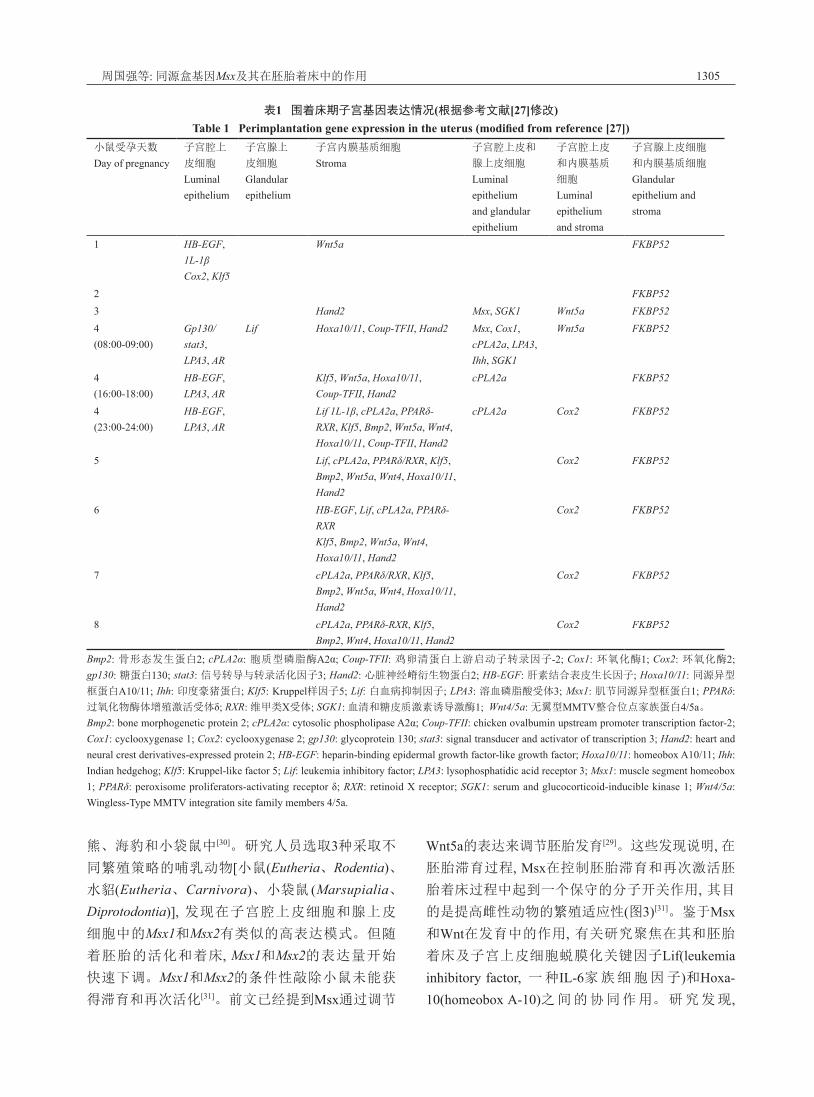
现出重叠表达模式来发挥功能[27](表1)。
5 Msx基因在着床中的作用早期胚胎发育过程中, 起重要调节作用的基因
在胚胎发育的不同阶段起作用, 显示出重叠表达模
式, 这使得理解某基因在某个阶段的作用显得十分
困难。在小鼠受孕第4 d, 此时其子宫处于接受态, Msx1特异表达在子宫腔上皮细胞和腺上皮细胞中, 但在随后胚胎发育的其他阶段里, 子宫中检测不到
Msx1的表达。 Msx1的特异性表达提示其与子宫的
接受态存在着必要的联系。在Msx1基因敲除小鼠
中, 其子宫接受窗口时期被推迟, 表现出延迟着床
现象, 影响胚胎发育过程。同时敲除Msx1和Msx2的小鼠表现出着床的失败, 这说明Msx2对于Msx1功能
的缺陷有一定的补偿作用[28]。正常情况下, Msx基因表达将抑制Wnt5a的表达, 从而抑制E-钙黏蛋白
(E-cadherin)和β-联蛋白(β-catenin)复合物的形成, 使子宫腔上皮细胞从其高极性状态转变为低极性状
态, 子宫腔上皮细胞丢失极性变为立方上皮, 这有利
于囊胚黏附。在Msx基因缺失条件下, Wnt5a表达将
提高E-钙黏蛋白和β-联蛋白复合物的形成, 从而维
持子宫腔上皮细胞的极性, 维持极性的子宫腔上皮
细胞, 将创建一种屏障, 这种屏障将阻碍囊胚滋养层
细胞黏附和侵入(图2)[29]。
6 Msx基因在胚胎滞育中的作用哺乳动物胚胎滞育是胚胎的生长和代谢活力
短暂受到抑制而处于静止状态的一种现象。这种现
象是当外界环境和母体的生理状况不适宜胚胎发育
时, 短暂延迟分娩的一种繁殖策略[27]。胚胎滞育被
定义为兼性滞育和专性滞育。兼性滞育主要存在于
啮齿目动物和有袋目动物。专性滞育主要发现在貂、
周国强等: 同源盒基因Msx及其在胚胎着床中的作用 1305
表1 围着床期子宫基因表达情况(根据参考文献[27]修改)Table 1 Perimplantation gene expression in the uterus (modified from reference [27])
小鼠受孕天数
Day of pregnancy子宫腔上
皮细胞
Luminal epithelium
子宫腺上
皮细胞
Glandular epithelium
子宫内膜基质细胞
Stroma子宫腔上皮和
腺上皮细胞
Luminal epithelium and glandular epithelium
子宫腔上皮
和内膜基质
细胞
Luminal epithelium and stroma
子宫腺上皮细胞
和内膜基质细胞
Glandular epithelium and stroma
1 HB-EGF, 1L-1βCox2, Klf5
Wnt5a FKBP52
2 FKBP523 Hand2 Msx, SGK1 Wnt5a FKBP524(08:00-09:00)
Gp130/stat3,LPA3, AR
Lif Hoxa10/11, Coup-TFII, Hand2 Msx, Cox1, cPLA2a, LPA3, Ihh, SGK1
Wnt5a FKBP52
4(16:00-18:00)
HB-EGF, LPA3, AR
Klf5, Wnt5a, Hoxa10/11,Coup-TFII, Hand2
cPLA2a FKBP52
4(23:00-24:00)
HB-EGF, LPA3, AR
Lif 1L-1β, cPLA2a, PPARδ-RXR, Klf5, Bmp2, Wnt5a, Wnt4, Hoxa10/11, Coup-TFII, Hand2
cPLA2a Cox2 FKBP52
5 Lif, cPLA2a, PPARδ/RXR, Klf5, Bmp2, Wnt5a, Wnt4, Hoxa10/11, Hand2
Cox2 FKBP52
6 HB-EGF, Lif, cPLA2a, PPARδ-RXRKlf5, Bmp2, Wnt5a, Wnt4, Hoxa10/11, Hand2
Cox2 FKBP52
7 cPLA2a, PPARδ/RXR, Klf5, Bmp2, Wnt5a, Wnt4, Hoxa10/11, Hand2
Cox2 FKBP52
8 cPLA2a, PPARδ-RXR, Klf5, Bmp2, Wnt4, Hoxa10/11, Hand2
Cox2 FKBP52
Bmp2: 骨形态发生蛋白2; cPLA2α: 胞质型磷脂酶A2α; Coup-TFII: 鸡卵清蛋白上游启动子转录因子-2; Cox1: 环氧化酶1; Cox2: 环氧化酶2; gp130: 糖蛋白130; stat3: 信号转导与转录活化因子3; Hand2: 心脏神经嵴衍生物蛋白2; HB-EGF: 肝素结合表皮生长因子; Hoxa10/11: 同源异型
框蛋白A10/11; Ihh: 印度豪猪蛋白; Klf5: Kruppel样因子5; Lif: 白血病抑制因子; LPA3: 溶血磷脂酸受体3; Msx1: 肌节同源异型框蛋白1; PPARδ: 过氧化物酶体增殖激活受体δ; RXR: 维甲类X受体; SGK1: 血清和糖皮质激素诱导激酶1; Wnt4/5a: 无翼型MMTV整合位点家族蛋白4/5a。Bmp2: bone morphogenetic protein 2; cPLA2α: cytosolic phospholipase A2α; Coup-TFII: chicken ovalbumin upstream promoter transcription factor-2; Cox1: cyclooxygenase 1; Cox2: cyclooxygenase 2; gp130: glycoprotein 130; stat3: signal transducer and activator of transcription 3; Hand2: heart and neural crest derivatives-expressed protein 2; HB-EGF: heparin-binding epidermal growth factor-like growth factor; Hoxa10/11: homeobox A10/11; Ihh: Indian hedgehog; Klf5: Kruppel-like factor 5; Lif: leukemia inhibitory factor; LPA3: lysophosphatidic acid receptor 3; Msx1: muscle segment homeobox 1; PPARδ: peroxisome proliferators-activating receptor δ; RXR: retinoid X receptor; SGK1: serum and glucocorticoid-inducible kinase 1; Wnt4/5a: Wingless-Type MMTV integration site family members 4/5a.
熊、海豹和小袋鼠中[30]。研究人员选取3种采取不
同繁殖策略的哺乳动物[小鼠(Eutheria、Rodentia)、水貂(Eutheria、Carnivora)、小袋鼠 (Marsupialia、Diprotodontia)], 发现在子宫腔上皮细胞和腺上皮
细胞中的Msx1和Msx2有类似的高表达模式。但随
着胚胎的活化和着床, Msx1和Msx2的表达量开始
快速下调。Msx1和Msx2的条件性敲除小鼠未能获
得滞育和再次活化[31]。前文已经提到Msx通过调节
Wnt5a的表达来调节胚胎发育[29]。这些发现说明, 在胚胎滞育过程, Msx在控制胚胎滞育和再次激活胚
胎着床过程中起到一个保守的分子开关作用, 其目
的是提高雌性动物的繁殖适应性(图3)[31]。鉴于Msx和Wnt在发育中的作用, 有关研究聚焦在其和胚胎
着床及子宫上皮细胞蜕膜化关键因子Lif(leukemia inhibitory factor, 一种IL-6家族细胞因子)和Hoxa-10(homeobox A-10)之间的协同作用。研究发现,

1306 · 综述 ·
在Lif敲除小鼠和Hoxa-10敲除小鼠中, 围着床期时
Msx1、Wnt4以及Wnt拮抗物sFRP4在子宫中差异表
达, 这表明它们在着床中起关键作用。此外, 在Lif敲除小鼠中, Msx1和sFRP4异常表达; 在Hoxa-10敲除
小鼠中, Wnt4和sFRP4异常表达。尽管具体的分子
机制目前仍不明确, 但这些发现提示, 可能存在一个
新的信号通路即cytokin-homeotic-Wnt, 在胚胎发育
过程中起关键作用[32]。
7 结语与展望在生命过程中, Msx作为转录因子调控细胞分
化, 对于其在体内特异性地调控靶基因的机制已有
充分了解。在胚胎着床过程中, Msx在复杂的调控
网络中起到一个关键的分子开关作用, 其不仅在正
常着床过程中起调节作用, 也在胚胎滞育中起关键
作用。着床过程紊乱或失败是女性不孕的重要病因。
探讨Msx基因在胚胎着床中的作用将为体外受精临
床研究提供一种新的策略, 在胚胎植入前短暂地提
高子宫中Msx的表达量, 将会延长子宫的接受态。同
时, 市售的避孕药多为性激素类避孕药, 对人体有一
定的副作用, 进一步揭示Msx的作用可能有助于开
发非激素类避孕药。Msx在胚胎发育中已知的相关
分子机制还未研究透彻, 除本文列举出的一些, 也许
还有更多复杂机制存在。未来的研究应针对继续鉴
定Msx靶基因和与Msx相互作用的蛋白质为线索, 充分了解其潜在分子机制。关于Msx功能的研究充满
未知但值得探讨, 对Msx基因的研究可能为研究同
源盒基因在不同生命过程中的精确分子作用提供理
论基础。
参考文献 (References)1 Holland PWH, Takahashi T. The evolution of homeobox ge-
图2 Msx基因调控着床模式图
Fig.2 Proposed model of regulation of implantation by Msx genes
Msx expression Wnt5a
E-cadherin/β-catenin
formation
Wnt5aE-cadherin/β-catenin
complex formation
Msx deficiency
To maintain uterine luminal epithelial cell polarity does not benefit to
blastocyst attachment
To lose uterine luminal epithelial cell polarity benefits to blastocyst
attachment
complex
PRL: 催乳素; P4: 孕酮; E2: β-雌二醇。
PRL: prolactin; P4: progesterone; E2: estradiol-17β.图3 Msx在小鼠、水貂和小袋鼠中调节胚胎滞育的模式图(根据参考文献[31]修改)
Fig.3 Potential schematic of the regulation of diapause in mouse, mink and wallaby (modified from reference [31])
Inhibitory photoperiod
in mink
Suckling/inhibitory
photoperiod in wallaby
P4
P4
E2PRL
PRL
PRL
DIAPAUS
E2
P4
P4 PRL
PRL
PRL No suckling in mouse
Stimulatory photoperiod
in mink
No suckling/stimulatory
photoperiod in wallaby
REACTIVATI
Msx
Wnt5a
Msx
uterus
Suckling in mouse

周国强等: 同源盒基因Msx及其在胚胎着床中的作用 1307
nes: Implications for the study of brain development. Brain Res Bull 2005; 66(4/5/6): 484-90.
2 Banerjee-Basu S, Baxevanis AD. Molecular evolution of the homeodomain family of transcription factors. Nucleic Acids Res 2001; 29(15): 3258-69.
3 Müller M, Affolter M, Leupin W, Otting G, Wüthrich K, Gehring W. Isolation and sequence-specific DNA binding of the Antennapedia homeodomain. EMBO J 1988; 7(13): 4299.
4 Scott MP, Tamkun JW, Hartzell GW. The structure and func-tion of the homeodomain. Biochim Biophys Acta 1989; 989(1): 25-48.
5 Sun J, Ting M C, Ishii M, Maxson R. Msx1 and Msx2 fun-ction together in the regulation of primordial germ cell migration in the mouse. Dev Biol 2016; 417(1): 11-24.
6 Bendall AJ, Abate-Shen C. Roles for Msx and Dlx homeo-proteins in vertebrate development. Gene 2000; 247(1/2): 17-31.
7 Duval N, Daubas P, de Carbon CB, St Cloment C, Tinevez JY, Lopes M, et al. Msx1 and Msx2 act as essential activators of Atoh1 expression in the murine spinal cord. Development 2014; 141(8): 1726-36.
8 Wang W, Chen X, Xu H, Lufkin T. Msx3: A novel murine homologue of the Drosophila msh homeobox gene restricted to the dorsal embryonic central nervous system. Mech Develop 1996; 58(1/2): 203-15.
9 Wang J, Kumar RM, Biggs VJ, Lee H, Chen Y, Kagey MH, et al. The Msx1 homeoprotein recruits polycomb to the nuclear periphery during development. Dev Cell 2011; 21(3): 575-88.
10 Wang JQ, Abate-Shen C. Transcriptional repression by the Msx1 homeoprotein is associated with global redistribution of the H3K27me3 repressive mark to the nuclear periphery. Nucleus-Austin 2012; 3(2): 155-61.
11 Wang JQ, Abate-Shen C. The MSX1 homeoprotein recruits G9a methyltransferase to repressed target genes in myoblast cells. PLoS One 2012; 7(5): e37647.
12 Kachhap S, Singh B. Role of DNA conformation & energetic insights in Msx-1-DNA recognition as revealed by molecular dynamics studies on specific and nonspecific complexes. J Biomol Struct Dyn 2015; 33(10): 2069-82.
13 Houzelstein D, Auda-Boucher G, Chéraud Y, Rouaud T, Blanc I, Tajbakhsh S, et al. The homeobox gene Msx1 is expressed in a subset of somites, and in muscle progenitor cells migrating into the forelimb. Development 1999; 126(12): 2689-701.
14 Singh N, Gupta M, Trivedi CM, Singh MK, Li L, Epstein JA. Murine craniofacial development requires Hdac3-mediated repression of Msx gene expression. Dev Biol 2013; 377(2): 333-44.
15 Yilmaz A, Engeler R, Constantinescu S, Kokkaliaris KD, Dimitrakopoulos C, Schroeder T, et al. Ectopic expression of Msx2 in mammalian myotubes recapitulates aspects of amphibian muscle dedifferentiation. Stem Cell Res 2015; 15(3): 542-53.
16 Davidson D. The function and evolution of Msx genes poin-ters and paradoxes. Trends Genet 1995; 11(10): 405-11.
17 Han M, Yang X, Taylor G, Burdsal CA, Anderson RA,
Muneoka K. Limb regeneration in higher vertebrates: Developing a roadmap. Anat Rec B New Anat 2005; 287(1): 14-24.
18 Wang J, Kobayashi T, Floc’h N, Kinkade CW, Aytes A, Dankort D, et al. B-Raf activation cooperates with PTEN loss to drive c-Myc expression in advanced prostate cancer. Cancer Res 2012; 72(18): 4765-76.
19 Lallemand Y, Nicola MA, Ramos C, Bach A, Saint Cloment C, Robert B. Analysis of Msx1; Msx2 double mutants reveals multiple roles for Msx genes in limb development. Development 2005; 132(13): 3003-14.
20 Catron KM, Wang H, Hu G, Shen MM, Abate-Shen C. Comparison of MSX-1 and MSX-2 suggests a molecular basis for functional redundancy. Mech Dev 1996; 55(2): 185-99.
21 Satokata I, Maas R. Msx1 deficient mice exhibit cleft-palate and abnormalities of craniofacial and tooth development. Nat Genet 1994; 6(4): 348-56.
22 Bach A, Lallemand Y, Nicola MA, Ramos C, Mathis L, Maufras M, et al. Msx1 is required for dorsal diencephalon patterning. Development 2003; 130(17): 4025-36.
23 Hu G, Lee H, Price SM, Shen MM, Abate-Shen C. Msx homeobox genes inhibit differentiation through upregulation of cyclin D1. Development 2001; 128(12): 2373-84.
24 Satokata I, Ma L, Ohshima H, Bei M, Woo I, Nishizawa K, et al. Msx2 deficiency in mice causes pleiotropic defects in bone growth and ectodermal organ formation. Nat Genet 2000; 24(4): 391-5.
25 Winograd J, Reilly MP, Roe R, Lutz J, Laughner E, Xu X, et al. Perinatal lethality and multiple craniofacial malformations in MSX2 transgenic mice. Hum Mol Genet 1997; 6(3): 369-79.
26 Carson DD, Bagchi I, Dey SK, Enders AC, Fazleabas AT, Lessey BA, et al. Embryo implantation. Dev Biol 2000; 223(2): 217-37.
27 Cha JY, Sun XF, Dey SK. Mechanisms of implantation: Strategies for successful pregnancy. Nat Med 2012; 18(12): 1754-67.
28 Sun X, Park CB, Deng W, Potter SS, Dey SK. Uterine inactivation of muscle segment homeobox (Msx) genes alters epithelial cell junction proteins during embryo implantation. FASEB J 2015; 30(4): 1425-35.
29 Daikoku T, Cha J, Sun X, Tranguch S, Xie H, Fujita T, et al. Conditional deletion of Msx homeobox genes in the uterus inhibits blastocyst implantation by altering uterine receptivity. Dev Cell 2011; 21(6): 1014-25.
30 Lopes FL, Desmarais JA, Murphy BD. Embryonic diapause and its regulation. Reproduction 2004; 128(6): 669-78.
31 Cha J, Sun X, Bartos A, Fenelon J, Lefèvre P, Daikoku T, et al. A new role for muscle segment homeobox genes in mammalian embryonic diapause. Open Biol 2013; 3(4): 130035.
32 Daikoku T, Song H, Guo Y, Riesewijk A, Mosselman S, Das SK, et al. Uterine Msx-1 and Wnt4 signaling becomes aberrant in mice with the loss of leukemia inhibitory factor or Hoxa-10: Evidence for a novel cytokine-homeobox-Wnt signaling in implantation. Mol Endocrinol 2004; 18(5): 1238-50.
33 Cha J, Dey SK. Cadence of procreation: Orchestrating embryo-uterine interactions. Semin Cell Dev Biol 2014; 34: 56-64.

1308 · 综述 ·
34 Thompson IE. Reproductive endocrinology, surgery and technology. JAMA 1997; 277(16): 1328-9.
35 Psychoyos A. Hormonal control of ovoimplantation. Vitam Horm 1974; 31: 201-56.
36 Aplin JD. Adhesion molecules in implantation. Rev Reprod 1997; 2(2): 84-93.
37 Enders AC, Schlafke S. A morphological analysis of the early implantation stages in the rat. Am J Anat 1967; 120(2): 185-225.
38 Kirby DRS. Blastocyst-uterine relationship before and during Implantation. Chicago, IL: The University of Chicago Press
1971, 393-412.39 Finn CA, Porter DG. Implantation of ova. In: Porter DG, Finn
CA, editors. Theuterus, reproductive biology handbooks. London, UK: Scientific Books Ltd. 1975, 57-73.
40 Enders AC, Schlafke S. Implantation in nonhuman primates and in the human. Comp Prim Biol 1986, 3: 291-310.
41 Enders AC, Lopata A. Implantation in the marmoset monkey: Expansion of the early implantation site. Anat Rec 1999; 256(3): 279-99.