The Hippo pathway: regulators and regulationsgenesdev.cshlp.org/content/27/4/355.full.pdf · The...
18
REVIEW The Hippo pathway: regulators and regulations Fa-Xing Yu and Kun-Liang Guan 1 Department of Pharmacology, Moores Cancer Center, University of California at San Diego, La Jolla, California 92093, USA Control of cell number is crucial in animal development and tissue homeostasis, and its dysregulation may result in tumor formation or organ degeneration. The Hippo pathway in both Drosophila and mammals regulates cell number by modulating cell proliferation, cell death, and cell differentiation. Recently, numerous upstream com- ponents involved in the Hippo pathway have been iden- tified, such as cell polarity, mechanotransduction, and G-protein-coupled receptor (GPCR) signaling. Actin cyto- skeleton or cellular tension appears to be the master mediator that integrates and transmits upstream sig- nals to the core Hippo signaling cascade. Here, we review regulatory mechanisms of the Hippo pathway and discuss potential implications involved in different physiological and pathological conditions. Cell proliferation, death, and differentiation are funda- mental biological processes. Coordination of these pro- cesses is critical for a wide range of physiological and pathological conditions (Pellettieri and Sanchez Alvarado 2007; Galliot and Ghila 2010). During development, an increase in cell number is required to boost organ and body size; meanwhile, proper differentiation of multiple cell types will assure the appropriate function of devel- oped organs. In adulthood, most tissues undergo contin- uous cell turnover to maintain functionality. Aged or damaged cells are programmed to cell death, whereas adult stem cells may divide and differentiate to replace those dysfunctional cells. Under pathological conditions, such as wound healing and organ regeneration, cell division and differentiation of tissue-specific progenitor cells will be up-regulated to compensate for the lost cells. On the other hand, uncontrolled cell proliferation and decreased cell death lead to hyperplasia or tumorigenesis. Detailed mechanisms underlying cell proliferation, cell death, and cell differentiation have been extensively studied; however, how these processes are coordinated and integrated is poorly understood. Recently, the Hippo pathway has been shown to pro- mote cell death and differentiation and inhibit cell pro- liferation; therefore, the Hippo pathway may function as a key node to coordinate these cellular processes (Fig. 1). The Hippo pathway was first defined in Drosophila by genetic mosaic screens for tumor suppressor genes. Ge- netic inactivation of genes, including Warts (Wts) (Justice et al. 1995; Xu et al. 1995), Hippo (Hpo) (Harvey et al. 2003; Jia et al. 2003; Pantalacci et al. 2003; Udan et al. 2003; Wu et al. 2003), Salvador (Sav; also known as Shar-Pei) (Kango- Singh et al. 2002; Tapon et al. 2002), and Mats (Lai et al. 2005), all resulted in a similar phenotype with robust tissue overgrowth. Yorkie (Yki) is the major downstream effector of the Hippo pathway (Huang et al. 2005), which regulates a transcription program by interacting with the transcription factor Scalloped (Sd) (Fig. 2; Goulev et al. 2008; Wu et al. 2008; Zhang et al. 2008; Zhao et al. 2008). The Hippo pathway is highly conserved in mammals: MST1/2 (Hpo orthologs), Sav1, Lats1/2 (Wts orthologs), and Mob1 (MOBKL1A and MOBKL1B, Mats orthologs) form a kinase cascade that phosphorylates and inhibits YAP/TAZ (Yki orthologs). YAP/TAZ in conjunction with TEAD1–4 (Sd orthologs) mediate major physiological functions of the Hippo pathway (Fig. 2; for reviews, see Pan 2010; Zhao et al. 2010a). The nomenclature of many components of the Hippo pathway in Drosophila and mammals is different, and a summary of these compo- nents is shown in Table 1. The core Hippo pathway has been well established in both Drosophila and mammals; however, the regulatory mechanisms for this signaling pathway are less under- stood. Recently, by using both genetic and biochemical approaches, many additional components have been iden- tified to modulate the core Hippo pathway (Table 1). In this review, we briefly describe the components of the Hippo pathway and summarize recent advances with respect to Hippo pathway regulation. In addition, we also discuss the implications of Hippo pathway regulation in different physiological and pathological conditions. The mammalian Hippo pathway is the main focus, al- though some Drosophila works are also covered. For a detailed review on the Drosophila Hippo pathway, please refer to Staley and Irvine (2012). Core Hippo pathway: a kinase cascade MST1/2 are STE20 family protein kinases and can phos- phorylate Sav1, Lats1/2, and Mob1 (Wu et al. 2003; Chan et al. 2005; Callus et al. 2006; Praskova et al. 2008). The [Keywords: actin; GPCR; Hippo; YAP; mechanotransduction; polarity] 1 Corresponding author E-mail [email protected] Article is online at http://www.genesdev.org/cgi/doi/10.1101/gad.210773.112. GENES & DEVELOPMENT 27:355–371 Ó 2013 by Cold Spring Harbor Laboratory Press ISSN 0890-9369/13; www.genesdev.org 355 Cold Spring Harbor Laboratory Press on June 8, 2019 - Published by genesdev.cshlp.org Downloaded from
-
Upload
nguyenthien -
Category
Documents
-
view
226 -
download
0
Transcript of The Hippo pathway: regulators and regulationsgenesdev.cshlp.org/content/27/4/355.full.pdf · The...
REVIEW
The Hippo pathway: regulatorsand regulations
Fa-Xing Yu and Kun-Liang Guan1
Department of Pharmacology, Moores Cancer Center, University of California at San Diego, La Jolla, California 92093, USA
Control of cell number is crucial in animal developmentand tissue homeostasis, and its dysregulation may resultin tumor formation or organ degeneration. The Hippopathway in both Drosophila and mammals regulates cellnumber by modulating cell proliferation, cell death, andcell differentiation. Recently, numerous upstream com-ponents involved in the Hippo pathway have been iden-tified, such as cell polarity, mechanotransduction, andG-protein-coupled receptor (GPCR) signaling. Actin cyto-skeleton or cellular tension appears to be the mastermediator that integrates and transmits upstream sig-nals to the core Hippo signaling cascade. Here, we reviewregulatory mechanisms of the Hippo pathway and discusspotential implications involved in different physiologicaland pathological conditions.
Cell proliferation, death, and differentiation are funda-mental biological processes. Coordination of these pro-cesses is critical for a wide range of physiological andpathological conditions (Pellettieri and Sanchez Alvarado2007; Galliot and Ghila 2010). During development, anincrease in cell number is required to boost organ andbody size; meanwhile, proper differentiation of multiplecell types will assure the appropriate function of devel-oped organs. In adulthood, most tissues undergo contin-uous cell turnover to maintain functionality. Aged ordamaged cells are programmed to cell death, whereasadult stem cells may divide and differentiate to replacethose dysfunctional cells. Under pathological conditions,such as wound healing and organ regeneration, celldivision and differentiation of tissue-specific progenitorcells will be up-regulated to compensate for the lost cells.On the other hand, uncontrolled cell proliferation anddecreased cell death lead to hyperplasia or tumorigenesis.Detailed mechanisms underlying cell proliferation, celldeath, and cell differentiation have been extensivelystudied; however, how these processes are coordinatedand integrated is poorly understood.
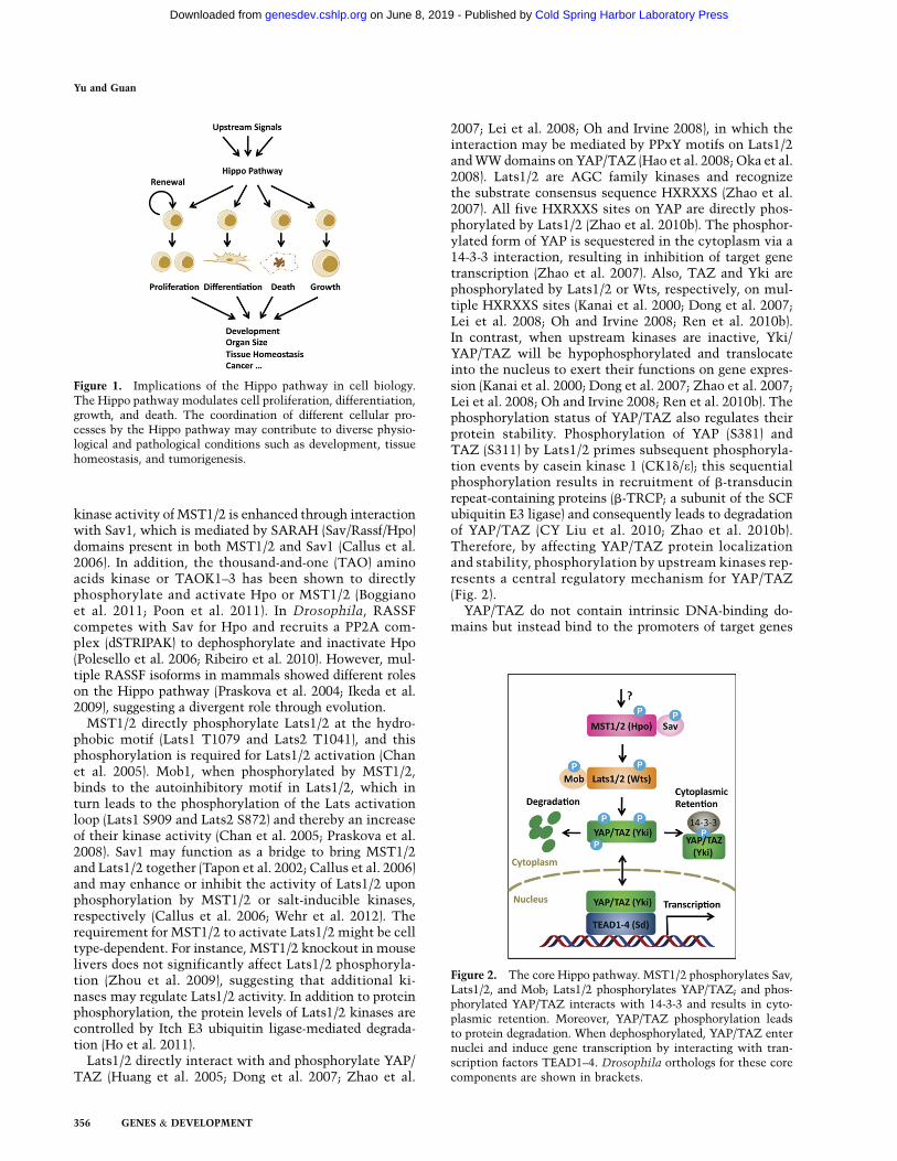
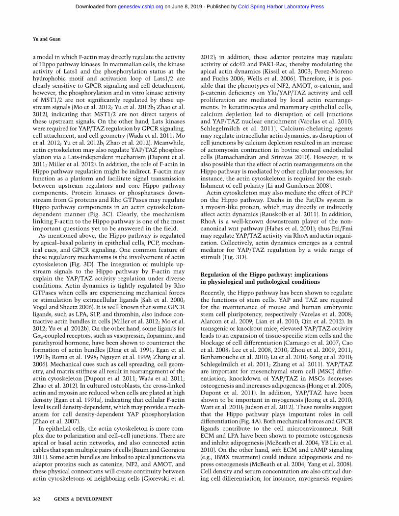
Recently, the Hippo pathway has been shown to pro-mote cell death and differentiation and inhibit cell pro-liferation; therefore, the Hippo pathway may function as
a key node to coordinate these cellular processes (Fig. 1).The Hippo pathway was first defined in Drosophila bygenetic mosaic screens for tumor suppressor genes. Ge-netic inactivation of genes, including Warts (Wts) (Justiceet al. 1995; Xu et al. 1995), Hippo (Hpo) (Harvey et al. 2003;Jia et al. 2003; Pantalacci et al. 2003; Udan et al. 2003; Wuet al. 2003), Salvador (Sav; also known as Shar-Pei) (Kango-Singh et al. 2002; Tapon et al. 2002), and Mats (Lai et al.2005), all resulted in a similar phenotype with robusttissue overgrowth. Yorkie (Yki) is the major downstreameffector of the Hippo pathway (Huang et al. 2005), whichregulates a transcription program by interacting with thetranscription factor Scalloped (Sd) (Fig. 2; Goulev et al.2008; Wu et al. 2008; Zhang et al. 2008; Zhao et al. 2008).The Hippo pathway is highly conserved in mammals:MST1/2 (Hpo orthologs), Sav1, Lats1/2 (Wts orthologs),and Mob1 (MOBKL1A and MOBKL1B, Mats orthologs)form a kinase cascade that phosphorylates and inhibitsYAP/TAZ (Yki orthologs). YAP/TAZ in conjunction withTEAD1–4 (Sd orthologs) mediate major physiologicalfunctions of the Hippo pathway (Fig. 2; for reviews, seePan 2010; Zhao et al. 2010a). The nomenclature of manycomponents of the Hippo pathway in Drosophila andmammals is different, and a summary of these compo-nents is shown in Table 1.
The core Hippo pathway has been well established inboth Drosophila and mammals; however, the regulatorymechanisms for this signaling pathway are less under-stood. Recently, by using both genetic and biochemicalapproaches, many additional components have been iden-tified to modulate the core Hippo pathway (Table 1). Inthis review, we briefly describe the components of theHippo pathway and summarize recent advances withrespect to Hippo pathway regulation. In addition, we alsodiscuss the implications of Hippo pathway regulationin different physiological and pathological conditions.The mammalian Hippo pathway is the main focus, al-though some Drosophila works are also covered. For adetailed review on the Drosophila Hippo pathway, pleaserefer to Staley and Irvine (2012).
Core Hippo pathway: a kinase cascade
MST1/2 are STE20 family protein kinases and can phos-phorylate Sav1, Lats1/2, and Mob1 (Wu et al. 2003; Chanet al. 2005; Callus et al. 2006; Praskova et al. 2008). The
[Keywords: actin; GPCR; Hippo; YAP; mechanotransduction; polarity]1Corresponding authorE-mail [email protected] is online at http://www.genesdev.org/cgi/doi/10.1101/gad.210773.112.
GENES & DEVELOPMENT 27:355–371 � 2013 by Cold Spring Harbor Laboratory Press ISSN 0890-9369/13; www.genesdev.org 355
Cold Spring Harbor Laboratory Press on June 8, 2019 - Published by genesdev.cshlp.orgDownloaded from
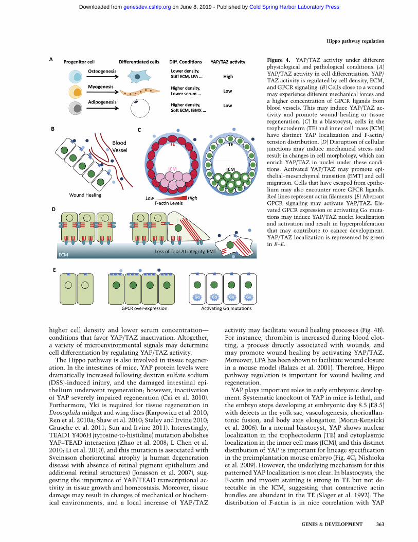
kinase activity of MST1/2 is enhanced through interactionwith Sav1, which is mediated by SARAH (Sav/Rassf/Hpo)domains present in both MST1/2 and Sav1 (Callus et al.2006). In addition, the thousand-and-one (TAO) aminoacids kinase or TAOK1–3 has been shown to directlyphosphorylate and activate Hpo or MST1/2 (Boggianoet al. 2011; Poon et al. 2011). In Drosophila, RASSFcompetes with Sav for Hpo and recruits a PP2A com-plex (dSTRIPAK) to dephosphorylate and inactivate Hpo(Polesello et al. 2006; Ribeiro et al. 2010). However, mul-tiple RASSF isoforms in mammals showed different roleson the Hippo pathway (Praskova et al. 2004; Ikeda et al.2009), suggesting a divergent role through evolution.
MST1/2 directly phosphorylate Lats1/2 at the hydro-phobic motif (Lats1 T1079 and Lats2 T1041), and thisphosphorylation is required for Lats1/2 activation (Chanet al. 2005). Mob1, when phosphorylated by MST1/2,binds to the autoinhibitory motif in Lats1/2, which inturn leads to the phosphorylation of the Lats activationloop (Lats1 S909 and Lats2 S872) and thereby an increaseof their kinase activity (Chan et al. 2005; Praskova et al.2008). Sav1 may function as a bridge to bring MST1/2and Lats1/2 together (Tapon et al. 2002; Callus et al. 2006)and may enhance or inhibit the activity of Lats1/2 uponphosphorylation by MST1/2 or salt-inducible kinases,respectively (Callus et al. 2006; Wehr et al. 2012). Therequirement for MST1/2 to activate Lats1/2 might be celltype-dependent. For instance, MST1/2 knockout in mouselivers does not significantly affect Lats1/2 phosphoryla-tion (Zhou et al. 2009), suggesting that additional ki-nases may regulate Lats1/2 activity. In addition to proteinphosphorylation, the protein levels of Lats1/2 kinases arecontrolled by Itch E3 ubiquitin ligase-mediated degrada-tion (Ho et al. 2011).
Lats1/2 directly interact with and phosphorylate YAP/TAZ (Huang et al. 2005; Dong et al. 2007; Zhao et al.
2007; Lei et al. 2008; Oh and Irvine 2008), in which theinteraction may be mediated by PPxY motifs on Lats1/2and WW domains on YAP/TAZ (Hao et al. 2008; Oka et al.2008). Lats1/2 are AGC family kinases and recognizethe substrate consensus sequence HXRXXS (Zhao et al.2007). All five HXRXXS sites on YAP are directly phos-phorylated by Lats1/2 (Zhao et al. 2010b). The phosphor-ylated form of YAP is sequestered in the cytoplasm via a14-3-3 interaction, resulting in inhibition of target genetranscription (Zhao et al. 2007). Also, TAZ and Yki arephosphorylated by Lats1/2 or Wts, respectively, on mul-tiple HXRXXS sites (Kanai et al. 2000; Dong et al. 2007;Lei et al. 2008; Oh and Irvine 2008; Ren et al. 2010b).In contrast, when upstream kinases are inactive, Yki/YAP/TAZ will be hypophosphorylated and translocateinto the nucleus to exert their functions on gene expres-sion (Kanai et al. 2000; Dong et al. 2007; Zhao et al. 2007;Lei et al. 2008; Oh and Irvine 2008; Ren et al. 2010b). Thephosphorylation status of YAP/TAZ also regulates theirprotein stability. Phosphorylation of YAP (S381) andTAZ (S311) by Lats1/2 primes subsequent phosphoryla-tion events by casein kinase 1 (CK1d/e); this sequentialphosphorylation results in recruitment of b-transducinrepeat-containing proteins (b-TRCP; a subunit of the SCFubiquitin E3 ligase) and consequently leads to degradationof YAP/TAZ (CY Liu et al. 2010; Zhao et al. 2010b).Therefore, by affecting YAP/TAZ protein localizationand stability, phosphorylation by upstream kinases rep-resents a central regulatory mechanism for YAP/TAZ(Fig. 2).
YAP/TAZ do not contain intrinsic DNA-binding do-mains but instead bind to the promoters of target genes
Figure 1. Implications of the Hippo pathway in cell biology.The Hippo pathway modulates cell proliferation, differentiation,growth, and death. The coordination of different cellular pro-cesses by the Hippo pathway may contribute to diverse physio-logical and pathological conditions such as development, tissuehomeostasis, and tumorigenesis.
Figure 2. The core Hippo pathway. MST1/2 phosphorylates Sav,Lats1/2, and Mob; Lats1/2 phosphorylates YAP/TAZ; and phos-phorylated YAP/TAZ interacts with 14-3-3 and results in cyto-plasmic retention. Moreover, YAP/TAZ phosphorylation leadsto protein degradation. When dephosphorylated, YAP/TAZ enternuclei and induce gene transcription by interacting with tran-scription factors TEAD1–4. Drosophila orthologs for these corecomponents are shown in brackets.
Yu and Guan
356 GENES & DEVELOPMENT
Cold Spring Harbor Laboratory Press on June 8, 2019 - Published by genesdev.cshlp.orgDownloaded from
by interacting with DNA-binding transcription factors.YAP/TAZ mainly bind to the transcription factorsTEAD1–4 to regulate genes involved in cell proliferationand cell death (Vassilev et al. 2001; Goulev et al. 2008; Wuet al. 2008; Zhang et al. 2008; Zhao et al. 2008). BesidesTEADs, YAP/TAZ may also interact with other tran-scription factors, such as Smad1 (Alarcon et al. 2009),Smad2/3 (Varelas et al. 2008), Smad7 (Ferrigno et al.2002), RUNX1/2 (Yagi et al. 1999), p63/p73 (Stranoet al. 2001), and ErbB4 (Komuro et al. 2003; Omerovicet al. 2004); these interactions may mediate transcrip-tion of diverse genes involved in proliferation, differ-entiation, and development.
Apical–basal polarity: the polarized localization of hippocomponents
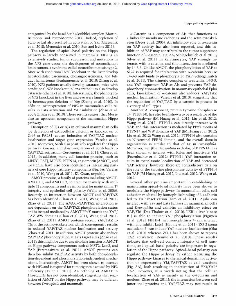
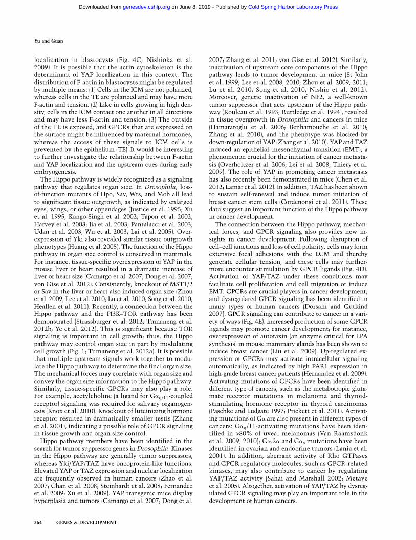
Epithelial cells usually adhere to one another throughcell–cell junctions such as adherens junctions (AJs), des-mosomes, and tight junctions (TJs). TJs and AJs, with helpfrom different polarity complexes, divide the plasmamembrane into an apical domain and a basolateral domainand thereby establish an apical–basal polarity in epithelialcells (Martin-Belmonte and Perez-Moreno 2012). Interest-ingly, many upstream regulators identified for the Hippopathway are known components of TJs, AJs, or apical–basal polarity protein complexes (Fig. 3A).
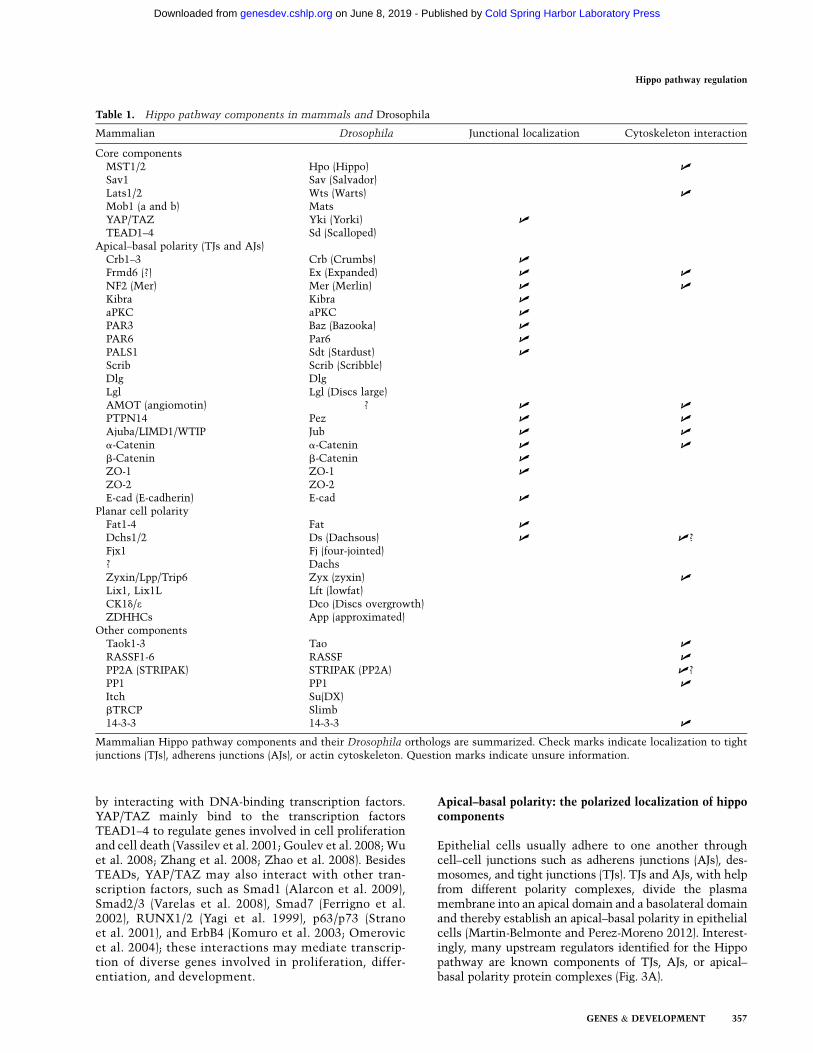
Table 1. Hippo pathway components in mammals and Drosophila
Mammalian Drosophila Junctional localization Cytoskeleton interaction
Core componentsMST1/2 Hpo (Hippo) U
Sav1 Sav (Salvador)Lats1/2 Wts (Warts) U
Mob1 (a and b) MatsYAP/TAZ Yki (Yorki) U
TEAD1–4 Sd (Scalloped)Apical–basal polarity (TJs and AJs)
Crb1–3 Crb (Crumbs) U
Frmd6 (?) Ex (Expanded) U U
NF2 (Mer) Mer (Merlin) U U
Kibra Kibra U
aPKC aPKC U
PAR3 Baz (Bazooka) U
PAR6 Par6 U
PALS1 Sdt (Stardust) U
Scrib Scrib (Scribble)Dlg DlgLgl Lgl (Discs large)AMOT (angiomotin) ? U U
PTPN14 Pez U U
Ajuba/LIMD1/WTIP Jub U U
a-Catenin a-Catenin U U
b-Catenin b-Catenin U
ZO-1 ZO-1 U
ZO-2 ZO-2E-cad (E-cadherin) E-cad U
Planar cell polarityFat1-4 Fat U
Dchs1/2 Ds (Dachsous) U U?Fjx1 Fj (four-jointed)? DachsZyxin/Lpp/Trip6 Zyx (zyxin) U
Lix1, Lix1L Lft (lowfat)CK1d/e Dco (Discs overgrowth)ZDHHCs App (approximated)
Other componentsTaok1-3 Tao U
RASSF1-6 RASSF U
PP2A (STRIPAK) STRIPAK (PP2A) U?PP1 PP1 U
Itch Su(DX)bTRCP Slimb14-3-3 14-3-3 U
Mammalian Hippo pathway components and their Drosophila orthologs are summarized. Check marks indicate localization to tightjunctions (TJs), adherens junctions (AJs), or actin cytoskeleton. Question marks indicate unsure information.
Hippo pathway regulation
GENES & DEVELOPMENT 357
Cold Spring Harbor Laboratory Press on June 8, 2019 - Published by genesdev.cshlp.orgDownloaded from

Mer (Merlin; also known as NF2 for neurofibromatosis-2) and Ex (Expanded) are two proteins that belong to theFERM (4.1, Ezrin, Radxin, and Moesin) domain-containingfamily of proteins. Both Mer and Ex have tumor suppressorfunctions and work together to regulate cell proliferationand differentiation (McCartney et al. 2000). In Drosophila,genetic inactivation of both Mer and Ex revealed a dra-matic overgrowth phenotype similar to that of the Hpomutants (Hamaratoglu et al. 2006). Later, Kibra (a WW andC2 domain-containing protein) was identified to physi-cally interact with Mer and Ex, and these three proteinsactivate Wts in a cooperative manner (Baumgartner et al.2010; Genevet et al. 2010; Yu et al. 2010).
Mer, Ex, and Kibra colocalize at the apical domain ofpolarized epithelial cells (Fig. 3A; Boedigheimer andLaughon 1993; Boedigheimer et al. 1997; Yu et al. 2010).Mer and Ex have been considered as a linker for the apicalplasma membrane and actin cytoskeleton (Bretscher et al.2002). Kibra contains a C2 domain that interacts withphospholipids and may target interacting proteins to thecell surface (Kremerskothen et al. 2003). Sav and Hpophysically associate with Mer and Ex (Yu et al. 2010), andKibra interacts with Wts (Genevet et al. 2010), suggestingthat the Mer/Ex/Kibra complex may recruit the Hippopathway kinases to the apical plasma membrane for acti-vation. Indeed, it has been shown that Mats is activatedat the plasma membrane (Ho et al. 2010), and targetingMST1 to the plasma membrane by adding a myristoylation
signal enhances MST1 kinase activity (Khokhlatchev et al.2002).
Drosophila crumbs (Crb) has been identified as a cellsurface regulator for the Hippo pathway (Fig. 3A; CLChen et al. 2010; Grzeschik et al. 2010; Ling et al. 2010;Robinson et al. 2010). In Drosophila embryos, Crb islocalized at the subapical plasma membrane and plays animportant role in organizing apical–basal polarity (Tepasset al. 1990). As a transmembrane protein, Crb has a largeextracellular domain and a short intracellular domain.The short intracellular domain contains a FERM-bindingmotif (FBM) that can interact with Ex, and this interac-tion modulates Ex localization and stability, which inturn regulates the activity of Hippo pathway kinases andYki (CL Chen et al. 2010; Ling et al. 2010; Robinson et al.2010). The connection of Crb with Ex and core compo-nents of the Hippo pathway is also reflected by an over-growth phenotype corresponding to Crb deficiency (Linget al. 2010). Interestingly, overexpression of Crb leads toEx mislocalization and inactivation of the Hippo path-way, likely due to a dominant-negative effect of overex-pressed Crb (CL Chen et al. 2010; Grzeschik et al. 2010;Robinson et al. 2010).
Similar to the Crb complex, the Par apical complex alsoregulates the Hippo pathway (Fig. 3A). Overexpression ofaPKC, a component of the Par complex, can induce Ykiactivity and tissue overgrowth (Grzeschik et al. 2010;Sun and Irvine 2011). The activity of the Par complex is
Figure 3. Regulatory mechanisms for the Hippo pathway. Regulation of the Hippo pathway by apical–basal polarity (A), PCP (B),mechanical cues and GPCR signaling (C), and actin cytoskeleton (D). Arrowed or blunted ends indicate activation or inhibition,respectively. Dashed lines indicate indirect or unknown mechanisms. Red lines in D represent actin filaments.
Yu and Guan
358 GENES & DEVELOPMENT
Cold Spring Harbor Laboratory Press on June 8, 2019 - Published by genesdev.cshlp.orgDownloaded from
antagonized by the basal Scrib (Scribble) complex (Martin-Belmonte and Perez-Moreno 2012). Indeed, depletion ofScrib or Lgl also resulted in activation of Yki (Grzeschiket al. 2010; Menendez et al. 2010; Sun and Irvine 2011).
The regulation of apical–basal polarity on the Hippopathway is largely conserved in mammals. NF2 is anextensively studied tumor suppressor, and mutations inthe NF2 gene cause the development of nonmalignantbrain tumors, a syndrome called neurofibromatosis type 2.Mice with conditional NF2 knockout in the liver develophepatocellular carcinoma, cholangiocarcinoma, and bileduct hamartomas (Benhamouche et al. 2010; Zhang et al.2010). NF2 patients usually develop cataracts; mice withconditional NF2 knockout in lens epithelium also developcataracts (Zhang et al. 2010). Interestingly, the phenotypesof NF2 knockout in the liver and eye were largely blockedby heterozygous deletion of Yap (Zhang et al. 2010). Inaddition, overexpression of NF2 in mammalian cells re-sults in Lats activation and YAP inhibition (Zhao et al.2007; Zhang et al. 2010). These results suggest that Mer isalso an upstream component of the mammalian Hippopathway.
Disruption of TJs or AJs in cultured mammalian cells(by depletion of extracellular calcium or knockdown ofCrb3 or PALS1) causes induction of YAP/TAZ nuclearlocalization and target gene expression (Varelas et al.2010). Moreover, Scrib also positively regulates the Hippopathway kinases, and down-regulation of Scrib leads toYAP/TAZ activation (Cordenonsi et al. 2011; Chen et al.2012). In addition, many cell junction proteins, such asLIN7C, PATJ, MPDZ, PTPN14, angiomotin (AMOT), anda-catenin, have also been identified as interacting part-ners of core Hippo pathway components (Fig. 3A; Varelaset al. 2010; Wang et al. 2011; KL Guan, unpubl.).
AMOT proteins, a family of proteins including AMOT,AMOTL1, and AMOTL2, interact extensively with mul-tiple TJ components and are important for maintaining TJintegrity and epithelial cell polarity (Wells et al. 2006).Recently, an interaction between AMOT and YAP/TAZhas been identified (Chan et al. 2011; Wang et al. 2011;Zhao et al. 2011). The AMOT–YAP/TAZ interaction isnot dependent on the YAP/TAZ phosphorylation statusand is instead mediated by AMOT PPxY motifs and YAP/TAZ WW domains (Chan et al. 2011; Wang et al. 2011;Zhao et al. 2011). AMOT proteins recruit YAP/TAZ toTJs or the actin cytoskeleton, which consequently resultsin reduced YAP/TAZ nuclear localization and activity(Zhao et al. 2011). In addition, AMOT proteins also induceYAP/TAZ phosphorylation at Lats target sites (Zhao et al.2011); this might be due to a scaffolding function of AMOTon Hippo pathway components such as MST2, Lats2, andYAP (Paramasivam et al. 2011). AMOT proteins cantherefore inhibit YAP/TAZ activity by both phosphoryla-tion-dependent and phosphorylation-independent mecha-nisms. Interestingly, AMOT has been shown to interactwith NF2 and is required for tumorigenesis caused by NF2deficiency (Yi et al. 2011). An ortholog of AMOT inDrosophila has not been identified, suggesting that regu-lation of AMOT on the Hippo pathway may be differentbetween Drosophila and mammals.
a-Catenin is a component of AJs that functions asa linker for membrane cadherins and the actin cytoskel-eton (Drees et al. 2005). An inhibitory role of a-cateninon YAP activity has also been reported, and this in-hibition of YAP may contribute to the tumor suppressorfunction of a-catenin (Fig. 3A; Schlegelmilch et al. 2011;Silvis et al. 2011). In keratinocytes, YAP strongly in-teracts with a-catenin, and this interaction is mediatedby 14-3-3. Unlike AMOT, the phosphorylation of YAP atS127 is required for interaction with a-catenin because14-3-3 only binds to phosphorylated YAP (Schlegelmilchet al. 2011). The trimeric complex of a-catenin, 14-3-3,and YAP sequesters YAP at AJs and prevents YAP de-phosphorylation/activation. In mammary epithelial Eph4cells, knockdown of a-catenin also induces YAP/TAZnuclear localization (Varelas et al. 2010), suggesting thatthe regulation of YAP/TAZ by a-catenin is present ina variety of cell types.
Another AJ component, protein tyrosine phosphatase14 (PTPN14), has also been shown to be a regulator of theHippo pathway (JM Huang et al. 2012; Liu et al. 2012;Wang et al. 2012). PTPN14 can directly interact withYAP, and this interaction is mediated by PPxY motifs ofPTPN14 and WW domains of YAP (JM Huang et al. 2012;Liu et al. 2012; Wang et al. 2012). PTPN14 also containsan N-terminal FERM domain, and the overall domainorganization is similar to that of Ex in Drosophila.Moreover, Pez (the Drosophila ortholog of PTPN14) hasbeen shown to interact with Kibra and inactivate Yki(Poernbacher et al. 2012). PTPN14–YAP interaction re-sults in cytoplasmic localization of YAP and decreasedYAP activity; however, there are contradictory data onthe role of the tyrosine phosphatase activity of PTPN14on YAP (JM Huang et al. 2012; Liu et al. 2012; Wang et al.2012).
Several other proteins important in establishing ormaintaining apical–basal polarity have been shown tomodulate the Hippo pathway. In mammalian cells, celladhesion mediated by homophilic binding of E-cadherinled to YAP inactivation (Kim et al. 2011). Ajuba caninteract with Sav and Lats kinases in mammalian cellsand Drosophila and exhibits an inhibitory effect onYAP/Yki (Das Thakur et al. 2010). LKB1 (liver kinaseB1) is able to induce YAP phosphorylation (Nguyenet al. 2012). NPHP4 (nephronophthisis 4) can interactwith and inhibit Lats1 (Habbig et al. 2011). ZO-2 (zonaoccludens-2) can induce YAP nuclear localization (Okaet al. 2010), whereas ZO-1 has been shown to repressTAZ activation (Remue et al. 2010). These resultsindicate that cell–cell contact, integrity of cell junc-tions, and apical–basal polarity are important in regu-lation of the Hippo pathway. Apical–basal polarity canregulate the Hippo pathway by either recruiting theHippo pathway kinases to the apical domain for activa-tion or sequestering Yki/YAP/TAZ at cell junctions(Fig. 3A), both resulting in inactivation of YKi/YAP/TAZ. However, it is worth noting that the cellularlocalization of YAP is mainly in the cytoplasm andnucleus (Zhao et al. 2011); the interaction between celljunctional proteins and YAP/TAZ may not result in
Hippo pathway regulation
GENES & DEVELOPMENT 359
Cold Spring Harbor Laboratory Press on June 8, 2019 - Published by genesdev.cshlp.orgDownloaded from
a predominant localization of YAP/TAZ at the cellularapical domain.
Planar cell polarity (PCP): coordinates for the Hippopathway
Epithelial cells are also polarized along an axis perpen-dicular to the apical–basal axis, in which clustered cellswithin an epithelial plate are coordinated, aligned, andorientated to the same direction, and this cell polarity istermed PCP (Simons and Mlodzik 2008). In addition toepithelial cells, PCP is also present in many other celltypes, such as mesenchymal cells, and is important in cellmigration and cell interchalation (Simons and Mlodzik2008). Two molecular networks are critical in establish-ing PCP: One is the Frizzled/Flamingo (Fzi/Fmi) system,and the other is the Fat/Dachsous (Ft/Ds) system (Simonsand Mlodzik 2008). The Ft/Ds PCP system has been shownto regulate the Hippo pathway in Drosophila (Fig. 3B).
Ft is a tumor suppressor and affects tissue growth(Mahoney et al. 1991). Loss of Ft results in activation ofYki by inactivating either Ex or Wts (Bennett and Harvey2006; Cho et al. 2006; Silva et al. 2006; Willecke et al.2006; Feng and Irvine 2007; Tyler and Baker 2007). Ftand Ds are both atypical cadherins, which form intercel-lular heterodimers (Cho and Irvine 2004; Matakatsu andBlair 2004), and this dimerization is regulated by Fj (four-jointed)-mediated phosphorylation (Ishikawa et al. 2008).Ds and Fj show gradient expression with opposite direc-tions in many tissues, and this expression pattern mightbe critical for Ft activity (Rogulja et al. 2008; Willecke et al.2008; Zecca and Struhl 2010). A sharp Ds gradient mayinhibit Ft activity, which leads to localization of atypicalmyosin Dachs to subapical regions (Cho et al. 2006; Fengand Irvine 2007). Polarized Dachs promotes interactionbetween Zyxin and Wts, which in turn leads to Wts deg-radation (Fig. 3B; Rauskolb et al. 2011). Several proteinshave been reported to modulate the inhibitory effect of Fton Yki. Dco (discs overgrown), a CK1 homolog, is able tophosphorylate the intracellular domain of Ft and induce Ftactivity (Sopko et al. 2009). App (approximated), a palmi-toyltransferase, can relieve the Ft inhibition on Dachs andpromote its apical localization (Matakatsu and Blair 2008).Lft (lowfat) can bind to Ft and Ds and thereby increasestheir protein stability (Mao et al. 2009).
The effect of the Ft/Ds PCP system on the Hippo path-way may be modulated by different morphogens. Dpp(decapentaplegic; a BMP homolog) and Wingless (a Wnthomolog) were shown to regulate the expression of Dsand Fj (Rogulja et al. 2008; Zecca and Struhl 2010), sug-gesting that these morphogens may help in establishingthe gradient of Ds and Fj. In addition, Fj is secreted andmay function as a morphogen to regulate Ft/Ds phosphor-ylation (Ishikawa et al. 2008; Tagliabracci et al. 2012).
Our understanding of the Ft/Ds PCP system in themammalian Hippo pathway is limited. There are two Dsorthologs (Hchs1–2) and four Ft orthologs (Fat1–4) inmammals. Among the four Fat genes in vertebrates, Fat4has the highest homology with Drosophila Ft. However,defects in YAP and Lats1 have not been observed in Dchs1
and Fat4 knockout mice with abnormal PCP (Mao et al.2011). In zebrafish, Fat1 depletion was shown to activateYAP in a Scrib-dependent manner (Skouloudaki et al.2009). In mammals, an obvious Dachs ortholog is lacking,suggesting that the connection between the Ft/Ds systemand the Hippo pathway may not be conserved in mam-mals. The effect of the Fzi/Fmi PCP system on the Hippopathway is less well understood. Recently, it has beenshown that overexpression of Frizzled 4 can activate YAPand that wnt signaling can activate TAZ in mammaliancells (Azzolin et al. 2012; W Huang et al. 2012; Yu et al.2012b), suggesting that the Fzi/Fmi system may alsoregulate the Hippo pathway.
G-protein-coupled receptor (GPCR) signaling: sensingdiffusible signals
A large number of growth factors regulate cell prolifera-tion by activating membrane receptors and intracellularsignaling pathways. It is reasonable to speculate that theYAP/TAZ oncoproteins are regulated by growth factors.However, several well-known growth factors, such as in-sulin and EGF, have no significant effect on YAP phos-phorylation (Zhao et al. 2007; Yu et al. 2012b). In mam-mals, the potential identity of extracellular ligands andtheir cognate receptors that regulate the Hippo pathwayremained elusive until recently. Two independent groupshave reported that serum could rapidly activate YAP/TAZin cultured cells. By extensive biochemical analysis, LPAand S1P were identified as the major components inserum responsible for YAP/TAZ activation (Miller et al.2012; Yu et al. 2012b). Both reports showed that LPA orS1P bound to their corresponding membrane GPCRs andact through Rho GTPases to activate YAP/TAZ. Consis-tently, another report showed that thrombin, which acti-vates protease-activated receptors (PARs; a GPCR), alsostimulated YAP/TAZ activity via Rho GTPases (Mo et al.2012). These results suggest that YAP/TAZ can be regu-lated by diffusible extracellular signals and cell surfacereceptors (Fig. 3C).
Yu et al. (2012b) further showed that YAP/TAZ isrobustly regulated by many GPCRs and their cognateligands and established a general function of GPCR inYAP/TAZ regulation. GPCRs usually activate downstreamsignaling through heterotrimeric G proteins. Ga12/13-,Gaq/11-, or Gai/o-coupled signals induce YAP/TAZ activ-ity, whereas Gas-coupled signals repress YAP/TAZ activ-ity (Fig. 3C). In the latter scenario, glucagon, epinephrine,and a dopamine receptor agonist induce YAP/TAZ phos-phorylation. Interestingly, in a screen for YAP inhibitors,dobutamine has been shown to inhibit YAP (Bao et al.2011). Dobutamine is an agonist for the b1 adrenergicreceptor, which likely inhibits YAP by activating Gas.These results suggest that the activity of YAP/TAZ can beeither up-regulated or down-regulated by GPCR signaling,depending on which Ga protein is activated. How up-stream G-protein signals are transmitted to the Hippopathway is not fully understood, it is likely that thesesignals regulate YAP/TAZ by modulating the actin cyto-skeleton (also see below).
Yu and Guan
360 GENES & DEVELOPMENT
Cold Spring Harbor Laboratory Press on June 8, 2019 - Published by genesdev.cshlp.orgDownloaded from
GPCR represents the largest family of plasma mem-brane receptors that can be activated or inactivated bya wide range of physiological ligands or pharmaceuticaldrugs (Lappano and Maggiolini 2011). Therefore, YAP/TAZ activity might be fine-tuned by multiple GPCRsignals in a given cellular environment (Fig. 3C). Sincemost extracellular signals identified in these studies arehormonal factors, it is possible that cells adjacent toblood vessels will be preferentially regulated. Interest-ingly, YAP activation has been observed at perivascularregions (Fernandez et al. 2009). YAP/TAZ could mediatethe physiological functions of LPA or thrombin in induc-ing gene expression, cell migration, and/or cell prolifera-tion, and YAP/TAZ are activated in breast cancer inducedby transgenic expression of LPA receptor in mice (Moet al. 2012; Yu et al. 2012b). As a downstream branch ofGPCR signaling, the Hippo pathway may mediate manybiological functions of GPCRs, particularly those relatedto cell proliferation, cell survival, and tissue growth (Yuet al. 2012a; also see below).
Extracellular matrix (ECM) and cytoskeleton: sensingmechanical cues
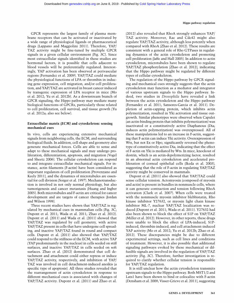
In vivo, cells are experiencing extensive mechanicalsignals from neighboring cells, the ECM, and surroundingbiological fluids. In addition, cell shape and geometry alsogenerate mechanical forces. Cells are able to sense andadapt to these mechanical signals and may undergo pro-liferation, differentiation, apoptosis, and migration (Vogeland Sheetz 2006). The cellular cytoskeleton can respondto and integrate extracellular mechanical signals. For in-stance, actin filaments (F-actin) have been considered asimportant regulators of cell proliferation (Provenzano andKeely 2011), and the dynamics of microtubules are essen-tial for cell division (Sorger et al. 1997). Mechanotransduc-tion is involved in not only normal physiology, but alsotumorigenesis and cancer metastasis (Huang and Ingber2005). Both microtubules and F-actin are critical for cancerdevelopment and are targets of cancer therapies (Jordanand Wilson 1998).
Three recent studies have shown that YAP/TAZ is reg-ulated by mechanical cues in mammalian cells (Fig. 3C;Dupont et al. 2011; Wada et al. 2011; Zhao et al. 2012).Dupont et al. (2011) and Wada et al. (2011) showed thatYAP/TAZ was regulated by cell geometry, with activeYAP/TAZ present in cells that have undergone cell spread-ing, and inactive YAP/TAZ found in round and compactcells. Dupont et al. (2011) also showed that YAP/TAZcould respond to the stiffness of the ECM, with active YAP(YAP predominantly in the nucleus) in cells seeded on stiffsurfaces, and inactive YAP/TAZ in cells seeded on softsurfaces. Zhao et al. (2012) demonstrated that cell de-tachment and attachment could either repress or induceYAP/TAZ activity, respectively, and inhibition of YAP/TAZ was involved in cell detachment-induced anoikis (aspecific type of apoptosis). All three studies revealed thatthe rearrangement of actin cytoskeleton in response todifferent mechanical cues is associated with changes ofYAP/TAZ activity. Dupont et al. (2011) and Zhao et al.
(2012) also revealed that RhoA strongly enhances YAP/TAZ activity. Moreover, Rac and Cdc42 might alsoregulate YAP/TAZ activity, although less potently whencompared with RhoA (Zhao et al. 2012). These results areconsistent with a general role of Rho GTPases in regulat-ing dynamics of the actin cytoskeleton and promotingcell proliferation (Jaffe and Hall 2005). In addition to actincytoskeleton, microtubules have been shown to regulateYAP/TAZ phosphorylation (Zhao et al. 2012), indicatingthat the Hippo pathway might be regulated by differenttypes of cellular cytoskeleton.
The regulation of the Hippo pathway by GPCR signal-ing and mechanical cues strongly suggests that the actincytoskeleton may function as a mediator and integratorof various upstream signals to the Hippo pathway. In-deed, two studies in Drosophila have revealed a linkbetween the actin cytoskeleton and the Hippo pathway(Fernandez et al. 2011; Sansores-Garcia et al. 2011). De-pletion of actin-capping protein, which inhibits actinpolymerization, resulted in Yki activation and tissue out-growth. Similar phenotypes were observed when Capulet(an actin-binding protein that inhibits polymerization) wasinactivated or a constitutively active Diaphanous (Dia;induces actin polymerization) was overexpressed. All ofthese manipulations led to an increase in F-actin, suggest-ing that F-actin can induce Yki activity. Overexpression ofWts, but not Ex or Hpo, significantly reversed the pheno-type of constitutively active Dia, indicating that the effectof F-actin on Yki is mediated by Wts. In mice, knockout ofdestrin, which is an actin-depolymerizing factor, resultedin an abnormal actin cytoskeleton and accelerated pro-liferation of corneal epithelial cells (Ikeda et al. 2003),suggesting that the role of F-actin in regulating YAP/TAZactivity might be conserved in mammals.
Dupont et al. (2011) also showed that YAP/TAZ couldsense cellular tension. Actomyosin (composed of myosinand actin) is present in bundles in nonmuscle cells, whereit can generate contraction and tension following RhoAactivation (Clark et al. 2007). When cells were treatedwith the nonmuscle myosin inhibitor blebbistatin, Rhokinase inhibitor Y27632, or myosin light chain kinaseinhibitor ML-7, nuclear YAP/TAZ localization was re-duced (Dupont et al. 2011; Wada et al. 2011). Y27632 hadalso been shown to block the effect of S1P on YAP/TAZ(Miller et al. 2012). However, in other reports, these drugswere unable to block the effect of LPA-induced, S1P-induced, thrombin-induced, and cell attachment-inducedYAP activity (Mo et al. 2012; Yu et al. 2012b; Zhao et al.2012). These discrepancies might be due to differentexperimental settings, such as cell lines and conditionsof treatment. However, it is also possible that additionalsignaling pathways evoked by these mechanical or dif-fusible signals are involved in the regulation of YAP/TAZactivity (Fig. 3C). Therefore, further investigation is re-quired to clarify whether cellular tension is responsiblefor YAP/TAZ regulation.
It is still unclear how the actin cytoskeleton transmitsupstream signals to the Hippo pathway. Both MST1/2 andLats1 have been shown to bind or colocalize with F-actin(Densham et al. 2009; Visser-Grieve et al. 2011), suggesting
Hippo pathway regulation
GENES & DEVELOPMENT 361
Cold Spring Harbor Laboratory Press on June 8, 2019 - Published by genesdev.cshlp.orgDownloaded from
a model in which F-actin may directly regulate the activityof Hippo pathway kinases. In mammalian cells, the kinaseactivity of Lats1 and the phosphorylation status at thehydrophobic motif and activation loop of Lats1/2 areclearly sensitive to GPCR signaling and cell detachment;however, the phosphorylation and in vitro kinase activityof MST1/2 are not significantly regulated by these up-stream signals (Mo et al. 2012; Yu et al. 2012b; Zhao et al.2012), indicating that MST1/2 are not direct targets ofthese upstream signals. On the other hand, Lats kinaseswere required for YAP/TAZ regulation by GPCR signaling,cell attachment, and cell geometry (Wada et al. 2011; Moet al. 2012; Yu et al. 2012b; Zhao et al. 2012). Meanwhile,actin cytoskeleton may also regulate YAP/TAZ phosphor-ylation via a Lats-independent mechanism (Dupont et al.2011; Miller et al. 2012). In addition, the role of F-actin inHippo pathway regulation might be indirect. F-actin mayfunction as a platform and facilitate signal transmissionbetween upstream regulators and core Hippo pathwaycomponents. Protein kinases or phosphatases down-stream from G proteins and Rho GTPases may regulateHippo pathway components in an actin cytoskeleton-dependent manner (Fig. 3C). Clearly, the mechanismlinking F-actin to the Hippo pathway is one of the mostimportant questions yet to be answered in the field.
As mentioned above, the Hippo pathway is regulatedby apical–basal polarity in epithelial cells, PCP, mechan-ical cues, and GPCR signaling. One common feature ofthese regulatory mechanisms is the involvement of actincytoskeleton (Fig. 3D). The integration of multiple up-stream signals to the Hippo pathway by F-actin mayexplain the YAP/TAZ activity regulation under diverseconditions. Actin dynamics is tightly regulated by RhoGTPases when cells are experiencing mechanical forcesor stimulation by extracellular ligands (Sah et al. 2000;Vogel and Sheetz 2006). It is well known that some GPCRligands, such as LPA, S1P, and thrombin, also induce con-tractive actin bundles in cells (Miller et al. 2012; Mo et al.2012; Yu et al. 2012b). On the other hand, some ligands forGas-coupled receptors, such as vasopressin, dopamine, andparathyroid hormone, have been shown to counteract theformation of actin bundles (Ding et al. 1991; Egan et al.1991b; Roma et al. 1998; Nguyen et al. 1999; Zhang et al.2006). Mechanical cues such as cell spreading, cell geom-etry, and matrix stiffness all result in rearrangement of theactin cytoskeleton (Dupont et al. 2011; Wada et al. 2011;Zhao et al. 2012). In cultured osteoblasts, the cross-linkedactin and myosin are reduced when cells are plated at highdensity (Egan et al. 1991a), indicating that cellular F-actinlevel is cell density-dependent, which may provide a mech-anism for cell density-dependent YAP phosphorylation(Zhao et al. 2007).
In epithelial cells, the actin cytoskeleton is more com-plex due to polarization and cell–cell junctions. There areapical or basal actin networks, and also connected actincables that span multiple pairs of cells (Baum and Georgiou2011). Some actin bundles are linked to apical junctions viaadaptor proteins such as catenins, NF2, and AMOT, andthese physical connections will create continuity betweenactin cytoskeletons of neighboring cells (Gjorevski et al.
2012); in addition, these adaptor proteins may regulateactivity of cdc42 and PAK1-Rac, thereby modulating theapical actin dynamics (Kissil et al. 2003; Perez-Morenoand Fuchs 2006; Wells et al. 2006). Therefore, it is pos-sible that the phenotypes of NF2, AMOT, a-catenin, andb-catenin deficiency on Yki/YAP/TAZ activity and cellproliferation are mediated by local actin rearrange-ments. In keratinocytes and mammary epithelial cells,calcium depletion led to disruption of cell junctionsand YAP/TAZ nuclear enrichment (Varelas et al. 2010;Schlegelmilch et al. 2011). Calcium-chelating agentsmay regulate intracellular actin dynamics, as disruption ofcell junctions by calcium depletion resulted in an increaseof actomyosin contraction in bovine corneal endothelialcells (Ramachandran and Srinivas 2010). However, it isalso possible that the effect of actin rearrangements on theHippo pathway is mediated by other cellular processes; forinstance, the actin cytoskeleton is required for the estab-lishment of cell polarity (Li and Gundersen 2008).
Actin cytoskeleton may also mediate the effect of PCPon the Hippo pathway. Dachs in the Fat/Ds system isa myosin-like protein, which may directly or indirectlyaffect actin dynamics (Rauskolb et al. 2011). In addition,RhoA is a well-known downstream player of the non-canonical wnt pathway (Habas et al. 2001); thus Fzi/Fmimay regulate YAP/TAZ activity via RhoA and actin organi-zation. Collectively, actin dynamics emerges as a centralmediator for YAP/TAZ regulation by a wide range ofstimuli (Fig. 3D).
Regulation of the Hippo pathway: implicationsin physiological and pathological conditions
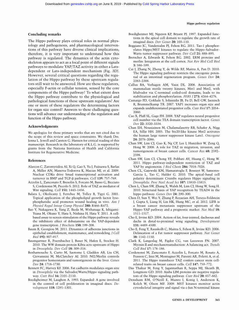
Recently, the Hippo pathway has been shown to regulatethe functions of stem cells. YAP and TAZ are requiredfor the maintenance of mouse and human embryonicstem cell pluripotency, respectively (Varelas et al. 2008;Alarcon et al. 2009; Lian et al. 2010; Qin et al. 2012). Intransgenic or knockout mice, elevated YAP/TAZ activityleads to an expansion of tissue-specific stem cells and theblockage of cell differentiation (Camargo et al. 2007; Caoet al. 2008; Lee et al. 2008, 2010; Zhou et al. 2009, 2011;Benhamouche et al. 2010; Lu et al. 2010; Song et al. 2010;Schlegelmilch et al. 2011; Zhang et al. 2011). YAP/TAZare important for mesenchymal stem cell (MSC) differ-entiation; knockdown of YAP/TAZ in MSCs decreasesosteogenesis and increases adipogenesis (Hong et al. 2005;Dupont et al. 2011). In addition, YAP/TAZ have beenshown to be important in myogenesis (Jeong et al. 2010;Watt et al. 2010; Judson et al. 2012). These results suggestthat the Hippo pathway plays important roles in celldifferentiation (Fig. 4A). Both mechanical forces and GPCRligands contribute to the cell microenvironment. StiffECM and LPA have been shown to promote osteogenesisand inhibit adipogenesis (McBeath et al. 2004; YB Liu et al.2010). On the other hand, soft ECM and cAMP signaling(e.g., IBMX treatment) could induce adipogenesis and re-press osteogenesis (McBeath et al. 2004; Yang et al. 2008).Cell density and serum concentration are also critical dur-ing cell differentiation; for instance, myogenesis requires
Yu and Guan
362 GENES & DEVELOPMENT
Cold Spring Harbor Laboratory Press on June 8, 2019 - Published by genesdev.cshlp.orgDownloaded from
higher cell density and lower serum concentration—conditions that favor YAP/TAZ inactivation. Altogether,a variety of microenvironmental signals may determinecell differentiation by regulating YAP/TAZ activity.
The Hippo pathway is also involved in tissue regener-ation. In the intestines of mice, YAP protein levels weredramatically increased following dextran sulfate sodium(DSS)-induced injury, and the damaged intestinal epi-thelium underwent regeneration; however, inactivationof YAP severely impaired regeneration (Cai et al. 2010).Furthermore, Yki is required for tissue regeneration inDrosophila midgut and wing discs (Karpowicz et al. 2010;Ren et al. 2010a; Shaw et al. 2010; Staley and Irvine 2010;Grusche et al. 2011; Sun and Irvine 2011). Interestingly,TEAD1 Y406H (tyrosine-to-histidine) mutation abolishesYAP–TEAD interaction (Zhao et al. 2008; L Chen et al.2010; Li et al. 2010), and this mutation is associated withSveinsson chorioretinal atrophy (a human degenerationdisease with absence of retinal pigment epithelium andadditional retinal structures) (Jonasson et al. 2007), sug-gesting the importance of YAP/TEAD transcriptional ac-tivity in tissue growth and homeostasis. Moreover, tissuedamage may result in changes of mechanical or biochem-ical environments, and a local increase of YAP/TAZ
activity may facilitate wound healing processes (Fig. 4B).For instance, thrombin is increased during blood clot-ting, a process directly associated with wounds, andmay promote wound healing by activating YAP/TAZ.Moreover, LPA has been shown to facilitate wound closurein a mouse model (Balazs et al. 2001). Therefore, Hippopathway regulation is important for wound healing andregeneration.
YAP plays important roles in early embryonic develop-ment. Systematic knockout of YAP in mice is lethal, andthe embryo stops developing at embryonic day 8.5 (E8.5)with defects in the yolk sac, vasculogenesis, chorioallan-tonic fusion, and body axis elongation (Morin-Kensickiet al. 2006). In a normal blastocyst, YAP shows nuclearlocalization in the trophectoderm (TE) and cytoplasmiclocalization in the inner cell mass (ICM), and this distinctdistribution of YAP is important for lineage specificationin the preimplantation mouse embryo (Fig. 4C; Nishiokaet al. 2009). However, the underlying mechanism for thispatterned YAP localization is not clear. In blastocysts, theF-actin and myosin staining is strong in TE but not de-tectable in the ICM, suggesting that contractive actinbundles are abundant in the TE (Slager et al. 1992). Thedistribution of F-actin is in nice correlation with YAP
Figure 4. YAP/TAZ activity under differentphysiological and pathological conditions. (A)YAP/TAZ activity in cell differentiation. YAP/TAZ activity is regulated by cell density, ECM,and GPCR signaling. (B) Cells close to a woundmay experience different mechanical forces anda higher concentration of GPCR ligands fromblood vessels. This may induce YAP/TAZ ac-tivity and promote wound healing or tissueregeneration. (C) In a blastocyst, cells in thetrophectoderm (TE) and inner cell mass (ICM)have distinct YAP localization and F-actin/tension distribution. (D) Disruption of cellularjunctions may induce mechanical stress andresult in changes in cell morphology, which canenrich YAP/TAZ in nuclei under these condi-tions. Activated YAP/TAZ may promote epi-thelial–mesenchymal transition (EMT) and cellmigration. Cells that have escaped from epithe-lium may also encounter more GPCR ligands.Red lines represent actin filaments. (E) AberrantGPCR signaling may activate YAP/TAZ. Ele-vated GPCR expression or activating Ga muta-tions may induce YAP/TAZ nuclei localizationand activation and result in hyperproliferationthat may contribute to cancer development.YAP/TAZ localization is represented by greenin B–E.
Hippo pathway regulation
GENES & DEVELOPMENT 363
Cold Spring Harbor Laboratory Press on June 8, 2019 - Published by genesdev.cshlp.orgDownloaded from
localization in blastocysts (Fig. 4C; Nishioka et al.2009). It is possible that the actin cytoskeleton is thedeterminant of YAP localization in this context. Thedistribution of F-actin in blastocysts might be regulatedby multiple means: (1) Cells in the ICM are not polarized,whereas cells in the TE are polarized and may have moreF-actin and tension. (2) Like in cells growing in high den-sity, cells in the ICM contact one another in all directionsand may have less F-actin and tension. (3) The outsideof the TE is exposed, and GPCRs that are expressed onthe surface might be influenced by maternal hormones,whereas the access of these signals to ICM cells isprevented by the epithelium (TE). It would be interestingto further investigate the relationship between F-actinand YAP localization and the upstream cues during earlyembryogenesis.
The Hippo pathway is widely recognized as a signalingpathway that regulates organ size. In Drosophila, loss-of-function mutants of Hpo, Sav, Wts, and Mob all leadto significant tissue outgrowth, as indicated by enlargedeyes, wings, or other appendages (Justice et al. 1995; Xuet al. 1995; Kango-Singh et al. 2002; Tapon et al. 2002;Harvey et al. 2003; Jia et al. 2003; Pantalacci et al. 2003;Udan et al. 2003; Wu et al. 2003; Lai et al. 2005). Over-expression of Yki also revealed similar tissue outgrowthphenotypes (Huang et al. 2005). The function of the Hippopathway in organ size control is conserved in mammals.For instance, tissue-specific overexpression of YAP in themouse liver or heart resulted in a dramatic increase ofliver or heart size (Camargo et al. 2007; Dong et al. 2007;von Gise et al. 2012). Consistently, knockout of MST1/2or Sav in the liver or heart also induced organ size (Zhouet al. 2009; Lee et al. 2010; Lu et al. 2010; Song et al. 2010;Heallen et al. 2011). Recently, a connection between theHippo pathway and the PI3K–TOR pathway has beendemonstrated (Strassburger et al. 2012; Tumaneng et al.2012b; Ye et al. 2012). This is significant because TORsignaling is important in cell growth; thus, the Hippopathway may control organ size in part by modulatingcell growth (Fig. 1; Tumaneng et al. 2012a). It is possiblethat multiple upstream signals work together to modu-late the Hippo pathway to determine the final organ size.The mechanical forces may correlate with organ size andconvey the organ size information to the Hippo pathway.Similarly, tissue-specific GPCRs may also play a role.For example, acetylcholine (a ligand for Gaq/11-coupledreceptor) signaling was required for salivary organogen-esis (Knox et al. 2010). Knockout of luteinizing hormonereceptor resulted in dramatically smaller testis (Zhanget al. 2001), indicating a possible role of GPCR signalingin tissue growth and organ size control.
Hippo pathway members have been identified in thesearch for tumor suppressor genes in Drosophila. Kinasesin the Hippo pathway are generally tumor suppressors,whereas Yki/YAP/TAZ have oncoprotein-like functions.Elevated YAP or TAZ expression and nuclear localizationare frequently observed in human cancers (Zhao et al.2007; Chan et al. 2008; Steinhardt et al. 2008; Fernandezet al. 2009; Xu et al. 2009). YAP transgenic mice displayhyperplasia and tumors (Camargo et al. 2007; Dong et al.
2007; Zhang et al. 2011; von Gise et al. 2012). Similarly,inactivation of upstream core components of the Hippopathway leads to tumor development in mice (St Johnet al. 1999; Lee et al. 2008, 2010; Zhou et al. 2009, 2011;Lu et al. 2010; Song et al. 2010; Nishio et al. 2012).Moreover, genetic inactivation of NF2, a well-knowntumor suppressor that acts upstream of the Hippo path-way (Rouleau et al. 1993; Ruttledge et al. 1994), resultedin tissue overgrowth in Drosophila and cancers in mice(Hamaratoglu et al. 2006; Benhamouche et al. 2010;Zhang et al. 2010), and the phenotype was blocked bydown-regulation of YAP (Zhang et al. 2010). YAP and TAZinduced an epithelial–mesenchymal transition (EMT), aphenomenon crucial for the initiation of cancer metasta-sis (Overholtzer et al. 2006; Lei et al. 2008; Thiery et al.2009). The role of YAP in promoting cancer metastasishas also recently been demonstrated in mice (Chen et al.2012; Lamar et al. 2012). In addition, TAZ has been shownto sustain self-renewal and induce tumor initiation ofbreast cancer stem cells (Cordenonsi et al. 2011). Thesedata suggest an important function of the Hippo pathwayin cancer development.
The connection between the Hippo pathway, mechan-ical forces, and GPCR signaling also provides new in-sights in cancer development. Following disruption ofcell–cell junctions and loss of cell polarity, cells may formextensive focal adhesions with the ECM and therebygenerate cellular tension, and these cells may further-more encounter stimulation by GPCR ligands (Fig. 4D).Activation of YAP/TAZ under these conditions mayfacilitate cell proliferation and cell migration or induceEMT. GPCRs are crucial players in cancer development,and dysregulated GPCR signaling has been identified inmany types of human cancers (Dorsam and Gutkind2007). GPCR signaling can contribute to cancer in a vari-ety of ways (Fig. 4E). Increased production of some GPCRligands may promote cancer development; for instance,overexpression of autotaxin (an enzyme critical for LPAsynthesis) in mouse mammary glands has been shown toinduce breast cancer (Liu et al. 2009). Up-regulated ex-pression of GPCRs may activate intracellular signalingautomatically, as indicated by high PAR1 expression inhigh-grade breast cancer patients (Hernandez et al. 2009).Activating mutations of GPCRs have been identified indifferent type of cancers, such as the metabotropic gluta-mate receptor mutations in melanoma and thyroid-stimulating hormone receptor in thyroid carcinomas(Paschke and Ludgate 1997; Prickett et al. 2011). Activat-ing mutations of Ga are also present in different types ofcancers: Gaq/11-activating mutations have been iden-tified in >80% of uveal melanomas (Van Raamsdonket al. 2009, 2010); Gai2a and Gas mutations have beenidentified in ovarian and endocrine tumors (Lania et al.2001). In addition, aberrant activity of Rho GTPasesand GPCR regulatory molecules, such as GPCR-relatedkinases, may also contribute to cancer by regulatingYAP/TAZ activity (Sahai and Marshall 2002; Metayeet al. 2005). Altogether, activation of YAP/TAZ by dysreg-ulated GPCR signaling may play an important role in thedevelopment of human cancers.
Yu and Guan
364 GENES & DEVELOPMENT
Cold Spring Harbor Laboratory Press on June 8, 2019 - Published by genesdev.cshlp.orgDownloaded from
Concluding remarks
The Hippo pathway plays critical roles in normal phys-iology and pathogenesis, and pharmacological interven-tions of this pathway have diverse clinical implications;therefore, it is very important to understand how thispathway is regulated. The dynamics of the actin cyto-skeleton appears to act as a focal point of different signalspathways to modulate YAP/TAZ activity in either a Lats-dependent or Lats-independent mechanism (Fig. 3D).However, several critical questions regarding the regu-lation of the Hippo pathway by these upstream regula-tors still wait to be answered. How are these regulators,especially F-actin or cellular tension, sensed by the corecomponents of the Hippo pathway? To what extent doesthe Hippo pathway contribute to the physiological andpathological functions of these upstream regulators? Areone or more of these regulators the determining factorsfor organ size control? Answers to these challenging ques-tions will advance our understanding of the regulation andfunction of the Hippo pathway.
Acknowledgments
We apologize for those primary works that are not cited due tothe scope of this review and space constraints. We thank Drs.Jenna L. Jewell and Carsten G. Hansen for critical reading of thismanuscript. Research in the laboratory of K.L.G. is supported bygrants from the Nationa Institutes of Health and CaliforniaInstitute for Regenerative Medicine.
References
Alarcon C, Zaromytidou AI, Xi Q, Gao S, Yu J, Fujisawa S, BarlasA, Miller AN, Manova-Todorova K, Macias MJ, et al. 2009.Nuclear CDKs drive Smad transcriptional activation andturnover in BMP and TGF-b pathways. Cell 139: 757–769.
Azzolin L, Zanconato F, Bresolin S, Forcato M, Basso G, BicciatoS, Cordenonsi M, Piccolo S. 2012. Role of TAZ as mediator ofWnt signaling. Cell 151: 1443–1456.
Balazs L, Okolicany J, Ferrebee M, Tolley B, Tigyi G. 2001.Topical application of the phospholipid growth factor lyso-phosphatidic acid promotes wound healing in vivo. Am JPhysiol Regul Integr Comp Physiol 280: R466–R472.
Bao Y, Nakagawa K, Yang Z, Ikeda M, Withanage K, Ishigami-Yuasa M, Okuno Y, Hata S, Nishina H, Hata Y. 2011. A cell-based assay to screen stimulators of the Hippo pathway revealsthe inhibitory effect of dobutamine on the YAP-dependentgene transcription. J Biochem 150: 199–208.
Baum B, Georgiou M. 2011. Dynamics of adherens junctions inepithelial establishment, maintenance, and remodeling. J Cell
Biol 192: 907–917.Baumgartner R, Poernbacher I, Buser N, Hafen E, Stocker H.
2010. The WW domain protein Kibra acts upstream of Hippoin Drosophila. Dev Cell 18: 309–316.
Benhamouche S, Curto M, Saotome I, Gladden AB, Liu CH,Giovannini M, McClatchey AI. 2010. Nf2/Merlin controlsprogenitor homeostasis and tumorigenesis in the liver. Genes
Dev 24: 1718–1730.Bennett FC, Harvey KF. 2006. Fat cadherin modulates organ size
in Drosophila via the Salvador/Warts/Hippo signaling path-way. Curr Biol 16: 2101–2110.
Boedigheimer M, Laughon A. 1993. Expanded: A gene involvedin the control of cell proliferation in imaginal discs. De-
velopment 118: 1291–1301.
Boedigheimer MJ, Nguyen KP, Bryant PJ. 1997. Expanded func-tions in the apical cell domain to regulate the growth rate ofimaginal discs. Dev Genet 20: 103–110.
Boggiano JC, Vanderzalm PJ, Fehon RG. 2011. Tao-1 phosphor-ylates Hippo/MST kinases to regulate the Hippo–Salvador–Warts tumor suppressor pathway. Dev Cell 21: 888–895.
Bretscher A, Edwards K, Fehon RG. 2002. ERM proteins andmerlin: Integrators at the cell cortex. Nat Rev Mol Cell Biol
3: 586–599.Cai J, Zhang N, Zheng Y, de Wilde RF, Maitra A, Pan D. 2010.
The Hippo signaling pathway restricts the oncogenic poten-tial of an intestinal regeneration program. Genes Dev 24:2383–2388.
Callus BA, Verhagen AM, Vaux DL. 2006. Association ofmammalian sterile twenty kinases, Mst1 and Mst2, withhSalvador via C-terminal coiled-coil domains, leads to itsstabilization and phosphorylation. FEBS J 273: 4264–4276.
Camargo FD, Gokhale S, Johnnidis JB, Fu D, Bell GW, JaenischR, Brummelkamp TR. 2007. YAP1 increases organ size andexpands undifferentiated progenitor cells. Curr Biol 17: 2054–2060.
Cao X, Pfaff SL, Gage FH. 2008. YAP regulates neural progenitorcell number via the TEA domain transcription factor. Genes
Dev 22: 3320–3334.Chan EH, Nousiainen M, Chalamalasetty RB, Schafer A, Nigg
EA, Sillje HH. 2005. The Ste20-like kinase Mst2 activatesthe human large tumor suppressor kinase Lats1. Oncogene
24: 2076–2086.Chan SW, Lim CJ, Guo K, Ng CP, Lee I, Hunziker W, Zeng Q,
Hong W. 2008. A role for TAZ in migration, invasion, andtumorigenesis of breast cancer cells. Cancer Res 68: 2592–2598.
Chan SW, Lim CJ, Chong YF, Pobbati AV, Huang C, Hong W.2011. Hippo pathway-independent restriction of TAZ andYAP by angiomotin. J Biol Chem 286: 7018–7026.
Chen CL, Gajewski KM, Hamaratoglu F, Bossuyt W, Sansores-Garcia L, Tao C, Halder G. 2010. The apical–basal cellpolarity determinant Crumbs regulates Hippo signaling inDrosophila. Proc Natl Acad Sci 107: 15810–15815.
Chen L, Chan SW, Zhang X, Walsh M, Lim CJ, Hong W, Song H.2010. Structural basis of YAP recognition by TEAD4 in thehippo pathway. Genes Dev 24: 290–300.
Chen D, Sun Y, Wei Y, Zhang P, Rezaeian AH, Teruya-FeldsteinJ, Gupta S, Liang H, Lin HK, Hung MC, et al. 2012. LIFR isa breast cancer metastasis suppressor upstream of theHippo–YAP pathway and a prognostic marker. Nat Med 18:1511–1517.
Cho E, Irvine KD. 2004. Action of fat, four-jointed, dachsous anddachs in distal-to-proximal wing signaling. Development
131: 4489–4500.Cho E, Feng Y, Rauskolb C, Maitra S, Fehon R, Irvine KD. 2006.
Delineation of a Fat tumor suppressor pathway. Nat Genet
38: 1142–1150.Clark K, Langeslag M, Figdor CG, van Leeuwen FN. 2007.
Myosin II and mechanotransduction: A balancing act. Trends
Cell Biol 17: 178–186.Cordenonsi M, Zanconato F, Azzolin L, Forcato M, Rosato A,
Frasson C, Inui M, Montagner M, Parenti AR, Poletti A, et al.2011. The Hippo transducer TAZ confers cancer stem cell-related traits on breast cancer cells. Cell 147: 759–772.
Das Thakur M, Feng Y, Jagannathan R, Seppa MJ, Skeath JB,Longmore GD. 2010. Ajuba LIM proteins are negative regula-tors of the Hippo signaling pathway. Curr Biol 20: 657–662.
Densham RM, O’Neill E, Munro J, Konig I, Anderson K,Kolch W, Olson MF. 2009. MST kinases monitor actincytoskeletal integrity and signal via c-Jun N-terminal kinase
Hippo pathway regulation
GENES & DEVELOPMENT 365
Cold Spring Harbor Laboratory Press on June 8, 2019 - Published by genesdev.cshlp.orgDownloaded from
stress-activated kinase to regulate p21Waf1/Cip1 stability.Mol Cell Biol 29: 6380–6390.
Ding GH, Franki N, Condeelis J, Hays RM. 1991. Vasopressindepolymerizes F-actin in toad bladder epithelial cells. Am J
Physiol 260: C9–C16.Dong J, Feldmann G, Huang J, Wu S, Zhang N, Comerford SA,
Gayyed MF, Anders RA, Maitra A, Pan D. 2007. Elucidationof a universal size-control mechanism in Drosophila andmammals. Cell 130: 1120–1133.
Dorsam RT, Gutkind JS. 2007. G-protein-coupled receptors andcancer. Nat Rev Cancer 7: 79–94.
Drees F, Pokutta S, Yamada S, Nelson WJ, Weis WI. 2005.a-Catenin is a molecular switch that binds E-cadherin–b-catenin and regulates actin-filament assembly. Cell 123:903–915.
Dupont S, Morsut L, Aragona M, Enzo E, Giulitti S, CordenonsiM, Zanconato F, Le Digabel J, Forcato M, Bicciato S, et al.2011. Role of YAP/TAZ in mechanotransduction. Nature
474: 179–183.Egan JJ, Gronowicz G, Rodan GA. 1991a. Cell density-dependent
decrease in cytoskeletal actin and myosin in cultured osteo-blastic cells: Correlation with cyclic AMP changes. J Cell
Biochem 45: 93–100.Egan JJ, Gronowicz G, Rodan GA. 1991b. Parathyroid hormone
promotes the disassembly of cytoskeletal actin and myosinin cultured osteoblastic cells: Mediation by cyclic AMP. J
Cell Biochem 45: 101–111.Feng Y, Irvine KD. 2007. Fat and expanded act in parallel to
regulate growth through warts. Proc Natl Acad Sci 104:20362–20367.
Fernandez LA, Northcott PA, Dalton J, Fraga C, Ellison D,Angers S, Taylor MD, Kenney AM. 2009. YAP1 is amplifiedand up-regulated in hedgehog-associated medulloblastomasand mediates Sonic hedgehog-driven neural precursor pro-liferation. Genes Dev 23: 2729–2741.
Fernandez BG, Gaspar P, Bras-Pereira C, Jezowska B, Rebelo SR,Janody F. 2011. Actin-capping protein and the Hippo path-way regulate F-actin and tissue growth in Drosophila. De-
velopment 138: 2337–2346.Ferrigno O, Lallemand F, Verrecchia F, L’Hoste S, Camonis J,
Atfi A, Mauviel A. 2002. Yes-associated protein (YAP65)interacts with Smad7 and potentiates its inhibitory activityagainst TGF-b/Smad signaling. Oncogene 21: 4879–4884.
Galliot B, Ghila L. 2010. Cell plasticity in homeostasis andregeneration. Mol Reprod Dev 77: 837–855.
Genevet A, Wehr MC, Brain R, Thompson BJ, Tapon N. 2010.Kibra is a regulator of the Salvador/Warts/Hippo signalingnetwork. Dev Cell 18: 300–308.
Gjorevski N, Boghaert E, Nelson CM. 2012. Regulation ofepithelial–mesenchymal transition by transmission of me-chanical stress through epithelial tissues. Cancer Microen-
viron 5: 29–38.Goulev Y, Fauny JD, Gonzalez-Marti B, Flagiello D, Silber J,
Zider A. 2008. SCALLOPED interacts with YORKIE, thenuclear effector of the hippo tumor-suppressor pathway inDrosophila. Curr Biol 18: 435–441.
Grusche FA, Degoutin JL, Richardson HE, Harvey KF. 2011. TheSalvador/Warts/Hippo pathway controls regenerative tissuegrowth in Drosophila melanogaster. Dev Biol 350: 255–266.
Grzeschik NA, Parsons LM, Allott ML, Harvey KF, RichardsonHE. 2010. Lgl, aPKC, and Crumbs regulate the Salvador/Warts/Hippo pathway through two distinct mechanisms.Curr Biol 20: 573–581.
Habas R, Kato Y, He X. 2001. Wnt/Frizzled activation of Rhoregulates vertebrate gastrulation and requires a novel Forminhomology protein Daam1. Cell 107: 843–854.
Habbig S, Bartram MP, Muller RU, Schwarz R, Andriopoulos N,Chen S, Sagmuller JG, Hoehne M, Burst V, Liebau MC, et al.2011. NPHP4, a cilia-associated protein, negatively regulatesthe Hippo pathway. J Cell Biol 193: 633–642.
Hamaratoglu F, Willecke M, Kango-Singh M, Nolo R, Hyun E,Tao C, Jafar-Nejad H, Halder G. 2006. The tumour-suppressorgenes NF2/Merlin and Expanded act through Hippo signallingto regulate cell proliferation and apoptosis. Nat Cell Biol 8:27–36.
Hao Y, Chun A, Cheung K, Rashidi B, Yang X. 2008. Tumorsuppressor LATS1 is a negative regulator of oncogene YAP.J Biol Chem 283: 5496–5509.
Harvey KF, Pfleger CM, Hariharan IK. 2003. The Drosophila Mstortholog, hippo, restricts growth and cell proliferation andpromotes apoptosis. Cell 114: 457–467.
Heallen T, Zhang M, Wang J, Bonilla-Claudio M, Klysik E,Johnson RL, Martin JF. 2011. Hippo pathway inhibits Wntsignaling to restrain cardiomyocyte proliferation and heartsize. Science 332: 458–461.
Hernandez NA, Correa E, Avila EP, Vela TA, Perez VM. 2009.PAR1 is selectively over expressed in high grade breastcancer patients: A cohort study. J Transl Med 7: 47.
Ho LL, Wei X, Shimizu T, Lai ZC. 2010. Mob as tumor sup-pressor is activated at the cell membrane to control tissuegrowth and organ size in Drosophila. Dev Biol 337: 274–283.
Ho KC, Zhou Z, She YM, Chun A, Cyr TD, Yang X. 2011. ItchE3 ubiquitin ligase regulates large tumor suppressor 1stability. Proc Natl Acad Sci 108: 4870–4875.
Hong JH, Hwang ES, McManus MT, Amsterdam A, Tian Y,Kalmukova R, Mueller E, Benjamin T, Spiegelman BM, SharpPA, et al. 2005. TAZ, a transcriptional modulator of mesen-chymal stem cell differentiation. Science 309: 1074–1078.
Huang S, Ingber DE. 2005. Cell tension, matrix mechanics, andcancer development. Cancer Cell 8: 175–176.
Huang J, Wu S, Barrera J, Matthews K, Pan D. 2005. The Hipposignaling pathway coordinately regulates cell proliferationand apoptosis by inactivating Yorkie, the Drosophila homo-log of YAP. Cell 122: 421–434.
Huang JM, Nagatomo I, Suzuki E, Mizuno T, Kumagai T,Berezov A, Zhang H, Karlan B, Greene MI, Wang Q. 2012.YAP modifies cancer cell sensitivity to EGFR and survivininhibitors and is negatively regulated by the non-receptortype protein tyrosine phosphatase 14. Oncogene doi: 10.1038/onc.2012.231.
Huang W, Lv X, Liu C, Zha Z, Zhang H, Jiang Y, Xiong Y, LeiQY, Guan KL. 2012. The N-terminal phosphodegron targetsTAZ/WWTR1 for SCFb–TrCP dependent degradation in re-sponse to PI3K inhibition. J Biol Chem 287: 26245–26253.
Ikeda S, Cunningham LA, Boggess D, Hawes N, Hobson CD,Sundberg JP, Naggert JK, Smith RS, Nishina PM. 2003.Aberrant actin cytoskeleton leads to accelerated prolifera-tion of corneal epithelial cells in mice deficient for destrin(actin depolymerizing factor). Hum Mol Genet 12: 1029–1037.
Ikeda M, Kawata A, Nishikawa M, Tateishi Y, Yamaguchi M,Nakagawa K, Hirabayashi S, Bao Y, Hidaka S, Hirata Y, et al.2009. Hippo pathway-dependent and -independent roles ofRASSF6. Sci Signal 2: ra59.
Ishikawa HO, Takeuchi H, Haltiwanger RS, Irvine KD. 2008.Four-jointed is a Golgi kinase that phosphorylates a subset ofcadherin domains. Science 321: 401–404.
Jaffe AB, Hall A. 2005. Rho GTPases: Biochemistry and biology.Annu Rev Cell Dev Biol 21: 247–269.
Jeong H, Bae S, An SY, Byun MR, Hwang JH, Yaffe MB, Hong JH,Hwang ES. 2010. TAZ as a novel enhancer of MyoD-mediatedmyogenic differentiation. FASEB J 24: 3310–3320.
Yu and Guan
366 GENES & DEVELOPMENT
Cold Spring Harbor Laboratory Press on June 8, 2019 - Published by genesdev.cshlp.orgDownloaded from
Jia J, Zhang W, Wang B, Trinko R, Jiang J. 2003. The Drosophila
Ste20 family kinase dMST functions as a tumor suppressorby restricting cell proliferation and promoting apoptosis.Genes Dev 17: 2514–2519.
Jonasson F, Hardarson S, Olafsson BM, Klintworth GK. 2007.Sveinsson chorioretinal atrophy/helicoid peripapillary cho-rioretinal degeneration: First histopathology report. Oph-
thalmology 114: 1541–1546.Jordan MA, Wilson L. 1998. Microtubules and actin filaments:
Dynamic targets for cancer chemotherapy. Curr Opin Cell
Biol 10: 123–130.Judson RN, Tremblay AM, Knopp P, White RB, Urcia R, Bari
CD, Zammit PS, Camargo FD, Wackerhage H. 2012. TheHippo pathway member Yap plays a key role in influencingfate decisions in muscle satellite cells. J Cell Sci doi: 10.1242/jcs.109546.
Justice RW, Zilian O, Woods DF, Noll M, Bryant PJ. 1995. TheDrosophila tumor suppressor gene warts encodes a homologof human myotonic dystrophy kinase and is required for thecontrol of cell shape and proliferation. Genes Dev 9: 534–546.
Kanai F, Marignani PA, Sarbassova D, Yagi R, Hall RA, DonowitzM, Hisaminato A, Fujiwara T, Ito Y, Cantley LC, et al. 2000.TAZ: A novel transcriptional co-activator regulated by in-teractions with 14-3-3 and PDZ domain proteins. EMBO J 19:6778–6791.
Kango-Singh M, Nolo R, Tao C, Verstreken P, Hiesinger PR,Bellen HJ, Halder G. 2002. Shar-pei mediates cell prolifera-tion arrest during imaginal disc growth in Drosophila. De-
velopment 129: 5719–5730.Karpowicz P, Perez J, Perrimon N. 2010. The Hippo tumor
suppressor pathway regulates intestinal stem cell regenera-tion. Development 137: 4135–4145.
Khokhlatchev A, Rabizadeh S, Xavier R, Nedwidek M, Chen T,Zhang XF, Seed B, Avruch J. 2002. Identification of a novelRas-regulated proapoptotic pathway. Curr Biol 12: 253–265.
Kim NG, Koh E, Chen X, Gumbiner BM. 2011. E-cadherinmediates contact inhibition of proliferation through Hipposignaling-pathway components. Proc Natl Acad Sci 108:11930–11935.
Kissil JL, Wilker EW, Johnson KC, Eckman MS, Yaffe MB, JacksT. 2003. Merlin, the product of the Nf2 tumor suppressorgene, is an inhibitor of the p21-activated kinase, Pak1. Mol
Cell 12: 841–849.Knox SM, Lombaert IM, Reed X, Vitale-Cross L, Gutkind JS,
Hoffman MP. 2010. Parasympathetic innervation maintainsepithelial progenitor cells during salivary organogenesis.Science 329: 1645–1647.
Komuro A, Nagai M, Navin NE, Sudol M. 2003. WW domain-containing protein YAP associates with ErbB-4 and acts asa co-transcriptional activator for the carboxyl-terminal frag-ment of ErbB-4 that translocates to the nucleus. J Biol Chem
278: 33334–33341.Kremerskothen J, Plaas C, Buther K, Finger I, Veltel S, Matanis
T, Liedtke T, Barnekow A. 2003. Characterization of KIBRA,a novel WW domain-containing protein. Biochem Biophys
Res Commun 300: 862–867.Lai ZC, Wei X, Shimizu T, Ramos E, Rohrbaugh M, Nikolaidis N,
Ho LL, Li Y. 2005. Control of cell proliferation and apoptosisby mob as tumor suppressor, mats. Cell 120: 675–685.
Lamar JM, Stern P, Liu H, Schindler JW, Jiang ZG, Hynes RO.2012. The Hippo pathway target, YAP, promotes metastasisthrough its TEAD-interaction domain. Proc Natl Acad Sci
109: E2441–E2450.Lania A, Mantovani G, Spada A. 2001. G protein mutations in
endocrine diseases. Eur J Endocrinol 145: 543–559.
Lappano R, Maggiolini M. 2011. G protein-coupled receptors:Novel targets for drug discovery in cancer. Nat Rev DrugDiscov 10: 47–60.
Lee JH, Kim TS, Yang TH, Koo BK, Oh SP, Lee KP, Oh HJ, LeeSH, Kong YY, Kim JM, et al. 2008. A crucial role of WW45 indeveloping epithelial tissues in the mouse. EMBO J 27: 1231–1242.
Lee KP, Lee JH, Kim TS, Kim TH, Park HD, Byun JS, Kim MC,Jeong WI, Calvisi DF, Kim JM, et al. 2010. The Hippo–Salvador pathway restrains hepatic oval cell proliferation,liver size, and liver tumorigenesis. Proc Natl Acad Sci 107:8248–8253.
Lei QY, Zhang H, Zhao B, Zha ZY, Bai F, Pei XH, Zhao S, XiongY, Guan KL. 2008. TAZ promotes cell proliferation andepithelial–mesenchymal transition and is inhibited by thehippo pathway. Mol Cell Biol 28: 2426–2436.
Li R, Gundersen GG. 2008. Beyond polymer polarity: How thecytoskeleton builds a polarized cell. Nat Rev Mol Cell Biol 9:860–873.
Li Z, Zhao B, Wang P, Chen F, Dong Z, Yang H, Guan KL, Xu Y.2010. Structural insights into the YAP and TEAD complex.Genes Dev 24: 235–240.
Lian I, Kim J, Okazawa H, Zhao J, Zhao B, Yu J, Chinnaiyan A,Israel MA, Goldstein LS, Abujarour R, et al. 2010. The role ofYAP transcription coactivator in regulating stem cell self-renewal and differentiation. Genes Dev 24: 1106–1118.
Ling C, Zheng Y, Yin F, Yu J, Huang J, Hong Y, Wu S, Pan D.2010. The apical transmembrane protein Crumbs func-tions as a tumor suppressor that regulates Hippo signalingby binding to Expanded. Proc Natl Acad Sci 107: 10532–10537.
Liu S, Umezu-Goto M, Murph M, Lu Y, Liu W, Zhang F, Yu S,Stephens LC, Cui X, Murrow G, et al. 2009. Expression ofautotaxin and lysophosphatidic acid receptors increases mam-mary tumorigenesis, invasion, and metastases. Cancer Cell15: 539–550.
Liu CY, Zha ZY, Zhou X, Zhang H, Huang W, Zhao D, Li T,Chan SW, Lim CJ, Hong W, et al. 2010. The hippo tumorpathway promotes TAZ degradation by phosphorylatinga phosphodegron and recruiting the SCFb–TrCP E3 ligase.J Biol Chem 285: 37159–37169.
Liu YB, Kharode Y, Bodine PV, Yaworsky PJ, Robinson JA,Billiard J. 2010. LPA induces osteoblast differentiation throughinterplay of two receptors: LPA1 and LPA4. J Cell Biochem
109: 794–800.Liu X, Yang N, Figel SA, Wilson KE, Morrison CD, Gelman IH,
Zhang J. 2012. PTPN14 interacts with and negatively regu-lates the oncogenic function of YAP. Oncogene doi: 10.1038/onc.2012.147.
Lu L, Li Y, Kim SM, Bossuyt W, Liu P, Qiu Q, Wang Y, Halder G,Finegold MJ, Lee JS, et al. 2010. Hippo signaling is a potent invivo growth and tumor suppressor pathway in the mamma-lian liver. Proc Natl Acad Sci 107: 1437–1442.
Mahoney PA, Weber U, Onofrechuk P, Biessmann H, Bryant PJ,Goodman CS. 1991. The fat tumor suppressor gene inDrosophila encodes a novel member of the cadherin genesuperfamily. Cell 67: 853–868.
Mao Y, Kucuk B, Irvine KD. 2009. Drosophila lowfat, a novelmodulator of Fat signaling. Development 136: 3223–3233.
Mao Y, Mulvaney J, Zakaria S, Yu T, Morgan KM, Allen S,Basson MA, Francis-West P, Irvine KD. 2011. Characteriza-tion of a Dchs1 mutant mouse reveals requirements forDchs1–Fat4 signaling during mammalian development. De-
velopment 138: 947–957.Martin-Belmonte F, Perez-Moreno M. 2012. Epithelial cell polar-
ity, stem cells and cancer. Nat Rev Cancer 12: 23–38.
Hippo pathway regulation
GENES & DEVELOPMENT 367
Cold Spring Harbor Laboratory Press on June 8, 2019 - Published by genesdev.cshlp.orgDownloaded from
Matakatsu H, Blair SS. 2004. Interactions between Fat andDachsous and the regulation of planar cell polarity in theDrosophila wing. Development 131: 3785–3794.
Matakatsu H, Blair SS. 2008. The DHHC palmitoyltransferaseapproximated regulates Fat signaling and Dachs localizationand activity. Curr Biol 18: 1390–1395.
McBeath R, Pirone DM, Nelson CM, Bhadriraju K, Chen CS.2004. Cell shape, cytoskeletal tension, and RhoA regulatestem cell lineage commitment. Dev Cell 6: 483–495.
McCartney BM, Kulikauskas RM, LaJeunesse DR, Fehon RG.2000. The neurofibromatosis-2 homologue, Merlin, and thetumor suppressor expanded function together in Drosophila
to regulate cell proliferation and differentiation. Develop-ment 127: 1315–1324.
Menendez J, Perez-Garijo A, Calleja M, Morata G. 2010. Atumor-suppressing mechanism in Drosophila involving cellcompetition and the Hippo pathway. Proc Natl Acad Sci 107:14651–14656.
Metaye T, Gibelin H, Perdrisot R, Kraimps JL. 2005. Pathophys-iological roles of G-protein-coupled receptor kinases. CellSignal 17: 917–928.
Miller E, Yang J, Deran M, Wu C, Su AI, Bonamy GM, Liu J,Peters EC, Wu X. 2012. Identification of serum-derivedsphingosine-1-phosphate as a small molecule regulator ofYAP. Chem Biol 19: 955–962.
Mo JS, Yu FX, Gong R, Brown JH, Guan KL. 2012. Regulation ofthe Hippo–YAP pathway by protease-activated receptors(PARs). Genes Dev 26: 2138–2143.
Morin-Kensicki EM, Boone BN, Howell M, Stonebraker JR, TeedJ, Alb JG, Magnuson TR, O’Neal W, Milgram SL. 2006.Defects in yolk sac vasculogenesis, chorioallantoic fusion,and embryonic axis elongation in mice with targeted disrup-tion of Yap65. Mol Cell Biol 26: 77–87.
Nguyen B, Carbajal ME, Vitale ML. 1999. Intracellular mecha-nisms involved in dopamine-induced actin cytoskeleton or-ganization and maintenance of a round phenotype in culturedrat lactotrope cells. Endocrinology 140: 3467–3477.
Nguyen HB, Babcock JT, Wells CD, Quilliam LA. 2012. LKB1tumor suppressor regulates AMP kinase/mTOR-independentcell growth and proliferation via the phosphorylation of Yap.Oncogene doi: 10.1038/onc.2012.431.
Nishio M, Hamada K, Kawahara K, Sasaki M, Noguchi F, ChibaS, Mizuno K, Suzuki SO, Dong Y, Tokuda M, et al. 2012.Cancer susceptibility and embryonic lethality in Mob1a/1bdouble-mutant mice. J Clin Invest 122: 4505–4518.
Nishioka N, Inoue K, Adachi K, Kiyonari H, Ota M, Ralston A,Yabuta N, Hirahara S, Stephenson RO, Ogonuki N, et al.2009. The Hippo signaling pathway components Lats andYap pattern Tead4 activity to distinguish mouse trophecto-derm from inner cell mass. Dev Cell 16: 398–410.
Oh H, Irvine KD. 2008. In vivo regulation of Yorkie phosphor-ylation and localization. Development 135: 1081–1088.
Oka T, Mazack V, Sudol M. 2008. Mst2 and Lats kinasesregulate apoptotic function of Yes kinase-associated protein(YAP). J Biol Chem 283: 27534–27546.
Oka T, Remue E, Meerschaert K, Vanloo B, Boucherie C, GfellerD, Bader GD, Sidhu SS, Vandekerckhove J, Gettemans J,et al. 2010. Functional complexes between YAP2 and ZO-2are PDZ domain-dependent, and regulate YAP2 nuclearlocalization and signalling. Biochem J 432: 461–472.
Omerovic J, Puggioni EM, Napoletano S, Visco V, Fraioli R, FratiL, Gulino A, Alimandi M. 2004. Ligand-regulated associationof ErbB-4 to the transcriptional co-activator YAP65 controlstranscription at the nuclear level. Exp Cell Res 294: 469–479.
Overholtzer M, Zhang J, Smolen GA, Muir B, Li W, Sgroi DC,Deng CX, Brugge JS, Haber DA. 2006. Transforming properties
of YAP, a candidate oncogene on the chromosome 11q22amplicon. Proc Natl Acad Sci 103: 12405–12410.
Pan D. 2010. The hippo signaling pathway in development andcancer. Dev Cell 19: 491–505.
Pantalacci S, Tapon N, Leopold P. 2003. The Salvador partnerHippo promotes apoptosis and cell-cycle exit in Drosophila.Nat Cell Biol 5: 921–927.
Paramasivam M, Sarkeshik A, Yates JR 3rd, Fernandes MJ,McCollum D. 2011. Angiomotin family proteins are novelactivators of the LATS2 kinase tumor suppressor. Mol Biol
Cell 22: 3725–3733.Paschke R, Ludgate M. 1997. The thyrotropin receptor in
thyroid diseases. N Engl J Med 337: 1675–1681.Pellettieri J, Sanchez Alvarado A. 2007. Cell turnover and adult
tissue homeostasis: From humans to planarians. Annu Rev
Genet 41: 83–105.Perez-Moreno M, Fuchs E. 2006. Catenins: Keeping cells from
getting their signals crossed. Dev Cell 11: 601–612.Poernbacher I, Baumgartner R, Marada SK, Edwards K, Stocker
H. 2012. Drosophila Pez acts in Hippo signaling to restrictintestinal stem cell proliferation. Curr Biol 22: 389–396.
Polesello C, Huelsmann S, Brown NH, Tapon N. 2006. TheDrosophila RASSF homolog antagonizes the hippo pathway.Curr Biol 16: 2459–2465.
Poon CL, Lin JI, Zhang X, Harvey KF. 2011. The sterile 20-likekinase Tao-1 controls tissue growth by regulating the Salvador-Warts-Hippo pathway. Dev Cell 21: 896–906.
Praskova M, Khoklatchev A, Ortiz-Vega S, Avruch J. 2004.Regulation of the MST1 kinase by autophosphorylation, bythe growth inhibitory proteins, RASSF1 and NORE1, and byRas. Biochem J 381: 453–462.
Praskova M, Xia F, Avruch J. 2008. MOBKL1A/MOBKL1Bphosphorylation by MST1 and MST2 inhibits cell prolifera-tion. Curr Biol 18: 311–321.
Prickett TD, Wei X, Cardenas-Navia I, Teer JK, Lin JC, Walia V,Gartner J, Jiang J, Cherukuri PF, Molinolo A, et al. 2011.Exon capture analysis of G protein-coupled receptors iden-tifies activating mutations in GRM3 in melanoma. Nat
Genet 43: 1119–1126.Provenzano PP, Keely PJ. 2011. Mechanical signaling through
the cytoskeleton regulates cell proliferation by coordinatedfocal adhesion and Rho GTPase signaling. J Cell Sci 124:1195–1205.
Qin H, Blaschke K, Wei G, Ohi Y, Blouin L, Qi Z, Yu J, Yeh RF,Hebrok M, Ramalho-Santos M. 2012. Transcriptional analy-sis of pluripotency reveals the Hippo pathway as a barrier toreprogramming. Hum Mol Genet 21: 2054–2067.
Ramachandran C, Srinivas SP. 2010. Formation and disassemblyof adherens and tight junctions in the corneal endothelium:Regulation by actomyosin contraction. Invest OphthalmolVis Sci 51: 2139–2148.
Rauskolb C, Pan G, Reddy BV, Oh H, Irvine KD. 2011. Zyxin linksfat signaling to the hippo pathway. PLoS Biol 9: e1000624.
Remue E, Meerschaert K, Oka T, Boucherie C, VandekerckhoveJ, Sudol M, Gettemans J. 2010. TAZ interacts with zonulaoccludens-1 and -2 proteins in a PDZ-1 dependent manner.FEBS Lett 584: 4175–4180.
Ren F, Wang B, Yue T, Yun EY, Ip YT, Jiang J. 2010a. Hipposignaling regulates Drosophila intestine stem cell prolifera-tion through multiple pathways. Proc Natl Acad Sci 107:21064–21069.
Ren F, Zhang L, Jiang J. 2010b. Hippo signaling regulates Yorkienuclear localization and activity through 14-3-3 dependentand independent mechanisms. Dev Biol 337: 303–312.
Ribeiro PS, Josue F, Wepf A, Wehr MC, Rinner O, Kelly G, TaponN, Gstaiger M. 2010. Combined functional genomic and
Yu and Guan
368 GENES & DEVELOPMENT
Cold Spring Harbor Laboratory Press on June 8, 2019 - Published by genesdev.cshlp.orgDownloaded from
proteomic approaches identify a PP2A complex as a negativeregulator of Hippo signaling. Mol Cell 39: 521–534.
Robinson BS, Huang J, Hong Y, Moberg KH. 2010. Crumbsregulates Salvador/Warts/Hippo signaling in Drosophila viathe FERM-domain protein Expanded. Curr Biol 20: 582–590.
Rogulja D, Rauskolb C, Irvine KD. 2008. Morphogen control ofwing growth through the Fat signaling pathway. Dev Cell 15:309–321.
Roma MG, Stone V, Shaw R, Coleman R. 1998. Vasopressin-induced disruption of actin cytoskeletal organization andcanalicular function in isolated rat hepatocyte couplets:Possible involvement of protein kinase C. Hepatology 28:1031–1041.
Rouleau GA, Merel P, Lutchman M, Sanson M, Zucman J,Marineau C, Hoang-Xuan K, Demczuk S, Desmaze C,Plougastel B, et al. 1993. Alteration in a new gene encodinga putative membrane-organizing protein causes neuro-fibromatosis type 2. Nature 363: 515–521.
Ruttledge MH, Sarrazin J, Rangaratnam S, Phelan CM, Twist E,Merel P, Delattre O, Thomas G, Nordenskjold M, Collins VP,et al. 1994. Evidence for the complete inactivation of theNF2 gene in the majority of sporadic meningiomas. Nat Genet
6: 180–184.Sah VP, Seasholtz TM, Sagi SA, Brown JH. 2000. The role of Rho
in G protein-coupled receptor signal transduction. Annu Rev
Pharmacol Toxicol 40: 459–489.Sahai E, Marshall CJ. 2002. RHO-GTPases and cancer. Nat Rev
Cancer 2: 133–142.Sansores-Garcia L, Bossuyt W, Wada K, Yonemura S, Tao C,
Sasaki H, Halder G. 2011. Modulating F-actin organizationinduces organ growth by affecting the Hippo pathway.EMBO J 30: 2325–2335.
Schlegelmilch K, Mohseni M, Kirak O, Pruszak J, Rodriguez JR,Zhou D, Kreger BT, Vasioukhin V, Avruch J, BrummelkampTR, et al. 2011. Yap1 acts downstream of a-catenin to controlepidermal proliferation. Cell 144: 782–795.
Shaw RL, Kohlmaier A, Polesello C, Veelken C, Edgar BA, TaponN. 2010. The Hippo pathway regulates intestinal stem cellproliferation during Drosophila adult midgut regeneration.Development 137: 4147–4158.
Silva E, Tsatskis Y, Gardano L, Tapon N, McNeill H. 2006. Thetumor-suppressor gene fat controls tissue growth upstreamof expanded in the hippo signaling pathway. Curr Biol 16:2081–2089.
Silvis MR, Kreger BT, Lien WH, Klezovitch O, Rudakova GM,Camargo FD, Lantz DM, Seykora JT, Vasioukhin V. 2011.a-Catenin is a tumor suppressor that controls cell accumu-lation by regulating the localization and activity of thetranscriptional coactivator Yap1. Sci Signal 4: ra33.
Simons M, Mlodzik M. 2008. Planar cell polarity signaling:From fly development to human disease. Annu Rev Genet
42: 517–540.Skouloudaki K, Puetz M, Simons M, Courbard JR, Boehlke C,
Hartleben B, Engel C, Moeller MJ, Englert C, Bollig F, et al.2009. Scribble participates in Hippo signaling and is requiredfor normal zebrafish pronephros development. Proc Natl
Acad Sci 106: 8579–8584.Slager HG, Good MJ, Schaart G, Groenewoud JS, Mummery CL.
1992. Organization of non-muscle myosin during early mu-rine embryonic differentiation. Differentiation 50: 47–56.
Song H, Mak KK, Topol L, Yun K, Hu J, Garrett L, Chen Y, ParkO, Chang J, Simpson RM, et al. 2010. Mammalian Mst1 andMst2 kinases play essential roles in organ size control andtumor suppression. Proc Natl Acad Sci 107: 1431–1436.
Sopko R, Silva E, Clayton L, Gardano L, Barrios-Rodiles M,Wrana J, Varelas X, Arbouzova NI, Shaw S, Saburi S, et al.
2009. Phosphorylation of the tumor suppressor fat is regu-lated by its ligand Dachsous and the kinase discs overgrown.Curr Biol 19: 1112–1117.
Sorger PK, Dobles M, Tournebize R, Hyman AA. 1997. Couplingcell division and cell death to microtubule dynamics. CurrOpin Cell Biol 9: 807–814.
Staley BK, Irvine KD. 2010. Warts and Yorkie mediate intestinalregeneration by influencing stem cell proliferation. Curr Biol
20: 1580–1587.Staley BK, Irvine KD. 2012. Hippo signaling in Drosophila:
Recent advances and insights. Dev Dyn 241: 3–15.Steinhardt AA, Gayyed MF, Klein AP, Dong J, Maitra A, Pan D,
Montgomery EA, Anders RA. 2008. Expression of Yes-asso-ciated protein in common solid tumors. Hum Pathol 39:1582–1589.
St. John MA, Tao W, Fei X, Fukumoto R, Carcangiu ML,Brownstein DG, Parlow AF, McGrath J, Xu T. 1999. Micedeficient of Lats1 develop soft-tissue sarcomas, ovariantumours and pituitary dysfunction. Nat Genet 21: 182–186.
Strano S, Munarriz E, Rossi M, Castagnoli L, Shaul Y, Sacchi A,Oren M, Sudol M, Cesareni G, Blandino G. 2001. Physicalinteraction with Yes-associated protein enhances p73 tran-scriptional activity. J Biol Chem 276: 15164–15173.
Strassburger K, Tiebe M, Pinna F, Breuhahn K, Teleman AA.2012. Insulin/IGF signaling drives cell proliferation in partvia Yorkie/YAP. Dev Biol 367: 187–196.
Sun G, Irvine KD. 2011. Regulation of Hippo signaling by Junkinase signaling during compensatory cell proliferation andregeneration, and in neoplastic tumors. Dev Biol 350: 139–151.
Tagliabracci VS, Engel JL, Wen J, Wiley SE, Worby CA, KinchLN, Xiao J, Grishin NV, Dixon JE. 2012. Secreted kinasephosphorylates extracellular proteins that regulate biomin-eralization. Science 336: 1150–1153.
Tapon N, Harvey KF, Bell DW, Wahrer DC, Schiripo TA, HaberDA, Hariharan IK. 2002. salvador promotes both cell cycleexit and apoptosis in Drosophila and is mutated in humancancer cell lines. Cell 110: 467–478.
Tepass U, Theres C, Knust E. 1990. crumbs encodes an EGF-likeprotein expressed on apical membranes of Drosophila epi-thelial cells and required for organization of epithelia. Cell
61: 787–799.Thiery JP, Acloque H, Huang RY, Nieto MA. 2009. Epithelial–
mesenchymal transitions in development and disease. Cell
139: 871–890.Tumaneng K, Russell RC, Guan KL. 2012a. Organ size control
by Hippo and TOR pathways. Curr Biol 22: R368–R379.Tumaneng K, Schlegelmilch K, Russell RC, Yimlamai D, Basnet
H, Mahadevan N, Fitamant J, Bardeesy N, Camargo FD,Guan KL. 2012b. YAP mediates crosstalk between the Hippoand PI(3)K–TOR pathways by suppressing PTEN via miR-29.Nat Cell Biol 14: 1322–1329.
Tyler DM, Baker NE. 2007. Expanded and fat regulate growthand differentiation in the Drosophila eye through multiplesignaling pathways. Dev Biol 305: 187–201.
Udan RS, Kango-Singh M, Nolo R, Tao C, Halder G. 2003. Hippopromotes proliferation arrest and apoptosis in the Salvador/Warts pathway. Nat Cell Biol 5: 914–920.
Van Raamsdonk CD, Bezrookove V, Green G, Bauer J, Gaugler L,O’Brien JM, Simpson EM, Barsh GS, Bastian BC. 2009.Frequent somatic mutations of GNAQ in uveal melanomaand blue naevi. Nature 457: 599–602.
Van Raamsdonk CD, Griewank KG, Crosby MB, Garrido MC,Vemula S, Wiesner T, Obenauf AC, Wackernagel W, GreenG, Bouvier N, et al. 2010. Mutations in GNA11 in uvealmelanoma. N Engl J Med 363: 2191–2199.
Hippo pathway regulation
GENES & DEVELOPMENT 369
Cold Spring Harbor Laboratory Press on June 8, 2019 - Published by genesdev.cshlp.orgDownloaded from
Varelas X, Sakuma R, Samavarchi-Tehrani P, Peerani R, Rao BM,Dembowy J, Yaffe MB, Zandstra PW, Wrana JL. 2008. TAZcontrols Smad nucleocytoplasmic shuttling and regulateshuman embryonic stem-cell self-renewal. Nat Cell Biol 10:837–848.
Varelas X, Samavarchi-Tehrani P, Narimatsu M, Weiss A,Cockburn K, Larsen BG, Rossant J, Wrana JL. 2010. TheCrumbs complex couples cell density sensing to Hippo-dependent control of the TGF-b–SMAD pathway. Dev Cell19: 831–844.
Vassilev A, Kaneko KJ, Shu H, Zhao Y, DePamphilis ML. 2001.TEAD/TEF transcription factors utilize the activation do-main of YAP65, a Src/Yes-associated protein localized in thecytoplasm. Genes Dev 15: 1229–1241.
Visser-Grieve S, Zhou Z, She YM, Huang H, Cyr TD, Xu T, YangX. 2011. LATS1 tumor suppressor is a novel actin-bindingprotein and negative regulator of actin polymerization. Cell
Res 21: 1513–1516.Vogel V, Sheetz M. 2006. Local force and geometry sensing
regulate cell functions. Nat Rev Mol Cell Biol 7: 265–275.von Gise A, Lin Z, Schlegelmilch K, Honor LB, Pan GM, Buck
JN, Ma Q, Ishiwata T, Zhou B, Camargo FD, et al. 2012.YAP1, the nuclear target of Hippo signaling, stimulates heartgrowth through cardiomyocyte proliferation but not hyper-trophy. Proc Natl Acad Sci 109: 2394–2399.
Wada K, Itoga K, Okano T, Yonemura S, Sasaki H. 2011. Hippopathway regulation by cell morphology and stress fibers.Development 138: 3907–3914.
Wang W, Huang J, Chen J. 2011. Angiomotin-like proteinsassociate with and negatively regulate YAP1. J Biol Chem
286: 4364–4370.Wang W, Huang J, Wang X, Yuan J, Li X, Feng L, Park JI, Chen J.
2012. PTPN14 is required for the density-dependent controlof YAP1. Genes Dev 26: 1959–1971.
Watt KI, Judson R, Medlow P, Reid K, Kurth TB, Burniston JG,Ratkevicius A, De Bari C, Wackerhage H. 2010. Yap is a novelregulator of C2C12 myogenesis. Biochem Biophys Res Com-
mun 393: 619–624.Wehr MC, Holder MV, Gailite I, Saunders RE, Maile TM,
Ciirdaeva E, Instrell R, Jiang M, Howell M, Rossner MJ,et al. 2012. Salt-inducible kinases regulate growth throughthe Hippo signalling pathway in Drosophila. Nat Cell Biol(in press). doi: 10.1038/ncb2658.
Wells CD, Fawcett JP, Traweger A, Yamanaka Y, Goudreault M,Elder K, Kulkarni S, Gish G, Virag C, Lim C, et al. 2006.A Rich1/Amot complex regulates the Cdc42 GTPase andapical-polarity proteins in epithelial cells. Cell 125: 535–548.
Willecke M, Hamaratoglu F, Kango-Singh M, Udan R, Chen CL,Tao C, Zhang X, Halder G. 2006. The fat cadherin actsthrough the hippo tumor-suppressor pathway to regulatetissue size. Curr Biol 16: 2090–2100.
Willecke M, Hamaratoglu F, Sansores-Garcia L, Tao C, HalderG. 2008. Boundaries of Dachsous cadherin activity modulatethe Hippo signaling pathway to induce cell proliferation.Proc Natl Acad Sci 105: 14897–14902.
Wu S, Huang J, Dong J, Pan D. 2003. hippo encodes a Ste-20family protein kinase that restricts cell proliferation andpromotes apoptosis in conjunction with salvador and warts.Cell 114: 445–456.
Wu S, Liu Y, Zheng Y, Dong J, Pan D. 2008. The TEAD/TEFfamily protein Scalloped mediates transcriptional output ofthe Hippo growth-regulatory pathway. Dev Cell 14: 388–398.
Xu T, Wang W, Zhang S, Stewart RA, Yu W. 1995. Identifyingtumor suppressors in genetic mosaics: The Drosophila latsgene encodes a putative protein kinase. Development 121:1053–1063.
Xu MZ, Yao TJ, Lee NP, Ng IO, Chan YT, Zender L, Lowe SW,Poon RT, Luk JM. 2009. Yes-associated protein is an in-dependent prognostic marker in hepatocellular carcinoma.Cancer 115: 4576–4585.
Yagi R, Chen LF, Shigesada K, Murakami Y, Ito Y. 1999. A WWdomain-containing yes-associated protein (YAP) is a noveltranscriptional co-activator. EMBO J 18: 2551–2562.
Yang DC, Tsay HJ, Lin SY, Chiou SH, Li MJ, Chang TJ, Hung SC.2008. cAMP/PKA regulates osteogenesis, adipogenesis andratio of RANKL/OPG mRNA expression in mesenchymalstem cells by suppressing leptin. PLoS One 3: e1540.
Ye X, Deng Y, Lai ZC. 2012. Akt is negatively regulated byHippo signaling for growth inhibition in Drosophila. DevBiol 369: 115–123.
Yi C, Troutman S, Fera D, Stemmer-Rachamimov A, Avila JL,Christian N, Persson NL, Shimono A, Speicher DW,Marmorstein R, et al. 2011. A tight junction-associatedMerlin–angiomotin complex mediates Merlin’s regulationof mitogenic signaling and tumor suppressive functions.Cancer Cell 19: 527–540.
Yu J, Zheng Y, Dong J, Klusza S, Deng WM, Pan D. 2010. Kibrafunctions as a tumor suppressor protein that regulates Hipposignaling in conjunction with Merlin and Expanded. Dev
Cell 18: 288–299.Yu FX, Mo JS, Guan KL. 2012a. Upstream regulators of the
Hippo pathway. Cell Cycle 11: 4097–4098.Yu FX, Zhao B, Panupinthu N, Jewell JL, Lian I, Wang LH, Zhao
J, Yuan H, Tumaneng K, Li H, et al. 2012b. Regulation of theHippo–YAP pathway by G-protein-coupled receptor signal-ing. Cell 150: 780–791.
Zecca M, Struhl G. 2010. A feed-forward circuit linking wing-less, fat–dachsous signaling, and the warts–hippo pathway toDrosophila wing growth. PLoS Biol 8: e1000386.
Zhang FP, Poutanen M, Wilbertz J, Huhtaniemi I. 2001. Normalprenatal but arrested postnatal sexual development of lutein-izing hormone receptor knockout (LuRKO) mice. Mol Endo-
crinol 15: 172–183.Zhang J, Ryder KD, Bethel JA, Ramirez R, Duncan RL. 2006.
PTH-induced actin depolymerization increases mechanosen-sitive channel activity to enhance mechanically stimulatedCa2+ signaling in osteoblasts. J Bone Miner Res 21: 1729–1737.
Zhang L, Ren F, Zhang Q, Chen Y, Wang B, Jiang J. 2008. TheTEAD/TEF family of transcription factor Scalloped mediatesHippo signaling in organ size control. Dev Cell 14: 377–387.
Zhang N, Bai H, David KK, Dong J, Zheng Y, Cai J, GiovanniniM, Liu P, Anders RA, Pan D. 2010. The Merlin/NF2 tumorsuppressor functions through the YAP oncoprotein to regu-late tissue homeostasis in mammals. Dev Cell 19: 27–38.
Zhang H, Pasolli HA, Fuchs E. 2011. Yes-associated protein(YAP) transcriptional coactivator functions in balancinggrowth and differentiation in skin. Proc Natl Acad Sci 108:2270–2275.
Zhao B, Wei X, Li W, Udan RS, Yang Q, Kim J, Xie J, Ikenoue T,Yu J, Li L, et al. 2007. Inactivation of YAP oncoprotein by theHippo pathway is involved in cell contact inhibition andtissue growth control. Genes Dev 21: 2747–2761.
Zhao B, Ye X, Yu J, Li L, Li W, Li S, Yu J, Lin JD, Wang CY,Chinnaiyan AM, et al. 2008. TEAD mediates YAP-dependentgene induction and growth control. Genes Dev 22: 1962–1971.
Zhao B, Li L, Lei Q, Guan KL. 2010a. The Hippo–YAP pathwayin organ size control and tumorigenesis: An updated version.Genes Dev 24: 862–874.
Zhao B, Li L, Tumaneng K, Wang CY, Guan KL. 2010b. A co-ordinated phosphorylation by Lats and CK1 regulates YAPstability through SCF(b-TRCP). Genes Dev 24: 72–85.
Yu and Guan
370 GENES & DEVELOPMENT
Cold Spring Harbor Laboratory Press on June 8, 2019 - Published by genesdev.cshlp.orgDownloaded from
Zhao B, Li L, Lu Q, Wang LH, Liu CY, Lei Q, Guan KL. 2011.Angiomotin is a novel Hippo pathway component that in-hibits YAP oncoprotein. Genes Dev 25: 51–63.
Zhao B, Li L, Wang L, Wang CY, Yu J, Guan KL. 2012. Celldetachment activates the Hippo pathway via cytoskeletonreorganization to induce anoikis. Genes Dev 26: 54–68.
Zhou D, Conrad C, Xia F, Park JS, Payer B, Yin Y, Lauwers GY,Thasler W, Lee JT, Avruch J, et al. 2009. Mst1 and Mst2maintain hepatocyte quiescence and suppress hepatocellularcarcinoma development through inactivation of the Yap1oncogene. Cancer Cell 16: 425–438.
Zhou D, Zhang Y, Wu H, Barry E, Yin Y, Lawrence E, Dawson D,Willis JE, Markowitz SD, Camargo FD, et al. 2011. Mst1 andMst2 protein kinases restrain intestinal stem cell proliferationand colonic tumorigenesis by inhibition of Yes-associatedprotein (Yap) overabundance. Proc Natl Acad Sci 108:E1312–E1320.
Hippo pathway regulation
GENES & DEVELOPMENT 371
Cold Spring Harbor Laboratory Press on June 8, 2019 - Published by genesdev.cshlp.orgDownloaded from
10.1101/gad.210773.112Access the most recent version at doi: 27:2013, Genes Dev.
Fa-Xing Yu and Kun-Liang Guan The Hippo pathway: regulators and regulations
References
http://genesdev.cshlp.org/content/27/4/355.full.html#ref-list-1
This article cites 205 articles, 75 of which can be accessed free at:
License
ServiceEmail Alerting
click here.right corner of the article or
Receive free email alerts when new articles cite this article - sign up in the box at the top
Copyright © 2013 by Cold Spring Harbor Laboratory Press
Cold Spring Harbor Laboratory Press on June 8, 2019 - Published by genesdev.cshlp.orgDownloaded from